User login
MDedge conference coverage features onsite reporting of the latest study results and expert perspectives from leading researchers.
VOYAGER PAD: Paclitaxel-coated devices don’t increase mortality
a multithousand-patient randomized trial with long-term follow-up and ascertainment of vital status in 99.6% of participants.
Observers opined that the VOYAGER PAD findings effectively put to rest a nearly 2-year-old controversy over whether paclitaxel-coated devices for treatment of peripheral artery disease (PAD) carry an increased mortality risk. The imbroglio, which was ignited by a meta-analysis of clinical trials with substantial amounts of missing follow-up data, triggered an Food and Drug Administration warning letter to health care providers which threw the field of vascular medicine into disarray.
“Although as a community we’ve continued to struggle with this issue of paclitaxel and mortality, VOYAGER PAD does fill many of the gaps and addresses many of the limitations of currently available data,” Connie N. Hess, MD, said in reporting results of a prespecified analysis of the trial at the Transcatheter Cardiovascular Research Therapeutics virtual annual meeting. “I think these are the most definitive data to date supporting the safety of drug-coated device use.”
VOYAGER PAD was a double-blind, placebo-controlled clinical trial in which 6,564 patients undergoing lower-extremity revascularization for symptomatic PAD were randomized to rivaroxaban at 2.5 mg twice daily or placebo on top of background low-dose aspirin. In the previously reported primary outcome, the group on rivaroxaban plus aspirin had a significant 15% reduction in the risk of the composite endpoint of cardiovascular death, acute limb ischemia, MI, ischemic stroke, or major amputation for vascular causes.
Of the 4,316 patients included in the prespecified analysis by Dr. Hess, a cardiologist at the University of Colorado at Denver, Aurora, 31% received a paclitaxel-coated device. At 3.5 years of follow-up, they had a 10.2% all-cause mortality rate, significantly less than the 13.5% rate in patients who didn’t get a drug-coated device. But since study participants weren’t randomized for drug-coated device use, the investigators utilized a rigorous form of propensity adjustment called inverse probability treatment weighting to neutralize all between-group differences in potentially confounding baseline characteristics, including statin use, prevalence of claudication, and target lesion length.
In the weighted analysis, the all-cause mortality rate at 3.5 years was 12.1% in paclitaxel-coated device recipients and 12.6% in those who didn’t get such devices. The difference was not statistically significant, and the hazard ratio of 0.95 had tight confidence intervals.
“We don’t see a mortality benefit, but I think more importantly, we don’t see any risk for mortality,” the cardiologist observed at the meeting sponsored by the Cardiovascular Research Foundation.
There was no between-group difference in causes of mortality. Nor did all-cause mortality differ by device type, be it paclitaxel-coated balloon versus plain balloon angioplasty, or drug-eluting stent versus bare-metal stent.
Also, the benefit of rivaroxaban plus aspirin over aspirin alone in terms of cardiovascular and ischemic limb outcomes was consistent regardless of whether patients got a drug-coated device or not.
Discussant Robert Lookstein, MD, praised Dr. Hess for “a really enlightening presentation.”
“The entire vascular community has been waiting for a prospective, independently adjudicated trial to try to make determinations of whether we can put this issue behind us, and I think this trial is it,” said Dr. Lookstein, professor of interventional radiology and surgery at the Icahn School of Medicine at Mount Sinai, New York.
“Personally, I think this is probably the most impactful data seen regarding the paclitaxel issue in almost 2 years because it is randomized data, it’s prospectively collected data, and – most importantly from my perspective – they were able to collect vital statistics on more than 99.5% of the patients,” he added. “I think this is incredibly impactful to my practice.”
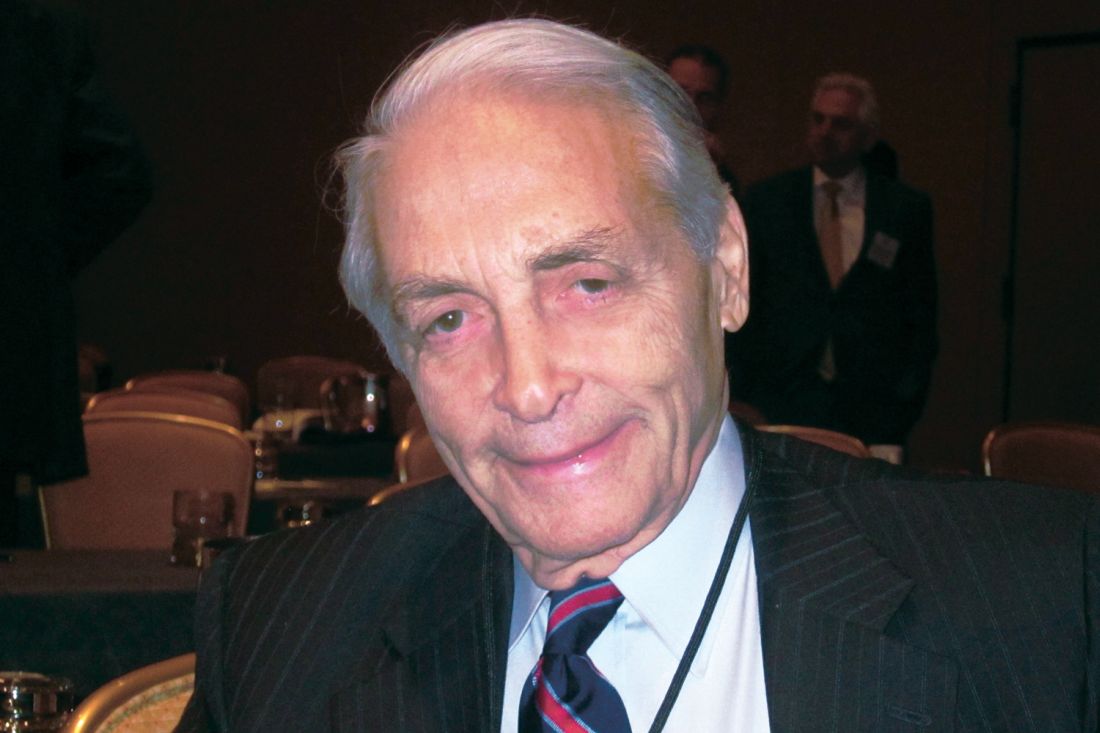
Frank Veith, MD, professor of surgery at New York University, concurred, declaring, “I think this study is a game changer. And I think the paclitaxel game is over.”
The VOYAGER PAD study was funded by institutional research grants from Bayer and Janssen.
a multithousand-patient randomized trial with long-term follow-up and ascertainment of vital status in 99.6% of participants.
Observers opined that the VOYAGER PAD findings effectively put to rest a nearly 2-year-old controversy over whether paclitaxel-coated devices for treatment of peripheral artery disease (PAD) carry an increased mortality risk. The imbroglio, which was ignited by a meta-analysis of clinical trials with substantial amounts of missing follow-up data, triggered an Food and Drug Administration warning letter to health care providers which threw the field of vascular medicine into disarray.
“Although as a community we’ve continued to struggle with this issue of paclitaxel and mortality, VOYAGER PAD does fill many of the gaps and addresses many of the limitations of currently available data,” Connie N. Hess, MD, said in reporting results of a prespecified analysis of the trial at the Transcatheter Cardiovascular Research Therapeutics virtual annual meeting. “I think these are the most definitive data to date supporting the safety of drug-coated device use.”
VOYAGER PAD was a double-blind, placebo-controlled clinical trial in which 6,564 patients undergoing lower-extremity revascularization for symptomatic PAD were randomized to rivaroxaban at 2.5 mg twice daily or placebo on top of background low-dose aspirin. In the previously reported primary outcome, the group on rivaroxaban plus aspirin had a significant 15% reduction in the risk of the composite endpoint of cardiovascular death, acute limb ischemia, MI, ischemic stroke, or major amputation for vascular causes.
Of the 4,316 patients included in the prespecified analysis by Dr. Hess, a cardiologist at the University of Colorado at Denver, Aurora, 31% received a paclitaxel-coated device. At 3.5 years of follow-up, they had a 10.2% all-cause mortality rate, significantly less than the 13.5% rate in patients who didn’t get a drug-coated device. But since study participants weren’t randomized for drug-coated device use, the investigators utilized a rigorous form of propensity adjustment called inverse probability treatment weighting to neutralize all between-group differences in potentially confounding baseline characteristics, including statin use, prevalence of claudication, and target lesion length.
In the weighted analysis, the all-cause mortality rate at 3.5 years was 12.1% in paclitaxel-coated device recipients and 12.6% in those who didn’t get such devices. The difference was not statistically significant, and the hazard ratio of 0.95 had tight confidence intervals.
“We don’t see a mortality benefit, but I think more importantly, we don’t see any risk for mortality,” the cardiologist observed at the meeting sponsored by the Cardiovascular Research Foundation.
There was no between-group difference in causes of mortality. Nor did all-cause mortality differ by device type, be it paclitaxel-coated balloon versus plain balloon angioplasty, or drug-eluting stent versus bare-metal stent.
Also, the benefit of rivaroxaban plus aspirin over aspirin alone in terms of cardiovascular and ischemic limb outcomes was consistent regardless of whether patients got a drug-coated device or not.
Discussant Robert Lookstein, MD, praised Dr. Hess for “a really enlightening presentation.”
“The entire vascular community has been waiting for a prospective, independently adjudicated trial to try to make determinations of whether we can put this issue behind us, and I think this trial is it,” said Dr. Lookstein, professor of interventional radiology and surgery at the Icahn School of Medicine at Mount Sinai, New York.
“Personally, I think this is probably the most impactful data seen regarding the paclitaxel issue in almost 2 years because it is randomized data, it’s prospectively collected data, and – most importantly from my perspective – they were able to collect vital statistics on more than 99.5% of the patients,” he added. “I think this is incredibly impactful to my practice.”
Frank Veith, MD, professor of surgery at New York University, concurred, declaring, “I think this study is a game changer. And I think the paclitaxel game is over.”
The VOYAGER PAD study was funded by institutional research grants from Bayer and Janssen.
a multithousand-patient randomized trial with long-term follow-up and ascertainment of vital status in 99.6% of participants.
Observers opined that the VOYAGER PAD findings effectively put to rest a nearly 2-year-old controversy over whether paclitaxel-coated devices for treatment of peripheral artery disease (PAD) carry an increased mortality risk. The imbroglio, which was ignited by a meta-analysis of clinical trials with substantial amounts of missing follow-up data, triggered an Food and Drug Administration warning letter to health care providers which threw the field of vascular medicine into disarray.
“Although as a community we’ve continued to struggle with this issue of paclitaxel and mortality, VOYAGER PAD does fill many of the gaps and addresses many of the limitations of currently available data,” Connie N. Hess, MD, said in reporting results of a prespecified analysis of the trial at the Transcatheter Cardiovascular Research Therapeutics virtual annual meeting. “I think these are the most definitive data to date supporting the safety of drug-coated device use.”
VOYAGER PAD was a double-blind, placebo-controlled clinical trial in which 6,564 patients undergoing lower-extremity revascularization for symptomatic PAD were randomized to rivaroxaban at 2.5 mg twice daily or placebo on top of background low-dose aspirin. In the previously reported primary outcome, the group on rivaroxaban plus aspirin had a significant 15% reduction in the risk of the composite endpoint of cardiovascular death, acute limb ischemia, MI, ischemic stroke, or major amputation for vascular causes.
Of the 4,316 patients included in the prespecified analysis by Dr. Hess, a cardiologist at the University of Colorado at Denver, Aurora, 31% received a paclitaxel-coated device. At 3.5 years of follow-up, they had a 10.2% all-cause mortality rate, significantly less than the 13.5% rate in patients who didn’t get a drug-coated device. But since study participants weren’t randomized for drug-coated device use, the investigators utilized a rigorous form of propensity adjustment called inverse probability treatment weighting to neutralize all between-group differences in potentially confounding baseline characteristics, including statin use, prevalence of claudication, and target lesion length.
In the weighted analysis, the all-cause mortality rate at 3.5 years was 12.1% in paclitaxel-coated device recipients and 12.6% in those who didn’t get such devices. The difference was not statistically significant, and the hazard ratio of 0.95 had tight confidence intervals.
“We don’t see a mortality benefit, but I think more importantly, we don’t see any risk for mortality,” the cardiologist observed at the meeting sponsored by the Cardiovascular Research Foundation.
There was no between-group difference in causes of mortality. Nor did all-cause mortality differ by device type, be it paclitaxel-coated balloon versus plain balloon angioplasty, or drug-eluting stent versus bare-metal stent.
Also, the benefit of rivaroxaban plus aspirin over aspirin alone in terms of cardiovascular and ischemic limb outcomes was consistent regardless of whether patients got a drug-coated device or not.
Discussant Robert Lookstein, MD, praised Dr. Hess for “a really enlightening presentation.”
“The entire vascular community has been waiting for a prospective, independently adjudicated trial to try to make determinations of whether we can put this issue behind us, and I think this trial is it,” said Dr. Lookstein, professor of interventional radiology and surgery at the Icahn School of Medicine at Mount Sinai, New York.
“Personally, I think this is probably the most impactful data seen regarding the paclitaxel issue in almost 2 years because it is randomized data, it’s prospectively collected data, and – most importantly from my perspective – they were able to collect vital statistics on more than 99.5% of the patients,” he added. “I think this is incredibly impactful to my practice.”
Frank Veith, MD, professor of surgery at New York University, concurred, declaring, “I think this study is a game changer. And I think the paclitaxel game is over.”
The VOYAGER PAD study was funded by institutional research grants from Bayer and Janssen.
FROM TCT 2020
Melancholic, psychotic depression may protect against ECT cognitive effects
Patients with severe melancholic or psychotic depression are more likely to respond to ECT, and preliminary evidence indicates they’re also protected against ECT-induced cognitive impairment, Linda van Diermen, MD, PhD, reported at the virtual congress of the European College of Neuropsychopharmacology.

Over the decades many small, underpowered studies have looked at possible predictors of ECT response and remission, with no consensus being reached. In an effort to bring a measure of clarity, Dr. van Diermen and her coinvestigators performed a meta-analysis of 34 published studies in accord with the PRISMA-P (Preferred Reporting Items for Systematic Review and Meta-analysis Protocols) guidelines and published their findings in the British Journal of Psychiatry. They scrutinized three potential predictors of response: the presence of psychotic features, melancholic depression with psychomotor symptoms, and older age.
Psychotic depression was associated with a 1.7-fold increased likelihood of response to ECT and a 1.5-fold increased odds of remission, compared with that of ECT-treated patients without psychotic depression. Older age was also a statistically significant predictor of response. However, the findings on melancholic depression were inconclusive, with only five studies with inconsistent results being available, said Dr. van Diermen, a psychiatrist at the University of Antwerp (Belgium).
She was quick to point out that, although psychotic depression and older age were statistically significant predictors of heightened likelihood of ECT response, they are of only limited clinical significance in treatment decision-making. The ECT response rate was 79% in patients with psychotic depression but still quite good at 71% in those without psychotic depression. Moreover, the average age of remitters was 59.7 years, compared with 55.4 years in nonresponders, a difference too small to be useful in guiding clinical treatment decisions.
“Although we did a meta-analysis in more than 3,200 patients that confirmed the superior effects of ECT in older patients and we recommended it at that time as one of the elements to guide decision-making when you consider ECT, our present, more detailed look at the interdependence of the predictors leads us to reconsider this statement. We now venture that age has been given too much weight in the past decades.”
A closer look at ECT response predictors
The studies included in the meta-analysis assessed psychotic depression and melancholic features as ECT response predictors in the typical binary way employed in clinical practice: yes/no, either present or absent. Dr. van Diermer hypothesized that a more in-depth assessment of the severity of those factors would boost their predictive power.
She found that this was indeed the case for melancholic depression as evaluated by three tools for measuring psychomotor symptoms, a core feature of this form of depression. She and her coinvestigators assessed psychomotor functioning in 65 adults with major depressive disorder before, during, and after ECT using the clinician-rated CORE scale, which measures psychomotor retardation, agitation, and noninteractiveness. In addition, the investigators had the subjects wear an accelerometer and complete a timed fine-motor drawing test.
The 41 patients with melancholic depression with psychomotor symptoms as defined by a CORE score of 8 or more were 4.9-fold more likely to reach an ECT response than were those with nonmelancholic depression. A lower baseline daytime activity level as assessed by accelerometer was also a significant predictor of increased likelihood of response, as were slower times on the drawing test.
In contrast, the investigators found that more detailed assessment of psychotic depression using the validated Psychotic Depression Assessment Scale (PDAS) was predictive of the likelihood of ECT response, but not any more so than the simple presence or absence of psychotic symptoms (J ECT. 2019 Dec;35[4]:238-44).
“In our sample, better measurement of psychotic symptoms did not improve prediction, but better measurement of psychomotor symptoms did seem to be valuable,” according to the psychiatrist.
Protection against ECT’s cognitive side effects?
Dr. van Diermen and colleagues assessed short- and long-term changes in global cognitive functioning in 65 consecutive patients treated with ECT for a major depressive episode by administering the Montreal Cognitive Assessment (MoCA) at baseline, before the third ECT session, and 1 week, 3 months, and 6 months after completing their treatment course.
During ECT, the investigators documented a limited decrease in cognitive functioning at the group level, which rebounded during the 6 months after ECT. But although there was no significant difference between MoCA scores at baseline and 6 months follow-up after ECT in the overall group of study participants, that doesn’t tell the full story. Six months after completing their course of ECT, 18% of patients demonstrated improved cognitive functioning, compared with baseline, but 8% had significantly worse cognitive functioning than pretreatment.
“Saying that ECT has no cognitive effects seems to be somewhat wrong to me. It has cognitive effects for certain people, and it will be interesting to know which people,” Dr. van Diermen said.
In what she termed “a very, very preliminary analysis,” she found that the patients with psychotic or melancholic depression were markedly less likely to have long-term cognitive impairment as defined by a worse MoCA score, compared with baseline, both at 6 months and one or more intermediate time points. Only 1 of 31 patients with psychotic depression fell into that poor cognitive outcome category, as did 4 patients with melancholic depression, compared with 12 patients without psychotic depression and 9 without melancholic depression. This, Dr. van Diermen believes, is the first report of an apparent protective effect of melancholic or psychotic depression against ECT-induced long-term cognitive worsening.
“Replication of our results is definitely necessary in larger patient samples,” she cautioned.
Dr. van Diermen reported having no financial conflicts regarding her presentation.
SOURCE: van Diermen L. ECNP 2020, Session EDU03.
Patients with severe melancholic or psychotic depression are more likely to respond to ECT, and preliminary evidence indicates they’re also protected against ECT-induced cognitive impairment, Linda van Diermen, MD, PhD, reported at the virtual congress of the European College of Neuropsychopharmacology.
Over the decades many small, underpowered studies have looked at possible predictors of ECT response and remission, with no consensus being reached. In an effort to bring a measure of clarity, Dr. van Diermen and her coinvestigators performed a meta-analysis of 34 published studies in accord with the PRISMA-P (Preferred Reporting Items for Systematic Review and Meta-analysis Protocols) guidelines and published their findings in the British Journal of Psychiatry. They scrutinized three potential predictors of response: the presence of psychotic features, melancholic depression with psychomotor symptoms, and older age.
Psychotic depression was associated with a 1.7-fold increased likelihood of response to ECT and a 1.5-fold increased odds of remission, compared with that of ECT-treated patients without psychotic depression. Older age was also a statistically significant predictor of response. However, the findings on melancholic depression were inconclusive, with only five studies with inconsistent results being available, said Dr. van Diermen, a psychiatrist at the University of Antwerp (Belgium).
She was quick to point out that, although psychotic depression and older age were statistically significant predictors of heightened likelihood of ECT response, they are of only limited clinical significance in treatment decision-making. The ECT response rate was 79% in patients with psychotic depression but still quite good at 71% in those without psychotic depression. Moreover, the average age of remitters was 59.7 years, compared with 55.4 years in nonresponders, a difference too small to be useful in guiding clinical treatment decisions.
“Although we did a meta-analysis in more than 3,200 patients that confirmed the superior effects of ECT in older patients and we recommended it at that time as one of the elements to guide decision-making when you consider ECT, our present, more detailed look at the interdependence of the predictors leads us to reconsider this statement. We now venture that age has been given too much weight in the past decades.”
A closer look at ECT response predictors
The studies included in the meta-analysis assessed psychotic depression and melancholic features as ECT response predictors in the typical binary way employed in clinical practice: yes/no, either present or absent. Dr. van Diermer hypothesized that a more in-depth assessment of the severity of those factors would boost their predictive power.
She found that this was indeed the case for melancholic depression as evaluated by three tools for measuring psychomotor symptoms, a core feature of this form of depression. She and her coinvestigators assessed psychomotor functioning in 65 adults with major depressive disorder before, during, and after ECT using the clinician-rated CORE scale, which measures psychomotor retardation, agitation, and noninteractiveness. In addition, the investigators had the subjects wear an accelerometer and complete a timed fine-motor drawing test.
The 41 patients with melancholic depression with psychomotor symptoms as defined by a CORE score of 8 or more were 4.9-fold more likely to reach an ECT response than were those with nonmelancholic depression. A lower baseline daytime activity level as assessed by accelerometer was also a significant predictor of increased likelihood of response, as were slower times on the drawing test.
In contrast, the investigators found that more detailed assessment of psychotic depression using the validated Psychotic Depression Assessment Scale (PDAS) was predictive of the likelihood of ECT response, but not any more so than the simple presence or absence of psychotic symptoms (J ECT. 2019 Dec;35[4]:238-44).
“In our sample, better measurement of psychotic symptoms did not improve prediction, but better measurement of psychomotor symptoms did seem to be valuable,” according to the psychiatrist.
Protection against ECT’s cognitive side effects?
Dr. van Diermen and colleagues assessed short- and long-term changes in global cognitive functioning in 65 consecutive patients treated with ECT for a major depressive episode by administering the Montreal Cognitive Assessment (MoCA) at baseline, before the third ECT session, and 1 week, 3 months, and 6 months after completing their treatment course.
During ECT, the investigators documented a limited decrease in cognitive functioning at the group level, which rebounded during the 6 months after ECT. But although there was no significant difference between MoCA scores at baseline and 6 months follow-up after ECT in the overall group of study participants, that doesn’t tell the full story. Six months after completing their course of ECT, 18% of patients demonstrated improved cognitive functioning, compared with baseline, but 8% had significantly worse cognitive functioning than pretreatment.
“Saying that ECT has no cognitive effects seems to be somewhat wrong to me. It has cognitive effects for certain people, and it will be interesting to know which people,” Dr. van Diermen said.
In what she termed “a very, very preliminary analysis,” she found that the patients with psychotic or melancholic depression were markedly less likely to have long-term cognitive impairment as defined by a worse MoCA score, compared with baseline, both at 6 months and one or more intermediate time points. Only 1 of 31 patients with psychotic depression fell into that poor cognitive outcome category, as did 4 patients with melancholic depression, compared with 12 patients without psychotic depression and 9 without melancholic depression. This, Dr. van Diermen believes, is the first report of an apparent protective effect of melancholic or psychotic depression against ECT-induced long-term cognitive worsening.
“Replication of our results is definitely necessary in larger patient samples,” she cautioned.
Dr. van Diermen reported having no financial conflicts regarding her presentation.
SOURCE: van Diermen L. ECNP 2020, Session EDU03.
Patients with severe melancholic or psychotic depression are more likely to respond to ECT, and preliminary evidence indicates they’re also protected against ECT-induced cognitive impairment, Linda van Diermen, MD, PhD, reported at the virtual congress of the European College of Neuropsychopharmacology.
Over the decades many small, underpowered studies have looked at possible predictors of ECT response and remission, with no consensus being reached. In an effort to bring a measure of clarity, Dr. van Diermen and her coinvestigators performed a meta-analysis of 34 published studies in accord with the PRISMA-P (Preferred Reporting Items for Systematic Review and Meta-analysis Protocols) guidelines and published their findings in the British Journal of Psychiatry. They scrutinized three potential predictors of response: the presence of psychotic features, melancholic depression with psychomotor symptoms, and older age.
Psychotic depression was associated with a 1.7-fold increased likelihood of response to ECT and a 1.5-fold increased odds of remission, compared with that of ECT-treated patients without psychotic depression. Older age was also a statistically significant predictor of response. However, the findings on melancholic depression were inconclusive, with only five studies with inconsistent results being available, said Dr. van Diermen, a psychiatrist at the University of Antwerp (Belgium).
She was quick to point out that, although psychotic depression and older age were statistically significant predictors of heightened likelihood of ECT response, they are of only limited clinical significance in treatment decision-making. The ECT response rate was 79% in patients with psychotic depression but still quite good at 71% in those without psychotic depression. Moreover, the average age of remitters was 59.7 years, compared with 55.4 years in nonresponders, a difference too small to be useful in guiding clinical treatment decisions.
“Although we did a meta-analysis in more than 3,200 patients that confirmed the superior effects of ECT in older patients and we recommended it at that time as one of the elements to guide decision-making when you consider ECT, our present, more detailed look at the interdependence of the predictors leads us to reconsider this statement. We now venture that age has been given too much weight in the past decades.”
A closer look at ECT response predictors
The studies included in the meta-analysis assessed psychotic depression and melancholic features as ECT response predictors in the typical binary way employed in clinical practice: yes/no, either present or absent. Dr. van Diermer hypothesized that a more in-depth assessment of the severity of those factors would boost their predictive power.
She found that this was indeed the case for melancholic depression as evaluated by three tools for measuring psychomotor symptoms, a core feature of this form of depression. She and her coinvestigators assessed psychomotor functioning in 65 adults with major depressive disorder before, during, and after ECT using the clinician-rated CORE scale, which measures psychomotor retardation, agitation, and noninteractiveness. In addition, the investigators had the subjects wear an accelerometer and complete a timed fine-motor drawing test.
The 41 patients with melancholic depression with psychomotor symptoms as defined by a CORE score of 8 or more were 4.9-fold more likely to reach an ECT response than were those with nonmelancholic depression. A lower baseline daytime activity level as assessed by accelerometer was also a significant predictor of increased likelihood of response, as were slower times on the drawing test.
In contrast, the investigators found that more detailed assessment of psychotic depression using the validated Psychotic Depression Assessment Scale (PDAS) was predictive of the likelihood of ECT response, but not any more so than the simple presence or absence of psychotic symptoms (J ECT. 2019 Dec;35[4]:238-44).
“In our sample, better measurement of psychotic symptoms did not improve prediction, but better measurement of psychomotor symptoms did seem to be valuable,” according to the psychiatrist.
Protection against ECT’s cognitive side effects?
Dr. van Diermen and colleagues assessed short- and long-term changes in global cognitive functioning in 65 consecutive patients treated with ECT for a major depressive episode by administering the Montreal Cognitive Assessment (MoCA) at baseline, before the third ECT session, and 1 week, 3 months, and 6 months after completing their treatment course.
During ECT, the investigators documented a limited decrease in cognitive functioning at the group level, which rebounded during the 6 months after ECT. But although there was no significant difference between MoCA scores at baseline and 6 months follow-up after ECT in the overall group of study participants, that doesn’t tell the full story. Six months after completing their course of ECT, 18% of patients demonstrated improved cognitive functioning, compared with baseline, but 8% had significantly worse cognitive functioning than pretreatment.
“Saying that ECT has no cognitive effects seems to be somewhat wrong to me. It has cognitive effects for certain people, and it will be interesting to know which people,” Dr. van Diermen said.
In what she termed “a very, very preliminary analysis,” she found that the patients with psychotic or melancholic depression were markedly less likely to have long-term cognitive impairment as defined by a worse MoCA score, compared with baseline, both at 6 months and one or more intermediate time points. Only 1 of 31 patients with psychotic depression fell into that poor cognitive outcome category, as did 4 patients with melancholic depression, compared with 12 patients without psychotic depression and 9 without melancholic depression. This, Dr. van Diermen believes, is the first report of an apparent protective effect of melancholic or psychotic depression against ECT-induced long-term cognitive worsening.
“Replication of our results is definitely necessary in larger patient samples,” she cautioned.
Dr. van Diermen reported having no financial conflicts regarding her presentation.
SOURCE: van Diermen L. ECNP 2020, Session EDU03.
FROM ECNP 2020

Link between vitamin D and ICU outcomes unclear
We can “stop putting money on vitamin D” to help patients who require critical care, said Todd Rice, MD, FCCP.
“Results from vitamin D trials have not been uniformly one way, but they have been pretty uniformly disappointing,” Dr. Rice, from Vanderbilt University Medical Center, Nashville, Tenn., reported at the annual meeting of the American College of Chest Physicians.
Low levels of vitamin D in critically ill COVID-19 patients have been reported in numerous recent studies, and researchers are looking for ways to boost those levels and improve outcomes.
We are seeing “the exact same story” in the critically ill COVID-19 population as we see in the general ICU population, said Dr. Rice. “The whole scenario is repeating itself. I’m pessimistic.”
Still, vitamin D levels can be elevated so, in theory, “the concept makes sense,” he said. There is evidence that, “when given enterally, the levels rise nicely” and vitamin D is absorbed reasonably well.” But is that enough?
When patients are admitted to the ICU, some biomarkers in the body are too high and others are too low. Vitamin D is often too low. So far, though, “supplementing vitamin D in the ICU has not significantly improved outcomes,” said Dr. Rice.
In the Vitamin D to Improve Outcomes by Leveraging Early Treatment (VIOLET) trial, Dr. Rice and colleagues found no statistical benefit when a 540,000 IU boost of vitamin D was administered to 2,624 critically ill patients, as reported by Medscape Medical News.
“Early administration of high-dose enteral vitamin D3 did not provide an advantage over placebo with respect to 90-day mortality or other nonfatal outcomes among critically ill, vitamin D–deficient patients,” the researchers write in their recent report.
In fact, VIOLET ended before enrollment had reached the planned 3,000-patient cohort because the statistical analysis clearly did not show benefit. Those enrolled were in the ICU because of, among other things, pneumonia, sepsis, the need for mechanical ventilation or vasopressors, and risk for acute respiratory distress syndrome.
“It doesn’t look like vitamin D is going to be the answer to our critical care problems,” Dr. Rice said in an interview.
Maintenance dose needed?
One theory suggests that VIOLET might have failed because a maintenance dose is needed after the initial boost of vitamin D.
In the ongoing VITDALIZE trial, critically ill patients with severe vitamin D deficiency (12 ng/mL or less at admission) receive an initial 540,000-IU dose followed by 4,000 IU per day.
The highly anticipated VITDALIZE results are expected in the middle of next year, Dr. Rice reported, so “let’s wait to see.”
“Vitamin D may not have an acute effect,” he theorized. “We can raise your levels, but that doesn’t give you all the benefits of having a sufficient level for a long period of time.”
Another theory suggests that a low level of vitamin D is simply a signal of the severity of disease, not a direct influence on disease pathology.
Some observational data have shown an association between low levels of vitamin D and outcomes in COVID-19 patients (Nutrients. 2020 May 9;12[5]:1359; medRxiv 2020 Apr 24. doi: 10.1101/2020.04.24.20075838; JAMA Netw Open. 2020;3[9]:e2019722; FEBS J. 2020 Jul 23;10.1111/febs.15495; Clin Endocrinol [Oxf]. 2020 Jul 3;10.1111/cen.14276), but some have shown no association (medRxiv. 2020 Jun 26. doi: 10.1101/2020.06.26.20140921; J Public Health [Oxf]. 2020 Aug 18;42[3]:451-60).
Dr. Rice conducted a search of Clinicaltrials.gov immediately before his presentation on Sunday, and found 41 ongoing interventional studies – “not observational studies” – looking at COVID-19 and vitamin D.
“They’re recruiting, they’re enrolling; hopefully we’ll have data soon,” he said.
Researchers have checked a lot of boxes with a resounding yes on the vitamin D question, so there’s reason to think an association does exist for ICU patients, whether or not they have COVID-19.
“Is there a theoretical benefit of vitamin D in the ICU?” Dr. Rice asked. “Yes. Is vitamin D deficient in patients in the ICU? Yes. Is that deficiency associated with poor outcomes? Yes. Can it be replaced safely? Yes.”
However, “we’re not really sure that it improves outcomes,” he said.
A chronic issue?
“Do you think it’s really an issue of the patients being critically ill with vitamin D,” or is it “a chronic issue of having low vitamin D?” asked session moderator Antine Stenbit, MD, PhD, from the University of California, San Diego.
“We don’t know for sure,” Dr. Rice said. Vitamin D might not have a lot of acute effects; it might have effects that are chronic, that work with levels over a period of time, he explained.
“It’s not clear we can correct that with a single dose or with a few days of giving a level that is adequate,” he acknowledged.
Dr. Rice is an investigator in the PETAL network. Dr. Stenbit disclosed no relevant financial relationships.
A version of this article originally appeared on Medscape.com.
We can “stop putting money on vitamin D” to help patients who require critical care, said Todd Rice, MD, FCCP.
“Results from vitamin D trials have not been uniformly one way, but they have been pretty uniformly disappointing,” Dr. Rice, from Vanderbilt University Medical Center, Nashville, Tenn., reported at the annual meeting of the American College of Chest Physicians.
Low levels of vitamin D in critically ill COVID-19 patients have been reported in numerous recent studies, and researchers are looking for ways to boost those levels and improve outcomes.
We are seeing “the exact same story” in the critically ill COVID-19 population as we see in the general ICU population, said Dr. Rice. “The whole scenario is repeating itself. I’m pessimistic.”
Still, vitamin D levels can be elevated so, in theory, “the concept makes sense,” he said. There is evidence that, “when given enterally, the levels rise nicely” and vitamin D is absorbed reasonably well.” But is that enough?
When patients are admitted to the ICU, some biomarkers in the body are too high and others are too low. Vitamin D is often too low. So far, though, “supplementing vitamin D in the ICU has not significantly improved outcomes,” said Dr. Rice.
In the Vitamin D to Improve Outcomes by Leveraging Early Treatment (VIOLET) trial, Dr. Rice and colleagues found no statistical benefit when a 540,000 IU boost of vitamin D was administered to 2,624 critically ill patients, as reported by Medscape Medical News.
“Early administration of high-dose enteral vitamin D3 did not provide an advantage over placebo with respect to 90-day mortality or other nonfatal outcomes among critically ill, vitamin D–deficient patients,” the researchers write in their recent report.
In fact, VIOLET ended before enrollment had reached the planned 3,000-patient cohort because the statistical analysis clearly did not show benefit. Those enrolled were in the ICU because of, among other things, pneumonia, sepsis, the need for mechanical ventilation or vasopressors, and risk for acute respiratory distress syndrome.
“It doesn’t look like vitamin D is going to be the answer to our critical care problems,” Dr. Rice said in an interview.
Maintenance dose needed?
One theory suggests that VIOLET might have failed because a maintenance dose is needed after the initial boost of vitamin D.
In the ongoing VITDALIZE trial, critically ill patients with severe vitamin D deficiency (12 ng/mL or less at admission) receive an initial 540,000-IU dose followed by 4,000 IU per day.
The highly anticipated VITDALIZE results are expected in the middle of next year, Dr. Rice reported, so “let’s wait to see.”
“Vitamin D may not have an acute effect,” he theorized. “We can raise your levels, but that doesn’t give you all the benefits of having a sufficient level for a long period of time.”
Another theory suggests that a low level of vitamin D is simply a signal of the severity of disease, not a direct influence on disease pathology.
Some observational data have shown an association between low levels of vitamin D and outcomes in COVID-19 patients (Nutrients. 2020 May 9;12[5]:1359; medRxiv 2020 Apr 24. doi: 10.1101/2020.04.24.20075838; JAMA Netw Open. 2020;3[9]:e2019722; FEBS J. 2020 Jul 23;10.1111/febs.15495; Clin Endocrinol [Oxf]. 2020 Jul 3;10.1111/cen.14276), but some have shown no association (medRxiv. 2020 Jun 26. doi: 10.1101/2020.06.26.20140921; J Public Health [Oxf]. 2020 Aug 18;42[3]:451-60).
Dr. Rice conducted a search of Clinicaltrials.gov immediately before his presentation on Sunday, and found 41 ongoing interventional studies – “not observational studies” – looking at COVID-19 and vitamin D.
“They’re recruiting, they’re enrolling; hopefully we’ll have data soon,” he said.
Researchers have checked a lot of boxes with a resounding yes on the vitamin D question, so there’s reason to think an association does exist for ICU patients, whether or not they have COVID-19.
“Is there a theoretical benefit of vitamin D in the ICU?” Dr. Rice asked. “Yes. Is vitamin D deficient in patients in the ICU? Yes. Is that deficiency associated with poor outcomes? Yes. Can it be replaced safely? Yes.”
However, “we’re not really sure that it improves outcomes,” he said.
A chronic issue?
“Do you think it’s really an issue of the patients being critically ill with vitamin D,” or is it “a chronic issue of having low vitamin D?” asked session moderator Antine Stenbit, MD, PhD, from the University of California, San Diego.
“We don’t know for sure,” Dr. Rice said. Vitamin D might not have a lot of acute effects; it might have effects that are chronic, that work with levels over a period of time, he explained.
“It’s not clear we can correct that with a single dose or with a few days of giving a level that is adequate,” he acknowledged.
Dr. Rice is an investigator in the PETAL network. Dr. Stenbit disclosed no relevant financial relationships.
A version of this article originally appeared on Medscape.com.
We can “stop putting money on vitamin D” to help patients who require critical care, said Todd Rice, MD, FCCP.
“Results from vitamin D trials have not been uniformly one way, but they have been pretty uniformly disappointing,” Dr. Rice, from Vanderbilt University Medical Center, Nashville, Tenn., reported at the annual meeting of the American College of Chest Physicians.
Low levels of vitamin D in critically ill COVID-19 patients have been reported in numerous recent studies, and researchers are looking for ways to boost those levels and improve outcomes.
We are seeing “the exact same story” in the critically ill COVID-19 population as we see in the general ICU population, said Dr. Rice. “The whole scenario is repeating itself. I’m pessimistic.”
Still, vitamin D levels can be elevated so, in theory, “the concept makes sense,” he said. There is evidence that, “when given enterally, the levels rise nicely” and vitamin D is absorbed reasonably well.” But is that enough?
When patients are admitted to the ICU, some biomarkers in the body are too high and others are too low. Vitamin D is often too low. So far, though, “supplementing vitamin D in the ICU has not significantly improved outcomes,” said Dr. Rice.
In the Vitamin D to Improve Outcomes by Leveraging Early Treatment (VIOLET) trial, Dr. Rice and colleagues found no statistical benefit when a 540,000 IU boost of vitamin D was administered to 2,624 critically ill patients, as reported by Medscape Medical News.
“Early administration of high-dose enteral vitamin D3 did not provide an advantage over placebo with respect to 90-day mortality or other nonfatal outcomes among critically ill, vitamin D–deficient patients,” the researchers write in their recent report.
In fact, VIOLET ended before enrollment had reached the planned 3,000-patient cohort because the statistical analysis clearly did not show benefit. Those enrolled were in the ICU because of, among other things, pneumonia, sepsis, the need for mechanical ventilation or vasopressors, and risk for acute respiratory distress syndrome.
“It doesn’t look like vitamin D is going to be the answer to our critical care problems,” Dr. Rice said in an interview.
Maintenance dose needed?
One theory suggests that VIOLET might have failed because a maintenance dose is needed after the initial boost of vitamin D.
In the ongoing VITDALIZE trial, critically ill patients with severe vitamin D deficiency (12 ng/mL or less at admission) receive an initial 540,000-IU dose followed by 4,000 IU per day.
The highly anticipated VITDALIZE results are expected in the middle of next year, Dr. Rice reported, so “let’s wait to see.”
“Vitamin D may not have an acute effect,” he theorized. “We can raise your levels, but that doesn’t give you all the benefits of having a sufficient level for a long period of time.”
Another theory suggests that a low level of vitamin D is simply a signal of the severity of disease, not a direct influence on disease pathology.
Some observational data have shown an association between low levels of vitamin D and outcomes in COVID-19 patients (Nutrients. 2020 May 9;12[5]:1359; medRxiv 2020 Apr 24. doi: 10.1101/2020.04.24.20075838; JAMA Netw Open. 2020;3[9]:e2019722; FEBS J. 2020 Jul 23;10.1111/febs.15495; Clin Endocrinol [Oxf]. 2020 Jul 3;10.1111/cen.14276), but some have shown no association (medRxiv. 2020 Jun 26. doi: 10.1101/2020.06.26.20140921; J Public Health [Oxf]. 2020 Aug 18;42[3]:451-60).
Dr. Rice conducted a search of Clinicaltrials.gov immediately before his presentation on Sunday, and found 41 ongoing interventional studies – “not observational studies” – looking at COVID-19 and vitamin D.
“They’re recruiting, they’re enrolling; hopefully we’ll have data soon,” he said.
Researchers have checked a lot of boxes with a resounding yes on the vitamin D question, so there’s reason to think an association does exist for ICU patients, whether or not they have COVID-19.
“Is there a theoretical benefit of vitamin D in the ICU?” Dr. Rice asked. “Yes. Is vitamin D deficient in patients in the ICU? Yes. Is that deficiency associated with poor outcomes? Yes. Can it be replaced safely? Yes.”
However, “we’re not really sure that it improves outcomes,” he said.
A chronic issue?
“Do you think it’s really an issue of the patients being critically ill with vitamin D,” or is it “a chronic issue of having low vitamin D?” asked session moderator Antine Stenbit, MD, PhD, from the University of California, San Diego.
“We don’t know for sure,” Dr. Rice said. Vitamin D might not have a lot of acute effects; it might have effects that are chronic, that work with levels over a period of time, he explained.
“It’s not clear we can correct that with a single dose or with a few days of giving a level that is adequate,” he acknowledged.
Dr. Rice is an investigator in the PETAL network. Dr. Stenbit disclosed no relevant financial relationships.
A version of this article originally appeared on Medscape.com.
FROM CHEST 2020
Include irritability in ADHD suicidality risk assessments
Irritability appears to be a potent independent predictor of increased risk for suicidality in children and adolescents with ADHD, Tomer Levy, MD, said at the virtual congress of the European College of Neuropsychopharmacology.
While there is ample evidence that ADHD is associated with increased suicidality, Dr. Levy’s recent study involving 1,516 youths aged 6-17 years attending an outpatient ADHD clinic demonstrated that this increased risk is mediated by depression and irritability in roughly equal measures. Moreover, upon controlling for those two factors in a multivariate analysis, ADHD symptoms, per se, had no direct effect on risk of suicidality as defined by suidical ideation, attempts, or self-harm.
The clinical take-home message is that assessing irritability, as well as depression, may bolster an estimate of suicidality and help in managing suicidal risk in ADHD, according to Dr. Levy, a child and adolescent psychiatrist at the Hospital for Sick Children, Toronto, and head of behavioral regulation services at the Geha Mental Health Center in Petah Tikva, Israel.
The study included separate parent- and teacher-structured reports of the youths’ ADHD symptoms, suicidality, depression, irritability, and anxiety.
In multivariate analyses, parent-reported depression accounted for 39.1% of the association between ADHD symptoms and suicidality, while irritability symptoms mediated 36.8% of the total effect. In the teachers’ reports, depression and irritability symptoms accounted for 45.3% and 38.4% of the association. Anxiety symptoms mediated 19% of the relationship between ADHD and suicidality by parental report but had no significant impact on the association according to teacher report in the recently published study.
Dr. Levy noted that, in the DSM-5, irritability cuts across diagnostic categories. It is not only a core dimension of ADHD, but of the other externalizing disorders – conduct disorder and oppositional defiant disorder – as well, and also of neurodevelopmental, internalizing, and stress-related disorders.
Interventional studies aimed at dampening irritability as a potential strategy to reduce suicidality haven’t yet been done, but they deserve research priority status, in Dr. Levy’s view. Numerous functional dimensions that influence irritability are potential targets, including aggression, negative affect, low tolerance of frustration, skewed threat perception, and impaired self-regulation, according to the psychiatrist.
Most suicidal youths are attempting to cope with mental disorders. The most prevalent of these are major depressive disorder and dysthymia, followed by externalizing disorders. And among the externalizing disorders, conduct disorder stands out in terms of the magnitude of associated suicidality risk. In a large Taiwanese national study including 3,711 adolescents with conduct disorder and 14,844 age- and sex-matched controls, conduct disorder was associated with an adjusted 5.17-fold increased risk of subsequent suicide attempts over the next 10 years in a multivariate regression analysis adjusted for other psychiatric comorbidities and demographics.
In addition to depression, irritability symptoms, and conduct problems, other risk factors that should be part of a suicidality assessment in children and adolescents with ADHD include substance use, anxiety, poor family support, and bullying and/or being bullied. But, perhaps surprisingly, not impulsivity, Dr. Levy said.
“There is a widely held perception that impulsivity imparts a risk for suicidality, and especially in the transition from ideation to attempt. However, more recent evidence fails to show a convincing association,” according to Dr. Levy.
He reported having no financial conflicts regarding his presentation.
SOURCE: Levy T. ECNP 2020, Session EDU.02.
Irritability appears to be a potent independent predictor of increased risk for suicidality in children and adolescents with ADHD, Tomer Levy, MD, said at the virtual congress of the European College of Neuropsychopharmacology.
While there is ample evidence that ADHD is associated with increased suicidality, Dr. Levy’s recent study involving 1,516 youths aged 6-17 years attending an outpatient ADHD clinic demonstrated that this increased risk is mediated by depression and irritability in roughly equal measures. Moreover, upon controlling for those two factors in a multivariate analysis, ADHD symptoms, per se, had no direct effect on risk of suicidality as defined by suidical ideation, attempts, or self-harm.
The clinical take-home message is that assessing irritability, as well as depression, may bolster an estimate of suicidality and help in managing suicidal risk in ADHD, according to Dr. Levy, a child and adolescent psychiatrist at the Hospital for Sick Children, Toronto, and head of behavioral regulation services at the Geha Mental Health Center in Petah Tikva, Israel.
The study included separate parent- and teacher-structured reports of the youths’ ADHD symptoms, suicidality, depression, irritability, and anxiety.
In multivariate analyses, parent-reported depression accounted for 39.1% of the association between ADHD symptoms and suicidality, while irritability symptoms mediated 36.8% of the total effect. In the teachers’ reports, depression and irritability symptoms accounted for 45.3% and 38.4% of the association. Anxiety symptoms mediated 19% of the relationship between ADHD and suicidality by parental report but had no significant impact on the association according to teacher report in the recently published study.
Dr. Levy noted that, in the DSM-5, irritability cuts across diagnostic categories. It is not only a core dimension of ADHD, but of the other externalizing disorders – conduct disorder and oppositional defiant disorder – as well, and also of neurodevelopmental, internalizing, and stress-related disorders.
Interventional studies aimed at dampening irritability as a potential strategy to reduce suicidality haven’t yet been done, but they deserve research priority status, in Dr. Levy’s view. Numerous functional dimensions that influence irritability are potential targets, including aggression, negative affect, low tolerance of frustration, skewed threat perception, and impaired self-regulation, according to the psychiatrist.
Most suicidal youths are attempting to cope with mental disorders. The most prevalent of these are major depressive disorder and dysthymia, followed by externalizing disorders. And among the externalizing disorders, conduct disorder stands out in terms of the magnitude of associated suicidality risk. In a large Taiwanese national study including 3,711 adolescents with conduct disorder and 14,844 age- and sex-matched controls, conduct disorder was associated with an adjusted 5.17-fold increased risk of subsequent suicide attempts over the next 10 years in a multivariate regression analysis adjusted for other psychiatric comorbidities and demographics.
In addition to depression, irritability symptoms, and conduct problems, other risk factors that should be part of a suicidality assessment in children and adolescents with ADHD include substance use, anxiety, poor family support, and bullying and/or being bullied. But, perhaps surprisingly, not impulsivity, Dr. Levy said.
“There is a widely held perception that impulsivity imparts a risk for suicidality, and especially in the transition from ideation to attempt. However, more recent evidence fails to show a convincing association,” according to Dr. Levy.
He reported having no financial conflicts regarding his presentation.
SOURCE: Levy T. ECNP 2020, Session EDU.02.
Irritability appears to be a potent independent predictor of increased risk for suicidality in children and adolescents with ADHD, Tomer Levy, MD, said at the virtual congress of the European College of Neuropsychopharmacology.
While there is ample evidence that ADHD is associated with increased suicidality, Dr. Levy’s recent study involving 1,516 youths aged 6-17 years attending an outpatient ADHD clinic demonstrated that this increased risk is mediated by depression and irritability in roughly equal measures. Moreover, upon controlling for those two factors in a multivariate analysis, ADHD symptoms, per se, had no direct effect on risk of suicidality as defined by suidical ideation, attempts, or self-harm.
The clinical take-home message is that assessing irritability, as well as depression, may bolster an estimate of suicidality and help in managing suicidal risk in ADHD, according to Dr. Levy, a child and adolescent psychiatrist at the Hospital for Sick Children, Toronto, and head of behavioral regulation services at the Geha Mental Health Center in Petah Tikva, Israel.
The study included separate parent- and teacher-structured reports of the youths’ ADHD symptoms, suicidality, depression, irritability, and anxiety.
In multivariate analyses, parent-reported depression accounted for 39.1% of the association between ADHD symptoms and suicidality, while irritability symptoms mediated 36.8% of the total effect. In the teachers’ reports, depression and irritability symptoms accounted for 45.3% and 38.4% of the association. Anxiety symptoms mediated 19% of the relationship between ADHD and suicidality by parental report but had no significant impact on the association according to teacher report in the recently published study.
Dr. Levy noted that, in the DSM-5, irritability cuts across diagnostic categories. It is not only a core dimension of ADHD, but of the other externalizing disorders – conduct disorder and oppositional defiant disorder – as well, and also of neurodevelopmental, internalizing, and stress-related disorders.
Interventional studies aimed at dampening irritability as a potential strategy to reduce suicidality haven’t yet been done, but they deserve research priority status, in Dr. Levy’s view. Numerous functional dimensions that influence irritability are potential targets, including aggression, negative affect, low tolerance of frustration, skewed threat perception, and impaired self-regulation, according to the psychiatrist.
Most suicidal youths are attempting to cope with mental disorders. The most prevalent of these are major depressive disorder and dysthymia, followed by externalizing disorders. And among the externalizing disorders, conduct disorder stands out in terms of the magnitude of associated suicidality risk. In a large Taiwanese national study including 3,711 adolescents with conduct disorder and 14,844 age- and sex-matched controls, conduct disorder was associated with an adjusted 5.17-fold increased risk of subsequent suicide attempts over the next 10 years in a multivariate regression analysis adjusted for other psychiatric comorbidities and demographics.
In addition to depression, irritability symptoms, and conduct problems, other risk factors that should be part of a suicidality assessment in children and adolescents with ADHD include substance use, anxiety, poor family support, and bullying and/or being bullied. But, perhaps surprisingly, not impulsivity, Dr. Levy said.
“There is a widely held perception that impulsivity imparts a risk for suicidality, and especially in the transition from ideation to attempt. However, more recent evidence fails to show a convincing association,” according to Dr. Levy.
He reported having no financial conflicts regarding his presentation.
SOURCE: Levy T. ECNP 2020, Session EDU.02.
FROM ECNP 2020
Key clinical point: Assessment of irritability symptoms and depression may be helpful in managing suicidality risk in ADHD.
Major finding: Parent- and teacher-reported depression and irritability symptoms mediated up to 84% of the association between pediatric ADHD and suicidality.
Study details: This cross-sectional study examined the role of irritability, depression, and anxiety in suicidality among 1,516 children and adolescents at an outpatient ADHD clinic.
Disclosures: The presenter reported having no financial conflicts regarding his study.
Source: Levy T. ECNP 2020, Session EDU.02.
Efforts to close the ‘AYA gap’ in lymphoma
In the 1970s, cancer survival was poor for young children and older adults in the United States, as shown by data published in the Journal of the National Cancer Institute.
Great progress has been made since the 1970s, but improvements in outcome have been less impressive for cancer patients aged 15-39 years, as shown by research published in Cancer.

Patients aged 15-39 years have been designated by the National Institutes of Health (NIH) as “adolescents and young adults (AYAs),” and the lag in survival benefit has been termed “the AYA gap.”
The AYA gap persists in lymphoma patients, and an expert panel recently outlined differences between lymphoma in AYAs and lymphoma in other age groups.
The experts spoke at a special session of the AACR Virtual Meeting: Advances in Malignant Lymphoma moderated by Somali M. Smith, MD, of the University of Chicago.
Factors that contribute to the AYA gap
About 89,000 AYAs are diagnosed with cancer each year in the United States, according to data from the National Cancer Institute (NCI). Lymphomas and thyroid cancer are the most common cancers among younger AYAs, aged 15-24 years.
In a report commissioned by the NIH in 2006, many factors contributing to the AYA gap were identified. Chief among them were:
- Limitations in access to care.
- Delayed diagnosis.
- Inconsistency in treatment and follow-up.
- Long-term toxicity (fertility, second malignancies, and cardiovascular disease).
These factors compromise health-related survival, even when cancer-specific survival is improved.
Panelist Kara Kelly, MD, of Roswell Park Comprehensive Cancer Center in Buffalo, N.Y., noted that there are additional unique challenges for AYAs with cancer. These include:
- Pubertal changes.
- Developmental transition to independence.
- Societal impediments such as insurance coverage and disparities in access to specialized centers.
- Psychosocial factors such as health literacy and adherence to treatment and follow-up.
Focusing on lymphoma specifically, Dr. Kelly noted that lymphoma biology differs across the age spectrum and by race and ethnicity. Both tumor and host factors require further study, she said.
Clinical trial access for AYAs
Dr. Kelly emphasized that, unfortunately, clinical research participation is low among AYAs. A major impediment is that adult clinical trials historically required participants to be at least 18 years old.
In addition, there has not been a focused effort to educate AYAs about regulatory safeguards to ensure safety and the promise of enhanced benefit to them in NCI Cancer Trials Network (NCTN) trials. As a result, the refusal rate is high.
A multi-stakeholder workshop, convened in May 2016 by the American Society of Clinical Oncology and Friends of Cancer Research, outlined opportunities for expanding trial eligibility to include children younger than 18 years in first-in-human and other adult cancer clinical trials, enhancing their access to new agents, without compromising safety.
Recently, collaborative efforts between the adult and children’s NCTN research groups have included AYAs in studies addressing cancers that span the age spectrum, including lymphoma.
However, as Dr. Kelly noted, there are differences in AYA lymphoid malignancy types with a transition from more pediatric to more adult types.
Hodgkin lymphoma and primary mediastinal B-cell lymphoma
Panelist Lisa G. Roth, MD, of Weill Cornell Medicine, New York, reviewed the genomic landscape of Hodgkin lymphoma (HL) and primary mediastinal B-cell lymphoma (PMBCL).
Dr. Roth explained that both HL and PMBCL are derived from thymic B cells, predominantly affect the mediastinum, and are CD30-positive lymphomas. Both are characterized by upregulation of JAK/STAT and NF-kappaB as well as overexpression of PD-L1.
Dr. Roth noted that HL is challenging to sequence by standard methods because Reed Sternberg (HRS) cells represent less than 1% of the cellular infiltrate. Recurrently mutated genes in HL cluster by histologic subtype.
Whole-exome sequencing of HRS cells show loss of beta-2 microglobulin and MHC-1 expression, HLA-B, NF-kappaB signaling, and JAK-STAT signaling, according to data published in Blood Advances in 2019.
Dr. Roth’s lab performed immunohistochemistry on tissue microarrays in 145 cases of HL (unpublished data). Results showed that loss of beta-2 microglobulin is more common in younger HL patients. For other alterations, there were too few cases to know.
Dr. Roth’s lab is a member of a pediatric/AYA HL sequencing multi-institutional consortium that has been able to extract DNA and RNA from samples submitted for whole-exome sequencing. The consortium’s goal is to shed light on implications of other genomic alterations that may differ by age in HL patients.
Dr. Roth cited research showing that PMBCL shares molecular alterations similar to those of HL. Alterations in PMBCL suggest dysregulated cellular signaling and immune evasion mechanisms (e.g., deletions in MHC type 1 and 2, beta-2 microglobulin, JAK-STAT, and NF-kappaB mutations) that provide opportunities to study novel agents, according to data published in Blood in 2019.
By early 2021, the S1826 and ANHL1931 studies, which have no age restriction, will be available to AYA lymphoma patients with HL and PMBCL, respectively, Dr. Roth said.
Follicular lymphoma: Clinical features by age
Panelist Abner Louissaint Jr, MD, PhD, of Massachusetts General Hospital in Boston, discussed age-related differences in follicular lymphoma (FL).
He noted that FL typically presents at an advanced stage, with low- or high-grade histology. It is increasingly common in adults in their 50s and 60s, representing 20% of all lymphomas. FL is rare in children and AYAs.
Dr. Louissaint explained that the typical flow cytometric findings in FL are BCL2 translocations, occurring in up to 85%-90% of low-grade and 50% of high-grade cases. The t(14;18)(q32;q21) translocation juxtaposes BCL2 on 18q21 to regulatory sequences and enhances the expression of elements of the Ig heavy chain.
Malignant cells in FL patients express CD20, CD10, CD21, and BCL2 (in contrast to normal germinal centers) and overexpress BCL6 (in contrast to normal follicles), Dr. Louissaint noted. He said the Ki-67 proliferative index of the malignant cells is typically low.
Pediatric-type FL is rare, but case series show clinical, pathologic, and molecular features that are distinctive from adult FL, Dr. Louissaint explained.
He then discussed the features of pediatric-type FL in multiple domains. In the clinical domain, there is a male predilection, and stage tends to be low. There is frequent involvement of nodes of the head and neck region and rare involvement of internal lymph node chains.
Pathologically, the malignant cells appear high grade, with architectural effacement, expansile follicular pattern, large lymphocyte size, and an elevated proliferation index. In contrast to adult FL, malignant cells in pediatric-type FL lack aberrant BCL2 expression.
Most importantly, for pediatric-type FL, the prognosis is excellent with durable remissions after surgical excision, Dr. Louissaint said.
Follicular lymphoma: Molecular features by age
Because of the excellent prognosis in pediatric-type FL, it is important to assess whether young adults with FL have adult-type or pediatric-type lesions, Dr. Louissaint said.
He cited many studies showing differences in adult and pediatric-type FL. In adult FL, the mutational landscape is characterized by frequent chromatin-modifying mutations in genes such as CREBBP, KM22D, and EP300.
In contrast, in pediatric-type FL, there are frequent activating MAPK pathway mutations, including mutations in the negative regulatory domain of MAP2K1. These mutations are not seen in adult FL.
Dr. Louissaint noted that there may be mutations in epigenetic modifiers (CREBBP, TNFRSF14) in both adult and pediatric-type FL. However, CREBBP is very unusual in pediatric-type FL and common in adult FL. This suggests the alterations in pediatric-type FL do not simply represent an early stage of the same disease as adult FL.
Despite a high proliferating fraction and absence of BCL2/BCL6/IRF4 rearrangements in pediatric-type FL, the presence of these features was associated with dramatic difference in progression-free survival, according to research published in Blood in 2012.
A distinct entity
In 2016, the World Health Organization recognized pediatric-type FL as a distinct entity, with the following diagnostic criteria (published in Blood):
- At least partial effacement of nodal architecture, expansile follicles, intermediate-size blastoid cells, and no component of diffuse large B-cell lymphoma.
- Immunohistochemistry showing BCL6 positivity, BCL2 negativity or weak positivity, and a high proliferative fraction.
- Genomic studies showing no BCL2 amplification.
- Clinical features of nodal disease in the head and neck region, early clinical stage, age younger than 40 years, typically in a male with no internal nodes involved.
When FL occurs in AYAs, the diagnostic findings of pediatric-type FL suggest the patient will do well with conservative management (e.g., excision alone), Dr. Louissaint noted.
Two sizes do not fit all
The strategies that have improved cancer outcomes since the 1970s for children and older adults have been much less successful for AYAs with cancer.
As an oncologic community, we should not allow the AYA gap to persist. As always, the solutions are likely to involve focused clinical research, education, and communication. Effort will need to be targeted specifically to the AYA population.
Since health-related mortality is high even when cancer-specific outcomes improve, adopting and maintaining a healthy lifestyle must be a key part of the discussion with these young patients.
The biologic differences associated with AYA lymphomas demand participation in clinical trials.
Oncologists should vigorously support removing impediments to the participation of AYAs in prospective clinical trials, stratified (but unrestricted) by age, with careful analysis of patient-reported outcomes, late adverse effects, and biospecimen collection.
As Dr. Kelly noted in the question-and-answer period, the Children’s Oncology Group has an existing biobank of paraffin-embedded tumor samples, DNA from lymphoma specimens, plasma, and sera with clinically annotated data that can be given to investigators upon request and justification.
Going beyond eligibility for clinical trials
Unfortunately, we will likely find that broadening eligibility criteria is the “low-hanging fruit.” There are protocol-, patient-, and physician-related obstacles, according to a review published in Cancer in 2019.
Patient-related obstacles include fear of toxicity, uncertainty about placebos, a steep learning curve for health literacy, insurance-related impediments, and other access-related issues.
Discussions will need to be tailored to the AYA population. Frank, early conversations about fertility, sexuality, financial hardship, career advancement, work-life balance, and cognitive risks may not only facilitate treatment planning but also encourage the trust that is essential for patients to enroll in trials.
The investment in time, multidisciplinary staff and physician involvement, and potential delays in treatment initiation may be painful and inconvenient, but the benefits for long-term health outcomes and personal-professional relationships will be gratifying beyond measure.
Dr. Smith disclosed relationships with Genentech/Roche, Celgene, TGTX, Karyopharm, Janssen, and Bantem. Dr. Roth disclosed relationships with Janssen, ADC Therapeutics, and Celgene. Dr. Kelly and Dr. Louissaint had no financial relationships to disclose.
Dr. Lyss was a community-based medical oncologist and clinical researcher for more than 35 years before his recent retirement. His clinical and research interests were focused on breast and lung cancers, as well as expanding clinical trial access to medically underserved populations. He is based in St. Louis. He has no conflicts of interest.
In the 1970s, cancer survival was poor for young children and older adults in the United States, as shown by data published in the Journal of the National Cancer Institute.
Great progress has been made since the 1970s, but improvements in outcome have been less impressive for cancer patients aged 15-39 years, as shown by research published in Cancer.
Patients aged 15-39 years have been designated by the National Institutes of Health (NIH) as “adolescents and young adults (AYAs),” and the lag in survival benefit has been termed “the AYA gap.”
The AYA gap persists in lymphoma patients, and an expert panel recently outlined differences between lymphoma in AYAs and lymphoma in other age groups.
The experts spoke at a special session of the AACR Virtual Meeting: Advances in Malignant Lymphoma moderated by Somali M. Smith, MD, of the University of Chicago.
Factors that contribute to the AYA gap
About 89,000 AYAs are diagnosed with cancer each year in the United States, according to data from the National Cancer Institute (NCI). Lymphomas and thyroid cancer are the most common cancers among younger AYAs, aged 15-24 years.
In a report commissioned by the NIH in 2006, many factors contributing to the AYA gap were identified. Chief among them were:
- Limitations in access to care.
- Delayed diagnosis.
- Inconsistency in treatment and follow-up.
- Long-term toxicity (fertility, second malignancies, and cardiovascular disease).
These factors compromise health-related survival, even when cancer-specific survival is improved.
Panelist Kara Kelly, MD, of Roswell Park Comprehensive Cancer Center in Buffalo, N.Y., noted that there are additional unique challenges for AYAs with cancer. These include:
- Pubertal changes.
- Developmental transition to independence.
- Societal impediments such as insurance coverage and disparities in access to specialized centers.
- Psychosocial factors such as health literacy and adherence to treatment and follow-up.
Focusing on lymphoma specifically, Dr. Kelly noted that lymphoma biology differs across the age spectrum and by race and ethnicity. Both tumor and host factors require further study, she said.
Clinical trial access for AYAs
Dr. Kelly emphasized that, unfortunately, clinical research participation is low among AYAs. A major impediment is that adult clinical trials historically required participants to be at least 18 years old.
In addition, there has not been a focused effort to educate AYAs about regulatory safeguards to ensure safety and the promise of enhanced benefit to them in NCI Cancer Trials Network (NCTN) trials. As a result, the refusal rate is high.
A multi-stakeholder workshop, convened in May 2016 by the American Society of Clinical Oncology and Friends of Cancer Research, outlined opportunities for expanding trial eligibility to include children younger than 18 years in first-in-human and other adult cancer clinical trials, enhancing their access to new agents, without compromising safety.
Recently, collaborative efforts between the adult and children’s NCTN research groups have included AYAs in studies addressing cancers that span the age spectrum, including lymphoma.
However, as Dr. Kelly noted, there are differences in AYA lymphoid malignancy types with a transition from more pediatric to more adult types.
Hodgkin lymphoma and primary mediastinal B-cell lymphoma
Panelist Lisa G. Roth, MD, of Weill Cornell Medicine, New York, reviewed the genomic landscape of Hodgkin lymphoma (HL) and primary mediastinal B-cell lymphoma (PMBCL).
Dr. Roth explained that both HL and PMBCL are derived from thymic B cells, predominantly affect the mediastinum, and are CD30-positive lymphomas. Both are characterized by upregulation of JAK/STAT and NF-kappaB as well as overexpression of PD-L1.
Dr. Roth noted that HL is challenging to sequence by standard methods because Reed Sternberg (HRS) cells represent less than 1% of the cellular infiltrate. Recurrently mutated genes in HL cluster by histologic subtype.
Whole-exome sequencing of HRS cells show loss of beta-2 microglobulin and MHC-1 expression, HLA-B, NF-kappaB signaling, and JAK-STAT signaling, according to data published in Blood Advances in 2019.
Dr. Roth’s lab performed immunohistochemistry on tissue microarrays in 145 cases of HL (unpublished data). Results showed that loss of beta-2 microglobulin is more common in younger HL patients. For other alterations, there were too few cases to know.
Dr. Roth’s lab is a member of a pediatric/AYA HL sequencing multi-institutional consortium that has been able to extract DNA and RNA from samples submitted for whole-exome sequencing. The consortium’s goal is to shed light on implications of other genomic alterations that may differ by age in HL patients.
Dr. Roth cited research showing that PMBCL shares molecular alterations similar to those of HL. Alterations in PMBCL suggest dysregulated cellular signaling and immune evasion mechanisms (e.g., deletions in MHC type 1 and 2, beta-2 microglobulin, JAK-STAT, and NF-kappaB mutations) that provide opportunities to study novel agents, according to data published in Blood in 2019.
By early 2021, the S1826 and ANHL1931 studies, which have no age restriction, will be available to AYA lymphoma patients with HL and PMBCL, respectively, Dr. Roth said.
Follicular lymphoma: Clinical features by age
Panelist Abner Louissaint Jr, MD, PhD, of Massachusetts General Hospital in Boston, discussed age-related differences in follicular lymphoma (FL).
He noted that FL typically presents at an advanced stage, with low- or high-grade histology. It is increasingly common in adults in their 50s and 60s, representing 20% of all lymphomas. FL is rare in children and AYAs.
Dr. Louissaint explained that the typical flow cytometric findings in FL are BCL2 translocations, occurring in up to 85%-90% of low-grade and 50% of high-grade cases. The t(14;18)(q32;q21) translocation juxtaposes BCL2 on 18q21 to regulatory sequences and enhances the expression of elements of the Ig heavy chain.
Malignant cells in FL patients express CD20, CD10, CD21, and BCL2 (in contrast to normal germinal centers) and overexpress BCL6 (in contrast to normal follicles), Dr. Louissaint noted. He said the Ki-67 proliferative index of the malignant cells is typically low.
Pediatric-type FL is rare, but case series show clinical, pathologic, and molecular features that are distinctive from adult FL, Dr. Louissaint explained.
He then discussed the features of pediatric-type FL in multiple domains. In the clinical domain, there is a male predilection, and stage tends to be low. There is frequent involvement of nodes of the head and neck region and rare involvement of internal lymph node chains.
Pathologically, the malignant cells appear high grade, with architectural effacement, expansile follicular pattern, large lymphocyte size, and an elevated proliferation index. In contrast to adult FL, malignant cells in pediatric-type FL lack aberrant BCL2 expression.
Most importantly, for pediatric-type FL, the prognosis is excellent with durable remissions after surgical excision, Dr. Louissaint said.
Follicular lymphoma: Molecular features by age
Because of the excellent prognosis in pediatric-type FL, it is important to assess whether young adults with FL have adult-type or pediatric-type lesions, Dr. Louissaint said.
He cited many studies showing differences in adult and pediatric-type FL. In adult FL, the mutational landscape is characterized by frequent chromatin-modifying mutations in genes such as CREBBP, KM22D, and EP300.
In contrast, in pediatric-type FL, there are frequent activating MAPK pathway mutations, including mutations in the negative regulatory domain of MAP2K1. These mutations are not seen in adult FL.
Dr. Louissaint noted that there may be mutations in epigenetic modifiers (CREBBP, TNFRSF14) in both adult and pediatric-type FL. However, CREBBP is very unusual in pediatric-type FL and common in adult FL. This suggests the alterations in pediatric-type FL do not simply represent an early stage of the same disease as adult FL.
Despite a high proliferating fraction and absence of BCL2/BCL6/IRF4 rearrangements in pediatric-type FL, the presence of these features was associated with dramatic difference in progression-free survival, according to research published in Blood in 2012.
A distinct entity
In 2016, the World Health Organization recognized pediatric-type FL as a distinct entity, with the following diagnostic criteria (published in Blood):
- At least partial effacement of nodal architecture, expansile follicles, intermediate-size blastoid cells, and no component of diffuse large B-cell lymphoma.
- Immunohistochemistry showing BCL6 positivity, BCL2 negativity or weak positivity, and a high proliferative fraction.
- Genomic studies showing no BCL2 amplification.
- Clinical features of nodal disease in the head and neck region, early clinical stage, age younger than 40 years, typically in a male with no internal nodes involved.
When FL occurs in AYAs, the diagnostic findings of pediatric-type FL suggest the patient will do well with conservative management (e.g., excision alone), Dr. Louissaint noted.
Two sizes do not fit all
The strategies that have improved cancer outcomes since the 1970s for children and older adults have been much less successful for AYAs with cancer.
As an oncologic community, we should not allow the AYA gap to persist. As always, the solutions are likely to involve focused clinical research, education, and communication. Effort will need to be targeted specifically to the AYA population.
Since health-related mortality is high even when cancer-specific outcomes improve, adopting and maintaining a healthy lifestyle must be a key part of the discussion with these young patients.
The biologic differences associated with AYA lymphomas demand participation in clinical trials.
Oncologists should vigorously support removing impediments to the participation of AYAs in prospective clinical trials, stratified (but unrestricted) by age, with careful analysis of patient-reported outcomes, late adverse effects, and biospecimen collection.
As Dr. Kelly noted in the question-and-answer period, the Children’s Oncology Group has an existing biobank of paraffin-embedded tumor samples, DNA from lymphoma specimens, plasma, and sera with clinically annotated data that can be given to investigators upon request and justification.
Going beyond eligibility for clinical trials
Unfortunately, we will likely find that broadening eligibility criteria is the “low-hanging fruit.” There are protocol-, patient-, and physician-related obstacles, according to a review published in Cancer in 2019.
Patient-related obstacles include fear of toxicity, uncertainty about placebos, a steep learning curve for health literacy, insurance-related impediments, and other access-related issues.
Discussions will need to be tailored to the AYA population. Frank, early conversations about fertility, sexuality, financial hardship, career advancement, work-life balance, and cognitive risks may not only facilitate treatment planning but also encourage the trust that is essential for patients to enroll in trials.
The investment in time, multidisciplinary staff and physician involvement, and potential delays in treatment initiation may be painful and inconvenient, but the benefits for long-term health outcomes and personal-professional relationships will be gratifying beyond measure.
Dr. Smith disclosed relationships with Genentech/Roche, Celgene, TGTX, Karyopharm, Janssen, and Bantem. Dr. Roth disclosed relationships with Janssen, ADC Therapeutics, and Celgene. Dr. Kelly and Dr. Louissaint had no financial relationships to disclose.
Dr. Lyss was a community-based medical oncologist and clinical researcher for more than 35 years before his recent retirement. His clinical and research interests were focused on breast and lung cancers, as well as expanding clinical trial access to medically underserved populations. He is based in St. Louis. He has no conflicts of interest.
In the 1970s, cancer survival was poor for young children and older adults in the United States, as shown by data published in the Journal of the National Cancer Institute.
Great progress has been made since the 1970s, but improvements in outcome have been less impressive for cancer patients aged 15-39 years, as shown by research published in Cancer.
Patients aged 15-39 years have been designated by the National Institutes of Health (NIH) as “adolescents and young adults (AYAs),” and the lag in survival benefit has been termed “the AYA gap.”
The AYA gap persists in lymphoma patients, and an expert panel recently outlined differences between lymphoma in AYAs and lymphoma in other age groups.
The experts spoke at a special session of the AACR Virtual Meeting: Advances in Malignant Lymphoma moderated by Somali M. Smith, MD, of the University of Chicago.
Factors that contribute to the AYA gap
About 89,000 AYAs are diagnosed with cancer each year in the United States, according to data from the National Cancer Institute (NCI). Lymphomas and thyroid cancer are the most common cancers among younger AYAs, aged 15-24 years.
In a report commissioned by the NIH in 2006, many factors contributing to the AYA gap were identified. Chief among them were:
- Limitations in access to care.
- Delayed diagnosis.
- Inconsistency in treatment and follow-up.
- Long-term toxicity (fertility, second malignancies, and cardiovascular disease).
These factors compromise health-related survival, even when cancer-specific survival is improved.
Panelist Kara Kelly, MD, of Roswell Park Comprehensive Cancer Center in Buffalo, N.Y., noted that there are additional unique challenges for AYAs with cancer. These include:
- Pubertal changes.
- Developmental transition to independence.
- Societal impediments such as insurance coverage and disparities in access to specialized centers.
- Psychosocial factors such as health literacy and adherence to treatment and follow-up.
Focusing on lymphoma specifically, Dr. Kelly noted that lymphoma biology differs across the age spectrum and by race and ethnicity. Both tumor and host factors require further study, she said.
Clinical trial access for AYAs
Dr. Kelly emphasized that, unfortunately, clinical research participation is low among AYAs. A major impediment is that adult clinical trials historically required participants to be at least 18 years old.
In addition, there has not been a focused effort to educate AYAs about regulatory safeguards to ensure safety and the promise of enhanced benefit to them in NCI Cancer Trials Network (NCTN) trials. As a result, the refusal rate is high.
A multi-stakeholder workshop, convened in May 2016 by the American Society of Clinical Oncology and Friends of Cancer Research, outlined opportunities for expanding trial eligibility to include children younger than 18 years in first-in-human and other adult cancer clinical trials, enhancing their access to new agents, without compromising safety.
Recently, collaborative efforts between the adult and children’s NCTN research groups have included AYAs in studies addressing cancers that span the age spectrum, including lymphoma.
However, as Dr. Kelly noted, there are differences in AYA lymphoid malignancy types with a transition from more pediatric to more adult types.
Hodgkin lymphoma and primary mediastinal B-cell lymphoma
Panelist Lisa G. Roth, MD, of Weill Cornell Medicine, New York, reviewed the genomic landscape of Hodgkin lymphoma (HL) and primary mediastinal B-cell lymphoma (PMBCL).
Dr. Roth explained that both HL and PMBCL are derived from thymic B cells, predominantly affect the mediastinum, and are CD30-positive lymphomas. Both are characterized by upregulation of JAK/STAT and NF-kappaB as well as overexpression of PD-L1.
Dr. Roth noted that HL is challenging to sequence by standard methods because Reed Sternberg (HRS) cells represent less than 1% of the cellular infiltrate. Recurrently mutated genes in HL cluster by histologic subtype.
Whole-exome sequencing of HRS cells show loss of beta-2 microglobulin and MHC-1 expression, HLA-B, NF-kappaB signaling, and JAK-STAT signaling, according to data published in Blood Advances in 2019.
Dr. Roth’s lab performed immunohistochemistry on tissue microarrays in 145 cases of HL (unpublished data). Results showed that loss of beta-2 microglobulin is more common in younger HL patients. For other alterations, there were too few cases to know.
Dr. Roth’s lab is a member of a pediatric/AYA HL sequencing multi-institutional consortium that has been able to extract DNA and RNA from samples submitted for whole-exome sequencing. The consortium’s goal is to shed light on implications of other genomic alterations that may differ by age in HL patients.
Dr. Roth cited research showing that PMBCL shares molecular alterations similar to those of HL. Alterations in PMBCL suggest dysregulated cellular signaling and immune evasion mechanisms (e.g., deletions in MHC type 1 and 2, beta-2 microglobulin, JAK-STAT, and NF-kappaB mutations) that provide opportunities to study novel agents, according to data published in Blood in 2019.
By early 2021, the S1826 and ANHL1931 studies, which have no age restriction, will be available to AYA lymphoma patients with HL and PMBCL, respectively, Dr. Roth said.
Follicular lymphoma: Clinical features by age
Panelist Abner Louissaint Jr, MD, PhD, of Massachusetts General Hospital in Boston, discussed age-related differences in follicular lymphoma (FL).
He noted that FL typically presents at an advanced stage, with low- or high-grade histology. It is increasingly common in adults in their 50s and 60s, representing 20% of all lymphomas. FL is rare in children and AYAs.
Dr. Louissaint explained that the typical flow cytometric findings in FL are BCL2 translocations, occurring in up to 85%-90% of low-grade and 50% of high-grade cases. The t(14;18)(q32;q21) translocation juxtaposes BCL2 on 18q21 to regulatory sequences and enhances the expression of elements of the Ig heavy chain.
Malignant cells in FL patients express CD20, CD10, CD21, and BCL2 (in contrast to normal germinal centers) and overexpress BCL6 (in contrast to normal follicles), Dr. Louissaint noted. He said the Ki-67 proliferative index of the malignant cells is typically low.
Pediatric-type FL is rare, but case series show clinical, pathologic, and molecular features that are distinctive from adult FL, Dr. Louissaint explained.
He then discussed the features of pediatric-type FL in multiple domains. In the clinical domain, there is a male predilection, and stage tends to be low. There is frequent involvement of nodes of the head and neck region and rare involvement of internal lymph node chains.
Pathologically, the malignant cells appear high grade, with architectural effacement, expansile follicular pattern, large lymphocyte size, and an elevated proliferation index. In contrast to adult FL, malignant cells in pediatric-type FL lack aberrant BCL2 expression.
Most importantly, for pediatric-type FL, the prognosis is excellent with durable remissions after surgical excision, Dr. Louissaint said.
Follicular lymphoma: Molecular features by age
Because of the excellent prognosis in pediatric-type FL, it is important to assess whether young adults with FL have adult-type or pediatric-type lesions, Dr. Louissaint said.
He cited many studies showing differences in adult and pediatric-type FL. In adult FL, the mutational landscape is characterized by frequent chromatin-modifying mutations in genes such as CREBBP, KM22D, and EP300.
In contrast, in pediatric-type FL, there are frequent activating MAPK pathway mutations, including mutations in the negative regulatory domain of MAP2K1. These mutations are not seen in adult FL.
Dr. Louissaint noted that there may be mutations in epigenetic modifiers (CREBBP, TNFRSF14) in both adult and pediatric-type FL. However, CREBBP is very unusual in pediatric-type FL and common in adult FL. This suggests the alterations in pediatric-type FL do not simply represent an early stage of the same disease as adult FL.
Despite a high proliferating fraction and absence of BCL2/BCL6/IRF4 rearrangements in pediatric-type FL, the presence of these features was associated with dramatic difference in progression-free survival, according to research published in Blood in 2012.
A distinct entity
In 2016, the World Health Organization recognized pediatric-type FL as a distinct entity, with the following diagnostic criteria (published in Blood):
- At least partial effacement of nodal architecture, expansile follicles, intermediate-size blastoid cells, and no component of diffuse large B-cell lymphoma.
- Immunohistochemistry showing BCL6 positivity, BCL2 negativity or weak positivity, and a high proliferative fraction.
- Genomic studies showing no BCL2 amplification.
- Clinical features of nodal disease in the head and neck region, early clinical stage, age younger than 40 years, typically in a male with no internal nodes involved.
When FL occurs in AYAs, the diagnostic findings of pediatric-type FL suggest the patient will do well with conservative management (e.g., excision alone), Dr. Louissaint noted.
Two sizes do not fit all
The strategies that have improved cancer outcomes since the 1970s for children and older adults have been much less successful for AYAs with cancer.
As an oncologic community, we should not allow the AYA gap to persist. As always, the solutions are likely to involve focused clinical research, education, and communication. Effort will need to be targeted specifically to the AYA population.
Since health-related mortality is high even when cancer-specific outcomes improve, adopting and maintaining a healthy lifestyle must be a key part of the discussion with these young patients.
The biologic differences associated with AYA lymphomas demand participation in clinical trials.
Oncologists should vigorously support removing impediments to the participation of AYAs in prospective clinical trials, stratified (but unrestricted) by age, with careful analysis of patient-reported outcomes, late adverse effects, and biospecimen collection.
As Dr. Kelly noted in the question-and-answer period, the Children’s Oncology Group has an existing biobank of paraffin-embedded tumor samples, DNA from lymphoma specimens, plasma, and sera with clinically annotated data that can be given to investigators upon request and justification.
Going beyond eligibility for clinical trials
Unfortunately, we will likely find that broadening eligibility criteria is the “low-hanging fruit.” There are protocol-, patient-, and physician-related obstacles, according to a review published in Cancer in 2019.
Patient-related obstacles include fear of toxicity, uncertainty about placebos, a steep learning curve for health literacy, insurance-related impediments, and other access-related issues.
Discussions will need to be tailored to the AYA population. Frank, early conversations about fertility, sexuality, financial hardship, career advancement, work-life balance, and cognitive risks may not only facilitate treatment planning but also encourage the trust that is essential for patients to enroll in trials.
The investment in time, multidisciplinary staff and physician involvement, and potential delays in treatment initiation may be painful and inconvenient, but the benefits for long-term health outcomes and personal-professional relationships will be gratifying beyond measure.
Dr. Smith disclosed relationships with Genentech/Roche, Celgene, TGTX, Karyopharm, Janssen, and Bantem. Dr. Roth disclosed relationships with Janssen, ADC Therapeutics, and Celgene. Dr. Kelly and Dr. Louissaint had no financial relationships to disclose.
Dr. Lyss was a community-based medical oncologist and clinical researcher for more than 35 years before his recent retirement. His clinical and research interests were focused on breast and lung cancers, as well as expanding clinical trial access to medically underserved populations. He is based in St. Louis. He has no conflicts of interest.
FROM AACR ADVANCES IN MALIGNANT LYMPHOMA 2020
National lung cancer screening guidelines may miss younger African American individuals at high risk
in a recent retrospective study, the lead author reported at the annual meeting of the American College of Chest Physicians.
The finding highlights a health disparity issue that may be addressed through an update of those guidelines that is in the works, said Carol Velez Martinez, MD, a third-year internal medicine resident at Louisiana State University Health Sciences Center in Shreveport, La.
About one-third of the lung cancer patients in the retrospective cohort study were diagnosed before the age of 55 years, which means they would not have been recommended for screening with low-dose computed tomography (LDCT) based on the 2013 lung cancer guidelines from the United States Preventive Services Task Force (USPSTF), said Dr. Velez Martinez.
By contrast, 12.5% of screening-ineligible patients would have been counted as LDCT eligible based on guidelines from the National Comprehensive Cancer Network (NCCN), Dr. Velez Martinez and coauthors found in their analysis.
In a draft recommendation statement posted July 7, the USPSTF said they would now recommend that screening at age 50 years, rather than 55, and that the pack-years of smoking history that would make an individual eligible for screening would be dropped from 30 pack-years to 20, changes that task force members said would be more inclusive of African Americans and women.
Dr. Velez Martinez said she is looking forward to a formal recommendation from USPSTF soon: “I’m hoping that’s where they’re heading,” she said in an interview. “When I’m in practice as a resident, I actually bring it up to my patients, and if I have to call the insurance I don’t have a problem – but I still have to call them because they’re still going by the prior guidelines.”
“I think there are going to be a lot of other health disparities,” Dr. Revelo said in an interview. “[Dr. Velez Martinez’s] study was limited by the fact that she cared mostly for Caucasians and also African Americans, but maybe no Latinos or Hispanics that I’m sure would also be affected if we were looking to that in a bigger or national study.”
The 2013 USPSTF guidelines were based on benefits observed in the National Lung Screening Trial (NLST), which indicated a 20% relative risk reduction in death from lung cancer; however, the generalizability of the study beyond White males has been questioned, said Dr. Velez Martinez in a presentation at the CHEST annual meeting.
About 90% of NSLT participants were White and 59% were male, according to results published in 2011.
Other studies have shown that African Americans are more likely to get lung cancer than Whites, despite comparable smoking rates between the races, and that African American men are more likely to die from lung cancer than White men, Dr. Velez Martinez said. Many African Americans live below the poverty line, which means they have limited resources for insurance and health providers, and they also participate less often in clinical trials, she added.
In their retrospective observational cohort study, Dr. Velez Martinez and coinvestigators reviewed 1,500 medical records of patients with newly diagnosed stage 1-4 lung cancers from the LSU Health Science Center Shreveport between 2011 and 2015.
They found that 33% of those lung cancer patients were diagnosed before the age of 55 years, meaning they did not meet the 2013 USPSTF screening guidelines, which recommend annual LDCT in adults aged 55-80 years with a 30 pack-year smoking history who currently smoke or have quit within the past 15 years.
Next, they sought to classify those screening-ineligible patients based on NCCN guidelines, which recommend LDCT in patients 50 years of age or older with at least a 20 pack-year smoking history and a 6-year risk of lung cancer of at least 1.3% based on the Tammemagi lung cancer risk calculator. The Tammemagi calculator considers factors such as age, education, body mass index, prior lung disease, familial cancer history, race and ethnicity, and smoking history.
After applying the risk stratification, the investigators found that 12.5% of these patients would have been categorized as high risk and therefore recommended for LDCT, and of that group, more than 65% were African American, Dr. Velez Martinez reported.
Dr. Revelo, who chaired the CHEST session where the findings were reported, said that shared decision-making will still be as important regardless of any changes to lung screening guidelines given the recognized potential harms of LDCT screening, such as false positives, radiation exposure, and psychological distress.
“I think we will continue to have a very personal conversation and make important decisions focused on what the patient wants,” he said.
Authors reported no disclosures.
in a recent retrospective study, the lead author reported at the annual meeting of the American College of Chest Physicians.
The finding highlights a health disparity issue that may be addressed through an update of those guidelines that is in the works, said Carol Velez Martinez, MD, a third-year internal medicine resident at Louisiana State University Health Sciences Center in Shreveport, La.
About one-third of the lung cancer patients in the retrospective cohort study were diagnosed before the age of 55 years, which means they would not have been recommended for screening with low-dose computed tomography (LDCT) based on the 2013 lung cancer guidelines from the United States Preventive Services Task Force (USPSTF), said Dr. Velez Martinez.
By contrast, 12.5% of screening-ineligible patients would have been counted as LDCT eligible based on guidelines from the National Comprehensive Cancer Network (NCCN), Dr. Velez Martinez and coauthors found in their analysis.
In a draft recommendation statement posted July 7, the USPSTF said they would now recommend that screening at age 50 years, rather than 55, and that the pack-years of smoking history that would make an individual eligible for screening would be dropped from 30 pack-years to 20, changes that task force members said would be more inclusive of African Americans and women.
Dr. Velez Martinez said she is looking forward to a formal recommendation from USPSTF soon: “I’m hoping that’s where they’re heading,” she said in an interview. “When I’m in practice as a resident, I actually bring it up to my patients, and if I have to call the insurance I don’t have a problem – but I still have to call them because they’re still going by the prior guidelines.”
“I think there are going to be a lot of other health disparities,” Dr. Revelo said in an interview. “[Dr. Velez Martinez’s] study was limited by the fact that she cared mostly for Caucasians and also African Americans, but maybe no Latinos or Hispanics that I’m sure would also be affected if we were looking to that in a bigger or national study.”
The 2013 USPSTF guidelines were based on benefits observed in the National Lung Screening Trial (NLST), which indicated a 20% relative risk reduction in death from lung cancer; however, the generalizability of the study beyond White males has been questioned, said Dr. Velez Martinez in a presentation at the CHEST annual meeting.
About 90% of NSLT participants were White and 59% were male, according to results published in 2011.
Other studies have shown that African Americans are more likely to get lung cancer than Whites, despite comparable smoking rates between the races, and that African American men are more likely to die from lung cancer than White men, Dr. Velez Martinez said. Many African Americans live below the poverty line, which means they have limited resources for insurance and health providers, and they also participate less often in clinical trials, she added.
In their retrospective observational cohort study, Dr. Velez Martinez and coinvestigators reviewed 1,500 medical records of patients with newly diagnosed stage 1-4 lung cancers from the LSU Health Science Center Shreveport between 2011 and 2015.
They found that 33% of those lung cancer patients were diagnosed before the age of 55 years, meaning they did not meet the 2013 USPSTF screening guidelines, which recommend annual LDCT in adults aged 55-80 years with a 30 pack-year smoking history who currently smoke or have quit within the past 15 years.
Next, they sought to classify those screening-ineligible patients based on NCCN guidelines, which recommend LDCT in patients 50 years of age or older with at least a 20 pack-year smoking history and a 6-year risk of lung cancer of at least 1.3% based on the Tammemagi lung cancer risk calculator. The Tammemagi calculator considers factors such as age, education, body mass index, prior lung disease, familial cancer history, race and ethnicity, and smoking history.
After applying the risk stratification, the investigators found that 12.5% of these patients would have been categorized as high risk and therefore recommended for LDCT, and of that group, more than 65% were African American, Dr. Velez Martinez reported.
Dr. Revelo, who chaired the CHEST session where the findings were reported, said that shared decision-making will still be as important regardless of any changes to lung screening guidelines given the recognized potential harms of LDCT screening, such as false positives, radiation exposure, and psychological distress.
“I think we will continue to have a very personal conversation and make important decisions focused on what the patient wants,” he said.
Authors reported no disclosures.
in a recent retrospective study, the lead author reported at the annual meeting of the American College of Chest Physicians.
The finding highlights a health disparity issue that may be addressed through an update of those guidelines that is in the works, said Carol Velez Martinez, MD, a third-year internal medicine resident at Louisiana State University Health Sciences Center in Shreveport, La.
About one-third of the lung cancer patients in the retrospective cohort study were diagnosed before the age of 55 years, which means they would not have been recommended for screening with low-dose computed tomography (LDCT) based on the 2013 lung cancer guidelines from the United States Preventive Services Task Force (USPSTF), said Dr. Velez Martinez.
By contrast, 12.5% of screening-ineligible patients would have been counted as LDCT eligible based on guidelines from the National Comprehensive Cancer Network (NCCN), Dr. Velez Martinez and coauthors found in their analysis.
In a draft recommendation statement posted July 7, the USPSTF said they would now recommend that screening at age 50 years, rather than 55, and that the pack-years of smoking history that would make an individual eligible for screening would be dropped from 30 pack-years to 20, changes that task force members said would be more inclusive of African Americans and women.
Dr. Velez Martinez said she is looking forward to a formal recommendation from USPSTF soon: “I’m hoping that’s where they’re heading,” she said in an interview. “When I’m in practice as a resident, I actually bring it up to my patients, and if I have to call the insurance I don’t have a problem – but I still have to call them because they’re still going by the prior guidelines.”
“I think there are going to be a lot of other health disparities,” Dr. Revelo said in an interview. “[Dr. Velez Martinez’s] study was limited by the fact that she cared mostly for Caucasians and also African Americans, but maybe no Latinos or Hispanics that I’m sure would also be affected if we were looking to that in a bigger or national study.”
The 2013 USPSTF guidelines were based on benefits observed in the National Lung Screening Trial (NLST), which indicated a 20% relative risk reduction in death from lung cancer; however, the generalizability of the study beyond White males has been questioned, said Dr. Velez Martinez in a presentation at the CHEST annual meeting.
About 90% of NSLT participants were White and 59% were male, according to results published in 2011.
Other studies have shown that African Americans are more likely to get lung cancer than Whites, despite comparable smoking rates between the races, and that African American men are more likely to die from lung cancer than White men, Dr. Velez Martinez said. Many African Americans live below the poverty line, which means they have limited resources for insurance and health providers, and they also participate less often in clinical trials, she added.
In their retrospective observational cohort study, Dr. Velez Martinez and coinvestigators reviewed 1,500 medical records of patients with newly diagnosed stage 1-4 lung cancers from the LSU Health Science Center Shreveport between 2011 and 2015.
They found that 33% of those lung cancer patients were diagnosed before the age of 55 years, meaning they did not meet the 2013 USPSTF screening guidelines, which recommend annual LDCT in adults aged 55-80 years with a 30 pack-year smoking history who currently smoke or have quit within the past 15 years.
Next, they sought to classify those screening-ineligible patients based on NCCN guidelines, which recommend LDCT in patients 50 years of age or older with at least a 20 pack-year smoking history and a 6-year risk of lung cancer of at least 1.3% based on the Tammemagi lung cancer risk calculator. The Tammemagi calculator considers factors such as age, education, body mass index, prior lung disease, familial cancer history, race and ethnicity, and smoking history.
After applying the risk stratification, the investigators found that 12.5% of these patients would have been categorized as high risk and therefore recommended for LDCT, and of that group, more than 65% were African American, Dr. Velez Martinez reported.
Dr. Revelo, who chaired the CHEST session where the findings were reported, said that shared decision-making will still be as important regardless of any changes to lung screening guidelines given the recognized potential harms of LDCT screening, such as false positives, radiation exposure, and psychological distress.
“I think we will continue to have a very personal conversation and make important decisions focused on what the patient wants,” he said.
Authors reported no disclosures.
FROM CHEST 2020

Strategies offered for optimizing ECT anesthesia
General anesthesia for ECT gets short shrift in the psychiatric literature, yet it’s an indispensable part of the procedure, with a major impact on its safety and outcomes, Alexander Sartorius, MD, asserted at the virtual congress of the European College of Neuropsychopharmacology.

Just how neglected is the topic?
“The two bibles of ECT – the American Psychiatric Association’s ‘The Practice of Electroconvulsive Therapy’ and Richard Abrams’s ‘Electroconvulsive Therapy,’ contain only three pages on anesthesia out of several hundred pages,” noted Dr. Sartorius, a psychiatrist at the Central Institute of Mental Health in Mannheim, Germany.
Dr. Sartorius, who has published extensively on the management of general anesthesia in ECT, offered fresh insights into its optimization. He also shared how to swiftly identify and deal with its main side effects.
General anesthesia is an essential part of ECT for only one reason: Not to spare the patient from pain or trauma, as is widely supposed, but simply to avoid awareness of the muscle relaxant that’s given to prevent bone fractures and other injuries caused by motor seizure, the psychiatrist explained.
Four anesthetic agents traditionally used for ECT have fallen by the wayside. The two barbiturates, thiopental and methohexital, have problematic anticonvulsant properties that complicate their use in a procedure whose whole purpose is to induce a seizure. Plus, they have black-box warnings in some countries. Etomidate, in contrast, has no anticonvulsant effect; however, anesthesiologists are increasingly leery of the drug. A single dose completely suppresses the hypothalamic-pituitary-adrenal axis for more than 24 hours, and mounting evidence suggests that etomidate may be associated with increased mortality.
Dr. Sartorius is a fan of ketofol, a combination of two anesthetic agents – ketamine and propofol – that provide rapid onset and cessation of action, pharmacokinetic predictability, synergistic efficacy, and minimal adverse effects when the two drugs are given in doses lower than standard as monotherapy.
Propofol has attractive qualities as an anesthetic, but it is a very potent anticonvulsant with an adverse effect on seizure quality and duration. When used alone for general anesthesia in ECT, a higher stimulation dose is often necessary to achieve adequate seizure quality, which in turn may produce worse cognitive side effects. In contrast, ketamine, which is listed as an essential drug by the World Health Organization, has no anticonvulsive effects.
“My conclusion about ketamine alone is it has less side effects than feared, and it’s probably not more but definitely not less effective than the grand old four anesthetic agents,” Dr. Sartorius said.
Plus, ketamine shows promise as an antidepressant agent in and of itself. Moreover, the fact that patients require a lower ECT stimulation dose while under the influence of ketamine could result in fewer cognitive side effects, although that’s conjecture at this point, he added.
Ketofol is often administered in a 1:1 ratio of propofol to ketamine. That’s not optimum for each individual patient undergoing ECT, as in many cases it results in so much propofol that seizure quality is diminished, in Dr. Sartorius’s experience. He, therefore, recently published a retrospective study of 52 patients who received 919 ECT sessions with empirically determined doses of S-ketamine plus propofol for anesthesia. The endpoints were time in the recovery room and seizure duration and quality. Seizure quality was assessed as a composite of the ratio of duration of motor response to EEG seizure duration, peak heart rate, midictal amplitude, maximal interhemispheric coherence, and postictal suppression index.
The optimal S-ketamine/propofol ratio in terms of seizure quality was 1.52:1, with a mean relative dose of 0.72 mg/kg of S-ketamine and 0.54 mg/kg of propofol.
His team uses only the S-enantiomer of ketamine, not the racemic mixture known as ketamine, but his study results would translate to a 3:1 ratio of racemic ketamine to propofol, Dr. Sartorius said.
Time in the recovery room was dependent upon return of cardiorespiratory function and orientation status to baseline pre-ECT levels. Longer recovery room time proved to be significantly related to older age. The S-ketamine dose wasn’t a significant factor.
Propofol was injected prior to S-ketamine in all patients. This was followed 1-2 minutes later by administration of succinylcholine as a muscle relaxant. It’s important to then wait for at least another 2-3 minutes before delivering the ECT stimulation. Dr. Sartorius and others have demonstrated that waiting at least 4 minutes between anesthesia induction and delivery of the ECT charge results in a better-quality seizure.
“We have a timer running so we can be sure to wait longer than 4 minutes. That’s a large advantage if you want to reduce the anticonvulsant property of propofol,” he explained.
Anesthesia-related side effects
Dr. Sartorius addressed postictal agitation syndrome, postanesthetic shivering, cardiac arrhythmias, and hypersalivation.
Postictal agitation syndrome: The deeper the level of sedation, the less likely this complication. Historically, in ECT without anesthesia, the incidence of postictal agitation was as high as 50%. At the center where Dr. Sartorius works, it’s 2%-3%. The use of intraprocedural bispectral index monitoring of the achieved deepest level of sedation allows highly accurate prediction of postictal agitation.
“Do not restrain,” he advised. “Patients are aware of this problematic situation. You have to keep everything calm and use the least possible amount of physical limitation. The good thing is that it’s self-limited within 20 minutes in most cases. But in severe cases you have to escalate staff immediately, and you may want to use 10 mg of IV diazepam. ; a lower dose of anesthetic is not the solution.”
It is also important to watch for these possible complications:
- Postanesthetic shivering: This is a rare but potentially fatal complication. It’s important to be familiar with the grading system, and to recognize that grade 3 or 4 post-anesthetic shivering requires treatment. “The treatment of choice is clonidine. That should always be with you when you do ECT,” Dr. Sartorius observed.
- Cardiac arrhythmias: “ECT is a proarrhythmic intervention; don’t forget that,” he said.
- Poststimulation asystole: This occurs in more than half of treated patients. It’s caused by the current, not the seizure, and it stops within a few seconds after the current halts. If the asystoles bother the patient, try switching to bifrontal electrode placement. Right unilateral stimulation has been shown to increase the likelihood of asystole by 207-fold, compared with bifrontal stimulation.
- Tachycardia: This is another common complication of ECT. It responds well to a short-acting beta-blocker.
- Hypersalivation: The treatment of choice is glycopyrrolate, a muscarinic receptor antagonist that doesn’t cross the blood-brain barrier.
Dr. Sartorius reported having no financial conflicts regarding his presentation.
SOURCE: Sartorius A et al. ECNP 2020, Session EDU03.02.
General anesthesia for ECT gets short shrift in the psychiatric literature, yet it’s an indispensable part of the procedure, with a major impact on its safety and outcomes, Alexander Sartorius, MD, asserted at the virtual congress of the European College of Neuropsychopharmacology.
Just how neglected is the topic?
“The two bibles of ECT – the American Psychiatric Association’s ‘The Practice of Electroconvulsive Therapy’ and Richard Abrams’s ‘Electroconvulsive Therapy,’ contain only three pages on anesthesia out of several hundred pages,” noted Dr. Sartorius, a psychiatrist at the Central Institute of Mental Health in Mannheim, Germany.
Dr. Sartorius, who has published extensively on the management of general anesthesia in ECT, offered fresh insights into its optimization. He also shared how to swiftly identify and deal with its main side effects.
General anesthesia is an essential part of ECT for only one reason: Not to spare the patient from pain or trauma, as is widely supposed, but simply to avoid awareness of the muscle relaxant that’s given to prevent bone fractures and other injuries caused by motor seizure, the psychiatrist explained.
Four anesthetic agents traditionally used for ECT have fallen by the wayside. The two barbiturates, thiopental and methohexital, have problematic anticonvulsant properties that complicate their use in a procedure whose whole purpose is to induce a seizure. Plus, they have black-box warnings in some countries. Etomidate, in contrast, has no anticonvulsant effect; however, anesthesiologists are increasingly leery of the drug. A single dose completely suppresses the hypothalamic-pituitary-adrenal axis for more than 24 hours, and mounting evidence suggests that etomidate may be associated with increased mortality.
Dr. Sartorius is a fan of ketofol, a combination of two anesthetic agents – ketamine and propofol – that provide rapid onset and cessation of action, pharmacokinetic predictability, synergistic efficacy, and minimal adverse effects when the two drugs are given in doses lower than standard as monotherapy.
Propofol has attractive qualities as an anesthetic, but it is a very potent anticonvulsant with an adverse effect on seizure quality and duration. When used alone for general anesthesia in ECT, a higher stimulation dose is often necessary to achieve adequate seizure quality, which in turn may produce worse cognitive side effects. In contrast, ketamine, which is listed as an essential drug by the World Health Organization, has no anticonvulsive effects.
“My conclusion about ketamine alone is it has less side effects than feared, and it’s probably not more but definitely not less effective than the grand old four anesthetic agents,” Dr. Sartorius said.
Plus, ketamine shows promise as an antidepressant agent in and of itself. Moreover, the fact that patients require a lower ECT stimulation dose while under the influence of ketamine could result in fewer cognitive side effects, although that’s conjecture at this point, he added.
Ketofol is often administered in a 1:1 ratio of propofol to ketamine. That’s not optimum for each individual patient undergoing ECT, as in many cases it results in so much propofol that seizure quality is diminished, in Dr. Sartorius’s experience. He, therefore, recently published a retrospective study of 52 patients who received 919 ECT sessions with empirically determined doses of S-ketamine plus propofol for anesthesia. The endpoints were time in the recovery room and seizure duration and quality. Seizure quality was assessed as a composite of the ratio of duration of motor response to EEG seizure duration, peak heart rate, midictal amplitude, maximal interhemispheric coherence, and postictal suppression index.
The optimal S-ketamine/propofol ratio in terms of seizure quality was 1.52:1, with a mean relative dose of 0.72 mg/kg of S-ketamine and 0.54 mg/kg of propofol.
His team uses only the S-enantiomer of ketamine, not the racemic mixture known as ketamine, but his study results would translate to a 3:1 ratio of racemic ketamine to propofol, Dr. Sartorius said.
Time in the recovery room was dependent upon return of cardiorespiratory function and orientation status to baseline pre-ECT levels. Longer recovery room time proved to be significantly related to older age. The S-ketamine dose wasn’t a significant factor.
Propofol was injected prior to S-ketamine in all patients. This was followed 1-2 minutes later by administration of succinylcholine as a muscle relaxant. It’s important to then wait for at least another 2-3 minutes before delivering the ECT stimulation. Dr. Sartorius and others have demonstrated that waiting at least 4 minutes between anesthesia induction and delivery of the ECT charge results in a better-quality seizure.
“We have a timer running so we can be sure to wait longer than 4 minutes. That’s a large advantage if you want to reduce the anticonvulsant property of propofol,” he explained.
Anesthesia-related side effects
Dr. Sartorius addressed postictal agitation syndrome, postanesthetic shivering, cardiac arrhythmias, and hypersalivation.
Postictal agitation syndrome: The deeper the level of sedation, the less likely this complication. Historically, in ECT without anesthesia, the incidence of postictal agitation was as high as 50%. At the center where Dr. Sartorius works, it’s 2%-3%. The use of intraprocedural bispectral index monitoring of the achieved deepest level of sedation allows highly accurate prediction of postictal agitation.
“Do not restrain,” he advised. “Patients are aware of this problematic situation. You have to keep everything calm and use the least possible amount of physical limitation. The good thing is that it’s self-limited within 20 minutes in most cases. But in severe cases you have to escalate staff immediately, and you may want to use 10 mg of IV diazepam. ; a lower dose of anesthetic is not the solution.”
It is also important to watch for these possible complications:
- Postanesthetic shivering: This is a rare but potentially fatal complication. It’s important to be familiar with the grading system, and to recognize that grade 3 or 4 post-anesthetic shivering requires treatment. “The treatment of choice is clonidine. That should always be with you when you do ECT,” Dr. Sartorius observed.
- Cardiac arrhythmias: “ECT is a proarrhythmic intervention; don’t forget that,” he said.
- Poststimulation asystole: This occurs in more than half of treated patients. It’s caused by the current, not the seizure, and it stops within a few seconds after the current halts. If the asystoles bother the patient, try switching to bifrontal electrode placement. Right unilateral stimulation has been shown to increase the likelihood of asystole by 207-fold, compared with bifrontal stimulation.
- Tachycardia: This is another common complication of ECT. It responds well to a short-acting beta-blocker.
- Hypersalivation: The treatment of choice is glycopyrrolate, a muscarinic receptor antagonist that doesn’t cross the blood-brain barrier.
Dr. Sartorius reported having no financial conflicts regarding his presentation.
SOURCE: Sartorius A et al. ECNP 2020, Session EDU03.02.
General anesthesia for ECT gets short shrift in the psychiatric literature, yet it’s an indispensable part of the procedure, with a major impact on its safety and outcomes, Alexander Sartorius, MD, asserted at the virtual congress of the European College of Neuropsychopharmacology.
Just how neglected is the topic?
“The two bibles of ECT – the American Psychiatric Association’s ‘The Practice of Electroconvulsive Therapy’ and Richard Abrams’s ‘Electroconvulsive Therapy,’ contain only three pages on anesthesia out of several hundred pages,” noted Dr. Sartorius, a psychiatrist at the Central Institute of Mental Health in Mannheim, Germany.
Dr. Sartorius, who has published extensively on the management of general anesthesia in ECT, offered fresh insights into its optimization. He also shared how to swiftly identify and deal with its main side effects.
General anesthesia is an essential part of ECT for only one reason: Not to spare the patient from pain or trauma, as is widely supposed, but simply to avoid awareness of the muscle relaxant that’s given to prevent bone fractures and other injuries caused by motor seizure, the psychiatrist explained.
Four anesthetic agents traditionally used for ECT have fallen by the wayside. The two barbiturates, thiopental and methohexital, have problematic anticonvulsant properties that complicate their use in a procedure whose whole purpose is to induce a seizure. Plus, they have black-box warnings in some countries. Etomidate, in contrast, has no anticonvulsant effect; however, anesthesiologists are increasingly leery of the drug. A single dose completely suppresses the hypothalamic-pituitary-adrenal axis for more than 24 hours, and mounting evidence suggests that etomidate may be associated with increased mortality.
Dr. Sartorius is a fan of ketofol, a combination of two anesthetic agents – ketamine and propofol – that provide rapid onset and cessation of action, pharmacokinetic predictability, synergistic efficacy, and minimal adverse effects when the two drugs are given in doses lower than standard as monotherapy.
Propofol has attractive qualities as an anesthetic, but it is a very potent anticonvulsant with an adverse effect on seizure quality and duration. When used alone for general anesthesia in ECT, a higher stimulation dose is often necessary to achieve adequate seizure quality, which in turn may produce worse cognitive side effects. In contrast, ketamine, which is listed as an essential drug by the World Health Organization, has no anticonvulsive effects.
“My conclusion about ketamine alone is it has less side effects than feared, and it’s probably not more but definitely not less effective than the grand old four anesthetic agents,” Dr. Sartorius said.
Plus, ketamine shows promise as an antidepressant agent in and of itself. Moreover, the fact that patients require a lower ECT stimulation dose while under the influence of ketamine could result in fewer cognitive side effects, although that’s conjecture at this point, he added.
Ketofol is often administered in a 1:1 ratio of propofol to ketamine. That’s not optimum for each individual patient undergoing ECT, as in many cases it results in so much propofol that seizure quality is diminished, in Dr. Sartorius’s experience. He, therefore, recently published a retrospective study of 52 patients who received 919 ECT sessions with empirically determined doses of S-ketamine plus propofol for anesthesia. The endpoints were time in the recovery room and seizure duration and quality. Seizure quality was assessed as a composite of the ratio of duration of motor response to EEG seizure duration, peak heart rate, midictal amplitude, maximal interhemispheric coherence, and postictal suppression index.
The optimal S-ketamine/propofol ratio in terms of seizure quality was 1.52:1, with a mean relative dose of 0.72 mg/kg of S-ketamine and 0.54 mg/kg of propofol.
His team uses only the S-enantiomer of ketamine, not the racemic mixture known as ketamine, but his study results would translate to a 3:1 ratio of racemic ketamine to propofol, Dr. Sartorius said.
Time in the recovery room was dependent upon return of cardiorespiratory function and orientation status to baseline pre-ECT levels. Longer recovery room time proved to be significantly related to older age. The S-ketamine dose wasn’t a significant factor.
Propofol was injected prior to S-ketamine in all patients. This was followed 1-2 minutes later by administration of succinylcholine as a muscle relaxant. It’s important to then wait for at least another 2-3 minutes before delivering the ECT stimulation. Dr. Sartorius and others have demonstrated that waiting at least 4 minutes between anesthesia induction and delivery of the ECT charge results in a better-quality seizure.
“We have a timer running so we can be sure to wait longer than 4 minutes. That’s a large advantage if you want to reduce the anticonvulsant property of propofol,” he explained.
Anesthesia-related side effects
Dr. Sartorius addressed postictal agitation syndrome, postanesthetic shivering, cardiac arrhythmias, and hypersalivation.
Postictal agitation syndrome: The deeper the level of sedation, the less likely this complication. Historically, in ECT without anesthesia, the incidence of postictal agitation was as high as 50%. At the center where Dr. Sartorius works, it’s 2%-3%. The use of intraprocedural bispectral index monitoring of the achieved deepest level of sedation allows highly accurate prediction of postictal agitation.
“Do not restrain,” he advised. “Patients are aware of this problematic situation. You have to keep everything calm and use the least possible amount of physical limitation. The good thing is that it’s self-limited within 20 minutes in most cases. But in severe cases you have to escalate staff immediately, and you may want to use 10 mg of IV diazepam. ; a lower dose of anesthetic is not the solution.”
It is also important to watch for these possible complications:
- Postanesthetic shivering: This is a rare but potentially fatal complication. It’s important to be familiar with the grading system, and to recognize that grade 3 or 4 post-anesthetic shivering requires treatment. “The treatment of choice is clonidine. That should always be with you when you do ECT,” Dr. Sartorius observed.
- Cardiac arrhythmias: “ECT is a proarrhythmic intervention; don’t forget that,” he said.
- Poststimulation asystole: This occurs in more than half of treated patients. It’s caused by the current, not the seizure, and it stops within a few seconds after the current halts. If the asystoles bother the patient, try switching to bifrontal electrode placement. Right unilateral stimulation has been shown to increase the likelihood of asystole by 207-fold, compared with bifrontal stimulation.
- Tachycardia: This is another common complication of ECT. It responds well to a short-acting beta-blocker.
- Hypersalivation: The treatment of choice is glycopyrrolate, a muscarinic receptor antagonist that doesn’t cross the blood-brain barrier.
Dr. Sartorius reported having no financial conflicts regarding his presentation.
SOURCE: Sartorius A et al. ECNP 2020, Session EDU03.02.
FROM ECNP 2020
Adjunctive pimavanserin looks promising for anxious depression
Adjunctive pimavanserin brought clinically meaningful improvement in patients with anxious major depressive disorder inadequately responsive to standard antidepressants alone in a post hoc analysis of the CLARITY trial, Bryan Dirks, MD, reported at the virtual congress of the European College of Neuropsychopharmacology.
This is an intriguing observation, because it’s estimated that roughly 50% of individuals with major depressive disorder (MDD) have comorbid anxiety disorders or a high level of anxiety symptoms. Moreover, anxious depression has been associated with increased risk of suicidality, high unemployment, and impaired functioning.
CLARITY was a phase 2, multicenter, randomized, double-blind, placebo-controlled clinical trial whose positive results for the primary outcome have been published (J Clin Psychiatry. 2019 Sep 24;80[6]:19m12928. doi: 10.4088/JCP.19m12928). Because the encouraging findings regarding pimavanserin’s impact on anxious depression came from a post hoc analysis, the results need replication. That’s ongoing in a phase 3 trial of adjunctive pimavanserin versus placebo in patients with MDD, according to Dr. Dirks, director of clinical research at Acadia Pharmaceuticals, San Diego.
The CLARITY post hoc analysis included 104 patients with baseline MDD inadequately responsive to an SSRI or a serotonin norepinephrine reuptake inhibitor and anxious depression as defined by a Hamilton Depression Rating Scale (HAMD-17) anxiety/somatization factor subscale score of 7 or more. Twenty-nine of the patients were randomized to 34 mg of adjunctive oral pimavanserin once daily, and 75 to placebo. At 5 weeks, the HAMD-17 anxiety/somatization factor score in the pimavanserin group had dropped by a mean of 5 points from a baseline of 8.8, a significantly greater effect than the 2.8-point drop in placebo-treated controls.
By week 5, the treatment response rate as defined by at least a 50% reduction in HAMD-17 total score from baseline was 55% with pimavanserin and 22% with placebo. The remission rate as indicated by a HAMD-17 total score below 7 was 24% in the pimavanserin group, compared with 5% with placebo. These results translated into an effect size of 0.78, considered by statisticians to be on the border between medium and large. Those response and remission rates in patients with anxious depression were higher with pimavanserin and lower with placebo than in the overall CLARITY trial.
as defined by a HAMD-17 total score of 24 or more plus an anxiety/somatization factor score of 7 or greater. Seventeen such patients were randomized to adjunctive pimavanserin, 36 to placebo. At 5 weeks, the mean HAMD total score had dropped by 17.4 points from a baseline of 27.6 in the pimavanserin group, compared with a 9.3-point reduction in controls.
“Of note, significant differences from placebo were observed as early as week 2 with pimavanserin,” Dr. Dirks said.
Pimavanserin is a novel selective serotonin inverse agonist with a high affinity for 5-HT2A receptors and low affinity for 5-HT2C receptors. At present pimavanserin is Food Drug Administration–approved as Nuplazid only for treatment of hallucinations and delusions associated with Parkinson’s disease psychosis, but because of the drug’s unique mechanism of action it is under study for a variety of other mental disorders. Indeed, pimavanserin is now under FDA review for a possible expanded indication for treatment of dementia-related psychosis. The drug is also under study for schizophrenia as well as for MDD.
The CLARITY trial and this post hoc analysis were sponsored by Acadia Pharmaceuticals.
SOURCE: Dirks B. ECNP 2020. Abstract P 094.
Adjunctive pimavanserin brought clinically meaningful improvement in patients with anxious major depressive disorder inadequately responsive to standard antidepressants alone in a post hoc analysis of the CLARITY trial, Bryan Dirks, MD, reported at the virtual congress of the European College of Neuropsychopharmacology.
This is an intriguing observation, because it’s estimated that roughly 50% of individuals with major depressive disorder (MDD) have comorbid anxiety disorders or a high level of anxiety symptoms. Moreover, anxious depression has been associated with increased risk of suicidality, high unemployment, and impaired functioning.
CLARITY was a phase 2, multicenter, randomized, double-blind, placebo-controlled clinical trial whose positive results for the primary outcome have been published (J Clin Psychiatry. 2019 Sep 24;80[6]:19m12928. doi: 10.4088/JCP.19m12928). Because the encouraging findings regarding pimavanserin’s impact on anxious depression came from a post hoc analysis, the results need replication. That’s ongoing in a phase 3 trial of adjunctive pimavanserin versus placebo in patients with MDD, according to Dr. Dirks, director of clinical research at Acadia Pharmaceuticals, San Diego.
The CLARITY post hoc analysis included 104 patients with baseline MDD inadequately responsive to an SSRI or a serotonin norepinephrine reuptake inhibitor and anxious depression as defined by a Hamilton Depression Rating Scale (HAMD-17) anxiety/somatization factor subscale score of 7 or more. Twenty-nine of the patients were randomized to 34 mg of adjunctive oral pimavanserin once daily, and 75 to placebo. At 5 weeks, the HAMD-17 anxiety/somatization factor score in the pimavanserin group had dropped by a mean of 5 points from a baseline of 8.8, a significantly greater effect than the 2.8-point drop in placebo-treated controls.
By week 5, the treatment response rate as defined by at least a 50% reduction in HAMD-17 total score from baseline was 55% with pimavanserin and 22% with placebo. The remission rate as indicated by a HAMD-17 total score below 7 was 24% in the pimavanserin group, compared with 5% with placebo. These results translated into an effect size of 0.78, considered by statisticians to be on the border between medium and large. Those response and remission rates in patients with anxious depression were higher with pimavanserin and lower with placebo than in the overall CLARITY trial.
as defined by a HAMD-17 total score of 24 or more plus an anxiety/somatization factor score of 7 or greater. Seventeen such patients were randomized to adjunctive pimavanserin, 36 to placebo. At 5 weeks, the mean HAMD total score had dropped by 17.4 points from a baseline of 27.6 in the pimavanserin group, compared with a 9.3-point reduction in controls.
“Of note, significant differences from placebo were observed as early as week 2 with pimavanserin,” Dr. Dirks said.
Pimavanserin is a novel selective serotonin inverse agonist with a high affinity for 5-HT2A receptors and low affinity for 5-HT2C receptors. At present pimavanserin is Food Drug Administration–approved as Nuplazid only for treatment of hallucinations and delusions associated with Parkinson’s disease psychosis, but because of the drug’s unique mechanism of action it is under study for a variety of other mental disorders. Indeed, pimavanserin is now under FDA review for a possible expanded indication for treatment of dementia-related psychosis. The drug is also under study for schizophrenia as well as for MDD.
The CLARITY trial and this post hoc analysis were sponsored by Acadia Pharmaceuticals.
SOURCE: Dirks B. ECNP 2020. Abstract P 094.
Adjunctive pimavanserin brought clinically meaningful improvement in patients with anxious major depressive disorder inadequately responsive to standard antidepressants alone in a post hoc analysis of the CLARITY trial, Bryan Dirks, MD, reported at the virtual congress of the European College of Neuropsychopharmacology.
This is an intriguing observation, because it’s estimated that roughly 50% of individuals with major depressive disorder (MDD) have comorbid anxiety disorders or a high level of anxiety symptoms. Moreover, anxious depression has been associated with increased risk of suicidality, high unemployment, and impaired functioning.
CLARITY was a phase 2, multicenter, randomized, double-blind, placebo-controlled clinical trial whose positive results for the primary outcome have been published (J Clin Psychiatry. 2019 Sep 24;80[6]:19m12928. doi: 10.4088/JCP.19m12928). Because the encouraging findings regarding pimavanserin’s impact on anxious depression came from a post hoc analysis, the results need replication. That’s ongoing in a phase 3 trial of adjunctive pimavanserin versus placebo in patients with MDD, according to Dr. Dirks, director of clinical research at Acadia Pharmaceuticals, San Diego.
The CLARITY post hoc analysis included 104 patients with baseline MDD inadequately responsive to an SSRI or a serotonin norepinephrine reuptake inhibitor and anxious depression as defined by a Hamilton Depression Rating Scale (HAMD-17) anxiety/somatization factor subscale score of 7 or more. Twenty-nine of the patients were randomized to 34 mg of adjunctive oral pimavanserin once daily, and 75 to placebo. At 5 weeks, the HAMD-17 anxiety/somatization factor score in the pimavanserin group had dropped by a mean of 5 points from a baseline of 8.8, a significantly greater effect than the 2.8-point drop in placebo-treated controls.
By week 5, the treatment response rate as defined by at least a 50% reduction in HAMD-17 total score from baseline was 55% with pimavanserin and 22% with placebo. The remission rate as indicated by a HAMD-17 total score below 7 was 24% in the pimavanserin group, compared with 5% with placebo. These results translated into an effect size of 0.78, considered by statisticians to be on the border between medium and large. Those response and remission rates in patients with anxious depression were higher with pimavanserin and lower with placebo than in the overall CLARITY trial.
as defined by a HAMD-17 total score of 24 or more plus an anxiety/somatization factor score of 7 or greater. Seventeen such patients were randomized to adjunctive pimavanserin, 36 to placebo. At 5 weeks, the mean HAMD total score had dropped by 17.4 points from a baseline of 27.6 in the pimavanserin group, compared with a 9.3-point reduction in controls.
“Of note, significant differences from placebo were observed as early as week 2 with pimavanserin,” Dr. Dirks said.
Pimavanserin is a novel selective serotonin inverse agonist with a high affinity for 5-HT2A receptors and low affinity for 5-HT2C receptors. At present pimavanserin is Food Drug Administration–approved as Nuplazid only for treatment of hallucinations and delusions associated with Parkinson’s disease psychosis, but because of the drug’s unique mechanism of action it is under study for a variety of other mental disorders. Indeed, pimavanserin is now under FDA review for a possible expanded indication for treatment of dementia-related psychosis. The drug is also under study for schizophrenia as well as for MDD.
The CLARITY trial and this post hoc analysis were sponsored by Acadia Pharmaceuticals.
SOURCE: Dirks B. ECNP 2020. Abstract P 094.
FROM ECNP 2020
Key clinical point: Pimavanserin may have a future as a novel treatment for anxious depression.
Major finding: Twenty-four percent of patients with anxious major depressive disorder inadequately responsive to standard antidepressant therapy achieved remission with 5 weeks of adjunctive pimavanserin, compared with 5% with placebo.
Study details: This was a post hoc analysis of the phase 2, multicenter, randomized, double-blind CLARITY trial.
Disclosures: The study was sponsored by Acadia Pharmaceuticals and presented by a company employee.
Source: Dirks B. ECNP 2020. Abstract P 094.
MitraClip effective for post-MI acute mitral regurgitation with cardiogenic shock
Percutaneous mitral valve repair with the MitraClip appears to be a safe, effective, and life-saving new treatment for severe acute mitral regurgitation (MR) secondary to MI in surgical noncandidates, even when accompanied by cardiogenic shock, according to data from the international IREMMI registry.
“Cardiogenic shock, when adequately supported, does not seem to influence short- and mid-term outcomes, so the development of cardiogenic shock should not preclude percutaneous mitral valve repair in this scenario,” Rodrigo Estevez-Loureiro, MD, PhD, said in presenting the IREMMI (International Registry of MitraClip in Acute Myocardial Infarction) findings reported at the Transcatheter Cardiovascular Research Therapeutics virtual annual meeting.
Commentators hailed the prospective IREMMI data as potentially practice changing in light of the dire prognosis of such patients when surgery is deemed unacceptably high risk because medical management, the traditionally the only alternative, has a 30-day mortality of up to 50%.
Severe acute MR occurs in an estimated 3% of acute MIs, and in roughly 10% of patients who present with acute MI complicated by cardiogenic shock (CS). The impact of intervening with the MitraClip in an effort to correct the acute MR arising from MI with CS has previously been addressed only in sparse case reports. The new IREMMI study is easily the largest dataset to date detailing clinical and echocardiographic outcomes, Dr. Estevez-Loureiro of Alvaro Cunqueiro Hospital in Vigo, Spain, said at the meeting, sponsored by the Cardiovascular Research Foundation.
He reported on 93 consecutive patients who underwent MitraClip implantation for acute MR arising in the setting of MI, including 50 patients in CS at the time of the procedure. All 93 patients had been turned down by their surgical team because of extreme surgical risk. Three-quarters of the MIs showed ST-segment elevation. Only six patients had a papillary muscle rupture; in the rest, the mechanism of acute MR involved left ventricular global remodeling associated with mitral valve leaflet tethering. Percutaneous valve repair was performed at 18 expert valvular heart centers in the United States, Canada, Israel, and five European countries.
Procedural success
Time from MI to MitraClip implantation averaged 24 days in the CS patients and 33 days in the comparator arm without CS.
“These patients had been turned down for surgery, so the attending physicians generally followed a strategy of trying to cool them down with mechanical circulatory support and vasopressors. MitraClip wasn’t an option at the beginning, but after two or three failed weanings from all the possible therapies, then MitraClip becomes an option. This is one of the reasons why the time lapse between MI and the clip is so large,” the cardiologist explained.
Procedural success rates were similar in the two groups: 90% in those with CS and 93% in those without. However, average procedure time was significantly longer in the CS patients: 143 minutes versus 83 minutes in the patients without CS.
At baseline, 86% of the CS group had grade 4+ MR, similar to the 79% rate in the non-CS patients. Postprocedurally, 60% of the CS group were MR grade 0/1 and 34% were grade 2, comparable to the rates of 65% and 23% in the non-CS group.
At 3 months’ follow-up, 83.4% of the CS group had MR grade 2 or less, again not significantly different from the 90.5% rate in non-CS patients. Systolic pulmonary artery pressure was also similar: 39.6 mm Hg in the CS patients, 44 mm Hg in those without. While everyone was New York Heart Association functional class IV preprocedurally, 79.5% of the CS group were NYHA class I or II at 3 months, not significantly different from the 86.5% prevalence in the comparator arm.
Longer-term clinical outcomes
At a median follow-up of 7 months, the composite primary clinical outcome composed of all-cause mortality or heart failure rehospitalization did not differ between the two groups: a 28% rate in the CS group and 25.6% in non-CS patients. All-cause mortality occurred in 16% with CS and 9.3% without, again not a significant difference.
In a Cox regression analysis, neither surgical risk score, patient age, left ventricular geometry, nor CS was independently associated with the primary composite endpoint. Indeed, the only independent predictor of freedom from mortality or heart failure readmission at follow-up was procedural success, which is very much a function of the experience of the heart team, Dr. Estevez-Loureiro continued.
Michael A. Borger, MD, PhD, who comoderated the late-breaking clinical science session, was wowed by the IREMMI results.
“The mortality rates, I can tell you, compared to traditional surgical series of acute MR in the face of ACS [acute cardiogenic shock] are very, very respectable,” commented Dr. Borger, director of the cardiac surgery clinic at the Leipzig (Ger.) University Heart Center.
“Extremely impressive,” agreed discussant Vinayak N. Bapat, MD, a cardiothoracic surgeon and valve scientist at the Minneapolis Heart Institute Foundation. He posed a practical question: “Should we take from this presentation that patients should be stabilized with something like ECMO [extracorporeal membrane oxygenation] or Impella [left ventricular assist device], then transferred to an expert center for the procedure?”
“I think that the stabilization is essential in the patients with cardiogenic shock,” Dr. Estevez-Loureiro replied. “Unlike with surgery, it’s very difficult to establish a MitraClip procedure in a couple of hours in the middle of the night. You have to stabilize them and then treat for shock with ECMO, Impella, or both. I think they should be transferred to a center than can deliver the best treatment. In centers with less experience, patients can be put on mechanical support and transferred to an expert valve center, not only for MitraClip implantation, but for discussion of all the treatment possibilities, including surgery.”
At a press conference in which Dr. Estevez-Loureiro presented highlights of the IREMMI study, discussant Dee Dee Wang, MD, said the international coinvestigators “need to be applauded” for this study.
“Having these outcomes is incredible,” declared Dr. Wang, a structural heart disease specialist at the Henry Ford Health System, Detroit.
While this is an observational study, it’s a high-quality dataset with excellent methodology. And conducting a randomized trial in patients with such high surgical risk scores – the CS group had an average EuroSCORE II of 21 – would be extremely difficult, according to the cardiologist.
Dr. Estevez-Loureiro reported receiving research grants from Abbott and serving as a consultant to that company as well as Boston Scientific.
SOURCE: Estevez-Loureiro, R. TCT 2020, LBCS session IV.
Percutaneous mitral valve repair with the MitraClip appears to be a safe, effective, and life-saving new treatment for severe acute mitral regurgitation (MR) secondary to MI in surgical noncandidates, even when accompanied by cardiogenic shock, according to data from the international IREMMI registry.
“Cardiogenic shock, when adequately supported, does not seem to influence short- and mid-term outcomes, so the development of cardiogenic shock should not preclude percutaneous mitral valve repair in this scenario,” Rodrigo Estevez-Loureiro, MD, PhD, said in presenting the IREMMI (International Registry of MitraClip in Acute Myocardial Infarction) findings reported at the Transcatheter Cardiovascular Research Therapeutics virtual annual meeting.
Commentators hailed the prospective IREMMI data as potentially practice changing in light of the dire prognosis of such patients when surgery is deemed unacceptably high risk because medical management, the traditionally the only alternative, has a 30-day mortality of up to 50%.
Severe acute MR occurs in an estimated 3% of acute MIs, and in roughly 10% of patients who present with acute MI complicated by cardiogenic shock (CS). The impact of intervening with the MitraClip in an effort to correct the acute MR arising from MI with CS has previously been addressed only in sparse case reports. The new IREMMI study is easily the largest dataset to date detailing clinical and echocardiographic outcomes, Dr. Estevez-Loureiro of Alvaro Cunqueiro Hospital in Vigo, Spain, said at the meeting, sponsored by the Cardiovascular Research Foundation.
He reported on 93 consecutive patients who underwent MitraClip implantation for acute MR arising in the setting of MI, including 50 patients in CS at the time of the procedure. All 93 patients had been turned down by their surgical team because of extreme surgical risk. Three-quarters of the MIs showed ST-segment elevation. Only six patients had a papillary muscle rupture; in the rest, the mechanism of acute MR involved left ventricular global remodeling associated with mitral valve leaflet tethering. Percutaneous valve repair was performed at 18 expert valvular heart centers in the United States, Canada, Israel, and five European countries.
Procedural success
Time from MI to MitraClip implantation averaged 24 days in the CS patients and 33 days in the comparator arm without CS.
“These patients had been turned down for surgery, so the attending physicians generally followed a strategy of trying to cool them down with mechanical circulatory support and vasopressors. MitraClip wasn’t an option at the beginning, but after two or three failed weanings from all the possible therapies, then MitraClip becomes an option. This is one of the reasons why the time lapse between MI and the clip is so large,” the cardiologist explained.
Procedural success rates were similar in the two groups: 90% in those with CS and 93% in those without. However, average procedure time was significantly longer in the CS patients: 143 minutes versus 83 minutes in the patients without CS.
At baseline, 86% of the CS group had grade 4+ MR, similar to the 79% rate in the non-CS patients. Postprocedurally, 60% of the CS group were MR grade 0/1 and 34% were grade 2, comparable to the rates of 65% and 23% in the non-CS group.
At 3 months’ follow-up, 83.4% of the CS group had MR grade 2 or less, again not significantly different from the 90.5% rate in non-CS patients. Systolic pulmonary artery pressure was also similar: 39.6 mm Hg in the CS patients, 44 mm Hg in those without. While everyone was New York Heart Association functional class IV preprocedurally, 79.5% of the CS group were NYHA class I or II at 3 months, not significantly different from the 86.5% prevalence in the comparator arm.
Longer-term clinical outcomes
At a median follow-up of 7 months, the composite primary clinical outcome composed of all-cause mortality or heart failure rehospitalization did not differ between the two groups: a 28% rate in the CS group and 25.6% in non-CS patients. All-cause mortality occurred in 16% with CS and 9.3% without, again not a significant difference.
In a Cox regression analysis, neither surgical risk score, patient age, left ventricular geometry, nor CS was independently associated with the primary composite endpoint. Indeed, the only independent predictor of freedom from mortality or heart failure readmission at follow-up was procedural success, which is very much a function of the experience of the heart team, Dr. Estevez-Loureiro continued.
Michael A. Borger, MD, PhD, who comoderated the late-breaking clinical science session, was wowed by the IREMMI results.
“The mortality rates, I can tell you, compared to traditional surgical series of acute MR in the face of ACS [acute cardiogenic shock] are very, very respectable,” commented Dr. Borger, director of the cardiac surgery clinic at the Leipzig (Ger.) University Heart Center.
“Extremely impressive,” agreed discussant Vinayak N. Bapat, MD, a cardiothoracic surgeon and valve scientist at the Minneapolis Heart Institute Foundation. He posed a practical question: “Should we take from this presentation that patients should be stabilized with something like ECMO [extracorporeal membrane oxygenation] or Impella [left ventricular assist device], then transferred to an expert center for the procedure?”
“I think that the stabilization is essential in the patients with cardiogenic shock,” Dr. Estevez-Loureiro replied. “Unlike with surgery, it’s very difficult to establish a MitraClip procedure in a couple of hours in the middle of the night. You have to stabilize them and then treat for shock with ECMO, Impella, or both. I think they should be transferred to a center than can deliver the best treatment. In centers with less experience, patients can be put on mechanical support and transferred to an expert valve center, not only for MitraClip implantation, but for discussion of all the treatment possibilities, including surgery.”
At a press conference in which Dr. Estevez-Loureiro presented highlights of the IREMMI study, discussant Dee Dee Wang, MD, said the international coinvestigators “need to be applauded” for this study.
“Having these outcomes is incredible,” declared Dr. Wang, a structural heart disease specialist at the Henry Ford Health System, Detroit.
While this is an observational study, it’s a high-quality dataset with excellent methodology. And conducting a randomized trial in patients with such high surgical risk scores – the CS group had an average EuroSCORE II of 21 – would be extremely difficult, according to the cardiologist.
Dr. Estevez-Loureiro reported receiving research grants from Abbott and serving as a consultant to that company as well as Boston Scientific.
SOURCE: Estevez-Loureiro, R. TCT 2020, LBCS session IV.
Percutaneous mitral valve repair with the MitraClip appears to be a safe, effective, and life-saving new treatment for severe acute mitral regurgitation (MR) secondary to MI in surgical noncandidates, even when accompanied by cardiogenic shock, according to data from the international IREMMI registry.
“Cardiogenic shock, when adequately supported, does not seem to influence short- and mid-term outcomes, so the development of cardiogenic shock should not preclude percutaneous mitral valve repair in this scenario,” Rodrigo Estevez-Loureiro, MD, PhD, said in presenting the IREMMI (International Registry of MitraClip in Acute Myocardial Infarction) findings reported at the Transcatheter Cardiovascular Research Therapeutics virtual annual meeting.
Commentators hailed the prospective IREMMI data as potentially practice changing in light of the dire prognosis of such patients when surgery is deemed unacceptably high risk because medical management, the traditionally the only alternative, has a 30-day mortality of up to 50%.
Severe acute MR occurs in an estimated 3% of acute MIs, and in roughly 10% of patients who present with acute MI complicated by cardiogenic shock (CS). The impact of intervening with the MitraClip in an effort to correct the acute MR arising from MI with CS has previously been addressed only in sparse case reports. The new IREMMI study is easily the largest dataset to date detailing clinical and echocardiographic outcomes, Dr. Estevez-Loureiro of Alvaro Cunqueiro Hospital in Vigo, Spain, said at the meeting, sponsored by the Cardiovascular Research Foundation.
He reported on 93 consecutive patients who underwent MitraClip implantation for acute MR arising in the setting of MI, including 50 patients in CS at the time of the procedure. All 93 patients had been turned down by their surgical team because of extreme surgical risk. Three-quarters of the MIs showed ST-segment elevation. Only six patients had a papillary muscle rupture; in the rest, the mechanism of acute MR involved left ventricular global remodeling associated with mitral valve leaflet tethering. Percutaneous valve repair was performed at 18 expert valvular heart centers in the United States, Canada, Israel, and five European countries.
Procedural success
Time from MI to MitraClip implantation averaged 24 days in the CS patients and 33 days in the comparator arm without CS.
“These patients had been turned down for surgery, so the attending physicians generally followed a strategy of trying to cool them down with mechanical circulatory support and vasopressors. MitraClip wasn’t an option at the beginning, but after two or three failed weanings from all the possible therapies, then MitraClip becomes an option. This is one of the reasons why the time lapse between MI and the clip is so large,” the cardiologist explained.
Procedural success rates were similar in the two groups: 90% in those with CS and 93% in those without. However, average procedure time was significantly longer in the CS patients: 143 minutes versus 83 minutes in the patients without CS.
At baseline, 86% of the CS group had grade 4+ MR, similar to the 79% rate in the non-CS patients. Postprocedurally, 60% of the CS group were MR grade 0/1 and 34% were grade 2, comparable to the rates of 65% and 23% in the non-CS group.
At 3 months’ follow-up, 83.4% of the CS group had MR grade 2 or less, again not significantly different from the 90.5% rate in non-CS patients. Systolic pulmonary artery pressure was also similar: 39.6 mm Hg in the CS patients, 44 mm Hg in those without. While everyone was New York Heart Association functional class IV preprocedurally, 79.5% of the CS group were NYHA class I or II at 3 months, not significantly different from the 86.5% prevalence in the comparator arm.
Longer-term clinical outcomes
At a median follow-up of 7 months, the composite primary clinical outcome composed of all-cause mortality or heart failure rehospitalization did not differ between the two groups: a 28% rate in the CS group and 25.6% in non-CS patients. All-cause mortality occurred in 16% with CS and 9.3% without, again not a significant difference.
In a Cox regression analysis, neither surgical risk score, patient age, left ventricular geometry, nor CS was independently associated with the primary composite endpoint. Indeed, the only independent predictor of freedom from mortality or heart failure readmission at follow-up was procedural success, which is very much a function of the experience of the heart team, Dr. Estevez-Loureiro continued.
Michael A. Borger, MD, PhD, who comoderated the late-breaking clinical science session, was wowed by the IREMMI results.
“The mortality rates, I can tell you, compared to traditional surgical series of acute MR in the face of ACS [acute cardiogenic shock] are very, very respectable,” commented Dr. Borger, director of the cardiac surgery clinic at the Leipzig (Ger.) University Heart Center.
“Extremely impressive,” agreed discussant Vinayak N. Bapat, MD, a cardiothoracic surgeon and valve scientist at the Minneapolis Heart Institute Foundation. He posed a practical question: “Should we take from this presentation that patients should be stabilized with something like ECMO [extracorporeal membrane oxygenation] or Impella [left ventricular assist device], then transferred to an expert center for the procedure?”
“I think that the stabilization is essential in the patients with cardiogenic shock,” Dr. Estevez-Loureiro replied. “Unlike with surgery, it’s very difficult to establish a MitraClip procedure in a couple of hours in the middle of the night. You have to stabilize them and then treat for shock with ECMO, Impella, or both. I think they should be transferred to a center than can deliver the best treatment. In centers with less experience, patients can be put on mechanical support and transferred to an expert valve center, not only for MitraClip implantation, but for discussion of all the treatment possibilities, including surgery.”
At a press conference in which Dr. Estevez-Loureiro presented highlights of the IREMMI study, discussant Dee Dee Wang, MD, said the international coinvestigators “need to be applauded” for this study.
“Having these outcomes is incredible,” declared Dr. Wang, a structural heart disease specialist at the Henry Ford Health System, Detroit.
While this is an observational study, it’s a high-quality dataset with excellent methodology. And conducting a randomized trial in patients with such high surgical risk scores – the CS group had an average EuroSCORE II of 21 – would be extremely difficult, according to the cardiologist.
Dr. Estevez-Loureiro reported receiving research grants from Abbott and serving as a consultant to that company as well as Boston Scientific.
SOURCE: Estevez-Loureiro, R. TCT 2020, LBCS session IV.
FROM TCT 2020
Intravascular lithotripsy hailed as ‘game changer’ for coronary calcification
aimed at gaining U.S. regulatory approval.
The technology is basically the same as in extracorporeal lithotripsy, used for the treatment of kidney stones for more than 30 years: namely, transmission of pulsed acoustic pressure waves in order to fracture calcium. For interventional cardiology purposes, however, the transmitter is located within a balloon angioplasty catheter, Dean J. Kereiakes, MD, explained in presenting the study results at the Transcatheter Cardiovascular Research Therapeutics virtual annual meeting.
In Disrupt CAD III, intravascular lithotripsy far exceeded the procedural success and 30-day freedom from major adverse cardiovascular event (MACE) performance targets set in conjunction with the Food and Drug Administration. In so doing, the intravascular lithotripsy device developed by Shockwave Medical successfully addressed one of the banes of contemporary interventional cardiology: heavily calcified coronary lesions.
Currently available technologies targeting such lesions, including noncompliant high-pressure balloons, intravascular lasers, cutting balloons, and orbital and rotational atherectomy, often yield suboptimal results, noted Dr. Kereiakes, medical director of the Christ Hospital Heart and Cardiovascular Center in Cincinnati.
Severe vascular calcifications are becoming more common, due in part to an aging population and the growing prevalence of hypertension, diabetes, and renal insufficiency. Severely calcified coronary lesions complicate percutaneous coronary intervention. They’re associated with increased risks of dissection, perforation, and periprocedural MI. Moreover, heavily calcified lesions impede stent delivery and expansion – and stent underexpansion is the leading predictor of restenosis and stent thrombosis, he observed at the meeting, sponsored by the Cardiovascular Research Foundation. Disrupt CAD III was a prospective single-arm study of 384 patients at 47 sites in the United States and several European countries. All participants had de novo coronary calcifications graded as severe by core laboratory assessment, with a mean calcified length of 47.9 mm by quantitative coronary angiography and a mean calcium angle and thickness of 292.5 degrees and 0.96 mm by optical coherence tomography.
“It’s staggering, the level of calcification these patients had. It’s jaw dropping,” Dr. Kereiakes observed.
Intravascular lithotripsy was used to prepare these severely calcified lesions for stenting. The intervention entailed transmission of acoustic waves circumferentially and transmurally at 1 pulse per second through tissue at an effective pressure of about 50 atm. Patients received an average of 69 pulses.
This was not a randomized trial; there was no sham-treated control arm. Instead, the comparator group selected under regulatory guidance was comprised of patients who had received orbital atherectomy for severe coronary calcifications in the earlier, similarly designed ORBIT II trial, which led to FDA marketing approval of that technology.
Key outcomes
The procedural success rate, defined as successful stent delivery with less than a 50% residual stenosis and no in-hospital MACE, was 92.4% in Disrupt CAD III, compared to 83.4% for orbital atherectomy in ORBIT II. The primary safety endpoint of freedom from cardiac death, MI, or target vessel revascularization at 30 days was achieved in 92.2% of patients in the intravascular lithotripsy trial, versus 84.4% in ORBIT II.
The 30-day MACE rate of 7.8% in Disrupt CAD III was primarily driven by periprocedural MIs, which occurred in 6.8% of participants. Only one-third of the MIs were clinically relevant by the Society for Coronary Angiography and Intervention definition. There were two cardiac deaths and three cases of stent thrombosis, all of which were associated with known predictors of the complication. There was 1 case each of dissection, abrupt closure, and perforation, but no instances of slow flow or no reflow at the procedure’s end. Transient lithotripsy-induced left ventricular capture occurred in 41% of patients, but they were benign events with no lasting consequences.
The device was able to cross and deliver acoustic pressure wave therapy to 98.2% of lesions. The mean diameter stenosis preprocedure was 65.1%, dropping to 37.2% post lithotripsy, with a final in-stent residual stenosis diameter of 11.9%, with a 1.7-mm acute gain. The average stent expansion at the site of maximum calcification was 102%, with a minimum stent area of 6.5 mm2.
Optical coherence imaging revealed that 67% of treated lesions had circumferential and transmural fractures of both deep and superficial calcium post lithotripsy. Yet outcomes were the same regardless of whether fractures were evident on imaging.
At 30-day follow-up, 72.9% of patients had no angina, up from just 12.6% of participants pre-PCI. Follow-up will continue for 2 years.
Outcomes were similar for the first case done at each participating center and all cases thereafter.
“The ease of use was remarkable,” Dr. Kereiakes recalled. “The learning curve is virtually nonexistent.”
The reaction
At a press conference where Dr. Kereiakes presented the Disrupt CAD III results, discussant Allen Jeremias, MD, said he found the results compelling.
“The success rate is high, I think it’s relatively easy to use, as demonstrated, and I think the results are spectacular,” said Dr. Jeremias, director of interventional cardiology research and associate director of the cardiac catheterization laboratory at St. Francis Hospital in Roslyn, N.Y.

Cardiologists “really don’t do a good job most of the time” with severely calcified coronary lesions, added Dr. Jeremias, who wasn’t involved in the trial.
“A lot of times these patients have inadequate stent outcomes when we do intravascular imaging. So to do something to try to basically crack the calcium and expand the stent is, I think, critically important in these patients, and this is an amazing technology that accomplishes that,” the cardiologist said.
Juan F. Granada, MD, of Columbia University, New York, who moderated the press conference, said, “Some of the debulking techniques used for calcified stenoses actually require a lot of training, knowledge, experience, and hospital infrastructure.

I really think having a technology that is easy to use and familiar to all interventional cardiologists, such as a balloon, could potentially be a disruptive change in our field.”
“It’s an absolute game changer,” agreed Dr. Jeremias.
Dr. Kereiakes reported serving as a consultant to a handful of medical device companies, including Shockwave Medical, which sponsored Disrupt CAD III.
SOURCE: Kereiakes DJ. TCT 2020. Late Breaking Clinical Science session 2.
aimed at gaining U.S. regulatory approval.
The technology is basically the same as in extracorporeal lithotripsy, used for the treatment of kidney stones for more than 30 years: namely, transmission of pulsed acoustic pressure waves in order to fracture calcium. For interventional cardiology purposes, however, the transmitter is located within a balloon angioplasty catheter, Dean J. Kereiakes, MD, explained in presenting the study results at the Transcatheter Cardiovascular Research Therapeutics virtual annual meeting.
In Disrupt CAD III, intravascular lithotripsy far exceeded the procedural success and 30-day freedom from major adverse cardiovascular event (MACE) performance targets set in conjunction with the Food and Drug Administration. In so doing, the intravascular lithotripsy device developed by Shockwave Medical successfully addressed one of the banes of contemporary interventional cardiology: heavily calcified coronary lesions.
Currently available technologies targeting such lesions, including noncompliant high-pressure balloons, intravascular lasers, cutting balloons, and orbital and rotational atherectomy, often yield suboptimal results, noted Dr. Kereiakes, medical director of the Christ Hospital Heart and Cardiovascular Center in Cincinnati.
Severe vascular calcifications are becoming more common, due in part to an aging population and the growing prevalence of hypertension, diabetes, and renal insufficiency. Severely calcified coronary lesions complicate percutaneous coronary intervention. They’re associated with increased risks of dissection, perforation, and periprocedural MI. Moreover, heavily calcified lesions impede stent delivery and expansion – and stent underexpansion is the leading predictor of restenosis and stent thrombosis, he observed at the meeting, sponsored by the Cardiovascular Research Foundation. Disrupt CAD III was a prospective single-arm study of 384 patients at 47 sites in the United States and several European countries. All participants had de novo coronary calcifications graded as severe by core laboratory assessment, with a mean calcified length of 47.9 mm by quantitative coronary angiography and a mean calcium angle and thickness of 292.5 degrees and 0.96 mm by optical coherence tomography.
“It’s staggering, the level of calcification these patients had. It’s jaw dropping,” Dr. Kereiakes observed.
Intravascular lithotripsy was used to prepare these severely calcified lesions for stenting. The intervention entailed transmission of acoustic waves circumferentially and transmurally at 1 pulse per second through tissue at an effective pressure of about 50 atm. Patients received an average of 69 pulses.
This was not a randomized trial; there was no sham-treated control arm. Instead, the comparator group selected under regulatory guidance was comprised of patients who had received orbital atherectomy for severe coronary calcifications in the earlier, similarly designed ORBIT II trial, which led to FDA marketing approval of that technology.
Key outcomes
The procedural success rate, defined as successful stent delivery with less than a 50% residual stenosis and no in-hospital MACE, was 92.4% in Disrupt CAD III, compared to 83.4% for orbital atherectomy in ORBIT II. The primary safety endpoint of freedom from cardiac death, MI, or target vessel revascularization at 30 days was achieved in 92.2% of patients in the intravascular lithotripsy trial, versus 84.4% in ORBIT II.
The 30-day MACE rate of 7.8% in Disrupt CAD III was primarily driven by periprocedural MIs, which occurred in 6.8% of participants. Only one-third of the MIs were clinically relevant by the Society for Coronary Angiography and Intervention definition. There were two cardiac deaths and three cases of stent thrombosis, all of which were associated with known predictors of the complication. There was 1 case each of dissection, abrupt closure, and perforation, but no instances of slow flow or no reflow at the procedure’s end. Transient lithotripsy-induced left ventricular capture occurred in 41% of patients, but they were benign events with no lasting consequences.
The device was able to cross and deliver acoustic pressure wave therapy to 98.2% of lesions. The mean diameter stenosis preprocedure was 65.1%, dropping to 37.2% post lithotripsy, with a final in-stent residual stenosis diameter of 11.9%, with a 1.7-mm acute gain. The average stent expansion at the site of maximum calcification was 102%, with a minimum stent area of 6.5 mm2.
Optical coherence imaging revealed that 67% of treated lesions had circumferential and transmural fractures of both deep and superficial calcium post lithotripsy. Yet outcomes were the same regardless of whether fractures were evident on imaging.
At 30-day follow-up, 72.9% of patients had no angina, up from just 12.6% of participants pre-PCI. Follow-up will continue for 2 years.
Outcomes were similar for the first case done at each participating center and all cases thereafter.
“The ease of use was remarkable,” Dr. Kereiakes recalled. “The learning curve is virtually nonexistent.”
The reaction
At a press conference where Dr. Kereiakes presented the Disrupt CAD III results, discussant Allen Jeremias, MD, said he found the results compelling.
“The success rate is high, I think it’s relatively easy to use, as demonstrated, and I think the results are spectacular,” said Dr. Jeremias, director of interventional cardiology research and associate director of the cardiac catheterization laboratory at St. Francis Hospital in Roslyn, N.Y.
Cardiologists “really don’t do a good job most of the time” with severely calcified coronary lesions, added Dr. Jeremias, who wasn’t involved in the trial.
“A lot of times these patients have inadequate stent outcomes when we do intravascular imaging. So to do something to try to basically crack the calcium and expand the stent is, I think, critically important in these patients, and this is an amazing technology that accomplishes that,” the cardiologist said.
Juan F. Granada, MD, of Columbia University, New York, who moderated the press conference, said, “Some of the debulking techniques used for calcified stenoses actually require a lot of training, knowledge, experience, and hospital infrastructure.
I really think having a technology that is easy to use and familiar to all interventional cardiologists, such as a balloon, could potentially be a disruptive change in our field.”
“It’s an absolute game changer,” agreed Dr. Jeremias.
Dr. Kereiakes reported serving as a consultant to a handful of medical device companies, including Shockwave Medical, which sponsored Disrupt CAD III.
SOURCE: Kereiakes DJ. TCT 2020. Late Breaking Clinical Science session 2.
aimed at gaining U.S. regulatory approval.
The technology is basically the same as in extracorporeal lithotripsy, used for the treatment of kidney stones for more than 30 years: namely, transmission of pulsed acoustic pressure waves in order to fracture calcium. For interventional cardiology purposes, however, the transmitter is located within a balloon angioplasty catheter, Dean J. Kereiakes, MD, explained in presenting the study results at the Transcatheter Cardiovascular Research Therapeutics virtual annual meeting.
In Disrupt CAD III, intravascular lithotripsy far exceeded the procedural success and 30-day freedom from major adverse cardiovascular event (MACE) performance targets set in conjunction with the Food and Drug Administration. In so doing, the intravascular lithotripsy device developed by Shockwave Medical successfully addressed one of the banes of contemporary interventional cardiology: heavily calcified coronary lesions.
Currently available technologies targeting such lesions, including noncompliant high-pressure balloons, intravascular lasers, cutting balloons, and orbital and rotational atherectomy, often yield suboptimal results, noted Dr. Kereiakes, medical director of the Christ Hospital Heart and Cardiovascular Center in Cincinnati.
Severe vascular calcifications are becoming more common, due in part to an aging population and the growing prevalence of hypertension, diabetes, and renal insufficiency. Severely calcified coronary lesions complicate percutaneous coronary intervention. They’re associated with increased risks of dissection, perforation, and periprocedural MI. Moreover, heavily calcified lesions impede stent delivery and expansion – and stent underexpansion is the leading predictor of restenosis and stent thrombosis, he observed at the meeting, sponsored by the Cardiovascular Research Foundation. Disrupt CAD III was a prospective single-arm study of 384 patients at 47 sites in the United States and several European countries. All participants had de novo coronary calcifications graded as severe by core laboratory assessment, with a mean calcified length of 47.9 mm by quantitative coronary angiography and a mean calcium angle and thickness of 292.5 degrees and 0.96 mm by optical coherence tomography.
“It’s staggering, the level of calcification these patients had. It’s jaw dropping,” Dr. Kereiakes observed.
Intravascular lithotripsy was used to prepare these severely calcified lesions for stenting. The intervention entailed transmission of acoustic waves circumferentially and transmurally at 1 pulse per second through tissue at an effective pressure of about 50 atm. Patients received an average of 69 pulses.
This was not a randomized trial; there was no sham-treated control arm. Instead, the comparator group selected under regulatory guidance was comprised of patients who had received orbital atherectomy for severe coronary calcifications in the earlier, similarly designed ORBIT II trial, which led to FDA marketing approval of that technology.
Key outcomes
The procedural success rate, defined as successful stent delivery with less than a 50% residual stenosis and no in-hospital MACE, was 92.4% in Disrupt CAD III, compared to 83.4% for orbital atherectomy in ORBIT II. The primary safety endpoint of freedom from cardiac death, MI, or target vessel revascularization at 30 days was achieved in 92.2% of patients in the intravascular lithotripsy trial, versus 84.4% in ORBIT II.
The 30-day MACE rate of 7.8% in Disrupt CAD III was primarily driven by periprocedural MIs, which occurred in 6.8% of participants. Only one-third of the MIs were clinically relevant by the Society for Coronary Angiography and Intervention definition. There were two cardiac deaths and three cases of stent thrombosis, all of which were associated with known predictors of the complication. There was 1 case each of dissection, abrupt closure, and perforation, but no instances of slow flow or no reflow at the procedure’s end. Transient lithotripsy-induced left ventricular capture occurred in 41% of patients, but they were benign events with no lasting consequences.
The device was able to cross and deliver acoustic pressure wave therapy to 98.2% of lesions. The mean diameter stenosis preprocedure was 65.1%, dropping to 37.2% post lithotripsy, with a final in-stent residual stenosis diameter of 11.9%, with a 1.7-mm acute gain. The average stent expansion at the site of maximum calcification was 102%, with a minimum stent area of 6.5 mm2.
Optical coherence imaging revealed that 67% of treated lesions had circumferential and transmural fractures of both deep and superficial calcium post lithotripsy. Yet outcomes were the same regardless of whether fractures were evident on imaging.
At 30-day follow-up, 72.9% of patients had no angina, up from just 12.6% of participants pre-PCI. Follow-up will continue for 2 years.
Outcomes were similar for the first case done at each participating center and all cases thereafter.
“The ease of use was remarkable,” Dr. Kereiakes recalled. “The learning curve is virtually nonexistent.”
The reaction
At a press conference where Dr. Kereiakes presented the Disrupt CAD III results, discussant Allen Jeremias, MD, said he found the results compelling.
“The success rate is high, I think it’s relatively easy to use, as demonstrated, and I think the results are spectacular,” said Dr. Jeremias, director of interventional cardiology research and associate director of the cardiac catheterization laboratory at St. Francis Hospital in Roslyn, N.Y.
Cardiologists “really don’t do a good job most of the time” with severely calcified coronary lesions, added Dr. Jeremias, who wasn’t involved in the trial.
“A lot of times these patients have inadequate stent outcomes when we do intravascular imaging. So to do something to try to basically crack the calcium and expand the stent is, I think, critically important in these patients, and this is an amazing technology that accomplishes that,” the cardiologist said.
Juan F. Granada, MD, of Columbia University, New York, who moderated the press conference, said, “Some of the debulking techniques used for calcified stenoses actually require a lot of training, knowledge, experience, and hospital infrastructure.
I really think having a technology that is easy to use and familiar to all interventional cardiologists, such as a balloon, could potentially be a disruptive change in our field.”
“It’s an absolute game changer,” agreed Dr. Jeremias.
Dr. Kereiakes reported serving as a consultant to a handful of medical device companies, including Shockwave Medical, which sponsored Disrupt CAD III.
SOURCE: Kereiakes DJ. TCT 2020. Late Breaking Clinical Science session 2.
FROM TCT 2020
Key clinical point: Intravascular lithotripsy was safe and effective for treatment of severely calcified coronary stenoses in a pivotal trial.
Major finding: The 30-day rate of freedom from major adverse cardiovascular events was 92.2%, well above the prespecified performance goal of 84.4%.
Study details: Disrupt CAD III study is a multicenter, single-arm, prospective study of intravascular lithotripsy in 384 patients with severe coronary calcification.
Disclosures: The presenter reported serving as a consultant to Shockwave Medical Inc., the study sponsor, as well as several other medical device companies.
Source: Kereiakes DJ. TCT 2020. Late Breaking Clinical Science session 2.