User login
Direct from San Antonio: SGS Fellow Scholar reports from society’s 2017 annual meeting
3/28/17. DAY 3 AT SGS
Exciting presentations continue, society groups provide updates
The team from Mayo Clinic was still riding high this morning after winning last night’s armadillo race, which was part of the Texas hoedown fundraiser for SHARE.
The seventh scientific session started with a nice presentation by Cara Grimes, MD, entitled, “Evaluating ureteral patency in the post-indigo carmine era: a randomized controlled trial.” Several quality presentations followed before Ike Rahn, MD, updated the society on the work of the Fellows Pelvic Research Network, a group currently celebrating its 10th anniversary.
Drs. Star Hampton and Peter Jeppson then took the stage to share the progress of the SGS Pelvic Anatomy Group as they undertake the daunting task of a systematic review of anatomic terms used in the medical literature.
Leadership transition
Surely the moment that SGS President Vivian Sung, MD, had been waiting for all week was the passing of the gavel to incoming president John Gebhart, MD. On acceptance of his role as the incoming president, Dr. Gebhart’s remarks focused on the honor of serving in that role, and he stated that Dr. Sung, in her usual fashion, set the bar for performance very high.
After the midmorning break, where attendants usually say their temporary goodbyes to friends and mentors, old and new, the eighth and final scientific session began.
Come to next year’s meeting!
If you consider yourself a gynecologic surgeon (and if you’re reading this you probably do), please consider adding attendance at the next SGS meeting to your list of “things to do” in 2018. This family-friendly meeting is filled with opportunities for surgical teaching, learning, rest, relaxation, networking, and reconnecting. Most of all, it is a place where mentoring relationships begin and are nurtured, recognized, and appreciated.
See you in Orlando!
3/28/17. DAY 2 AT SGS
Morning highlights: Prize-winning paper, presidential address
Scholarly activity continued this morning at the Society of Gynecologic Surgeons Annual Scientific meeting at the La Cantera Resort in San Antonio, Texas. After early morning reviews and coffee with good friends, the scientific program began with a comparison of barbed and nonbarbed sutures, after which Eric Jelovsek, MD, presented the prize-winning paper for the Pelvic Floor Disorders Network, “A Randomized Trial of Uterosacral Ligament Suspension or Sacrospinous Ligament Fixation for Apical Pelvic Organ Prolapse: Five-year Outcomes.”
The highlight of the morning was almost certainly the presidential address in which Dr. Vivian Sung shared with the audience the recipe for the “secret sauce” that makes SGS a special organization:
- Be everyday leaders and mentors. Do the little things to teach, coach, and encourage others in the field.
- Maintain a safe environment. Allow others to be brave, be creative, and make mistakes. It will make them more effective.
- Consider the “WHY.” Focus on why SGS exists and continue to strive for that mission.
Stimulating scientific sessions
After the fifth scientific session, where we learned that prophylactic salpingectomy at the time of vaginal hysterectomy not only is feasible but also cost-effective, participants were treated to the TeLinde Lecture. Dr. Richard Reznick, Dean of Health Sciences and Professor in the Department of Surgery at Queen’s University in Kingston, Ontario, shared exciting and intriguing data regarding competency-based learning in surgical training. The lecture, entitled “Great Expectations: The Promise of Competency-based Education,” sparked questions and conversation that could have gone on for hours. Alas, program director Rob Gutman, MD, kept the program on track and, after a brief break for lunch, the sixth scientific session was underway.
In the sixth session, Dr. Gutman moderated a lively panel discussion that set out to answer the question, “How can we increase the percentage and quality of minimally invasive hysterectomy for benign disease among low/intermediate volume gynecologic surgeons?” Panelists shared thoughts and information—from organizations and institutions around the country—outlining the data on current hysterectomy rates, trends in policymaking, learning through simulation, incremental quality improvement planning, and surgical pathways.
Afternoon fun and a Texas hoedown
The scientific meeting was then adjourned, making way for the SGS business meeting and then an afternoon of well-deserved fun in the Texas sun.
Evening events included an old-fashioned Texas hoedown—a time for two-stepping, armadillo racing, and camaraderie to raise money for SHARE. My money’s on the armadillo from the University of New Mexico!
3/27/17. DAY 1 AT SGS
Debate, postgrad courses, videos galore
The Annual Scientific Meeting of the Society of Gynecologic Surgeons, held in San Antonio, Texas, opened to an energetic crowd when SGS President, Vivan Sung, MD, welcomed participants from 10 countries before introducing the society’s 10 newest members. The first scientific session then quickly got underway with oral presentations and videos covering a variety of topics.
Janet Bickel, MS, a national leader in mentorship and faculty development, reinforced the meeting’s theme with her keynote lecture, “Hard Work and Talent Aren’t Enough: Mentoring and Finding Mentors across Career Stages.” She shared with attendees the keys to mentoring women and minorities before outlining the characteristics associated with both effective mentors and mentees. It turns out that many of her key points had been on display just a day earlier during the postgraduate courses in which physicians from around the country were coached by experts on surgical complications, pelvic anatomy and computer modeling, surgical teaching, and enhanced surgical recovery.
After a brief break for lunch at the beautiful La Cantera Resort, attendees returned for a lively debate between Kim Kenton, MD and Geoff Cundiff, MD entitled “Should we separate the O from the G in Obstetrics and Gynecology?” Dee Fenner, MD acted as moderator and referee as both sides passionately shared their convincing arguments. In the end, both parties agreed that this century-old debate would continue as we constantly evaluate the best approach to caring for the female patient.
Promises of popcorn brought attendees back to the meeting hall for the afternoon videofest where 13 videos were presented on a variety of surgical topics.
Specialists learn from each other
Meanwhile, the Fellows Pelvic Research Network had the pleasure a special lecture and Q&A session with Linda Brubaker, MD. Among the many pearls of wisdom she shared was an evergreen piece of advice, “Enthusiasm is good. Focus is better.” The fellows then turned their focus to the review of current projects and evaluation of proposals for new research.
Eight academic roundtables hosted by experts from across the country provided an opportunity for attendees to discuss best practices in various areas of pelvic surgery including bladder pain syndrome, chronic pelvic pain, transgender care, billing and coding, social media, and more.
The day ended with a delightful awards ceremony in which Dr. Sung recognized outstanding scholarly and service activity in the gynecologic surgery community. Notably, Dr. Peter Jeppson was presented with the 2017 Distinguished Service Award. Members then joined meeting sponsors and staff in the exhibit hall for an evening reception—a fitting ending to a phenomenal first day.
Click for more…
For more details about the scientific presentations and to read abstracts of presentations, videos, and posters, see the March 2017 supplemental issue of the American Journal of Obstetrics and Gynecology.
For an up-to-the minute report of the week’s events, follow the #SGS2017 hashtag on Twitter. Be sure to follow @obgmanagement, @gynsurg, and @stuboo as well.

J. Ryan Stewart, DO
Fellow, Female Pelvic Medicine & Reconstructive Surgery
University of Louisville School of Medicine
Louisville, Kentucky
J. Ryan Stewart, DO
Fellow, Female Pelvic Medicine & Reconstructive Surgery
University of Louisville School of Medicine
Louisville, Kentucky
J. Ryan Stewart, DO
Fellow, Female Pelvic Medicine & Reconstructive Surgery
University of Louisville School of Medicine
Louisville, Kentucky
3/28/17. DAY 3 AT SGS
Exciting presentations continue, society groups provide updates
The team from Mayo Clinic was still riding high this morning after winning last night’s armadillo race, which was part of the Texas hoedown fundraiser for SHARE.
The seventh scientific session started with a nice presentation by Cara Grimes, MD, entitled, “Evaluating ureteral patency in the post-indigo carmine era: a randomized controlled trial.” Several quality presentations followed before Ike Rahn, MD, updated the society on the work of the Fellows Pelvic Research Network, a group currently celebrating its 10th anniversary.
Drs. Star Hampton and Peter Jeppson then took the stage to share the progress of the SGS Pelvic Anatomy Group as they undertake the daunting task of a systematic review of anatomic terms used in the medical literature.
Leadership transition
Surely the moment that SGS President Vivian Sung, MD, had been waiting for all week was the passing of the gavel to incoming president John Gebhart, MD. On acceptance of his role as the incoming president, Dr. Gebhart’s remarks focused on the honor of serving in that role, and he stated that Dr. Sung, in her usual fashion, set the bar for performance very high.
After the midmorning break, where attendants usually say their temporary goodbyes to friends and mentors, old and new, the eighth and final scientific session began.
Come to next year’s meeting!
If you consider yourself a gynecologic surgeon (and if you’re reading this you probably do), please consider adding attendance at the next SGS meeting to your list of “things to do” in 2018. This family-friendly meeting is filled with opportunities for surgical teaching, learning, rest, relaxation, networking, and reconnecting. Most of all, it is a place where mentoring relationships begin and are nurtured, recognized, and appreciated.
See you in Orlando!
3/28/17. DAY 2 AT SGS
Morning highlights: Prize-winning paper, presidential address
Scholarly activity continued this morning at the Society of Gynecologic Surgeons Annual Scientific meeting at the La Cantera Resort in San Antonio, Texas. After early morning reviews and coffee with good friends, the scientific program began with a comparison of barbed and nonbarbed sutures, after which Eric Jelovsek, MD, presented the prize-winning paper for the Pelvic Floor Disorders Network, “A Randomized Trial of Uterosacral Ligament Suspension or Sacrospinous Ligament Fixation for Apical Pelvic Organ Prolapse: Five-year Outcomes.”
The highlight of the morning was almost certainly the presidential address in which Dr. Vivian Sung shared with the audience the recipe for the “secret sauce” that makes SGS a special organization:
- Be everyday leaders and mentors. Do the little things to teach, coach, and encourage others in the field.
- Maintain a safe environment. Allow others to be brave, be creative, and make mistakes. It will make them more effective.
- Consider the “WHY.” Focus on why SGS exists and continue to strive for that mission.
Stimulating scientific sessions
After the fifth scientific session, where we learned that prophylactic salpingectomy at the time of vaginal hysterectomy not only is feasible but also cost-effective, participants were treated to the TeLinde Lecture. Dr. Richard Reznick, Dean of Health Sciences and Professor in the Department of Surgery at Queen’s University in Kingston, Ontario, shared exciting and intriguing data regarding competency-based learning in surgical training. The lecture, entitled “Great Expectations: The Promise of Competency-based Education,” sparked questions and conversation that could have gone on for hours. Alas, program director Rob Gutman, MD, kept the program on track and, after a brief break for lunch, the sixth scientific session was underway.
In the sixth session, Dr. Gutman moderated a lively panel discussion that set out to answer the question, “How can we increase the percentage and quality of minimally invasive hysterectomy for benign disease among low/intermediate volume gynecologic surgeons?” Panelists shared thoughts and information—from organizations and institutions around the country—outlining the data on current hysterectomy rates, trends in policymaking, learning through simulation, incremental quality improvement planning, and surgical pathways.
Afternoon fun and a Texas hoedown
The scientific meeting was then adjourned, making way for the SGS business meeting and then an afternoon of well-deserved fun in the Texas sun.
Evening events included an old-fashioned Texas hoedown—a time for two-stepping, armadillo racing, and camaraderie to raise money for SHARE. My money’s on the armadillo from the University of New Mexico!
3/27/17. DAY 1 AT SGS
Debate, postgrad courses, videos galore
The Annual Scientific Meeting of the Society of Gynecologic Surgeons, held in San Antonio, Texas, opened to an energetic crowd when SGS President, Vivan Sung, MD, welcomed participants from 10 countries before introducing the society’s 10 newest members. The first scientific session then quickly got underway with oral presentations and videos covering a variety of topics.
Janet Bickel, MS, a national leader in mentorship and faculty development, reinforced the meeting’s theme with her keynote lecture, “Hard Work and Talent Aren’t Enough: Mentoring and Finding Mentors across Career Stages.” She shared with attendees the keys to mentoring women and minorities before outlining the characteristics associated with both effective mentors and mentees. It turns out that many of her key points had been on display just a day earlier during the postgraduate courses in which physicians from around the country were coached by experts on surgical complications, pelvic anatomy and computer modeling, surgical teaching, and enhanced surgical recovery.
After a brief break for lunch at the beautiful La Cantera Resort, attendees returned for a lively debate between Kim Kenton, MD and Geoff Cundiff, MD entitled “Should we separate the O from the G in Obstetrics and Gynecology?” Dee Fenner, MD acted as moderator and referee as both sides passionately shared their convincing arguments. In the end, both parties agreed that this century-old debate would continue as we constantly evaluate the best approach to caring for the female patient.
Promises of popcorn brought attendees back to the meeting hall for the afternoon videofest where 13 videos were presented on a variety of surgical topics.
Specialists learn from each other
Meanwhile, the Fellows Pelvic Research Network had the pleasure a special lecture and Q&A session with Linda Brubaker, MD. Among the many pearls of wisdom she shared was an evergreen piece of advice, “Enthusiasm is good. Focus is better.” The fellows then turned their focus to the review of current projects and evaluation of proposals for new research.
Eight academic roundtables hosted by experts from across the country provided an opportunity for attendees to discuss best practices in various areas of pelvic surgery including bladder pain syndrome, chronic pelvic pain, transgender care, billing and coding, social media, and more.
The day ended with a delightful awards ceremony in which Dr. Sung recognized outstanding scholarly and service activity in the gynecologic surgery community. Notably, Dr. Peter Jeppson was presented with the 2017 Distinguished Service Award. Members then joined meeting sponsors and staff in the exhibit hall for an evening reception—a fitting ending to a phenomenal first day.
Click for more…
For more details about the scientific presentations and to read abstracts of presentations, videos, and posters, see the March 2017 supplemental issue of the American Journal of Obstetrics and Gynecology.
For an up-to-the minute report of the week’s events, follow the #SGS2017 hashtag on Twitter. Be sure to follow @obgmanagement, @gynsurg, and @stuboo as well.
3/28/17. DAY 3 AT SGS
Exciting presentations continue, society groups provide updates
The team from Mayo Clinic was still riding high this morning after winning last night’s armadillo race, which was part of the Texas hoedown fundraiser for SHARE.
The seventh scientific session started with a nice presentation by Cara Grimes, MD, entitled, “Evaluating ureteral patency in the post-indigo carmine era: a randomized controlled trial.” Several quality presentations followed before Ike Rahn, MD, updated the society on the work of the Fellows Pelvic Research Network, a group currently celebrating its 10th anniversary.
Drs. Star Hampton and Peter Jeppson then took the stage to share the progress of the SGS Pelvic Anatomy Group as they undertake the daunting task of a systematic review of anatomic terms used in the medical literature.
Leadership transition
Surely the moment that SGS President Vivian Sung, MD, had been waiting for all week was the passing of the gavel to incoming president John Gebhart, MD. On acceptance of his role as the incoming president, Dr. Gebhart’s remarks focused on the honor of serving in that role, and he stated that Dr. Sung, in her usual fashion, set the bar for performance very high.
After the midmorning break, where attendants usually say their temporary goodbyes to friends and mentors, old and new, the eighth and final scientific session began.
Come to next year’s meeting!
If you consider yourself a gynecologic surgeon (and if you’re reading this you probably do), please consider adding attendance at the next SGS meeting to your list of “things to do” in 2018. This family-friendly meeting is filled with opportunities for surgical teaching, learning, rest, relaxation, networking, and reconnecting. Most of all, it is a place where mentoring relationships begin and are nurtured, recognized, and appreciated.
See you in Orlando!
3/28/17. DAY 2 AT SGS
Morning highlights: Prize-winning paper, presidential address
Scholarly activity continued this morning at the Society of Gynecologic Surgeons Annual Scientific meeting at the La Cantera Resort in San Antonio, Texas. After early morning reviews and coffee with good friends, the scientific program began with a comparison of barbed and nonbarbed sutures, after which Eric Jelovsek, MD, presented the prize-winning paper for the Pelvic Floor Disorders Network, “A Randomized Trial of Uterosacral Ligament Suspension or Sacrospinous Ligament Fixation for Apical Pelvic Organ Prolapse: Five-year Outcomes.”
The highlight of the morning was almost certainly the presidential address in which Dr. Vivian Sung shared with the audience the recipe for the “secret sauce” that makes SGS a special organization:
- Be everyday leaders and mentors. Do the little things to teach, coach, and encourage others in the field.
- Maintain a safe environment. Allow others to be brave, be creative, and make mistakes. It will make them more effective.
- Consider the “WHY.” Focus on why SGS exists and continue to strive for that mission.
Stimulating scientific sessions
After the fifth scientific session, where we learned that prophylactic salpingectomy at the time of vaginal hysterectomy not only is feasible but also cost-effective, participants were treated to the TeLinde Lecture. Dr. Richard Reznick, Dean of Health Sciences and Professor in the Department of Surgery at Queen’s University in Kingston, Ontario, shared exciting and intriguing data regarding competency-based learning in surgical training. The lecture, entitled “Great Expectations: The Promise of Competency-based Education,” sparked questions and conversation that could have gone on for hours. Alas, program director Rob Gutman, MD, kept the program on track and, after a brief break for lunch, the sixth scientific session was underway.
In the sixth session, Dr. Gutman moderated a lively panel discussion that set out to answer the question, “How can we increase the percentage and quality of minimally invasive hysterectomy for benign disease among low/intermediate volume gynecologic surgeons?” Panelists shared thoughts and information—from organizations and institutions around the country—outlining the data on current hysterectomy rates, trends in policymaking, learning through simulation, incremental quality improvement planning, and surgical pathways.
Afternoon fun and a Texas hoedown
The scientific meeting was then adjourned, making way for the SGS business meeting and then an afternoon of well-deserved fun in the Texas sun.
Evening events included an old-fashioned Texas hoedown—a time for two-stepping, armadillo racing, and camaraderie to raise money for SHARE. My money’s on the armadillo from the University of New Mexico!
3/27/17. DAY 1 AT SGS
Debate, postgrad courses, videos galore
The Annual Scientific Meeting of the Society of Gynecologic Surgeons, held in San Antonio, Texas, opened to an energetic crowd when SGS President, Vivan Sung, MD, welcomed participants from 10 countries before introducing the society’s 10 newest members. The first scientific session then quickly got underway with oral presentations and videos covering a variety of topics.
Janet Bickel, MS, a national leader in mentorship and faculty development, reinforced the meeting’s theme with her keynote lecture, “Hard Work and Talent Aren’t Enough: Mentoring and Finding Mentors across Career Stages.” She shared with attendees the keys to mentoring women and minorities before outlining the characteristics associated with both effective mentors and mentees. It turns out that many of her key points had been on display just a day earlier during the postgraduate courses in which physicians from around the country were coached by experts on surgical complications, pelvic anatomy and computer modeling, surgical teaching, and enhanced surgical recovery.
After a brief break for lunch at the beautiful La Cantera Resort, attendees returned for a lively debate between Kim Kenton, MD and Geoff Cundiff, MD entitled “Should we separate the O from the G in Obstetrics and Gynecology?” Dee Fenner, MD acted as moderator and referee as both sides passionately shared their convincing arguments. In the end, both parties agreed that this century-old debate would continue as we constantly evaluate the best approach to caring for the female patient.
Promises of popcorn brought attendees back to the meeting hall for the afternoon videofest where 13 videos were presented on a variety of surgical topics.
Specialists learn from each other
Meanwhile, the Fellows Pelvic Research Network had the pleasure a special lecture and Q&A session with Linda Brubaker, MD. Among the many pearls of wisdom she shared was an evergreen piece of advice, “Enthusiasm is good. Focus is better.” The fellows then turned their focus to the review of current projects and evaluation of proposals for new research.
Eight academic roundtables hosted by experts from across the country provided an opportunity for attendees to discuss best practices in various areas of pelvic surgery including bladder pain syndrome, chronic pelvic pain, transgender care, billing and coding, social media, and more.
The day ended with a delightful awards ceremony in which Dr. Sung recognized outstanding scholarly and service activity in the gynecologic surgery community. Notably, Dr. Peter Jeppson was presented with the 2017 Distinguished Service Award. Members then joined meeting sponsors and staff in the exhibit hall for an evening reception—a fitting ending to a phenomenal first day.
Click for more…
For more details about the scientific presentations and to read abstracts of presentations, videos, and posters, see the March 2017 supplemental issue of the American Journal of Obstetrics and Gynecology.
For an up-to-the minute report of the week’s events, follow the #SGS2017 hashtag on Twitter. Be sure to follow @obgmanagement, @gynsurg, and @stuboo as well.
Patient Transfer Before Thrombectomy Worsens Stroke Outcomes
HOUSTON—The drip-and-ship technique may not be the most time-effective way to treat patients with acute ischemic stroke who are candidates for endovascular thrombectomy, according to research presented at the International Stroke Conference 2017.
Results from two separate real-world, observational studies showed that patients with acute ischemic stroke and large-vessel occlusions amenable to mechanical thrombectomy had significantly worse clinical outcomes when their management path included a stop at a primary stroke center, followed by transfer to a comprehensive stroke center that had the capacity to perform thrombectomy, compared with going straight to the thrombectomy site.

The findings show that “the system of care has room for improvement. Patients with large-vessel occlusions clearly do better when we get them to mechanical thrombectomy as quickly as possible,” said Dr. Froehler. Thrombectomy “has a more powerful treatment effect than t-PA, and we need to adjust our standard of care to best deliver” thrombectomy, he said.

Researchers Observed Better Outcomes Among Direct Patients
Dr. Froehler and colleagues used data collected in the Systematic Evaluation of Patients Treated With Stroke Devices for Acute Ischemic Stroke (STRATIS) registry, which began in 2014 and includes data for 984 patients with acute ischemic stroke with large-vessel occlusions treated by mechanical thrombectomy at any of 55 US centers. The series included 445 (45%) patients first seen at a primary stroke center and then transferred to a comprehensive center, and 539 (55%) who went directly to a comprehensive stroke center (ie, direct patients). Prior to thrombectomy, 628 patients (64%) received t-PA, with roughly similar percentages in the transferred and direct patients.
The median time from symptom onset to revascularization was 202 minutes among the direct patients and 312 minutes among patients first seen at a primary stroke center and then transferred, a statistically significant difference. The average time difference per patient between the two subgroups was 100 minutes, said Dr. Froehler.
This difference in time to reperfusion led directly to significant differences in functional outcomes after 90 days, as measured on the modified Rankin Scale (mRS). The percentage of patients with an mRS score of 0 or 1 was 38% among patients first seen at primary stroke centers, and 47% in direct patients. Overall, there was a 47% relative increase in excellent outcomes among the direct patients, compared with the patients who had been transferred. The percentage of patients with an mRS score of 0 to 2, which identifies functional independence post stroke, was 52% among transferred patients, and 60% in direct patients, indicating a 38% relative improvement among direct patients.
Stroke Transport and Delays at Primary Stroke Centers
The second study of stroke transfer times and outcomes used data from 562 patients with acute ischemic stroke and large-vessel occlusions treated in the Providence Health & Services system in five western US states between 2012 and 2016. Nearly half of the patients required a transfer, and the other half went directly to a center that performs thrombectomy. The analysis used clinical outcomes scored on the mRS at the time of hospital discharge.
Results from analyses that adjusted for baseline differences among the patients showed that patients who underwent an acute transfer were five times more likely to die during their index hospitalization or be discharged with moderate or severe disability, compared with direct patients. Patients initially seen at a primary stroke center were more than three times more likely to have these adverse outcomes, compared with direct patients. Further analyses showed that transferred patients and individuals initially treated at a primary stroke center were also significantly more likely to be discharged to a hospice, inpatient rehabilitation facility, or a skilled nursing facility, compared with direct patients, said Jason W. Tarpley, MD, a vascular neurologist at Providence Health & Services in Santa Monica, California.

“The big delay at primary stroke centers is the door-in–door-out time,” said Ryan McTaggart, MD, an interventional neuroradiologist at Rhode Island Hospital in Providence. He helped organize a partnership with 14 primary stroke centers in Rhode Island that uses a streamlined imaging, treatment (with t-PA), and transfer protocol that reduced transfer times by dozens of minutes and achieved a median time from onset of symptoms to revascularization by thrombectomy of 184 minutes in patients first seen at a primary stroke center. This result is shorter than the 202-minute median time from stroke onset to revascularization in the direct patients in Dr. Froehler’s study.
The best way to improve outcomes for patients with large-vessel occlusion is not to bypass primary stroke centers, but to make the primary centers more time efficient, said Dr. McTaggart. “Door-in–door-out time is the key metric for primary stroke centers, and they must try to keep it to less than 45 minutes,” he said.
Stroke transport and treatment networks are undergoing refinement in Tennessee, said Dr. Froehler. Considerations in Tennessee include how emergency medical service (EMS) workers assess patients with suspected stroke, decisions by EMS on where to take patients, and how quality of care is measured at primary and comprehensive stroke centers.
The STRATIS registry is sponsored by Medtronic. Dr. Froehler is a consultant to Medtronic, Blockade, Stryker, and Control Medical. Dr. Smith, Dr. Tarpley, and Dr. McTaggart had no disclosures.
—Mitchel L. Zoler
Suggested Reading
Sonig A, Lin N, Krishna C, et al. Impact of transfer status on hospitalization cost and discharge disposition for acute ischemic stroke across the US. J Neurosurg. 2016;124(5):1228-1237.
HOUSTON—The drip-and-ship technique may not be the most time-effective way to treat patients with acute ischemic stroke who are candidates for endovascular thrombectomy, according to research presented at the International Stroke Conference 2017.
Results from two separate real-world, observational studies showed that patients with acute ischemic stroke and large-vessel occlusions amenable to mechanical thrombectomy had significantly worse clinical outcomes when their management path included a stop at a primary stroke center, followed by transfer to a comprehensive stroke center that had the capacity to perform thrombectomy, compared with going straight to the thrombectomy site.
The findings show that “the system of care has room for improvement. Patients with large-vessel occlusions clearly do better when we get them to mechanical thrombectomy as quickly as possible,” said Dr. Froehler. Thrombectomy “has a more powerful treatment effect than t-PA, and we need to adjust our standard of care to best deliver” thrombectomy, he said.
Researchers Observed Better Outcomes Among Direct Patients
Dr. Froehler and colleagues used data collected in the Systematic Evaluation of Patients Treated With Stroke Devices for Acute Ischemic Stroke (STRATIS) registry, which began in 2014 and includes data for 984 patients with acute ischemic stroke with large-vessel occlusions treated by mechanical thrombectomy at any of 55 US centers. The series included 445 (45%) patients first seen at a primary stroke center and then transferred to a comprehensive center, and 539 (55%) who went directly to a comprehensive stroke center (ie, direct patients). Prior to thrombectomy, 628 patients (64%) received t-PA, with roughly similar percentages in the transferred and direct patients.
The median time from symptom onset to revascularization was 202 minutes among the direct patients and 312 minutes among patients first seen at a primary stroke center and then transferred, a statistically significant difference. The average time difference per patient between the two subgroups was 100 minutes, said Dr. Froehler.
This difference in time to reperfusion led directly to significant differences in functional outcomes after 90 days, as measured on the modified Rankin Scale (mRS). The percentage of patients with an mRS score of 0 or 1 was 38% among patients first seen at primary stroke centers, and 47% in direct patients. Overall, there was a 47% relative increase in excellent outcomes among the direct patients, compared with the patients who had been transferred. The percentage of patients with an mRS score of 0 to 2, which identifies functional independence post stroke, was 52% among transferred patients, and 60% in direct patients, indicating a 38% relative improvement among direct patients.
Stroke Transport and Delays at Primary Stroke Centers
The second study of stroke transfer times and outcomes used data from 562 patients with acute ischemic stroke and large-vessel occlusions treated in the Providence Health & Services system in five western US states between 2012 and 2016. Nearly half of the patients required a transfer, and the other half went directly to a center that performs thrombectomy. The analysis used clinical outcomes scored on the mRS at the time of hospital discharge.
Results from analyses that adjusted for baseline differences among the patients showed that patients who underwent an acute transfer were five times more likely to die during their index hospitalization or be discharged with moderate or severe disability, compared with direct patients. Patients initially seen at a primary stroke center were more than three times more likely to have these adverse outcomes, compared with direct patients. Further analyses showed that transferred patients and individuals initially treated at a primary stroke center were also significantly more likely to be discharged to a hospice, inpatient rehabilitation facility, or a skilled nursing facility, compared with direct patients, said Jason W. Tarpley, MD, a vascular neurologist at Providence Health & Services in Santa Monica, California.
“The big delay at primary stroke centers is the door-in–door-out time,” said Ryan McTaggart, MD, an interventional neuroradiologist at Rhode Island Hospital in Providence. He helped organize a partnership with 14 primary stroke centers in Rhode Island that uses a streamlined imaging, treatment (with t-PA), and transfer protocol that reduced transfer times by dozens of minutes and achieved a median time from onset of symptoms to revascularization by thrombectomy of 184 minutes in patients first seen at a primary stroke center. This result is shorter than the 202-minute median time from stroke onset to revascularization in the direct patients in Dr. Froehler’s study.
The best way to improve outcomes for patients with large-vessel occlusion is not to bypass primary stroke centers, but to make the primary centers more time efficient, said Dr. McTaggart. “Door-in–door-out time is the key metric for primary stroke centers, and they must try to keep it to less than 45 minutes,” he said.
Stroke transport and treatment networks are undergoing refinement in Tennessee, said Dr. Froehler. Considerations in Tennessee include how emergency medical service (EMS) workers assess patients with suspected stroke, decisions by EMS on where to take patients, and how quality of care is measured at primary and comprehensive stroke centers.
The STRATIS registry is sponsored by Medtronic. Dr. Froehler is a consultant to Medtronic, Blockade, Stryker, and Control Medical. Dr. Smith, Dr. Tarpley, and Dr. McTaggart had no disclosures.
—Mitchel L. Zoler
Suggested Reading
Sonig A, Lin N, Krishna C, et al. Impact of transfer status on hospitalization cost and discharge disposition for acute ischemic stroke across the US. J Neurosurg. 2016;124(5):1228-1237.
HOUSTON—The drip-and-ship technique may not be the most time-effective way to treat patients with acute ischemic stroke who are candidates for endovascular thrombectomy, according to research presented at the International Stroke Conference 2017.
Results from two separate real-world, observational studies showed that patients with acute ischemic stroke and large-vessel occlusions amenable to mechanical thrombectomy had significantly worse clinical outcomes when their management path included a stop at a primary stroke center, followed by transfer to a comprehensive stroke center that had the capacity to perform thrombectomy, compared with going straight to the thrombectomy site.
The findings show that “the system of care has room for improvement. Patients with large-vessel occlusions clearly do better when we get them to mechanical thrombectomy as quickly as possible,” said Dr. Froehler. Thrombectomy “has a more powerful treatment effect than t-PA, and we need to adjust our standard of care to best deliver” thrombectomy, he said.
Researchers Observed Better Outcomes Among Direct Patients
Dr. Froehler and colleagues used data collected in the Systematic Evaluation of Patients Treated With Stroke Devices for Acute Ischemic Stroke (STRATIS) registry, which began in 2014 and includes data for 984 patients with acute ischemic stroke with large-vessel occlusions treated by mechanical thrombectomy at any of 55 US centers. The series included 445 (45%) patients first seen at a primary stroke center and then transferred to a comprehensive center, and 539 (55%) who went directly to a comprehensive stroke center (ie, direct patients). Prior to thrombectomy, 628 patients (64%) received t-PA, with roughly similar percentages in the transferred and direct patients.
The median time from symptom onset to revascularization was 202 minutes among the direct patients and 312 minutes among patients first seen at a primary stroke center and then transferred, a statistically significant difference. The average time difference per patient between the two subgroups was 100 minutes, said Dr. Froehler.
This difference in time to reperfusion led directly to significant differences in functional outcomes after 90 days, as measured on the modified Rankin Scale (mRS). The percentage of patients with an mRS score of 0 or 1 was 38% among patients first seen at primary stroke centers, and 47% in direct patients. Overall, there was a 47% relative increase in excellent outcomes among the direct patients, compared with the patients who had been transferred. The percentage of patients with an mRS score of 0 to 2, which identifies functional independence post stroke, was 52% among transferred patients, and 60% in direct patients, indicating a 38% relative improvement among direct patients.
Stroke Transport and Delays at Primary Stroke Centers
The second study of stroke transfer times and outcomes used data from 562 patients with acute ischemic stroke and large-vessel occlusions treated in the Providence Health & Services system in five western US states between 2012 and 2016. Nearly half of the patients required a transfer, and the other half went directly to a center that performs thrombectomy. The analysis used clinical outcomes scored on the mRS at the time of hospital discharge.
Results from analyses that adjusted for baseline differences among the patients showed that patients who underwent an acute transfer were five times more likely to die during their index hospitalization or be discharged with moderate or severe disability, compared with direct patients. Patients initially seen at a primary stroke center were more than three times more likely to have these adverse outcomes, compared with direct patients. Further analyses showed that transferred patients and individuals initially treated at a primary stroke center were also significantly more likely to be discharged to a hospice, inpatient rehabilitation facility, or a skilled nursing facility, compared with direct patients, said Jason W. Tarpley, MD, a vascular neurologist at Providence Health & Services in Santa Monica, California.
“The big delay at primary stroke centers is the door-in–door-out time,” said Ryan McTaggart, MD, an interventional neuroradiologist at Rhode Island Hospital in Providence. He helped organize a partnership with 14 primary stroke centers in Rhode Island that uses a streamlined imaging, treatment (with t-PA), and transfer protocol that reduced transfer times by dozens of minutes and achieved a median time from onset of symptoms to revascularization by thrombectomy of 184 minutes in patients first seen at a primary stroke center. This result is shorter than the 202-minute median time from stroke onset to revascularization in the direct patients in Dr. Froehler’s study.
The best way to improve outcomes for patients with large-vessel occlusion is not to bypass primary stroke centers, but to make the primary centers more time efficient, said Dr. McTaggart. “Door-in–door-out time is the key metric for primary stroke centers, and they must try to keep it to less than 45 minutes,” he said.
Stroke transport and treatment networks are undergoing refinement in Tennessee, said Dr. Froehler. Considerations in Tennessee include how emergency medical service (EMS) workers assess patients with suspected stroke, decisions by EMS on where to take patients, and how quality of care is measured at primary and comprehensive stroke centers.
The STRATIS registry is sponsored by Medtronic. Dr. Froehler is a consultant to Medtronic, Blockade, Stryker, and Control Medical. Dr. Smith, Dr. Tarpley, and Dr. McTaggart had no disclosures.
—Mitchel L. Zoler
Suggested Reading
Sonig A, Lin N, Krishna C, et al. Impact of transfer status on hospitalization cost and discharge disposition for acute ischemic stroke across the US. J Neurosurg. 2016;124(5):1228-1237.

New and Noteworthy Information—April 2017
More than 80% of patients with stroke and a history of atrial fibrillation received inadequate or no anticoagulation therapy before the stroke, despite the drugs’ proven record of reducing stroke risk, according to a study published March 14 in JAMA. This retrospective observational study included 94,474 patients with acute ischemic stroke and a known history of atrial fibrillation enrolled in the Get With the Guidelines-Stroke program registry. Approximately 16% of patients with atrial fibrillation had received the recommended anticoagulation medication prior to having a stroke. In addition, 84% of patients were not treated according to the guidelines prior to stroke. Thirty percent of patients were not taking any antithrombotic treatment, 40% were taking an antiplatelet drug, and 13.5% of patients were taking a subtherapeutic dose of warfarin.
Microwave measurements can enable rapid detection of intracranial bleeding in traumatic brain injuries, according to a study published online ahead of print March 13 in the Journal of Neurotrauma. The study compared 20 patients hospitalized for surgery of chronic subdural hematoma with 20 healthy volunteers. The patients were examined with microwave measurements that were compared with CT scans. The researchers assessed whether these measurements, together with a diagnostic algorithm, could distinguish between groups. The accuracy of the diagnostic algorithm was assessed using a leave-one-out analysis. At 100% sensitivity, the algorithm’s specificity was 75%. “The result indicates that the microwave measurements can be useful in ambulances and in other care settings,” said the researchers. Further studies of patients with acute head injury are ongoing.
People with epilepsy use cannabis products when antiepileptic drug side-effects are intolerable and their epilepsy is uncontrolled, according to Australian survey results published online ahead of print February 24 in Epilepsy & Behavior. The Epilepsy Action Australia survey consisted of 39 questions assessing demographics; clinical factors, including diagnosis and seizure types; and experiences with and opinions of cannabis use in epilepsy. In all, 976 responses met the inclusion criteria. Approximately 15% of adults with epilepsy and 13% of parents and guardians of children with epilepsy were currently using or had previously used cannabis products to treat epilepsy. Of those with a history of cannabis product use, 90% of adults and 71% of parents reported success in reducing seizure frequency after using cannabis products.
Interhemispheric transfer time may help predict which children will take longer to recover from a traumatic brain injury (TBI), according to a study published online ahead of print March 15 in Neurology. Researchers studied 21 children with moderate to severe TBI at two to five months post injury and at 13 to 19 months post injury. Twenty well-matched healthy control children also were studied. Investigators assessed corpus callosum function through interhemispheric transfer time, and related it to diffusion-weighted MRI measures of white matter microstructure. Children with TBI and normal interhemispheric transfer time did not differ significantly from healthy controls in white matter organization in the chronic phase or between the two evaluations. Children with TBI and slow interhemispheric transfer time had low and progressively declining white matter organization, compared with controls.
Dietary factors are associated with approximately half of deaths from heart disease, stroke, and type 2 diabetes, according to a study published March 7 in JAMA. Researchers used data from the National Health and Nutrition Examination Surveys, estimated associations of diet and disease from studies and clinical trials, and estimated disease-specific national mortality from the National Center for Health Statistics. The authors focused on the consumption of 10 foods and nutrients associated with cardiometabolic diseases. The largest numbers of estimated diet-related cardiometabolic deaths were related to high sodium intake, low nut and seed intake, high processed meat intake, low seafood omega-3 fat intake, low vegetable intake, low fruit intake, and high sugar-sweetened beverage intake. Declines in cardiometabolic deaths were associated with increased consumption of polyunsaturated fats.
Monoamine oxidase type B (MAO-B) inhibitors may reduce clinical decline in Parkinson’s disease, according to a study published March 6 in the Journal of Parkinson’s Disease. Researchers performed a secondary analysis of the NET-PD LS1 trial. They used a linear mixed model to explore the association between the cumulative duration of MAO-B inhibitor exposure and the Global Outcome, which included five clinical measures. The investigators found a significant association between longer duration of MAO-B inhibitor exposure and slower clinical decline. Each additional year of MAO-B inhibitor exposure reduced the annual clinical decline by approximately 20%. Significant associations between duration of MAO-B inhibitor exposure and less progression were observed for the Activities of Daily Living scale, ambulatory capacity, and the modified Rankin scale.
Topological data analysis has identified a multivariate phenotype associated with unfavorable outcome at three and six months after mild traumatic brain injury (TBI), according to a study published online ahead of print March 3 in PLOS ONE. The Transforming Research and Clinical Knowledge in TBI Pilot multicenter study included 586 patients with acute TBI and collected diverse common data elements from them. Researchers applied topology-based data-driven discovery to identify subgroups of patients. The analysis identified a multivariate phenotype with high rates of posttraumatic stress disorder that was enriched for PARP1, ANKK1, and COMT. Machine learning methods such as topological data analysis may provide a robust method for patient stratification and treatment planning that targets identified biomarkers in future clinical trials in patients with TBI, said the investigators.
In patients ages 18 to 45 with ischemic stroke, migraine, especially migraine without aura, is consistently associated with cervical artery dissection (CEAD), according to a study published online ahead of print March 6 in JAMA Neurology. In all, 2,485 patients enrolled in the multicenter Italian Project on Stroke in Young Adults study that was conducted between January 1, 2000, and June 30, 2015. Of the people included in the study, 13.4% had CEAD ischemic stroke and 86.6% had non-CEAD ischemic stroke. Migraine was more common in the CEAD ischemic stroke group, mainly because of migraine without aura. Compared with migraine with aura, migraine without aura was independently associated with CEAD ischemic stroke. The strength of this association was higher in men and in patients age 39 or younger.
Data are insufficient to support a recommendation of cognitive training in the treatment of patients with dementia, according to a study published in the Journal of Alzheimer’s Disease. Researchers systematically reviewed the current evidence from randomized controlled trials to determine whether cognitive training improves or stabilizes cognition or everyday functioning in patients with mild and moderate Alzheimer’s disease. The investigators examined 31 randomized controlled trials with cognitive training as either the primary intervention or part of a broader cognitive or multicomponent intervention. A positive effect was reported in 24 trials, mainly on global cognition and training-specific tasks. The trials yielded little evidence of improved everyday functioning, however. Future randomized controlled trials with appropriate classification and specification of cognitive interventions are needed to confirm the latter’s benefit, said the authors.
Patients who infer a correlation between data collected on wearable sleep-tracking devices and daytime fatigue may begin a perfectionistic quest for ideal sleep to optimize daytime function, according to a case series published February 15 in the Journal of Clinical Sleep Medicine. Many patients believe that the devices are more reliable than they are. A male patient went to bed feeling the pressure of ensuring that the tracker would display eight hours of sleep the next day. He thus had self-induced anxiety that made achieving sound sleep more difficult. A female patient complained about feeling unrefreshed when awakening after what she perceived had been a poor night’s sleep. Polysomnography revealed that the woman had had deep sleep, but the woman dismissed the finding in favor of her device’s data.
Changes in brain connectivity at the time of mild traumatic brain injury (mTBI) may predict cognitive and behavioral performance at six months, according to a study published online ahead of print January 13 in the Journal of Neurotrauma. Seventy-five patients with mTBI were recruited into a pilot study and compared with 47 matched healthy subjects. Resting-state functional MRI data were acquired and processed using probabilistic independent component analysis. The investigators found alterations in the spatial maps of the resting-state networks between patients with mTBI and healthy controls in networks involved in behavioral and cognition processes. These alterations predicted outcomes at six months post injury in patients with mTBI. Compared with controls, patients with mTBI and lesions and those with mTBI without lesions had different patterns of reduced network interactions.
Patients with the BChE-K genotype who receive donepezil for mild cognitive impairment (MCI) may have faster cognitive decline, according to a study published in the January issue of the Journal of Alzheimer’s Disease. Researchers examined the association between BChE-K genotype and changes in cognitive function using data collected during a study of people with amnestic MCI who were treated with vitamin E, donepezil, or placebo. They found significant interactions between BChE-K genotype and the duration of donepezil treatment, with increased changes in Mini-Mental State Examination (MMSE) and Clinical Dementia Rating Scale Sum of Boxes (CDR-SB) scores, compared with the common allele. BChE-K homozygous people treated with donepezil had faster decline on MMSE score and faster rise in CDR-SB score, compared with untreated BChE-K homozygous individuals.
The FDA has approved Xadago (safinamide) tablets as an add-on treatment for patients with Parkinson’s disease who are taking levodopa–carbidopa and experiencing off episodes. In a clinical trial of 645 participants with Parkinson’s disease taking levodopa and experiencing off time, participants who received Xadago had more on time without troublesome dyskinesia, compared with controls. The increase in on time was accompanied by a reduction in off time and better scores on a measure of motor function assessed during on time. Patients with severe liver problems and those who take dextromethorphan should not take Xadago. Patients who take a monoamine oxidase inhibitor or St. John’s wort also should not take Xadago. The most common adverse reactions observed in patients taking Xadago were uncontrolled involuntary movement, falls, nausea, and insomnia.
—Kimberly Williams
More than 80% of patients with stroke and a history of atrial fibrillation received inadequate or no anticoagulation therapy before the stroke, despite the drugs’ proven record of reducing stroke risk, according to a study published March 14 in JAMA. This retrospective observational study included 94,474 patients with acute ischemic stroke and a known history of atrial fibrillation enrolled in the Get With the Guidelines-Stroke program registry. Approximately 16% of patients with atrial fibrillation had received the recommended anticoagulation medication prior to having a stroke. In addition, 84% of patients were not treated according to the guidelines prior to stroke. Thirty percent of patients were not taking any antithrombotic treatment, 40% were taking an antiplatelet drug, and 13.5% of patients were taking a subtherapeutic dose of warfarin.
Microwave measurements can enable rapid detection of intracranial bleeding in traumatic brain injuries, according to a study published online ahead of print March 13 in the Journal of Neurotrauma. The study compared 20 patients hospitalized for surgery of chronic subdural hematoma with 20 healthy volunteers. The patients were examined with microwave measurements that were compared with CT scans. The researchers assessed whether these measurements, together with a diagnostic algorithm, could distinguish between groups. The accuracy of the diagnostic algorithm was assessed using a leave-one-out analysis. At 100% sensitivity, the algorithm’s specificity was 75%. “The result indicates that the microwave measurements can be useful in ambulances and in other care settings,” said the researchers. Further studies of patients with acute head injury are ongoing.
People with epilepsy use cannabis products when antiepileptic drug side-effects are intolerable and their epilepsy is uncontrolled, according to Australian survey results published online ahead of print February 24 in Epilepsy & Behavior. The Epilepsy Action Australia survey consisted of 39 questions assessing demographics; clinical factors, including diagnosis and seizure types; and experiences with and opinions of cannabis use in epilepsy. In all, 976 responses met the inclusion criteria. Approximately 15% of adults with epilepsy and 13% of parents and guardians of children with epilepsy were currently using or had previously used cannabis products to treat epilepsy. Of those with a history of cannabis product use, 90% of adults and 71% of parents reported success in reducing seizure frequency after using cannabis products.
Interhemispheric transfer time may help predict which children will take longer to recover from a traumatic brain injury (TBI), according to a study published online ahead of print March 15 in Neurology. Researchers studied 21 children with moderate to severe TBI at two to five months post injury and at 13 to 19 months post injury. Twenty well-matched healthy control children also were studied. Investigators assessed corpus callosum function through interhemispheric transfer time, and related it to diffusion-weighted MRI measures of white matter microstructure. Children with TBI and normal interhemispheric transfer time did not differ significantly from healthy controls in white matter organization in the chronic phase or between the two evaluations. Children with TBI and slow interhemispheric transfer time had low and progressively declining white matter organization, compared with controls.
Dietary factors are associated with approximately half of deaths from heart disease, stroke, and type 2 diabetes, according to a study published March 7 in JAMA. Researchers used data from the National Health and Nutrition Examination Surveys, estimated associations of diet and disease from studies and clinical trials, and estimated disease-specific national mortality from the National Center for Health Statistics. The authors focused on the consumption of 10 foods and nutrients associated with cardiometabolic diseases. The largest numbers of estimated diet-related cardiometabolic deaths were related to high sodium intake, low nut and seed intake, high processed meat intake, low seafood omega-3 fat intake, low vegetable intake, low fruit intake, and high sugar-sweetened beverage intake. Declines in cardiometabolic deaths were associated with increased consumption of polyunsaturated fats.
Monoamine oxidase type B (MAO-B) inhibitors may reduce clinical decline in Parkinson’s disease, according to a study published March 6 in the Journal of Parkinson’s Disease. Researchers performed a secondary analysis of the NET-PD LS1 trial. They used a linear mixed model to explore the association between the cumulative duration of MAO-B inhibitor exposure and the Global Outcome, which included five clinical measures. The investigators found a significant association between longer duration of MAO-B inhibitor exposure and slower clinical decline. Each additional year of MAO-B inhibitor exposure reduced the annual clinical decline by approximately 20%. Significant associations between duration of MAO-B inhibitor exposure and less progression were observed for the Activities of Daily Living scale, ambulatory capacity, and the modified Rankin scale.
Topological data analysis has identified a multivariate phenotype associated with unfavorable outcome at three and six months after mild traumatic brain injury (TBI), according to a study published online ahead of print March 3 in PLOS ONE. The Transforming Research and Clinical Knowledge in TBI Pilot multicenter study included 586 patients with acute TBI and collected diverse common data elements from them. Researchers applied topology-based data-driven discovery to identify subgroups of patients. The analysis identified a multivariate phenotype with high rates of posttraumatic stress disorder that was enriched for PARP1, ANKK1, and COMT. Machine learning methods such as topological data analysis may provide a robust method for patient stratification and treatment planning that targets identified biomarkers in future clinical trials in patients with TBI, said the investigators.
In patients ages 18 to 45 with ischemic stroke, migraine, especially migraine without aura, is consistently associated with cervical artery dissection (CEAD), according to a study published online ahead of print March 6 in JAMA Neurology. In all, 2,485 patients enrolled in the multicenter Italian Project on Stroke in Young Adults study that was conducted between January 1, 2000, and June 30, 2015. Of the people included in the study, 13.4% had CEAD ischemic stroke and 86.6% had non-CEAD ischemic stroke. Migraine was more common in the CEAD ischemic stroke group, mainly because of migraine without aura. Compared with migraine with aura, migraine without aura was independently associated with CEAD ischemic stroke. The strength of this association was higher in men and in patients age 39 or younger.
Data are insufficient to support a recommendation of cognitive training in the treatment of patients with dementia, according to a study published in the Journal of Alzheimer’s Disease. Researchers systematically reviewed the current evidence from randomized controlled trials to determine whether cognitive training improves or stabilizes cognition or everyday functioning in patients with mild and moderate Alzheimer’s disease. The investigators examined 31 randomized controlled trials with cognitive training as either the primary intervention or part of a broader cognitive or multicomponent intervention. A positive effect was reported in 24 trials, mainly on global cognition and training-specific tasks. The trials yielded little evidence of improved everyday functioning, however. Future randomized controlled trials with appropriate classification and specification of cognitive interventions are needed to confirm the latter’s benefit, said the authors.
Patients who infer a correlation between data collected on wearable sleep-tracking devices and daytime fatigue may begin a perfectionistic quest for ideal sleep to optimize daytime function, according to a case series published February 15 in the Journal of Clinical Sleep Medicine. Many patients believe that the devices are more reliable than they are. A male patient went to bed feeling the pressure of ensuring that the tracker would display eight hours of sleep the next day. He thus had self-induced anxiety that made achieving sound sleep more difficult. A female patient complained about feeling unrefreshed when awakening after what she perceived had been a poor night’s sleep. Polysomnography revealed that the woman had had deep sleep, but the woman dismissed the finding in favor of her device’s data.
Changes in brain connectivity at the time of mild traumatic brain injury (mTBI) may predict cognitive and behavioral performance at six months, according to a study published online ahead of print January 13 in the Journal of Neurotrauma. Seventy-five patients with mTBI were recruited into a pilot study and compared with 47 matched healthy subjects. Resting-state functional MRI data were acquired and processed using probabilistic independent component analysis. The investigators found alterations in the spatial maps of the resting-state networks between patients with mTBI and healthy controls in networks involved in behavioral and cognition processes. These alterations predicted outcomes at six months post injury in patients with mTBI. Compared with controls, patients with mTBI and lesions and those with mTBI without lesions had different patterns of reduced network interactions.
Patients with the BChE-K genotype who receive donepezil for mild cognitive impairment (MCI) may have faster cognitive decline, according to a study published in the January issue of the Journal of Alzheimer’s Disease. Researchers examined the association between BChE-K genotype and changes in cognitive function using data collected during a study of people with amnestic MCI who were treated with vitamin E, donepezil, or placebo. They found significant interactions between BChE-K genotype and the duration of donepezil treatment, with increased changes in Mini-Mental State Examination (MMSE) and Clinical Dementia Rating Scale Sum of Boxes (CDR-SB) scores, compared with the common allele. BChE-K homozygous people treated with donepezil had faster decline on MMSE score and faster rise in CDR-SB score, compared with untreated BChE-K homozygous individuals.
The FDA has approved Xadago (safinamide) tablets as an add-on treatment for patients with Parkinson’s disease who are taking levodopa–carbidopa and experiencing off episodes. In a clinical trial of 645 participants with Parkinson’s disease taking levodopa and experiencing off time, participants who received Xadago had more on time without troublesome dyskinesia, compared with controls. The increase in on time was accompanied by a reduction in off time and better scores on a measure of motor function assessed during on time. Patients with severe liver problems and those who take dextromethorphan should not take Xadago. Patients who take a monoamine oxidase inhibitor or St. John’s wort also should not take Xadago. The most common adverse reactions observed in patients taking Xadago were uncontrolled involuntary movement, falls, nausea, and insomnia.
—Kimberly Williams
More than 80% of patients with stroke and a history of atrial fibrillation received inadequate or no anticoagulation therapy before the stroke, despite the drugs’ proven record of reducing stroke risk, according to a study published March 14 in JAMA. This retrospective observational study included 94,474 patients with acute ischemic stroke and a known history of atrial fibrillation enrolled in the Get With the Guidelines-Stroke program registry. Approximately 16% of patients with atrial fibrillation had received the recommended anticoagulation medication prior to having a stroke. In addition, 84% of patients were not treated according to the guidelines prior to stroke. Thirty percent of patients were not taking any antithrombotic treatment, 40% were taking an antiplatelet drug, and 13.5% of patients were taking a subtherapeutic dose of warfarin.
Microwave measurements can enable rapid detection of intracranial bleeding in traumatic brain injuries, according to a study published online ahead of print March 13 in the Journal of Neurotrauma. The study compared 20 patients hospitalized for surgery of chronic subdural hematoma with 20 healthy volunteers. The patients were examined with microwave measurements that were compared with CT scans. The researchers assessed whether these measurements, together with a diagnostic algorithm, could distinguish between groups. The accuracy of the diagnostic algorithm was assessed using a leave-one-out analysis. At 100% sensitivity, the algorithm’s specificity was 75%. “The result indicates that the microwave measurements can be useful in ambulances and in other care settings,” said the researchers. Further studies of patients with acute head injury are ongoing.
People with epilepsy use cannabis products when antiepileptic drug side-effects are intolerable and their epilepsy is uncontrolled, according to Australian survey results published online ahead of print February 24 in Epilepsy & Behavior. The Epilepsy Action Australia survey consisted of 39 questions assessing demographics; clinical factors, including diagnosis and seizure types; and experiences with and opinions of cannabis use in epilepsy. In all, 976 responses met the inclusion criteria. Approximately 15% of adults with epilepsy and 13% of parents and guardians of children with epilepsy were currently using or had previously used cannabis products to treat epilepsy. Of those with a history of cannabis product use, 90% of adults and 71% of parents reported success in reducing seizure frequency after using cannabis products.
Interhemispheric transfer time may help predict which children will take longer to recover from a traumatic brain injury (TBI), according to a study published online ahead of print March 15 in Neurology. Researchers studied 21 children with moderate to severe TBI at two to five months post injury and at 13 to 19 months post injury. Twenty well-matched healthy control children also were studied. Investigators assessed corpus callosum function through interhemispheric transfer time, and related it to diffusion-weighted MRI measures of white matter microstructure. Children with TBI and normal interhemispheric transfer time did not differ significantly from healthy controls in white matter organization in the chronic phase or between the two evaluations. Children with TBI and slow interhemispheric transfer time had low and progressively declining white matter organization, compared with controls.
Dietary factors are associated with approximately half of deaths from heart disease, stroke, and type 2 diabetes, according to a study published March 7 in JAMA. Researchers used data from the National Health and Nutrition Examination Surveys, estimated associations of diet and disease from studies and clinical trials, and estimated disease-specific national mortality from the National Center for Health Statistics. The authors focused on the consumption of 10 foods and nutrients associated with cardiometabolic diseases. The largest numbers of estimated diet-related cardiometabolic deaths were related to high sodium intake, low nut and seed intake, high processed meat intake, low seafood omega-3 fat intake, low vegetable intake, low fruit intake, and high sugar-sweetened beverage intake. Declines in cardiometabolic deaths were associated with increased consumption of polyunsaturated fats.
Monoamine oxidase type B (MAO-B) inhibitors may reduce clinical decline in Parkinson’s disease, according to a study published March 6 in the Journal of Parkinson’s Disease. Researchers performed a secondary analysis of the NET-PD LS1 trial. They used a linear mixed model to explore the association between the cumulative duration of MAO-B inhibitor exposure and the Global Outcome, which included five clinical measures. The investigators found a significant association between longer duration of MAO-B inhibitor exposure and slower clinical decline. Each additional year of MAO-B inhibitor exposure reduced the annual clinical decline by approximately 20%. Significant associations between duration of MAO-B inhibitor exposure and less progression were observed for the Activities of Daily Living scale, ambulatory capacity, and the modified Rankin scale.
Topological data analysis has identified a multivariate phenotype associated with unfavorable outcome at three and six months after mild traumatic brain injury (TBI), according to a study published online ahead of print March 3 in PLOS ONE. The Transforming Research and Clinical Knowledge in TBI Pilot multicenter study included 586 patients with acute TBI and collected diverse common data elements from them. Researchers applied topology-based data-driven discovery to identify subgroups of patients. The analysis identified a multivariate phenotype with high rates of posttraumatic stress disorder that was enriched for PARP1, ANKK1, and COMT. Machine learning methods such as topological data analysis may provide a robust method for patient stratification and treatment planning that targets identified biomarkers in future clinical trials in patients with TBI, said the investigators.
In patients ages 18 to 45 with ischemic stroke, migraine, especially migraine without aura, is consistently associated with cervical artery dissection (CEAD), according to a study published online ahead of print March 6 in JAMA Neurology. In all, 2,485 patients enrolled in the multicenter Italian Project on Stroke in Young Adults study that was conducted between January 1, 2000, and June 30, 2015. Of the people included in the study, 13.4% had CEAD ischemic stroke and 86.6% had non-CEAD ischemic stroke. Migraine was more common in the CEAD ischemic stroke group, mainly because of migraine without aura. Compared with migraine with aura, migraine without aura was independently associated with CEAD ischemic stroke. The strength of this association was higher in men and in patients age 39 or younger.
Data are insufficient to support a recommendation of cognitive training in the treatment of patients with dementia, according to a study published in the Journal of Alzheimer’s Disease. Researchers systematically reviewed the current evidence from randomized controlled trials to determine whether cognitive training improves or stabilizes cognition or everyday functioning in patients with mild and moderate Alzheimer’s disease. The investigators examined 31 randomized controlled trials with cognitive training as either the primary intervention or part of a broader cognitive or multicomponent intervention. A positive effect was reported in 24 trials, mainly on global cognition and training-specific tasks. The trials yielded little evidence of improved everyday functioning, however. Future randomized controlled trials with appropriate classification and specification of cognitive interventions are needed to confirm the latter’s benefit, said the authors.
Patients who infer a correlation between data collected on wearable sleep-tracking devices and daytime fatigue may begin a perfectionistic quest for ideal sleep to optimize daytime function, according to a case series published February 15 in the Journal of Clinical Sleep Medicine. Many patients believe that the devices are more reliable than they are. A male patient went to bed feeling the pressure of ensuring that the tracker would display eight hours of sleep the next day. He thus had self-induced anxiety that made achieving sound sleep more difficult. A female patient complained about feeling unrefreshed when awakening after what she perceived had been a poor night’s sleep. Polysomnography revealed that the woman had had deep sleep, but the woman dismissed the finding in favor of her device’s data.
Changes in brain connectivity at the time of mild traumatic brain injury (mTBI) may predict cognitive and behavioral performance at six months, according to a study published online ahead of print January 13 in the Journal of Neurotrauma. Seventy-five patients with mTBI were recruited into a pilot study and compared with 47 matched healthy subjects. Resting-state functional MRI data were acquired and processed using probabilistic independent component analysis. The investigators found alterations in the spatial maps of the resting-state networks between patients with mTBI and healthy controls in networks involved in behavioral and cognition processes. These alterations predicted outcomes at six months post injury in patients with mTBI. Compared with controls, patients with mTBI and lesions and those with mTBI without lesions had different patterns of reduced network interactions.
Patients with the BChE-K genotype who receive donepezil for mild cognitive impairment (MCI) may have faster cognitive decline, according to a study published in the January issue of the Journal of Alzheimer’s Disease. Researchers examined the association between BChE-K genotype and changes in cognitive function using data collected during a study of people with amnestic MCI who were treated with vitamin E, donepezil, or placebo. They found significant interactions between BChE-K genotype and the duration of donepezil treatment, with increased changes in Mini-Mental State Examination (MMSE) and Clinical Dementia Rating Scale Sum of Boxes (CDR-SB) scores, compared with the common allele. BChE-K homozygous people treated with donepezil had faster decline on MMSE score and faster rise in CDR-SB score, compared with untreated BChE-K homozygous individuals.
The FDA has approved Xadago (safinamide) tablets as an add-on treatment for patients with Parkinson’s disease who are taking levodopa–carbidopa and experiencing off episodes. In a clinical trial of 645 participants with Parkinson’s disease taking levodopa and experiencing off time, participants who received Xadago had more on time without troublesome dyskinesia, compared with controls. The increase in on time was accompanied by a reduction in off time and better scores on a measure of motor function assessed during on time. Patients with severe liver problems and those who take dextromethorphan should not take Xadago. Patients who take a monoamine oxidase inhibitor or St. John’s wort also should not take Xadago. The most common adverse reactions observed in patients taking Xadago were uncontrolled involuntary movement, falls, nausea, and insomnia.
—Kimberly Williams

Conference News Roundup—American College of Cardiology
Zika Virus May Harm the Heart
As the Zika virus continues to spread globally, new evidence has emerged about the virus’s potentially detrimental effects on the heart, according to the first study to report Zika-related heart troubles following infection. The investigation included adult patients with no prior history of cardiovascular disease who were treated at the Institute of Tropical Medicine in Caracas, Venezuela, one of the epicenters of the Zika virus outbreak. All but one patient developed a dangerous heart rhythm problem, and two-thirds had evidence of heart failure.
“Our report provides clear evidence that there is a relationship between the Zika virus infection and cardiovascular complications,” said Karina Gonzalez Carta, MD, a cardiologist and research fellow at the department of cardiovascular diseases at Mayo Clinic in Rochester, Minnesota, and the study’s lead author. “Based on these initial results, people need to be aware that if they travel to or live in a place with known Zika virus and develop a rash, fever, or conjunctivitis, and within a short timeframe also feel other symptoms such as fatigue, shortness of breath, or their heart skipping beats, they should see their doctor.”
Dr. Carta and her team were not entirely surprised by their findings, as they follow trends seen with other mosquito-borne diseases known to affect the heart, including the dengue and Chikungunya viruses. However, she noted that the burden and severity of heart problems, including rapidly progressive heart failure and potentially life-threatening arrhythmias, among these patients was unexpected.
Nine patients who were seen in the clinic in Caracas within one week of having Zika-type symptoms and who subsequently reported common symptoms of heart problems, most commonly palpitations followed by shortness of breath and fatigue, were included in this small, prospective case report. One patient had previous cardiovascular problems (ie, well-controlled high blood pressure), and laboratory tests confirmed that all had active Zika infection. Patients were asked to fill out a form to record their symptoms and underwent an initial ECG. These findings prompted researchers to perform a full cardiovascular work up using an echocardiogram, 24-hour Holter monitor, and a cardiac MRI study. Of the nine patients, six were female, with a mean age of 47. They were followed for an average of six months, beginning in July 2016.
Dangerous arrhythmias were detected in eight of the nine patients: three cases of atrial fibrillation, two cases of nonsustained atrial tachycardia, and two cases of ventricular arrhythmias, which can be deadly. Heart failure was present in six cases. Of these, five patients had heart failure with low ejection fraction, and one had heart failure with preserved ejection fraction along with pre-eclampsia and a moderate to severe amount of pericardial effusion. So far, none of the patients’ cardiac issues have resolved, though symptoms are much improved due to guideline-directed treatment for heart failure or atrial fibrillation. Cardiovascular symptoms tend to manifest later in the process. Data show an average lag of 10 days from patients’ initial complaints of Zika symptoms to reports of symptoms suggestive of heart problems.
“Since the majority of people with Zika virus infections present with mild or nonspecific symptoms, and symptoms of cardiovascular complications may not occur right away, we need to raise awareness about the possible association,” Dr. Carta said.
Serum Ceramide Levels Predict Cardiovascular Events
Measuring concentrations of a class of lipids known as ceramides in the blood may help clinicians identify individuals with suspected coronary heart disease who need treatment or should be followed more closely, according to research. Although previous research conducted outside the US has shown elevated ceramide levels among people with confirmed heart disease or post heart attack, this is the first study to show its predictive power among people with no blockages and in those with low levels of low-density lipoprotein (LDL).
Study data show that ceramides were able to predict major cardiovascular events (ie, heart attack, stroke, revascularization, and death) among patients with and without evidence of blockages and in those with low LDL. In fact, individuals with the highest levels of blood ceramides had a threefold to fourfold greater risk of having a cardiovascular event, compared with those with the lowest ceramide score, regardless of their LDL cholesterol level or the presence of a blockage in the heart’s arteries.
“Based on our findings, measuring ceramides in the blood appears to be a new, potentially better marker than LDL in predicting first and repeat cardiac events in both patients with and without established coronary blockages,” said Jeff Meeusen, PhD, a clinical chemist and codirector of Cardiovascular Laboratory Medicine at Mayo Clinic in Rochester, Minnesota, and the study’s lead author. “Heart disease remains the number one killer in the United States. Measuring ceramides offers another piece of information to help identify individuals who might need a little more attention, guide treatment decisions, and keep patients motivated to [live heart healthier].”
Unlike cholesterol, which is fairly inert, acting like a clog in the arteries, ceramides play an active role in the cardiovascular disease process by attracting and drawing inflammatory cells and promoting clotting. All cells have the ability to make ceramides; however, ceramide levels tend to accumulate in the blood when one has too much fat or consumes excess calories.
The study included 499 patients at Mayo Clinic who were referred for coronary angiography to check for possible blockages in the heart’s arteries. About 46% of participants had evidence of a blockage. Coronary artery disease was defined as 50% stenosis in one or more artery. Patients were similar in age and with regard to blood pressure, smoking status, and high-density lipoprotein (HDL). Those who had diabetes or a previous heart attack, stroke, or procedure to open narrowed coronary arteries were excluded. Researchers measured four types of ceramides in the blood at baseline and combined the values into a 12-point scale. Patients were grouped into the following four risk categories according to their ceramide levels: low (0–2), intermediate (3–6), moderate (7–9), and high (10–12).
Researchers prospectively followed study participants for an average of eight years and recorded occurrences of heart attack, stroke, revascularization, and death. Overall, 5.1% of patients had a major cardiovascular event during the study. The risk of having an event increased as the level of ceramides in the blood increased; for each one-point increase in the ceramide risk score, the risk rose by 9%—a trend that remained even after fully adjusting for other risk factors, including age, sex, high blood pressure, smoking, total cholesterol, HDL, and markers of inflammation. In fact, the rate of events was double among people with the highest ceramide score, compared with those with the lowest (8.1% vs 4.1%). Total cholesterol also increased with rising ceramide scores, and males were less likely to have high levels of ceramides.
Among those without coronary artery disease upon angiography, the rate of cardiovascular events was 3.1%, which was lower than the average. But when researchers examined cardiovascular disease in this population by ceramide scores, people with the highest levels of ceramides were four times more likely to have an event, compared with those with the lowest (7.8% vs 2.2%). A similar trend was seen among people with low LDL levels (<100 mg/dL). In this group, the rate of heart attack, stroke and revascularization, and death was 3.7% among those with a low ceramide score, but increased to 16.4% in people with the highest ceramide levels.
“Ceramides continued to be significant and independently associated with disease even after adjusting for traditional and novel cardiovascular risk factors,” said Dr. Meeusen. “[Based on what we are seeing,] ceramides appear to be much more important than previously recognized.”
Marijuana Increases Risks of Stroke and Heart Failure
Using marijuana increases the risk of stroke and heart failure, even after accounting for demographic factors, other health conditions, and lifestyle risk factors such as smoking and alcohol use, researchers reported. Coming at a time when marijuana, medically known as cannabis, soon may become legal for medical or recreational use in more than half of US states, this study sheds new light on how the drug affects cardiovascular health. While previous marijuana research has focused mostly on pulmonary and psychiatric complications, the new study is one of only a handful to investigate cardiovascular outcomes.
“Like all other drugs, whether they are prescribed or not prescribed, we want to know the effects and side effects of this drug,” said Aditi Kalla, MD, Cardiology Fellow at the Einstein Medical Center in Philadelphia and the study’s lead author. “It is important for physicians to know these effects so we can better educate patients, such as those who are inquiring about the safety of cannabis or even asking for a prescription for cannabis.”
The study drew data from the Nationwide Inpatient Sample, which includes the health records of patients admitted at more than 1,000 hospitals comprising about 20% of US medical centers. Researchers extracted records from young and middle-aged patients—ages 18 to 55—who were discharged from hospitals in 2009 and 2010, when marijuana use was illegal in most states.
Marijuana use was diagnosed in about 1.5% (316,000) of more than 20 million health records included in the analysis. Comparing cardiovascular disease rates in these patients to disease rates in patients not reporting marijuana use, researchers found that marijuana use was associated with a significantly increased risk for stroke, heart failure, coronary artery disease, and sudden cardiac death.
Marijuana use was also linked with various factors known to increase cardiovascular risk, such as obesity, high blood pressure, smoking, and alcohol use. After researchers adjusted the analysis to account for these factors, marijuana use was independently associated with a 26% increase in the risk of stroke and a 10% increase in the risk of developing heart failure.
“Even when we corrected for known risk factors, we still found a higher rate of both stroke and heart failure in these patients, so that leads us to believe that there is something else going on besides just obesity or diet-related cardiovascular side effects,” said Dr. Kalla. “More research will be needed to understand the pathophysiology behind this effect.”
Research in cell cultures shows that heart muscle cells have cannabis receptors relevant to contractility, thus suggesting that those receptors might be one mechanism through which marijuana use could affect the cardiovascular system. It is possible that other compounds could be developed to counteract that mechanism and reduce cardiovascular risk, said Dr. Kalla.
Because the study was based on hospital discharge records, the findings may not reflect the general population. The study was also limited by the researchers’ inability to account for quantity or frequency of marijuana use, purpose of use (ie, recreational or medical), or delivery mechanism (eg, smoking or ingestion).
The growing trend toward legalization of marijuana could mean that patients and doctors will become more comfortable speaking openly about marijuana use, which could allow for better data collection and further insights into the drug’s effects and side effects, said Dr. Kalla.
Study Examines Best Time to Screen for Sleep Apnea After Heart Attack
Conducting a diagnostic sleep test shortly after a heart attack can help doctors rule out sleep apnea in patients, but tests conducted in the immediate aftermath of a heart attack are somewhat unreliable for positively diagnosing sleep apnea, according to results from a single-center study. As a result, it may be best to repeat the test after a few months or to delay initial testing before making a definitive diagnosis and initiating treatment.
“In view of the strong association between sleep disordered breathing and heart attack and the established negative prognostic implications of untreated sleep apnea in these patients, cardiologists are becoming increasingly aware of the importance of screening for sleep disorders in their daily practice,” said Jeanette Ting, MBChB, senior resident at National University Heart Centre in Singapore, the study’s lead author. “Our aim was to determine if the screening should be performed during the acute phase soon after a heart attack or after a period of stabilization.”
Sleep apnea is thought to contribute to cardiovascular disease by increasing stress on the heart and blood vessels, causing inflammation, reducing available oxygen, and affecting hormones. Doctors can use questionnaires to identify patients who might have sleep apnea, but the only definitive test is an overnight sleep study, in which a specialist uses electrodes and sensors to monitor how often the patient stops breathing during sleep and the length of each pause.
For the study, researchers performed an overnight sleep test in 397 patients treated for heart attack at Singapore’s National University Heart Center. This initial test was conducted within five days of hospital admission. A subgroup of 102 patients underwent a second sleep test at home six months later.
In all, 52% of patients tested positive for sleep apnea in the initial test. Forty-two percent had obstructive sleep apnea, the most common form of the disorder. In addition, 10% had central sleep apnea.
About one-quarter of the patients underwent a second sleep study after six months. A majority of the patients initially found to have sleep apnea showed a change of status in the follow-up sleep study. Among those initially diagnosed with obstructive sleep apnea, 46% no longer had sleep apnea at the six-month test. Among those initially diagnosed with central sleep apnea, 83% were found to have obstructive sleep apnea at the six-month test. The vast majority (93%) of those initially found to have no sleep apnea remained apnea-free at six months.
Overall, patients with sleep apnea were older, had a higher BMI, and more often had high blood pressure, compared with those without sleep apnea. Patients showed no significant change in BMI between the first and second sleep tests.
Zika Virus May Harm the Heart
As the Zika virus continues to spread globally, new evidence has emerged about the virus’s potentially detrimental effects on the heart, according to the first study to report Zika-related heart troubles following infection. The investigation included adult patients with no prior history of cardiovascular disease who were treated at the Institute of Tropical Medicine in Caracas, Venezuela, one of the epicenters of the Zika virus outbreak. All but one patient developed a dangerous heart rhythm problem, and two-thirds had evidence of heart failure.
“Our report provides clear evidence that there is a relationship between the Zika virus infection and cardiovascular complications,” said Karina Gonzalez Carta, MD, a cardiologist and research fellow at the department of cardiovascular diseases at Mayo Clinic in Rochester, Minnesota, and the study’s lead author. “Based on these initial results, people need to be aware that if they travel to or live in a place with known Zika virus and develop a rash, fever, or conjunctivitis, and within a short timeframe also feel other symptoms such as fatigue, shortness of breath, or their heart skipping beats, they should see their doctor.”
Dr. Carta and her team were not entirely surprised by their findings, as they follow trends seen with other mosquito-borne diseases known to affect the heart, including the dengue and Chikungunya viruses. However, she noted that the burden and severity of heart problems, including rapidly progressive heart failure and potentially life-threatening arrhythmias, among these patients was unexpected.
Nine patients who were seen in the clinic in Caracas within one week of having Zika-type symptoms and who subsequently reported common symptoms of heart problems, most commonly palpitations followed by shortness of breath and fatigue, were included in this small, prospective case report. One patient had previous cardiovascular problems (ie, well-controlled high blood pressure), and laboratory tests confirmed that all had active Zika infection. Patients were asked to fill out a form to record their symptoms and underwent an initial ECG. These findings prompted researchers to perform a full cardiovascular work up using an echocardiogram, 24-hour Holter monitor, and a cardiac MRI study. Of the nine patients, six were female, with a mean age of 47. They were followed for an average of six months, beginning in July 2016.
Dangerous arrhythmias were detected in eight of the nine patients: three cases of atrial fibrillation, two cases of nonsustained atrial tachycardia, and two cases of ventricular arrhythmias, which can be deadly. Heart failure was present in six cases. Of these, five patients had heart failure with low ejection fraction, and one had heart failure with preserved ejection fraction along with pre-eclampsia and a moderate to severe amount of pericardial effusion. So far, none of the patients’ cardiac issues have resolved, though symptoms are much improved due to guideline-directed treatment for heart failure or atrial fibrillation. Cardiovascular symptoms tend to manifest later in the process. Data show an average lag of 10 days from patients’ initial complaints of Zika symptoms to reports of symptoms suggestive of heart problems.
“Since the majority of people with Zika virus infections present with mild or nonspecific symptoms, and symptoms of cardiovascular complications may not occur right away, we need to raise awareness about the possible association,” Dr. Carta said.
Serum Ceramide Levels Predict Cardiovascular Events
Measuring concentrations of a class of lipids known as ceramides in the blood may help clinicians identify individuals with suspected coronary heart disease who need treatment or should be followed more closely, according to research. Although previous research conducted outside the US has shown elevated ceramide levels among people with confirmed heart disease or post heart attack, this is the first study to show its predictive power among people with no blockages and in those with low levels of low-density lipoprotein (LDL).
Study data show that ceramides were able to predict major cardiovascular events (ie, heart attack, stroke, revascularization, and death) among patients with and without evidence of blockages and in those with low LDL. In fact, individuals with the highest levels of blood ceramides had a threefold to fourfold greater risk of having a cardiovascular event, compared with those with the lowest ceramide score, regardless of their LDL cholesterol level or the presence of a blockage in the heart’s arteries.
“Based on our findings, measuring ceramides in the blood appears to be a new, potentially better marker than LDL in predicting first and repeat cardiac events in both patients with and without established coronary blockages,” said Jeff Meeusen, PhD, a clinical chemist and codirector of Cardiovascular Laboratory Medicine at Mayo Clinic in Rochester, Minnesota, and the study’s lead author. “Heart disease remains the number one killer in the United States. Measuring ceramides offers another piece of information to help identify individuals who might need a little more attention, guide treatment decisions, and keep patients motivated to [live heart healthier].”
Unlike cholesterol, which is fairly inert, acting like a clog in the arteries, ceramides play an active role in the cardiovascular disease process by attracting and drawing inflammatory cells and promoting clotting. All cells have the ability to make ceramides; however, ceramide levels tend to accumulate in the blood when one has too much fat or consumes excess calories.
The study included 499 patients at Mayo Clinic who were referred for coronary angiography to check for possible blockages in the heart’s arteries. About 46% of participants had evidence of a blockage. Coronary artery disease was defined as 50% stenosis in one or more artery. Patients were similar in age and with regard to blood pressure, smoking status, and high-density lipoprotein (HDL). Those who had diabetes or a previous heart attack, stroke, or procedure to open narrowed coronary arteries were excluded. Researchers measured four types of ceramides in the blood at baseline and combined the values into a 12-point scale. Patients were grouped into the following four risk categories according to their ceramide levels: low (0–2), intermediate (3–6), moderate (7–9), and high (10–12).
Researchers prospectively followed study participants for an average of eight years and recorded occurrences of heart attack, stroke, revascularization, and death. Overall, 5.1% of patients had a major cardiovascular event during the study. The risk of having an event increased as the level of ceramides in the blood increased; for each one-point increase in the ceramide risk score, the risk rose by 9%—a trend that remained even after fully adjusting for other risk factors, including age, sex, high blood pressure, smoking, total cholesterol, HDL, and markers of inflammation. In fact, the rate of events was double among people with the highest ceramide score, compared with those with the lowest (8.1% vs 4.1%). Total cholesterol also increased with rising ceramide scores, and males were less likely to have high levels of ceramides.
Among those without coronary artery disease upon angiography, the rate of cardiovascular events was 3.1%, which was lower than the average. But when researchers examined cardiovascular disease in this population by ceramide scores, people with the highest levels of ceramides were four times more likely to have an event, compared with those with the lowest (7.8% vs 2.2%). A similar trend was seen among people with low LDL levels (<100 mg/dL). In this group, the rate of heart attack, stroke and revascularization, and death was 3.7% among those with a low ceramide score, but increased to 16.4% in people with the highest ceramide levels.
“Ceramides continued to be significant and independently associated with disease even after adjusting for traditional and novel cardiovascular risk factors,” said Dr. Meeusen. “[Based on what we are seeing,] ceramides appear to be much more important than previously recognized.”
Marijuana Increases Risks of Stroke and Heart Failure
Using marijuana increases the risk of stroke and heart failure, even after accounting for demographic factors, other health conditions, and lifestyle risk factors such as smoking and alcohol use, researchers reported. Coming at a time when marijuana, medically known as cannabis, soon may become legal for medical or recreational use in more than half of US states, this study sheds new light on how the drug affects cardiovascular health. While previous marijuana research has focused mostly on pulmonary and psychiatric complications, the new study is one of only a handful to investigate cardiovascular outcomes.
“Like all other drugs, whether they are prescribed or not prescribed, we want to know the effects and side effects of this drug,” said Aditi Kalla, MD, Cardiology Fellow at the Einstein Medical Center in Philadelphia and the study’s lead author. “It is important for physicians to know these effects so we can better educate patients, such as those who are inquiring about the safety of cannabis or even asking for a prescription for cannabis.”
The study drew data from the Nationwide Inpatient Sample, which includes the health records of patients admitted at more than 1,000 hospitals comprising about 20% of US medical centers. Researchers extracted records from young and middle-aged patients—ages 18 to 55—who were discharged from hospitals in 2009 and 2010, when marijuana use was illegal in most states.
Marijuana use was diagnosed in about 1.5% (316,000) of more than 20 million health records included in the analysis. Comparing cardiovascular disease rates in these patients to disease rates in patients not reporting marijuana use, researchers found that marijuana use was associated with a significantly increased risk for stroke, heart failure, coronary artery disease, and sudden cardiac death.
Marijuana use was also linked with various factors known to increase cardiovascular risk, such as obesity, high blood pressure, smoking, and alcohol use. After researchers adjusted the analysis to account for these factors, marijuana use was independently associated with a 26% increase in the risk of stroke and a 10% increase in the risk of developing heart failure.
“Even when we corrected for known risk factors, we still found a higher rate of both stroke and heart failure in these patients, so that leads us to believe that there is something else going on besides just obesity or diet-related cardiovascular side effects,” said Dr. Kalla. “More research will be needed to understand the pathophysiology behind this effect.”
Research in cell cultures shows that heart muscle cells have cannabis receptors relevant to contractility, thus suggesting that those receptors might be one mechanism through which marijuana use could affect the cardiovascular system. It is possible that other compounds could be developed to counteract that mechanism and reduce cardiovascular risk, said Dr. Kalla.
Because the study was based on hospital discharge records, the findings may not reflect the general population. The study was also limited by the researchers’ inability to account for quantity or frequency of marijuana use, purpose of use (ie, recreational or medical), or delivery mechanism (eg, smoking or ingestion).
The growing trend toward legalization of marijuana could mean that patients and doctors will become more comfortable speaking openly about marijuana use, which could allow for better data collection and further insights into the drug’s effects and side effects, said Dr. Kalla.
Study Examines Best Time to Screen for Sleep Apnea After Heart Attack
Conducting a diagnostic sleep test shortly after a heart attack can help doctors rule out sleep apnea in patients, but tests conducted in the immediate aftermath of a heart attack are somewhat unreliable for positively diagnosing sleep apnea, according to results from a single-center study. As a result, it may be best to repeat the test after a few months or to delay initial testing before making a definitive diagnosis and initiating treatment.
“In view of the strong association between sleep disordered breathing and heart attack and the established negative prognostic implications of untreated sleep apnea in these patients, cardiologists are becoming increasingly aware of the importance of screening for sleep disorders in their daily practice,” said Jeanette Ting, MBChB, senior resident at National University Heart Centre in Singapore, the study’s lead author. “Our aim was to determine if the screening should be performed during the acute phase soon after a heart attack or after a period of stabilization.”
Sleep apnea is thought to contribute to cardiovascular disease by increasing stress on the heart and blood vessels, causing inflammation, reducing available oxygen, and affecting hormones. Doctors can use questionnaires to identify patients who might have sleep apnea, but the only definitive test is an overnight sleep study, in which a specialist uses electrodes and sensors to monitor how often the patient stops breathing during sleep and the length of each pause.
For the study, researchers performed an overnight sleep test in 397 patients treated for heart attack at Singapore’s National University Heart Center. This initial test was conducted within five days of hospital admission. A subgroup of 102 patients underwent a second sleep test at home six months later.
In all, 52% of patients tested positive for sleep apnea in the initial test. Forty-two percent had obstructive sleep apnea, the most common form of the disorder. In addition, 10% had central sleep apnea.
About one-quarter of the patients underwent a second sleep study after six months. A majority of the patients initially found to have sleep apnea showed a change of status in the follow-up sleep study. Among those initially diagnosed with obstructive sleep apnea, 46% no longer had sleep apnea at the six-month test. Among those initially diagnosed with central sleep apnea, 83% were found to have obstructive sleep apnea at the six-month test. The vast majority (93%) of those initially found to have no sleep apnea remained apnea-free at six months.
Overall, patients with sleep apnea were older, had a higher BMI, and more often had high blood pressure, compared with those without sleep apnea. Patients showed no significant change in BMI between the first and second sleep tests.
Zika Virus May Harm the Heart
As the Zika virus continues to spread globally, new evidence has emerged about the virus’s potentially detrimental effects on the heart, according to the first study to report Zika-related heart troubles following infection. The investigation included adult patients with no prior history of cardiovascular disease who were treated at the Institute of Tropical Medicine in Caracas, Venezuela, one of the epicenters of the Zika virus outbreak. All but one patient developed a dangerous heart rhythm problem, and two-thirds had evidence of heart failure.
“Our report provides clear evidence that there is a relationship between the Zika virus infection and cardiovascular complications,” said Karina Gonzalez Carta, MD, a cardiologist and research fellow at the department of cardiovascular diseases at Mayo Clinic in Rochester, Minnesota, and the study’s lead author. “Based on these initial results, people need to be aware that if they travel to or live in a place with known Zika virus and develop a rash, fever, or conjunctivitis, and within a short timeframe also feel other symptoms such as fatigue, shortness of breath, or their heart skipping beats, they should see their doctor.”
Dr. Carta and her team were not entirely surprised by their findings, as they follow trends seen with other mosquito-borne diseases known to affect the heart, including the dengue and Chikungunya viruses. However, she noted that the burden and severity of heart problems, including rapidly progressive heart failure and potentially life-threatening arrhythmias, among these patients was unexpected.
Nine patients who were seen in the clinic in Caracas within one week of having Zika-type symptoms and who subsequently reported common symptoms of heart problems, most commonly palpitations followed by shortness of breath and fatigue, were included in this small, prospective case report. One patient had previous cardiovascular problems (ie, well-controlled high blood pressure), and laboratory tests confirmed that all had active Zika infection. Patients were asked to fill out a form to record their symptoms and underwent an initial ECG. These findings prompted researchers to perform a full cardiovascular work up using an echocardiogram, 24-hour Holter monitor, and a cardiac MRI study. Of the nine patients, six were female, with a mean age of 47. They were followed for an average of six months, beginning in July 2016.
Dangerous arrhythmias were detected in eight of the nine patients: three cases of atrial fibrillation, two cases of nonsustained atrial tachycardia, and two cases of ventricular arrhythmias, which can be deadly. Heart failure was present in six cases. Of these, five patients had heart failure with low ejection fraction, and one had heart failure with preserved ejection fraction along with pre-eclampsia and a moderate to severe amount of pericardial effusion. So far, none of the patients’ cardiac issues have resolved, though symptoms are much improved due to guideline-directed treatment for heart failure or atrial fibrillation. Cardiovascular symptoms tend to manifest later in the process. Data show an average lag of 10 days from patients’ initial complaints of Zika symptoms to reports of symptoms suggestive of heart problems.
“Since the majority of people with Zika virus infections present with mild or nonspecific symptoms, and symptoms of cardiovascular complications may not occur right away, we need to raise awareness about the possible association,” Dr. Carta said.
Serum Ceramide Levels Predict Cardiovascular Events
Measuring concentrations of a class of lipids known as ceramides in the blood may help clinicians identify individuals with suspected coronary heart disease who need treatment or should be followed more closely, according to research. Although previous research conducted outside the US has shown elevated ceramide levels among people with confirmed heart disease or post heart attack, this is the first study to show its predictive power among people with no blockages and in those with low levels of low-density lipoprotein (LDL).
Study data show that ceramides were able to predict major cardiovascular events (ie, heart attack, stroke, revascularization, and death) among patients with and without evidence of blockages and in those with low LDL. In fact, individuals with the highest levels of blood ceramides had a threefold to fourfold greater risk of having a cardiovascular event, compared with those with the lowest ceramide score, regardless of their LDL cholesterol level or the presence of a blockage in the heart’s arteries.
“Based on our findings, measuring ceramides in the blood appears to be a new, potentially better marker than LDL in predicting first and repeat cardiac events in both patients with and without established coronary blockages,” said Jeff Meeusen, PhD, a clinical chemist and codirector of Cardiovascular Laboratory Medicine at Mayo Clinic in Rochester, Minnesota, and the study’s lead author. “Heart disease remains the number one killer in the United States. Measuring ceramides offers another piece of information to help identify individuals who might need a little more attention, guide treatment decisions, and keep patients motivated to [live heart healthier].”
Unlike cholesterol, which is fairly inert, acting like a clog in the arteries, ceramides play an active role in the cardiovascular disease process by attracting and drawing inflammatory cells and promoting clotting. All cells have the ability to make ceramides; however, ceramide levels tend to accumulate in the blood when one has too much fat or consumes excess calories.
The study included 499 patients at Mayo Clinic who were referred for coronary angiography to check for possible blockages in the heart’s arteries. About 46% of participants had evidence of a blockage. Coronary artery disease was defined as 50% stenosis in one or more artery. Patients were similar in age and with regard to blood pressure, smoking status, and high-density lipoprotein (HDL). Those who had diabetes or a previous heart attack, stroke, or procedure to open narrowed coronary arteries were excluded. Researchers measured four types of ceramides in the blood at baseline and combined the values into a 12-point scale. Patients were grouped into the following four risk categories according to their ceramide levels: low (0–2), intermediate (3–6), moderate (7–9), and high (10–12).
Researchers prospectively followed study participants for an average of eight years and recorded occurrences of heart attack, stroke, revascularization, and death. Overall, 5.1% of patients had a major cardiovascular event during the study. The risk of having an event increased as the level of ceramides in the blood increased; for each one-point increase in the ceramide risk score, the risk rose by 9%—a trend that remained even after fully adjusting for other risk factors, including age, sex, high blood pressure, smoking, total cholesterol, HDL, and markers of inflammation. In fact, the rate of events was double among people with the highest ceramide score, compared with those with the lowest (8.1% vs 4.1%). Total cholesterol also increased with rising ceramide scores, and males were less likely to have high levels of ceramides.
Among those without coronary artery disease upon angiography, the rate of cardiovascular events was 3.1%, which was lower than the average. But when researchers examined cardiovascular disease in this population by ceramide scores, people with the highest levels of ceramides were four times more likely to have an event, compared with those with the lowest (7.8% vs 2.2%). A similar trend was seen among people with low LDL levels (<100 mg/dL). In this group, the rate of heart attack, stroke and revascularization, and death was 3.7% among those with a low ceramide score, but increased to 16.4% in people with the highest ceramide levels.
“Ceramides continued to be significant and independently associated with disease even after adjusting for traditional and novel cardiovascular risk factors,” said Dr. Meeusen. “[Based on what we are seeing,] ceramides appear to be much more important than previously recognized.”
Marijuana Increases Risks of Stroke and Heart Failure
Using marijuana increases the risk of stroke and heart failure, even after accounting for demographic factors, other health conditions, and lifestyle risk factors such as smoking and alcohol use, researchers reported. Coming at a time when marijuana, medically known as cannabis, soon may become legal for medical or recreational use in more than half of US states, this study sheds new light on how the drug affects cardiovascular health. While previous marijuana research has focused mostly on pulmonary and psychiatric complications, the new study is one of only a handful to investigate cardiovascular outcomes.
“Like all other drugs, whether they are prescribed or not prescribed, we want to know the effects and side effects of this drug,” said Aditi Kalla, MD, Cardiology Fellow at the Einstein Medical Center in Philadelphia and the study’s lead author. “It is important for physicians to know these effects so we can better educate patients, such as those who are inquiring about the safety of cannabis or even asking for a prescription for cannabis.”
The study drew data from the Nationwide Inpatient Sample, which includes the health records of patients admitted at more than 1,000 hospitals comprising about 20% of US medical centers. Researchers extracted records from young and middle-aged patients—ages 18 to 55—who were discharged from hospitals in 2009 and 2010, when marijuana use was illegal in most states.
Marijuana use was diagnosed in about 1.5% (316,000) of more than 20 million health records included in the analysis. Comparing cardiovascular disease rates in these patients to disease rates in patients not reporting marijuana use, researchers found that marijuana use was associated with a significantly increased risk for stroke, heart failure, coronary artery disease, and sudden cardiac death.
Marijuana use was also linked with various factors known to increase cardiovascular risk, such as obesity, high blood pressure, smoking, and alcohol use. After researchers adjusted the analysis to account for these factors, marijuana use was independently associated with a 26% increase in the risk of stroke and a 10% increase in the risk of developing heart failure.
“Even when we corrected for known risk factors, we still found a higher rate of both stroke and heart failure in these patients, so that leads us to believe that there is something else going on besides just obesity or diet-related cardiovascular side effects,” said Dr. Kalla. “More research will be needed to understand the pathophysiology behind this effect.”
Research in cell cultures shows that heart muscle cells have cannabis receptors relevant to contractility, thus suggesting that those receptors might be one mechanism through which marijuana use could affect the cardiovascular system. It is possible that other compounds could be developed to counteract that mechanism and reduce cardiovascular risk, said Dr. Kalla.
Because the study was based on hospital discharge records, the findings may not reflect the general population. The study was also limited by the researchers’ inability to account for quantity or frequency of marijuana use, purpose of use (ie, recreational or medical), or delivery mechanism (eg, smoking or ingestion).
The growing trend toward legalization of marijuana could mean that patients and doctors will become more comfortable speaking openly about marijuana use, which could allow for better data collection and further insights into the drug’s effects and side effects, said Dr. Kalla.
Study Examines Best Time to Screen for Sleep Apnea After Heart Attack
Conducting a diagnostic sleep test shortly after a heart attack can help doctors rule out sleep apnea in patients, but tests conducted in the immediate aftermath of a heart attack are somewhat unreliable for positively diagnosing sleep apnea, according to results from a single-center study. As a result, it may be best to repeat the test after a few months or to delay initial testing before making a definitive diagnosis and initiating treatment.
“In view of the strong association between sleep disordered breathing and heart attack and the established negative prognostic implications of untreated sleep apnea in these patients, cardiologists are becoming increasingly aware of the importance of screening for sleep disorders in their daily practice,” said Jeanette Ting, MBChB, senior resident at National University Heart Centre in Singapore, the study’s lead author. “Our aim was to determine if the screening should be performed during the acute phase soon after a heart attack or after a period of stabilization.”
Sleep apnea is thought to contribute to cardiovascular disease by increasing stress on the heart and blood vessels, causing inflammation, reducing available oxygen, and affecting hormones. Doctors can use questionnaires to identify patients who might have sleep apnea, but the only definitive test is an overnight sleep study, in which a specialist uses electrodes and sensors to monitor how often the patient stops breathing during sleep and the length of each pause.
For the study, researchers performed an overnight sleep test in 397 patients treated for heart attack at Singapore’s National University Heart Center. This initial test was conducted within five days of hospital admission. A subgroup of 102 patients underwent a second sleep test at home six months later.
In all, 52% of patients tested positive for sleep apnea in the initial test. Forty-two percent had obstructive sleep apnea, the most common form of the disorder. In addition, 10% had central sleep apnea.
About one-quarter of the patients underwent a second sleep study after six months. A majority of the patients initially found to have sleep apnea showed a change of status in the follow-up sleep study. Among those initially diagnosed with obstructive sleep apnea, 46% no longer had sleep apnea at the six-month test. Among those initially diagnosed with central sleep apnea, 83% were found to have obstructive sleep apnea at the six-month test. The vast majority (93%) of those initially found to have no sleep apnea remained apnea-free at six months.
Overall, patients with sleep apnea were older, had a higher BMI, and more often had high blood pressure, compared with those without sleep apnea. Patients showed no significant change in BMI between the first and second sleep tests.
Resuming Anticoagulation Therapy After ICH May Improve Outcomes

“This is one of the most vexing issues in vascular neurology nowadays, because we know [that] … patients in atrial fibrillation are at exceedingly high risk for one or more strokes, especially recurrent strokes,” said Mark J. Alberts, MD, Chief of Neurology at Hartford Hospital in Connecticut. Dr. Alberts did not participate in the study.
Although ICH location (ie, lobar vs nonlobar) provides information about etiology and the risk of ICH recurrence, previous studies have not examined the effect of ICH location on oral anticoagulation resumption. Previous studies also have not examined functional outcome following resumption of oral anticoagulation after ICH.
Meta-Analysis Included Three Trials
To address these questions, Dr. Biffi and colleagues conducted a meta-analysis of patient data from three studies of ICH. The studies were a German investigation that included 542 patients, an American investigation of 268 patients, and another American study of 217 patients. Eligible participants were 18 or older, had a diagnosis of acute ICH confirmed by CT, and were taking oral anticoagulants to prevent cardioembolic stroke resulting from nonvalvular atrial fibrillation. Patients with a history of prior ICH were excluded.
Dr. Biffi and colleagues assessed whether, at one year after ICH, resumption of oral anticoagulation was associated with mortality, favorable functional outcome (defined as modified Rankin Scale [mRS] score of 0 to 3), or recurrent ICH and ischemic stroke. They used multivariable Cox regression models to analyze nonlobar and lobar ICH cases separately.
Anticoagulation Resumption Reduced Mortality
In all, 641 patients had nonlobar ICH at baseline, and 386 participants had lobar ICH. Among patients with nonlobar ICH, 179 (28%) resumed oral anticoagulation therapy. Eighty-eight (23%) of patients with lobar ICH resumed anticoagulation therapy. ICH volume and CHADS2 and HAS-BLED scores were not associated with oral anticoagulation resumption in either lobar or nonlobar ICH. Discharge mRS was associated with oral anticoagulation resumption in lobar ICH only.
In multivariable analyses, resumption of oral anticoagulation after nonlobar ICH was associated with decreased mortality (hazard ratio [HR], 0.22) and improved functional outcome (HR, 5.12) at one year. Oral anticoagulation resumption after lobar ICH was associated with decreased mortality (HR, 0.25) and favorable functional outcome (HR, 4.89).
One limitation of the meta-analysis is that it examined observational studies, which are susceptible to ascertainment bias. “Physicians are going to do what they think is in the best interest of the patients, so you have bias in terms of who gets started and does not get started [on oral anticoagulation therapy],” said Dr. Alberts. Another limitation is that few patients in the meta-analysis used a new oral anticoagulant.
Many variables influence the decision about whether a patient should resume oral anticoagulation, including the severity of the initial stroke, the patient’s risk profile, and the size of the patient’s left atrium. “We need data from prospective randomized trials to figure out what the best approach is,” Dr. Alberts concluded.
Dr. Biffi’s meta-analysis was funded by a grant from the NIH.
—Erik Greb
Suggested Reading
Goldstein JN, Greenberg SM. Should anticoagulation be resumed after intracerebral hemorrhage? Cleve Clin J Med. 2010;77(11):791-799.
Nielsen PB, Larsen TB, Skjøth F, Lip GY. Outcomes associated with resuming warfarin treatment after hemorrhagic stroke or traumatic intracranial hemorrhage in patients with atrial fibrillation. JAMA Intern Med. 2017 Feb 20 [Epub ahead of print].
Poli D, Antonucci E, Dentali F, et al. Recurrence of ICH after resumption of anticoagulation with VK antagonists: CHIRONE study. Neurology. 2014;82(12):1020-1026.
“This is one of the most vexing issues in vascular neurology nowadays, because we know [that] … patients in atrial fibrillation are at exceedingly high risk for one or more strokes, especially recurrent strokes,” said Mark J. Alberts, MD, Chief of Neurology at Hartford Hospital in Connecticut. Dr. Alberts did not participate in the study.
Although ICH location (ie, lobar vs nonlobar) provides information about etiology and the risk of ICH recurrence, previous studies have not examined the effect of ICH location on oral anticoagulation resumption. Previous studies also have not examined functional outcome following resumption of oral anticoagulation after ICH.
Meta-Analysis Included Three Trials
To address these questions, Dr. Biffi and colleagues conducted a meta-analysis of patient data from three studies of ICH. The studies were a German investigation that included 542 patients, an American investigation of 268 patients, and another American study of 217 patients. Eligible participants were 18 or older, had a diagnosis of acute ICH confirmed by CT, and were taking oral anticoagulants to prevent cardioembolic stroke resulting from nonvalvular atrial fibrillation. Patients with a history of prior ICH were excluded.
Dr. Biffi and colleagues assessed whether, at one year after ICH, resumption of oral anticoagulation was associated with mortality, favorable functional outcome (defined as modified Rankin Scale [mRS] score of 0 to 3), or recurrent ICH and ischemic stroke. They used multivariable Cox regression models to analyze nonlobar and lobar ICH cases separately.
Anticoagulation Resumption Reduced Mortality
In all, 641 patients had nonlobar ICH at baseline, and 386 participants had lobar ICH. Among patients with nonlobar ICH, 179 (28%) resumed oral anticoagulation therapy. Eighty-eight (23%) of patients with lobar ICH resumed anticoagulation therapy. ICH volume and CHADS2 and HAS-BLED scores were not associated with oral anticoagulation resumption in either lobar or nonlobar ICH. Discharge mRS was associated with oral anticoagulation resumption in lobar ICH only.
In multivariable analyses, resumption of oral anticoagulation after nonlobar ICH was associated with decreased mortality (hazard ratio [HR], 0.22) and improved functional outcome (HR, 5.12) at one year. Oral anticoagulation resumption after lobar ICH was associated with decreased mortality (HR, 0.25) and favorable functional outcome (HR, 4.89).
One limitation of the meta-analysis is that it examined observational studies, which are susceptible to ascertainment bias. “Physicians are going to do what they think is in the best interest of the patients, so you have bias in terms of who gets started and does not get started [on oral anticoagulation therapy],” said Dr. Alberts. Another limitation is that few patients in the meta-analysis used a new oral anticoagulant.
Many variables influence the decision about whether a patient should resume oral anticoagulation, including the severity of the initial stroke, the patient’s risk profile, and the size of the patient’s left atrium. “We need data from prospective randomized trials to figure out what the best approach is,” Dr. Alberts concluded.
Dr. Biffi’s meta-analysis was funded by a grant from the NIH.
—Erik Greb
Suggested Reading
Goldstein JN, Greenberg SM. Should anticoagulation be resumed after intracerebral hemorrhage? Cleve Clin J Med. 2010;77(11):791-799.
Nielsen PB, Larsen TB, Skjøth F, Lip GY. Outcomes associated with resuming warfarin treatment after hemorrhagic stroke or traumatic intracranial hemorrhage in patients with atrial fibrillation. JAMA Intern Med. 2017 Feb 20 [Epub ahead of print].
Poli D, Antonucci E, Dentali F, et al. Recurrence of ICH after resumption of anticoagulation with VK antagonists: CHIRONE study. Neurology. 2014;82(12):1020-1026.
“This is one of the most vexing issues in vascular neurology nowadays, because we know [that] … patients in atrial fibrillation are at exceedingly high risk for one or more strokes, especially recurrent strokes,” said Mark J. Alberts, MD, Chief of Neurology at Hartford Hospital in Connecticut. Dr. Alberts did not participate in the study.
Although ICH location (ie, lobar vs nonlobar) provides information about etiology and the risk of ICH recurrence, previous studies have not examined the effect of ICH location on oral anticoagulation resumption. Previous studies also have not examined functional outcome following resumption of oral anticoagulation after ICH.
Meta-Analysis Included Three Trials
To address these questions, Dr. Biffi and colleagues conducted a meta-analysis of patient data from three studies of ICH. The studies were a German investigation that included 542 patients, an American investigation of 268 patients, and another American study of 217 patients. Eligible participants were 18 or older, had a diagnosis of acute ICH confirmed by CT, and were taking oral anticoagulants to prevent cardioembolic stroke resulting from nonvalvular atrial fibrillation. Patients with a history of prior ICH were excluded.
Dr. Biffi and colleagues assessed whether, at one year after ICH, resumption of oral anticoagulation was associated with mortality, favorable functional outcome (defined as modified Rankin Scale [mRS] score of 0 to 3), or recurrent ICH and ischemic stroke. They used multivariable Cox regression models to analyze nonlobar and lobar ICH cases separately.
Anticoagulation Resumption Reduced Mortality
In all, 641 patients had nonlobar ICH at baseline, and 386 participants had lobar ICH. Among patients with nonlobar ICH, 179 (28%) resumed oral anticoagulation therapy. Eighty-eight (23%) of patients with lobar ICH resumed anticoagulation therapy. ICH volume and CHADS2 and HAS-BLED scores were not associated with oral anticoagulation resumption in either lobar or nonlobar ICH. Discharge mRS was associated with oral anticoagulation resumption in lobar ICH only.
In multivariable analyses, resumption of oral anticoagulation after nonlobar ICH was associated with decreased mortality (hazard ratio [HR], 0.22) and improved functional outcome (HR, 5.12) at one year. Oral anticoagulation resumption after lobar ICH was associated with decreased mortality (HR, 0.25) and favorable functional outcome (HR, 4.89).
One limitation of the meta-analysis is that it examined observational studies, which are susceptible to ascertainment bias. “Physicians are going to do what they think is in the best interest of the patients, so you have bias in terms of who gets started and does not get started [on oral anticoagulation therapy],” said Dr. Alberts. Another limitation is that few patients in the meta-analysis used a new oral anticoagulant.
Many variables influence the decision about whether a patient should resume oral anticoagulation, including the severity of the initial stroke, the patient’s risk profile, and the size of the patient’s left atrium. “We need data from prospective randomized trials to figure out what the best approach is,” Dr. Alberts concluded.
Dr. Biffi’s meta-analysis was funded by a grant from the NIH.
—Erik Greb
Suggested Reading
Goldstein JN, Greenberg SM. Should anticoagulation be resumed after intracerebral hemorrhage? Cleve Clin J Med. 2010;77(11):791-799.
Nielsen PB, Larsen TB, Skjøth F, Lip GY. Outcomes associated with resuming warfarin treatment after hemorrhagic stroke or traumatic intracranial hemorrhage in patients with atrial fibrillation. JAMA Intern Med. 2017 Feb 20 [Epub ahead of print].
Poli D, Antonucci E, Dentali F, et al. Recurrence of ICH after resumption of anticoagulation with VK antagonists: CHIRONE study. Neurology. 2014;82(12):1020-1026.

Therapeutic window is narrow for steroids in alcoholic hepatitis
PHILADELPHIA – The main lesson for discordant data regarding the benefit of corticosteroids for alcoholic hepatitis is that mortality reductions accrue only to those patients who have advanced hepatitis but have not yet developed end-stage disease, according to a detailed look at published studies presented at Digestive Diseases: New Advances, held by Rutgers, the State University of New Jersey, and Global Academy for Medical Education.
The relative benefit even in appropriate candidates is modest, according to Kevin Mullen, MD, chief of digestive diseases, West Virginia University, Morgantown. By his calculations, there is a survival benefit for one of every five to seven patients treated, and that survival benefit endures only 6-12 months.
These calculations were extrapolated from a long list of studies published over the last 45 years, some of the largest of which concluded that steroids are ineffective, according to Dr. Mullen. The likely source of the conflicting data is the timing of steroids over the course of the disease and the disparity in the scales used to define severity.
Of the scales employed to select candidates for steroid therapy, the Maddrey Discriminant Function (MDF) may be the best supported, according to Dr. Mullen. He suggested that other options, such as the MELD (Model for End-Stage Liver Disease) score and the presence or absence of hepatic encephalopathy are also likely to have discriminatory value in selecting patients for steroid therapy, but these have been largely evaluated in retrospective studies using disparate methodologies.
“The problem arises from so many trials using different criteria for patient selection,” Dr. Mullen explained.
Nevertheless, drawing on the preponderance of data, Dr. Mullen concluded that there is likely to be a therapeutic window within which steroids are beneficial. Using one prednisolone study that stratified patients by MDF score to illustrate this point, he noted that 6-month survival on active therapy was no better than placebo in patients with an MDF less than 25 and numerically but not necessarily clinically significantly better in those scoring 25-34. In the groups with an MDF score of 35-44 or 44-54, the survival at 6 months was several times higher (greater than 60% vs. less than 20%), but there was no advantage with scores greater than 54. In this latter group, the mortality rate at 6 months was 100% in those receiving steroids but only 80% among those given placebo.
“In my mind, there is no question that steroids can be of benefit, but it is a question of picking the right patient. If steroids are given too late in the disease process, it can exacerbate end-stage problems, leading to death,” Dr. Mullen said.
The potential mechanisms of benefit from steroids in alcoholic hepatitis include a reduction in collagen formation and an increase in albumin production, according to Dr. Mullen. In addition, steroids have the potential to suppress the cytokine-mediated inflammation that drives progressive liver dysfunction. However, steroids also have the potential of exacerbating existing infections by suppressing immune function. Moreover, he cautioned that steroids are contraindicated in patients with gastrointestinal bleeding or pancreatitis.
Importantly, patients with alcoholic hepatitis who are going to respond to steroids typically demonstrate a reduction in bilirubin within the first week, according to Dr. Mullen. He cautioned that continuing steroids in the absence of a change in bilirubin should be weighed again potential harms, including the exacerbation of liver disease or comorbidities. Even in responders, he recommended no more than 3 weeks to preserve a favorable benefit-to-risk ratio.
“Four weeks may be too long,” Dr. Mullen advised, but he also suggested that the management of advanced alcoholic hepatitis may be best left to specialists.
“Patients with severe alcoholic hepatitis should be referred and the referral should be to a hepatologist accustomed to managing these patients,” said Dr. Mullen, who cautioned that this is a challenging disease. “We have not been making a huge amount of progress” in the treatment of alcoholic hepatitis, which can be a frustrating disease because of alcoholic recidivism and poor prognosis in advanced stages, he said.
“I would argue that severe alcoholic liver disease has been one of the barriers for recruiting physicians into hepatology, because it is a very arduous group of people to look after, they get very sick, and the treatments are often not very successful,” he noted.
Global Academy and this news organization are owned by the same company. Dr. Mullen had no disclosures to report.
PHILADELPHIA – The main lesson for discordant data regarding the benefit of corticosteroids for alcoholic hepatitis is that mortality reductions accrue only to those patients who have advanced hepatitis but have not yet developed end-stage disease, according to a detailed look at published studies presented at Digestive Diseases: New Advances, held by Rutgers, the State University of New Jersey, and Global Academy for Medical Education.
The relative benefit even in appropriate candidates is modest, according to Kevin Mullen, MD, chief of digestive diseases, West Virginia University, Morgantown. By his calculations, there is a survival benefit for one of every five to seven patients treated, and that survival benefit endures only 6-12 months.
These calculations were extrapolated from a long list of studies published over the last 45 years, some of the largest of which concluded that steroids are ineffective, according to Dr. Mullen. The likely source of the conflicting data is the timing of steroids over the course of the disease and the disparity in the scales used to define severity.
Of the scales employed to select candidates for steroid therapy, the Maddrey Discriminant Function (MDF) may be the best supported, according to Dr. Mullen. He suggested that other options, such as the MELD (Model for End-Stage Liver Disease) score and the presence or absence of hepatic encephalopathy are also likely to have discriminatory value in selecting patients for steroid therapy, but these have been largely evaluated in retrospective studies using disparate methodologies.
“The problem arises from so many trials using different criteria for patient selection,” Dr. Mullen explained.
Nevertheless, drawing on the preponderance of data, Dr. Mullen concluded that there is likely to be a therapeutic window within which steroids are beneficial. Using one prednisolone study that stratified patients by MDF score to illustrate this point, he noted that 6-month survival on active therapy was no better than placebo in patients with an MDF less than 25 and numerically but not necessarily clinically significantly better in those scoring 25-34. In the groups with an MDF score of 35-44 or 44-54, the survival at 6 months was several times higher (greater than 60% vs. less than 20%), but there was no advantage with scores greater than 54. In this latter group, the mortality rate at 6 months was 100% in those receiving steroids but only 80% among those given placebo.
“In my mind, there is no question that steroids can be of benefit, but it is a question of picking the right patient. If steroids are given too late in the disease process, it can exacerbate end-stage problems, leading to death,” Dr. Mullen said.
The potential mechanisms of benefit from steroids in alcoholic hepatitis include a reduction in collagen formation and an increase in albumin production, according to Dr. Mullen. In addition, steroids have the potential to suppress the cytokine-mediated inflammation that drives progressive liver dysfunction. However, steroids also have the potential of exacerbating existing infections by suppressing immune function. Moreover, he cautioned that steroids are contraindicated in patients with gastrointestinal bleeding or pancreatitis.
Importantly, patients with alcoholic hepatitis who are going to respond to steroids typically demonstrate a reduction in bilirubin within the first week, according to Dr. Mullen. He cautioned that continuing steroids in the absence of a change in bilirubin should be weighed again potential harms, including the exacerbation of liver disease or comorbidities. Even in responders, he recommended no more than 3 weeks to preserve a favorable benefit-to-risk ratio.
“Four weeks may be too long,” Dr. Mullen advised, but he also suggested that the management of advanced alcoholic hepatitis may be best left to specialists.
“Patients with severe alcoholic hepatitis should be referred and the referral should be to a hepatologist accustomed to managing these patients,” said Dr. Mullen, who cautioned that this is a challenging disease. “We have not been making a huge amount of progress” in the treatment of alcoholic hepatitis, which can be a frustrating disease because of alcoholic recidivism and poor prognosis in advanced stages, he said.
“I would argue that severe alcoholic liver disease has been one of the barriers for recruiting physicians into hepatology, because it is a very arduous group of people to look after, they get very sick, and the treatments are often not very successful,” he noted.
Global Academy and this news organization are owned by the same company. Dr. Mullen had no disclosures to report.
PHILADELPHIA – The main lesson for discordant data regarding the benefit of corticosteroids for alcoholic hepatitis is that mortality reductions accrue only to those patients who have advanced hepatitis but have not yet developed end-stage disease, according to a detailed look at published studies presented at Digestive Diseases: New Advances, held by Rutgers, the State University of New Jersey, and Global Academy for Medical Education.
The relative benefit even in appropriate candidates is modest, according to Kevin Mullen, MD, chief of digestive diseases, West Virginia University, Morgantown. By his calculations, there is a survival benefit for one of every five to seven patients treated, and that survival benefit endures only 6-12 months.
These calculations were extrapolated from a long list of studies published over the last 45 years, some of the largest of which concluded that steroids are ineffective, according to Dr. Mullen. The likely source of the conflicting data is the timing of steroids over the course of the disease and the disparity in the scales used to define severity.
Of the scales employed to select candidates for steroid therapy, the Maddrey Discriminant Function (MDF) may be the best supported, according to Dr. Mullen. He suggested that other options, such as the MELD (Model for End-Stage Liver Disease) score and the presence or absence of hepatic encephalopathy are also likely to have discriminatory value in selecting patients for steroid therapy, but these have been largely evaluated in retrospective studies using disparate methodologies.
“The problem arises from so many trials using different criteria for patient selection,” Dr. Mullen explained.
Nevertheless, drawing on the preponderance of data, Dr. Mullen concluded that there is likely to be a therapeutic window within which steroids are beneficial. Using one prednisolone study that stratified patients by MDF score to illustrate this point, he noted that 6-month survival on active therapy was no better than placebo in patients with an MDF less than 25 and numerically but not necessarily clinically significantly better in those scoring 25-34. In the groups with an MDF score of 35-44 or 44-54, the survival at 6 months was several times higher (greater than 60% vs. less than 20%), but there was no advantage with scores greater than 54. In this latter group, the mortality rate at 6 months was 100% in those receiving steroids but only 80% among those given placebo.
“In my mind, there is no question that steroids can be of benefit, but it is a question of picking the right patient. If steroids are given too late in the disease process, it can exacerbate end-stage problems, leading to death,” Dr. Mullen said.
The potential mechanisms of benefit from steroids in alcoholic hepatitis include a reduction in collagen formation and an increase in albumin production, according to Dr. Mullen. In addition, steroids have the potential to suppress the cytokine-mediated inflammation that drives progressive liver dysfunction. However, steroids also have the potential of exacerbating existing infections by suppressing immune function. Moreover, he cautioned that steroids are contraindicated in patients with gastrointestinal bleeding or pancreatitis.
Importantly, patients with alcoholic hepatitis who are going to respond to steroids typically demonstrate a reduction in bilirubin within the first week, according to Dr. Mullen. He cautioned that continuing steroids in the absence of a change in bilirubin should be weighed again potential harms, including the exacerbation of liver disease or comorbidities. Even in responders, he recommended no more than 3 weeks to preserve a favorable benefit-to-risk ratio.
“Four weeks may be too long,” Dr. Mullen advised, but he also suggested that the management of advanced alcoholic hepatitis may be best left to specialists.
“Patients with severe alcoholic hepatitis should be referred and the referral should be to a hepatologist accustomed to managing these patients,” said Dr. Mullen, who cautioned that this is a challenging disease. “We have not been making a huge amount of progress” in the treatment of alcoholic hepatitis, which can be a frustrating disease because of alcoholic recidivism and poor prognosis in advanced stages, he said.
“I would argue that severe alcoholic liver disease has been one of the barriers for recruiting physicians into hepatology, because it is a very arduous group of people to look after, they get very sick, and the treatments are often not very successful,” he noted.
Global Academy and this news organization are owned by the same company. Dr. Mullen had no disclosures to report.

Study supports NCCN recommendations on risk-reducing salpingo-oophorectomy
NATIONAL HARBOR, MD – A large hereditary cancer study supports National Comprehensive Cancer Network guidance to consider risk-reducing salpingo-oophorectomy (RRSO) between ages 45 and 50 years for women with BRIP1, RAD51C, or RAD51D mutations, Lydia Usha, MD, said at the annual meeting of the Society of Gynecologic Oncology.
The average ages for an ovarian cancer diagnosis were 56 years for women with RAD51D mutations, 61 years for RAD51C mutations, and 64 years for BRIP1 mutations, said Dr. Usha of Rush Medical College, Chicago. When appropriate, delaying RRSO “avoids the psychosocial and medical complications of premature menopause,” she said.

Among all women, mutation prevalence was 0.3% for BRIP1, 0.1% for RAD51C and RAD51D, 1.2% for BRCA1, and 1.3% for BRCA2 mutations, Dr. Usha reported. Among 18,719 women who had a personal history of ovarian cancer, the most common mutation was BRCA1 (3.5%), followed by BRCA2 (2.7%). In contrast, the combined prevalence of BRIP1, RAD51C, and RAD51D mutations among cancer patients was only 1.6%.
Cancer prevalence was highest among women who had mutations of RAD51C (22%), followed by RAD51D (19%), BRCA1 and BRIP1 (16% in each case), and BRCA2 (11%). Thus, while BRIP1 and RAD51D mutations were uncommon, their presence signified an ovarian cancer risk that was similar to that with BRCA1 mutations, and a greater risk than with BRCA2, Dr. Usha said.
The average ages for ovarian cancer diagnosis were 64 years for BRIP1, 61 years for RAD51C, 60 years for BRCA2, 56 years for RAD51C, and 54 years for BRCA1. “More than 80% of women with ovarian cancer who had a mutation in BRIP1, RAD51C, or BRCA2 were diagnosed after age 50,” Dr. Usha noted. These findings support considering RRSO closer to age 45 years for RAD51D mutation carriers and closer to age 50 years for women with pathogenic variants of BRIP1, added discussant Kari Ring, MD, of the University of Virginia, Charlottesville.
Mutation type did not significantly correlate with ethnicity or type of ovarian cancer, Dr. Usha noted. “Collectively, these findings may aid clinical decisions about the medical management of women with mutations in these genes,” she said. “Our data may also assist with reproductive decisions, such as age of childbearing.”
Dr. Usha did not report external funding sources, but disclosed travel expenses from Myriad Genetics.
NATIONAL HARBOR, MD – A large hereditary cancer study supports National Comprehensive Cancer Network guidance to consider risk-reducing salpingo-oophorectomy (RRSO) between ages 45 and 50 years for women with BRIP1, RAD51C, or RAD51D mutations, Lydia Usha, MD, said at the annual meeting of the Society of Gynecologic Oncology.
The average ages for an ovarian cancer diagnosis were 56 years for women with RAD51D mutations, 61 years for RAD51C mutations, and 64 years for BRIP1 mutations, said Dr. Usha of Rush Medical College, Chicago. When appropriate, delaying RRSO “avoids the psychosocial and medical complications of premature menopause,” she said.
Among all women, mutation prevalence was 0.3% for BRIP1, 0.1% for RAD51C and RAD51D, 1.2% for BRCA1, and 1.3% for BRCA2 mutations, Dr. Usha reported. Among 18,719 women who had a personal history of ovarian cancer, the most common mutation was BRCA1 (3.5%), followed by BRCA2 (2.7%). In contrast, the combined prevalence of BRIP1, RAD51C, and RAD51D mutations among cancer patients was only 1.6%.
Cancer prevalence was highest among women who had mutations of RAD51C (22%), followed by RAD51D (19%), BRCA1 and BRIP1 (16% in each case), and BRCA2 (11%). Thus, while BRIP1 and RAD51D mutations were uncommon, their presence signified an ovarian cancer risk that was similar to that with BRCA1 mutations, and a greater risk than with BRCA2, Dr. Usha said.
The average ages for ovarian cancer diagnosis were 64 years for BRIP1, 61 years for RAD51C, 60 years for BRCA2, 56 years for RAD51C, and 54 years for BRCA1. “More than 80% of women with ovarian cancer who had a mutation in BRIP1, RAD51C, or BRCA2 were diagnosed after age 50,” Dr. Usha noted. These findings support considering RRSO closer to age 45 years for RAD51D mutation carriers and closer to age 50 years for women with pathogenic variants of BRIP1, added discussant Kari Ring, MD, of the University of Virginia, Charlottesville.
Mutation type did not significantly correlate with ethnicity or type of ovarian cancer, Dr. Usha noted. “Collectively, these findings may aid clinical decisions about the medical management of women with mutations in these genes,” she said. “Our data may also assist with reproductive decisions, such as age of childbearing.”
Dr. Usha did not report external funding sources, but disclosed travel expenses from Myriad Genetics.
NATIONAL HARBOR, MD – A large hereditary cancer study supports National Comprehensive Cancer Network guidance to consider risk-reducing salpingo-oophorectomy (RRSO) between ages 45 and 50 years for women with BRIP1, RAD51C, or RAD51D mutations, Lydia Usha, MD, said at the annual meeting of the Society of Gynecologic Oncology.
The average ages for an ovarian cancer diagnosis were 56 years for women with RAD51D mutations, 61 years for RAD51C mutations, and 64 years for BRIP1 mutations, said Dr. Usha of Rush Medical College, Chicago. When appropriate, delaying RRSO “avoids the psychosocial and medical complications of premature menopause,” she said.
Among all women, mutation prevalence was 0.3% for BRIP1, 0.1% for RAD51C and RAD51D, 1.2% for BRCA1, and 1.3% for BRCA2 mutations, Dr. Usha reported. Among 18,719 women who had a personal history of ovarian cancer, the most common mutation was BRCA1 (3.5%), followed by BRCA2 (2.7%). In contrast, the combined prevalence of BRIP1, RAD51C, and RAD51D mutations among cancer patients was only 1.6%.
Cancer prevalence was highest among women who had mutations of RAD51C (22%), followed by RAD51D (19%), BRCA1 and BRIP1 (16% in each case), and BRCA2 (11%). Thus, while BRIP1 and RAD51D mutations were uncommon, their presence signified an ovarian cancer risk that was similar to that with BRCA1 mutations, and a greater risk than with BRCA2, Dr. Usha said.
The average ages for ovarian cancer diagnosis were 64 years for BRIP1, 61 years for RAD51C, 60 years for BRCA2, 56 years for RAD51C, and 54 years for BRCA1. “More than 80% of women with ovarian cancer who had a mutation in BRIP1, RAD51C, or BRCA2 were diagnosed after age 50,” Dr. Usha noted. These findings support considering RRSO closer to age 45 years for RAD51D mutation carriers and closer to age 50 years for women with pathogenic variants of BRIP1, added discussant Kari Ring, MD, of the University of Virginia, Charlottesville.
Mutation type did not significantly correlate with ethnicity or type of ovarian cancer, Dr. Usha noted. “Collectively, these findings may aid clinical decisions about the medical management of women with mutations in these genes,” she said. “Our data may also assist with reproductive decisions, such as age of childbearing.”
Dr. Usha did not report external funding sources, but disclosed travel expenses from Myriad Genetics.
AT THE ANNUAL MEETING ON WOMEN'S CANCER
Key clinical point: A large hereditary cancer study supports National Comprehensive Cancer Network guidance to consider risk-reducing salpingo-oophorectomy between age 45 and 50 years for women with BRIP1, RAD51C, or RAD51D mutations.
Major finding: Average ages for an ovarian cancer diagnosis were 56 years for women with RAD51D mutations, 61 years for RAD51C mutations, and 64 years for BRIP1 mutations.
Data source: Analyses of a 25-gene hereditary panel performed in 345,667 women.
Disclosures: Dr. Usha did not report external funding sources, but disclosed travel expenses from Myriad Genetics.
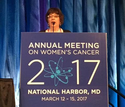
Dupilumab: FDA approves first biologic for atopic dermatitis
Dupilumab, a monoclonal antibody that targets both interleukin-4 and interleukin-13, has been approved for the treatment of moderate to severe atopic dermatitis in adults not adequately controlled with topical prescription therapies or for whom topicals are not appropriate.
The approval marks the first biologic approved for treating AD, according to a March 28 announcement from the Food and Drug Administration.
Dupilumab “inhibits signaling of IL-4 and IL-13, two key cytokines required for the type 2 (including Th2) immune response, which is believed to be a major driver in the pathogenesis of the disease,” according to Regeneron, which will market dupilumab.
Approval was based on three phase III pivotal studies of adults with moderate to severe AD whose disease was not adequately controlled with topical prescription treatments: SOLO-1 and SOLO-2, which evaluated dupilumab as monotherapy, and the CHRONOS study, which compared dupilumab with topical corticosteroids to treatment with topical corticosteroids alone.
The 16-week data from the SOLO-1 and -2 studies were presented at the 2016 annual congress of the European Academy for Dermatology and Venereology.
In two phase III trials of identical design involving patients with atopic dermatitis, dupilumab, administered weekly or every 2 weeks, improved the signs and symptoms of atopic dermatitis, including pruritus, symptoms of anxiety and depression, and quality of life, as compared with placebo. Eczema Area and Severity Index was reported in significantly more patients who received each regimen of dupilumab than in patients who received placebo (P less than .001 for all comparisons).
Dupilumab also was associated with improvement in other clinical endpoints, including reductions in pruritus and symptoms of anxiety or depression, and an improvement in quality of life. Injection-site reactions and conjunctivitis were more frequent in the dupilumab groups than in the placebo groups. The results were published in the New England Journal of Medicine (2016;375:2335-48).
More recently, 52-week data from the CHRONOS study were reported at the annual meeting of the American Academy of Dermatology in March. In that study of 740 adults with moderate to severe AD that was not controlled with topical medications – including corticosteroids with or without calcineurin inhibitors – those randomized to 300 mg of dupilumab once a week, plus topical corticosteroids, showed significantly greater improvements in measures of overall disease severity at 16 weeks and at 52 weeks, compared with those treated with steroids alone. Measures used included Eczema Area and Severity Index and the Pruritus Numerical Rating Scale, Patient Oriented Eczema Measure, Dermatology Life Quality Index.
Adverse events experienced with dupilumab included injection site reactions, eye and eyelid inflammation, and cold sores on the mouth or lips, according to a Regeneron statement.
Dupilumab will be available “later this week,” according to the statement, which noted the wholesale acquisition cost of the medication is expected to be $37,000 annually.
The FDA approval announcement noted that the safety and efficacy of dupilumab had not been established in patients with asthma.
Dupilumab currently is being studied for children with AD in phase II studies and is being studied for other indications: eosinophilic esophagitis in phase II studies, and asthma and nasal polyps in phase III studies.
Dupilumab will be marketed as Dupixent by Regeneron.
Dupilumab, a monoclonal antibody that targets both interleukin-4 and interleukin-13, has been approved for the treatment of moderate to severe atopic dermatitis in adults not adequately controlled with topical prescription therapies or for whom topicals are not appropriate.
The approval marks the first biologic approved for treating AD, according to a March 28 announcement from the Food and Drug Administration.
Dupilumab “inhibits signaling of IL-4 and IL-13, two key cytokines required for the type 2 (including Th2) immune response, which is believed to be a major driver in the pathogenesis of the disease,” according to Regeneron, which will market dupilumab.
Approval was based on three phase III pivotal studies of adults with moderate to severe AD whose disease was not adequately controlled with topical prescription treatments: SOLO-1 and SOLO-2, which evaluated dupilumab as monotherapy, and the CHRONOS study, which compared dupilumab with topical corticosteroids to treatment with topical corticosteroids alone.
The 16-week data from the SOLO-1 and -2 studies were presented at the 2016 annual congress of the European Academy for Dermatology and Venereology.
In two phase III trials of identical design involving patients with atopic dermatitis, dupilumab, administered weekly or every 2 weeks, improved the signs and symptoms of atopic dermatitis, including pruritus, symptoms of anxiety and depression, and quality of life, as compared with placebo. Eczema Area and Severity Index was reported in significantly more patients who received each regimen of dupilumab than in patients who received placebo (P less than .001 for all comparisons).
Dupilumab also was associated with improvement in other clinical endpoints, including reductions in pruritus and symptoms of anxiety or depression, and an improvement in quality of life. Injection-site reactions and conjunctivitis were more frequent in the dupilumab groups than in the placebo groups. The results were published in the New England Journal of Medicine (2016;375:2335-48).
More recently, 52-week data from the CHRONOS study were reported at the annual meeting of the American Academy of Dermatology in March. In that study of 740 adults with moderate to severe AD that was not controlled with topical medications – including corticosteroids with or without calcineurin inhibitors – those randomized to 300 mg of dupilumab once a week, plus topical corticosteroids, showed significantly greater improvements in measures of overall disease severity at 16 weeks and at 52 weeks, compared with those treated with steroids alone. Measures used included Eczema Area and Severity Index and the Pruritus Numerical Rating Scale, Patient Oriented Eczema Measure, Dermatology Life Quality Index.
Adverse events experienced with dupilumab included injection site reactions, eye and eyelid inflammation, and cold sores on the mouth or lips, according to a Regeneron statement.
Dupilumab will be available “later this week,” according to the statement, which noted the wholesale acquisition cost of the medication is expected to be $37,000 annually.
The FDA approval announcement noted that the safety and efficacy of dupilumab had not been established in patients with asthma.
Dupilumab currently is being studied for children with AD in phase II studies and is being studied for other indications: eosinophilic esophagitis in phase II studies, and asthma and nasal polyps in phase III studies.
Dupilumab will be marketed as Dupixent by Regeneron.
Dupilumab, a monoclonal antibody that targets both interleukin-4 and interleukin-13, has been approved for the treatment of moderate to severe atopic dermatitis in adults not adequately controlled with topical prescription therapies or for whom topicals are not appropriate.
The approval marks the first biologic approved for treating AD, according to a March 28 announcement from the Food and Drug Administration.
Dupilumab “inhibits signaling of IL-4 and IL-13, two key cytokines required for the type 2 (including Th2) immune response, which is believed to be a major driver in the pathogenesis of the disease,” according to Regeneron, which will market dupilumab.
Approval was based on three phase III pivotal studies of adults with moderate to severe AD whose disease was not adequately controlled with topical prescription treatments: SOLO-1 and SOLO-2, which evaluated dupilumab as monotherapy, and the CHRONOS study, which compared dupilumab with topical corticosteroids to treatment with topical corticosteroids alone.
The 16-week data from the SOLO-1 and -2 studies were presented at the 2016 annual congress of the European Academy for Dermatology and Venereology.
In two phase III trials of identical design involving patients with atopic dermatitis, dupilumab, administered weekly or every 2 weeks, improved the signs and symptoms of atopic dermatitis, including pruritus, symptoms of anxiety and depression, and quality of life, as compared with placebo. Eczema Area and Severity Index was reported in significantly more patients who received each regimen of dupilumab than in patients who received placebo (P less than .001 for all comparisons).
Dupilumab also was associated with improvement in other clinical endpoints, including reductions in pruritus and symptoms of anxiety or depression, and an improvement in quality of life. Injection-site reactions and conjunctivitis were more frequent in the dupilumab groups than in the placebo groups. The results were published in the New England Journal of Medicine (2016;375:2335-48).
More recently, 52-week data from the CHRONOS study were reported at the annual meeting of the American Academy of Dermatology in March. In that study of 740 adults with moderate to severe AD that was not controlled with topical medications – including corticosteroids with or without calcineurin inhibitors – those randomized to 300 mg of dupilumab once a week, plus topical corticosteroids, showed significantly greater improvements in measures of overall disease severity at 16 weeks and at 52 weeks, compared with those treated with steroids alone. Measures used included Eczema Area and Severity Index and the Pruritus Numerical Rating Scale, Patient Oriented Eczema Measure, Dermatology Life Quality Index.
Adverse events experienced with dupilumab included injection site reactions, eye and eyelid inflammation, and cold sores on the mouth or lips, according to a Regeneron statement.
Dupilumab will be available “later this week,” according to the statement, which noted the wholesale acquisition cost of the medication is expected to be $37,000 annually.
The FDA approval announcement noted that the safety and efficacy of dupilumab had not been established in patients with asthma.
Dupilumab currently is being studied for children with AD in phase II studies and is being studied for other indications: eosinophilic esophagitis in phase II studies, and asthma and nasal polyps in phase III studies.
Dupilumab will be marketed as Dupixent by Regeneron.

Liver disease likely to become increasing indication for bariatric surgery
PHILADELPHIA – There is a long list of benefits from bariatric surgery in the morbidly obese, but prevention of end-stage liver disease and the need for a first or second liver transplant is likely to grow as an indication, according to an overview of weight loss surgery at Digestive Diseases: New Advances, held by Rutgers, the State University of New Jersey, and Global Academy for Medical Education.
“Bariatric surgery is associated with significant improvement not just in diabetes, dyslipidemia, hypertension, and other complications of metabolic disorders but for me more interestingly, it is effective for treating fatty liver disease where you can see a 90% improvement in steatosis,” reported Subhashini Ayloo, MD, chief of minimally invasive robotic hepato-pancreato-biliary surgery and liver transplantation at New Jersey Medical School, Newark.
Trained in both bariatric surgery and liver transplant, Dr. Ayloo predicts that these fields will become increasingly connected because of the obesity epidemic and the related rise in nonalcoholic fatty liver disease (NAFLD). Dr. Ayloo reported that bariatric surgery is already being used in her center to avoid a second liver transplant in obese patients who are unable to lose sufficient weight to prevent progressive NAFLD after a first transplant.
The emphasis Dr. Ayloo placed on the role of bariatric surgery in preventing progression of NAFLD to nonalcoholic steatohepatitis and the inflammatory process that leads to fibrosis, cirrhosis, and liver decompensation, was drawn from her interest in these two fields. However, she did not ignore the potential of protection from obesity control for other diseases.
“Obesity adversely affects every organ in the body,” Dr. Ayloo pointed out. As a result of weight loss achieved with bariatric surgery, there is now a large body of evidence supporting broad benefits, not just those related to fat deposited in hepatocytes.
“We have a couple of decades of experience that has been published [with bariatric surgery], and this has shown that it maintains weight loss long term, it improves all the obesity-associated comorbidities, and it is cost effective,” Dr. Ayloo said. Now with long-term follow-up, “all of the studies are showing that bariatric surgery improves survival.”
Although most of the survival data have been generated by retrospective cohort studies, Dr. Ayloo cited nine sets of data showing odds ratios associating bariatric surgery with up to a 90% reduction in death over periods of up to 10 years of follow-up. In a summary slide presented by Dr. Ayloo, the estimated mortality benefit over 5 years was listed as 85%. The same summary slide listed large improvements in relevant measures of morbidity for more than 10 organ systems, such as improvement or resolution of dyslipidemia and hypertension in the circulatory system, improvement or resolution of asthma and other diseases affecting the respiratory system, and resolution or improvement of gastroesophageal reflux disease and other diseases affecting the gastrointestinal system.
Specific to the liver, these benefits included a nearly 40% reduction in liver inflammation and 20% reduction in fibrosis. According to Dr. Ayloo, who noted that NAFLD is expected to overtake hepatitis C virus as the No. 1 cause of liver transplant within the next 5 years, these data are important for drawing attention to bariatric surgery as a strategy to control liver disease. She suggested that there is a need to create a tighter link between efforts to treat morbid obesity and advanced liver disease.
“There is an established literature showing that if somebody is morbidly obese, the rate of liver transplant is lower than when compared to patients with normal weight,” Dr. Ayloo said. “There is a call out in the transplant community that we need to address this and we cannot just be throwing this under the table.”
Because of the strong relationship between obesity and NAFLD, a systematic approach is needed to consider liver disease in obese patients and obesity in patients with liver disease, she said. The close relationship is relevant when planning interventions for either. Liver disease should be assessed prior to bariatric surgery regardless of the indication and then monitored closely as part of postoperative care, she said.
Dr. Ayloo identified weight control as an essential part of posttransplant care to prevent hepatic fat deposition that threatens transplant-free survival.
Global Academy and this news organization are owned by the same company. Dr. Ayloo reports no relevant financial relationships.
PHILADELPHIA – There is a long list of benefits from bariatric surgery in the morbidly obese, but prevention of end-stage liver disease and the need for a first or second liver transplant is likely to grow as an indication, according to an overview of weight loss surgery at Digestive Diseases: New Advances, held by Rutgers, the State University of New Jersey, and Global Academy for Medical Education.
“Bariatric surgery is associated with significant improvement not just in diabetes, dyslipidemia, hypertension, and other complications of metabolic disorders but for me more interestingly, it is effective for treating fatty liver disease where you can see a 90% improvement in steatosis,” reported Subhashini Ayloo, MD, chief of minimally invasive robotic hepato-pancreato-biliary surgery and liver transplantation at New Jersey Medical School, Newark.
Trained in both bariatric surgery and liver transplant, Dr. Ayloo predicts that these fields will become increasingly connected because of the obesity epidemic and the related rise in nonalcoholic fatty liver disease (NAFLD). Dr. Ayloo reported that bariatric surgery is already being used in her center to avoid a second liver transplant in obese patients who are unable to lose sufficient weight to prevent progressive NAFLD after a first transplant.
The emphasis Dr. Ayloo placed on the role of bariatric surgery in preventing progression of NAFLD to nonalcoholic steatohepatitis and the inflammatory process that leads to fibrosis, cirrhosis, and liver decompensation, was drawn from her interest in these two fields. However, she did not ignore the potential of protection from obesity control for other diseases.
“Obesity adversely affects every organ in the body,” Dr. Ayloo pointed out. As a result of weight loss achieved with bariatric surgery, there is now a large body of evidence supporting broad benefits, not just those related to fat deposited in hepatocytes.
“We have a couple of decades of experience that has been published [with bariatric surgery], and this has shown that it maintains weight loss long term, it improves all the obesity-associated comorbidities, and it is cost effective,” Dr. Ayloo said. Now with long-term follow-up, “all of the studies are showing that bariatric surgery improves survival.”
Although most of the survival data have been generated by retrospective cohort studies, Dr. Ayloo cited nine sets of data showing odds ratios associating bariatric surgery with up to a 90% reduction in death over periods of up to 10 years of follow-up. In a summary slide presented by Dr. Ayloo, the estimated mortality benefit over 5 years was listed as 85%. The same summary slide listed large improvements in relevant measures of morbidity for more than 10 organ systems, such as improvement or resolution of dyslipidemia and hypertension in the circulatory system, improvement or resolution of asthma and other diseases affecting the respiratory system, and resolution or improvement of gastroesophageal reflux disease and other diseases affecting the gastrointestinal system.
Specific to the liver, these benefits included a nearly 40% reduction in liver inflammation and 20% reduction in fibrosis. According to Dr. Ayloo, who noted that NAFLD is expected to overtake hepatitis C virus as the No. 1 cause of liver transplant within the next 5 years, these data are important for drawing attention to bariatric surgery as a strategy to control liver disease. She suggested that there is a need to create a tighter link between efforts to treat morbid obesity and advanced liver disease.
“There is an established literature showing that if somebody is morbidly obese, the rate of liver transplant is lower than when compared to patients with normal weight,” Dr. Ayloo said. “There is a call out in the transplant community that we need to address this and we cannot just be throwing this under the table.”
Because of the strong relationship between obesity and NAFLD, a systematic approach is needed to consider liver disease in obese patients and obesity in patients with liver disease, she said. The close relationship is relevant when planning interventions for either. Liver disease should be assessed prior to bariatric surgery regardless of the indication and then monitored closely as part of postoperative care, she said.
Dr. Ayloo identified weight control as an essential part of posttransplant care to prevent hepatic fat deposition that threatens transplant-free survival.
Global Academy and this news organization are owned by the same company. Dr. Ayloo reports no relevant financial relationships.
PHILADELPHIA – There is a long list of benefits from bariatric surgery in the morbidly obese, but prevention of end-stage liver disease and the need for a first or second liver transplant is likely to grow as an indication, according to an overview of weight loss surgery at Digestive Diseases: New Advances, held by Rutgers, the State University of New Jersey, and Global Academy for Medical Education.
“Bariatric surgery is associated with significant improvement not just in diabetes, dyslipidemia, hypertension, and other complications of metabolic disorders but for me more interestingly, it is effective for treating fatty liver disease where you can see a 90% improvement in steatosis,” reported Subhashini Ayloo, MD, chief of minimally invasive robotic hepato-pancreato-biliary surgery and liver transplantation at New Jersey Medical School, Newark.
Trained in both bariatric surgery and liver transplant, Dr. Ayloo predicts that these fields will become increasingly connected because of the obesity epidemic and the related rise in nonalcoholic fatty liver disease (NAFLD). Dr. Ayloo reported that bariatric surgery is already being used in her center to avoid a second liver transplant in obese patients who are unable to lose sufficient weight to prevent progressive NAFLD after a first transplant.
The emphasis Dr. Ayloo placed on the role of bariatric surgery in preventing progression of NAFLD to nonalcoholic steatohepatitis and the inflammatory process that leads to fibrosis, cirrhosis, and liver decompensation, was drawn from her interest in these two fields. However, she did not ignore the potential of protection from obesity control for other diseases.
“Obesity adversely affects every organ in the body,” Dr. Ayloo pointed out. As a result of weight loss achieved with bariatric surgery, there is now a large body of evidence supporting broad benefits, not just those related to fat deposited in hepatocytes.
“We have a couple of decades of experience that has been published [with bariatric surgery], and this has shown that it maintains weight loss long term, it improves all the obesity-associated comorbidities, and it is cost effective,” Dr. Ayloo said. Now with long-term follow-up, “all of the studies are showing that bariatric surgery improves survival.”
Although most of the survival data have been generated by retrospective cohort studies, Dr. Ayloo cited nine sets of data showing odds ratios associating bariatric surgery with up to a 90% reduction in death over periods of up to 10 years of follow-up. In a summary slide presented by Dr. Ayloo, the estimated mortality benefit over 5 years was listed as 85%. The same summary slide listed large improvements in relevant measures of morbidity for more than 10 organ systems, such as improvement or resolution of dyslipidemia and hypertension in the circulatory system, improvement or resolution of asthma and other diseases affecting the respiratory system, and resolution or improvement of gastroesophageal reflux disease and other diseases affecting the gastrointestinal system.
Specific to the liver, these benefits included a nearly 40% reduction in liver inflammation and 20% reduction in fibrosis. According to Dr. Ayloo, who noted that NAFLD is expected to overtake hepatitis C virus as the No. 1 cause of liver transplant within the next 5 years, these data are important for drawing attention to bariatric surgery as a strategy to control liver disease. She suggested that there is a need to create a tighter link between efforts to treat morbid obesity and advanced liver disease.
“There is an established literature showing that if somebody is morbidly obese, the rate of liver transplant is lower than when compared to patients with normal weight,” Dr. Ayloo said. “There is a call out in the transplant community that we need to address this and we cannot just be throwing this under the table.”
Because of the strong relationship between obesity and NAFLD, a systematic approach is needed to consider liver disease in obese patients and obesity in patients with liver disease, she said. The close relationship is relevant when planning interventions for either. Liver disease should be assessed prior to bariatric surgery regardless of the indication and then monitored closely as part of postoperative care, she said.
Dr. Ayloo identified weight control as an essential part of posttransplant care to prevent hepatic fat deposition that threatens transplant-free survival.
Global Academy and this news organization are owned by the same company. Dr. Ayloo reports no relevant financial relationships.

What Is the Impact of a High-Salt Diet in Patients With MS?
ORLANDO—A high-salt diet might be a key environmental risk factor for multiple sclerosis (MS), according to an overview presented at the ACTRIMS 2017 Forum. Since most research has been performed in vitro and in animal models, it remains unclear how a high-salt diet affects patients with MS. Researchers have found in experimental models, however, that salt induces inflammation by several mechanisms: it increases frequency of inflammatory TH17 cells, decreases function of suppressor cells, and increases inflammation of antigen-presenting cells.
“There’s been an epidemic of human autoimmune disease over the past 70 years,” said Dr. Hafler. Researchers are working to discover why there has been such a significant rise in cases. “Genetics cannot allow this to happen. There must be something in the environment,” said Dr. Hafler. He and his colleagues sought to understand how a high-salt diet affects MS.
“What we found is if you added sodium chloride to cultures of T cells, you have autoimmune increases in the frequency of TH17 cells,” he said. In addition, salt decreases function of suppressor cells, the Tregs, and increases inflammation of antigen-presenting cells of the immune system, said Dr. Hafler.
Previous research found that mice fed a high-salt diet were more prone to severe experimental autoimmune encephalomyelitis. When the mice were given a high-salt, high-fat diet that mimicked fast food and probiotics, researchers observed a decrease in inflammation.
“It is really surprising to me how diet can really influence the degree of inflammation in these animals,” said Dr. Hafler. “We do not know if this will work in humans, but probiotics may decrease inflammation. We have no information [on] whether a diet with a probiotic would help prevent or treat MS.”
According to Farez et al, a higher sodium intake is associated with increased clinical and radiological disease activity in patients with MS. Other studies have found that environmental factors such as smoking and low vitamin D levels are associated with MS risk, and the risk is genetically mediated. What may be an environmental risk for one individual may not be a risk for another individual, said Dr. Hafler. “It is unlikely that any of these factors by themselves—smoking, vitamin D, fat, and salt—would be critically important, but together, they might have a strong effect,” he said.
Since there is limited research on a high-salt diet in humans, Dr. Hafler does not recommend his patients with MS go on a strict low-salt diet. However, he does advise patients to stay away from processed foods and fast food. He also encourages patients to get most of their calories from fruits and vegetables.
“We are now doing studies in which we put patients and control subjects on a high-salt diet and low[-salt] diet to observe the direct effect in those individuals,” said Dr. Hafler.
—Erica Tricarico
Suggested Reading
Farez MF, Fiol MP, Gaitán MI, et al. Sodium intake is associated with increased disease activity in multiple sclerosis. J Neurol Neurosurg Psychiatry.
Jörg S, Kissel J, Manzel A, et al. High salt drives Th17 responses in experimental autoimmune encephalomyelitis without impacting myeloid dendritic cells. Exp Neurol. 2016;279:212-222.
Kleinewietfeld M, Manzel A, Tize J, et al. Sodium chloride drives autoimmune disease by the induction of pathogenic TH17 cells. Nature. 2013;496(7446):518-522.
Paling D, Solanky BS, Riemer F, et al. Sodium accumulation is associated with disability and a progressive course in multiple sclerosis. Brain. 2013;136(Pt 7):2305-2317.
ORLANDO—A high-salt diet might be a key environmental risk factor for multiple sclerosis (MS), according to an overview presented at the ACTRIMS 2017 Forum. Since most research has been performed in vitro and in animal models, it remains unclear how a high-salt diet affects patients with MS. Researchers have found in experimental models, however, that salt induces inflammation by several mechanisms: it increases frequency of inflammatory TH17 cells, decreases function of suppressor cells, and increases inflammation of antigen-presenting cells.
“There’s been an epidemic of human autoimmune disease over the past 70 years,” said Dr. Hafler. Researchers are working to discover why there has been such a significant rise in cases. “Genetics cannot allow this to happen. There must be something in the environment,” said Dr. Hafler. He and his colleagues sought to understand how a high-salt diet affects MS.
“What we found is if you added sodium chloride to cultures of T cells, you have autoimmune increases in the frequency of TH17 cells,” he said. In addition, salt decreases function of suppressor cells, the Tregs, and increases inflammation of antigen-presenting cells of the immune system, said Dr. Hafler.
Previous research found that mice fed a high-salt diet were more prone to severe experimental autoimmune encephalomyelitis. When the mice were given a high-salt, high-fat diet that mimicked fast food and probiotics, researchers observed a decrease in inflammation.
“It is really surprising to me how diet can really influence the degree of inflammation in these animals,” said Dr. Hafler. “We do not know if this will work in humans, but probiotics may decrease inflammation. We have no information [on] whether a diet with a probiotic would help prevent or treat MS.”
According to Farez et al, a higher sodium intake is associated with increased clinical and radiological disease activity in patients with MS. Other studies have found that environmental factors such as smoking and low vitamin D levels are associated with MS risk, and the risk is genetically mediated. What may be an environmental risk for one individual may not be a risk for another individual, said Dr. Hafler. “It is unlikely that any of these factors by themselves—smoking, vitamin D, fat, and salt—would be critically important, but together, they might have a strong effect,” he said.
Since there is limited research on a high-salt diet in humans, Dr. Hafler does not recommend his patients with MS go on a strict low-salt diet. However, he does advise patients to stay away from processed foods and fast food. He also encourages patients to get most of their calories from fruits and vegetables.
“We are now doing studies in which we put patients and control subjects on a high-salt diet and low[-salt] diet to observe the direct effect in those individuals,” said Dr. Hafler.
—Erica Tricarico
Suggested Reading
Farez MF, Fiol MP, Gaitán MI, et al. Sodium intake is associated with increased disease activity in multiple sclerosis. J Neurol Neurosurg Psychiatry.
Jörg S, Kissel J, Manzel A, et al. High salt drives Th17 responses in experimental autoimmune encephalomyelitis without impacting myeloid dendritic cells. Exp Neurol. 2016;279:212-222.
Kleinewietfeld M, Manzel A, Tize J, et al. Sodium chloride drives autoimmune disease by the induction of pathogenic TH17 cells. Nature. 2013;496(7446):518-522.
Paling D, Solanky BS, Riemer F, et al. Sodium accumulation is associated with disability and a progressive course in multiple sclerosis. Brain. 2013;136(Pt 7):2305-2317.
ORLANDO—A high-salt diet might be a key environmental risk factor for multiple sclerosis (MS), according to an overview presented at the ACTRIMS 2017 Forum. Since most research has been performed in vitro and in animal models, it remains unclear how a high-salt diet affects patients with MS. Researchers have found in experimental models, however, that salt induces inflammation by several mechanisms: it increases frequency of inflammatory TH17 cells, decreases function of suppressor cells, and increases inflammation of antigen-presenting cells.
“There’s been an epidemic of human autoimmune disease over the past 70 years,” said Dr. Hafler. Researchers are working to discover why there has been such a significant rise in cases. “Genetics cannot allow this to happen. There must be something in the environment,” said Dr. Hafler. He and his colleagues sought to understand how a high-salt diet affects MS.
“What we found is if you added sodium chloride to cultures of T cells, you have autoimmune increases in the frequency of TH17 cells,” he said. In addition, salt decreases function of suppressor cells, the Tregs, and increases inflammation of antigen-presenting cells of the immune system, said Dr. Hafler.
Previous research found that mice fed a high-salt diet were more prone to severe experimental autoimmune encephalomyelitis. When the mice were given a high-salt, high-fat diet that mimicked fast food and probiotics, researchers observed a decrease in inflammation.
“It is really surprising to me how diet can really influence the degree of inflammation in these animals,” said Dr. Hafler. “We do not know if this will work in humans, but probiotics may decrease inflammation. We have no information [on] whether a diet with a probiotic would help prevent or treat MS.”
According to Farez et al, a higher sodium intake is associated with increased clinical and radiological disease activity in patients with MS. Other studies have found that environmental factors such as smoking and low vitamin D levels are associated with MS risk, and the risk is genetically mediated. What may be an environmental risk for one individual may not be a risk for another individual, said Dr. Hafler. “It is unlikely that any of these factors by themselves—smoking, vitamin D, fat, and salt—would be critically important, but together, they might have a strong effect,” he said.
Since there is limited research on a high-salt diet in humans, Dr. Hafler does not recommend his patients with MS go on a strict low-salt diet. However, he does advise patients to stay away from processed foods and fast food. He also encourages patients to get most of their calories from fruits and vegetables.
“We are now doing studies in which we put patients and control subjects on a high-salt diet and low[-salt] diet to observe the direct effect in those individuals,” said Dr. Hafler.
—Erica Tricarico
Suggested Reading
Farez MF, Fiol MP, Gaitán MI, et al. Sodium intake is associated with increased disease activity in multiple sclerosis. J Neurol Neurosurg Psychiatry.
Jörg S, Kissel J, Manzel A, et al. High salt drives Th17 responses in experimental autoimmune encephalomyelitis without impacting myeloid dendritic cells. Exp Neurol. 2016;279:212-222.
Kleinewietfeld M, Manzel A, Tize J, et al. Sodium chloride drives autoimmune disease by the induction of pathogenic TH17 cells. Nature. 2013;496(7446):518-522.
Paling D, Solanky BS, Riemer F, et al. Sodium accumulation is associated with disability and a progressive course in multiple sclerosis. Brain. 2013;136(Pt 7):2305-2317.