User login
Newer St. Jude leads last as long as Medtronic Sprint Quattro
SAN FRANCISCO – St. Jude Medical’s Durata and Riata ST Optim defibrillator leads performed comparably to Medtronic’s Sprint Quattro out to 7 years in a Veterans Affairs analysis of almost 18,000 patients in the VA National Cardiac Device Surveillance Program.
The “highly satisfactory electrical survival” of the Optim leads, at least until year 5, should be of some reassurance to cardiologists, especially since the findings come from the VA, not a device company, investigator Seema Pursnani, MD, said.
The investigators combined Durata and Riata ST Optim leads together in their analysis, since they are similar; both are 7 Fr leads with St. Jude’s silicone/polyurethane Optim coating. After a mean follow-up of 3.4 years in 4,091 Durata patients and 351 Riata ST Optim patients, there were 26 electrical lead failures, which translated to 0.17% failures per device-year.
The investigators compared those results with Medtronic’s Sprint Quattro, which “is sort of a gold standard. It’s been around for quite a long time, and people have confidence in it,” said Dr. Pursnani. After a mean follow-up of 3.8 years in 13,254 patients, there were 57 failures, translating to 0.11% failures per device-year.
Seven-year lead survival was 97.7% with St. Jude’s products, and 98.9% with Medtronic’s. Although the difference was not statistically significant, “we need a little more follow-up to see why the curves are diverging at years 6 and 7,” said Dr. Pursnani, a cardiologist at the San Francisco Veterans Affairs Medical Center when the study was done, but now with the Kaiser Permanente San Leandro (Calif.) Medical Center.
There’s been lingering concern about St. Jude leads ever since the recall of earlier versions of Riata – with silicone-only insulation – in 2011 because of lead abrasion and subsequent safety problems. Optim was developed to address the issue.
“One of the most common modes of lead failure that we saw” with all three leads “was a rise in the pace-sense conductor impedance from the baseline impedance. Sometimes, there is nonphysiologic noise that can also be a sign of early failure,” she said.
There was no industry funding for the work, and the investigators have no disclosures.
AT HEART RHYTHM 2016
Key clinical point: St. Jude may have solved the Riata lead problem.
Major finding: Seven-year lead survival was 97.7% with St. Jude’s products, and 98.9% with Medtronic’s; the difference was not statistically significant.
Data source: Veterans Affairs analysis of almost 18,000 patients
Disclosures: There was no industry funding for the work, and the investigators have no disclosures.
LAA excision of no benefit in persistent AF ablation
SAN FRANCISCO – Adding left atrial appendage excision to pulmonary vein isolation does not reduce the rate of recurrence in persistent atrial fibrillation, according to a Russian investigation.
Eighty-eight patients with persistent atrial fibrillation (AF) were randomized to thoracoscopic pulmonary vein isolation (PVI) with bilateral epicardial ganglia ablation and box lesion set of the posterior left atrial wall; 88 others were randomized to that approach plus left atrial appendage (LAA) amputation. After 18 months, 64 out of 87 patients in the LAA-excision group (73.6%) and 61 out of 86 patients (70.9%) in the control group were free from recurrent AF, meaning no episodes greater than 30 seconds (P = .73). Freedom from any atrial arrhythmia after a single procedure with or without follow-up antiarrhythmic drugs (AADs) was also similar, with 70.9% in the control and 74.7% in the treatment groups. “Both approaches had excellent” results with no differences in complication rates, but there “was no reduction in AF recurrence when LAA excision was performed,” said investigator Dr. Alexander Romanov of the State Research Institute of Circulation Pathology, Novosibirsk, Russia.
The results are a bit surprising because some previous studies have suggested that electrical isolation of the LAA improves AF ablation success, and surgical excision might be expected to have a similar effect. In many places in the United States, LAA excisions are routine in open heart surgery when patients have AF, to prevent stroke. Guidelines for AF management from the American Heart Association, American College of Cardiology, and Heart Rhythm Society published in 2014 give a class IIb recommendation, saying “surgical excision of the left atrial appendage may be considered in patients undergoing cardiac surgery,” with an evidence level of C, meaning there are no data to support the recommendation, only expert consensus (J Am Coll Cardiol. 2014;64[21]:2246-80).
There were no significant differences between the groups; patients were about 60 years old, on average, and more than 80% in both groups had baseline CHADS2 scores of 0 or 1. All patients had persistent AF for more than a week but no longer than a year; longer-standing cases were excluded, as were patients with prior heart surgeries or catheter ablations. There were no statistically significant differences in operative times or complications. A few patients in each arm needed sternotomies for hemostasis, and one in each arm had a stroke during follow-up. Patients were followed at regular intervals by ECG and Holter monitoring.
AADs were allowed during the blanking period; patients could continue them afterwards for AF recurrence or have endocardial redo ablations; 10 patients in the control group (12%) and 13 in the LAA group (15%) had repeat procedures (P = .55). Most were for right atrial flutter and a few for left atrial flutter. “Only one redo case was for true AF recurrence,” Dr. Romanov said.
The team did not test for exertion intolerance and other potential LAA excision problems.
Dr. Romanov is a speaker for Medtronic, Biosense Webster, and Boston Scientific.
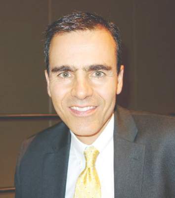 |
Dr. John Day |
This study is interesting because it goes against what other studies are showing, which is that LAA isolation increases the success rate with AF ablation. What makes me a little suspicious is that the success rates in both arms of this study were unusually high for persistent AF. If they were more in line with previous reports, I would feel a little bit better concluding that LAA isolation doesn’t’ help.
I know anecdotally from having done thousands of these ablations that there are some patients whose AF originates from the LAA, and if you treat it, you improve their outcomes.
Dr. John Day is the director of Intermountain Heart Rhythm Specialists in Murray, Utah, and the current president of the Hearth Rhythm Society. He has no disclosures.
SAN FRANCISCO – Adding left atrial appendage excision to pulmonary vein isolation does not reduce the rate of recurrence in persistent atrial fibrillation, according to a Russian investigation.
Eighty-eight patients with persistent atrial fibrillation (AF) were randomized to thoracoscopic pulmonary vein isolation (PVI) with bilateral epicardial ganglia ablation and box lesion set of the posterior left atrial wall; 88 others were randomized to that approach plus left atrial appendage (LAA) amputation. After 18 months, 64 out of 87 patients in the LAA-excision group (73.6%) and 61 out of 86 patients (70.9%) in the control group were free from recurrent AF, meaning no episodes greater than 30 seconds (P = .73). Freedom from any atrial arrhythmia after a single procedure with or without follow-up antiarrhythmic drugs (AADs) was also similar, with 70.9% in the control and 74.7% in the treatment groups. “Both approaches had excellent” results with no differences in complication rates, but there “was no reduction in AF recurrence when LAA excision was performed,” said investigator Dr. Alexander Romanov of the State Research Institute of Circulation Pathology, Novosibirsk, Russia.
The results are a bit surprising because some previous studies have suggested that electrical isolation of the LAA improves AF ablation success, and surgical excision might be expected to have a similar effect. In many places in the United States, LAA excisions are routine in open heart surgery when patients have AF, to prevent stroke. Guidelines for AF management from the American Heart Association, American College of Cardiology, and Heart Rhythm Society published in 2014 give a class IIb recommendation, saying “surgical excision of the left atrial appendage may be considered in patients undergoing cardiac surgery,” with an evidence level of C, meaning there are no data to support the recommendation, only expert consensus (J Am Coll Cardiol. 2014;64[21]:2246-80).
There were no significant differences between the groups; patients were about 60 years old, on average, and more than 80% in both groups had baseline CHADS2 scores of 0 or 1. All patients had persistent AF for more than a week but no longer than a year; longer-standing cases were excluded, as were patients with prior heart surgeries or catheter ablations. There were no statistically significant differences in operative times or complications. A few patients in each arm needed sternotomies for hemostasis, and one in each arm had a stroke during follow-up. Patients were followed at regular intervals by ECG and Holter monitoring.
AADs were allowed during the blanking period; patients could continue them afterwards for AF recurrence or have endocardial redo ablations; 10 patients in the control group (12%) and 13 in the LAA group (15%) had repeat procedures (P = .55). Most were for right atrial flutter and a few for left atrial flutter. “Only one redo case was for true AF recurrence,” Dr. Romanov said.
The team did not test for exertion intolerance and other potential LAA excision problems.
Dr. Romanov is a speaker for Medtronic, Biosense Webster, and Boston Scientific.
AT HEART RHYTHM 2016
Key clinical point: Adding left atrial appendage excision to pulmonary vein isolation does not reduce the rate of recurrence in persistent atrial fibrillation.
Major finding: After 18 months, 64 out of 87 patients in the LAA-excision group (73.6%) and 61 out of 86 patients (70.9%) in the control group were free from recurrent AF, meaning no episodes greater than 30 seconds (P = 0.73).
Data source: Randomized trial in 176 patients with persistent AF.
Disclosures: The lead investigator is a speaker for Medtronic, Biosense Webster, and Boston Scientific.
ICD same-day discharge safe, but not a money saver
San Francisco – Same day discharge is generally safe after cardioverter defibrillator implantation for primary prevention, but it doesn’t save money.
Furthermore, guidelines are needed to standardize the practice as it becomes increasingly common in the United States, according to a 25-site investigation.
After implantable cardioverter defibrillator (ICD) procedures, patients were monitored for 3-4 hours, and their devices were checked for proper functioning; 129 patients who were stable at that point were randomized to early discharge and 136 to next day discharge (NDD).
The overall 30-day procedural complication rate was 3.1% in the same day discharge (SDD) group and 1.6% in the NDD group, a nonsignificant difference (P = .37). Three patients in the SDD group developed hematomas that resolved on their own, and one had a cardiac perforation. One NDD patient dislodged a lead and another developed an infection. There were no differences in quality of life measures between the two groups at 30 days.
However, there were also no differences in procedural and perioperative direct costs, which was surprising because saving money is a major driver of SDD, and the most expensive part of ICD implantation is the first 24 hours. Direct per-patient medical costs in the study – estimated by applying hospital cost-to-charge ratios to the Medicare-reported charge – were $31,771 for SDD and $30,437 for NDD, but NDD was more expensive than SDD at several sites. The investigators suspect a flaw in their analysis related to the opaque nature of hospital accounting, and plan to look into the matter further with modeling to identify savings opportunities with SDD.
“We can insert ICDs on an outpatient basis, but this study will be difficult to replicate because clinical practice is moving towards SDD. In view of this, we think professional societies should be thinking of standardizing criteria for SDD; guidelines would help with the adoption of this approach. There are clinicians who are astute and have great clinical judgment, but there are others who need a scoring system. We believe that by using the 270,000 patients in the [American College of Cardiology’s ICD Registry], there is the ability to identify patients who have low periprocedural risk,” said lead investigator Dr. Ranjit Suri, a cardiologist at Mt. Sinai Hospital in New York.
The study excluded patients receiving an ICD for secondary prevention, as well as those on periprocedural heparin and patients who were pacemaker dependent. SDD seemed safe otherwise, but it’s unknown “if our concept of low risk is acceptable to all implanting physicians,” Dr. Suri said at the annual scientific sessions of the Heart Rhythm Society.
The study groups were well matched. About 75% in each arm were men, and ischemic cardiomyopathy was the leading ICD indication. Patients were amenable to the idea of SDD; the advent of remote monitoring “adds a certain sense of safety” for both patients and physicians, he said.
Dr. Suri is a speaker for Boehringer Ingelheim and St. Jude Medical. He is also a consultant for Biosense Webster and Zoll, and receives research funding from St. Jude.
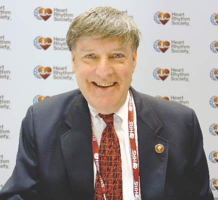 |
Dr. Thomas Deering |
The vast majority of primary prevention patients who are clinically stable enough to come in as outpatients can go home as outpatients if you watch them for a short period of time and make sure they are clinically stable. Most patients don’t want to be in the hospital, and many hospitals are crunched for available beds. It would be great to have guidelines on how to handle this, but we have to allow for clinical judgment.
Dr. Thomas Deering is chief of the Arrhythmia Center at the Piedmont Heart Institute in Atlanta, where he is also chairman of the Executive Council and the Clinical Centers for Excellence. He moderated Dr. Suri’s presentation and was not involved in the work.
San Francisco – Same day discharge is generally safe after cardioverter defibrillator implantation for primary prevention, but it doesn’t save money.
Furthermore, guidelines are needed to standardize the practice as it becomes increasingly common in the United States, according to a 25-site investigation.
After implantable cardioverter defibrillator (ICD) procedures, patients were monitored for 3-4 hours, and their devices were checked for proper functioning; 129 patients who were stable at that point were randomized to early discharge and 136 to next day discharge (NDD).
The overall 30-day procedural complication rate was 3.1% in the same day discharge (SDD) group and 1.6% in the NDD group, a nonsignificant difference (P = .37). Three patients in the SDD group developed hematomas that resolved on their own, and one had a cardiac perforation. One NDD patient dislodged a lead and another developed an infection. There were no differences in quality of life measures between the two groups at 30 days.
However, there were also no differences in procedural and perioperative direct costs, which was surprising because saving money is a major driver of SDD, and the most expensive part of ICD implantation is the first 24 hours. Direct per-patient medical costs in the study – estimated by applying hospital cost-to-charge ratios to the Medicare-reported charge – were $31,771 for SDD and $30,437 for NDD, but NDD was more expensive than SDD at several sites. The investigators suspect a flaw in their analysis related to the opaque nature of hospital accounting, and plan to look into the matter further with modeling to identify savings opportunities with SDD.
“We can insert ICDs on an outpatient basis, but this study will be difficult to replicate because clinical practice is moving towards SDD. In view of this, we think professional societies should be thinking of standardizing criteria for SDD; guidelines would help with the adoption of this approach. There are clinicians who are astute and have great clinical judgment, but there are others who need a scoring system. We believe that by using the 270,000 patients in the [American College of Cardiology’s ICD Registry], there is the ability to identify patients who have low periprocedural risk,” said lead investigator Dr. Ranjit Suri, a cardiologist at Mt. Sinai Hospital in New York.
The study excluded patients receiving an ICD for secondary prevention, as well as those on periprocedural heparin and patients who were pacemaker dependent. SDD seemed safe otherwise, but it’s unknown “if our concept of low risk is acceptable to all implanting physicians,” Dr. Suri said at the annual scientific sessions of the Heart Rhythm Society.
The study groups were well matched. About 75% in each arm were men, and ischemic cardiomyopathy was the leading ICD indication. Patients were amenable to the idea of SDD; the advent of remote monitoring “adds a certain sense of safety” for both patients and physicians, he said.
Dr. Suri is a speaker for Boehringer Ingelheim and St. Jude Medical. He is also a consultant for Biosense Webster and Zoll, and receives research funding from St. Jude.
AT HEART RHYTHM 2016
Key clinical point: Same-day discharge is generally safe after cardioverter defibrillator implantation for primary prevention, but it doesn’t save money and guidelines are needed to standardize the practice as it becomes increasingly common in the United States.
Major finding: The overall 30-day procedural complication rate was 3.1% in the same day discharge (SDD) group and 1.5% in the next-day discharge group, a nonsignificant difference (P = .37).
Data source: Randomized trial of 265 ICD patients.
Disclosures: The lead investigator is a speaker for Boehringer Ingelheim and St. Jude Medical. He is also a consultant for Biosense Webster and Zoll, and receives research funding from St. Jude.
Rotor ablation for atrial fibrillation strikes out in first randomized trial
SAN FRANCISCO – Focal impulse and rotor modulation-guided ablation for persistent atrial fibrillation – either alone or in conjunction with other procedures – increased procedural times without improving outcomes, according to the first randomized trial to assess its utility.
In fact, enrollment in the rotor ablation-only (RA) arm was halted early for futility. “There was 100% recurrence” of atrial fibrillation (AF), said senior investigator Dr. Andrea Natale, executive medical director of the Texas Cardiac Arrhythmia Institute, Austin.
“I’m surprised it took this long for a randomized study, because this system has been around for 5 or 6 years,” noted Dr. Natale. “Our community should demand these sorts of studies earlier, because it’s not fair for patients to go on with a procedure for years that has not been proven to be effective.
“For us, unless there is a new version of rotor mapping that I feel is significantly different, this will be the end of rotor ablation in my lab with this system [the Topera Physiologic Rotor Mapping Solution],” Dr. Natale said at the annual scientific sessions of the Heart Rhythm Society.
In the study, his team randomized 29 patients to RA only, 42 to RA plus pulmonary vein antral isolation (PVAI), and 42 to PVAI plus posterior wall and nonpulmonary vein trigger ablation.
At about 1 year, four RA-only patients (14%), 22 RA plus PVAI patients (52%), and 32 patients in the PVAI plus trigger group (76%) were free of AF and atrial tachycardias without antiarrhythmic drugs (P < .0001).
Meanwhile, RA alone and RA plus PVAI cases took about 230 minutes, while the more effective PVAI plus trigger approach took about 130 minutes (P < .001).
There was “a very poor outcome with rotor-only ablation,” Dr. Natale said. “There isn’t a benefit either alone or as an add-on strategy, at least with this mapping software.”
Perhaps “people who think rotors don’t exist are right,” he added. On the other hand, maybe the basket mapping catheter doesn’t touch enough of the left atrium, or the software that makes sense of what the catheter detects needs to be improved, Dr. Natale noted.
All the patients were undergoing their first ablation. They were in their early 60s, on average, and most were men. The mean left atrium diameter was about 47 mm, and mean left ventricle ejection fraction about 55%. There were no statistically significant differences between the study arms, and no significant differences in outcomes between the 70% of patients with persistent AF and the 30% with long-standing persistent AF.
There was no industry funding for the work. Dr. Natale disclosed relationships with Biosense Webster, Boston Scientific, Janssen, Medtronic, and St. Jude Medical.
My gut sense is that there’s something to rotor mapping, but we are not there yet. There are a lot of investment dollars and a lot of bright people working on this. It really is the Holy Grail to find the source of AF.
Dr. John Day is the director of Intermountain Heart Rhythm Specialists in Murray, Utah, and the current president of the Hearth Rhythm Society. He had no disclosures.
SAN FRANCISCO – Focal impulse and rotor modulation-guided ablation for persistent atrial fibrillation – either alone or in conjunction with other procedures – increased procedural times without improving outcomes, according to the first randomized trial to assess its utility.
In fact, enrollment in the rotor ablation-only (RA) arm was halted early for futility. “There was 100% recurrence” of atrial fibrillation (AF), said senior investigator Dr. Andrea Natale, executive medical director of the Texas Cardiac Arrhythmia Institute, Austin.
“I’m surprised it took this long for a randomized study, because this system has been around for 5 or 6 years,” noted Dr. Natale. “Our community should demand these sorts of studies earlier, because it’s not fair for patients to go on with a procedure for years that has not been proven to be effective.
“For us, unless there is a new version of rotor mapping that I feel is significantly different, this will be the end of rotor ablation in my lab with this system [the Topera Physiologic Rotor Mapping Solution],” Dr. Natale said at the annual scientific sessions of the Heart Rhythm Society.
In the study, his team randomized 29 patients to RA only, 42 to RA plus pulmonary vein antral isolation (PVAI), and 42 to PVAI plus posterior wall and nonpulmonary vein trigger ablation.
At about 1 year, four RA-only patients (14%), 22 RA plus PVAI patients (52%), and 32 patients in the PVAI plus trigger group (76%) were free of AF and atrial tachycardias without antiarrhythmic drugs (P < .0001).
Meanwhile, RA alone and RA plus PVAI cases took about 230 minutes, while the more effective PVAI plus trigger approach took about 130 minutes (P < .001).
There was “a very poor outcome with rotor-only ablation,” Dr. Natale said. “There isn’t a benefit either alone or as an add-on strategy, at least with this mapping software.”
Perhaps “people who think rotors don’t exist are right,” he added. On the other hand, maybe the basket mapping catheter doesn’t touch enough of the left atrium, or the software that makes sense of what the catheter detects needs to be improved, Dr. Natale noted.
All the patients were undergoing their first ablation. They were in their early 60s, on average, and most were men. The mean left atrium diameter was about 47 mm, and mean left ventricle ejection fraction about 55%. There were no statistically significant differences between the study arms, and no significant differences in outcomes between the 70% of patients with persistent AF and the 30% with long-standing persistent AF.
There was no industry funding for the work. Dr. Natale disclosed relationships with Biosense Webster, Boston Scientific, Janssen, Medtronic, and St. Jude Medical.
AT HEART RHYTHM 2016
Key clinical point: Focal impulse and rotor modulation-guided ablation for persistent atrial fibrillation – either alone or in conjunction with other procedures – increased procedural times without improving outcomes.
Major finding: At about 1 year, four rotor ablation-only patients (14%), 22 RA plus pulmonary vein antral isolation patients (52.4%), and 32 patients in the PVAI plus trigger group (76%) were free of atrial fibrillation and atrial tachycardias without antiarrhythmic drugs (P < .0001).
Data source: A randomized trial in 113 persistent AF patients.
Disclosures: There was no industry funding for the work. The senior investigator disclosed relationships with Biosense Webster, Boston Scientific, Janssen, Medtronic, and St. Jude Medical.
Ablation tops drug escalation for persistent VT
SAN FRANCISCO – Catheter ablation is a better option than antiarrhythmic drug escalation when patients with ventricular tachycardia (VT) fail standard first-line medical therapy, according to a 259-patient trial.
During a mean follow-up of 27.9 months, 59.1% (78) of patients randomized to ablation, but 68.5% (87) randomized to escalation, met the combined primary endpoint of death, ventricular tachycardia (VT) storm, or appropriate implantable cardioverter defibrillator shock (hazard ratio favoring ablation, 0.72; P = 0.04).
All the patients had ischemic cardiomyopathy secondary to myocardial infarct, as well as an implantable cardioverter defibrillator (ICD) and recurrent VT despite amiodarone in two-thirds and sotalol in the rest. “In this situation with the options we have available, catheter ablation is preferable,” lead investigator Dr. John Sapp, a cardiology professor at Dalhousie University in Halifax, N.S., said at the annual scientific sessions of the Heart Rhythm Society.
Guidelines already recommend ablation when antiarrhythmic drugs don’t do the job, but many clinicians opt for caution and escalate drug therapy instead; the two approaches have never been compared head to head. “This trial provides evidence that catheter ablation should be preferred over escalation of AAD [antiarrhythmic drug] therapy” to reduce “recurrent ventricular tachycardia,” Dr. Sapp and his colleagues concluded in their report, which was published online at the time of presentation (N Engl J Med. 2016 May 5. doi: 10.1056/NEJMoa1513614).
The benefit of ablation was seen only in patients on baseline amiodarone and was due entirely to fewer shocks and VT storms. There was no significant between-group difference in mortality, which was about 27% in both arms and mostly from cardiovascular causes. Dr. Sapp noted that the study wasn’t powered to detect a difference in mortality.
Three deaths were attributed to AAD in the escalation arm, two from pulmonary and one from hepatic complications. The authors didn’t attribute any deaths directly to ablation, but noted that there were two cardiac perforations and three cases of major bleeding in the ablation arm. Even so, treatment-related adverse events were significantly more common with escalation (51 vs. 22 in ablation patients) and occurred in more patients (39 and 20, respectively). VT that was undetectable by ICD persisted in 10.2% (13) of drug-escalated patient and 3% (4) of ablation patients.
In the patients randomly assigned to escalation, those on sotalol were switched to amiodarone 200 mg/day; amiodarone patients were either increased to more than 300 mg/day or, if already at that level, had mexiletine 600 mg/day added to their regimen.
The work was funded by the Canadian Institutes of Health Research, St. Jude Medical, and Biosense Webster. Dr. Sapp reported research grants from St. Jude, Biosense, Medtronic, and Philips.
AT HEART RHYTHM 2016
Key clinical point: Catheter ablation is a better option than antiarrhythmic drug escalation when ventricular tachycardia patients fail standard first-line medical therapy.
Major finding: During a mean follow-up of 27.9 months, 59.1% (78) of patients randomized to ablation in the study, but 68.5% (87) randomized to escalation, met the combined primary endpoint of death, ventricular tachycardia storm, or appropriate implantable cardioverter defibrillator shock (HR favoring ablation, 0.72; P = 0.04).
Disclosures: The work was funded by the Canadian Institutes of Health Research, St. Jude Medical, and Biosense Webster. The lead investigator reported research grants from St. Jude, Biosense, Medtronic, and Philips.
Epicardial GP ablation of no benefit in advanced atrial fibrillation
San Francisco – Routine ganglionic plexus ablation increases risk and offers no clinical benefit in patients undergoing thoracoscopic surgery for advanced atrial fibrillation, according to a randomized Dutch trial.
“Most surgeons who do epicardial ablation do GP [ganglionic plexus] ablation because of the assumption that they are doing something good; that assumption is wrong. GP ablation should not be performed in patients with advanced AF [atrial fibrillation],” said lead investigator Dr. Joris de Groot, a cardiologist at the University of Amsterdam.
Following pulmonary vein isolation (PVI), 117 patients were randomized to GP ablation, and 123 to no GP ablation. GP ablation eliminated 100% of evoked vagal responses; vagal responses remained intact in nearly all of the control subjects.
At 1 year, 70.9% in the GP group compared with 68.4% in control arm were free of recurrence (P = .7); there were no statistically significant differences when the analysis was limited to the 59% of patients who went into the trial with persistent AF or limited to the rest of the patients with paroxysmal AF. Recurrences constituted significantly more atrial tachycardia in the GP group than in the control group. Even after the researchers controlled for a wide variety of demographic, anatomical, and clinical variables, “GP ablation made no difference in atrial fibrillation recurrence at 1 year,” Dr. de Groot said at the annual scientific sessions of the Hearth Rhythm Society.
Meanwhile, major perioperative bleeding occurred in nine patients, all in the GP group, and one required a sternotomy for hemostatic control. Clinically relevant sinus node dysfunction occurred in 12 of the GP group, but only four control patients; six GP patients – but no one in the control arm – required subsequent pacemakers, three while in the hospital after surgery and three during follow-up. Almost 30 patients in each arm required cardioversion during the 3-month blanking period, and about 20 in each arm afterwards.
“The largest randomized study in thoracoscopic surgery for advanced AF to date demonstrates that GP ablation is associated with significantly more periprocedural major bleeding, sinus node dysfunction, and pacemaker implantation, but not with improved rhythm outcome,” the investigators concluded.
Procedure time was 185 +/– 54 minutes in the GP arm, and 168 +/– 54 minutes in the control arm (P = .015). In the GP group, four major GPs and the ligament of Marshall were ablated.
Patients were 60 years old, on average, and three-quarters were men. AF duration was a median of 4 years. Four patients had died at 1 year, all in the GP arm, but none related to the procedure. All antiarrhythmic drugs were stopped after the blanking period; any atrial arrhythmia lasting 30 seconds or longer thereafter was considered a recurrence.
Dr. de Groot disclosed payments for services from AtriCure, Daiichi, and St. Jude Medical and research funding from AtriCure and St. Jude.
AF ablation is an evolving field, and we are constantly trying to think of new ways to improve our success rates. Some of the things we try turn out to be advantageous and others do not. Negative studies like this have a very important clinical impact; they help us figure out what road to take.
 |
Dr. Thomas Deering |
Dr. Thomas Deering is chief of the Arrhythmia Center at the Piedmont Heart Institute in Atlanta, where he is also chairman of the Executive Council and the Clinical Centers for Excellence. He moderated Dr. de Groot’s presentation, and wasn’t involved in the work.
AF ablation is an evolving field, and we are constantly trying to think of new ways to improve our success rates. Some of the things we try turn out to be advantageous and others do not. Negative studies like this have a very important clinical impact; they help us figure out what road to take.
 |
Dr. Thomas Deering |
Dr. Thomas Deering is chief of the Arrhythmia Center at the Piedmont Heart Institute in Atlanta, where he is also chairman of the Executive Council and the Clinical Centers for Excellence. He moderated Dr. de Groot’s presentation, and wasn’t involved in the work.
AF ablation is an evolving field, and we are constantly trying to think of new ways to improve our success rates. Some of the things we try turn out to be advantageous and others do not. Negative studies like this have a very important clinical impact; they help us figure out what road to take.
 |
Dr. Thomas Deering |
Dr. Thomas Deering is chief of the Arrhythmia Center at the Piedmont Heart Institute in Atlanta, where he is also chairman of the Executive Council and the Clinical Centers for Excellence. He moderated Dr. de Groot’s presentation, and wasn’t involved in the work.
San Francisco – Routine ganglionic plexus ablation increases risk and offers no clinical benefit in patients undergoing thoracoscopic surgery for advanced atrial fibrillation, according to a randomized Dutch trial.
“Most surgeons who do epicardial ablation do GP [ganglionic plexus] ablation because of the assumption that they are doing something good; that assumption is wrong. GP ablation should not be performed in patients with advanced AF [atrial fibrillation],” said lead investigator Dr. Joris de Groot, a cardiologist at the University of Amsterdam.
Following pulmonary vein isolation (PVI), 117 patients were randomized to GP ablation, and 123 to no GP ablation. GP ablation eliminated 100% of evoked vagal responses; vagal responses remained intact in nearly all of the control subjects.
At 1 year, 70.9% in the GP group compared with 68.4% in control arm were free of recurrence (P = .7); there were no statistically significant differences when the analysis was limited to the 59% of patients who went into the trial with persistent AF or limited to the rest of the patients with paroxysmal AF. Recurrences constituted significantly more atrial tachycardia in the GP group than in the control group. Even after the researchers controlled for a wide variety of demographic, anatomical, and clinical variables, “GP ablation made no difference in atrial fibrillation recurrence at 1 year,” Dr. de Groot said at the annual scientific sessions of the Hearth Rhythm Society.
Meanwhile, major perioperative bleeding occurred in nine patients, all in the GP group, and one required a sternotomy for hemostatic control. Clinically relevant sinus node dysfunction occurred in 12 of the GP group, but only four control patients; six GP patients – but no one in the control arm – required subsequent pacemakers, three while in the hospital after surgery and three during follow-up. Almost 30 patients in each arm required cardioversion during the 3-month blanking period, and about 20 in each arm afterwards.
“The largest randomized study in thoracoscopic surgery for advanced AF to date demonstrates that GP ablation is associated with significantly more periprocedural major bleeding, sinus node dysfunction, and pacemaker implantation, but not with improved rhythm outcome,” the investigators concluded.
Procedure time was 185 +/– 54 minutes in the GP arm, and 168 +/– 54 minutes in the control arm (P = .015). In the GP group, four major GPs and the ligament of Marshall were ablated.
Patients were 60 years old, on average, and three-quarters were men. AF duration was a median of 4 years. Four patients had died at 1 year, all in the GP arm, but none related to the procedure. All antiarrhythmic drugs were stopped after the blanking period; any atrial arrhythmia lasting 30 seconds or longer thereafter was considered a recurrence.
Dr. de Groot disclosed payments for services from AtriCure, Daiichi, and St. Jude Medical and research funding from AtriCure and St. Jude.
AT HEART RHYTHM 2016
Key clinical point: Routine ganglionic plexus ablation increases risk and offers no clinical benefit in patients undergoing thoracoscopic surgery for advanced atrial fibrillation.
Major finding: At 1 year, 70.9% in the GP ablation group, but 68.4% in the control arm, were free of recurrence (P = .7)
Data source: Randomized trial of 240 AF patients, almost two-thirds with persistent disease
Disclosures: The lead investigator disclosed payments for services from AtriCure, Daiichi, and St. Jude Medical, and research funding from AtriCure and St. Jude.
PVI redo at 2 months drops 1 year AF recurrence by 30%
SAN FRANCISCO – Repeat, invasive electrophysiology studies conducted 2 months after pulmonary vein isolation – regardless of symptoms and, if necessary, with repeat ablations – substantially reduce atrial fibrillation recurrence and improve quality of life at 1 year, according to an investigation conducted at the Liverpool (Eng.) Heart and Chest Hospital.
After initial pulmonary vein isolation (PVI), 40 patients with drug-refractory, paroxysmal atrial fibrillation (AF) were randomized to the repeat approach, and 40 others to the current standard of care (SC), meaning repeat PVI based on recurrent AF symptoms.
Pulmonary vein reconnections were found in 25 patients (63%) checked at 2 months, and all 25 had repeat PVIs without complications. Meanwhile, nine (23%) patients in the SC group had repeat PVIs for clinical recurrence at a mean of about 7 months.
At one year, 33 patients in the repeat group (82.5%), but only 23 in the SC group (57.5%), were free of atrial tachyarrhythmia (AT) (P = .03), and total group AT burden was lower (91 versus 127 days, P = .03). Quality of life scores on the Atrial Fibrillation Effect on Quality of Life (AFEQT) questionnaire were higher in the repeat study group, too (mean 92.2 versus 79.1 out of 126 points, P = .030).
“A strategy of early assessment with re-isolation of PV reconnections can be deployed safely and improves freedom from AT recurrence and quality of life compared with current standard care. While the gold standard remains durable PVI from the initial procedure, until rates of this can be substantially improved, early re-intervention could be considered as a reasonable strategy to improve outcomes,” the investigators concluded.
“If it was my dad and I was doing a PVI today, I’d say, ‘Dad, let’s bring you back and look at how you’re doing in 2 months,’” lead investigator Dr. Dhiraj Gupta said during an interview after his presentation at the Heart Rhythm Society annual meeting.
“We can’t really afford to do everybody twice, but this has certainly lowered our threshold for reintervention,” said Dr. Gupta, a cardiologist at the Liverpool hospital. “We used to try antiarrhythmic drugs” for recurrence; “we don’t do that anymore. We complete the job that [we] set out to do in the first place. Our threshold is any recurrence beyond 1 month. A surprising number of patients agree with this [recheck] strategy, which is one of the reasons we didn’t have a single drop out in this study. I tell them that it’s highly likely that some of the pulmonary veins I isolated for them are going to reconnect.”
Audience members were concerned that 15 patients in the study arm (38%) ended up having an invasive test they didn’t need. Dr. Gupta said it’s a “glass half full or half empty” situation. “I see it as half full. These repeat procedures are short, safe, and quick [about 80 minutes], and even shorter for those patients who don’t require pulmonary reisolation.” For those patients who do, only a few have symptoms; the rest would have had to wait for remergent symptoms to trigger a second procedure. “I believe that these repeat procedures have become so safe that the risk is more than made up” for by the benefits.
There were no complications with early reinterventions, and there were just two complications with the original PVI; one patient who ended up in the repeat group had a spontaneously resolving phrenic nerve palsy with the first procedure, and one SC patient had a transient ischemic attack. In short, “the complication rates for the two groups were identical,” Dr. Gupta said.
Patients were split about evenly between men and women in both study arms, and patients were in their early 60s, on average. The mean baseline AFEQT score was 46.8 points; 78% in the standard care group, but 55% in the early reintervention group, were on baseline anticoagulants.
All of the subjects were given portable ECG recorders after their initial PVIs, and told to take a daily 30-second recording, and to record if they felt any heart symptoms. They followed the instructions and made more than 32,000 recordings.
Every PVI patient at the Liverpool hospital now gets a recorder at discharge, and physicians there base early interventions on the results, whether or not patients are symptomatic. “We’ve bought lots of them, and tell patients to have a low threshold for recording. I believe 24-hours Holters are a bit outdated,” Dr. Gupta said.
All the PVIs in the study were contact-force guided and used wide area circumferential ablation with the help of 3-D mapping and automated lesion tagging. Entrance and exit block were demonstrated, and adenosine was administered to unmask dormant reconnections after a waiting period of at least 20 minutes. Antiarrhythmic drugs were stopped at 4 weeks.
Biosense Webster funded the work. Dr. Gupta is a speaker for and receives research and fellowship funding from the company.
This is a provocative study, but the redo rate was very low in the standard of care arm [23%], much lower than we typically see. In my mind, they should have been more aggressive with that group. I would love to see them repeat this study but with a redo procedure in the standard of care arm with the first recurrence after 2 months.
Dr. John Day is the director of Intermountain Heart Rhythm Specialists in Murray, Utah, and the current president of the Hearth Rhythm Society. He has no disclosures.
SAN FRANCISCO – Repeat, invasive electrophysiology studies conducted 2 months after pulmonary vein isolation – regardless of symptoms and, if necessary, with repeat ablations – substantially reduce atrial fibrillation recurrence and improve quality of life at 1 year, according to an investigation conducted at the Liverpool (Eng.) Heart and Chest Hospital.
After initial pulmonary vein isolation (PVI), 40 patients with drug-refractory, paroxysmal atrial fibrillation (AF) were randomized to the repeat approach, and 40 others to the current standard of care (SC), meaning repeat PVI based on recurrent AF symptoms.
Pulmonary vein reconnections were found in 25 patients (63%) checked at 2 months, and all 25 had repeat PVIs without complications. Meanwhile, nine (23%) patients in the SC group had repeat PVIs for clinical recurrence at a mean of about 7 months.
At one year, 33 patients in the repeat group (82.5%), but only 23 in the SC group (57.5%), were free of atrial tachyarrhythmia (AT) (P = .03), and total group AT burden was lower (91 versus 127 days, P = .03). Quality of life scores on the Atrial Fibrillation Effect on Quality of Life (AFEQT) questionnaire were higher in the repeat study group, too (mean 92.2 versus 79.1 out of 126 points, P = .030).
“A strategy of early assessment with re-isolation of PV reconnections can be deployed safely and improves freedom from AT recurrence and quality of life compared with current standard care. While the gold standard remains durable PVI from the initial procedure, until rates of this can be substantially improved, early re-intervention could be considered as a reasonable strategy to improve outcomes,” the investigators concluded.
“If it was my dad and I was doing a PVI today, I’d say, ‘Dad, let’s bring you back and look at how you’re doing in 2 months,’” lead investigator Dr. Dhiraj Gupta said during an interview after his presentation at the Heart Rhythm Society annual meeting.
“We can’t really afford to do everybody twice, but this has certainly lowered our threshold for reintervention,” said Dr. Gupta, a cardiologist at the Liverpool hospital. “We used to try antiarrhythmic drugs” for recurrence; “we don’t do that anymore. We complete the job that [we] set out to do in the first place. Our threshold is any recurrence beyond 1 month. A surprising number of patients agree with this [recheck] strategy, which is one of the reasons we didn’t have a single drop out in this study. I tell them that it’s highly likely that some of the pulmonary veins I isolated for them are going to reconnect.”
Audience members were concerned that 15 patients in the study arm (38%) ended up having an invasive test they didn’t need. Dr. Gupta said it’s a “glass half full or half empty” situation. “I see it as half full. These repeat procedures are short, safe, and quick [about 80 minutes], and even shorter for those patients who don’t require pulmonary reisolation.” For those patients who do, only a few have symptoms; the rest would have had to wait for remergent symptoms to trigger a second procedure. “I believe that these repeat procedures have become so safe that the risk is more than made up” for by the benefits.
There were no complications with early reinterventions, and there were just two complications with the original PVI; one patient who ended up in the repeat group had a spontaneously resolving phrenic nerve palsy with the first procedure, and one SC patient had a transient ischemic attack. In short, “the complication rates for the two groups were identical,” Dr. Gupta said.
Patients were split about evenly between men and women in both study arms, and patients were in their early 60s, on average. The mean baseline AFEQT score was 46.8 points; 78% in the standard care group, but 55% in the early reintervention group, were on baseline anticoagulants.
All of the subjects were given portable ECG recorders after their initial PVIs, and told to take a daily 30-second recording, and to record if they felt any heart symptoms. They followed the instructions and made more than 32,000 recordings.
Every PVI patient at the Liverpool hospital now gets a recorder at discharge, and physicians there base early interventions on the results, whether or not patients are symptomatic. “We’ve bought lots of them, and tell patients to have a low threshold for recording. I believe 24-hours Holters are a bit outdated,” Dr. Gupta said.
All the PVIs in the study were contact-force guided and used wide area circumferential ablation with the help of 3-D mapping and automated lesion tagging. Entrance and exit block were demonstrated, and adenosine was administered to unmask dormant reconnections after a waiting period of at least 20 minutes. Antiarrhythmic drugs were stopped at 4 weeks.
Biosense Webster funded the work. Dr. Gupta is a speaker for and receives research and fellowship funding from the company.
AT HEART RHYTHM 2016
Key clinical point: Repeat, invasive electrophysiology studies 2 months after pulmonary vein isolation – regardless of symptoms and, if necessary, with repeat ablations – substantially reduce atrial fibrillation recurrence and improve quality of life at 1 year.
Major finding: Thirty-three patients in the repeat group (82.5%), but only 23 in the SC group (57.5%), were free of atrial tachyarrhythmias at 12 months (P = .03).
Data source: Randomized study in 80 drug-refractory, paroxysmal atrial fibrillation patients
Disclosures: Biosense Webster funded the work. Dr. Gupta is a speaker for and receives research and fellowship funding from Biosense Webster.
Refined technique eliminates phrenic nerve palsy with second-generation cryoablation device
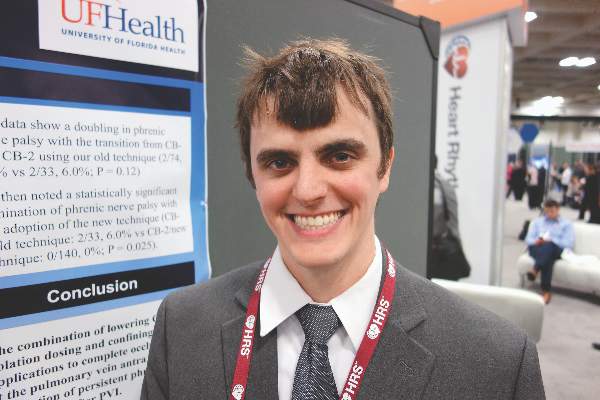
SAN FRANCISCO – Lower doses and conservative applications eliminated phrenic nerve palsy with Medtronic’s Arctic Front Advance cryoballoon ablation catheter at the University of Florida, Gainesville.
Cardiologists there were accustomed to performing atrial fibrillation pulmonary vein isolation with the first-generation device – the Arctic Front – when they switched to the second-generation Advance catheter in 2012; they had 2 phrenic nerve palsies in the first 33 patients (6%), a doubling from the 2 cases in 74 patients (2.7%) with the first-generation device.
The second-generation catheter is more powerful, with a cooling jet in the front of the balloon that delivers colder temperatures deeper into pulmonary veins. “We realized we needed to change the way we were using these catheters. You can use them safely, but you have to respect” their power, said lead investigator Dr. Robert Gibson.
“We reduced freezing times from 240 seconds to 180 seconds, and made that a hard rule. We limited the number of ablations” to two complete occlusions per vein, down from four to seven with the first-generation Arctic Front. “We also implemented a nadir cutoff to stop ablation when catheter temperatures fell below –55° C, and we tried to stay as proximal as possible to the pulmonary vein antra while still maintaining complete occlusion.” To help with that, cardiologists stopped using the 23-mm catheter, opting instead for the 28-mm catheter, Dr. Gibson said at the Heart Rhythm Society annual meeting.
Since making the changes, the team has performed 140 ablations, and there has not been a single phrenic nerve palsy. “We haven’t experienced a diaphragm paralysis” with the new approach. “We are very happy with these results. The changes make physiologic sense. You stay back; you use fewer freezes,” he said.
Phrenic nerve injury also decreased, from 3 cases in the first 33 second-generation patients (9%) to 9 in the 140 (6.4%) with the refined technique. Total phrenic nerve complications are now fewer in Gainesville than with the original, less powerful first-generation Arctic Front.
The team didn’t report ablation success with their new approach, but a 2015 review of Arctic Front Advance in more than 3,000 patients suggested long-term success with similar refinements (Heart Rhythm. 2015 Jul;12[7]:1658-66).
Patients in the University of Florida review were in their early 60s, on average, and about two-thirds were men. About 30% had prior ablations. Phrenic nerve injury was determined by continuous phrenic nerve stimulation and manual diaphragm palpation during cryoablation.
The investigators had no relevant financial disclosures. Medtronic helped with the statistical analysis.
AT HEART RHYTHM 2016
Key clinical point: Second-generation cryoablation devices require a lighter touch to avoid phrenic nerve palsy.
Major finding: A conservative approach dropped the phrenic nerve palsy rate from 6% to 0% (P = .025).
Data source: A review of 247 cryoballoon ablations for paroxysmal atrial fibrillation
Disclosures: The investigators had no relevant financial disclosures. Medtronic helped with the statistical analysis.
Anticoagulation therapy after VT ablation yields fewer thrombotic events
San Francisco – Anticoagulation therapy is probably a good idea after ventricular tachycardia ablation in patients with risk factors or stroke, even if they don’t have atrial fibrillation, according to investigators from the University of Kansas Medical Center in Kansas City.
The advice comes from a review of 2,235 ventricular tachycardia (VT) ablation cases from the university and other members of the International VT Ablation Center Collaborative; about a quarter of the patients (604) were prescribed oral anticoagulation therapy at baseline and at discharge, nearly all for atrial fibrillation (AF) and most with warfarin. Over the next year, just 0.3% (2) had a subsequent thromboembolic complication, one of which was an ischemic stroke.
The remaining patients (1,631) did not have a diagnosis of AF and were not on anticoagulants at baseline or after discharge. They were more likely to have New York Heart Association class I or II heart failure and higher ejection fractions, and to otherwise be in better shape compared with the patients who received anticoagulation therapy. Even so, within a year, 1.3% (21) had a thromboembolic event, almost half of which were ischemic strokes, a substantial increase in relative risk (P = .05).
Maybe those patients had undiagnosed AF at baseline, or perhaps a clot formed over the ablation scar, Dr. Rizwan Afzal said at the annual scientific sessions of the Heart Rhythm Society. Regardless, “this observation has changed our practice. If VT ablation patients have low ejection fractions, if they’re elderly, or have other risk factors for stroke, we put them on blood thinners [afterward] “even if they don’t have atrial fibrillation. We are not sure how long they should be on anticoagulation [therapy] to counteract the increased risk of stroke,” but probably at least for a few weeks, he said.
Dr. Afzal and his colleagues generally opt for warfarin; the use is off label for newer oral anticoagulants, and a tough sell to insurance companies.
There were no predictors of increased thromboembolic risk in the group that was not on anticoagulation therapy. During follow-up, about 2.2% (13) of patients on anticoagulation therapy had bleeding complications, including one intracranial hemorrhage, compared with 2.5% (41) of the patients not treated with an anticoagulant; most of them were on aspirin after the procedure, and the rest were on dual antiplatelet therapy (P = .7), reported Dr. Afzal, a cardiology fellow at the University of Kansas.
The median age of the study patients was 65 years, and 87% were men. In the group on anticoagulation therapy, the mean baseline left ventricular ejection fraction was 31%; 35% had prior cardiac surgery, 29% were on cardiac resynchronization therapy, and 44% had NYHA class III or IV heart failure. The mean baseline ejection fraction among patients who were not on anticoagulation therapy was 35%; 29% had prior heart surgery, 24% were on CRT, and 32.5% had NYHA class III or IV heart failure.
There was no industry funding for the work, and the investigators had no disclosures.
AT HEART RHYTHM 2016
Key clinical point: Anticoagulant therapy may be a good idea after ventricular tachycardia ablation in patients with risk factors for stroke, even if they don’t have atrial fibrillation.
Major finding: About 0.3% of patients on oral anticoagulant therapy after VT ablation had a thromboembolic event within a year, compared with 1.3% of those who were not on such therapy.
Data source: Review of 2,245 VT ablation cases.
Disclosures: There was no industry funding for the work, and the investigators had no disclosures.

