User login
For MD-IQ use only
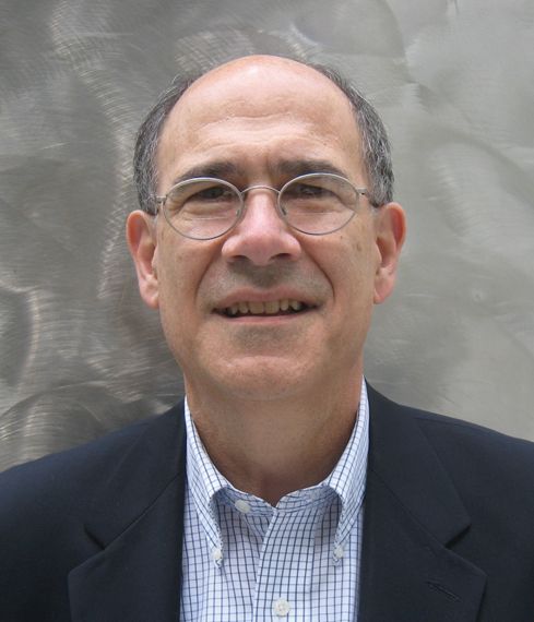
Extraordinary Patients Inspired Father of Cancer Immunotherapy
His pioneering research established interleukin-2 (IL-2) as the first U.S. Food and Drug Administration–approved cancer immunotherapy in 1992.
To recognize his trailblazing work and other achievements, the American Association for Cancer Research (AACR) will award Dr. Rosenberg with the 2024 AACR Award for Lifetime Achievement in Cancer Research at its annual meeting in April.

Dr. Rosenberg, a senior investigator for the Center for Cancer Research at the National Cancer Institute (NCI), and chief of the NCI Surgery Branch, shared the history behind his novel research and the patient stories that inspired his discoveries, during an interview.
Tell us a little about yourself and where you grew up.
Dr. Rosenberg: I grew up in the Bronx. My parents both immigrated to the United States from Poland as teenagers.
As a young boy, did you always want to become a doctor?
Dr. Rosenberg: I think some defining moments on why I decided to go into medicine occurred when I was 6 or 7 years old. The second world war was over, and many of the horrors of the Holocaust became apparent to me. I was brought up as an Orthodox Jew. My parents were quite religious, and I remember postcards coming in one after another about relatives that had died in the death camps. That had a profound influence on me.
How did that experience impact your aspirations?
Dr. Rosenberg: It was an example to me of how evil certain people and groups can be toward one another. I decided at that point, that I wanted to do something good for people, and medicine seemed the most likely way to do that. But also, I was developing a broad scientific interest. I ended up at the Bronx High School of Science and knew that I not only wanted to practice the medicine of today, but I wanted to play a role in helping develop the medicine.
What led to your interest in cancer treatment?
Dr. Rosenberg: Well, as a medical student and resident, it became clear that the field of cancer needed major improvement. We had three major ways to treat cancer: surgery, radiation therapy, and chemotherapy. That could cure about half of the people [who] had cancer. But despite the best application of those three specialties, there were over 600,000 deaths from cancer each year in the United States alone. It was clear to me that new approaches were needed, and I became very interested in taking advantage of the body’s immune system as a source of information to try to make progress.
Were there patients who inspired your research?
Dr. Rosenberg: There were two patients that I saw early in my career that impressed me a great deal. One was a patient that I saw when working in the emergency ward as a resident. A patient came in with right upper quadrant pain that looked like a gallbladder attack. That’s what it was. But when I went through his chart, I saw that he had been at that hospital 12 years earlier with a metastatic gastric cancer. The surgeons had operated. They saw tumor had spread to the liver and could not be removed. They closed the belly, not expecting him to survive. Yet he kept showing up for follow-up visits.
Here he was 12 years later. When I helped operate to take out his gallbladder, there was no evidence of any cancer. The cancer had disappeared in the absence of any external treatment. One of the rarest events in medicine, the spontaneous regression of a cancer. Somehow his body had learned how to destroy the tumor.
Was the second patient’s case as impressive?
Dr. Rosenberg: This patient had received a kidney transplant from a gentleman who died in an auto accident. [The donor’s] kidney contained a cancer deposit, a kidney cancer, unbeknownst to the transplant surgeons. [When the kidney was transplanted], the recipient developed widespread metastatic kidney cancer.
[The recipient] was on immunosuppressive drugs, and so the drugs had to be stopped. [When the immunosuppressive drugs were stopped], the patient’s body rejected the kidney and his cancer disappeared.
That showed me that, in fact, if you could stimulate a strong enough immune reaction, in this case, an [allogeneic] reaction, against foreign tissues from a different individual, that you could make large vascularized, invasive cancers disappear based on immune reactivities. Those were clues that led me toward studying the immune system’s impact on cancer.
From there, how did your work evolve?
Dr. Rosenberg: As chief of the surgery branch at NIH, I began doing research. It was very difficult to manipulate immune cells in the laboratory. They wouldn’t stay alive. But I tried to study immune reactions in patients with cancer to see if there was such a thing as an immune reaction against the cancer. There was no such thing known at the time. There were no cancer antigens and no known immune reactions against the disease in the human.
Around this time, investigators were publishing studies about interleukin-2 (IL-2), or white blood cells known as leukocytes. How did interleukin-2 further your research?
Dr. Rosenberg: The advent of interleukin-2 enabled scientists to grow lymphocytes outside the body. [This] enabled us to grow t-lymphocytes, which are some of the major warriors of the immune system against foreign tissue. After [studying] 66 patients in which we studied interleukin-2 and cells that would develop from it, we finally saw a disappearance of melanoma in a patient that received interleukin-2. And we went on to treat hundreds of patients with that hormone, interleukin-2. In fact, interleukin-2 became the first immunotherapy ever approved by the Food and Drug Administration for the treatment of cancer in humans.
How did this finding impact your future discoveries?
Dr. Rosenberg: [It] led to studies of the mechanism of action of interleukin-2 and to do that, we identified a kind of cell called a tumor infiltrating lymphocyte. What better place, intuitively to look for cells doing battle against the cancer than within the cancer itself?
In 1988, we demonstrated for the first time that transfer of lymphocytes with antitumor activity could cause the regression of melanoma. This was a living drug obtained from melanoma deposits that could be grown outside the body and then readministered to the patient under suitable conditions. Interestingly, [in February the FDA approved that drug as treatment for patients with melanoma]. A company developed it to the point where in multi-institutional studies, they reproduced our results.
And we’ve now emphasized the value of using T cell therapy, t cell transfer, for the treatment of patients with the common solid cancers, the cancers that start anywhere from the colon up through the intestine, the stomach, the pancreas, and the esophagus. Solid tumors such as ovarian cancer, uterine cancer and so on, are also potentially susceptible to this T cell therapy.
We’ve published several papers showing in isolated patients that you could cause major regressions, if not complete regressions, of these solid cancers in the liver, in the breast, the cervix, the colon. That’s a major aspect of what we’re doing now.
I think immunotherapy has come to be recognized as a major fourth arm that can be used to attack cancers, adding to surgery, radiation, and chemotherapy.
What guidance would you have for other physician-investigators or young doctors who want to follow in your path?
Dr. Rosenberg: You have to have a broad base of knowledge. You have to be willing to immerse yourself in a problem so that your mind is working on it when you’re doing things where you can only think. [When] you’re taking a shower, [or] waiting at a red light, your mind is working on this problem because you’re immersed in trying to understand it.
You need to have a laser focus on the goals that you have and not get sidetracked by issues that may be interesting but not directly related to the goals that you’re attempting to achieve.
His pioneering research established interleukin-2 (IL-2) as the first U.S. Food and Drug Administration–approved cancer immunotherapy in 1992.
To recognize his trailblazing work and other achievements, the American Association for Cancer Research (AACR) will award Dr. Rosenberg with the 2024 AACR Award for Lifetime Achievement in Cancer Research at its annual meeting in April.
Dr. Rosenberg, a senior investigator for the Center for Cancer Research at the National Cancer Institute (NCI), and chief of the NCI Surgery Branch, shared the history behind his novel research and the patient stories that inspired his discoveries, during an interview.
Tell us a little about yourself and where you grew up.
Dr. Rosenberg: I grew up in the Bronx. My parents both immigrated to the United States from Poland as teenagers.
As a young boy, did you always want to become a doctor?
Dr. Rosenberg: I think some defining moments on why I decided to go into medicine occurred when I was 6 or 7 years old. The second world war was over, and many of the horrors of the Holocaust became apparent to me. I was brought up as an Orthodox Jew. My parents were quite religious, and I remember postcards coming in one after another about relatives that had died in the death camps. That had a profound influence on me.
How did that experience impact your aspirations?
Dr. Rosenberg: It was an example to me of how evil certain people and groups can be toward one another. I decided at that point, that I wanted to do something good for people, and medicine seemed the most likely way to do that. But also, I was developing a broad scientific interest. I ended up at the Bronx High School of Science and knew that I not only wanted to practice the medicine of today, but I wanted to play a role in helping develop the medicine.
What led to your interest in cancer treatment?
Dr. Rosenberg: Well, as a medical student and resident, it became clear that the field of cancer needed major improvement. We had three major ways to treat cancer: surgery, radiation therapy, and chemotherapy. That could cure about half of the people [who] had cancer. But despite the best application of those three specialties, there were over 600,000 deaths from cancer each year in the United States alone. It was clear to me that new approaches were needed, and I became very interested in taking advantage of the body’s immune system as a source of information to try to make progress.
Were there patients who inspired your research?
Dr. Rosenberg: There were two patients that I saw early in my career that impressed me a great deal. One was a patient that I saw when working in the emergency ward as a resident. A patient came in with right upper quadrant pain that looked like a gallbladder attack. That’s what it was. But when I went through his chart, I saw that he had been at that hospital 12 years earlier with a metastatic gastric cancer. The surgeons had operated. They saw tumor had spread to the liver and could not be removed. They closed the belly, not expecting him to survive. Yet he kept showing up for follow-up visits.
Here he was 12 years later. When I helped operate to take out his gallbladder, there was no evidence of any cancer. The cancer had disappeared in the absence of any external treatment. One of the rarest events in medicine, the spontaneous regression of a cancer. Somehow his body had learned how to destroy the tumor.
Was the second patient’s case as impressive?
Dr. Rosenberg: This patient had received a kidney transplant from a gentleman who died in an auto accident. [The donor’s] kidney contained a cancer deposit, a kidney cancer, unbeknownst to the transplant surgeons. [When the kidney was transplanted], the recipient developed widespread metastatic kidney cancer.
[The recipient] was on immunosuppressive drugs, and so the drugs had to be stopped. [When the immunosuppressive drugs were stopped], the patient’s body rejected the kidney and his cancer disappeared.
That showed me that, in fact, if you could stimulate a strong enough immune reaction, in this case, an [allogeneic] reaction, against foreign tissues from a different individual, that you could make large vascularized, invasive cancers disappear based on immune reactivities. Those were clues that led me toward studying the immune system’s impact on cancer.
From there, how did your work evolve?
Dr. Rosenberg: As chief of the surgery branch at NIH, I began doing research. It was very difficult to manipulate immune cells in the laboratory. They wouldn’t stay alive. But I tried to study immune reactions in patients with cancer to see if there was such a thing as an immune reaction against the cancer. There was no such thing known at the time. There were no cancer antigens and no known immune reactions against the disease in the human.
Around this time, investigators were publishing studies about interleukin-2 (IL-2), or white blood cells known as leukocytes. How did interleukin-2 further your research?
Dr. Rosenberg: The advent of interleukin-2 enabled scientists to grow lymphocytes outside the body. [This] enabled us to grow t-lymphocytes, which are some of the major warriors of the immune system against foreign tissue. After [studying] 66 patients in which we studied interleukin-2 and cells that would develop from it, we finally saw a disappearance of melanoma in a patient that received interleukin-2. And we went on to treat hundreds of patients with that hormone, interleukin-2. In fact, interleukin-2 became the first immunotherapy ever approved by the Food and Drug Administration for the treatment of cancer in humans.
How did this finding impact your future discoveries?
Dr. Rosenberg: [It] led to studies of the mechanism of action of interleukin-2 and to do that, we identified a kind of cell called a tumor infiltrating lymphocyte. What better place, intuitively to look for cells doing battle against the cancer than within the cancer itself?
In 1988, we demonstrated for the first time that transfer of lymphocytes with antitumor activity could cause the regression of melanoma. This was a living drug obtained from melanoma deposits that could be grown outside the body and then readministered to the patient under suitable conditions. Interestingly, [in February the FDA approved that drug as treatment for patients with melanoma]. A company developed it to the point where in multi-institutional studies, they reproduced our results.
And we’ve now emphasized the value of using T cell therapy, t cell transfer, for the treatment of patients with the common solid cancers, the cancers that start anywhere from the colon up through the intestine, the stomach, the pancreas, and the esophagus. Solid tumors such as ovarian cancer, uterine cancer and so on, are also potentially susceptible to this T cell therapy.
We’ve published several papers showing in isolated patients that you could cause major regressions, if not complete regressions, of these solid cancers in the liver, in the breast, the cervix, the colon. That’s a major aspect of what we’re doing now.
I think immunotherapy has come to be recognized as a major fourth arm that can be used to attack cancers, adding to surgery, radiation, and chemotherapy.
What guidance would you have for other physician-investigators or young doctors who want to follow in your path?
Dr. Rosenberg: You have to have a broad base of knowledge. You have to be willing to immerse yourself in a problem so that your mind is working on it when you’re doing things where you can only think. [When] you’re taking a shower, [or] waiting at a red light, your mind is working on this problem because you’re immersed in trying to understand it.
You need to have a laser focus on the goals that you have and not get sidetracked by issues that may be interesting but not directly related to the goals that you’re attempting to achieve.
Combining Targeted Drugs and Radiation in Breast Cancer: What’s Safe?
One reason is studies of new drugs typically exclude concurrent radiotherapy, said Kathy Miller, MD, a contributor to this news organization and professor of oncology and medicine at the Indiana University School of Medicine, Indianapolis, Indiana.
If trials evaluating new targeted therapies included concurrent radiotherapy, it would be challenging to identify whether toxicities came from the drug itself, the radiation, or the combination, Dr. Miller explained.
Given the limited evidence, “we tend to be cautious and conservative” and not combine therapies that “we don’t know are safe or appropriate for patients,” said Chirag Shah, MD, director of breast radiology at the Cleveland Clinic, Cleveland, Ohio.
Below is a guide to what we do and don’t know about combining radiotherapy and systemic treatments in breast cancer.
1. Immunotherapy plus radiotherapy likely safe but evidence is limited
Safety data on combining immune checkpoint inhibitors and radiotherapy in breast cancer are limited because concurrent radiotherapy has typically been excluded in pivotal trials.
The 2020 KEYNOTE-522 trial did provide a rare look at concurrent radiotherapy and immunotherapy in early triple-negative breast cancer. The analysis found “no safety concerns” with concurrent radiotherapy and pembrolizumab, lead investigator Peter Schmid, MD, of Queen Mary University of London, England, told this news organization.
Research on other solid tumor types also suggests that radiotherapy “can be considered safe” alongside immunotherapy, the authors of a recent ESTRO consensus said.
Despite evidence indicating radiotherapy alongside immunotherapy can be safe in patients with breast cancer, “certain aspects, such as patient selection, total dose, and dose per fraction, remain open for debate to achieve the best therapeutic outcomes,” the ESTRO experts cautioned.
2. CDK4/6 inhibitors may be offered with radiotherapy in some settings, not others
CDK4/6 inhibitors are now standard of care for first- or second-line treatment in patients with advanced or metastatic hormone receptor–positive, human epidermal growth factor receptor 2 (HER2)–negative breast cancer.
“Unfortunately, we found no information regarding concurrent radiotherapy in the adjuvant setting” in pivotal trials for palbociclib, abemaciclib, and ribociclib, the ESTRO authors said. In the pivotal trials for palbociclib and abemaciclib, patients had to discontinue immunotherapy before initiating radiotherapy, and in the trial for ribociclib, palliative radiotherapy was allowed for relieving bone pain only.
However, in 2023, a team of experts from 12 countries attempted to piece together the available evidence, publishing a meta-analysis of 11 retrospective studies on the safety of CDK4/6 inhibitors given concurrently with radiotherapy in patients with metastatic disease.
Although most of these studies had small patient populations, the analysis revealed that CDK4/6 inhibitors given concurrently with radiotherapy in patients with metastatic breast cancer led to a similar side-effect profile to that observed in trials of the inhibitors given sequentially with adjuvant radiotherapy.
“These findings suggest that the simultaneous administration of CDK4/6 inhibitors and radiotherapy is generally well tolerated,” the ESTRO authors concluded but added that CDK4/6 inhibitors and concomitant radiotherapy should be investigated more in the adjuvant locoregional, whole brain, and intracranial stereotactic radiotherapy settings.
The expert panel did note, however, that CDK4/6 inhibitors and concomitant radiotherapy “could be offered” during palliative and ablative extracranial radiotherapy.
3. Only offer poly (ADP-ribose) polymerase (PARP) inhibitors plus radiotherapy in clinical trial setting
PARP inhibitors olaparib (Lynparza) and talazoprib (Talzenna) are standard of care in patients with metastatic breast cancer who have BRCA1/2 gene mutations. Olaparib is also indicated for high-risk early breast cancer following neoadjuvant or adjuvant chemotherapy.
But data on combining PARP inhibitors with radiotherapy in breast cancer also remain limited.
One ongoing phase 2 trial, comparing olaparib plus radiotherapy to radiotherapy alone in 300 people with inflammatory breast cancer, is aiming to tease out the safety of the combination and whether it improves local control in patients with aggressive disease.
“The desire is to explore the exciting possibility that low doses of PARP inhibition may radiosensitize tumor cells more than normal tissues,” Reshma Jagsi, MD, chair of the Department of Radiation Oncology at Emory University School of Medicine in Atlanta, Georgia, who is leading the study.
Because of potential good or bad interactions between new systemic therapies and radiotherapy, “intentional trial design” is important, Dr. Jagsi said, so we “know the best way to combine treatments in practice to optimize outcomes.”
But given the evidence to date, the ESTRO experts advised waiting until “further research provides more comprehensive safety and efficacy data” in the primary, adjuvant, and metastatic settings. The experts also advised not offering PARP inhibitors and concomitant radiotherapy to treat advanced breast cancer outside of clinical trials.
4. Phosphoinositide 3-kinase inhibitors (PI3K) inhibitors, mammalian target of rapamycin (mTOR) inhibitors, and newer targeted agents should not be offered concurrently with radiotherapy
Clinical trial data on the safety of combining PI3K and mTOR inhibitors with radiation are thin, especially in advanced breast cancer. Typically, radiotherapy within 4 weeks before randomization, or 2 weeks for palliative radiation, was excluded in pivotal trials.
For this reason, the ESTRO team recommended that concurrent radiation with either PI3K inhibitors or mTOR inhibitors “should not be offered.”
ESTRO also cautioned against providing radiation concurrently with newer anti-HER2 tyrosine-kinase drugs, such as neratinib or tucatinib, or newer antibody-drug conjugates such as trastuzumab deruxtecan, until more data emerge on the safety of these combinations.
5. Combining older HER2-targeted drugs and radiotherapy generally safe
The ESTRO authors agreed that older anti-HER2 drugs trastuzumab (Herceptin), pertuzumab (Perjeta), and lapatinib (Tykerb) can be safely used concurrently with locoregional radiotherapy as well.
One of the biggest concerns in the field is how to combine radiation with systemic therapies in the setting of brain metastases, and the data on these older anti-HER2 drugs are relatively clear that it’s safe, Dr. Miller said.
For instance, in a 2019 study of 84 patients with 487 brain metastases, stereotactic radiosurgery given alongside lapatinib led to significantly higher rates of complete responses than stereotactic radiosurgery alone (35% vs 11%) with no increased risk for radiation necrosis.
The ESTRO team agreed, noting that the latest evidence supports the use of trastuzumab, pertuzumab, or lapatinib alongside radiotherapy for whole brain and ablative intracranial stereotactic radiotherapy.
As for older antibody-drug conjugates, trastuzumab emtansine (T-DM1) plus radiotherapy “might be considered” during adjuvant locoregional radiotherapy for breast cancer but should not be offered for whole brain and ablative intracranial stereotactic radiotherapy, the ESTRO team said.
Dr. Jagsi declared the following conflicts in a recent 2024 publication: Stock options for advisory board role in Equity Quotient; grants or contracts from Genentech; and expert witness for Kleinbard, LLC, and Hawks Quindel Law. In the Keynote-522 trial publication Dr. Schmid declared relationships with AstraZeneca, Bayer, Boehringer Ingelheim, Celgene, Eisai, Hoffmann-La Roche, Genetech, Merck, Novartis, and Pfizer. Dr. Shah reported consulting for Impedimed, Videra Surgical, and PreludeDX.
A version of this article appeared on Medscape.com.
Neurologists Read Signs to Diagnose Functional Neurological Disorders
They have gone by many different names over the centuries: hysteria, psychosomatic illnesses, psychogenic neurological disorders, conversion disorders, dissociative neurological symptom disorders. The terminology may change, but functional neurological disorders by any other name are still real and serious yet treatable phenomena.
Functional neurological disorders, or FNDs, live at the crossroads of neurology and psychiatry, and they are as much a product of the body as they are of the brain, say neurologists who specialize in treating these complex and clinically challenging conditions.
“Whether they’re easily recognized or not depends on someone’s training and experience in this regard,” said Mark Hallett, MD, of the Human Motor Control Section of the National Institute of Neurological Disorders and Stroke in Bethesda, Maryland.
“The difficulty has been that there hasn’t been very good education about functional disorders over the last 50 years or so,” he said in an interview.
However, with training and experience, clinicians can learn to identify these common and disabling conditions, Dr. Hallett said.
Varying Definitions
The Diagnostic and Statistical Manual of Mental Disorders 5th edition (DSM-5) labels FND as “conversion disorder,” and lists diagnostic criteria that include “one or more symptoms of altered voluntary motor or sensory function; clinical findings provide evidence of incompatibility between the symptom and recognized neurological or medical conditions; the symptom or deficit is not better explained by another medical or mental disorder;” and “the symptom or deficit causes clinically significant distress or impairment in social, occupational, or other important areas of functioning or warrants medical evaluation.”
Dr. Hallett offers his own definition of FND, which includes the following characteristics:
- A neurological disorder, characterized by almost any type of neurological symptom
- Not voluntarily produced
- Caused by a brain network dysfunction that does not exclude the possibility of normal function
- Sometimes due in part to a psychological cause, and not explained by other neurological pathology that may or may not be present
- Symptoms may be inconsistent (variable) or incompatible (incongruent) with other known neurological disorders or human anatomy and physiology.
The two most common types of FND are psychogenic nonepileptic seizures and functional movement disorders, but patients may also have functional sensory, visual, auditory, speech, and urologic disorders, and even functional coma.
Dr. Hallett cited studies showing that an estimated 9% of neurology hospital admission are for FNDS, and that among patients in neurology clinics 5.4% had a diagnosis of FND, and 30% had an FND as part of the diagnosis.
Women comprise between 60% and 75% of the population with FNDs.
Diagnosis
FND is not, as once thought, a diagnosis of exclusion, but is based on signs and symptoms, which may be either inconsistent or irreversible and may occur in the absence of a stressor, said Sara Finkelstein, MD, MSc, of the Functional Neurological Disorder Unit in the Department of Neurology at Massachusetts General Hospital in Boston.
She emphasized that there are several diagnostic pitfalls that clinicians need to be aware of.
For example, “just because a patient has a psychiatric history does not mean that they have a functional neurological disorder,” she said in an interview.

Clinicians may also make unwarranted assumptions about a given patient, excluding an FND diagnosis in, say, a young woman with symptoms of anxiety. Alternatively, clinicians may either include or exclude a diagnosis based on personality factors or on a prior stressor, neither of which alone are sufficiently diagnostic.
Additionally, a clinician may be tempted to make the diagnosis of an FND based on the absence of findings on standard exams rather than on rule-in signs and symptoms, she emphasized.
Functional seizures
A definitive diagnosis can depend on the type of disorder.
“Many functional seizures have some clinical manifestations that are apparent, but as seizures are intermittent the doctor may not see one, and it may depend upon someone taking a video of the person with the seizure perhaps, or bringing them into a hospital and watching them until they do have the seizure,” Dr. Hallett said.
There are some manifestations that indicate the likelihood that a seizure has a functional origin, and when there is uncertainty EEG can help to nail down a diagnosis, he added.
Dr. Finkelstein noted that exam signs with good reliability for functional seizures include eye closure or resistance to opening; duration longer than 2 minutes; stopping and starting; asynchronous limb movements; patient maintenance of awareness during a generalized event; and ictal weeping.
Differential diagnoses included migraine with complex aura, dissociation related to posttraumatic stress disorder, or anxiety.
Functional movement disorders
Dr. Finkelstein cautioned that when evaluating patients for potential functional movement disorders, it’s important to not jump to conclusions.
For example, the amplitude of tremor can vary in Parkinson’s disease and essential tremor as well as in functional tremor. The clinician should not read too much into the observation that a patient’s tremor gets worse with increasing stress as stress can exacerbate most tremor types, she said.
One sign that tremor could be functional (dystonic tremor) is irregularity of amplitude and frequency, she noted.
When assessing patients with gait disorder, it’s important to understand that there is no single sign that is specially characteristic for a given disorder, and just because a patient has a “bizarre” gait, it doesn’t necessarily signal a functional disorder.
“A dystonic gait may improve with an alternate motor pattern or be inconsistent over time,” Dr. Finkelstein said.
Treatment
In a comprehensive review published in The Lancet: Neurology in 2022, Dr. Hallett and colleagues said that good doctor-patient communications and understanding of each patient’s needs and goals are essential for effective treatment of all FNDs.
“Neurologists have traditionally avoided taking responsibility for people with FND, although are often most appropriate to engage patients in treatment. Explaining the diagnosis with clarity, confidence, using the principles of a ‘rule in’ process, is a key step in treatment,” they wrote.
Treatment can take several forms, depending on the FND, and may include physiotherapy for patients with functional movement disorders and psychological therapy for patients with functional seizures.
“With increasing evidence-based treatment, the diagnosis of FND should be seen as a process of looking for potentially reversible cause of disability and distress whether or not an individual has abnormalities on conventional laboratory or radiological testing,” Dr. Hallett and colleagues concluded.
This article was based on interviews and from presentations by Dr. Hallett and Dr. Finkelstein at a 2023 meeting of the Indiana Neurological Society. Dr. Hallett and Dr. Finkelstein declared no conflicts of interest.
They have gone by many different names over the centuries: hysteria, psychosomatic illnesses, psychogenic neurological disorders, conversion disorders, dissociative neurological symptom disorders. The terminology may change, but functional neurological disorders by any other name are still real and serious yet treatable phenomena.
Functional neurological disorders, or FNDs, live at the crossroads of neurology and psychiatry, and they are as much a product of the body as they are of the brain, say neurologists who specialize in treating these complex and clinically challenging conditions.
“Whether they’re easily recognized or not depends on someone’s training and experience in this regard,” said Mark Hallett, MD, of the Human Motor Control Section of the National Institute of Neurological Disorders and Stroke in Bethesda, Maryland.
“The difficulty has been that there hasn’t been very good education about functional disorders over the last 50 years or so,” he said in an interview.
However, with training and experience, clinicians can learn to identify these common and disabling conditions, Dr. Hallett said.
Varying Definitions
The Diagnostic and Statistical Manual of Mental Disorders 5th edition (DSM-5) labels FND as “conversion disorder,” and lists diagnostic criteria that include “one or more symptoms of altered voluntary motor or sensory function; clinical findings provide evidence of incompatibility between the symptom and recognized neurological or medical conditions; the symptom or deficit is not better explained by another medical or mental disorder;” and “the symptom or deficit causes clinically significant distress or impairment in social, occupational, or other important areas of functioning or warrants medical evaluation.”
Dr. Hallett offers his own definition of FND, which includes the following characteristics:
- A neurological disorder, characterized by almost any type of neurological symptom
- Not voluntarily produced
- Caused by a brain network dysfunction that does not exclude the possibility of normal function
- Sometimes due in part to a psychological cause, and not explained by other neurological pathology that may or may not be present
- Symptoms may be inconsistent (variable) or incompatible (incongruent) with other known neurological disorders or human anatomy and physiology.
The two most common types of FND are psychogenic nonepileptic seizures and functional movement disorders, but patients may also have functional sensory, visual, auditory, speech, and urologic disorders, and even functional coma.
Dr. Hallett cited studies showing that an estimated 9% of neurology hospital admission are for FNDS, and that among patients in neurology clinics 5.4% had a diagnosis of FND, and 30% had an FND as part of the diagnosis.
Women comprise between 60% and 75% of the population with FNDs.
Diagnosis
FND is not, as once thought, a diagnosis of exclusion, but is based on signs and symptoms, which may be either inconsistent or irreversible and may occur in the absence of a stressor, said Sara Finkelstein, MD, MSc, of the Functional Neurological Disorder Unit in the Department of Neurology at Massachusetts General Hospital in Boston.
She emphasized that there are several diagnostic pitfalls that clinicians need to be aware of.
For example, “just because a patient has a psychiatric history does not mean that they have a functional neurological disorder,” she said in an interview.
Clinicians may also make unwarranted assumptions about a given patient, excluding an FND diagnosis in, say, a young woman with symptoms of anxiety. Alternatively, clinicians may either include or exclude a diagnosis based on personality factors or on a prior stressor, neither of which alone are sufficiently diagnostic.
Additionally, a clinician may be tempted to make the diagnosis of an FND based on the absence of findings on standard exams rather than on rule-in signs and symptoms, she emphasized.
Functional seizures
A definitive diagnosis can depend on the type of disorder.
“Many functional seizures have some clinical manifestations that are apparent, but as seizures are intermittent the doctor may not see one, and it may depend upon someone taking a video of the person with the seizure perhaps, or bringing them into a hospital and watching them until they do have the seizure,” Dr. Hallett said.
There are some manifestations that indicate the likelihood that a seizure has a functional origin, and when there is uncertainty EEG can help to nail down a diagnosis, he added.
Dr. Finkelstein noted that exam signs with good reliability for functional seizures include eye closure or resistance to opening; duration longer than 2 minutes; stopping and starting; asynchronous limb movements; patient maintenance of awareness during a generalized event; and ictal weeping.
Differential diagnoses included migraine with complex aura, dissociation related to posttraumatic stress disorder, or anxiety.
Functional movement disorders
Dr. Finkelstein cautioned that when evaluating patients for potential functional movement disorders, it’s important to not jump to conclusions.
For example, the amplitude of tremor can vary in Parkinson’s disease and essential tremor as well as in functional tremor. The clinician should not read too much into the observation that a patient’s tremor gets worse with increasing stress as stress can exacerbate most tremor types, she said.
One sign that tremor could be functional (dystonic tremor) is irregularity of amplitude and frequency, she noted.
When assessing patients with gait disorder, it’s important to understand that there is no single sign that is specially characteristic for a given disorder, and just because a patient has a “bizarre” gait, it doesn’t necessarily signal a functional disorder.
“A dystonic gait may improve with an alternate motor pattern or be inconsistent over time,” Dr. Finkelstein said.
Treatment
In a comprehensive review published in The Lancet: Neurology in 2022, Dr. Hallett and colleagues said that good doctor-patient communications and understanding of each patient’s needs and goals are essential for effective treatment of all FNDs.
“Neurologists have traditionally avoided taking responsibility for people with FND, although are often most appropriate to engage patients in treatment. Explaining the diagnosis with clarity, confidence, using the principles of a ‘rule in’ process, is a key step in treatment,” they wrote.
Treatment can take several forms, depending on the FND, and may include physiotherapy for patients with functional movement disorders and psychological therapy for patients with functional seizures.
“With increasing evidence-based treatment, the diagnosis of FND should be seen as a process of looking for potentially reversible cause of disability and distress whether or not an individual has abnormalities on conventional laboratory or radiological testing,” Dr. Hallett and colleagues concluded.
This article was based on interviews and from presentations by Dr. Hallett and Dr. Finkelstein at a 2023 meeting of the Indiana Neurological Society. Dr. Hallett and Dr. Finkelstein declared no conflicts of interest.
FROM THE INDIANA NEUROLOGICAL SOCIETY’S FUNCTIONAL NEUROLOGICAL DISORDERS CONFERENCE
Consider These Factors in an Academic Radiation Oncology Position
TOPLINE:
— and accept an offer if the practice is “great” in at least two of those areas and “good” in the third, experts say in a recent editorial.
METHODOLOGY:
- Many physicians choose to go into academic medicine because they want to stay involved in research and education while still treating patients.
- However, graduating radiation oncology residents often lack or have limited guidance on what to look for in a prospective job and how to assess their contract.
- This recent editorial provides guidance to radiation oncologists seeking academic positions. The authors advise prospective employees to evaluate three main factors — compensation, daily duties, and location — as well as provide tips for identifying red flags in each category.
TAKEAWAY:
- Compensation: Prospective faculty should assess both direct compensation, that is, salary, and indirect compensation, which typically includes retirement contributions and other perks. For direct compensation, what is the base salary? Is extra work compensated? How does the salary offer measure up to salary data reported by national agencies? Also: Don’t overlook uncompensated duties, such as time in tumor boards or in meetings, which may be time-consuming, and make sure compensation terms are clearly delineated in a contract and equitable among physicians in a specific rank.
- Daily duties: When it comes to daily life on the job, a prospective employee should consider many factors, including the cancer center’s excitement to hire you, the reputation of the faculty and leaders at the organization, employee turnover rates, diversity among faculty, and the time line of career advancement.
- Location: The location of the job encompasses the geography — such as distance from home to work, the number of practices covered, cost of living, and the area itself — as well as the atmosphere for conducting research and publishing.
- Finally, carefully review the job contract. All the key aspects of the job, including compensation and benefits, should be clearly stated in the contract to “improve communication of expectations.”
IN PRACTICE:
“A prospective faculty member can ask 100 questions, but they can’t make 100 demands; consideration of the three domains can help to focus negotiation efforts where the efforts are needed,” the authors noted.
SOURCE:
This editorial, led by Nicholas G. Zaorsky from the Department of Radiation Oncology, University Hospitals Seidman Cancer Center, Case Western Reserve School of Medicine, Cleveland, Ohio, was published online in Practical Radiation Oncology
DISCLOSURES:
The lead author declared being supported by the American Cancer Society and National Institutes of Health. He also reported having ties with many other sources.
A version of this article appeared on Medscape.com.
Look Beyond BMI: Metabolic Factors’ Link to Cancer Explained
The new research finds that adults with persistent metabolic syndrome that worsens over time are at increased risk for any type of cancer.
The conditions that make up metabolic syndrome (high blood pressure, high blood sugar, increased abdominal adiposity, and high cholesterol and triglycerides) have been associated with an increased risk of diseases, including heart disease, stroke, and type 2 diabetes, wrote Li Deng, PhD, of Capital Medical University, Beijing, and colleagues.
However, a single assessment of metabolic syndrome at one point in time is inadequate to show an association with cancer risk over time, they said. In the current study, the researchers used models to examine the association between trajectory patterns of metabolic syndrome over time and the risk of overall and specific cancer types. They also examined the impact of chronic inflammation concurrent with metabolic syndrome.
What We Know About Metabolic Syndrome and Cancer Risk
A systematic review and meta-analysis published in Diabetes Care in 2012 showed an association between the presence of metabolic syndrome and an increased risk of various cancers including liver, bladder, pancreatic, breast, and colorectal.
More recently, a 2020 study published in Diabetes showed evidence of increased risk for certain cancers (pancreatic, kidney, uterine, cervical) but no increased risk for cancer overall.
In addition, a 2022 study by some of the current study researchers of the same Chinese cohort focused on the role of inflammation in combination with metabolic syndrome on colorectal cancer specifically, and found an increased risk for cancer when both metabolic syndrome and inflammation were present.
However, the reasons for this association between metabolic syndrome and cancer remain unclear, and the effect of the fluctuating nature of metabolic syndrome over time on long-term cancer risk has not been explored, the researchers wrote.
“There is emerging evidence that even normal weight individuals who are metabolically unhealthy may be at an elevated cancer risk, and we need better metrics to define the underlying metabolic dysfunction in obesity,” Sheetal Hardikar, MBBS, PhD, MPH, an investigator at the Huntsman Cancer Institute, University of Utah, said in an interview.
Dr. Hardikar, who serves as assistant professor in the department of population health sciences at the University of Utah, was not involved in the current study. She and her colleagues published a research paper on data from the National Health and Nutrition Examination Survey in 2023 that showed an increased risk of obesity-related cancer.
What New Study Adds to Related Research
Previous studies have consistently reported an approximately 30% increased risk of cancer with metabolic syndrome, Dr. Hardikar said. “What is unique about this study is the examination of metabolic syndrome trajectories over four years, and not just the presence of metabolic syndrome at one point in time,” she said.
In the new study, published in Cancer on March 11 (doi: 10.1002/cncr.35235), 44,115 adults in China were separated into four trajectories based on metabolic syndrome scores for the period from 2006 to 2010. The scores were based on clinical evidence of metabolic syndrome, defined using the International Diabetes Federation criteria of central obesity and the presence of at least two other factors including increased triglycerides, decreased HDL cholesterol, high blood pressure (or treatment for previously diagnosed hypertension), and increased fasting plasma glucose (or previous diagnosis of type 2 diabetes).
The average age of the participants was 49 years; the mean body mass index ranged from approximately 22 kg/m2 in the low-stable group to approximately 28 kg/m2 in the elevated-increasing group.
The four trajectories of metabolic syndrome were low-stable (10.56% of participants), moderate-low (40.84%), moderate-high (41.46%), and elevated-increasing (7.14%), based on trends from the individuals’ initial physical exams on entering the study.
Over a median follow-up period of 9.4 years (from 2010 to 2021), 2,271 cancer diagnoses were reported in the study population. Those with an elevated-increasing metabolic syndrome trajectory had 1.3 times the risk of any cancer compared with those in the low-stable group. Risk for breast cancer, endometrial cancer, kidney cancer, colorectal cancer, and liver cancer in the highest trajectory group were 2.1, 3.3, 4.5, 2.5, and 1.6 times higher, respectively, compared to the lowest group. The increased risk in the elevated-trajectory group for all cancer types persisted when the low-stable, moderate-low, and moderate-high trajectory pattern groups were combined.
The researchers also examined the impact of chronic inflammation and found that individuals with persistently high metabolic syndrome scores and concurrent chronic inflammation had the highest risks of breast, endometrial, colon, and liver cancer. However, individuals with persistently high metabolic syndrome scores and no concurrent chronic inflammation had the highest risk of kidney cancer.
What Are the Limitations of This Research?
The researchers of the current study acknowledged the lack of information on other causes of cancer, including dietary habits, hepatitis C infection, and Helicobacter pylori infection. Other limitations include the focus only on individuals from a single community of mainly middle-aged men in China that may not generalize to other populations.
Also, the metabolic syndrome trajectories did not change much over time, which may be related to the short 4-year study period.
Using the International Diabetes Federation criteria was another limitation, because it prevented the assessment of cancer risk in normal weight individuals with metabolic dysfunction, Dr. Hardikar noted.
Does Metabolic Syndrome Cause Cancer?
“This research suggests that proactive and continuous management of metabolic syndrome may serve as an essential strategy in preventing cancer,” senior author Han-Ping Shi, MD, PhD, of Capital Medical University in Beijing, noted in a statement on the study.
More research is needed to assess the impact of these interventions on cancer risk. However, the data from the current study can guide future research that may lead to more targeted treatments and more effective preventive strategies, he continued.
“Current evidence based on this study and many other reports strongly suggests an increased risk for cancer associated with metabolic syndrome,” Dr. Hardikar said in an interview. The data serve as a reminder to clinicians to look beyond BMI as the only measure of obesity, and to consider metabolic factors together to identify individuals at increased risk for cancer, she said.
“We must continue to educate patients about obesity and all the chronic conditions it may lead to, but we cannot ignore this emerging phenotype of being of normal weight but metabolically unhealthy,” Dr. Hardikar emphasized.
What Additional Research is Needed?
Looking ahead, “we need well-designed interventions to test causality for metabolic syndrome and cancer risk, though the evidence from the observational studies is very strong,” Dr. Hardikar said.
In addition, a consensus is needed to better define metabolic dysfunction,and to explore cancer risk in normal weight but metabolically unhealthy individuals, she said.
The study was supported by the National Key Research and Development Program of China. The researchers and Dr. Hardikar had no financial conflicts to disclose.
FROM CANCER
ASTRO Pushes Return to Direct Supervision in RT: Needed or ‘Babysitting’?
Although serious errors during virtual supervision are rare, ASTRO said radiation treatments (RT) should be done with a radiation oncologist on site to ensure high-quality care. But some radiation oncologists do not agree with the proposal to move back to direct in-person supervision only.
Changes to Direct Supervision
Most radiation oncology treatments are delivered in an outpatient setting under a physician’s direction and control.
During the COVID-19 pandemic when social distancing mandates were in place, CMS temporarily changed the definition of “direct supervision” to include telehealth, specifying that a physician must be immediately available to assist and direct a procedure virtually using real-time audio and video. In other words, a physician did not need to be physically present in the room when the treatment was being performed.
CMS has extended this rule until the end of 2024 and is considering making it a permanent change. In the Calendar Year (CY) 2024 Medicare Physician Fee Schedule (PFS) Final Rule, CMS asked for comments on whether to extend the rule.
“We received input from interested parties on potential patient safety or quality concerns when direct supervision occurs virtually, which we will consider for future rulemaking,” a CMS spokesperson told this news organization. “CMS is currently considering the best approach that will protect patient access and safety as well as quality of care and program integrity concerns following CY 2024.”
CMS also noted its concerns that an abrupt transition back to requiring a physician’s physical presence could interrupt care from practitioners who have established new patterns of practice with telehealth.
What Are ASTRO’s Concerns?
Late last month, ASTRO sent CMS a letter, asking the agency to change the rules back to direct in-person supervision for all radiation services, citing that virtual supervision jeopardizes patient safety and quality of care.
Jeff Michalski, MD, MBA, chair of the ASTRO Board of Directors, said in an interview that radiation oncologists should be physically present to supervise the treatments.
“ASTRO is concerned that blanket policies of general or virtual supervision could lead to patients not having direct, in-person access to their doctors’ care,” he said. “While serious errors are rare, real-world experiences of radiation oncologists across practice settings demonstrate how an in-person radiation oncology physician is best suited to ensure high-quality care.”
What Do Radiation Oncologists Think?
According to ASTRO, most radiation oncologists would agree that in-person supervision is best for patients.
But that might not be the case.
Radiation oncologists took to X (formerly Twitter) to voice their opinions about ASTRO’s letter.
Jason Beckta, MD, PhD, of Rutland Regional’s Foley Cancer Center, Vermont, said “the February 26th ASTRO letter reads like an Onion article.”
“I’m struggling to understand the Luddite-level myopia around this topic,” he said in another tweet. “Virtual direct/outpatient general supervision has done nothing but boost my productivity and in particular, face-to-face patient contact.”
Join Y. Luh, MD, with the Providence Medical Network in Eureka, California, said he understands the challenges faced by clinicians working in more isolated rural settings. “For them, it’s either having virtual supervision or closing the center,” Dr. Luh said.
“Virtual care is definitely at my clinic and is not only an option but is critical to my patients who are 2+ snowy, mountainous hours away,” Dr. Luh wrote. “But I’m still in the clinic directly supervising treatments.”
Sidney Roberts, MD, with the CHI St. Luke’s Health-Memorial, Texas, tweeted that supervision does require some face-to-face care but contended that “babysitting trained therapists for every routine treatment is a farce.”
Another issue Dr. Luh brought up is reimbursement for virtual supervision, noting that “the elephant in the room is whether that level of service should be reimbursed at the same rate. Reimbursement has not changed — but will it stay that way?”
ASTRO has acknowledged that radiation oncologists will have varying opinions and says it is working to balance these challenges.
CMS has not reached a decision on whether the change will be implemented permanently. The organization will assess concern, patient safety, and quality of care at the end of the year.
A version of this article first appeared on Medscape.com
High Marks for New CAR T Toxicity Grading Tool
“Hematotoxicity after CAR T is common and clinically relevant, but it also remains poorly understood [with] a high degree of heterogeneity in terms of grading its clinical management,” said first author Kai Rejeski, MD, in presenting on the findings at the 6th European CAR T-cell Meeting, held in Spain and jointly sponsored by the European Society for Blood and Marrow Transplantation (EBMT) and the European Hematology Association (EHA).
“We hope that this novel grading system helps with this by enabling harmonized reporting using the same nomenclature and allowing the comparison of the expected incidence rates of grade 3 or higher [hematological toxicities] across several disease entities and CAR T products,” said Dr. Rejeski, of the Adult BMT (Blood Marrow Transplant) and Cellular Therapy Service, Memorial Sloan Kettering Cancer Center, New York City.
ICAHT Grading System
In a recent meta-analysis, Dr. Rejeski and his team found that infections are the cause of as many as 49% of non–relapse related deaths after CAR T-cell therapy, representing the most common cause of death and numbering significantly more than the more prominent causes of cytokine release syndrome (CRS) or immune effector cell–associated neurotoxicity (ICANS), which paradoxically have been the focus of significantly more research. In addition, the authors have reported substantial inconsistency among CAR T centers in the grading and management of the post–CAR T cytopenias that can cause those infections, underscoring the need for better guidelines.
“The narrative around CAR T toxicity has long centered on CRS and ICANS as novel and prototypical side effects with distinct management protocols,” Dr. Rejeski said in an interview. “However, it is cytopenias and the associated infections that drive nonrelapse mortality after CAR T.”
To address the need, the EHA and EBMT established the grading system for Immune Effector Cell–Associated HematoToxicity (ICAHT) that is applicable across disease types, indications, and treatment settings.
The details of the grading system were published in September 2023 in the journal Blood. The new system, which specifically focuses on neutrophil count and timing, importantly addresses the biphasic nature of ICAHT by distinguishing “early” ICAHT, occurring within 30 days of the CAR T administration, and “late” ICAHT, occurring more that 30 days following the treatment.
By contrast, conventional grading scales for CAR T–related cytopenias, such as the Common Terminology Criteria for Adverse Events (CTCAE) scale, “neither reflect the unique quality of post–CAR T neutrophil recovery, nor do they reflect the inherent risk of infections due to protracted neutropenia,” the authors report in the study.
Real-World Evaluation
To assess the ICAHT grading system’s relevance in a real-world clinical setting of CAR T-cell therapy recipients, Dr. Rejeski and colleagues conducted a multicenter observational study, published in January 2024 in Blood Advances.
The study involved 549 patients at 12 international CAR T centers treated with BCMA- or CD19- directed CAR T therapy for relapsed/refractory B-cell malignancies.
Of the patients, 112 were treated for multiple myeloma (MM), 334 for large B cell lymphoma (LBCL), and 103 for mantle cell lymphoma (MCL).
Using the grading system, grade 3 (severe) or 4 (life-threatening) ICAHT (n = 125), was found to be strongly associated with key factors including a cumulative duration of severe neutropenia (P < .0001), the presence of multilineage cytopenias, such as severe thrombocytopenia (90%, compared with 46% in nonsevere ICAHT) and severe anemia (92% vs 49%; both P < .001), as well as the use of platelet and red blood cell transfusions.
Grade 3 or higher ICAHT was more common in patients with MCL (28%), compared with LBCL (23%) and MM (15%).
Key factors at baseline that were independently associated with severe ICAHT after multivariate adjustment included the presence of bone marrow infiltration, increased serum LDH levels, elevated CAR-HEMATOTOX scores (all P < .001), and receipt of CD28z costimulatory domain products, including axi-cel or brexu-cel (P = .01).
Those with grade 3 or higher ICAHT scores had a significantly higher rate of severe infections, compared with lower ICAHT scores (49% vs 13%; P < .0001), as well as increased nonrelapse mortality (14% vs 4.5%; P < .0001), primarily attributable to fatal infections.
Survival outcomes were also worse with grade 3 or higher ICAHT, including significantly lower rates of 1-year progression-free survival (35% vs 51%) and 1-year overall survival (52% vs 73%; both P < .0001).
Grade 3 or higher ICAHT was also significantly associated with prolonged hospital stays (median 21 vs 16 days; P < .0001).
However, contrary to findings from some previous studies, the current study showed no association between ICAHT severity and the prior administration of autologous stem cell transplant.
The number of prior treatment lines was not associated with grade 3 or higher ICAHT. However, grade 3 or higher CRS was more common as a cotoxicity (15% vs 5% without severe ICAHT), as was severe ICANS (26% vs 13%; both P < .001).
Notably, ICAHT grading showed superiority in the prediction of severe infections, compared with CTCAE grading (c-index 0.73 vs 0.55, P < .0001 vs nonsignificant).
While mild to moderate toxicity after CAR T-cell therapy has been associated with more favorable outcomes, the poor survival rates associated with severe ICAHT “underscore that high-grade toxicity and inferior treatment outcomes often go hand-in-hand,” the authors write.
Conversely, “the patients with grade 1 or 2 ICAHT exhibited excellent treatment outcomes in our study,” they point out.
Recommendations in Clinical Practice
For clinical guidance, the ICAHT grading system provides best practice recommendations based on severity for diagnostic work-up and management, such as measures including use of granulocyte-colony stimulating factor (G-CSF), anti-infective prophylaxis and stem cell boosts.
The authors add that preinfusion scoring systems, including the CAR-HEMATOTOX prognostic score, may be optimized by ICAHT grading in terms of modeling for severe or life-threatening ICAHT as an important endpoint.
“We have had an absence of the standardized severity-based guidelines that we know very well for CRS and ICANS, both in terms of the diagnostic work-up and the grading but also the management,” Dr. Rejeski said at the meeting.
“We hope that the new ICAHT grading focuses future research efforts to not only understand this important side effect better, but also develop specific management strategies that mitigate the risk of infections in high-risk patients,” Dr. Rejeski added.
“The multiply validated CAR-HEMATOTOX score, assessed at time of lymphodepletion, may be helpful in this regard,” he added.
An accompanying editorial published with the guidelines underscored that “this is the first such guideline by a major organization and is a much-needed development for the management of this important CAR T-cell–associated toxicity.”
The improved standardized reporting of ICAHT “could also inform hematotoxicity management protocols,” said the editorial authors, David Qualls, MD, of the Memorial Sloan Kettering Cancer Center in New York City and Caron Jacobson, MD, of the Dana-Farber Cancer Institute, in Boston, Massachusetts.
“While providing comprehensive recommendations for ICAHT, the EHA/EBMT guidelines also highlight important gaps in our current knowledge of ICAHT, which are significant,” the editorial authors add.
Further commenting, Ulrich Jaeger, MD, a professor of hematology at the Medical University of Vienna, Vienna, Austria, agreed that the research fills an important need in post–CAR T-cell therapy management.
“Dr. Rejeski´s work is really seminal in the field and confirmed by validation cohorts in other centers,” he said in an interview. “I think the story is absolutely clear. It will be of increasing importance, with more patients surviving. [The system] will have to be adapted to novel indications as well.”
Dr. Rejeski disclosed ties with Kite/Gilead, Novartis, GMS/Celgene, and Pierre-Fabre. Jaeger reports relationships with Novartis, Gilead Sciences, Celgene/BMS, Janssen, Roche, Miltenyi Biotec, and Innovative Medicines Initiative.
FROM THE 6TH EUROPEAN CAR T-CELL MEETING
Does worsening metabolic syndrome increase the risk of developing cancer?
The conditions that comprise metabolic syndrome (high blood pressure, high blood sugar, increased abdominal adiposity, and high cholesterol and triglycerides) have been associated with an increased risk of diseases, including heart disease, stroke, and type 2 diabetes, wrote Li Deng, PhD, of Capital Medical University, Beijing, China, and colleagues.
A systematic review and meta-analysis published in Diabetes Care in 2012 showed an association between the presence of metabolic syndrome and an increased risk of various cancers including liver, bladder, pancreatic, breast, and colorectal.
More recently, a 2019 study published in Diabetes showed evidence of increased risk for certain cancers (pancreatic, kidney, uterine, cervical) but no increased risk for cancer overall.
However, the reasons for this association between metabolic syndrome and cancer remain unclear, and the effect of the fluctuating nature of metabolic syndrome over time on long-term cancer risk has not been explored, the researchers wrote.
What Does New Study Add to Other Research on Metabolic Syndrome and Cancer Risk?
In the new study, published in Cancer on March 11 (doi: 10.1002/cncr.35235), 44,115 adults in China were separated into four trajectories based on metabolic syndrome scores for the period from 2006 to 2010. The scores were based on clinical evidence of metabolic syndrome, defined using the International Diabetes Federation criteria of central obesity and the presence of at least two other factors including increased triglycerides, decreased HDL cholesterol, high blood pressure (or treatment for previously diagnosed hypertension), and increased fasting plasma glucose (or previous diagnosis of type 2 diabetes).
The average age of the participants was 49 years. The four trajectories of metabolic syndrome were low-stable (10.56% of participants), moderate-low (40.84%), moderate-high (41.46%), and elevated-increasing (7.14%), based on trends from the individuals’ initial physical exams on entering the study.
Over a median follow-up period of 9.4 years (from 2010 to 2021), 2,271 cancer diagnoses were reported in the study population. Those with an elevated-increasing metabolic syndrome trajectory had 1.3 times the risk of any cancer compared with those in the low-stable group. Risk for breast cancer, endometrial cancer, kidney cancer, colorectal cancer, and liver cancer in the highest trajectory group were 2.1, 3.3, 4.5, 2.5, and 1.6 times higher, respectively, compared to the lowest group. The increased risk in the elevated-trajectory group for all cancer types persisted when the low-stable, moderate-low, and moderate-high trajectory pattern groups were combined.
The researchers also examined the impact of chronic inflammation and found that individuals with persistently high metabolic syndrome scores and concurrent chronic inflammation had the highest risks of breast, endometrial, colon, and liver cancer. However, individuals with persistently high metabolic syndrome scores and no concurrent chronic inflammation had the highest risk of kidney cancer.
What Are the Limitations of This Research?
The researchers of the current study acknowledged the lack of information on other causes of cancer, including dietary habits, hepatitis C infection, and Helicobacter pylori infection. Other limitations include the focus only on individuals from a single community of mainly middle-aged men in China that may not generalize to other populations.
Also, the metabolic syndrome trajectories did not change much over time, which may be related to the short 4-year study period.
What Is the Takeaway Message for Clinical Practice?
The results suggest that monitoring and managing metabolic syndrome could help reduce cancer risk, the researchers concluded.
“This research suggests that proactive and continuous management of metabolic syndrome may serve as an essential strategy in preventing cancer,” senior author Han-Ping Shi, MD, PhD, of Capital Medical University in Beijing, said in a press release accompanying the study.
More research is needed to assess the impact of these interventions on cancer risk, he noted. However, the data from the current study can guide future research that may lead to more targeted treatments and more effective preventive strategies, he said in a statement.
The study was supported by the National Key Research and Development Program of China. The researchers had no financial conflicts to disclose.
FROM CANCER
Does Exercise Reduce Cancer Risk? It’s Just Not That Simple
“Exercise is medicine” has become something of a mantra, with good reason. There’s no doubt that regular physical activity has a broad range of health benefits. Exercise can improve circulation, help control weight, reduce stress, and boost mood — take your pick.
Lower cancer risk is also on the list — with exercise promoted as a risk-cutting strategy in government guidelines and in recommendations from professional groups such as the American Cancer Society.
The bulk of the data hangs on less rigorous, observational studies that have linked physical activity to lower risks for certain cancers, but plenty of questions remain.
What are the cancer types where exercise makes a difference? How significant is that impact? And what, exactly, defines a physical activity pattern powerful enough to move the needle on cancer risk?
Here’s an overview of the state of the evidence.
Exercise and Cancer Types: A Mixed Bag
When it comes to cancer prevention strategies, guidelines uniformly endorse less couch time and more movement. But a deeper look at the science reveals a complex and often poorly understood connection between exercise and cancer risk.
For certain cancer types, the benefits of exercise on cancer risk seem fairly well established.
The latest edition of the Physical Activity Guidelines for Americans, published in 2018, cites “strong evidence” that regular exercise might curb the risks for breast and colon cancers as well as bladder, endometrial, esophageal, kidney, and gastric cancers. These guidelines also point to “moderate”-strength evidence of a protective association with lung cancer.
The evidence of a protective effect, however, is strongest for breast and colon cancers, said Jennifer Ligibel, MD, senior physician in the Breast Oncology Center at Dana-Farber Cancer Institute, Boston, . “But,” she pointed out, “that may be because they’re some of the most common cancers, and it’s been easier to detect an association.”
Guidelines from the American Cancer Society, published in 2020, align with the 2018 recommendations.
“We believe there’s strong evidence to suggest at least eight different types of cancer are associated with physical activity,” said Erika Rees-Punia, PhD, MPH, senior principal scientist, epidemiology and behavioral research at the American Cancer Society.
That view is not universal, however. Current recommendations from the World Cancer Research Fund and American Institute for Cancer Research, for example, are more circumspect, citing only three cancers with good evidence of a protective effect from exercise: Breast (postmenopausal), colon, and endometrial.
“We definitely can’t say exercise reduces the risk of all cancers,” said Lee Jones, PhD, head of the Exercise Oncology Program at Memorial Sloan Kettering Cancer Center in New York City. “The data suggest it’s just not that simple.”
And it’s challenging to put all the evidence together, Dr. Jones added.
The physical activity guidelines are based on published systematic reviews, meta-analyses, and pooled analyses of data from observational studies that examined the relationship between physical activity — aerobic exercise, specifically — and cancer incidence. That means the evidence comes with all the limitations observational studies entail, such as how they collect information on participants’ exercise habits — which, Dr. Jones noted, is typically done via “monster questionnaires” that gauge physical activity in broad strokes.
Pooling all those findings into a meta-analysis is tricky, Dr. Jones added, because individual studies vary in important ways — from follow-up periods to how they quantify exercise and track cancer incidence.
In a study published in February in Cancer Cell, Dr. Jones and his colleagues attempted to address some of those issues by leveraging data from the PLCO screening trial.
The PLCO was a prospective study of over 60,000 US adults that compared the effects of annual screening vs usual care on cancer mortality. At enrollment, participants completed questionnaires that included an assessment of “vigorous” exercise. Based on that, Dr. Jones and his colleagues classified 55% as “exercisers” — meaning they reported 2 or more hours of vigorous exercise per week. The remaining 45%, who were in the 0 to 1 hour per week range, were deemed non-exercisers.
Over a median of 18 years, nearly 16,000 first-time invasive cancers were diagnosed, and some interesting differences between exercisers and non-exercisers emerged. The active group had lower risks for three cancers: Head and neck, with a 26% lower risk (hazard ratio [HR], 0.74), lung (a 20% lower risk), and breast (an 11% lower risk).
What was striking, however, was the lack of connection between exercise and many cancers cited in the guidelines, including colon, gastric, bladder, endometrial, and renal cancers.
Perhaps even more surprising — exercisers had higher risks for prostate cancer (12%) and melanoma (20%). This finding, Dr. Jones said, is in line with a previous pooled analysis of data from 12 US and European prospective cohorts. In this study, the most physically active participants (90th percentile) had higher risks for melanoma and prostate cancer, compared with the least active group (10th percentile).
The melanoma findings do make sense, Dr. Jones said, given that highly active people may spend a lot of time in the sun. “My advice,” Dr. Jones said, “is, if you’re exercising outside, wear sunscreen.” The prostate cancer findings, however, are more puzzling and warrant further research, he noted.
But the bottom line is that the relationship between exercise and cancer types is mixed and far from nailed down.
How Big Is the Effect?
Even if exercise reduces the risk for only certain cancers, that’s still important, particularly when those links appear strongest for common cancer types, such as breast and colon.
But how much of a difference can exercise make?
Based on the evidence, it may only be a modest one. A 2019 systematic review by the Physical Activity Guidelines Advisory Committee provided a rough estimate: Across hundreds of epidemiological studies, people with the highest physical activity levels had a 10%-20% lower risk for the cancers cited in the 2018 exercise guidelines compared with people who were least active.
These figures, however, are probably an underestimate, said Anne McTiernan, MD, PhD, a member of the advisory committee and professor of epidemiology, at Fred Hutchinson Cancer Center, Seattle.
“This is what we usually see when a factor is not measured very well,” said Dr. McTiernan, explaining that the individual studies differed in their categories of “highest” and “lowest” physical activity, such that one study’s “highest” could be another’s mid-range.
“In other words, the effects of physical activity are likely larger” than the review found, Dr. McTiernan said.
The next logical question is whether a bigger exercise “dose” — more time or higher intensity — would have a greater impact on cancer risk. A 2019 study published in the Journal of Clinical Oncology tried to clarify that by pooling data on over 750,000 participants from nine prospective cohorts.
Overall, people meeting government recommendations for exercise — equivalent to about 2.5-5 hours of weekly moderate activity, such as a brisk walk, or about 1.25-2.5 hours of more vigorous activities, like running — had lower risks for seven of 15 cancer types studied compared with less active people.
For cancers with positive findings, being on the higher end of the recommended 2.5- to 5-hour weekly range was better. Risk reductions for breast cancer, for instance, were 6% at 2.5 hours of physical activity per week and 10% at 5 hours per week. Similar trends emerged for other cancer types, including colon (8%-14%), endometrial (10%-18%), liver cancer (18%-27%), and non-Hodgkin lymphoma in women (11%-18%).
But there may be an exercise sweet spot that maximizes the cancer risk benefit.
Among people who surpassed the recommendations — exercising for more time or more intensely — the risk reduction benefit did not necessarily improve in a linear fashion. For certain cancer types, such as colon and endometrial, the benefits of more vigorous exercise “eroded at higher levels of activity,” the authors said.
The issue here is that most studies have not dug deeply into aerobic exercise habits. Often, studies present participants with a list of activities — walking, biking, and running — and ask them to estimate how often and for what duration they do each.
Plus, “we’ve usually lumped moderate and vigorous activities together,” Dr. Rees-Punia said, which means there’s a lack of “granular data” to say whether certain intensities or frequencies of exercise are optimal and for whom.
Why Exercise May Lower Cancer Risk
Exercise habits do not, of course, exist in a vacuum. Highly active people, Dr. Ligibel said, tend to be of higher socioeconomic status, leaner, and have generally healthier lifestyles than sedentary people.
Body weight is a big confounder as well. However, Dr. Rees-Punia noted, it’s also probably a reason that exercise is linked to lower cancer risks, particularly by preventing weight gain. Still, studies have found that the association between exercise and many cancers remains significant after adjusting for body mass index.
The why remains unclear, though some studies offer clues.
“There’s been some really interesting mechanistic research, suggesting that exercise may help inhibit tumor growth or upregulate the immune system,” Dr. Ligibel said.
That includes not only lab research but small intervention studies. While these studies have largely involved people who already have cancer, some have also focused on healthy individuals.
A 2019 study from Dr. Ligibel and her colleagues, which randomly assigned 49 women newly diagnosed with breast cancer to start either an exercise program or mind-body practices ahead of surgery, found exercisers, who had been active for about a month at the time of surgery, showed signs of immune system upregulation in their tumors, while the control group did not.
Among healthy postmenopausal women, a meta-analysis of six clinical trials from Dr. McTiernan and her colleagues found that exercise plus calorie reduction can reduce levels of breast cancer-related endogenous hormones, more so than calorie-cutting alone. And a 2023 study found that high-intensity exercise boosted the ranks of certain immune cells and reduced inflammation in the colon among people at high risk for colon and endometrial cancers due to Lynch syndrome.
Defining an Exercise ‘Prescription’
Despite the gaps and uncertainties in the research, government guidelines as well as those from the American Cancer Society and other medical groups are in lockstep in their exercise recommendations: Adults should strive for 150-300 minutes of moderate-intensity aerobic exercise (like brisk walking), 75-150 minutes of vigorous activity (like running), or some combination each week.
The guidelines also encourage strength training twice a week — advice that’s based on research tying those activity levels to lower risks for heart disease, diabetes, and other chronic conditions.
But there’s no “best” exercise prescription for lowering cancer risk specifically. Most epidemiological studies have examined only aerobic activity, Dr. Rees-Punia said, and there’s very little known about whether strength conditioning or other moderate heart rate-elevating activities, such as daily household chores, may reduce the risk for cancer.
Given the lack of nuance in the literature, it’s hard to say what intensities, types, or amounts of exercise are best for each individual.
Going forward, device-based measurements of physical activity could “help us sort out the effects of different intensities of exercise and possibly types,” Dr. Rees-Punia said.
But overall, Dr. McTiernan said, the data do show that the risks for several cancers are lower at the widely recommended activity levels.
“The bottom-line advice is still to exercise at least 150 minutes per week at a moderate-intensity level or greater,” Dr. McTiernan said.
Or put another way, moving beats being sedentary. It’s probably wise for everyone to sit less, noted Dr. Rees-Punia, for overall health and based on evidence tying sedentary time to the risks for certain cancers, including colon, endometrial, and lung.
There’s a practical element to consider in all of this: What physical activities will people actually do on the regular? In the big epidemiological studies, Dr. McTiernan noted, middle-aged and older adults most often report walking, suggesting that’s the preferred, or most accessible activity, for many.
“You can only benefit from the physical activity you’ll actually do,” Dr. Rees-Punia said.
Dr. Ligibel echoed that sentiment, saying she encourages patients to think about physical activity as a process: “You need to find things you like to do and work them into your daily life, in a sustainable way.
“People often talk about exercise being medicine,” Dr. Ligibel said. “But I think you could take that too far. If we get too prescriptive about it, that could take the joy away.”
A version of this article appeared on Medscape.com.
Nivolumab Wins First-Line Indication in Metastatic Urothelial Carcinoma
Approval was based on the CHECKMATE-901 trial in 608 patients randomized equally to either cisplatin and gemcitabine for ≤ six cycles or nivolumab plus cisplatin and gemcitabine for ≤ six cycles, followed by nivolumab alone for ≤ 2 years.
Median overall survival was 21.7 months with nivolumab add-on vs 18.9 months with cisplatin/gemcitabine alone (hazard ratio [HR], 0.78; P = .0171). The nivolumab group had a slightly higher median progression-free survival of 7.9 months vs 7.6 months in the cisplatin and gemcitabine group (HR, 0.72; P = .0012).
The most common adverse events, occurring in ≥ 15% of nivolumab patients, were nausea, fatigue, musculoskeletal pain, constipation, decreased appetite, rash, vomiting, peripheral neuropathy, urinary tract infection, diarrhea, edema, hypothyroidism, and pruritus.
Among numerous other oncology indications, nivolumab was previously approved for adjuvant treatment following urothelial carcinoma resection and for locally advanced or metastatic urothelial carcinoma that progresses during or following platinum-containing chemotherapy.
A version of this article appeared on Medscape.com .