User login
AYA cancer survivors have better social support than peers

Researchers have developed a new method to measure social networks of adolescent and young adult (AYA) cancer survivors.
This method indicated that AYA cancer survivors often have stronger social networks than their non-cancer peers.
However, the strength of the social network varied by diagnosis, with the lymphoma and leukemia survivors having the greatest support.
These findings were published in Cancer.
“Cancer survivors need healthy social connections, and, to the best of our knowledge, this is the first published study to quantify social networks of adolescent and young adult cancer survivors compared to their peers,” said study author I-Chan Huang, PhD, of St. Jude Children’s Research Hospital in Memphis, Tennessee.
“The study introduces a method we developed and validated for evaluating social networks of these cancer survivors.”
The method, called the functional social network index (FSNI), measures marital status, contact frequency with friends and relatives, and available resources for health support/advice, which includes emotional support, tangible support, physical activity advice, and weight management advice.
The researchers compared the FSNI to a pair of traditional social network indices—density and betweenness centrality.
Density represents the ratio of the existing relationships/connections within a network to all possible relationships/connections. And betweenness centrality represents the ratio of the existing shortest paths between 2 friends/relatives of the study participants to the shortest possible paths between 2 friends/relatives.
Subjects
The researchers used the 3 social network indices to analyze 102 AYA cancer survivors, ages 18 to 30, and 102 young adults with no cancer history who were matched to the survivors by age, sex, and race.
Subjects were recruited from a commercial national Internet survey panel. They reported detailed social connection information with up to 25 friends and relatives.
The cancer survivors were between 15 and 30 years old when their cancer was diagnosed, and all had completed treatment at least 5 years prior.
Results
Neither the density index nor the betweenness centrality index demonstrated significant differences between cancer survivors and controls (all P values were less than 0.05).
However, according to the FSNI, cancer survivors had more available resources for emotional support (beta [b]=3.02; P=0.003), tangible support (b=4.17; P<0.001), physical activity advice (b=3.94; P<0.001), and weight management advice (b=4.10; P<0.001).
“This makes sense,” Dr Huang said. “Because of their cancer, survivors often have strong networks of physicians, friends, and relatives to provide advice and support.”
However, the FSNI showed the strength of cancer survivors’ support network varied by diagnosis.
Lymphoma survivors ranked highest on the FSNI (b=2.765; P=0.02), followed by survivors of leukemia (b=2.542; P=0.03) and solid tumors (b=2.178; P=0.047), with central nervous system malignancies as the reference.
The researchers also found a higher FSNI was associated with better coping skills, including using emotional support (b=0.08; P=0.04), using instrumental support (b=0.12; P<0.001), venting of emotions (b=0.10; P=0.004), positive reframing (b=0.12; P=0.003), planning for the future (b=0.08; P=0.03), participating in religious activities (b=0.16; P<0.001), and less denial (b=0.10; P=0.01) and destructive behavior (b=0.08; P=0.04).
The researchers said long-term follow-up is needed to understand how social networks and social support may change over time.
“Adolescents and young adult cancer survivors are in a transitory stage of independence from parents,” Dr Huang said. “While this study suggests that survivors often report strong social connections, our previous studies have reported that childhood cancer survivors are more likely than their peers to struggle mentally and physically and report issues like distress and loneliness.”
Dr Huang and his colleagues are working to streamline the FSNI to make it easier for healthcare providers to assess support available to cancer survivors of any age.
Meanwhile, researchers are working to better understand how social connections affect health outcomes in order to design interventions to foster those connections.
“A lack of social connections with friends and relatives is associated with poor quality of life, risky health behaviors, chronic health conditions, and premature death,” Dr Huang said. “Once we identify the mechanism between social connections and health outcomes, we can start designing interventions to use social networks to improve health outcomes of cancer survivors.”
CCSs have greater risk of cardiovascular disease

Childhood cancer survivors (CCSs) have an increased risk of premature cardiovascular disease in adulthood, according to a new study.
Researchers found a nearly 2-fold increased risk of cardiovascular diseases in CCSs compared to the general population.
Cardiovascular disease was identified in 4.5% of CCSs and occurred in most before they reached the age of 40, nearly 8 years earlier than in the general population.
“Our results show that these survivors of childhood cancer have a substantially elevated burden of prematurely occurring traditional cardiovascular risk factors and cardiovascular diseases,” said study author Joerg Faber, MD, PhD, of the Johannes Gutenberg University Mainz in Germany.
Dr Faber and his colleagues reported these results in the European Heart Journal.
The researchers evaluated 951 adult CCSs (ages 23 to 48), referred to as the CVSS cohort (Cardiac and Vascular Late Sequelae in Long-Term Survivors of Childhood Cancer Study).
The patients had been diagnosed with cancer between 1980 and 1990. The most common diagnoses were leukemia (43.5%), central nervous system tumors (12.8%), and lymphoma (9.9%).
For this study, the patients underwent standardized clinical and laboratory cardiovascular screening. The mean time from cancer diagnosis to cardiovascular screening was 28.4 years (range, 23–36).
The researchers compared the incidence of cardiovascular risk factors and cardiovascular disease in the CVSS cohort and subjects from the Gutenberg Health Study (GHS), a population-based study including more than 15,000 subjects.
Risk factors
The CVSS cohort had a greater risk of 2 cardiovascular risk factors—arterial hypertension and dyslipidemia—than the GHS cohort. In the CVSS cohort, the incidence of dyslipidemia was 28.3%, and the incidence of hypertension was 23.0%.
CVSS subjects had an age-adjusted 38% increase in risk for hypertension (relative risk [RR]=1.38) and a 26% increase in risk for dyslipidemia (RR=1.26).
Hypertension occurred about 6 years earlier in CVSS subjects than GHS subjects (rate advancement period estimator [RAP]=5.75). And dyslipidemia occurred about 8 years earlier in the CVSS cohort (RAP=8.16).
“[T]he premature onset of high blood pressure and blood lipid disorders may play an important role in the development of severe cardiovascular conditions, such as heart disease and stroke, in the long-term,” said study author Philipp S. Wild, MD, of the German Center for Cardiovascular Research (DZHK) in Mainz, Germany.
“We also found that a remarkable number [of CVSS subjects] attended their clinical examination for this study with previously unidentified cardiovascular risk factors and cardiovascular disease. For example, only 62 out of 269 were aware of having dyslipidemia. Consequently, 207, approximately 80%, were only diagnosed at that point.”
Disease
In the CVSS cohort, 4.5% of patients had at least 1 type of cardiovascular disease. This included venous thromboembolism (2.0%), congestive heart failure (1.2%), stroke (0.5%), peripheral artery disease (0.5%), atrial fibrillation (0.4%), and coronary heart disease (0.3%).
CVSS subjects had nearly twice the risk of cardiovascular disease as GHS subjects. The age-adjusted RR was 1.89. And CVSS subjects developed cardiovascular disease roughly 8 years earlier than GHS subjects (RAP=7.9).
In the CVSS cohort, the probability of developing cardiovascular disease was estimated as 2.9% at age 30 and 9.6% at age 45.
The researchers said these findings show that CCSs have a greater risk of cardiovascular disease that continues to increase with age. This, in turn, means CCSs may be more likely to die earlier. However, this might be preventable, according to Dr Wild.
“Early systematic screening, particularly focusing on blood pressure and lipid measurements, might be suggested in all childhood cancer survivors irrespective of the type of cancer or treatment they had had,” Dr Wild said. “This might help to prevent long-term cardiovascular diseases by intervening early—for instance, by modifying lifestyles and having treatment for high blood pressure.”
“Usually, survivors are followed for only 5 to 10 years after completion of therapy, and this is focused on the risk of the cancer returning and the acute adverse effects of their treatment, rather than on other conditions,” added Dr Faber.
“Current guidelines recommend cardiovascular assessments only for subgroups known to be at risk, such as for patients who were treated with anthracycline therapy and/or radiation therapy. However, further investigations are needed to answer questions about the best follow-up care.”
Best options for treating relapsed/refractory PTCL
LA JOLLA, CALIF. – When patients with peripheral T-cell lymphoma (PTCL) experience relapse, consider an allogeneic stem cell transplant or clinical trial, investigators advised.
Patients with relapsed PTCL have generally dismal outcomes, with a median progression-free survival (PFS) of 3.7 months and a median overall survival (OS) of just 6.5 months, according to one study (J Clin Oncol. 2013 Jun 1;31[16]:1970-6).
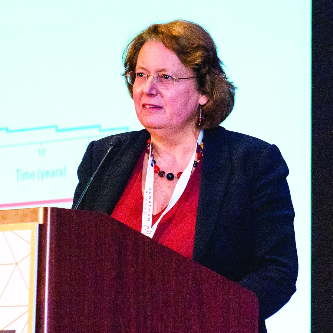
“Clearly the problem with most of the relapsed PTCL [cases] is that they don’t achieve a good response to salvage therapy. If they do, then they have much better chance of doing well,” she said at the annual T-cell Lymphoma Forum.
She outlined her center’s approach for treating patients with relapsed or refractory PTCL, following a case presentation by Royal Marsden fellow Matthew Cross, MD.
Complex disease, multiple therapies
The patient was a 71-year-old woman who in 2007 had a diagnosis of asymptomatic stage 4A follicular lymphoma managed with observation; in 2010, she was diagnosed with a CD30-positive PTCL not otherwise specified with ongoing low-level bone marrow involvement with follicular lymphoma.
She initially was treated elsewhere with R-CHOP chemotherapy (cyclophosphamide, doxorubicin, vincristine, and prednisone plus rituximab) and had a response after four cycles; however, she had progression with new intra-abdominal nodal sites by the sixth cycle and then was started on two cycles of ESHAP (etoposide, methylprednisolone, high-dose cytarabine, and cisplatin), but she had further progression by May 2011 and opted to forgo additional treatment.
By July 2011, however, she became highly symptomatic with new pruritic rashes on her legs, abdominal pain, and distention. She was referred to the Royal Marsden Hospital, where she was eventually diagnosed with angioimmunoblastic T-cell lymphoma (AITL) with an Epstein-Barr virus–negative clonal large B-cell proliferation in her bone marrow.
She was treated with gemcitabine plus methylprednisolone and prophylactic intrathecal methotrexate and had an “excellent clinical and radiological response,” Dr. Cross said.
A subsequent bone marrow biopsy showed marked hypocellularity but no evidence of either T-cell of B-cell lymphomas.
An autologous stem cell transplant was planned, but two attempts at harvesting peripheral blood stem cells – including one with plerixafor (Mozobil) – failed, and a PET scan within 3 months showed signs of early progression.
In April 2012, the patient was started on romidepsin (Istodax) and had a 1-year remission. But in April 2013, a repeat biopsy again showed CD30-positive AITL. Based on the CD30 positivity, the patient was started on brentuximab vedotin (Adcetris) in May 2013. She was observed to have progression in inguinal nodes in January 2014; she was treated with local radiotherapy and continued on brentuximab but had further progression in June 2014. At that time, she had additional gemcitabine-based combination chemotherapy and had stable disease for 10 months.
In March 2015, she received lenalidomide for further progression but could not tolerate the drug. She died in September 2015, 5 years after diagnosis and 4.5 years after frontline therapy failed.
Therapeutic rationale
Dr. Dearden walked through the choices that she, along with Dr. Cross and their colleagues, made in treating the patient. They chose gemcitabine-based regimens for salvage therapy because of the drug’s efficacy across various forms on non-Hodgkin and Hodgkin lymphoma, she said.
However, a randomized, phase 3, noninferiority trial in the United Kingdom comparing GEM-P (gemcitabine, cisplatin, and methylprednisolone) with CHOP for first-line therapy of PTCL was halted at the interim analysis because GEM-P had not meet the primary endpoint, she said. Results of that trial have not been published to date.
“Clearly, if it’s the patients who do well, often it’s because they achieve a good enough remission to be able to proceed to some sort of consolidation therapy with autologous or allogeneic stem cell transplants, and I think auto-graft is probably accepted for the younger, fitter patients with relapsed chemo-sensitive disease,” she said.
Three-year survival rates for autologous hematopoietic stem cell transplantation range from 36% to 58% and are better than those seen with chemotherapy alone, she said.
“The problem of course is that not many patients receive the planned auto-graft, even if that’s the intention, either because of failure to respond to salvage regimen or early disease progression, which happens before the transplant is able to take place,” she said,
A reasonable alternative for patients with relapsed/refractory PTCL is allogeneic transplantation, as shown in a 2008 study.
Among 77 patients – 57 of whom had received myeloablative conditioning, 31 of whom were in complete remission, and 26 of whom had partial response at the time of transplants – the 5-year treatment-related mortality rate was 33%. However, the 5-year event-free and overall survival rates were 53% and 57%, respectively. Patients with AITL had especially good outcomes (J Clin Oncol. 2008 May 10;26[14]:2264-71).
“In an ideal world, if our patient had been a suitable candidate for an allo-transplant, it’s what we would have tried to undertake,” Dr. Dearden said.
Dr. Dearden recommended that all patients with relapsed or refractory PTCL be considered for clinical trials. For fit patients in first relapse, combination platinum-based chemotherapy followed by autologous or allogeneic transplant may be effective.
For patients not eligible for transplant or with chemotherapy-refractory disease, she recommended trying the following monotherapy approaches: pralatrexate for patients with PTCL not otherwise specified, histone deacetylase inhibitors or 5-azacytidine for AITL, brentuximab vedotin for anaplastic large cell lymphoma, and pembrolizumab for natural killer/T-cell lymphomas.
Although two lines of intensive chemotherapy had failed the case patient within 6 months of diagnosis, she still survived for 5 years with sequential monotherapies, Dr. Dearden noted.
“I use to say to her, ‘You just need to stay one drug ahead of your disease.’ And she was well, she had a very good quality of life for a period of time, and if you can deliver a treatment that is effective for a patient, it will extend their survival,” Dr. Dearden said.
The T-cell Lymphoma Forum is held by Jonathan Wood & Associates, which is owned by the same company as this news organization. Dr. Dearden has consulted for MedImmune, Infinity Pharmaceuticals, Janssen, Gilead Sciences, and Roche, and has received honoraria from Janssen and Gilead. Dr. Cross reported no having no financial disclosures.
LA JOLLA, CALIF. – When patients with peripheral T-cell lymphoma (PTCL) experience relapse, consider an allogeneic stem cell transplant or clinical trial, investigators advised.
Patients with relapsed PTCL have generally dismal outcomes, with a median progression-free survival (PFS) of 3.7 months and a median overall survival (OS) of just 6.5 months, according to one study (J Clin Oncol. 2013 Jun 1;31[16]:1970-6).
“Clearly the problem with most of the relapsed PTCL [cases] is that they don’t achieve a good response to salvage therapy. If they do, then they have much better chance of doing well,” she said at the annual T-cell Lymphoma Forum.
She outlined her center’s approach for treating patients with relapsed or refractory PTCL, following a case presentation by Royal Marsden fellow Matthew Cross, MD.
Complex disease, multiple therapies
The patient was a 71-year-old woman who in 2007 had a diagnosis of asymptomatic stage 4A follicular lymphoma managed with observation; in 2010, she was diagnosed with a CD30-positive PTCL not otherwise specified with ongoing low-level bone marrow involvement with follicular lymphoma.
She initially was treated elsewhere with R-CHOP chemotherapy (cyclophosphamide, doxorubicin, vincristine, and prednisone plus rituximab) and had a response after four cycles; however, she had progression with new intra-abdominal nodal sites by the sixth cycle and then was started on two cycles of ESHAP (etoposide, methylprednisolone, high-dose cytarabine, and cisplatin), but she had further progression by May 2011 and opted to forgo additional treatment.
By July 2011, however, she became highly symptomatic with new pruritic rashes on her legs, abdominal pain, and distention. She was referred to the Royal Marsden Hospital, where she was eventually diagnosed with angioimmunoblastic T-cell lymphoma (AITL) with an Epstein-Barr virus–negative clonal large B-cell proliferation in her bone marrow.
She was treated with gemcitabine plus methylprednisolone and prophylactic intrathecal methotrexate and had an “excellent clinical and radiological response,” Dr. Cross said.
A subsequent bone marrow biopsy showed marked hypocellularity but no evidence of either T-cell of B-cell lymphomas.
An autologous stem cell transplant was planned, but two attempts at harvesting peripheral blood stem cells – including one with plerixafor (Mozobil) – failed, and a PET scan within 3 months showed signs of early progression.
In April 2012, the patient was started on romidepsin (Istodax) and had a 1-year remission. But in April 2013, a repeat biopsy again showed CD30-positive AITL. Based on the CD30 positivity, the patient was started on brentuximab vedotin (Adcetris) in May 2013. She was observed to have progression in inguinal nodes in January 2014; she was treated with local radiotherapy and continued on brentuximab but had further progression in June 2014. At that time, she had additional gemcitabine-based combination chemotherapy and had stable disease for 10 months.
In March 2015, she received lenalidomide for further progression but could not tolerate the drug. She died in September 2015, 5 years after diagnosis and 4.5 years after frontline therapy failed.
Therapeutic rationale
Dr. Dearden walked through the choices that she, along with Dr. Cross and their colleagues, made in treating the patient. They chose gemcitabine-based regimens for salvage therapy because of the drug’s efficacy across various forms on non-Hodgkin and Hodgkin lymphoma, she said.
However, a randomized, phase 3, noninferiority trial in the United Kingdom comparing GEM-P (gemcitabine, cisplatin, and methylprednisolone) with CHOP for first-line therapy of PTCL was halted at the interim analysis because GEM-P had not meet the primary endpoint, she said. Results of that trial have not been published to date.
“Clearly, if it’s the patients who do well, often it’s because they achieve a good enough remission to be able to proceed to some sort of consolidation therapy with autologous or allogeneic stem cell transplants, and I think auto-graft is probably accepted for the younger, fitter patients with relapsed chemo-sensitive disease,” she said.
Three-year survival rates for autologous hematopoietic stem cell transplantation range from 36% to 58% and are better than those seen with chemotherapy alone, she said.
“The problem of course is that not many patients receive the planned auto-graft, even if that’s the intention, either because of failure to respond to salvage regimen or early disease progression, which happens before the transplant is able to take place,” she said,
A reasonable alternative for patients with relapsed/refractory PTCL is allogeneic transplantation, as shown in a 2008 study.
Among 77 patients – 57 of whom had received myeloablative conditioning, 31 of whom were in complete remission, and 26 of whom had partial response at the time of transplants – the 5-year treatment-related mortality rate was 33%. However, the 5-year event-free and overall survival rates were 53% and 57%, respectively. Patients with AITL had especially good outcomes (J Clin Oncol. 2008 May 10;26[14]:2264-71).
“In an ideal world, if our patient had been a suitable candidate for an allo-transplant, it’s what we would have tried to undertake,” Dr. Dearden said.
Dr. Dearden recommended that all patients with relapsed or refractory PTCL be considered for clinical trials. For fit patients in first relapse, combination platinum-based chemotherapy followed by autologous or allogeneic transplant may be effective.
For patients not eligible for transplant or with chemotherapy-refractory disease, she recommended trying the following monotherapy approaches: pralatrexate for patients with PTCL not otherwise specified, histone deacetylase inhibitors or 5-azacytidine for AITL, brentuximab vedotin for anaplastic large cell lymphoma, and pembrolizumab for natural killer/T-cell lymphomas.
Although two lines of intensive chemotherapy had failed the case patient within 6 months of diagnosis, she still survived for 5 years with sequential monotherapies, Dr. Dearden noted.
“I use to say to her, ‘You just need to stay one drug ahead of your disease.’ And she was well, she had a very good quality of life for a period of time, and if you can deliver a treatment that is effective for a patient, it will extend their survival,” Dr. Dearden said.
The T-cell Lymphoma Forum is held by Jonathan Wood & Associates, which is owned by the same company as this news organization. Dr. Dearden has consulted for MedImmune, Infinity Pharmaceuticals, Janssen, Gilead Sciences, and Roche, and has received honoraria from Janssen and Gilead. Dr. Cross reported no having no financial disclosures.
EXPERT ANALYSIS FROM TCLF 2018
Researchers question validity of NCCN guidelines

New research suggests guidelines from the National Comprehensive Cancer Network (NCCN) may sometimes be supported by low-quality evidence or no evidence at all.
Researchers compared NCCN recommendations for cancer drugs to US cancer drug approvals over a 5-year period.
Thirty-nine percent of NCCN’s treatment recommendations did not coincide with uses approved by the US Food and Drug Administration (FDA).
For most of these recommendations (84%), NCCN did not provide supporting data from randomized, phase 3 trials.
For 36% of the recommendations, NCCN gave no supporting evidence.
Vinay Prasad, MD, of Oregon Health & Science University in Portland, Oregon, and his colleagues reported these findings in The BMJ.
Dr Prasad and his colleagues compared FDA approvals of cancer drugs between 2011 and 2015 with NCCN recommendations as of March 25, 2016.
When NCCN made recommendations beyond FDA approvals, the researchers evaluated the evidence used to support those recommendations.
Forty-seven new cancer drugs were approved by the FDA for 69 indications between 2011 and 2015. NCCN recommended the 47 drugs for 113 indications, including the 69 FDA-approved indications.
So 39% (n=44) of NCCN’s recommendations were not approved by the FDA, and NCCN gave the following evidence to support these recommendations:
- No evidence—36% (n=16)
- Phase 2 trial without randomization—30% (n=13)
- Randomized, phase 3 trial—16% (n=7)
- Phase 2 trial with randomization—7% (n=3)
- Case report or series of less than 5 patients—5% (n=2)
- Book chapter or review article—2% (n=1)
- Phase 1 trial—2% (n=1)
- Ongoing trial—2% (n=1).
Dr Prasad and his colleagues did point out that not all FDA approvals are supported by randomized, phase 3 trials.
And when the team followed-up 21 months after their initial analysis, they found that 6 of the 44 (14%) additional recommendations by NCCN had received FDA approval.
The researchers also noted that they did not search for independent evidence to support NCCN recommendations beyond the references NCCN provided. So some of the recommendations may have had more or better supporting evidence than what was provided.
Still, the team said these results suggest NCCN “frequently” makes recommendations that go beyond FDA approvals and “often fails to cite evidence or relies on low levels of evidence.” Therefore, NCCN should cite all evidence used to formulate its recommendations.
NCCN argues that it does provide ample evidence to support the recommendations in its guidelines.
“The NCCN guidelines contain more than 24,500 references to inform users of the evidence used in making its decisions,” said Robert W. Carlson, MD, chief executive officer of NCCN.
“These data are supplemented by the analysis of the available evidence by expert clinician researchers and patient advocates who evaluate each recommendation and come to consensus. Each recommendation is labeled with a Category of Evidence, and the vast majority of those for systemic therapies are accompanied by Evidence Blocks, which outline, on 1-5 scales, the efficacy, safety, quality of the evidence, consistency of the evidence, and affordability of the treatment.”
FDA approves label update for nivolumab
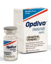
The US Food and Drug Administration (FDA) has updated the label for nivolumab (Opdivo®) to include new dosing and administration information.
Nivolumab can now be given at 480 mg infused every 4 weeks for most approved indications, in addition to the previously approved dosing schedule of 240 mg every 2 weeks.
The FDA also approved a shorter 30-minute infusion across all approved indications of nivolumab.
The 480 mg dose option can be used for nearly all approved indications of nivolumab. The exceptions are patients with microsatellite instability-high or mismatch repair-deficient metastatic colorectal cancer.
Nivolumab is FDA-approved for the following indications:
- To treat adults with classical Hodgkin lymphoma that has relapsed or progressed after autologous hematopoietic stem cell transplant (HSCT) and brentuximab vedotin or after 3 or more lines of systemic therapy that includes autologous HSCT.
- As monotherapy for patients with BRAF V600 mutation-positive unresectable or metastatic melanoma as well as BRAF V600 wild-type unresectable or metastatic melanoma.
- In combination with ipilimumab for the treatment of patients with unresectable or metastatic melanoma.
- To treat patients with metastatic non-small cell lung cancer with progression on or after platinum-based chemotherapy. Patients with EGFR or ALK genomic tumor aberrations should have disease progression on FDA-approved therapy for these aberrations prior to receiving nivolumab.
- For patients with advanced renal cell carcinoma who have received prior anti-angiogenic therapy.
- To treat patients with recurrent or metastatic squamous cell carcinoma of the head and neck with disease progression on or after platinum-based therapy.
- For patients with locally advanced or metastatic urothelial carcinoma who have disease progression during or after platinum-containing chemotherapy or have disease progression within 12 months of neoadjuvant or adjuvant treatment with platinum-containing chemotherapy.
- To treat adult and pediatric (12 years and older) patients with microsatellite instability-high or mismatch repair-deficient metastatic colorectal cancer that has progressed following treatment with a fluoropyrimidine, oxaliplatin, and irinotecan.
- For patients with hepatocellular carcinoma who have been previously treated with sorafenib.
- For the adjuvant treatment of patients with melanoma with involvement of lymph nodes or metastatic disease who have undergone complete resection.
High efficacy, no safety signals for herpes zoster vaccine post-HSCT
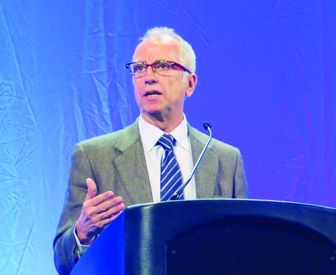
SALT LAKE CITY – A recently approved adjuvanted herpes zoster vaccine)(Shingrix) effectively and safely prevented herpes zoster in a population of patients with multiple myeloma and other hematologic malignancies who received autologous hematopoietic stem cell transplantation.
The use of recombinant varicella zoster virus glycoprotein E in combination with an adjuvant system gives immunosuppressed individuals who have received hematopoietic stem cell transplantation (HSCT) a safe option for prevention of herpes zoster (HZ), said Javier de la Serna, MD, PhD, speaking at the combined annual meetings of the Center for International Blood & Marrow Transplant Research and the American Society for Blood and Marrow Transplantation.
Presenting the findings at a late-breaking abstract session, Dr. de la Serna said that for the 1,721 participants in a placebo-controlled multicenter trial who received both doses of the vaccine, the incidence of HZ for vaccine recipients was 3.0%, compared with 9.4% of placebo recipients, for a vaccine efficacy of 68.2% (95% confidence interval, 55.6-77.5; P less than 0.0001). These results met the study’s primary objective.
Postherpetic neuralgia (PHN) prevention efficacy – a secondary endpoint – was 89.3% for those receiving the vaccine (HZ/su); the incidence of PHN was 0.5% in the HZ/su study arm, compared with 4.9% for those who received placebo (95% CI, 22.5-99.8). The study also tracked other HZ complications as a secondary endpoint, finding efficacy of 77.8% (95% CI, 19.1–95.0). “The vaccine was highly efficacious in preventing all the secondary outcomes,” said Dr. de la Serna of the Hospital Universitario 12 de Octubre, Madrid.
The randomized, observer-blind phase 3 trial was conducted in 28 countries.Adults who received autologous HSCT were randomized 1:1 to receive HZ/su (n = 922) or placebo (n = 924) within 50-70 days of their transplant. Patients were excluded if they were expected to receive more than 6 months of anti–varicella zoster prophylaxis posttransplant, Dr. de la Serna said.
Participants received the first dose of HZ/su at the first study visit, and the second dose 30-60 days later. Patients were seen 1 month after the last vaccine dose, and then again at months 13 and 25, with telephone follow-up between the later visits. All participants were followed for at least 1 year, Dr. de la Serna said.
Episodes of HZ were confirmed by polymerase chain reaction assay, or, when samples were lacking or indeterminate, by agreement of at least three members of an ascertainment committee.
Of the two components of the HZ/su vaccine, glycoprotein E triggers both humoral immunity and activity of varicella zoster–specific CD4+ T cells; the adjuvant system – dubbed ASO1 – boosts immune response. The vaccine was approved by the Food and Drug Administration in October 2017 for use in adults aged 50 years and older.
In addition to the primary endpoint of vaccine efficacy in prevention of HZ cases during the study period, secondary objectives included monitoring vaccine reactogenicity and safety, and evaluating vaccine efficacy for the prevention of PHN and other complications of HZ.
Tertiary objectives included vaccine efficacy in preventing HZ during the first year posttransplant (vaccine efficacy 84.7%; 95% CI, 32.2-96.6), as well as efficacy in preventing hospitalizations related to HZ (vaccine efficacy 76.2%, 95% CI 61.1-86.0).
An exploratory analysis found vaccine efficacy of 71.8% for participants younger than 50 years (95% CI, 38.8 – 88.3). For patients aged 50 years and older, vaccine efficacy was 67.3% (95% CI, 52.6–77.9).
The safety of HZ/su was determined by analyzing data for all participants, but efficacy data included only those who received the second dose and did not develop HZ within a month of receiving the second vaccine dose.
In the efficacy group (n = 1,721), patients were mostly (n = 1,296) aged 50 years or older. Most patients (n = 937) received HSCT for multiple myeloma. Overall, participants were about 63% male, and 77% were of Caucasian/European ancestry.
Adverse events, solicited for the first 7 days after injections, were mostly mild and related to the local site pain and inflammation expected with an adjuvanted vaccine; HZ/su recipients also experienced more fatigue and muscle aches than did those receiving placebo. Median duration of symptoms was up to 3 days, with grade 3 events lasting up to 2 days.
Unsolicited and serious adverse events were similar between study arms, with a median safety follow-up period of 29 months. The investigators judged that no deaths were related to the vaccine, and there were no signals for increased rate of relapse or immune-mediated diseases.
The study was funded by GlaxoSmithKline; HZ/su(Shingrix) is marketed by GlaxoSmithKline. Dr. de la Serna reported being on the advisory board or receiving honoraria from multiple pharmaceutical companies.
SOURCE: de la Serna J et al. 2018 BMT Tandem Meetings, Abstract LBA2.
REPORTING FROM THE 2018 BMT TANDEM MEETINGS
Key clinical point:
Major finding: Efficacy was 68.17% for preventing herpes zoster among HSCT recipients.
Study details: A randomized, observer blind, placebo-controlled phase 3 study of 1,846 post-HSCT recipients.
Disclosures: The study was sponsored by GlaxoSmithKline. Dr. de la Serna reported relationships with multiple pharmaceutical companies.
Source: de la Serna J et al. 2018 BMT Tandem Meetings, Abstract LBA2.
Export protein inhibitor shows activity against refractory myeloma
Think of it as a biological nuclear export ban: A combination of the nuclear export protein inhibitor selinexor and dexamethasone was associated with relatively good objective response rates and clinical benefit rates among some patients with relapsed or refractory multiple myeloma (MM).
In the dose-expansion portion of a phase 1 dose-finding trial, the objective response rate (ORR) among 12 patients treated with selinexor 45 mg/m2 and 20 mg dexamethasone twice weekly was 50%, and the clinical benefit rate – a composite of complete responses, very good partial responses, partial responses and minimal responses – was 58%, reported Christine Chen, MD, of Princess Margaret Cancer Centre in Toronto, and her colleagues.
“Selinexor is an oral agent with a completely novel mechanism of action and anti-MM activity in combination with dexamethasone that could provide a new option for patients suffering from this incurable disease,” wrote Dr. Chen and her colleagues. The report was published in Blood.
Selinexor is a selective oral inhibitor of the cellular nuclear export protein exportin 1 (XPO1). Inhibition of this protein causes tumor suppressor proteins to accumulate in the nuclei of malignant cells, leading to programmed cell death (apoptosis) of malignant cells, but with minimal effects on normal cells.
The study, performed in centers in Canada, the United States, and Denmark, was designed primarily to identify the recommended dose of oral selinexor for phase 2 trials, with or without corticosteroids.
A total of 84 patients were enrolled, including 22 with MM and 3 with Waldenstrom macroglobulinemia in the dose-escalation phase, and 59 with MM in the dose expansion phase.
In the dose-expansion phase, patients were treated with one of two dosing schemes: either selinexor at a dose of 45 or 60 mg/m2 plus dexamethasone 20 mg twice weekly in 28-day cycles, or selinexor in a 40 mg or 60 mg flat dose without corticosteroids in 21-day cycles.
As a single agent, selinexor showed minimal activity, with an ORR of 4%, and clinical benefit rate of 21% in 57 patients.
Among 12 patients assigned to the 45 mg/m2 selinexor dose plus dexamethasone, the ORR was 50%, consisting of one complete response and five partial responses. In addition, one patient at this dose level had a minimal response, three had stable disease, one had disease progression, and one was withdrawn from the study before disease assessment, with no evidence of progression.
There were no objective responses in the 60-mg/m2 selinexor dose group.
Among all 84 patients enrolled, the ORR with selinexor alone or in combination was 10%, and the clinical benefit rate was 25%. Of the patients with Waldenstrom macroglobulinemia, one had a partial response and one had a minimal response.
In the safety analysis, which included all patients who received at least one dose of selinexor, the most common grade 3 or 4 adverse events included thrombocytopenia in 45% of patients, hyponatremia in 26% of patients, and anemia and neutropenia in 23% each.
The most common nonhematologic adverse events – primarily grade 1 or 2 – included nausea, fatigue, anorexia, vomiting, and weight loss and diarrhea.
The combination of selinexor and dexamethasone is currently being investigated in the phase 2 Selinexor Treatment of Refractory Myeloma study, in combination with standard multiple myeloma therapies in the STOMP trial (Selinexor and Backbone Treatments of Multiple Myeloma Patients), and with bortezomib in the BOSTON trial (Bortezomib, Selinexor and Dexamethasone in Patients with Multiple Myeloma).
Dr. Chen reported no conflicts of interest. Her coauthors reported financial ties to Karyopharm Therapeutics, which funded the study.
hematologynews@frontlinemedcom.com
SOURCE: Chen C et al. Blood. 2018;131(8):855-63.
FROM BLOOD
Key clinical point:
Major finding: The objective response rate among 12 patients with multiple myeloma treated at a dose of 45 mg/m2 selinexor and 20 mg dexamethasone was 50%.
Data source: Phase 1 dose-escalation and expansion study of 81 patients with relapsed or refractory multiple myeloma and three patients with Waldenstrom macroglobulinemia.
Disclosures: Dr. Chen reported no conflicts of interest. Her coauthors reported financial ties to Karyopharm Therapeutics, which funded the study.
Source: Chen C et al. Blood. 2018;131(8):855-63.
Study confirms higher risk of infection with CB transplant
SALT LAKE CITY—Results of a large, retrospective analysis support the notion that patients who receive cord blood (CB) transplants have a higher risk of infection than other hematopoietic stem cell transplant (HSCT) recipients.
Investigators found that CB recipients had a significantly higher risk of bacterial, viral, and fungal infections in the early post-transplant period than patients who received peripheral blood (PB) or bone marrow (BM) transplants.
In addition, CB recipients had longer hospital stays, higher inpatient costs, and greater inpatient mortality than PB and BM recipients.
Amandeep Godara, MD, of Tufts Medical Center in Boston, Massachusetts, presented these results at the 2018 BMT Tandem Meetings (abstract 30*).
“Infections are considered more common in cord blood transplant recipients based on some prior retrospective analyses,” Dr Godara noted. “But there is limited data comparing these infectious complications between cord blood transplant and peripheral blood/bone marrow stem cell transplants during the inpatient stay for the stem cell transplant.”
With this in mind, Dr Godara and his colleagues analyzed data from the Healthcare Cost and Utilization Project National Inpatient Sample. This database covers 46 US states and contains data from more than 7 million hospital stays each year.
The investigators searched the database for hospital admissions for HSCT from 2002 to 2014. They identified 2979 CB transplants and 56,845 PB or BM transplants.
The CB recipients had a median age of 48, and 55% were male. Fifty-nine percent were white, 18% Hispanic, 13% black, 5% Asian, and 5% “other.” Sixty-six percent of patients had acute leukemia, 18% non-Hodgkin lymphoma, 5% Hodgkin lymphoma, and 11% “other” diseases.
The PB/BM recipients had a median age of 45, and 58% were male. Seventy-nine percent were white, 8% Hispanic, 6% black, 3% Asian, and 4% “other.” Sixty-one percent of patients had acute leukemia, 16% non-Hodgkin lymphoma, 4% Hodgkin lymphoma, and 19% “other” diseases.
Results
Dr Godara and his colleagues compared the rates and types of infection from the time of HSCT to hospital discharge in CB and PB/BM recipients. The team also compared early inpatient mortality, the cost of hospitalization, and the length of hospital stay.
“[W]e observed a higher risk for infections in cord blood transplant patients compared to peripheral blood and bone marrow stem cell transplant patients, and this risk for infection extended through a wide spectrum of pathogens,” Dr Godara said.
“We also observed a higher all-cause inpatient mortality in cord blood transplant compared to peripheral blood and bone marrow transplant, especially in patients who had bacterial sepsis or invasive fungal infection.”
The rate of bacterial sepsis was 34.87% in CB recipients and 20.20% in PB/BM recipients (P<0.0001). Rates of viral infection were 20.05% and 8.19%, respectively (P<0.0001). And rates of invasive fungal infection were 12.87% and 7.89% (P<0.0001).
There was a similar distribution of bacterial infections in CB and PB/BM recipients. The most common was pneumonia (47% and 41%, respectively), followed by abdominal infections (29% and 31%, respectively), urinary tract infections (17% and 21%, respectively), central line-associated bloodstream infections (4% and 6%, respectively), and acute sinusitis (3% and 1%, respectively).
The rate of Clostridium difficile infection was significantly higher in CB recipients than PB/BM recipients—11.75% and 8.90%, respectively (P=0.02). However, there was no significant difference in mortality related to C. difficile—14% and 10%, respectively (P=0.3).
On the other hand, all-cause inpatient mortality was significantly higher in CB recipients than PB/BM recipients—16% and 7%, respectively (P<0.0001).
Inpatient mortality rates were significantly higher for CB recipients with bacterial sepsis (33% vs 23%, P=0.001) and invasive fungal infections (29% vs 16%, P=0.0045) but not viral infections (19% vs 17%, P=0.5).
The median length of hospital stay was 36 days for CB recipients and 25 days for PB/BM recipients. The mean inpatient charges were $448,892 and $250,437 respectively.
*Data in the abstract differ from the presentation.
Company withdraws application for idelalisib
Gilead Sciences International Ltd. recently withdrew its application for European approval of idelalisib (Zydelig) in combination with rituximab and bendamustine for the treatment of relapsed/refractory chronic lymphocytic leukemia (CLL).
Idelalisib is currently approved in the European Union for use in combination with an anti-CD20 monoclonal antibody (rituximab or ofatumumab) to treat adults with CLL who have received at least 1 prior therapy or as first-line treatment of CLL in the presence of 17p deletion or TP53 mutation in patients who are not eligible for any other therapies.
Idelalisib is also approved as monotherapy for adults with follicular lymphoma that is refractory to 2 prior lines of treatment.
In seeking expanded approval for idelalisib, Gilead submitted data from a study (NCT01569295) comparing idelalisib plus bendamustine and rituximab to placebo plus bendamustine and rituximab.
Interim results from this study were published in The Lancet Oncology in March 2017.
Gilead withdrew the application for idelalisib after the European Medicines Agency’s Committee for Medicinal Products for Human Use (CHMP) had evaluated documentation provided by the company and formulated lists of questions.
Gilead had not responded to the last round of questions at the time of the withdrawal.
At that point, the CHMP was of the provisional opinion that idelalisib should not be approved for use in combination with rituximab and bendamustine in patients with relapsed/refractory CLL.
The CHMP said additional, longer-term data are needed to show the benefits of idelalisib plus rituximab and bendamustine outweigh the risks.
Gilead said its withdrawal of the application was based on the CHMP’s opinion that there was insufficient evidence of a favorable benefit-risk profile.
The company also said the withdrawal does not impact ongoing trials of idelalisib.
CHMP endorses drug delivery system
The European Medicines Agency’s Committee for Medicinal Products for Human Use (CHMP) has recommended a label variation for Neulasta® (pegfilgrastim) to include the Neulasta® Onpro® Kit.
The Neulasta Onpro Kit is an on-body injector (OBI) delivery system for Neulasta, which is approved in the European Union to reduce the duration of neutropenia and the incidence of febrile neutropenia in adults treated with cytotoxic chemotherapy for malignancy (with the exception of chronic myeloid leukemia and myelodysplastic syndromes).
The Neulasta Onpro Kit includes a specifically designed Neulasta pre-filled syringe along with a single-use OBI. The small, lightweight OBI is applied to a patient’s skin on the same day of chemotherapy.
The OBI is intended to facilitate timed delivery of the correct dose of Neulasta and eliminate the need for patients to return to a healthcare setting the day after they receive chemotherapy.
The CHMP’s opinion on the Neulasta Onpro Kit will be reviewed by the European Commission (EC).
If the EC agrees with the CHMP, the centralized marketing authorization of Neulasta will be updated to include information on the Neulasta Onpro Kit in the drug’s label.
This change will be valid throughout the European Union. Norway, Iceland, and Liechtenstein will make corresponding decisions about Neulasta’s label on the basis of the EC’s decision.
The EC typically makes a decision within 67 days of the CHMP’s recommendation.