User login
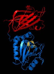
Protein is key to HSC recovery after chemo, radiation

in the bone marrow
The protein beta-catenin plays a critical role in promoting the recovery of hematopoietic stem cells (HSCs) after exposure to radiation or chemotherapy, according to preclinical research published in Genes & Development.
The study provides new insight into the impact of radiation and chemotherapy on cellular and molecular processes.
And it points to possibilities for improving HSC regeneration in cancer patients who have undergone these treatments.
Study investigators first used mouse models to show that exposure to radiation triggers activation of the Wnt signaling pathway in hematopoietic stem and progenitor cells.
“The Wnt pathway and its key mediator, beta catenin, are critical for embryonic development and establishment of the body plan,” explained Tannishtha Reya, PhD, of the University of California, San Diego.
“In addition, the Wnt pathway is activated in stem cells from many tissues and is needed for their continued maintenance.”
Dr Reya and her colleagues then found that mice deficient in beta-catenin lacked the ability to activate canonical Wnt signaling. They also suffered from impaired HSC regeneration and bone marrow recovery after radiation or chemotherapy.
Mouse HSCs without beta-catenin could not suppress the production of reactive oxygen species or resolve DNA double-strand breaks. As a result, they could not recover effectively after radiation exposure or treatment with the chemotherapeutic agent fluorouracil.
“Our work shows that Wnt signaling is important in the mammalian hematopoietic system and is critical for recovery from chemotherapy and radiation,” Dr Reya said. “There are 2 major reasons why accelerating regeneration is important clinically.”
“One is that, after cancer patients are irradiated and transplanted with stem cells, the rate and extent of recovery is often not sufficient to protect the patient from anemia or infections . . . . Identifying signals that can boost regeneration after the bone marrow is severely damaged may help improve outcomes after transplantation.”
“Second, doses of chemotherapy and radiation used to treat cancer are often limited by the collateral damage they cause to normal tissues. If we can improve and accelerate recovery, we might be able to use higher doses of radiation or chemotherapy and reduce the risk of cancer relapse.”
Dr Reya added that this research suggests HSC regeneration could be accelerated by modulating the Wnt pathway, either by delivering additional Wnt proteins directly to patients or through drugs that activate the pathway. ![]()
Stats show increase in cancer survival rates

Credit: NIH
New statistics suggest 10-year survival rates for cancer patients in England and Wales have more than doubled over a 40-year period.
And rates increased substantially for those with hematologic malignancies.
From 1971 to 2011, 10-year survival rates increased nearly 7-fold for patients with multiple myeloma and almost 6-fold for leukemia patients.
Rates nearly tripled for non-Hodgkin lymphoma patients and almost doubled for those with Hodgkin lymphoma.
These statistics were released by Cancer Research UK.
“These results come from detailed analysis of the survival of more than 7 million cancer patients diagnosed in England and Wales since the 1970s,” said Michel Coleman, BM BCh, head of Cancer Research UK’s Cancer Survival Group at the London School of Hygiene and Tropical Medicine.
“They show just how far we’ve come in improving cancer survival, but they also shine a spotlight on areas where much more needs to be done.”
The statistics include all adults (aged 15 to 99) diagnosed with cancer in England and Wales.
An analysis of the figures showed that, in 1971-1972, 24% of all cancer patients survived 10 years. By 2010-2011, that figure had increased to 50%.
For leukemia patients, 10-year survival increased from 8% in 1970-1971 to 46% in 2010-2011. For patients with multiple myeloma, it rose from 5% to 33%.
For patients with Hodgkin lymphoma, 10-year survival increased from 49% to 80%. And for non-Hodgkin lymphoma patients, it increased from 22% to 63%.
There were substantial increases in shorter-term survival rates (1-year and 5-year) as well. For details, see the Cancer Research UK website. ![]()
Diagnosis and Management of Acute and Chronic Graft-versus-Host Disease
Allogeneic hematopoietic stem cell transplantation (HSCT) is a potentially curative treatment option for several hematologic malignancies and other congenital diseases including immunodeficiencies or hemoglobinopathies. When the first allografts were performed, most patients given bone marrow (BM) from donors other than homozygotic twins developed skin, gut, and/or liver injury. This disease was defined by Billingham in 1966 as graft-versushost disease (GVHD). He also described 3 standard tenets for GVHD pathophysiology, which remain valid today even with rapid advances in this area: (1) donor graft must have immune-competent cells, (2) recipient must be incapable of rejecting the graft, and (3) recipient must have tissue antigens not present in the donor.
To read the full article in PDF:
Vitamin D may affect outcome in cancer patients

Cancer patients with higher levels of vitamin D at diagnosis tend to have better outcomes than patients who are vitamin D-deficient, a meta-analysis suggests.
Researchers found the association between vitamin D and prognosis was most pronounced in patients with breast cancer, colorectal cancer, and lymphomas.
The team observed an association between vitamin D and prognosis in other cancers as well, but the correlations were not significant.
Hui Wang, MD, PhD, of the Chinese Academy of Sciences in Shanghai, China, and colleagues conducted this research and reported their results in the Journal of Clinical Endocrinology & Metabolism.
The team analyzed data from 25 studies including 17,332 cancer patients. In most of the studies, patients had their vitamin D levels tested before they underwent any cancer treatment.
The analysis showed that, overall, a 10 nmol/L increase in vitamin D levels was associated with a 4% increase in survival.
The strongest links between vitamin D levels and survival were seen in breast cancer, colorectal cancer, and lymphomas. There was less evidence of a connection in patients with leukemias, lung cancer, gastric cancer, prostate cancer, melanoma, or Merkel cell carcinoma, but the available data were positive.
There were 2 studies evaluating the association between survival and vitamin D levels in leukemia patients.
Vitamin D insufficiency was associated with poor overall survival in patients with newly diagnosed chronic lymphocytic leukemia, although this was not statistically significant. The hazard ratio (HR) was 0.68 (P=0.07).
For acute myeloid leukemia, patients with vitamin D levels that were considered insufficient (20-31.9 ng/mL) or deficient (<20 ng/mL) had worse overall survival (HR=0.65) and disease-free survival (HR=0.65) than patients with normal vitamin D levels.
There were also 2 studies evaluating the association between survival and vitamin D levels in lymphoma patients. The results showed that patients with the highest vitamin D levels had better overall survival than those with the lowest vitamin D levels (HR=0.48, P<0.001).
Lymphoma patients in the highest quartile of vitamin D levels also had a lower risk of cancer-specific mortality (HR=0.50, P<0.001) and better disease-free survival (HR=0.80, P=0.04).
Similarly, higher vitamin D levels were significantly associated with reduced cancer-specific mortality for patients with colorectal cancer (P=0.005) and improved disease-free survival for patients with breast cancer (P<0.001).
For overall survival, the HR for the highest vs lowest quartile of vitamin D levels was 0.55 for colorectal cancer patients and 0.63 for breast cancer patients.
Based on these results, Dr Wang concluded, “Physicians need to pay close attention to vitamin D levels in people who have been diagnosed with cancer.” ![]()
FDA approves new formulation of mercaptopurine

Credit: Bill Branson
The US Food and Drug Administration (FDA) has approved an oral suspension of mercaptopurine (Purixan), a drug used in combination therapy to treat patients with acute lymphoblastic leukemia (ALL).
Mercaptopurine will now be available as a 20 mg/mL oral suspension.
The drug was originally approved as a 50 mg tablet in 1953 and, since that time, has only been commercially available in this form.
Unfortunately, a 50 mg tablet is not ideal because of the age and weight range of children with ALL.
The tablet does not allow for easy body-surface-area dosing and dose adjustments. In addition, tablets can be difficult to administer to children younger than 6 years of age.
To overcome these issues, physicians have used ad hoc local formulations of mercaptopurine compounded in pharmacies or recommended splitting tablets to provide children with the desired dose.
According to the FDA, offering mercaptopurine as a suspension will allow for more accurate delivery of the desired dose to children with a wide range of weights. And a commercially produced suspension is likely to provide a more consistent dose of 6-mercaptopurine than ad hoc compounded formulations.
The FDA’s approval of mercaptopurine as a 20 mg/mL oral suspension is based on results of a clinical pharmacology study. The goal of the study was to assess the bioequivalence of mercaptopurine from a tablet with that of the mercaptopurine oral suspension in a healthy adult population.
The starting dose of mercaptopurine in multi-agent combination chemotherapy maintenance regimens is 1.5 mg/kg to 2.5 mg/kg (50 mg/m2 to 75 mg/m2) as a single, daily dose.
After initiating mercaptopurine, continuation of appropriate dosing requires periodic monitoring of absolute neutrophil counts and platelet counts to assure sufficient drug exposure and to adjust for excessive hematologic toxicity.
Mercaptopurine is distributed by Rare Disease Therapeutics, Inc., in Nashville, Tennessee. For more details on the drug, see the prescribing information. ![]()
Approval of mercaptopurine suspension will facilitate pediatric dosing
More than 60 years after it first became available as a 50-mg tablet, an oral suspension formulation of mercaptopurine has been approved by the Food and Drug Administration.
The oral suspension formulation (20 mg/mL) – approved on April 28 for the treatment of patients with acute lymphoblastic leukemia as part of a combination regimen – will make it possible to more accurately dose pediatric patients with ALL, according to the FDA announcement of the approval.
Mercaptopurine, a nucleoside metabolic inhibitor, was initially approved in 1953, in a 50-mg tablet formulation. Until now, this has been the only formulation available, which the FDA statement points out is not an ideal dosage form for children under age 6. Compounded formulations provided by local pharmacies or splitting the 50-mg tablets have been used to dose children, but "a suspension offers the advantage of more accurately delivering the desired dose to children with a wide range of weights using a consistent administration schedule," and will make it possible to be more flexible in adjusting the dose, the FDA statement said.
Approval of the suspension was based on a clinical pharmacology study that evaluated the bioequivalence of mercaptopurine tablets with the oral suspension in healthy adults. The starting dose of mercaptopurine in multiagent combination chemotherapy maintenance regimens is 1.5 to 2.5 mg/kg (50-75 mg/m2) as a single daily dose.
The suspension is manufactured by NOVA Laboratories in the United Kingdom, and it will be distributed in the United States by Rare Disease Therapeutics under the trade name Purixan. The prescribing information is available here.
Serious adverse events associated with this product should be reported to the FDA’s MedWatch program at 800-332-1088 or www.fda.gov/medwatch/.
Team uncovers novel function of p53

Andrei Thomas Tikhonenko
Investigators have uncovered a novel role for the tumor suppressor p53, according to a paper published in Nature Cell Biology.
The research showed that loss of p53 function caused overproduction of the Aurora A kinase, an enzyme involved in cell division.
That overproduction led to mitotic spindle malformation and aberrant separation of duplicated chromosomes over daughter cells, a phenomenon that predicts tumor metastasis and poor patient outcomes.
“Attempts to identify which genetic defects drive chromosome reshuffling in human cancer led us to focus on cyclin B1 and B2, two key regulators of the stage in the cell cycle where duplicated chromosomes normally separate,” said principal investigator Jan van Deursen, PhD, of the Mayo Clinic in Rochester, Minnesota.
Dr van Deursen and his colleague, Hyun-Ja Nam, PhD, used mouse models to mimic the cyclin B1 and B2 gene defects observed in treatment-resistant human cancers. And the pair discovered that both cyclin B1 and B2 induce chromosome reshuffling and tumor formation.
Subsequent experiments investigating cyclin B2’s mechanism of action pinpointed Aurora A kinase hyperactivity as the main culprit and showed that damage or loss of p53 is a mimetic of cyclin B2 gene defects.
The investigators said the next step for this research will be testing whether anticancer drugs that inhibit Aurora A kinase can be effective in treating cancer patients whose tumors have defects in p53. ![]()
New insight into PTEN’s role in cancers
Researchers say they’ve uncovered new details that help explain how the PTEN gene exerts its anticancer effects and how PTEN loss or alteration can set cells on a cancerous course.
The team’s study, published in Cell, reveals that PTEN loss and PTEN mutations are not synonymous.
This discovery provides additional insight into basic tumor biology and offers a potential new direction in the pursuit of cancer therapies, according to the researchers.
“By characterizing the ways that 2 specific PTEN mutations regulate the tumor suppressor function of the normal PTEN protein, our findings suggest that different PTEN mutations contribute to tumorigenesis by regulating different aspects of PTEN biology,” said study author Pier Paolo Pandolfi, MD, PhD, of Beth Israel Deaconess Medical Center in Boston.
“It has been suggested that cancer patients harboring mutations in PTEN had poorer outcomes than cancer patients with PTEN loss. Now, using mouse modeling, we are able to demonstrate that this is indeed the case. Because PTEN mutations are extremely frequent in various types of tumors, this discovery could help pave the way for a new level of personalized cancer treatment.”
Several of the proteins that PTEN acts upon, both lipids and proteins, are known to promote cancer when bound to a phosphate. Consequently, when PTEN removes their phosphates, it is acting as a tumor suppressor to prevent cancer. When PTEN is mutated, it loses this suppressive ability, and the cancer-promoting proteins are left intact and uninhibited.
This new study showed that the PTEN mutant protein is not only functionally impaired, it also acquires the ability to affect the function of normal PTEN proteins, thereby gaining a pro-tumorigenic function. But the researchers also wanted to determine the difference between PTEN mutation and PTEN loss.
“We wanted to know, would outcomes differ in cases when PTEN was not expressed, compared with cases when PTEN was expressed but encoded a mutation within its sequence?” said study author Antonella Papa, PhD, an investigator in the Pandolfi lab.
To find out, the team created several genetically modified strains of mice to mimic the PTEN mutations found in human cancer patients.
“All mice [and humans] have 2 copies of the PTEN gene,” Dr Papa explained. “The genetically modified mice in our study had 1 copy of the PTEN gene that contained a cancer-associated mutation [either PTENC124S or PTENG129E] and 1 normal copy of PTEN. Other mice in the study had only 1 copy of the normal PTEN gene, and the second copy was removed.”
The researchers found that the mice with a single mutated copy of PTEN were more tumor-prone than the mice with a deleted copy of PTEN. They also discovered that the mutated protein that was produced by PTENC124S or PTENG129E was binding to and inhibiting the PTEN protein made from the normal copy of the PTEN gene.
“This was very surprising, as we were expecting a reduction in tumorigenesis,” Dr Papa said. “Instead, mechanistically, we found that PTEN exists as a dimer, and, in this new conformation, the mutated protein prevents the normal protein from functioning.”
At the molecular level, this generates an increased activation of a PTEN target—the protein Akt—which is what leads to the augmented tumorigenesis in the mice. Akt is part of a signaling pathway that regulates cell growth, division and metabolism.
When PTEN is prevented from inhibiting Akt, the pathway becomes overactive. As a result, targeting Akt and its pathway may be an effective treatment strategy for patients with PTEN mutations, the researchers said, adding that inhibitors to affect this pathway are currently being tested and developed.
“This defines a new working model for the function and regulation of PTEN and tells us that PTEN mutational status can be used to determine which cancer patients might benefit from earlier and more radical therapeutic interventions and, ultimately, better prognosis,” Dr Pandolfi said.
“Our findings may help to better identify and stratify patients and their response to treatment based on the different genetic alterations found in the PTEN gene. Importantly, our study shows that cancer therapy should be tailored on the basis of the very specific type of mutations that the tumor harbors.”
“This adds a new layer of complexity but also a new opportunity for precision medicine. I would say that, based on these thorough genetic analyses, this story represents the ultimate example of why personalized cancer medicine is so urgently needed.” ![]()
Drug confers benefits for subset of AML patients

Credit: Rhoda Baer
A drug that combines 2 chemotherapy agents into 1 can be more effective than treatment with the individual agents in combination, results of a phase 2 study suggest.
The drug, CPX-351, is a fixed-ratio combination of cytarabine and daunorubicin inside a lipid vesicle.
In older patients with acute myeloid leukemia (AML), CPX-351 elicited a higher response rate than combination treatment with cytarabine and daunorubicin, although the difference was not significant.
Likewise, there were no significant differences in event-free survival (EFS) or overall survival (OS) between the 2 treatment groups.
However, CPX-351 conferred a significant response benefit among patients with poor cytogenetics and a significant survival benefit in patients with secondary AML (sAML).
Jeffrey Lancet, MD, of the Moffitt Cancer Center in Tampa, Florida, and his colleagues reported these results in Blood. The study was funded by Celator Pharmaceuticals, the company developing CPX-351.
Treatment details
The researchers analyzed 126 newly diagnosed AML patients who were 60 to 75 years of age.
Patients were randomized to receive CPX-351 (n=85) or “control” treatment consisting of cytarabine and daunorubicin (n=41). The 2 treatment groups were well-balanced for disease and patient characteristics at baseline.
As induction, patients in the CPX-351 arm received a 90-minute infusion of the drug at 100 units/m2 on days 1, 3, and 5 (delivering 100 mg/m2 cytarabine and 44 mg/m2 daunorubicin with each dose). Second induction and consolidation courses were given at 100 units/m2 on days 1 and 3.
Patients in the control arm received induction therapy consisting of cytarabine at 100 mg/m2/day by 7-day continuous infusion and daunorubicin at 60 mg/m2/day on days 1, 2, and 3. Daunorubicin could be reduced to 45 mg/m2/day at the investigator’s discretion for patients with advanced age, poor performance status, or reduced liver/kidney function.
The choice of consolidation therapy was at the investigator’s discretion as well. The recommended regimens included cytarabine at 100 to 200 mg/m2 for 5 to 7 days, with or without daunorubicin or intermediate-dose cytarabine (1.0 to 1.5 g/m2/dose).
Response and survival
The response rate was higher in the CPX-351 arm than in the control arm—66.7% and 51.2%, respectively (P=0.07), which met the predefined criterion for success (P<0.1). Response was defined as a complete response (CR) or a complete response with incomplete blood count recovery (CRi).
CRs occurred in 48.8% of patients in both arms. But CRis favored the CPX-351 arm over the control arm—17.9% and 2.4%, respectively.
Likewise, response rates favoring CPX-351 occurred in patients with adverse cytogenetics and sAML.
Among patients with adverse cytogenetics, the response rate was 77.3% in the CPX-351 arm and 38.5% in the control arm (P=0.03). And among patients with sAML, the response rate was 57.6% in the CPX-351 arm and 31.6% in the control arm (P=0.06).
The median OS was 14.7 months in the CPX-351 arm and 12.9 months in the control arm. The median EFS was 6.5 months and 2.0 months, respectively. These differences were not statistically significant.
However, sAML patients treated with CPX-351 had significantly better OS than sAML patients in the control arm. The median OS was 12.1 months and 6.1 months, respectively (P=0.01). And the median EFS was 4.5 months and 1.3 months, respectively (P=0.08).
Safety results
By day 60, 4.7% of patients in the CPX-351 arm and 14.6% of patients in the control arm had died. All of these deaths occurred in high-risk patients, particularly those with sAML.
Two patients died of intracranial hemorrhage during CPX-351 consolidation. One of these deaths was associated with head trauma and relapsed AML, and the other was from chemotherapy-induced thrombocytopenia.
For many of the most common adverse events, there were minimal differences between the treatment arms. These events included febrile neutropenia, infection, rash, diarrhea, nausea, edema, and constipation.
Patients in the CPX-351 arm had a higher incidence of grade 3-4 infection than controls—70.6% and 43.9%, respectively—but not infection-related deaths—3.5% and 7.3%, respectively.
The median time to neutrophil recovery (to ≥ 1000/μL) was longer in the CPX-351 arm than the control arm—36 days and 32 days, respectively. The same was true for platelet recovery (to ≥ 100,000/μL)—37 days and 28 days, respectively.
Researchers are now conducting a phase 3 trial of CPX-351, which is open and recruiting patients. ![]()
Credit: Rhoda Baer
A drug that combines 2 chemotherapy agents into 1 can be more effective than treatment with the individual agents in combination, results of a phase 2 study suggest.
The drug, CPX-351, is a fixed-ratio combination of cytarabine and daunorubicin inside a lipid vesicle.
In older patients with acute myeloid leukemia (AML), CPX-351 elicited a higher response rate than combination treatment with cytarabine and daunorubicin, although the difference was not significant.
Likewise, there were no significant differences in event-free survival (EFS) or overall survival (OS) between the 2 treatment groups.
However, CPX-351 conferred a significant response benefit among patients with poor cytogenetics and a significant survival benefit in patients with secondary AML (sAML).
Jeffrey Lancet, MD, of the Moffitt Cancer Center in Tampa, Florida, and his colleagues reported these results in Blood. The study was funded by Celator Pharmaceuticals, the company developing CPX-351.
Treatment details
The researchers analyzed 126 newly diagnosed AML patients who were 60 to 75 years of age.
Patients were randomized to receive CPX-351 (n=85) or “control” treatment consisting of cytarabine and daunorubicin (n=41). The 2 treatment groups were well-balanced for disease and patient characteristics at baseline.
As induction, patients in the CPX-351 arm received a 90-minute infusion of the drug at 100 units/m2 on days 1, 3, and 5 (delivering 100 mg/m2 cytarabine and 44 mg/m2 daunorubicin with each dose). Second induction and consolidation courses were given at 100 units/m2 on days 1 and 3.
Patients in the control arm received induction therapy consisting of cytarabine at 100 mg/m2/day by 7-day continuous infusion and daunorubicin at 60 mg/m2/day on days 1, 2, and 3. Daunorubicin could be reduced to 45 mg/m2/day at the investigator’s discretion for patients with advanced age, poor performance status, or reduced liver/kidney function.
The choice of consolidation therapy was at the investigator’s discretion as well. The recommended regimens included cytarabine at 100 to 200 mg/m2 for 5 to 7 days, with or without daunorubicin or intermediate-dose cytarabine (1.0 to 1.5 g/m2/dose).
Response and survival
The response rate was higher in the CPX-351 arm than in the control arm—66.7% and 51.2%, respectively (P=0.07), which met the predefined criterion for success (P<0.1). Response was defined as a complete response (CR) or a complete response with incomplete blood count recovery (CRi).
CRs occurred in 48.8% of patients in both arms. But CRis favored the CPX-351 arm over the control arm—17.9% and 2.4%, respectively.
Likewise, response rates favoring CPX-351 occurred in patients with adverse cytogenetics and sAML.
Among patients with adverse cytogenetics, the response rate was 77.3% in the CPX-351 arm and 38.5% in the control arm (P=0.03). And among patients with sAML, the response rate was 57.6% in the CPX-351 arm and 31.6% in the control arm (P=0.06).
The median OS was 14.7 months in the CPX-351 arm and 12.9 months in the control arm. The median EFS was 6.5 months and 2.0 months, respectively. These differences were not statistically significant.
However, sAML patients treated with CPX-351 had significantly better OS than sAML patients in the control arm. The median OS was 12.1 months and 6.1 months, respectively (P=0.01). And the median EFS was 4.5 months and 1.3 months, respectively (P=0.08).
Safety results
By day 60, 4.7% of patients in the CPX-351 arm and 14.6% of patients in the control arm had died. All of these deaths occurred in high-risk patients, particularly those with sAML.
Two patients died of intracranial hemorrhage during CPX-351 consolidation. One of these deaths was associated with head trauma and relapsed AML, and the other was from chemotherapy-induced thrombocytopenia.
For many of the most common adverse events, there were minimal differences between the treatment arms. These events included febrile neutropenia, infection, rash, diarrhea, nausea, edema, and constipation.
Patients in the CPX-351 arm had a higher incidence of grade 3-4 infection than controls—70.6% and 43.9%, respectively—but not infection-related deaths—3.5% and 7.3%, respectively.
The median time to neutrophil recovery (to ≥ 1000/μL) was longer in the CPX-351 arm than the control arm—36 days and 32 days, respectively. The same was true for platelet recovery (to ≥ 100,000/μL)—37 days and 28 days, respectively.
Researchers are now conducting a phase 3 trial of CPX-351, which is open and recruiting patients.
Group maps B-cell development
New technology has allowed scientists to create the most comprehensive map of B-cell development to date, according to a paper published in Cell.
The team combined emerging technologies for studying single cells with an advanced computational algorithm to map human B-cell development.
They believe their approach could improve researchers’ ability to investigate development in all cells and make it possible to identify rare aberrations that lead to disease.
“There are so many diseases that result from malfunctions in the molecular programs that control the development of our cell repertoire and so many rare, yet important, regulatory cell types that we have yet to discover,” said study author Dana Pe’er, PhD, of Columbia University in New York.
“We can only truly understand what goes wrong in these diseases if we have a complete map of the progression in normal development.”
Combining technologies
Dr Pe’er and her colleagues used mass cytology to observe cells in a bone marrow sample. In a single experiment, mass cytology can measure 44 molecular markers simultaneously in millions of individual cells. This provides data that can be used to compare, categorize, and order cells, as well as identify the molecular systems responsible for development.
Taking advantage of this data required the researchers to develop new mathematical and computational methods for interpreting it. Just as one can represent a physical object in 3 dimensions, the Pe’er lab’s approach involved thinking of the 44 measurements as a 44-dimensional geometric object.
So they created a new computational algorithm called Wanderlust, which uses mathematical concepts from a field called graph theory to reduce this high-dimensional data into a simple form that is easier to interpret. Wanderlust converts the developmental marker measurements in each cell into a single, 1-dimensional value that corresponds to the cell’s place within the chronology of development.
“Our body has trillions of cells of countless different types, each type bearing different molecular features and behavior,” Dr Pe’er noted. “This complexity expands from a single cell in a carefully regulated process called development.”
“This regulation creates patterns and shapes in the high-dimensional data we measure. By using Wanderlust to analyze these data, we can find the pattern and trace the trajectory that cellular development follows.”
Mapping B-cell development
To test their approach, the researchers studied development in human B cells. The team used mass cytometry to profile 44 markers in a cohort of approximately 200,000 healthy immune cells that were gathered from a single bone marrow sample.
In each cell, they measured surface markers that help identify cell type, as well as markers inside the cell that can reveal what the cell is doing, including markers for signaling, the cell cycle, apoptosis, and genome rearrangement.
Using Wanderlust to analyze the high-dimensional data provided by mass cytometry, the researchers accurately ordered the entire trajectory of 200,000 cells according to their developmental chronology. Wanderlust captured and correctly ordered all of the primary molecular landmarks known to be present in human B-cell development.
The algorithm also pinpointed a number of previously unknown regulatory signaling checkpoints that are required for human B-cell development, as well as uncharacterized subtypes of B-cell progenitors that correspond to developmental stages.
The researchers identified rare, previously unknown signaling events involving STAT5 that occurred in just 7 out of 10,000 cells. The team found that disrupting these signaling events using kinase inhibitors fully stalled the development of B cells.
Identifying and characterizing the regulatory checkpoints that control and monitor cell fate can have many practical applications, the researchers said, including the development of new diagnostics and therapeutics.
Furthermore, the team’s mapping process can be applied to any type of cell. They believe their method offers the possibility of studying normal development as well as the processes responsible for any kind of developmental disease.
“This current project is a landmark, both in the study of development and in single-cell research, and has completely changed the way I think about science,” Dr Pe’er said. “A fire has been lit, and these findings are just the tip of the iceberg of what is now possible.”