User login
Molecular monitoring and minimal residual disease in the management of chronic myelogenous leukemia
The introduction of BCR-ABL1 tyrosine kinase inhibitors (TKIs) in 2001 for treatment of chronic myelogenous leukemia (CML) marked a paradigm shift in management of the disease. With that advance, CML has been largely managed as a chronic condition, with daily medication and frequent monitoring. Optimizing monitoring methods and identifying factors associated with response and long-term outcomes has thus been a major clinical research focus. Given the improved understanding of surveillance techniques in CML and the advent of several recently approved second- and third-generation TKIs, there have been recent updates to clinical practice guidelines.
Click on the PDF icon at the top of this introduction to read the full article.
Taking the data and findings into the real-world setting
Click on the PDF icon at the top of this introduction to read the full article.
Click on the PDF icon at the top of this introduction to read the full article.
Click on the PDF icon at the top of this introduction to read the full article.
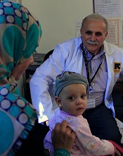
Refugees struggle to access cancer treatment
Credit: UK Department
for International Development
A new study published in The Lancet Oncology reveals a high demand for costly cancer treatment among refugees from the recent conflicts in Iraq and Syria, with host countries struggling to find the money and medicine to treat their new patients.
The findings prompted a call from study author Paul Spiegel, MD, the United Nations High Commissioner for Refugees (UNHCR) Chief Medical Expert, for schemes to improve access to affordable cancer care for refugees.
In the first study of its kind, Dr Spiegel and his colleagues examined data from funding applications made to the UNHCR Exceptional Care Committee (ECC) from refugees in Jordan and Syria whose cancer treatment costs were likely to exceed US$2000 a year.
The results indicate that cancer is an important public health problem in refugee settings, the researchers said. The study also highlights the challenges and costs national health systems and humanitarian organizations face when overwhelmed by massive influxes of refugees.
For example, in Jordan, the ECC assessed 1989 applications for treatment between 2010 and 2012. Roughly a quarter of these (511) were for cancer, with breast cancer and colorectal cancer being the most common. Around half (48%) of these cases were approved and funded.
Funding was often denied because the patient had a poor prognosis (43% of cases in 2011 and 31% in 2012) or the treatment was too costly (25% in 2011). The average amount requested from the ECC for cancer treatment was $11,540 in 2011 and $5151 in 2012. However, the amounts approved were substantially lower—$4626 in 2011 and $3501 in 2012.
“The countries in the Middle East have welcomed millions of refugees, first from Iraq and then Syria,” Dr Spiegel said. “This massive influx has strained health systems at all levels. Despite help from international organizations and donors to expand health facilities and pay for additional personnel and drugs, it has been insufficient.”
“The burden has fallen disproportionately on the host countries to absorb the costs. For example, the Jordanian Ministry of Health footed an estimated $53 million bill for medical care for refugees in the first 4 months of 2013.”
Dr Spiegel and his colleagues are therefore calling for improved cancer prevention and treatment in refugee settings through the use of innovative financing schemes; better primary care, including screening for common cancers (eg, colonoscopies and mammograms); and the development of web-based cancer registries to prevent the interruption of treatment.
“Until now, the responses to humanitarian crises have been primarily based on experiences from refugee camps in sub-Saharan Africa where infectious diseases and malnutrition have been the priority,” Dr Spiegel said. “In the 21st century, refugee situations are substantially longer and increasingly occur in middle-income countries where the levels of chronic diseases, including cancer, are higher.”
“Cancer diagnosis and care in humanitarian emergencies typifies a growing trend towards more costly chronic disease care, something that seems to have been overlooked but is of increasing importance because the number of refugees is growing.” ![]()
Drug gets orphan designation for CLL/SLL

The US Food and Drug Administration (FDA) has granted orphan designation for MOR208, an anti-CD19 monoclonal antibody, for the treatment of chronic lymphocytic leukemia (CLL) or small lymphocytic leukemia (SLL).
The European Medicines Agency (EMA) also announced a positive opinion of the orphan medicinal product application for MOR208 to treat patients with CLL/SLL.
Researchers said MOR208 showed promise in a phase 1 study of CLL/SLL patients.
Orphan drug and orphan medicinal product status are granted by the FDA and EMA to promote the development of promising therapeutics for the treatment of rare diseases affecting fewer than 200,000 people in the US annually and no more than 5 in 10,000 people in the European Union (EU).
Orphan drug designation includes benefits such as a 7-year period of marketing exclusivity in the US and 10 years of market exclusivity in the EU after approval. Other potential advantages come in the form of protocol assistance, the ability to apply for research funding, tax credits for certain research expenses, and fee waivers for the regulatory procedures.
MOR208 development, results
MOR208 is a humanized monoclonal antibody that targets the antigen CD19 for treatment of B-cell malignancies and autoimmune diseases. The development program for MOR208 is currently in phase 2 development in CLL, B-cell acute lymphoblastic leukemia, and non-Hodgkin lymphoma.
The drug is being developed by MorphoSys AG. The development program was in-licensed from Xencor in 2010.
Researchers evaluated MOR208 (formerly known as XmAb5574) in a phase 1 study of patients with CLL/SLL and reported their results at the 2012 ASH Annual Meeting (abstract 2894). The study was sponsored by Xencor.
The study included 27 patients with relapsed or refractory CLL/SLL. The median patient age was 66 years (range, 40-84), and patients were generally high-risk. The median number of prior therapies was 4 (range, 1-14).
Patients received MOR208 at a range of doses—0.3 mg/kg, 1 mg/kg, 3 mg/kg, 6 mg/kg, 9 mg/kg, and 12 mg/kg. MOR208 was administered as an intravenous infusion on days 1, 4, 8, 15, and 22 of cycle 1, and on days 1, 8, 15, and 22 of cycle 2.
Dose escalation continued without dose-limiting toxicities until the highest dose level. At this dose, 1 patient experienced grade 4 neutropenia associated with febrile neutropenia, and the patient was taken off treatment.
All 27 patients experienced adverse events, but the majority of them were grade 1-2. The most common events were infusion reactions. These occurred in 67% (n=18) of patients and were all grade 1 or 2.
Nineteen percent of patients (n=5) experienced grade 3-4 treatment-related adverse events, including neutropenia (n=3), thrombocytopenia (n=2), increased aspartate aminotransferase (n=1), febrile neutropenia (n=1), and tumor lysis syndrome (n=1). Four of these patients were on the 12 mg/kg dose, but 1 patient who experienced neutropenia was receiving 1 mg/kg.
Eleven percent of patients experienced a partial response according to IWCLL 2008 criteria. Responses occurred at the 6 mg/kg, 9 mg/kg, and 12 mg/kg dose levels.
All objective responses occurred in patients categorized as CLL as opposed to SLL, and none of the patients with lymph nodes greater than 5 cm responded. Two patients had progressed at the 8-week evaluation point. ![]()
Cancer trial publications often omit minority accrual rates

Credit: Rhoda Baer
A review of clinical trial data from 2012 suggests Hispanic patients are underrepresented in US cancer studies, and many trial publications fail to provide information on patients’ racial/ethnic backgrounds.
Researchers analyzed 159 reports of phase 2 and 3 trials and found that roughly 21% included information on the number of minority patients enrolled.
About 8% of the publications included data on the number of Hispanic patients enrolled.
And from this data, the investigators found the accrual rate for Hispanic patients was about 4%.
According to the researchers, this lack of information and low representation inhibits physicians’ ability to provide optimal treatment to Hispanic cancer patients and patients belonging to other minority groups.
“We have a major responsibility to ensure adequate representation,” said study author Ian M. Thompson Jr, MD, of The University of Texas Health Science Center at San Antonio.
“How else will we know how best to treat our patients, and how else are we going to reduce the health disparities in [the Hispanic] population?”
Dr Thompson and his colleagues have a particular interest in the Hispanic population because 58% of San Antonio residents are Hispanic, as are 68% of residents in South Texas as a whole.
So the investigators wanted to determine Hispanic accrual rates in randomized trials of cancer patients. The team evaluated data from phase 2 and 3 cancer trials published in 2012. They focused on studies that were considered likely to change the standard of care and were published in “high-impact” journals.
The researchers identified 159 trials—68 phase 2 studies and 91 phase 3 studies. They discovered that 33 of the trial publications—about 21%—disclosed data on minority accrual. And 13 publications—about 8%—included data on the accrual of Hispanic cancer patients.
Of the 4154 patients enrolled on those 13 trials, 162 were Hispanic, which translates to an overall accrual rate of 3.9%. The enrollment of Hispanic patients ranged from 1 patient (0.5%) in a phase 2 trial of lung cancer to 17 patients (26%) in a phase 2 study of acute lymphoblastic leukemia.
“Fundamentally, in the most recent published cancer clinical trials, either the number and proportion of Hispanics are not reported or are far below their actual representation in the national population,” Dr Thompson summarized.
“For institutions like ours that serve a ‘minority-majority’ population, it’s a major responsibility for us to ensure adequate representation so that we can tell our patients how they can best be treated and how we can reduce the disparities of this rapidly growing population.”
Dr Thompson and his colleagues described this research in the Journal of Clinical Oncology. ![]()
Drug granted orphan designation for AML

The US Food and Drug Administration (FDA) has granted selinexor (KPT-330) orphan designation for the treatment of acute myeloid leukemia (AML).
Selinexor is a selective inhibitor of nuclear transport that functions by binding to the nuclear export protein XPO1 (also called CRM1).
This leads to the accumulation of tumor suppressor proteins in the cell nucleus, which is thought to cause apoptosis in cancer cells while largely sparing normal cells.
The FDA grants orphan designation to promote the development of drugs that target conditions affecting 200,000 or fewer US patients annually and are expected to provide significant therapeutic advantage over existing treatments.
Selinexor’s orphan designation qualifies the drug’s developer, Karyopharm Therapeutics, Inc., for benefits that apply across all stages of development, including an accelerated approval process, 7 years of market exclusivity following marketing approval, tax credits on US clinical trials, eligibility for orphan drug grants, and a waiver of certain administrative fees.
Promising early results
Selinexor has shown promising results in a phase 1 trial of older patients with relapsed or refractory AML. The study, which was sponsored by Karyopharm, was published in Blood.
Researchers enrolled 16 patients with relapsed or refractory AML. The median age was 71 years, and the median number of prior therapeutic regimens was 2.
Patients received 8 to 10 doses of selinexor on a 4-week cycle across 2 dose levels, 16.8 mg/m2 to 23 mg/m2 (with additional cohorts ongoing).
The researchers reported no dose-limiting toxicity, no clinically significant cumulative toxicities, and no major organ dysfunction.
However, 4 patients experienced drug-related grade 3/4 adverse events, including hypotension (n=1), increased AST (n=1), hypokalemia (n=1), nausea (n=1), headache (n=1), and fatigue (n=1).
The most common grade 1/2 toxicities were nausea (9/17; 53%), anorexia (8/17; 47%), vomiting (6/17; 35%), fatigue (5/17; 29%), weight loss (5/17; 29%), and diarrhea (3/17; 18%). But these events were manageable.
Fourteen of the patients were evaluable for response. Two (14%) achieved a complete response with full hematologic recovery, and 2 (14%) achieved a complete response without hematologic recovery. Four patients (29%) had stable disease for more than 30 days, and 6 (43%) experienced progression.
Other trials of selinexor in AML
Karyopharm’s development plans for selinexor in AML include a number of additional studies.
In a phase 2 trial, researchers will evaluate selinexor monotherapy in older patients with AML. The study will enroll patients 60 years of age or older with relapsed or refractory AML who are ineligible for intensive chemotherapy and/or transplant.
In another study, researchers will evaluate selinexor in combination with decitabine for patients with relapsed, refractory, or newly diagnosed AML. The study will enroll up to 42 patients aged 60 or older who are ineligible for intensive chemotherapy.
Lastly, researchers are planning a study of selinexor in pediatric leukemia patients. The goal of this study is to determine the oral dosing, toxicity, and preliminary clinical activity of selinexor in pediatric patients. It will enroll up to 28 children with relapsed or refractory AML or acute lymphoblastic leukemia. ![]()
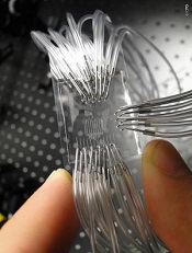
Chip may allow for early cancer detection
Institute of Photonic Sciences
Scientists say they’ve developed a lab-on-a-chip device capable of detecting protein markers for cancer.
The device can detect very low concentrations of protein markers in the blood, enabling cancer diagnosis in its earliest stages, the team says.
Romain Quidant, PhD, of The Institute of Photonic Sciences in Barcelona, Spain, and his colleagues described the device in Nano Letters.
The lab on a chip hosts 32 sensing sites distributed across a network of 8 fluidic microchannels that enables it to conduct multiple analyses.
Gold nanoparticles lie on the surface of the chip and are chemically programed with an antibody receptor in such a way that they are capable of specifically attracting the protein markers circulating in blood.
When a drop of blood is injected into the chip, it circulates through the microchannels, and, if cancer markers are present in the blood, they will stick to the nanoparticles located on the microchannels as they pass by, setting off changes in what is known as the plasmonic resonance.
The device monitors these changes, the magnitude of which is directly related to the concentration/number of markers in the patient’s blood. In this way, it provides a direct assessment of the patient’s risk of developing cancer.
“The most fascinating finding is that we are capable of detecting extremely low concentrations of this protein in a matter of minutes, making this device an ultra-high-sensitivity, state-of-the-art, powerful instrument that will benefit early detection and treatment monitoring of cancer,” Dr Quidant said. ![]()
CHF screening guidelines need another look, group says

patient and her father
Credit: Rhoda Baer
New research suggests a need to revisit cardiac screening guidelines for survivors of childhood cancers.
The study indicates that less frequent screening for early signs of impending congestive heart failure (CHF) may yield a similar clinical benefit as current screening recommendations.
Furthermore, some survivors might be better served by a different method of screening than the one currently used. And early treatment of patients at high risk of CHF may be beneficial.
The researchers reported these findings in the Annals of Internal Medicine.
Current CHF screening guidelines recommend that childhood cancer survivors treated with chemotherapeutic agents known to affect long-term heart health be screened as often as every year, with a schedule dependent on their level of CHF risk.
The Children’s Oncology Group (COG) recommends that survivors undergo screening by echocardiography for asymptomatic left ventricular dysfunction (ALVD). If left untreated, this clinically silent condition can progress to CHF, so clinicians typically prescribe beta blockers and ACE inhibitors to patients with signs of ALVD.
The COG recommends that patients at high risk of developing CHF be screened every year or 2 and those at low risk be screened every 2 or 5 years
“It is important to monitor survivors so we can reduce the late effects of treatment whenever possible, but we may be asking them to be tested too often, which burdens both individuals and the healthcare system,” said study author Lisa Diller, MD, of the Dana-Farber/Boston Children’s Cancer and Blood Disorders Center in Massachusetts. “We think it is worthwhile to review the current CHF screening guidelines.”
To estimate the clinical benefits and cost-effectiveness of the current heart screening guidelines, Dr Diller and her colleagues constructed a computer model of a virtual cohort of 15-year-olds who had survived cancer at least 5 years.
Using data from the Childhood Cancer Survivors Study and the Framingham Heart Study, the researchers modeled the cohort’s CHF risk and clinical progression over the course of survivors’ lifetimes. Results suggested that routine screening may prevent as many as 1 in 12 cases of CHF.
The team then used Medicare data to estimate the costs and value (expressed in cost per quality-adjusted life-year [QALY]) of different screening schedules—every 1, 2, 5, or 10 years—and methods—echocardiography vs cardiac magnetic resonance imaging (cMRI)—for the different CHF risk groups.
At a cost-effectiveness threshold of $100,000/QALY, the model’s results indicated that echocardiographic screening might not be the best value for resources invested to reduce lifetime CHF risk among survivors at low risk of developing the disease.
On the other hand, the data suggested that biennial echocardiography screening may be a high-value strategy for high-risk survivors.
The simulation’s data also suggested that cMRI may be preferable to echocardiography as a screening method, with cMRI’s greater cost per test balanced by its greater sensitivity. According to the model, cMRI-based screening of low-risk survivors every 10 years and high-risk survivors every 5 years was more cost-effective than any echocardiography-based schedule.
Lastly, the data suggested it may be most beneficial to treat high-risk survivors before signs of ALVD even appear. For instance, proactively treating all high-risk patients in the virtual cohort with ACE inhibitors and beta blockers reduced their lifetime CHF risk more than if they received an echocardiograph every 2 years.
The researchers relied on simulation modeling using the best available clinical and epidemiologic data because of the logistical obstacles to conducting a prospective, randomized, clinical trial.
They said enrolling the number of survivors needed for such a study would be challenging, given how rare childhood cancers are. Yet guidance on the health benefits associated with current recommendations is needed.
“Our findings suggest that there is a long-term benefit in screening survivors at elevated risk for CHF,” said study author Jennifer Yeh, PhD, of the Harvard School of Public Health in Boston.
“Yet less frequent screening than currently recommended may be reasonable when other factors are considered. We hope these results can help inform the ongoing discussion about screening childhood cancer survivors.” ![]()
Protein may be therapeutic target for AML

Credit: Lance Liotta
Researchers have found evidence to suggest that the phosphoinositide (PI) modulator PIP4K2A plays a key role in acute myeloid leukemia (AML).
PIs appear to control the transformation of hematopoietic stem cells into leukemic cells.
Some of these PIs switch on specific cell-signaling pathways, resulting in rapid growth and enhanced survival. Regulation of the PIs is carried out by a variety of proteins known as PI modulators.
“Little is known about the role of PI modulators in leukemia,” said Tim Somervaille, PhD, of The University of Manchester in the UK.
“We wanted to find out which ones were responsible for cell growth or survival in acute myeloid leukemia.”
He and his colleagues described this research in Oncogene.
The researchers performed a targeted knockdown screen of PI modulator genes in human AML cells, looking for genes required to sustain proliferation or prevent apoptosis.
They found that one PI modulator, PIP4K2A, was essential for the growth of leukemia. PIP4K2A knockdown resulted in leukemic cell death. This occurred in both murine MLL-AF9 AML cells and primary human AML cells.
Additional investigation showed that PIP4K2A knockdown resulted in the accumulation of the cyclin-dependent kinase inhibitors CDKN1A and CDKN1B, as well as G1 cell-cycle arrest and apoptosis. CDKN1A accumulation and apoptosis were partially dependent on activation of the mTOR pathway.
Fortunately, PIP4K2A knockdown did not adversely affect normal hematopoietic stem and progenitor cells from mice or human subjects. Neither clonogenic nor multilineage differentiation potential was affected.
“This makes [PIP4K2A] an ideal target for future drug development in leukemia,” Dr Somervaille concluded. ![]()
Credit: Lance Liotta
Researchers have found evidence to suggest that the phosphoinositide (PI) modulator PIP4K2A plays a key role in acute myeloid leukemia (AML).
PIs appear to control the transformation of hematopoietic stem cells into leukemic cells.
Some of these PIs switch on specific cell-signaling pathways, resulting in rapid growth and enhanced survival. Regulation of the PIs is carried out by a variety of proteins known as PI modulators.
“Little is known about the role of PI modulators in leukemia,” said Tim Somervaille, PhD, of The University of Manchester in the UK.
“We wanted to find out which ones were responsible for cell growth or survival in acute myeloid leukemia.”
He and his colleagues described this research in Oncogene.
The researchers performed a targeted knockdown screen of PI modulator genes in human AML cells, looking for genes required to sustain proliferation or prevent apoptosis.
They found that one PI modulator, PIP4K2A, was essential for the growth of leukemia. PIP4K2A knockdown resulted in leukemic cell death. This occurred in both murine MLL-AF9 AML cells and primary human AML cells.
Additional investigation showed that PIP4K2A knockdown resulted in the accumulation of the cyclin-dependent kinase inhibitors CDKN1A and CDKN1B, as well as G1 cell-cycle arrest and apoptosis. CDKN1A accumulation and apoptosis were partially dependent on activation of the mTOR pathway.
Fortunately, PIP4K2A knockdown did not adversely affect normal hematopoietic stem and progenitor cells from mice or human subjects. Neither clonogenic nor multilineage differentiation potential was affected.
“This makes [PIP4K2A] an ideal target for future drug development in leukemia,” Dr Somervaille concluded.
How cancer-fighting protein is held in check

Credit: A.T. Tikhonenko
A new study reveals how the protein p53 attaches to its regulatory molecule, BCL-xL.
Understanding how these molecular puzzle pieces fit together could help scientists design drugs that would unleash p53 to battle a range of cancers, according to study author Richard Kriwacki, PhD, of St Jude Children’s Research Hospital in Memphis, Tennessee.
He and his colleagues described this research in Nature Structural & Molecular Biology.
In guarding the cell against genetic damage, the p53 machinery functions both in the nucleus of the cell and in the cytosol. When this machinery detects irreparable damage to the cell, p53 is unleashed to trigger apoptosis.
In about half of all cancers, this machinery is rendered inoperable by mutation, enabling cancer cells to proliferate despite their genetic malfunctions. The protein BCL-xL is a central inhibitor of the p53 machinery, binding both p53 and BH3 proteins, which also drive apoptosis.
“The molecular details of how BCL-xl performs this dual inhibitory function were not understood,” Dr Kriwacki said. “Having those details has enabled us to determine exactly how BCL-xL can restrain or inhibit apoptosis through interactions with BH3-domain-containing proteins, as well as p53.”
He and his colleagues used a structural analysis technique called NMR spectroscopy to map the 3-D structure of p53 binding to BCL-xL.
Their experiments revealed how the DNA-binding domain of the p53 protein serves double duty in the machinery. It enables p53 to attach to DNA in the cell’s nucleus, helping the cell repair genetic damage. The same domain also acts as an attachment point for BCL-xL in the cytosol.
“The structural details that we report are novel,” Dr Kriwacki said. “And they provide the key insights for really dissecting the dual roles of BCL-xL in inhibiting apoptosis . . ., inhibiting the BH3-containing proteins on the one side and p53 on the other. Also, through these studies, we solidified the mechanistic understanding for how p53 functions in the cytosol, which complements its pro-apoptotic role in the nucleus.”
Dr Kriwacki added that these findings could help scientists design better anticancer agents. In many cancers, p53 is prevented from triggering apoptosis by its attachment to BCL-xL.
Drugs are currently being tested that bind to BCL-xL to free BH3 proteins to trigger apoptosis. However, Dr Kriwacki said new drugs could be developed that also block BCL-xL from binding p53.
“Our hypothesis is that many cancers have normal p53, but it is being tied up by BCL-xL,” he said. “If it could be released, it could play its role in triggering apoptosis. A drug that could block both of BCL-xL’s anti-apoptotic functions could potentially more profoundly induce apoptosis in cancer cells.”