User login
Systemic corticosteroids not recommended for long-term treatment of eczema
Systemic corticosteroids (SCSs) should be limited to short courses as a transition to steroid-sparing therapies in patients with atopic dermatitis because of the potential for adverse effects, reported Sherry Yu, MD, of Massachusetts General Hospital, Boston, and her associates.

Another AE is adrenal insufficiency, defined as a cortisol level less than or equal to 500 nmol/L. In a meta-analysis of 74 articles with 3,753 participants, there was a significant increase in absolute risk of adrenal insufficiency with medium term use (1 month to less than 1 year) and long term use (more than 1 year) of SCSs, as well as medium and high doses of SCSs. Adrenal insufficiency can occur in as little as 4 weeks and may be subclinical. This can leave patients vulnerable to infection and stress from surgery. Adrenal insufficiency also can manifest itself as weakness, fatigue, and shock in more severe cases.
Dr. Yu and her colleagues noted that tapering may minimize the risk of adrenal insufficiency and that a single morning dose or alternate dosing strategy may minimize this risk.
Another finding from this study was the adverse effects the SCSs have on pediatric patients. One study showed that 7 of 10 (70%) of children taking maintenance doses of beclomethasone dipropionate had growth impairment after 6 months of therapy. Significant growth disruption also was observed in children who had taken beclomethasone dipropionate only 4 weeks.
Dr. Yu and her colleagues warned that SCS should not be used to treat pediatric AD “because of growth retardation and other AEs.”
In conclusion, Dr. Yu and her colleagues said, “the literature supports short-term use of short courses of oral corticosteroids (less than 3 weeks) to interrupt acute flares, but rebound flaring is commonly observed after discontinuation. If SCSs are used at all, their use should be short term when bridging to other systemic therapies or phototherapy.” Despite the support for short-term courses of SCSs, the “duration of an optimal short course is not well defined” in the literature.
Dr. Yu reported no relevant financial disclosures. The other three authors reported relationships with various companies in the industry.
SOURCE: Yu S et al. J Am Acad Dermatol. 2017 Oct 13. doi: 10.1016/j.jaad.2017.09.074.
Systemic corticosteroids (SCSs) should be limited to short courses as a transition to steroid-sparing therapies in patients with atopic dermatitis because of the potential for adverse effects, reported Sherry Yu, MD, of Massachusetts General Hospital, Boston, and her associates.
Another AE is adrenal insufficiency, defined as a cortisol level less than or equal to 500 nmol/L. In a meta-analysis of 74 articles with 3,753 participants, there was a significant increase in absolute risk of adrenal insufficiency with medium term use (1 month to less than 1 year) and long term use (more than 1 year) of SCSs, as well as medium and high doses of SCSs. Adrenal insufficiency can occur in as little as 4 weeks and may be subclinical. This can leave patients vulnerable to infection and stress from surgery. Adrenal insufficiency also can manifest itself as weakness, fatigue, and shock in more severe cases.
Dr. Yu and her colleagues noted that tapering may minimize the risk of adrenal insufficiency and that a single morning dose or alternate dosing strategy may minimize this risk.
Another finding from this study was the adverse effects the SCSs have on pediatric patients. One study showed that 7 of 10 (70%) of children taking maintenance doses of beclomethasone dipropionate had growth impairment after 6 months of therapy. Significant growth disruption also was observed in children who had taken beclomethasone dipropionate only 4 weeks.
Dr. Yu and her colleagues warned that SCS should not be used to treat pediatric AD “because of growth retardation and other AEs.”
In conclusion, Dr. Yu and her colleagues said, “the literature supports short-term use of short courses of oral corticosteroids (less than 3 weeks) to interrupt acute flares, but rebound flaring is commonly observed after discontinuation. If SCSs are used at all, their use should be short term when bridging to other systemic therapies or phototherapy.” Despite the support for short-term courses of SCSs, the “duration of an optimal short course is not well defined” in the literature.
Dr. Yu reported no relevant financial disclosures. The other three authors reported relationships with various companies in the industry.
SOURCE: Yu S et al. J Am Acad Dermatol. 2017 Oct 13. doi: 10.1016/j.jaad.2017.09.074.
FROM THE JOURNAL OF THE AMERICAN ACADEMY OF DERMATOLOGY
Key clinical point:
Major finding: Growth impairment was seen after 6 months in 7 out of 10 children with eczema taking maintenance doses of oral beclomethasone dipropionate.
Study details: A systematic review of 52 reviews and 12 studies concerning systemic corticosteroid use in atopic dermatitis patients from journal databases including PubMed and Medline
Disclosures: Dr. Yu reported no relevant financial disclosures. The other three authors reported relationships with various companies in the industry.
Source: Yu S et al. J Am Acad Dermatol. 2017 Oct 13. doi: 10.1016/j.jaad.2017.09.074.
Very preterm birth is linked to reduced risk of eczema
according to data from a meta-analysis of 18 studies.
Previous research suggests that low birth weight is protective against the development of atopic dermatitis, said Tingting Zhu, PhD, of West China Second University Hospital, Chengdu, and colleagues.
Preterm birth (before 37 completed weeks’ gestation) was divided into subgroups of extremely preterm (less than 28 weeks’ gestation), very preterm (28 weeks’ to less than 32 weeks’ gestation), and moderate/late preterm (32 weeks’ gestation to less than 37 weeks’ gestation).
In an analysis based on gestational age, children had a significantly reduced risk of eczema if they were very preterm (relative risk, 0.77; 95% confidence interval, 0.70-0.84, P less than .01; adjusted RR, 0.73; 95% CI, 0.64-0.82; P less than 0.01), compared with children born full term. The association between eczema and preterm birth was no longer significant among children born moderately preterm, Dr. Zhu and associates reported.
The reasons for the impact of very preterm birth on eczema are unclear, but maturation of the stratum corneum at 29-37 weeks’ gestational age could play a role, the researchers noted. Also, limited microflora in very preterm infants could affect acquiring immune tolerance and lead to reduced risk of eczema. The study was limited by several factors, including variations in gestational age and inconsistent assessments of eczema among the studies.
However, the large sample size lends strength to the results, and further studies are needed to explore how the environment, nutrition, immune system development, and skin barrier function impact the risk of eczema in very preterm infants, Dr. Zhu and associates said.
The researchers had no relevant financial disclosures. The researchers had no financial conflicts to disclose. The study was funded in part by the National Science Foundation of China, the Ministry of Health of China, and various other grants.
SOURCE: Zhu T et al. J Amer Dermatol. 2018. doi: 10.1016/j.jaad.2017.12.015.
FROM JOURNAL OF THE AMERICAN ACADEMY OF DERMATOLOGY
Key clinical point: Very preterm birth was associated with a significantly reduced risk of eczema, compared with full-term birth, but no difference in risk appeared between moderate preterm and full-term birth.
Major finding: Children had a significantly reduced risk of eczema if they were very preterm (RR, 0.77; 95% CI, 0.70-0.84; P less than .01; aRR 0.73, 95% CI, 0.64-0.82; P less than .01), compared with children born full term.
Data source: The data come from a meta-analysis of 18 studies.
Disclosures: The researchers had no financial conflicts to disclose. The study was funded in part by the National Science Foundation of China, the Ministry of Health of China, and various other grants.
Source: Zhu T et al. J Amer Dermatol. 2018. doi: 10.1016/j.jaad.2017.12.015.
Study: Atopic dermatitis subgroups identified in children
Identification of subphenotypes of atopic dermatitis (AD) in children, with differing risk factors, prognoses, and comorbidities, could lead to a stratified approach to managing pediatric AD, said Lavinia Paternoster, PhD, of the University of Bristol, England, and her associates.

The study identified six classes of AD in these children. Early-onset/early-resolving AD, occurring in 13%-15% of the children, was most prevalent and was associated with male gender. Children in this class had a favorable prognosis, and there was only a very weak association with asthma in later life.
Two classes of persistent disease were identified: early-onset persistent AD (rash occurring in most of this class by 30 months and resolving in half by 16.5 years) and early-onset/late-resolving AD (rash occurring in most by 30 months and resolving in most by 16.5 years). These classes, occurring in about 7% of the children, had the strongest association with an AD genetic risk score; a strong link with personal and parental history of atopic disease; and a strong tie to asthma.
An unrecognized class of mid-onset-resolving AD, occurring in 7% of children, was not significantly linked to FLG mutations but was tied to asthma. In those children, AD prevalence rose sharply from 2.5 years of age and peaked at about 6 years, Dr. Paternoster and her associates said.
The investigators also found an unaffected/transient AD class in which children either had never reported rash; had one or two isolated occasions of rash; or reported a rash consistent with AD at 6-18 months that declined with age. In the two cohorts, 58%-63% children fell into this class. Late-onset-resolving AD occurred in 7%-8% of children, with most developing rash by 12 years and declining by 16.5 years.
There was a preponderance of females in the early-onset persistent AD and the late onset classes, and more males in the early-onset resolving class. “The associations with asthma at ages 7 and 11-13 years were strongest with the persistent class, but all AD classes showed evidence of some increased risk of asthma at these ages,” Dr. Paternoster and her associates wrote.
“There was evidence that FLG null mutations were associated with all classes, however ... the association was strongest in the group with early-onset-persistent disease,” the researchers said. The “heterogeneity of effect of genetic variants on different disease profiles, emphasizes the need for patient stratification in future genetic studies. Stratification may be used to increase the power to detect variants associated with specific classes; stratification could also allow the identification of phenotype-specific mechanistic pathways as future therapeutic targets.”
Read more at (J Allerg Clin Immunol. 2017 Nov 10. doi: 10.1016/j.jaci.2017.09.044).
Identification of subphenotypes of atopic dermatitis (AD) in children, with differing risk factors, prognoses, and comorbidities, could lead to a stratified approach to managing pediatric AD, said Lavinia Paternoster, PhD, of the University of Bristol, England, and her associates.
The study identified six classes of AD in these children. Early-onset/early-resolving AD, occurring in 13%-15% of the children, was most prevalent and was associated with male gender. Children in this class had a favorable prognosis, and there was only a very weak association with asthma in later life.
Two classes of persistent disease were identified: early-onset persistent AD (rash occurring in most of this class by 30 months and resolving in half by 16.5 years) and early-onset/late-resolving AD (rash occurring in most by 30 months and resolving in most by 16.5 years). These classes, occurring in about 7% of the children, had the strongest association with an AD genetic risk score; a strong link with personal and parental history of atopic disease; and a strong tie to asthma.
An unrecognized class of mid-onset-resolving AD, occurring in 7% of children, was not significantly linked to FLG mutations but was tied to asthma. In those children, AD prevalence rose sharply from 2.5 years of age and peaked at about 6 years, Dr. Paternoster and her associates said.
The investigators also found an unaffected/transient AD class in which children either had never reported rash; had one or two isolated occasions of rash; or reported a rash consistent with AD at 6-18 months that declined with age. In the two cohorts, 58%-63% children fell into this class. Late-onset-resolving AD occurred in 7%-8% of children, with most developing rash by 12 years and declining by 16.5 years.
There was a preponderance of females in the early-onset persistent AD and the late onset classes, and more males in the early-onset resolving class. “The associations with asthma at ages 7 and 11-13 years were strongest with the persistent class, but all AD classes showed evidence of some increased risk of asthma at these ages,” Dr. Paternoster and her associates wrote.
“There was evidence that FLG null mutations were associated with all classes, however ... the association was strongest in the group with early-onset-persistent disease,” the researchers said. The “heterogeneity of effect of genetic variants on different disease profiles, emphasizes the need for patient stratification in future genetic studies. Stratification may be used to increase the power to detect variants associated with specific classes; stratification could also allow the identification of phenotype-specific mechanistic pathways as future therapeutic targets.”
Read more at (J Allerg Clin Immunol. 2017 Nov 10. doi: 10.1016/j.jaci.2017.09.044).
FROM THE JOURNAL OF ALLERGY AND CLINICAL IMMUNOLOGY
Gene mutations tied to viral skin infection in pediatric AD patients
Filaggrin (FLG) polymorphism appears to be linked with Molluscum contagiosum virus skin infection in children with atopic dermatitis (AD), reported Sara Manti, MD, of the University of Messina (Italy), and her associates.
“To the best of our knowledge, this is the first study” to describe this association, they said.
In an Italian study of 100 children with AD and 97 healthy children who served as controls, both clinical and laboratory data showed that FLG gene mutations were linked to early AD onset, a more severe clinical course of disease, and a significantly higher risk of Molluscum contagiosum virus (MCV)–associated skin infection.
The data “suggest that FLG gene may serve as a potential biomarker to screen the atopic population, identifying patients at high risk of AD, to adopt preventive measures that can restore the barrier function of the skin and reduce patients’ susceptibility to recurrent skin infection,” they wrote. “Our findings also indicate that the identified SNPs [single-nucleotide polymorphisms] might have clinically relevant implications with respect to a major MCV [M. contagiosum virus] colonization of skin, also permitting the identification of a new AD phenotype in children. However, to date, because of the existence of controversial data, we believe that further and needed insights into AD pathophysiology may also be gained by studying people with the FLG gene who do not have atopic predisposition and/or AD. Thus, a large population-based study is necessary to further validate these early interpretations and to gain detailed information about the role of FLG variants,” Dr. Manti and her coinvestigators concluded.
Read more in Manti S et al. Ann Allergy Asthma Immunol. 2017;119:446-51.
cnellist@frontlinemedcom.com
FROM THE ANNALS OF ALLERGY, ASTHMA & IMMUNOLOGY
JAK inhibitors for atopic dermatitis might hit JAK-pot
GENEVA – at the annual congress of the European Academy of Dermatology and Venereology, with three positive phase 2 randomized trials featuring one topical and two oral agents presented to enthusiastic audiences.
The way has already been paved for dermatologic researchers by veterinarians, who developed oclacitinib (Apoquel), a relatively selective Janus kinase 1 (JAK1) inhibitor, for canine AD. The medication was approved by the Food and Drug Administration in 2013 for treating AD and for controlling pruritus associated with allergic dermatitis in dogs.
PF-04965842
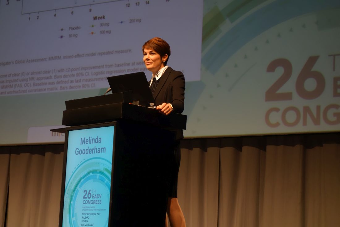
“Get out your pencils, everyone. This is why you’re all here at 8 o’clock on a Sunday morning,” Melinda Gooderham, MD, said, standing before a packed house at the main arena of the Geneva Convention Center, as she launched into the results of a phase II randomized, double-blind, placebo-controlled, 12-week trial of a JAK inhibitor known for now as PF-04965842. This is a JAK1-selective agent with a good effect on interleukin-4 and -13, key mediators of the Th2 cytokines implicated in the pathogenesis of AD.
The dose-ranging study included 250 adults with AD and an inadequate response to or intolerance of topical therapy. Their mean baseline Eczema Area and Severity Index (EASI) score was 25 with a 60/40 ratio of moderate to severe AD. The five-arm trial randomized patients to PF-04965842 at 10 mg, 30 mg, 100 mg, or 200 mg once daily or placebo.
The primary endpoint was the proportion of patients achieving an Investigator Global Assessment (IGA) score of 0 or 1 – clear or almost clear – along with at least a 2-grade improvement from baseline at week 12. A clear dose-response effect was evident, with the 100- and 200-mg doses achieving response rates of 28% and 45%, respectively, compared with 6% in placebo-treated controls, reported Dr. Gooderham, medical director of the Skin Center for Dermatology in Peterborough, Ont., and a dermatologist at Queen’s University in Kingston, Ont.
Onset of action was speedy: patients in the 200-mg group reached their full improvement in IGA score by week 4 and maintained that response through week 12. Maximum improvement in EASI score – a mean 80% reduction – was achieved by week 6 and sustained thereafter. The proportion of patients in the 200-mg group achieving at least a 4-point improvement on the Pruritus Numeric Rating Scale significantly exceeded that in the placebo group as early as day 2 of the trial. At week 12, 64% of patients in the 200-mg group had achieved this level of improvement in itch, compared with 26% of controls.
A dose-dependent drop in platelet count occurred in the study, reaching a 30% decline at the 4-week nadir in the 200-mg group, followed by gradual on-treatment recovery. Both LDL and HDL cholesterol rose on active therapy – a class effect of JAK inhibitors – but the ratio between the two lipid levels remained unchanged. The two serious adverse events deemed treatment related were a case of eczema herpeticum in a patient on the 100-mg dose and pneumonia in a patient on the 200-mg dose.
Baricitinib
This once-daily oral JAK1/2 inhibitor is approved for treatment of rheumatoid arthritis in Europe and Japan. Emma Guttman-Yassky, MD, PhD, presented a phase 2 study of baricitinib in 124 adults with moderate to severe AD. Notably, prior to enrollment, all participants had to have failed to respond to a 4-week run-in period of supervised treatment with 0.1% triamcinolone cream, a midpotency topical steroid. They were then randomized to 2 mg or 4 mg of once-daily baricitinib or placebo, in all cases supplemented as needed with the topical steroid. Their median baseline EASI score was 21.
The primary endpoint was the proportion of patients achieving at least a 50% improvement in EASI score, or EASI 50 response, by week 16 from baseline in a nonresponder imputation analysis. This was achieved in 65% of patients on the 4-mg dose of baricitinib, 64% on the 2-mg dose, and 46% of controls on placebo plus the topical steroid. A statistically significant difference in EASI 50 response between the baricitinib groups and controls was seen at 1 week, with nearly the maximum effect achieved at week 4. Patients with a baseline EASI score above the median had a much more impressive treatment response because the placebo effect was smaller in participants with more severe AD.
“I think this drug can be an exciting new addition to the field,” declared Dr. Guttman-Yassky, professor and vice chair of the department of dermatology at the Icahn School of Medicine at Mount Sinai, New York.
From a baseline total SCORAD (Scoring Atopic Dermatitis) score of 55, major improvements were seen in the JAK inhibitor–treated patients by week 4. At week 16, the average reduction from baseline was 47% in the 4-mg group, 41% in the 2-mg group, and 21% in the placebo group. Both the SCORAD pruritus and sleep loss subscores showed significantly more robust improvement in the baricitinib groups than in controls. Indeed, a significant drop in pruritus scores was noted within the first week.
The 4-mg dose was associated with greater improvement and faster onset of action than the 2-mg dose on some but not all disease measures.
Dr. Guttman-Yassky described baricitinib as having “an overall acceptable safety profile,” with no serious treatment-related adverse events noted. Headache, nasopharyngitis, and asymptomatic increases in serum creatinine phosphokinase were more common in baricitinib-treated patients than with placebo.
JTE-052
Hidemi Nakagawa, MD, presented a phase 2 study of topical JTE-052 ointment in 327 Japanese adults with moderate to severe AD. The drug inhibits JAK1/2/3 as well as the tyrosine kinase pathway. It also promotes keratinocyte production of filaggrin in the skin barrier. Participants were randomized to twice-daily application of JTE-052 ointment (at 0.25%, 0.5%, 1%, or 3%), vehicle ointment, or 0.1% tacrolimus ointment twice a day for 4 weeks. The primary outcome was the change from baseline in modified EASI score in the active treatment groups compared with placebo. All doses of JTE-052 proved significantly more effective than vehicle. A dose-response effect was noted, with a 42% reduction from baseline in modified EASI score in the 0.25% JTE-052 group, a 57% reduction with 0.5%, a 55% reduction with 1% ointment, and a 73% reduction with 3%, compared with a 12% reduction decrease in patients who received vehicle. The topical tacrolimus group showed a 62% reduction from baseline, reported Dr. Nakagawa, professor and head of the division of dermatology at Jikei University, Tokyo.
All doses of JTE-052 were also significantly more effective than placebo on all secondary endpoints, which included IGA, percent body surface area affected, and Pruritus Numeric Rating Scale score.
At all but the weakest concentration, JTE-052 resulted in significant reduction in pruritus starting with the second dose on day 1 of the trial, he added.
Mild nasopharyngitis occurred in 3.4% of JTE-052–treated patients. There were no serious adverse events and no changes in laboratory parameters in the study. One patient discontinued JTE-052 because of application-site contact dermatitis, another because of application-site irritation. The results of this study were recently published in the British Journal of Dermatology (Br J Dermatol. 2017 Sep 28. doi: 10.1111/bjd.16014).
Dr. Nakagawa reported receiving research grants from and serving as a consultant to Japan Tobacco, which is developing JTE-052. Dr. Guttman-Yassky reported having financial relationships with Eli Lilly and Incyte, which sponsored the baricitinib study, as well as most other pharmaceutical companies developing therapies for AD. Dr. Gooderham reported receiving research funding from and serving as a consultant to Pfizer, which sponsored the PF-04965842 study, as well as numerous other pharmaceutical companies.
GENEVA – at the annual congress of the European Academy of Dermatology and Venereology, with three positive phase 2 randomized trials featuring one topical and two oral agents presented to enthusiastic audiences.
The way has already been paved for dermatologic researchers by veterinarians, who developed oclacitinib (Apoquel), a relatively selective Janus kinase 1 (JAK1) inhibitor, for canine AD. The medication was approved by the Food and Drug Administration in 2013 for treating AD and for controlling pruritus associated with allergic dermatitis in dogs.
PF-04965842
“Get out your pencils, everyone. This is why you’re all here at 8 o’clock on a Sunday morning,” Melinda Gooderham, MD, said, standing before a packed house at the main arena of the Geneva Convention Center, as she launched into the results of a phase II randomized, double-blind, placebo-controlled, 12-week trial of a JAK inhibitor known for now as PF-04965842. This is a JAK1-selective agent with a good effect on interleukin-4 and -13, key mediators of the Th2 cytokines implicated in the pathogenesis of AD.
The dose-ranging study included 250 adults with AD and an inadequate response to or intolerance of topical therapy. Their mean baseline Eczema Area and Severity Index (EASI) score was 25 with a 60/40 ratio of moderate to severe AD. The five-arm trial randomized patients to PF-04965842 at 10 mg, 30 mg, 100 mg, or 200 mg once daily or placebo.
The primary endpoint was the proportion of patients achieving an Investigator Global Assessment (IGA) score of 0 or 1 – clear or almost clear – along with at least a 2-grade improvement from baseline at week 12. A clear dose-response effect was evident, with the 100- and 200-mg doses achieving response rates of 28% and 45%, respectively, compared with 6% in placebo-treated controls, reported Dr. Gooderham, medical director of the Skin Center for Dermatology in Peterborough, Ont., and a dermatologist at Queen’s University in Kingston, Ont.
Onset of action was speedy: patients in the 200-mg group reached their full improvement in IGA score by week 4 and maintained that response through week 12. Maximum improvement in EASI score – a mean 80% reduction – was achieved by week 6 and sustained thereafter. The proportion of patients in the 200-mg group achieving at least a 4-point improvement on the Pruritus Numeric Rating Scale significantly exceeded that in the placebo group as early as day 2 of the trial. At week 12, 64% of patients in the 200-mg group had achieved this level of improvement in itch, compared with 26% of controls.
A dose-dependent drop in platelet count occurred in the study, reaching a 30% decline at the 4-week nadir in the 200-mg group, followed by gradual on-treatment recovery. Both LDL and HDL cholesterol rose on active therapy – a class effect of JAK inhibitors – but the ratio between the two lipid levels remained unchanged. The two serious adverse events deemed treatment related were a case of eczema herpeticum in a patient on the 100-mg dose and pneumonia in a patient on the 200-mg dose.
Baricitinib
This once-daily oral JAK1/2 inhibitor is approved for treatment of rheumatoid arthritis in Europe and Japan. Emma Guttman-Yassky, MD, PhD, presented a phase 2 study of baricitinib in 124 adults with moderate to severe AD. Notably, prior to enrollment, all participants had to have failed to respond to a 4-week run-in period of supervised treatment with 0.1% triamcinolone cream, a midpotency topical steroid. They were then randomized to 2 mg or 4 mg of once-daily baricitinib or placebo, in all cases supplemented as needed with the topical steroid. Their median baseline EASI score was 21.
The primary endpoint was the proportion of patients achieving at least a 50% improvement in EASI score, or EASI 50 response, by week 16 from baseline in a nonresponder imputation analysis. This was achieved in 65% of patients on the 4-mg dose of baricitinib, 64% on the 2-mg dose, and 46% of controls on placebo plus the topical steroid. A statistically significant difference in EASI 50 response between the baricitinib groups and controls was seen at 1 week, with nearly the maximum effect achieved at week 4. Patients with a baseline EASI score above the median had a much more impressive treatment response because the placebo effect was smaller in participants with more severe AD.
“I think this drug can be an exciting new addition to the field,” declared Dr. Guttman-Yassky, professor and vice chair of the department of dermatology at the Icahn School of Medicine at Mount Sinai, New York.
From a baseline total SCORAD (Scoring Atopic Dermatitis) score of 55, major improvements were seen in the JAK inhibitor–treated patients by week 4. At week 16, the average reduction from baseline was 47% in the 4-mg group, 41% in the 2-mg group, and 21% in the placebo group. Both the SCORAD pruritus and sleep loss subscores showed significantly more robust improvement in the baricitinib groups than in controls. Indeed, a significant drop in pruritus scores was noted within the first week.
The 4-mg dose was associated with greater improvement and faster onset of action than the 2-mg dose on some but not all disease measures.
Dr. Guttman-Yassky described baricitinib as having “an overall acceptable safety profile,” with no serious treatment-related adverse events noted. Headache, nasopharyngitis, and asymptomatic increases in serum creatinine phosphokinase were more common in baricitinib-treated patients than with placebo.
JTE-052
Hidemi Nakagawa, MD, presented a phase 2 study of topical JTE-052 ointment in 327 Japanese adults with moderate to severe AD. The drug inhibits JAK1/2/3 as well as the tyrosine kinase pathway. It also promotes keratinocyte production of filaggrin in the skin barrier. Participants were randomized to twice-daily application of JTE-052 ointment (at 0.25%, 0.5%, 1%, or 3%), vehicle ointment, or 0.1% tacrolimus ointment twice a day for 4 weeks. The primary outcome was the change from baseline in modified EASI score in the active treatment groups compared with placebo. All doses of JTE-052 proved significantly more effective than vehicle. A dose-response effect was noted, with a 42% reduction from baseline in modified EASI score in the 0.25% JTE-052 group, a 57% reduction with 0.5%, a 55% reduction with 1% ointment, and a 73% reduction with 3%, compared with a 12% reduction decrease in patients who received vehicle. The topical tacrolimus group showed a 62% reduction from baseline, reported Dr. Nakagawa, professor and head of the division of dermatology at Jikei University, Tokyo.
All doses of JTE-052 were also significantly more effective than placebo on all secondary endpoints, which included IGA, percent body surface area affected, and Pruritus Numeric Rating Scale score.
At all but the weakest concentration, JTE-052 resulted in significant reduction in pruritus starting with the second dose on day 1 of the trial, he added.
Mild nasopharyngitis occurred in 3.4% of JTE-052–treated patients. There were no serious adverse events and no changes in laboratory parameters in the study. One patient discontinued JTE-052 because of application-site contact dermatitis, another because of application-site irritation. The results of this study were recently published in the British Journal of Dermatology (Br J Dermatol. 2017 Sep 28. doi: 10.1111/bjd.16014).
Dr. Nakagawa reported receiving research grants from and serving as a consultant to Japan Tobacco, which is developing JTE-052. Dr. Guttman-Yassky reported having financial relationships with Eli Lilly and Incyte, which sponsored the baricitinib study, as well as most other pharmaceutical companies developing therapies for AD. Dr. Gooderham reported receiving research funding from and serving as a consultant to Pfizer, which sponsored the PF-04965842 study, as well as numerous other pharmaceutical companies.
EXPERT ANALYSIS FROM THE EADV CONGRESS
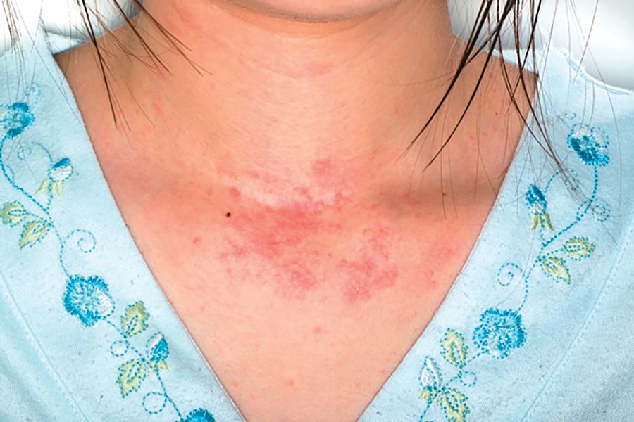
Pediatric Periorificial Dermatitis
Perioral dermatitis is an acneform eruption presenting with erythematous papules, vesicles, and rarely pustules clustered around the orifices of the face. 1 Lesions may be found near the eyes, mouth, and nose but typically spare the vermilion border of the lips. 2 Nguyen and Eichenfield 3 preferred the term periorificial dermatitis (POD), which has since been adopted by others. 4 Patients may report pruritus, but there generally are no systemic symptoms unless patients have comorbid conditions such as atopic dermatitis. 5 Although this condition has been well examined in the literature on adults, data in the pediatric population are far more limited, consisting of case series and retrospective chart reviews. In 1979, Wilkinson et al 6 published a study of more than 200 patients with perioral dermatitis, but only 15 patients younger than 12 years were included.
Etiology
Although the exact pathogenesis of POD is unknown, a common denominator among many patients is prior exposure to topical corticosteroids.3,7-9 Periorificial dermatitis also has been linked to the use of systemic corticosteroids in pediatric patients.10 The exact relationship between steroid use and dermatitis is unknown; it may be related to a change in the flora of hair follicles and in particular an association with fusiform bacteria–rich conditions.11 Aside from steroid exposure, POD has been associated with the use of physical sunscreen in pediatric patients with dry skin,12 rosin in chewing gum,13 and inhaled corticosteroids in those with asthma.14 In one case, a 15-year-old adolescent girl developed POD and swelling of the lips after 2 years of playing a flute made of cocus wood.15,16
Epidemiology
Comorbidities and Family History
Goel et al17 (N=222) reported the following comorbidities associated with pediatric POD: atopic dermatitis (29.3%), asthma (14.9%), and allergies (9.9%). Steroid exposure was noted in 58.1% of patients.17 Similarly, Nguyen and Eichenfield3 (N=79) found that the most common comorbidities were atopic dermatitis (14%), keratosis pilaris (14%), viral infections (14%), acne (10%), and seborrheic dermatitis (10%). Family history of atopy was noted in 55% of patients and family history of rosacea was noted in 3%. In a case series of 11 pediatric patients, 3 (27%) had keratosis pilaris, 7 (64%) had a family history of atopy, and 2 (18%) had a family history of rosacea.8 Weston and Morelli9 found a much higher incidence of familial rosacea (20%) in 106 children with steroid rosacea.
Clinical Presentation
Periorificial dermatitis generally presents with small, pink- to flesh-colored papules in a perioral, periocular, and perinasal distribution. Although many patients are white, a particularly prominent variant has been noted in black children with papules that may be hyperpigmented.18 In a 2006 chart review in 79 pediatric POD patients aged 6 months to 18 years, Nguyen and Eichenfield3 reported that 92% (73/79) of patients presented for a facial rash with an average duration ranging from 2 weeks to 4 years.
Boeck et al19 described 7 pediatric patients with perioral dermatitis. Six (86%) patients had perioral lesions, and 6 (86%) had previously been treated with moderate- to high-potency topical corticosteroids. Skin prick tests were negative in 6 (86%) patients.19 In one case report, a 6-year-old boy did not present with the classic acneform lesions but rather sharply demarcated eczematous patches around the eyes, nose, and mouth. The rash began to fade after 2 weeks of using metronidazole gel 1%, and after 4 months he was only left with mild hyperpigmentation.4
Periorificial dermatitis was once thought to be a juvenile form of rosacea.5 In 1972, Savin et al8 described 11 pediatric patients with “rosacea-like” facial flushing, papules, pustules, and scaling over the cheeks, forehead, and chin. In some patients, the eyelids also were involved. At least 8 patients had been using potent topical corticosteroids and had noticed exacerbation of their skin lesions after stopping therapy.8
Variants of POD
Several other variants of POD have been described in pediatric patients including childhood granulomatous periorificial dermatitis (CGPD)(also known as facial Afro-Caribbean [childhood] eruption) and lupus miliaris disseminatus faciei. Childhood granulomatous periorificial dermatitis presents in prepubertal children as dome-shaped, red to yellow-brown, monomorphous papules around the eyes, nose, and mouth; there are no systemic findings.20,21 It occurs equally in males and females and is more commonly seen in dark-skinned patients. Childhood granulomatous periorificial dermatitis usually resolves within a few months to years but may be associated with blepharitis or conjunctivitis.20 Urbatsch et al20 analyzed extrafacial lesions in 8 patients (aged 2–12 years) with CGPD. Lesions were found on the trunk (38% [3/8]), neck (25% [2/8]), ears (25% [2/8]), extremities (50% [4/8]), labia majora (38% [3/8]), and abdomen (13% [1/8]). In addition, 2 (25% [2/8]) patients had blepharitis.20
Lupus miliaris disseminatus faciei, which occurs in adolescents and adults, commonly involves the eyelids and central areas of the face such as the nose and upper lips. Patients typically present with erythematous or flesh-colored papules.1
Diagnosis
Diagnosis of POD is made clinically based on the observation of papules (and sometimes pustules) around the orifices of the face, sparing the vermilion border, together with a lack of comedones.17 Laboratory tests are not useful.5 Biopsies rarely are performed, and the results mimic those of rosacea, demonstrating a perifollicular lymphohistiocytic infiltrate, epithelioid cells, and occasionally giant cells.5,22,23 Early papular lesions can show mild acanthosis, epidermal edema, and parakeratosis.23 Biopsies in patients with CGPD reveal noncaseating perifollicular granulomas.20
Treatment and Clinical Outcome
Although topical corticosteroids can improve facial lesions in pediatric POD, the eruption often rebounds when therapy is discontinued.1 One therapy frequently used in adults is oral tetracyclines; however, these agents must not be used in patients younger than 9 years due to potential dental staining.4 The standards are either topical metronidazole twice daily with clearance in 3 to 8 weeks or oral erythromycin.7
In the review conducted by Goel et al,17 treatment included azithromycin (44.6%), topical metronidazole (42.3%), sodium sulfacetamide lotion (35.6%), oral antibiotic monotherapy (15.3%), topical agent monotherapy (44.6%), and combined oral and topical agent therapy (40.1%). Of those patients who presented for a follow-up visit (59%), 72% of cases resolved and 10.7% showed some improvement. For those patients who returned for follow-up, the average duration until symptom resolution was approximately 4 months. The most common side effects were pigmentation changes (1.8%), worsening of symptoms (1.8%), gastrointestinal upset (0.9%), irritant dermatitis (0.9%), and xerosis (0.5%).17
Changes were made to the treatment plans for 16 patients, most often due to inadequate treatment response.17 Five patients treated with sodium sulfacetamide lotion also were started on oral azithromycin. Four patients treated with oral antibiotics were given a topical agent (metronidazole or sodium sulfacetamide lotion). Other modifications included replacing sodium sulfacetamide lotion with topical metronidazole and an oral antibiotic (azithromycin or doxycycline, n=3), adjusting the doses of oral or topical medications (n=2), adding tacrolimus (n=1), and replacing topical metronidazole with sodium sulfacetamide lotion (n=1). Of the patients who underwent a change in treatment plan, 5 experienced symptom recurrence, 4 had mild improvement, and 1 patient had no improvement. Six patients were lost to follow-up.17
In the study conducted by Nguyen and Eichenfield,3 follow-up visits occurred approximately 3 months after the first visit.
In the case series by Boeck et al,19 all patients were started on metronidazole gel 1% applied once daily for the first week, and then twice daily until the lesions resolved. All patients showed improvement after 4 to 6 weeks, and eventually the disease cleared between 3 and 6 months. All patients were still symptom free during a 2-year observation period.19
Manders and Lucky7 described 14 patients with POD (aged 9 months to 6.5 years). Eight patients used only metronidazole gel 0.75%, while 5 used the gel in combination with topical corticosteroids (21% [3/14]), oral erythromycin (7% [1/14]), or topical erythromycin (7% [1/14]); 1 patient remained on hydrocortisone 1% and cleared. Patients responded well within 1 to 8 weeks and were symptom free for up to 16 months. Mid- to high-potency steroids were discontinued in all patients.7
In some pediatric patients with CGPD, recovery occurs faster with the use of oral macrolides or tetracyclines, either alone or in combination with topical antibiotics or sulfur-based lotions.20 Extrafacial lesions associated with CGPD do not appear to negatively impact treatment response or duration of disease. In the review conducted by Urbatsch et al,20 7 of 8 (88%) CGPD patients with extrafacial lesions were treated with oral agents including erythromycin, hydroxychloroquine, cyclosporine, minocycline, and azithromycin. Most of these patients also were using topical agents such as triamcinolone acetonide, desonide, metronidazole, and erythromycin. The time to resolution ranged from several weeks to 6 months.20
Weston and Morelli9 described a treatment regimen for steroid rosacea. The study included data on 106 children (60 females, 46 males) who had been exposed to mostly class 7 low-potency agents. All patients were advised to immediately stop topical steroid therapy without gradual withdrawal and to begin oral erythromycin stearate 30 mg/kg daily in 2 doses per day for 4 weeks. Patients who were unable to tolerate erythromycin were advised to use topical clindamycin phosphate twice daily for 4 weeks (n=6). Eighty-six percent of patients showed resolution within 4 weeks, and 100% showed clearance by 8 weeks. Twenty-two percent of patients had clearance within 3 weeks. There was no difference in the duration until resolution for those who had used oral or topical antibiotics.9 A different study suggested that low-potency topical steroids can be used to control inflammation when weaning patients off of strong steroids.5
Differential Diagnosis
The differential diagnosis should include acne vulgaris, allergic contact dermatitis, irritant contact dermatitis, seborrheic dermatitis, impetigo, dermatophyte infection, rosacea, and angiofibromas.4
Acne vulgaris commonly is found in older adolescents, and unlike POD, it will present with open or closed comedones.2 In patients aged 1 to 7 years, acne is a reason to consider endocrine evaluation. Allergic contact dermatitis is extremely pruritic, and the lesions often are papulovesicular with active weeping or crusting. Patients with irritant contact dermatitis often report burning and pain, and papules and pustules typically are absent. A thorough history can help rule out allergic or irritant contact dermatitis. Seborrheic dermatitis presents with erythema and scaling of the scalp, eyebrows, and nasolabial folds; it tends to spare the perioral regions and also lacks papules.2 The lesions of impetigo typically have a yellow-brown exudate, which forms a honey-colored crust.24 Tinea faciei, unlike the other tinea infections, can have an extremely variable presentation. Lesions usually begin as scaly macules that develop raised borders with central hypopigmentation, but papules, vesicles, and crusts can be seen.25 Potassium hydroxide
Conclusion
Diagnosis of POD is clinical and rests upon the finding of erythematous papules on the face near the eyes, mouth, and nose. Extrafacial lesions also have been described, particularly in pediatric patients with CGPD. Many patients will report a history of atopic dermatitis and asthma. Therapy for POD includes both topical and systemic agents. For those with mild disease, topical metronidazole commonly is used. For patients requiring oral antibiotics, tetracyclines or macrolides can be prescribed based on the age of the patient. Many pediatric patients who begin with both oral and topical agents can later be maintained on topical therapy, sometimes with a low-dose oral antibiotic. Periorificial dermatitis has an excellent prognosis and most pediatric patients show marked improvement within weeks to months.
- Tempark T, Shwayder TA. Perioral dermatitis: a review of the condition with special attention to treatment options. Am J Clin Dermatol. 2014;15:101-113.
- McFarland SL, Polcari IC. Morphology-based diagnosis of acneiform eruptions. Pediatr Ann. 2015;44:E188-E193.
- Nguyen V, Eichenfield LF. Periorificial dermatitis in children and adolescents. J Am Acad Dermatol. 2006;55:781-785.
- Kihiczak GG, Cruz MA, Schwartz RA. Periorificial dermatitis in children: an update and description of a child with striking features. Int J Dermatol. 2009;48:304-306.
- Laude TA, Salvemini JN. Perioral dermatitis in children. Sem Cutan Med Surg. 1999;18:206-209.
- Wilkinson DS, Kirton V, Wilkinson JD. Perioral dermatitis: a 12-year review. Br J Dermatol. 1979;101:245-257.
- Manders SM, Lucky AW. Perioral dermatitis in childhood. J Am Acad Dermatol. 1992;27(5 pt 1):688-692.
- Savin JA, Alexander S, Marks R. A rosacea-like eruption of children. Br J Dermatol. 1972;87:425-429.
- Weston WL, Morelli JG. Steroid rosacea in prepubertal children. Arch Pediatr Adolesc Med. 2000;154:62-64.
- Clementson B, Smidt AC. Periorificial dermatitis due to systemic corticosteroids in children: report of two cases. Pediatr Dermatol. 2012;29:331-332.
- Takiwaki H, Tsuda H, Arase S, et al. Differences between intrafollicular microorganism profiles in perioral and seborrhoeic dermatitis. Clin Exp Dermatol. 2003;28:531-534.
- Abeck D, Geisenfelder B, Brandt O. Physical sunscreens with high sun protection factor may cause perioral dermatitis in children. J Dtsch Dermatol Ges. 2009;7:701-703.
- Satyawan I, Oranje AP, van Joost T. Perioral dermatitis in a child due to rosin in chewing gum. Contact Dermatitis. 1990;22:182-183.
- Dubus JC, Marguet C, Deschildre A, et al. Local side-effects of inhaled corticosteroids in asthmatic children: influence of drug, dose, age, and device. Allergy. 2001;56:944-948.
- Hausen BM, Bruhn G, Koenig WA. New hydroxyisoflavans as contact sensitizers in cocus wood Brya ebenus DC (Fabaceae). Contact Dermatitis. 1991;25:149-155.
- Dirschka T, Weber K, Tronnier H. Topical cosmetics and perioral dermatitis. J Dtsch Dermatol Ges. 2004;2:194-199.
- Goel NS, Burkhart CN, Morrell DS. Pediatric periorificial dermatitis: clinical course and treatment outcomes in 222 patients. Pediatr Dermatol. 2015;32:333-336.
- Cribier B, Lieber-Mbomeyo A, Lipsker D. Clinical and histological study of a case of facial Afro-Caribbean childhood eruption (FACE) [in French][published online July 23, 2008]. Ann Dermatol Venerol. 2008;135:663-667.
- Boeck K, Abeck D, Werfel S, et al. Perioral dermatitis in children—clinical presentation, pathogenesis-related factors and response to topical metronidazole. Dermatology. 1997;195:235-238.
- Urbatsch AJ, Frieden I, Williams ML, et al. Extrafacial and generalized granulomatous periorificial dermatitis. Arch Dermatol. 2002;138:1354-1358.
- Kroshinsky D, Glick SA. Pediatric rosacea. Dermatol Ther. 2006;19:196-201.
- Ramelet AA, Delacrétaz J. Histopathologic study of perioral dermatitis [in French]. Dermatologica. 1981;163:361-369.
- Ljubojevi´c S, Lipozenci´c J, Turci´c P. Perioral dermatitis. Acta Dermatovenerol Croat. 2008;16:96-100.
- Nichols RL, Florman S. Clinical presentations of soft-tissue infections and surgical site infections. Clin Infect Dis. 2001;33(suppl 2):S84-S93.
- Lin RL, Szepietowski JC, Schwartz RA. Tinea faciei, an often deceptive facial eruption. Int J Dermatol. 2004;43:437-440.
Perioral dermatitis is an acneform eruption presenting with erythematous papules, vesicles, and rarely pustules clustered around the orifices of the face. 1 Lesions may be found near the eyes, mouth, and nose but typically spare the vermilion border of the lips. 2 Nguyen and Eichenfield 3 preferred the term periorificial dermatitis (POD), which has since been adopted by others. 4 Patients may report pruritus, but there generally are no systemic symptoms unless patients have comorbid conditions such as atopic dermatitis. 5 Although this condition has been well examined in the literature on adults, data in the pediatric population are far more limited, consisting of case series and retrospective chart reviews. In 1979, Wilkinson et al 6 published a study of more than 200 patients with perioral dermatitis, but only 15 patients younger than 12 years were included.
Etiology
Although the exact pathogenesis of POD is unknown, a common denominator among many patients is prior exposure to topical corticosteroids.3,7-9 Periorificial dermatitis also has been linked to the use of systemic corticosteroids in pediatric patients.10 The exact relationship between steroid use and dermatitis is unknown; it may be related to a change in the flora of hair follicles and in particular an association with fusiform bacteria–rich conditions.11 Aside from steroid exposure, POD has been associated with the use of physical sunscreen in pediatric patients with dry skin,12 rosin in chewing gum,13 and inhaled corticosteroids in those with asthma.14 In one case, a 15-year-old adolescent girl developed POD and swelling of the lips after 2 years of playing a flute made of cocus wood.15,16
Epidemiology
Comorbidities and Family History
Goel et al17 (N=222) reported the following comorbidities associated with pediatric POD: atopic dermatitis (29.3%), asthma (14.9%), and allergies (9.9%). Steroid exposure was noted in 58.1% of patients.17 Similarly, Nguyen and Eichenfield3 (N=79) found that the most common comorbidities were atopic dermatitis (14%), keratosis pilaris (14%), viral infections (14%), acne (10%), and seborrheic dermatitis (10%). Family history of atopy was noted in 55% of patients and family history of rosacea was noted in 3%. In a case series of 11 pediatric patients, 3 (27%) had keratosis pilaris, 7 (64%) had a family history of atopy, and 2 (18%) had a family history of rosacea.8 Weston and Morelli9 found a much higher incidence of familial rosacea (20%) in 106 children with steroid rosacea.
Clinical Presentation
Periorificial dermatitis generally presents with small, pink- to flesh-colored papules in a perioral, periocular, and perinasal distribution. Although many patients are white, a particularly prominent variant has been noted in black children with papules that may be hyperpigmented.18 In a 2006 chart review in 79 pediatric POD patients aged 6 months to 18 years, Nguyen and Eichenfield3 reported that 92% (73/79) of patients presented for a facial rash with an average duration ranging from 2 weeks to 4 years.
Boeck et al19 described 7 pediatric patients with perioral dermatitis. Six (86%) patients had perioral lesions, and 6 (86%) had previously been treated with moderate- to high-potency topical corticosteroids. Skin prick tests were negative in 6 (86%) patients.19 In one case report, a 6-year-old boy did not present with the classic acneform lesions but rather sharply demarcated eczematous patches around the eyes, nose, and mouth. The rash began to fade after 2 weeks of using metronidazole gel 1%, and after 4 months he was only left with mild hyperpigmentation.4
Periorificial dermatitis was once thought to be a juvenile form of rosacea.5 In 1972, Savin et al8 described 11 pediatric patients with “rosacea-like” facial flushing, papules, pustules, and scaling over the cheeks, forehead, and chin. In some patients, the eyelids also were involved. At least 8 patients had been using potent topical corticosteroids and had noticed exacerbation of their skin lesions after stopping therapy.8
Variants of POD
Several other variants of POD have been described in pediatric patients including childhood granulomatous periorificial dermatitis (CGPD)(also known as facial Afro-Caribbean [childhood] eruption) and lupus miliaris disseminatus faciei. Childhood granulomatous periorificial dermatitis presents in prepubertal children as dome-shaped, red to yellow-brown, monomorphous papules around the eyes, nose, and mouth; there are no systemic findings.20,21 It occurs equally in males and females and is more commonly seen in dark-skinned patients. Childhood granulomatous periorificial dermatitis usually resolves within a few months to years but may be associated with blepharitis or conjunctivitis.20 Urbatsch et al20 analyzed extrafacial lesions in 8 patients (aged 2–12 years) with CGPD. Lesions were found on the trunk (38% [3/8]), neck (25% [2/8]), ears (25% [2/8]), extremities (50% [4/8]), labia majora (38% [3/8]), and abdomen (13% [1/8]). In addition, 2 (25% [2/8]) patients had blepharitis.20
Lupus miliaris disseminatus faciei, which occurs in adolescents and adults, commonly involves the eyelids and central areas of the face such as the nose and upper lips. Patients typically present with erythematous or flesh-colored papules.1
Diagnosis
Diagnosis of POD is made clinically based on the observation of papules (and sometimes pustules) around the orifices of the face, sparing the vermilion border, together with a lack of comedones.17 Laboratory tests are not useful.5 Biopsies rarely are performed, and the results mimic those of rosacea, demonstrating a perifollicular lymphohistiocytic infiltrate, epithelioid cells, and occasionally giant cells.5,22,23 Early papular lesions can show mild acanthosis, epidermal edema, and parakeratosis.23 Biopsies in patients with CGPD reveal noncaseating perifollicular granulomas.20
Treatment and Clinical Outcome
Although topical corticosteroids can improve facial lesions in pediatric POD, the eruption often rebounds when therapy is discontinued.1 One therapy frequently used in adults is oral tetracyclines; however, these agents must not be used in patients younger than 9 years due to potential dental staining.4 The standards are either topical metronidazole twice daily with clearance in 3 to 8 weeks or oral erythromycin.7
In the review conducted by Goel et al,17 treatment included azithromycin (44.6%), topical metronidazole (42.3%), sodium sulfacetamide lotion (35.6%), oral antibiotic monotherapy (15.3%), topical agent monotherapy (44.6%), and combined oral and topical agent therapy (40.1%). Of those patients who presented for a follow-up visit (59%), 72% of cases resolved and 10.7% showed some improvement. For those patients who returned for follow-up, the average duration until symptom resolution was approximately 4 months. The most common side effects were pigmentation changes (1.8%), worsening of symptoms (1.8%), gastrointestinal upset (0.9%), irritant dermatitis (0.9%), and xerosis (0.5%).17
Changes were made to the treatment plans for 16 patients, most often due to inadequate treatment response.17 Five patients treated with sodium sulfacetamide lotion also were started on oral azithromycin. Four patients treated with oral antibiotics were given a topical agent (metronidazole or sodium sulfacetamide lotion). Other modifications included replacing sodium sulfacetamide lotion with topical metronidazole and an oral antibiotic (azithromycin or doxycycline, n=3), adjusting the doses of oral or topical medications (n=2), adding tacrolimus (n=1), and replacing topical metronidazole with sodium sulfacetamide lotion (n=1). Of the patients who underwent a change in treatment plan, 5 experienced symptom recurrence, 4 had mild improvement, and 1 patient had no improvement. Six patients were lost to follow-up.17
In the study conducted by Nguyen and Eichenfield,3 follow-up visits occurred approximately 3 months after the first visit.
In the case series by Boeck et al,19 all patients were started on metronidazole gel 1% applied once daily for the first week, and then twice daily until the lesions resolved. All patients showed improvement after 4 to 6 weeks, and eventually the disease cleared between 3 and 6 months. All patients were still symptom free during a 2-year observation period.19
Manders and Lucky7 described 14 patients with POD (aged 9 months to 6.5 years). Eight patients used only metronidazole gel 0.75%, while 5 used the gel in combination with topical corticosteroids (21% [3/14]), oral erythromycin (7% [1/14]), or topical erythromycin (7% [1/14]); 1 patient remained on hydrocortisone 1% and cleared. Patients responded well within 1 to 8 weeks and were symptom free for up to 16 months. Mid- to high-potency steroids were discontinued in all patients.7
In some pediatric patients with CGPD, recovery occurs faster with the use of oral macrolides or tetracyclines, either alone or in combination with topical antibiotics or sulfur-based lotions.20 Extrafacial lesions associated with CGPD do not appear to negatively impact treatment response or duration of disease. In the review conducted by Urbatsch et al,20 7 of 8 (88%) CGPD patients with extrafacial lesions were treated with oral agents including erythromycin, hydroxychloroquine, cyclosporine, minocycline, and azithromycin. Most of these patients also were using topical agents such as triamcinolone acetonide, desonide, metronidazole, and erythromycin. The time to resolution ranged from several weeks to 6 months.20
Weston and Morelli9 described a treatment regimen for steroid rosacea. The study included data on 106 children (60 females, 46 males) who had been exposed to mostly class 7 low-potency agents. All patients were advised to immediately stop topical steroid therapy without gradual withdrawal and to begin oral erythromycin stearate 30 mg/kg daily in 2 doses per day for 4 weeks. Patients who were unable to tolerate erythromycin were advised to use topical clindamycin phosphate twice daily for 4 weeks (n=6). Eighty-six percent of patients showed resolution within 4 weeks, and 100% showed clearance by 8 weeks. Twenty-two percent of patients had clearance within 3 weeks. There was no difference in the duration until resolution for those who had used oral or topical antibiotics.9 A different study suggested that low-potency topical steroids can be used to control inflammation when weaning patients off of strong steroids.5
Differential Diagnosis
The differential diagnosis should include acne vulgaris, allergic contact dermatitis, irritant contact dermatitis, seborrheic dermatitis, impetigo, dermatophyte infection, rosacea, and angiofibromas.4
Acne vulgaris commonly is found in older adolescents, and unlike POD, it will present with open or closed comedones.2 In patients aged 1 to 7 years, acne is a reason to consider endocrine evaluation. Allergic contact dermatitis is extremely pruritic, and the lesions often are papulovesicular with active weeping or crusting. Patients with irritant contact dermatitis often report burning and pain, and papules and pustules typically are absent. A thorough history can help rule out allergic or irritant contact dermatitis. Seborrheic dermatitis presents with erythema and scaling of the scalp, eyebrows, and nasolabial folds; it tends to spare the perioral regions and also lacks papules.2 The lesions of impetigo typically have a yellow-brown exudate, which forms a honey-colored crust.24 Tinea faciei, unlike the other tinea infections, can have an extremely variable presentation. Lesions usually begin as scaly macules that develop raised borders with central hypopigmentation, but papules, vesicles, and crusts can be seen.25 Potassium hydroxide
Conclusion
Diagnosis of POD is clinical and rests upon the finding of erythematous papules on the face near the eyes, mouth, and nose. Extrafacial lesions also have been described, particularly in pediatric patients with CGPD. Many patients will report a history of atopic dermatitis and asthma. Therapy for POD includes both topical and systemic agents. For those with mild disease, topical metronidazole commonly is used. For patients requiring oral antibiotics, tetracyclines or macrolides can be prescribed based on the age of the patient. Many pediatric patients who begin with both oral and topical agents can later be maintained on topical therapy, sometimes with a low-dose oral antibiotic. Periorificial dermatitis has an excellent prognosis and most pediatric patients show marked improvement within weeks to months.
- Tempark T, Shwayder TA. Perioral dermatitis: a review of the condition with special attention to treatment options. Am J Clin Dermatol. 2014;15:101-113.
- McFarland SL, Polcari IC. Morphology-based diagnosis of acneiform eruptions. Pediatr Ann. 2015;44:E188-E193.
- Nguyen V, Eichenfield LF. Periorificial dermatitis in children and adolescents. J Am Acad Dermatol. 2006;55:781-785.
- Kihiczak GG, Cruz MA, Schwartz RA. Periorificial dermatitis in children: an update and description of a child with striking features. Int J Dermatol. 2009;48:304-306.
- Laude TA, Salvemini JN. Perioral dermatitis in children. Sem Cutan Med Surg. 1999;18:206-209.
- Wilkinson DS, Kirton V, Wilkinson JD. Perioral dermatitis: a 12-year review. Br J Dermatol. 1979;101:245-257.
- Manders SM, Lucky AW. Perioral dermatitis in childhood. J Am Acad Dermatol. 1992;27(5 pt 1):688-692.
- Savin JA, Alexander S, Marks R. A rosacea-like eruption of children. Br J Dermatol. 1972;87:425-429.
- Weston WL, Morelli JG. Steroid rosacea in prepubertal children. Arch Pediatr Adolesc Med. 2000;154:62-64.
- Clementson B, Smidt AC. Periorificial dermatitis due to systemic corticosteroids in children: report of two cases. Pediatr Dermatol. 2012;29:331-332.
- Takiwaki H, Tsuda H, Arase S, et al. Differences between intrafollicular microorganism profiles in perioral and seborrhoeic dermatitis. Clin Exp Dermatol. 2003;28:531-534.
- Abeck D, Geisenfelder B, Brandt O. Physical sunscreens with high sun protection factor may cause perioral dermatitis in children. J Dtsch Dermatol Ges. 2009;7:701-703.
- Satyawan I, Oranje AP, van Joost T. Perioral dermatitis in a child due to rosin in chewing gum. Contact Dermatitis. 1990;22:182-183.
- Dubus JC, Marguet C, Deschildre A, et al. Local side-effects of inhaled corticosteroids in asthmatic children: influence of drug, dose, age, and device. Allergy. 2001;56:944-948.
- Hausen BM, Bruhn G, Koenig WA. New hydroxyisoflavans as contact sensitizers in cocus wood Brya ebenus DC (Fabaceae). Contact Dermatitis. 1991;25:149-155.
- Dirschka T, Weber K, Tronnier H. Topical cosmetics and perioral dermatitis. J Dtsch Dermatol Ges. 2004;2:194-199.
- Goel NS, Burkhart CN, Morrell DS. Pediatric periorificial dermatitis: clinical course and treatment outcomes in 222 patients. Pediatr Dermatol. 2015;32:333-336.
- Cribier B, Lieber-Mbomeyo A, Lipsker D. Clinical and histological study of a case of facial Afro-Caribbean childhood eruption (FACE) [in French][published online July 23, 2008]. Ann Dermatol Venerol. 2008;135:663-667.
- Boeck K, Abeck D, Werfel S, et al. Perioral dermatitis in children—clinical presentation, pathogenesis-related factors and response to topical metronidazole. Dermatology. 1997;195:235-238.
- Urbatsch AJ, Frieden I, Williams ML, et al. Extrafacial and generalized granulomatous periorificial dermatitis. Arch Dermatol. 2002;138:1354-1358.
- Kroshinsky D, Glick SA. Pediatric rosacea. Dermatol Ther. 2006;19:196-201.
- Ramelet AA, Delacrétaz J. Histopathologic study of perioral dermatitis [in French]. Dermatologica. 1981;163:361-369.
- Ljubojevi´c S, Lipozenci´c J, Turci´c P. Perioral dermatitis. Acta Dermatovenerol Croat. 2008;16:96-100.
- Nichols RL, Florman S. Clinical presentations of soft-tissue infections and surgical site infections. Clin Infect Dis. 2001;33(suppl 2):S84-S93.
- Lin RL, Szepietowski JC, Schwartz RA. Tinea faciei, an often deceptive facial eruption. Int J Dermatol. 2004;43:437-440.
Practice Points
- Periorificial dermatitis (POD) affects young children and presents as flesh-colored papules around the mouth, nose, and even groin.
- Periorificial dermatitis has been associated with prior use of topical or inhaled steroids.
- Children with POD can be treated with oral erythromycin.
International survey sheds new light on adult atopic dermatitis
GENEVA – The prevalence of atopic dermatitis (AD) among adults ages 18-65 years varies across countries in North America, Europe, and Asia, and regionally within those countries as well, according to an unprecedented eight-country survey of roughly 90,000 subjects.
The industry-supported web-based survey included roughly 20,000 U.S. respondents along with 10,000 from each of seven other countries: Italy, Spain, France, Germany, United Kingdom, Canada, and Japan. Most prior studies have focused on pediatric AD, Laurent Eckert, PhD, observed at the annual congress of the European Academy of Dermatology and Venereology.
The prevalence of adult AD was highest in Italy (8.1%) and Spain (7.2%), followed by the United States (4.9%), France (3.6%), Canada (3.5%), United Kingdom (2.5%), Germany (2.2%), and Japan (2.1%). Other investigators had previously reported a lower figure for the United States: 3.2% versus the 4.9% found in the new international survey, noted Dr. Eckert, an epidemiologist at Sanofi in Chilly-Mazarin, France.
The United States was the only country in which the prevalence of AD was higher in men than women, albeit by the narrow margin of 5.1% versus 4.9%. The rate was similar in men and women in the United Kingdom, and significantly greater in women in the other six participating countries. For example, the male:female prevalence ratio in Canada was 3.0%:4:0%, while in Italy it was 6.0%:10.0%.
Some degree of regional variability in the prevalence of AD was seen within each country. The biggest regional differences were seen within Italy and France. “The regional variability within Italy was in accord with a previous study that showed higher rates in Mediterranean regions relative to those in a more northern, continental climate,” Dr. Eckert said.
Two-stage criteria had to be met to label a respondent as having AD in this web-based survey. The participant had to be positive on the basis of the U.K. Working Party’s diagnostic criteria for AD (Br J Dermatol. 1994 Sep;131[3]:406-16), the key element of which is an affirmative answer to the question, “In the past 12 months, did you ever have an itchy rash that was coming and going for at least 6 months?” And the subject also had to self-report having received a physician diagnosis of AD.
Dr. Eckert and his coinvestigators employed three different validated methods of assessing AD severity: Physician Global Assessment, the Patient-Oriented Eczema Measure, and the Patient-Oriented Scoring AD. These three methods yielded wide variability in the distribution of individuals labeled as having severe AD. The Physician Global Assessment categorized 3%-8% of adult AD patients as having severe disease, depending upon the country, while the Patient-Oriented Eczema Measure yielded a 9%-17% prevalence of severe disease, and Patient-Oriented Scoring AD rated 12%-21% of adults with AD as having severe disease.
“The variability is severity distribution based on the outcome measure used suggests a need for standardization of severity assessment,” Dr. Eckert said.
In most countries, the peak prevalence of adult AD occurred in the 35- to 44-year-old age group, then fell steadily. In the United States, however, the peak came a decade earlier: The prevalence was 4.5% among 18- to 24-year-olds, it was 7.2% in the 25-34 age bracket, and it declined to 6.0% at age 35-44, 3.8% at 45-54, and 2.7% among 55- to 65-year-olds.
Dr. Eckert was also first author of a new study of the burden of adult AD in the United States. The study, which analyzed health care resource utilization data from the 2013 National Health and Wellness Survey, showed that the cost burden of adult AD was comparable to that of psoriasis, although adults with AD had more emergency department visits and higher rates of asthma and other atopic comorbidities (J Am Acad Dermatol. 2017 Oct 7. pii: S0190-9622[17]32181-3. doi: 10.1016/j.jaad.2017.08.002. [Epub ahead of print]).
The international survey was supported by Sanofi, which markets dupilumab with Regeneron.
AT THE EADV CONGRESS
Key clinical point:
Major finding: The prevalence of atopic dermatitis among adults ages 18-65 ranged from a high of 8.1% in Italy to 2.1% in Japan.
Data source: A web-based survey of roughly 90,000 adults in the United States and seven other countries in North America, Europe, and Asia.
Disclosures: The survey was supported by Sanofi and presented by a company employee.
Recommending efficacious cleansers for your patients
Cleansing is one of the most important steps in any skin care routine, but the surfeit of products on the market can lead to patients selecting an inappropriate cleanser for their skin type. This can engender various adverse cutaneous effects, including xerosis, flaking, acne, and flare-ups of chronic skin conditions such as eczema and rosacea. For example, acne medications are better tolerated when the proper cleanser is used. Cleanser choice is particularly important for individuals with dry skin who have an impaired barrier and those with sensitive skin who are susceptible to inflammation. The following discussion focuses on the factors that practitioners should address with patients when recommending cleansing products to help them maximize their outcomes and maintain clear, healthy-looking skin.
TYPES OF CLEANSERS
Foaming agents
Anionic surface acting agents (surfactants or detergents) produce foam and display the greatest cleansing potency. (Table 1). Because these detergents remove lipids from the skin’s surface and protective bilayer membrane barrier, they should only be used only by individuals with increased sebum production. Ingredients in this category injure the skin barrier and make the skin more susceptible to irritant reactions.1 For example, the widely used compound sodium lauryl sulfate (SLS), which strips lipids from the skin, irritates the skin to such an extent that it is used in research labs to hinder the skin barrier to test “barrier repair products.” The “sulfate- free” trend originates from the irritation caused by SLS. The barrier disruption caused by SLS can be used to intentionally damage the skin barrier to allow increased penetration of chemical peeling products and other therapeutic agents. An alternative to SLS is sodium laureth sulfate (or sodium lauryl ether sulfate, also known as SLES), which exhibits foaming attributes but is less likely than SLS to cause irritation. We often use a foaming cleanser in our practice prior to injectable procedures to ensure that makeup and debris are removed from the skin, and to decrease the time needed for topical lidocaine to penetrate into the skin. If you adopt this strategy, you should follow the injectable procedure with a barrier repair moisturizer.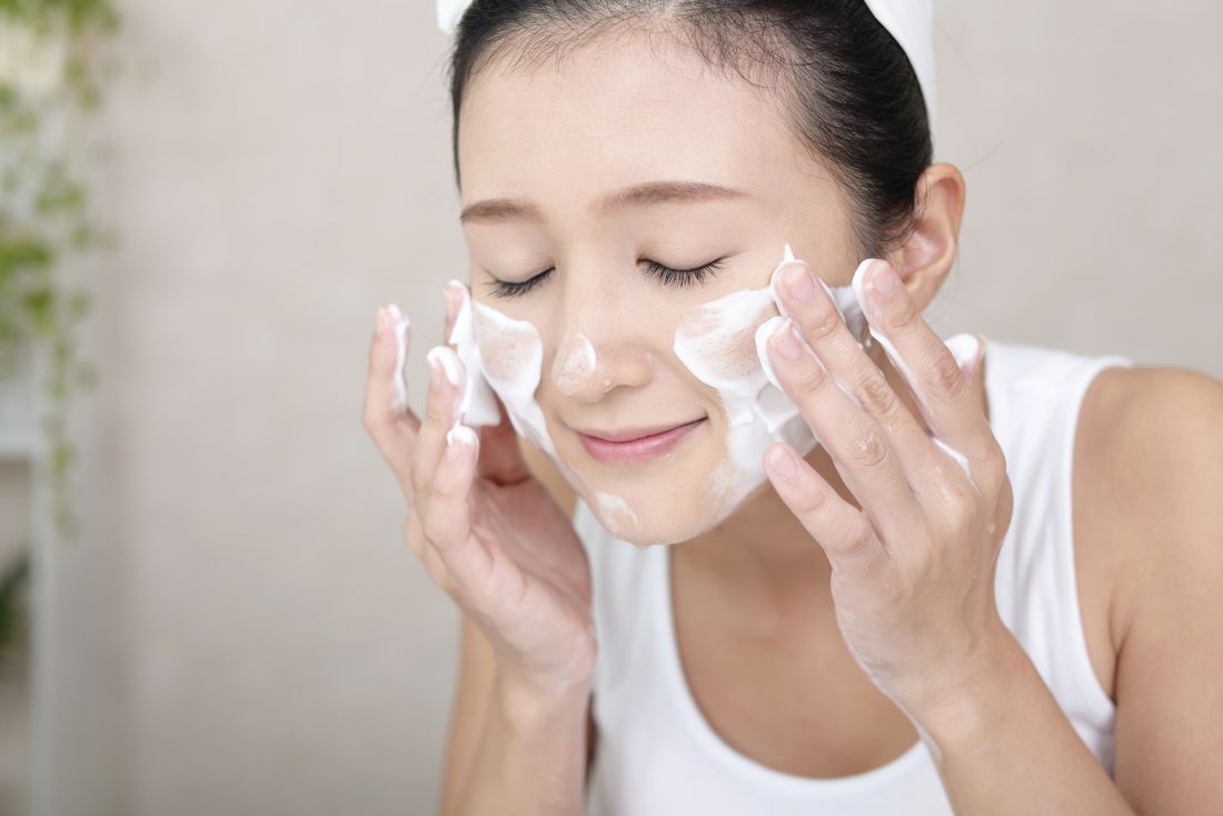
Nonfoaming agents
These agents were developed through efforts to reduce detergent irritancy. This class of cleansers includes superfatted soaps, combination bars (“combars”), syndet bars (composed of synthetic surfactants) and compounds that deposit lipids on the skin, such as creams, lotions and oils. Cream, milk, cold creams, and oil cleansers fall into this category. These products usually have a neutral pH, and include ingredients such as alkyl glyceryl, ether sulfonate, alpha olefin sulfonates, betaines, sulfosuccinates, sodium cocoyl monoglyceride sulfate, and sodium cocoyl isethionate. Organic nonfoaming agents are also available, and may include saponins, a large family of structurally related compounds derived from plant, and sucrose laurate. Nonfoaming cleansers are most appropriate for dry skin types. Oily skin types often report that they “do not feel clean” when they use these cleansers.
Hydroxy acid cleansers
Alpha hydroxy acids (AHAs) are well suited for use by individuals with dry skin because hydroxy acids act as humectants (water-soluble materials with high water absorption capabilities). These hydrophilic cleansers provide exfoliation, and are appropriate for individuals with dry skin and acne because their low pH contributes to an inhospitable microbiome for Propionibacterium acnes, making it harder for the bacteria to thrive. Importantly, the exfoliating activity imparted by hydroxy acids sets the stage for better penetration into the stratum corneum by ingredients applied subsequent to the cleanser. Alpha hydroxy acid cleansers do not dry out the skin the way that salicylic acid cleansers do because their hydrophilic nature makes them unable to penetrate through sebum.
Salicylic acid (SA) cleansers are a member of the aspirin family and therefore confer anti-inflammatory properties. Salicylic acid is lipophilic and can penetrate through the sebum derived lipids into pores. They are the most effective cleansers to unclog pores. Therefore, SA cleansers are ideal for use by individuals with oily, sensitive skin prone to acne, seborrheic dermatitis, or rosacea. The exfoliation yielded by salicylic acid also enhances skin barrier penetration by ingredients applied after its use and is well tolerated by individuals with oily skin. Dry skin types, especially those on retinoids and benzoyl peroxide, will not tolerate SA as well as they will AHA cleansers.
Antibacterial cleansers
Antibacterial cleansers contain ingredients that reduce P. acnes and other types of bacteria on the skin. These products include benzoyl peroxide (BP), silver, hypochlorous acid, and sodium hypochlorite. Benzoyl peroxide can be highly irritating and is not well tolerated by patients with dry skin. Silver has a long history, having been used as an antibacterial agent since the times of King Herod. On the other hand, hypochlorous acid and sodium hypochlorite are novel entrants in the cleansing realm, particularly for individuals with acne. In fact, sodium hypochlorite is formulated to be mild enough for daily use while still sufficiently effective for acne-prone skin.
CLEANSER CHOICE BY SKIN ISSUE
Acne
Recommending the right cleanser for acne-prone skin first depends on whether the patient has oily or dry skin. Individuals with dry skin and acne cannot tolerate drying acne medications. Choosing the correct cleanser and moisturizer can help acne patients be more compliant with the acne treatment plan because of fewer side effects. Dry skin acne types often need two different cleansers. For the morning cleanser, AHA cleansers such as glycolic acid are effective at managing dry. acne-prone skin because glycolic acid has a relatively low pH. P. acnes is less likely to grow on skin with a lower pH.
Hydroxy acids help prevent clogged pores and exfoliate dead skin, which helps prevent acne comedones. Glycolic acid also serves as a humectant ingredient. Creamy cleansers should be used once daily, preferably at night for patients who use makeup since these products are effective at makeup removal. Foaming cleansers should never be used on dry, acne-prone skin. Individuals with the acne subtype of sensitive skin should avoid using scrubs, loofahs, and other forms of mechanical exfoliation.
Patients with oily skin and acne are easier to treat than are dry types because they can better tolerate acne medications. I recommend a salicylic acid cleanser in the morning to unclog pores. The anti-inflammatory properties of salicylic acid help prevent the formation of papules and pustules that characterize acne. Twice-daily use of salicylic acid by patients with oily skin and acne may feel too drying when combined with acne medications such as a retinoid and benzoyl peroxide. If this is the case, a foaming cleanser can be used in the evening to remove dirt, makeup, sunscreen, and debris that can clog pores and exacerbate acne.
Rosacea
Most dry skin type rosacea patients flush red when they wash their face, even if they only use water. The friction alone is enough to cause them to react. Rosacea patients can skip the morning cleanse to help reduce this skin irritation and flushing. Instead they should apply their a.m. anti-redness products followed by a sunscreen appropriate for their skin type. In the evening, a soothing, nonfoaming cleanser with anti-inflammatory ingredients is the best choice to remove makeup, sunscreen, and any built-up dirt or bacteria from the skin’s surface. This should be followed by an anti-redness product that targets the inflammation caused by rosacea.
Anti-inflammatory ingredients that can be found in soothing cleansers and moisturizers for rosacea prone skin include argan oil, green tea, feverfew, chamomile, licorice extract, and aloe.
Patients with very oily skin who have rosacea need to cleanse twice daily to remove excess oil to prevent comedones and acne lesions. A foaming cleanser that contains anti-inflammatory ingredients such as green tea, feverfew, licorice extract, aloe, niacinamide, green tea, and salicylic acid are a good choice for oily rosacea prone skin types.
All rosacea patients should be counseled to avoid mechanical exfoliation, including cleansing scrubs, chemical exfoliants, and abrasive loofahs or cloths.
Eczema
Patients with eczema should choose the same nonfoaming cleansers recommended for dry skin. For patients with frequent skin infections, hypochlorite and silver are beneficial ingredients found in cleansers to help decrease skin bacteria and prevent infections. Foaming cleansers should never be used in eczema prone types.
Conclusion
Cleansers play an important role in skin care because they affect the skin barrier, pH of the skin, presence of bacteria, condition of the pores, and penetration of the post cleanser–applied ingredients. Knowing which cleansing product to use based on a patient’s skin type is critical to recommending the proper ingredients so that patients can achieve and maintain healthy skin.
Table 1. Ingredients used in foaming cleansers
Acyl glycinates
Acylglutamates
Alkyl acyl isethionates
Alkyl carboxylates
Alkyl ether sulfates
Alkyl ethoxy sulfates
Alkyl phosphates
Alkyl sulfates
Alkyl sulfonates
Alkyl sulfosuccinates
Alkyl taurates

Reference
Cleansing is one of the most important steps in any skin care routine, but the surfeit of products on the market can lead to patients selecting an inappropriate cleanser for their skin type. This can engender various adverse cutaneous effects, including xerosis, flaking, acne, and flare-ups of chronic skin conditions such as eczema and rosacea. For example, acne medications are better tolerated when the proper cleanser is used. Cleanser choice is particularly important for individuals with dry skin who have an impaired barrier and those with sensitive skin who are susceptible to inflammation. The following discussion focuses on the factors that practitioners should address with patients when recommending cleansing products to help them maximize their outcomes and maintain clear, healthy-looking skin.
TYPES OF CLEANSERS
Foaming agents
Anionic surface acting agents (surfactants or detergents) produce foam and display the greatest cleansing potency. (Table 1). Because these detergents remove lipids from the skin’s surface and protective bilayer membrane barrier, they should only be used only by individuals with increased sebum production. Ingredients in this category injure the skin barrier and make the skin more susceptible to irritant reactions.1 For example, the widely used compound sodium lauryl sulfate (SLS), which strips lipids from the skin, irritates the skin to such an extent that it is used in research labs to hinder the skin barrier to test “barrier repair products.” The “sulfate- free” trend originates from the irritation caused by SLS. The barrier disruption caused by SLS can be used to intentionally damage the skin barrier to allow increased penetration of chemical peeling products and other therapeutic agents. An alternative to SLS is sodium laureth sulfate (or sodium lauryl ether sulfate, also known as SLES), which exhibits foaming attributes but is less likely than SLS to cause irritation. We often use a foaming cleanser in our practice prior to injectable procedures to ensure that makeup and debris are removed from the skin, and to decrease the time needed for topical lidocaine to penetrate into the skin. If you adopt this strategy, you should follow the injectable procedure with a barrier repair moisturizer.
Nonfoaming agents
These agents were developed through efforts to reduce detergent irritancy. This class of cleansers includes superfatted soaps, combination bars (“combars”), syndet bars (composed of synthetic surfactants) and compounds that deposit lipids on the skin, such as creams, lotions and oils. Cream, milk, cold creams, and oil cleansers fall into this category. These products usually have a neutral pH, and include ingredients such as alkyl glyceryl, ether sulfonate, alpha olefin sulfonates, betaines, sulfosuccinates, sodium cocoyl monoglyceride sulfate, and sodium cocoyl isethionate. Organic nonfoaming agents are also available, and may include saponins, a large family of structurally related compounds derived from plant, and sucrose laurate. Nonfoaming cleansers are most appropriate for dry skin types. Oily skin types often report that they “do not feel clean” when they use these cleansers.
Hydroxy acid cleansers
Alpha hydroxy acids (AHAs) are well suited for use by individuals with dry skin because hydroxy acids act as humectants (water-soluble materials with high water absorption capabilities). These hydrophilic cleansers provide exfoliation, and are appropriate for individuals with dry skin and acne because their low pH contributes to an inhospitable microbiome for Propionibacterium acnes, making it harder for the bacteria to thrive. Importantly, the exfoliating activity imparted by hydroxy acids sets the stage for better penetration into the stratum corneum by ingredients applied subsequent to the cleanser. Alpha hydroxy acid cleansers do not dry out the skin the way that salicylic acid cleansers do because their hydrophilic nature makes them unable to penetrate through sebum.
Salicylic acid (SA) cleansers are a member of the aspirin family and therefore confer anti-inflammatory properties. Salicylic acid is lipophilic and can penetrate through the sebum derived lipids into pores. They are the most effective cleansers to unclog pores. Therefore, SA cleansers are ideal for use by individuals with oily, sensitive skin prone to acne, seborrheic dermatitis, or rosacea. The exfoliation yielded by salicylic acid also enhances skin barrier penetration by ingredients applied after its use and is well tolerated by individuals with oily skin. Dry skin types, especially those on retinoids and benzoyl peroxide, will not tolerate SA as well as they will AHA cleansers.
Antibacterial cleansers
Antibacterial cleansers contain ingredients that reduce P. acnes and other types of bacteria on the skin. These products include benzoyl peroxide (BP), silver, hypochlorous acid, and sodium hypochlorite. Benzoyl peroxide can be highly irritating and is not well tolerated by patients with dry skin. Silver has a long history, having been used as an antibacterial agent since the times of King Herod. On the other hand, hypochlorous acid and sodium hypochlorite are novel entrants in the cleansing realm, particularly for individuals with acne. In fact, sodium hypochlorite is formulated to be mild enough for daily use while still sufficiently effective for acne-prone skin.
CLEANSER CHOICE BY SKIN ISSUE
Acne
Recommending the right cleanser for acne-prone skin first depends on whether the patient has oily or dry skin. Individuals with dry skin and acne cannot tolerate drying acne medications. Choosing the correct cleanser and moisturizer can help acne patients be more compliant with the acne treatment plan because of fewer side effects. Dry skin acne types often need two different cleansers. For the morning cleanser, AHA cleansers such as glycolic acid are effective at managing dry. acne-prone skin because glycolic acid has a relatively low pH. P. acnes is less likely to grow on skin with a lower pH.
Hydroxy acids help prevent clogged pores and exfoliate dead skin, which helps prevent acne comedones. Glycolic acid also serves as a humectant ingredient. Creamy cleansers should be used once daily, preferably at night for patients who use makeup since these products are effective at makeup removal. Foaming cleansers should never be used on dry, acne-prone skin. Individuals with the acne subtype of sensitive skin should avoid using scrubs, loofahs, and other forms of mechanical exfoliation.
Patients with oily skin and acne are easier to treat than are dry types because they can better tolerate acne medications. I recommend a salicylic acid cleanser in the morning to unclog pores. The anti-inflammatory properties of salicylic acid help prevent the formation of papules and pustules that characterize acne. Twice-daily use of salicylic acid by patients with oily skin and acne may feel too drying when combined with acne medications such as a retinoid and benzoyl peroxide. If this is the case, a foaming cleanser can be used in the evening to remove dirt, makeup, sunscreen, and debris that can clog pores and exacerbate acne.
Rosacea
Most dry skin type rosacea patients flush red when they wash their face, even if they only use water. The friction alone is enough to cause them to react. Rosacea patients can skip the morning cleanse to help reduce this skin irritation and flushing. Instead they should apply their a.m. anti-redness products followed by a sunscreen appropriate for their skin type. In the evening, a soothing, nonfoaming cleanser with anti-inflammatory ingredients is the best choice to remove makeup, sunscreen, and any built-up dirt or bacteria from the skin’s surface. This should be followed by an anti-redness product that targets the inflammation caused by rosacea.
Anti-inflammatory ingredients that can be found in soothing cleansers and moisturizers for rosacea prone skin include argan oil, green tea, feverfew, chamomile, licorice extract, and aloe.
Patients with very oily skin who have rosacea need to cleanse twice daily to remove excess oil to prevent comedones and acne lesions. A foaming cleanser that contains anti-inflammatory ingredients such as green tea, feverfew, licorice extract, aloe, niacinamide, green tea, and salicylic acid are a good choice for oily rosacea prone skin types.
All rosacea patients should be counseled to avoid mechanical exfoliation, including cleansing scrubs, chemical exfoliants, and abrasive loofahs or cloths.
Eczema
Patients with eczema should choose the same nonfoaming cleansers recommended for dry skin. For patients with frequent skin infections, hypochlorite and silver are beneficial ingredients found in cleansers to help decrease skin bacteria and prevent infections. Foaming cleansers should never be used in eczema prone types.
Conclusion
Cleansers play an important role in skin care because they affect the skin barrier, pH of the skin, presence of bacteria, condition of the pores, and penetration of the post cleanser–applied ingredients. Knowing which cleansing product to use based on a patient’s skin type is critical to recommending the proper ingredients so that patients can achieve and maintain healthy skin.
Table 1. Ingredients used in foaming cleansers
Acyl glycinates
Acylglutamates
Alkyl acyl isethionates
Alkyl carboxylates
Alkyl ether sulfates
Alkyl ethoxy sulfates
Alkyl phosphates
Alkyl sulfates
Alkyl sulfonates
Alkyl sulfosuccinates
Alkyl taurates
Reference
Topical tapinarof heads for phase 3 in atopic dermatitis and psoriasis
GENEVA – Tapinarof cream, a first-in-class topical nonsteroidal anti-inflammatory agent, successfully met its primary and secondary efficacy endpoints in a large international, phase 2, dose-ranging study and is moving on to a phase 3 trial for atopic dermatitis.
Tapinarof is a naturally derived compound whose therapeutic mechanism of action has recently been shown to involve activation of the aryl hydrocarbon receptor, Johnny Peppers, PhD, said at the annual congress of the European Academy of Dermatology and Venereology.

GlaxoSmithKline is also developing tapinarof cream for mild to moderate plaque psoriasis, a disease that hasn’t seen a novel nonsteroidal topical therapy approved in more than 25 years. After a strong showing in a phase 2 study, a phase 3 trial in psoriasis is now scheduled.
Dr. Peppers presented a phase 2, double-blind, vehicle-controlled randomized trial including 247 adolescent and adult patients with mild, moderate, or severe atopic dermatitis. The six study arms were tapinarof cream at 1% or 0.5% or vehicle, self-administered at a frequency of either once or twice daily. Participants had a mean baseline Investigator’s Global Assessment (IGA) score of 3.1 on a 5-point scale, an Eczema Area and Severity Index (EASI) score of 9.8-13.1 in the various study arms, and a 5.1-5.8 score on an 11-point self-rated itch severity score recorded weekly.
“The 1% tapinarof arm showed higher efficacy and had a quicker onset of action than the 0.5% arm or vehicle,” he reported.
Indeed, the 1% tapinarof cream groups separated from controls in terms of the efficacy endpoints as early as week 1, with the maximum treatment effect seen at weeks 8-12, Dr. Peppers added.
The primary endpoint was a composite requiring both an IGA of 0 or 1, meaning clear or almost clear, at 12 weeks, along with a minimum 2-point improvement on the IGA from baseline to week 12. This was achieved in 46% of patients on tapinarof cream 1% applied once daily, 53% of those on tapinarof cream 1% twice a day, and in about 25% of controls on vehicle.
Eighty percent of subjects who achieved the primary endpoint maintained that level of treatment effect 2 weeks post treatment, and 70% still held their treatment response 4 weeks after they stopped using the medication.
There were two secondary endpoints. One was achievement of a 75% improvement from baseline on EASI scores (EASI-75) response. This was seen in 51% of the tapinarof 1% once-daily group, 60% on twice a day therapy, and 26% and 25% of controls. Onset of action was fastest with tapinarof cream 1% once daily.
The other secondary endpoint was at least a 3-point improvement from baseline to week 4 on the 11-point self-rated itch scale. This was achieved by 37% and 33% of patients on tapinarof cream 1% once daily and twice daily, respectively, a success rate twice that seen in controls.
Four percent of patients on tapinarof cream and 7% on vehicle discontinued the trial because of treatment-emergent adverse events. There were no serious treatment-related adverse events. The most frequent adverse events associated with tapinarof were folliculitis and contact dermatitis. The phase 3 trial will incorporate patch testing for contact dermatitis.
“We are very excited about this program. This will be the first topical therapy – if we’re able to achieve treatment success and ultimately regulatory approval – that would be able to treat both psoriasis and atopic dermatitis since topical steroids,” Dr. Peppers said.
The study was funded by GlaxoSmithKline and presented by a company employee.
bjancin@frontlinemedcom.com
GENEVA – Tapinarof cream, a first-in-class topical nonsteroidal anti-inflammatory agent, successfully met its primary and secondary efficacy endpoints in a large international, phase 2, dose-ranging study and is moving on to a phase 3 trial for atopic dermatitis.
Tapinarof is a naturally derived compound whose therapeutic mechanism of action has recently been shown to involve activation of the aryl hydrocarbon receptor, Johnny Peppers, PhD, said at the annual congress of the European Academy of Dermatology and Venereology.
GlaxoSmithKline is also developing tapinarof cream for mild to moderate plaque psoriasis, a disease that hasn’t seen a novel nonsteroidal topical therapy approved in more than 25 years. After a strong showing in a phase 2 study, a phase 3 trial in psoriasis is now scheduled.
Dr. Peppers presented a phase 2, double-blind, vehicle-controlled randomized trial including 247 adolescent and adult patients with mild, moderate, or severe atopic dermatitis. The six study arms were tapinarof cream at 1% or 0.5% or vehicle, self-administered at a frequency of either once or twice daily. Participants had a mean baseline Investigator’s Global Assessment (IGA) score of 3.1 on a 5-point scale, an Eczema Area and Severity Index (EASI) score of 9.8-13.1 in the various study arms, and a 5.1-5.8 score on an 11-point self-rated itch severity score recorded weekly.
“The 1% tapinarof arm showed higher efficacy and had a quicker onset of action than the 0.5% arm or vehicle,” he reported.
Indeed, the 1% tapinarof cream groups separated from controls in terms of the efficacy endpoints as early as week 1, with the maximum treatment effect seen at weeks 8-12, Dr. Peppers added.
The primary endpoint was a composite requiring both an IGA of 0 or 1, meaning clear or almost clear, at 12 weeks, along with a minimum 2-point improvement on the IGA from baseline to week 12. This was achieved in 46% of patients on tapinarof cream 1% applied once daily, 53% of those on tapinarof cream 1% twice a day, and in about 25% of controls on vehicle.
Eighty percent of subjects who achieved the primary endpoint maintained that level of treatment effect 2 weeks post treatment, and 70% still held their treatment response 4 weeks after they stopped using the medication.
There were two secondary endpoints. One was achievement of a 75% improvement from baseline on EASI scores (EASI-75) response. This was seen in 51% of the tapinarof 1% once-daily group, 60% on twice a day therapy, and 26% and 25% of controls. Onset of action was fastest with tapinarof cream 1% once daily.
The other secondary endpoint was at least a 3-point improvement from baseline to week 4 on the 11-point self-rated itch scale. This was achieved by 37% and 33% of patients on tapinarof cream 1% once daily and twice daily, respectively, a success rate twice that seen in controls.
Four percent of patients on tapinarof cream and 7% on vehicle discontinued the trial because of treatment-emergent adverse events. There were no serious treatment-related adverse events. The most frequent adverse events associated with tapinarof were folliculitis and contact dermatitis. The phase 3 trial will incorporate patch testing for contact dermatitis.
“We are very excited about this program. This will be the first topical therapy – if we’re able to achieve treatment success and ultimately regulatory approval – that would be able to treat both psoriasis and atopic dermatitis since topical steroids,” Dr. Peppers said.
The study was funded by GlaxoSmithKline and presented by a company employee.
bjancin@frontlinemedcom.com
AT THE EADV CONGRESS
Key clinical point:
Major finding: Forty-six percent of atopic dermatitis patients on tapinarof cream 1% applied once daily, and 53% of atopic dermatitis patients on tapinarof cream applied twice daily, met the primary study endpoint, rates twice those in vehicle-treated controls.
Data source: A phase 2, double-blind, vehicle-controlled, international 12-week clinical trial in 247 adolescents and adults with moderate to severe atopic dermatitis.
Disclosures: The study was funded by GlaxoSmithKline and presented by a company employee.
Nemolizumab continues to crush itch in 64-week atopic dermatitis study
GENEVA – Nemolizumab, a humanized monoclonal antibody that inhibits interleukin-31 signaling, maintained its early dramatic antipruritic effect in patients with moderate to severe atopic dermatitis (AD) throughout a year-long unblinded extension of a large, high-profile, 12-week, phase 2 randomized trial, Thomas Ruzicka, MD, reported at the annual congress of the European Academy of Dermatology and Venereology.
In contrast, the 52-week extension study also showed the improvement in the dermatitis aspect of AD as measured by Eczema Area and Severity Index (EASI) scores was more gradual and less robust.

The study, 64 weeks in total, also demonstrated that what really matters to patients with moderate or severe AD is relief from the itch. Scores on the Dermatology Life Quality Index showed marked improvement even if their improvement in EASI scores was suboptimal.
“The dermatitis scores improved. But the most concerning symptom to patients is the pruritus; that’s what worsens their quality of life. And this improves dramatically very early in the course of the study,” Dr. Ruzicka explained. “If they drop their itch they are very happy. They don’t care about the redness of the skin so much as the pruritus. This is what really bothers them, as has been shown in epidemiologic studies.”
The original 12-week, phase 2, randomized, double-blind, dose-ranging trial drew extensive attention because it demonstrated convincingly for the first time that the inflammatory cytokine interleukin-31 plays a key role in the pathobiology of AD by promoting skin barrier dysfunction, pruritus, and the inflammatory response in AD – and that a biologic agent, nemolizumab, could inhibit those disease mechanisms (N Engl J Med. 2017 Mar 2;376[9]:826-35).
At a dose of 0.5 mg/kg administered by subcutaneous injection every 4 weeks, nemolizumab, a humanized monoclonal antibody directed against interleukin-31 receptor A, reduced scores on the pruritus visual-analogue scale by 60% as early as 4 weeks and maintained that effect through 12 weeks. At that point, the double-blind study ended and the 52-week extension began. Patients on nemolizumab in the double-blind phase stayed on the same dosage for the extension, while those who’d been on placebo were switched to nemolizumab at 0.1, 0.5, or 2.0 mg/kg every 4 weeks.
A total of 211 patients with moderate to severe AD inadequately controlled by topical therapies enrolled in the extension study. The combined 64-week experience convinced investigators and the sponsor, Chugai Pharmaceutical, that 0.5 mg/kg every 4 weeks is the optimal dose to take forward into planned advanced-stage clinical trials.
“At this dosage the IL-31 receptor is saturated, so higher dosages aren’t needed,” according to Dr. Ruzicka.
By week 64, patients in the 0.5-mg/kg arm of the study showed further gradual improvement in their pruritus visual-analogue score, from a 60% reduction from baseline at 12 weeks to close to an 80% reduction at week 64.
Roughly half of nemolizumab-treated patients achieved an EASI-50 response within the first 4 weeks of the double-blind phase of the study and stayed in that response zone throughout the extension study.
The EASI-75 and -90 responses followed a pattern different from EASI-50. Patients didn’t leap to those more robust levels of response early and then plateau. Instead, the EASI-75 and -90 responses were achieved via a gradual climb in efficacy throughout the study, such that by week 64 roughly 35%-40% of patients on the various nemolizumab dosages had reached EASI-75.
In the extension study, patients were permitted to use a mild topical steroid or topical calcineurin inhibitor as needed, and a potent or very potent topical steroid as rescue medication. Roughly half of participants resorted to any topical steroid at least once during the 64 months. An important new observation gleaned in the long-term study was that the patients who did use topical steroids were twice as likely to reach an EASI 75 response.
Roughly 20% of patients on the three highest dosages of nemolizumab achieved a static Investigator’s Global Assessment score of 0 or 1, meaning clear or almost clear, by week 64.
Scores on the Dermatology Life Quality Index fell steadily over the course of 64 weeks, from a baseline of about 16 to 5 at study’s close.
The side effect profile of nemolizumab was essentially the same as seen in the placebo arm during the double-blind phase. About one-quarter of patients experienced nasopharyngitis over the course of 64 weeks of treatment. A similar fraction reported exacerbations of their atopic dermatitis; however, these events were front-loaded in the first weeks of the initial double-blind study phase and appeared to have been triggered by the drug washout period prior to the patients’ receiving the first dose of nemolizumab, according to Dr. Ruzicka. No new safety signals emerged during the additional 52 weeks of treatment.
The study was funded by Chugai. Dr. Ruzicka reported serving as a paid company adviser.
GENEVA – Nemolizumab, a humanized monoclonal antibody that inhibits interleukin-31 signaling, maintained its early dramatic antipruritic effect in patients with moderate to severe atopic dermatitis (AD) throughout a year-long unblinded extension of a large, high-profile, 12-week, phase 2 randomized trial, Thomas Ruzicka, MD, reported at the annual congress of the European Academy of Dermatology and Venereology.
In contrast, the 52-week extension study also showed the improvement in the dermatitis aspect of AD as measured by Eczema Area and Severity Index (EASI) scores was more gradual and less robust.
The study, 64 weeks in total, also demonstrated that what really matters to patients with moderate or severe AD is relief from the itch. Scores on the Dermatology Life Quality Index showed marked improvement even if their improvement in EASI scores was suboptimal.
“The dermatitis scores improved. But the most concerning symptom to patients is the pruritus; that’s what worsens their quality of life. And this improves dramatically very early in the course of the study,” Dr. Ruzicka explained. “If they drop their itch they are very happy. They don’t care about the redness of the skin so much as the pruritus. This is what really bothers them, as has been shown in epidemiologic studies.”
The original 12-week, phase 2, randomized, double-blind, dose-ranging trial drew extensive attention because it demonstrated convincingly for the first time that the inflammatory cytokine interleukin-31 plays a key role in the pathobiology of AD by promoting skin barrier dysfunction, pruritus, and the inflammatory response in AD – and that a biologic agent, nemolizumab, could inhibit those disease mechanisms (N Engl J Med. 2017 Mar 2;376[9]:826-35).
At a dose of 0.5 mg/kg administered by subcutaneous injection every 4 weeks, nemolizumab, a humanized monoclonal antibody directed against interleukin-31 receptor A, reduced scores on the pruritus visual-analogue scale by 60% as early as 4 weeks and maintained that effect through 12 weeks. At that point, the double-blind study ended and the 52-week extension began. Patients on nemolizumab in the double-blind phase stayed on the same dosage for the extension, while those who’d been on placebo were switched to nemolizumab at 0.1, 0.5, or 2.0 mg/kg every 4 weeks.
A total of 211 patients with moderate to severe AD inadequately controlled by topical therapies enrolled in the extension study. The combined 64-week experience convinced investigators and the sponsor, Chugai Pharmaceutical, that 0.5 mg/kg every 4 weeks is the optimal dose to take forward into planned advanced-stage clinical trials.
“At this dosage the IL-31 receptor is saturated, so higher dosages aren’t needed,” according to Dr. Ruzicka.
By week 64, patients in the 0.5-mg/kg arm of the study showed further gradual improvement in their pruritus visual-analogue score, from a 60% reduction from baseline at 12 weeks to close to an 80% reduction at week 64.
Roughly half of nemolizumab-treated patients achieved an EASI-50 response within the first 4 weeks of the double-blind phase of the study and stayed in that response zone throughout the extension study.
The EASI-75 and -90 responses followed a pattern different from EASI-50. Patients didn’t leap to those more robust levels of response early and then plateau. Instead, the EASI-75 and -90 responses were achieved via a gradual climb in efficacy throughout the study, such that by week 64 roughly 35%-40% of patients on the various nemolizumab dosages had reached EASI-75.
In the extension study, patients were permitted to use a mild topical steroid or topical calcineurin inhibitor as needed, and a potent or very potent topical steroid as rescue medication. Roughly half of participants resorted to any topical steroid at least once during the 64 months. An important new observation gleaned in the long-term study was that the patients who did use topical steroids were twice as likely to reach an EASI 75 response.
Roughly 20% of patients on the three highest dosages of nemolizumab achieved a static Investigator’s Global Assessment score of 0 or 1, meaning clear or almost clear, by week 64.
Scores on the Dermatology Life Quality Index fell steadily over the course of 64 weeks, from a baseline of about 16 to 5 at study’s close.
The side effect profile of nemolizumab was essentially the same as seen in the placebo arm during the double-blind phase. About one-quarter of patients experienced nasopharyngitis over the course of 64 weeks of treatment. A similar fraction reported exacerbations of their atopic dermatitis; however, these events were front-loaded in the first weeks of the initial double-blind study phase and appeared to have been triggered by the drug washout period prior to the patients’ receiving the first dose of nemolizumab, according to Dr. Ruzicka. No new safety signals emerged during the additional 52 weeks of treatment.
The study was funded by Chugai. Dr. Ruzicka reported serving as a paid company adviser.
AT THE EADV CONGRESS
Key clinical point:
Major finding: The early dramatic antipruritic effect demonstrated by nemolizumab for atopic dermatitis in a 12-week randomized trial was maintained throughout an additional 52 weeks in an extension study.
Data source: This analysis focused on 211 patients with moderate to severe atopic dermatitis who participated in a 52-week open-label extension study after completing a previously reported 12-week, double-blind, placebo-controlled phase.
Disclosures: The presenter is a paid medical adviser to Chugai Pharmaceutical, which funded the study.