User login
Microbead ban could impede nanomaterial development
The new law that will ban the sale or distribution of plastic microbeads in over-the-counter and personal care products beginning in July 2017 could have an unintended effect: a negative impact on the development and acceptance of micro- and nanotechnology–based medical and diagnostic products.
Environmentalists and others cheered the new law for its potentially protective effects on the environment and ultimately on public health. And the unavailability of such products is not expected to adversely affect consumers, as there are alternatives to the scrubs and other products that contain these microbeads – and manufacturers have started to reformulate their products that contain plastic microbeads.

However, the ban could potentially do more harm than good if the message consumers hear is that microtechnology – and, by extension, nanotechnology – is bad, according to Dr. Adam Friedman, director of translational research in the department of dermatology at George Washington University, Washington, who is a Dermatology News Board Member.
“What I’m most concerned about is the impact on public perception, that this ban infers that micro- and nanotechnology is inherently bad, and therefore, how it might impact approval of both over-the-counter and prescription medications that incorporate microscopic carriers,” said Dr. Friedman, who has a particular interest in nanotechnology. He explained that nanomaterials have enormous potential for helping to deliver drugs that are unstable, difficult to administer, or even toxic in their bulk form.
How such materials can be evaluated from a safety and efficacy standpoint in order to facilitate approval is currently under investigation, and negativity toward this field of research could hinder the progress of related research, he added, noting that “not all nano- and microtechnology is created equal.”
“This [ban] is about microplastics specifically,” not microspheres, -particles, or -beads, overall, he pointed out.
The basis of the Microbead-Free Waters Act of 2015 – which was signed into law by President Barack Obama on Dec. 28 after it sailed through Congress with an unusual level of bipartisan consensus among lawmakers regarding its importance – is that the plastic microbeads used in products such as facial scrubs and toothpaste pose a threat to marine life and ultimately to humans via the food chain.
Researchers have found that tiny microbeads – an estimated 11 billion daily – slip through wastewater treatment systems into the environment, where they appear to attract harmful chemicals that could make them toxic to marine life and ultimately to humans.
In fact, the biological dangers associated with microbeads upon which the ban was based are hypothetical, Dr. Friedman countered, noting that much of the argument against microbeads is based on an oft-cited publication that is actually a non-peer-reviewed editorial in support of a microbead ban (Environ Sci Technol. 2015;49:10759-61). The authors cited studies demonstrating the inability of current sanitation measures to effectively remove the microbeads from the water supply. They also observed that “the argument has been raised that there is not yet enough scientific evidence to support banning microbeads,” but added, “though there are gaps in our understanding of the precise impact of microbeads on aquatic ecosystems, this should not delay action.”

Indeed, there is reason to believe that microplastics could contain endocrine-disrupting chemicals linked with a host of intractable human diseases, and that they may otherwise pose a threat to human health – particularly by attracting and collecting pesticides, bisphenol A (BPA), and phthalates and binding to them, then entering the food chain, noted Deborah M. Kurrasch, Ph.D., of the department of medical genetics at the University of Calgary, Alberta, in an interview. However, there is no direct evidence of this, she agreed.
“Common sense says that might be the case, but there are no data to support that,” said Dr. Kurrasch, who is currently studying how environmental insults, including those from plastics and herbicides, affect the brain.
“There is lots of interest in plastics, and this ban is more of an environmental toxicology ban ... the leap to a human problem is very vague,” she commented.
In fact, the plastics in microbeads are generally considered “safe plastics,” as opposed to those containing BPA, for example, Dr. Kurrasch said. However, she added that she doesn’t think there is such a thing as a safe plastic.
“Ninety-one percent of plastics have been shown to leach compounds that affect estrogen signaling, and the fact that microbeads are made of plastic means they are likely to have some sort of effect down the road,” she said. “But the route of exposure? We just don’t know.”
Dr. Kurrasch believes the ban is a step in the right direction, and that it’s good that attention is being paid to protecting wildlife and humans from the potential harms of plastics.
Dr. Naissan O. Wesley, a dermatologist in private practice in Beverly Hills, noted that she, too is pleased about the concern regarding potential detrimental effects of microbeads.
“This is a huge step in skin care for products that are healthy for us and our environment,” she said, adding that while the quality of some products may be affected by the microbead ban, there are plenty of alternative scrubs, including sugar- or salt-based products.
“There are also other ways to achieve exfoliation, such as with things like the Clarisonic brush once a day, glycolic acid washes, Retin-A [tretinoin], chemical peels, microdermabrasion, or hydrofacials, and lasers,” Dr. Wesley, who is also a Dermatology News Board Member, noted. “Some women also do dermaplaning.”
Dr. Friedman also agreed with the importance of protecting the environment and the minimal impact the ban will have on products.
“I’m all for putting appropriate measures in place to prevent inappropriate exposures and damage to the natural setting. That said, I’m also for preserving the scientific method.”
Taking microbead-containing products off the shelf is not a life-altering process, Dr. Friedman said, noting that he doesn’t usually even recommend such products for patients. However, it will be expensive for the industry as it adapts to the microbead ban – and the money that will be spent is money that could have been applied to initiatives and efforts that may have had a bigger impact, he added.
“What frustrates me is that no opportunity was given to improve the technology,” he said, explaining that nano- and microparticles could be augmented to prevent them from entering the water system. For example, surface modifications applied to the microbeads could be used to increase aggregation under specific environmental conditions, and thereby prevent filtration failure. “There was a missed opportunity to engage industry and scientists alike to better understand how materials at this scale behave and how they can be manipulated for good,” he maintained.
“The ban opens the door to adversely impact more important technology that could be life altering. Now, we have an uphill battle.”
Dr. Friedman, Dr. Kurrasch, and Dr. Wesley reported having no relevant disclosures.
The new law that will ban the sale or distribution of plastic microbeads in over-the-counter and personal care products beginning in July 2017 could have an unintended effect: a negative impact on the development and acceptance of micro- and nanotechnology–based medical and diagnostic products.
Environmentalists and others cheered the new law for its potentially protective effects on the environment and ultimately on public health. And the unavailability of such products is not expected to adversely affect consumers, as there are alternatives to the scrubs and other products that contain these microbeads – and manufacturers have started to reformulate their products that contain plastic microbeads.
However, the ban could potentially do more harm than good if the message consumers hear is that microtechnology – and, by extension, nanotechnology – is bad, according to Dr. Adam Friedman, director of translational research in the department of dermatology at George Washington University, Washington, who is a Dermatology News Board Member.
“What I’m most concerned about is the impact on public perception, that this ban infers that micro- and nanotechnology is inherently bad, and therefore, how it might impact approval of both over-the-counter and prescription medications that incorporate microscopic carriers,” said Dr. Friedman, who has a particular interest in nanotechnology. He explained that nanomaterials have enormous potential for helping to deliver drugs that are unstable, difficult to administer, or even toxic in their bulk form.
How such materials can be evaluated from a safety and efficacy standpoint in order to facilitate approval is currently under investigation, and negativity toward this field of research could hinder the progress of related research, he added, noting that “not all nano- and microtechnology is created equal.”
“This [ban] is about microplastics specifically,” not microspheres, -particles, or -beads, overall, he pointed out.
The basis of the Microbead-Free Waters Act of 2015 – which was signed into law by President Barack Obama on Dec. 28 after it sailed through Congress with an unusual level of bipartisan consensus among lawmakers regarding its importance – is that the plastic microbeads used in products such as facial scrubs and toothpaste pose a threat to marine life and ultimately to humans via the food chain.
Researchers have found that tiny microbeads – an estimated 11 billion daily – slip through wastewater treatment systems into the environment, where they appear to attract harmful chemicals that could make them toxic to marine life and ultimately to humans.
In fact, the biological dangers associated with microbeads upon which the ban was based are hypothetical, Dr. Friedman countered, noting that much of the argument against microbeads is based on an oft-cited publication that is actually a non-peer-reviewed editorial in support of a microbead ban (Environ Sci Technol. 2015;49:10759-61). The authors cited studies demonstrating the inability of current sanitation measures to effectively remove the microbeads from the water supply. They also observed that “the argument has been raised that there is not yet enough scientific evidence to support banning microbeads,” but added, “though there are gaps in our understanding of the precise impact of microbeads on aquatic ecosystems, this should not delay action.”
Indeed, there is reason to believe that microplastics could contain endocrine-disrupting chemicals linked with a host of intractable human diseases, and that they may otherwise pose a threat to human health – particularly by attracting and collecting pesticides, bisphenol A (BPA), and phthalates and binding to them, then entering the food chain, noted Deborah M. Kurrasch, Ph.D., of the department of medical genetics at the University of Calgary, Alberta, in an interview. However, there is no direct evidence of this, she agreed.
“Common sense says that might be the case, but there are no data to support that,” said Dr. Kurrasch, who is currently studying how environmental insults, including those from plastics and herbicides, affect the brain.
“There is lots of interest in plastics, and this ban is more of an environmental toxicology ban ... the leap to a human problem is very vague,” she commented.
In fact, the plastics in microbeads are generally considered “safe plastics,” as opposed to those containing BPA, for example, Dr. Kurrasch said. However, she added that she doesn’t think there is such a thing as a safe plastic.
“Ninety-one percent of plastics have been shown to leach compounds that affect estrogen signaling, and the fact that microbeads are made of plastic means they are likely to have some sort of effect down the road,” she said. “But the route of exposure? We just don’t know.”
Dr. Kurrasch believes the ban is a step in the right direction, and that it’s good that attention is being paid to protecting wildlife and humans from the potential harms of plastics.
Dr. Naissan O. Wesley, a dermatologist in private practice in Beverly Hills, noted that she, too is pleased about the concern regarding potential detrimental effects of microbeads.
“This is a huge step in skin care for products that are healthy for us and our environment,” she said, adding that while the quality of some products may be affected by the microbead ban, there are plenty of alternative scrubs, including sugar- or salt-based products.
“There are also other ways to achieve exfoliation, such as with things like the Clarisonic brush once a day, glycolic acid washes, Retin-A [tretinoin], chemical peels, microdermabrasion, or hydrofacials, and lasers,” Dr. Wesley, who is also a Dermatology News Board Member, noted. “Some women also do dermaplaning.”
Dr. Friedman also agreed with the importance of protecting the environment and the minimal impact the ban will have on products.
“I’m all for putting appropriate measures in place to prevent inappropriate exposures and damage to the natural setting. That said, I’m also for preserving the scientific method.”
Taking microbead-containing products off the shelf is not a life-altering process, Dr. Friedman said, noting that he doesn’t usually even recommend such products for patients. However, it will be expensive for the industry as it adapts to the microbead ban – and the money that will be spent is money that could have been applied to initiatives and efforts that may have had a bigger impact, he added.
“What frustrates me is that no opportunity was given to improve the technology,” he said, explaining that nano- and microparticles could be augmented to prevent them from entering the water system. For example, surface modifications applied to the microbeads could be used to increase aggregation under specific environmental conditions, and thereby prevent filtration failure. “There was a missed opportunity to engage industry and scientists alike to better understand how materials at this scale behave and how they can be manipulated for good,” he maintained.
“The ban opens the door to adversely impact more important technology that could be life altering. Now, we have an uphill battle.”
Dr. Friedman, Dr. Kurrasch, and Dr. Wesley reported having no relevant disclosures.
Global approach to hand rejuvenation gives patients and clinicians options
HOLLYWOOD, FLA. – As more patients seek facial rejuvenation, they find themselves concerned with the mismatch between their refreshed facial appearance and the weathered, aged appearance of their hands. As more resurfacing treatments and fillers are being used beyond the face, options for hand rejuvenation expand.
Dr. Rania Agha, a dermatologist in private practice in Oakbrook Terrace, Ill., discussed a comprehensive approach to hand rejuvenation that addresses photoaging, vein prominence, and atrophy.
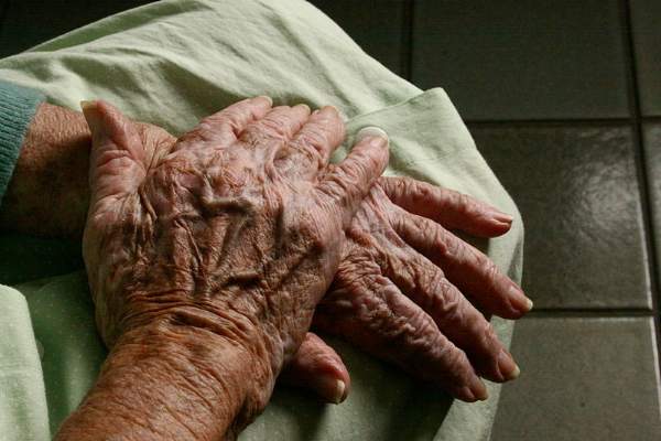
Sun exposure is the largest extrinsic factor that affects the appearance of hands. Resultant changes can include actinic keratosis, seborrheic keratosis, dermatoheliosis, and solar lentigines.
Intrinsic factors that contribute to hand aging include loss of collagen and fatty tissue, resulting in epidermal and dermal atrophy and loss of elasticity. Reduced hydration follows as well. Though hyaluronic acid is less than 1% of dermal dry weight, the loss of glycosaminoglycan-proteoglycan complexes means dermal tissue is much less effective at binding and retaining water, said Dr. Agha.
The consequences are wrinkles and prominence of bones and tendons, large intermetacarpal space, and prominent reticular veins. For many of her patients, “there will be a discordance of the face and the hands,” Dr. Agha nnoted.
Grading scales are available to evaluate hand aging, better to compare apparent hand age before and after treatment. The Merz Hand Grading Scale is used commonly and ranges from 0-4, with increasing scores for more wrinkles, bone and tendon prominence, and photodamage.
Where protuberant veins are a concern, sclerotherapy of the veins on the dorsum of the hand will reduce their prominence. Sclerotherapy options for the hand, where typical vessel size ranges from 1mm to 6 mm, are 0.5% sodium tetradecyl sulfate or polidocanol 1.5% or 3%. Post-sclerotherapy compression is recommended. An option of foam sclerotherapy, mixing the sclerosing agent with carbon dioxide or room air, can have greater efficacy because it prolongs contact between the sclerosant and the vein wall, but it theoretically carries some of the systemic risks associated with air emboli.
Adverse events from sclerotherapy can include telangiectatic matting, or microscopic revascularization; Dr. Agha said the reported incidence neared 15% in one study. Some pain, edema, and ecchymosis may occur; rare radial nerve neuropraxia has been reported. Other rare complications can include ulceration and hyperpigmentation.
Many energy-based, mechanical, and chemical rejuvenation techniques that are used on the face can also be used on the hands, though intensity or duration of treatment has to be modulated in some cases.
Superficial chemical peels only penetrate the epidermis; examples include 70% glycolic acid, salicylic acid, 50% resorcinol, Jessner’s solution, and trichloroacetic acid 10%-20%. Trichloroacetic acid at 30% or greater is an example of a medium-depth peel that extends to the papillary dermis. Deep peels that extend to the midreticular dermis, such as phenol, are not for use on the hands.
Even with light to medium peels, though, caution should be used. Reported complications can include maceration, desquamation, and delayed healing; also, practitioners should take care not to create a line of demarcation between the hand and forearm. “Be conservative and perform these gradually and serially over several weeks,” said Dr. Agha.
A wide range of laser and light therapies can be useful. Q-switched lasers are useful for solar lentigines and other photodamage; intense pulsed light, photodynamic therapy, Nd:YAG lasers, fractionated lasers, and the 2940 erbium:YAG laser are all options, depending on patient preference and what options are available.
“With light therapy, start at lower fluences,” said Dr. Agha. “Limit the number of passes, and the density, if you’re using a fractionated laser.” Complications can include erythema, hypo- or hyper-pigmentation, scarring, textural changes, crusting, and prolonged bleeding. Prompt local wound care can help mitigate long-term effects if any complications are seen post-treatment.
“Strict sun protection, whether with physical block or sunscreen, is a must after hand resurfacing,” whether the procedures are chemical or energy-based, said Dr. Agha.
Another option for hand rejuvenation is to use dermal fillers to plump the dorsum of the hand and obscure some of the exposed hand anatomy. Autologous fat can be used, but more demand is being seen for calcium hydroxylapatite microsphere (Radiesse) fillers, said Dr. Agha. “It is highly biocompatible; therefore, allergic reactions are extremely rare,” she said.
Calcium hydroxylapatite was approved by the Food and Drug Administration for hand rejuvenation in 2015. The particle size of 25-50 micrometers stimulates collagen formation and fibrosis when injected, and the product’s effects can last up to 18 months. In clinical trials, the product produced significant improvement in hand appearance without serious side effects in followup through 1 year.
Dr. Agha reviewed recommended administration of calcium hydroxylapatite, which is meant to be injected in bolus fashion midway between the dorsal crease of the wrist and the metacarpophalangeal joints, in a zone between the second and the fifth metacarpal bones. Dr. Agha says that in her practice, she has the patient make a tight fist, and then she performs vigorous massage of the hand immediately to distribute the product.
This is followed by application of a soapy cleanser, icing, and hand elevation for the first 24 hours post-injection. It’s important that patients be advised to expect moderate edema for up to 10 days, and that they avoid vigorous manual activities in the first week, said Dr. Agha.
Results from clinical trials indicate that between 70% and 80% of patients can expect an improvement of at least one point on a clinician-rated hand appearance scale, while over 98% of patients consider their hands subjectively improved at 1 month after treatment, and 86% of patients still feeling their hands are improved at 1 year post-treatment with calcium hydroxylapatite.
Dr. Agha reported being on the advisory boards of Taro Pharmaceutical Industries and Aqua Pharmaceutical.
On Twitter @karioakes
HOLLYWOOD, FLA. – As more patients seek facial rejuvenation, they find themselves concerned with the mismatch between their refreshed facial appearance and the weathered, aged appearance of their hands. As more resurfacing treatments and fillers are being used beyond the face, options for hand rejuvenation expand.
Dr. Rania Agha, a dermatologist in private practice in Oakbrook Terrace, Ill., discussed a comprehensive approach to hand rejuvenation that addresses photoaging, vein prominence, and atrophy.
Sun exposure is the largest extrinsic factor that affects the appearance of hands. Resultant changes can include actinic keratosis, seborrheic keratosis, dermatoheliosis, and solar lentigines.
Intrinsic factors that contribute to hand aging include loss of collagen and fatty tissue, resulting in epidermal and dermal atrophy and loss of elasticity. Reduced hydration follows as well. Though hyaluronic acid is less than 1% of dermal dry weight, the loss of glycosaminoglycan-proteoglycan complexes means dermal tissue is much less effective at binding and retaining water, said Dr. Agha.
The consequences are wrinkles and prominence of bones and tendons, large intermetacarpal space, and prominent reticular veins. For many of her patients, “there will be a discordance of the face and the hands,” Dr. Agha nnoted.
Grading scales are available to evaluate hand aging, better to compare apparent hand age before and after treatment. The Merz Hand Grading Scale is used commonly and ranges from 0-4, with increasing scores for more wrinkles, bone and tendon prominence, and photodamage.
Where protuberant veins are a concern, sclerotherapy of the veins on the dorsum of the hand will reduce their prominence. Sclerotherapy options for the hand, where typical vessel size ranges from 1mm to 6 mm, are 0.5% sodium tetradecyl sulfate or polidocanol 1.5% or 3%. Post-sclerotherapy compression is recommended. An option of foam sclerotherapy, mixing the sclerosing agent with carbon dioxide or room air, can have greater efficacy because it prolongs contact between the sclerosant and the vein wall, but it theoretically carries some of the systemic risks associated with air emboli.
Adverse events from sclerotherapy can include telangiectatic matting, or microscopic revascularization; Dr. Agha said the reported incidence neared 15% in one study. Some pain, edema, and ecchymosis may occur; rare radial nerve neuropraxia has been reported. Other rare complications can include ulceration and hyperpigmentation.
Many energy-based, mechanical, and chemical rejuvenation techniques that are used on the face can also be used on the hands, though intensity or duration of treatment has to be modulated in some cases.
Superficial chemical peels only penetrate the epidermis; examples include 70% glycolic acid, salicylic acid, 50% resorcinol, Jessner’s solution, and trichloroacetic acid 10%-20%. Trichloroacetic acid at 30% or greater is an example of a medium-depth peel that extends to the papillary dermis. Deep peels that extend to the midreticular dermis, such as phenol, are not for use on the hands.
Even with light to medium peels, though, caution should be used. Reported complications can include maceration, desquamation, and delayed healing; also, practitioners should take care not to create a line of demarcation between the hand and forearm. “Be conservative and perform these gradually and serially over several weeks,” said Dr. Agha.
A wide range of laser and light therapies can be useful. Q-switched lasers are useful for solar lentigines and other photodamage; intense pulsed light, photodynamic therapy, Nd:YAG lasers, fractionated lasers, and the 2940 erbium:YAG laser are all options, depending on patient preference and what options are available.
“With light therapy, start at lower fluences,” said Dr. Agha. “Limit the number of passes, and the density, if you’re using a fractionated laser.” Complications can include erythema, hypo- or hyper-pigmentation, scarring, textural changes, crusting, and prolonged bleeding. Prompt local wound care can help mitigate long-term effects if any complications are seen post-treatment.
“Strict sun protection, whether with physical block or sunscreen, is a must after hand resurfacing,” whether the procedures are chemical or energy-based, said Dr. Agha.
Another option for hand rejuvenation is to use dermal fillers to plump the dorsum of the hand and obscure some of the exposed hand anatomy. Autologous fat can be used, but more demand is being seen for calcium hydroxylapatite microsphere (Radiesse) fillers, said Dr. Agha. “It is highly biocompatible; therefore, allergic reactions are extremely rare,” she said.
Calcium hydroxylapatite was approved by the Food and Drug Administration for hand rejuvenation in 2015. The particle size of 25-50 micrometers stimulates collagen formation and fibrosis when injected, and the product’s effects can last up to 18 months. In clinical trials, the product produced significant improvement in hand appearance without serious side effects in followup through 1 year.
Dr. Agha reviewed recommended administration of calcium hydroxylapatite, which is meant to be injected in bolus fashion midway between the dorsal crease of the wrist and the metacarpophalangeal joints, in a zone between the second and the fifth metacarpal bones. Dr. Agha says that in her practice, she has the patient make a tight fist, and then she performs vigorous massage of the hand immediately to distribute the product.
This is followed by application of a soapy cleanser, icing, and hand elevation for the first 24 hours post-injection. It’s important that patients be advised to expect moderate edema for up to 10 days, and that they avoid vigorous manual activities in the first week, said Dr. Agha.
Results from clinical trials indicate that between 70% and 80% of patients can expect an improvement of at least one point on a clinician-rated hand appearance scale, while over 98% of patients consider their hands subjectively improved at 1 month after treatment, and 86% of patients still feeling their hands are improved at 1 year post-treatment with calcium hydroxylapatite.
Dr. Agha reported being on the advisory boards of Taro Pharmaceutical Industries and Aqua Pharmaceutical.
On Twitter @karioakes
EXPERT ANALYSIS FROM THE AMERICAN ACADEMY OF COSMETIC SURGERY ANNUAL SCIENTIFIC MEETING

Dermal Fillers for Aesthetic Rejuvenation
What does your patient need to know at the first consultation?
Several things are important. First, I have a discussion with the patient to find out exactly what bothers him or her the most. Some patients have very specific areas they would like to address while others simply come in and say, “Please make me look better/less tired/younger.” It’s very important to review all of the treatment options with the patient. Not only are there many different types of fillers, but there also are differences among the products within each category; for example, some hyaluronic acid (HA) fillers have similar clinical properties and applications (eg, Juvéderm Voluma XC [Allergan, Inc], Restylane Lyft [Galderma Laboratories, LP]), but they differ from other similar HA fillers (eg, Juvéderm Ultra XC [Allergan, Inc], Restylane [Galderma Laboratories, LP], Belotero Balance [Merz North America, Inc]) with regard to G′, molecular weight, and crosslinking. I also discuss longer-lasting filler materials such as calcium hydroxylapatite (eg, Radiesse [Merz North America, Inc]) and injectable poly-L-lactic acid (Sculptra Aesthetic [Galderma Laboratories, LP]), which stimulates collagen production.
For patients that have never had filler treatments before, I may try to steer them in the direction of using an HA filler simply because the effects can be reversed if they aren’t happy with the results. It’s also important to discuss how much filler the patient will need to achieve the desired effect. It’s important to take the patient’s budget into account when formulating a treatment plan. I also tell my patients that fillers alone may not achieve the desired results and that they also may need toxin treatment (eg, onabotulinumtoxinA [Botox Cosmetic (Allergan, Inc)], incobotulinumtoxinA [Xeomin (Merz North America, Inc)], abobotulinumtoxinA [Dysport (Galderma Laboratories, LP)]), and possibly laser treatment to improve the overall skin appearance. Additionally, I always discuss a skin care routine and the need for daily sunscreen use.
What procedures are most commonly requested in your practice?
In my practice, patients present with several common complaints. Thin, downturned lips are a common treatment area, and many patients are concerned about jowls and flattened cheeks. Patients also often seek treatment for prominent nasolabial and melolabial folds and “smoker’s lines.” I typically discuss contouring and shaping more than simply filling lines. We try to take a wholistic approach to improve the overall appearance of the face as opposed to just focusing on certain lines and wrinkles.
What are your go-to injection techniques?
All fillers have a place in my practice. I use Juvéderm Ultra XC, Restylane, and Belotero Balance to improve the appearance of tear troughs. Juvéderm Ultra Plus XC and Restylane are really great for deep creases like nasolabial folds. Belotero Balance and Restylane Silk are especially good for treating perioral wrinkles and lines. I use Juvéderm Voluma XC, Restylane Lyft, and Radiesse more for shaping and contouring, but these products also work great for adding volume. I use Sculptra Aesthetic as a foundation for patients who need volume and collagen stimulation. Radiesse is a great option for hand rejuvenation and was recently approved for this treatment by the US Food and Drug Administration.
There are numerous injection techniques that I find useful, including depot, serial puncture, fanning, and tower techniques. I recommend learning all of these and then picking what works for you. As an overall principle, I try to minimize tissue trauma and the possibility of bruising. Most importantly, one has to know the anatomic location of the injection site and stay away from danger zones. It’s also very important to always draw back to ensure that one isn’t injecting into a vessel.
I think it’s smart to start with HA fillers since the effects are reversible. After the physician becomes more comfortable with performing filler procedures, I would recommend moving on to longer-lasting fillers.
What complications/side effects should physicians be aware of?
The most common complications associated with dermal fillers are bruising and swelling. The risks for these side effects can be decreased by icing the treatment area immediately before and after the procedure. Also, I often recommend products containing arnica (topical and/or oral) for patients who tend to bruise. Nodule formation, skin necrosis, infection, and vascular occlusion in the immediate or distal areas can be avoided with proper training and knowledge of local anatomy; for example, it’s important to always draw back before injecting to ensure you aren’t injecting into a vascular structure. Knowledge of local anatomy and its variations also is important in order to avoid these danger zones. In very rare cases, blindness and stroke may occur following treatment with dermal fillers.
Suggested Readings
Sadick N, ed. Augmentation Fillers. New York, NY: Cambridge University Press; 2010.
Small R, Hoang D. A Practical Guide to Dermal Filler Procedures. Philadelphia, PA: Lippincott Willams & Wilkins; 2011.

Royal jelly
Used for centuries by humans for its health-promoting qualities, royal jelly is a yellowish, viscous secretion from the hypopharyngeal and mandibular glands of worker bees that nourishes bee larvae of all kinds (i.e., drones, workers, queens) after which it becomes the exclusive nourishment for queens throughout their development.1-3 A wide range of biologic activity has been attributed to royal jelly, including antitumor, antibacterial, anti-inflammatory, antioxidant, collagen production-promoting, immunomodulatory, and wound healing.3-8 Royal jelly is used in cosmetics, health tonics (particularly in Asia), dietary supplements, and beverages.2,9
Produced from pollen, royal jelly contains water, proteins (82%-90% of which are known as the major royal jelly proteins, with five primary members), lipids – including its primary unsaturated fatty acid, 10-hydroxy-2-decenoic acid (10-HDA) – sugars, carbohydrates, free amino acids, vitamins, and minerals.4,7,10 Many of the benefits to human health linked to royal jelly can be partly attributed to the activity of its lipids, particularly 10-HDA, which render the royal jelly emulsion highly acidic and impart antimicrobial properties.10 These and other constituents of royal jelly operate in ways that are thought to yield broad protection against skin aging and cancer development, modulation of the immune system, induction of neurogenesis, and alleviation of menopausal symptoms.1 This column will focus on recent studies pertaining to the topical use of royal jelly.

Wound healing
In 2008, Abdelatif et al. conducted a pilot study to determine the safety and effectiveness of a then-new ointment combining royal jelly and panthenol (Pedyphar) in 60 patients with limb-threatening diabetic foot infections. After 9 weeks of treatment and through 6 months of follow-up, 96% of subjects with full-thickness skin ulcers (Wagner grades 1 and 2) or deep tissue infection and suspected osteomyelitis (grade 3) responded well, with all grade 1 and 2 ulcers healing and 92% of grade 3 ulcers healing. All patients with gangrenous lesions (grades 4 and 5) healed after surgical excision, debridement, and conservative treatment with the royal jelly/panthenol product. The researchers called for more double-blind, randomized controlled studies to confirm their promising findings of the safety and efficacy of the royal jelly/panthenol combination.11
Two years later, Kim et al. treated freshly scratched normal human dermal fibroblasts with different concentrations of royal jelly (0.1 mg/mL, 1.0 mg/mL, or 5 mg/mL) for up to 48 hours. Fibroblast migration was found to have peaked at 24 hours after wound induction, with royal jelly significantly and dose-dependently accelerating the migration at the 8-hour mark. Royal jelly also influenced several fibroblast lipids involved in the wound healing process, with a decrease in cholesterol level and an increase in sphinganines.12
A small study with eight subjects was done in 2011 by Siavash et al. to evaluate the efficacy of topically applied royal jelly for diabetic foot ulcers. Seven of the eight ulcers treated healed, with a mean healing time of 41 days. The eighth ulcer improved, diminishing significantly in size. The researchers concluded that a royal jelly dressing is an effective alternative for treatment of diabetic foot ulcers.13 However, the same team conducted a double-blind, placebo-controlled clinical trial of topical royal jelly on diabetic foot ulcers in 25 patients (6 females, 19 males) and found no significant differences between 5% sterile topical royal jelly or placebo.6
Collagen production

A decade ago, Koya-Miyata et al. showed that royal jelly promotes collagen synthesis by skin fibroblasts in the presence of ascorbic acid-2-O-alpha-glucoside. They also showed that its primary fatty acid constituent, 10-HDA, facilitates the collagen production by fibroblasts treated with ascorbic acid-2-O-alpha-glucoside through activation of transforming growth factor-beta 1 production.5
Photoprotection
Park et al. measured the 10-HDA content of royal jelly in 2011 and studied its effects on UVB-induced skin photoaging in normal human dermal fibroblasts. The introduction of royal jelly (0.211% 10-HDA) promoted the production of procollagen type I and transforming growth factor (TGF)-beta-1 without affecting matrix metalloproteinase (MMP)-1 levels. The investigators concluded that the impact of royal jelly on collagen production positioned the bee product as a potential photoprotectant against UVB-induced photoaging.14 The next year, Park et al. observed that the production of type I collagen in the dorsal skin of ovariectomized Sprague-Dawley rats was enhanced by the dietary supplementation of 1% royal jelly extract. Although MMP-1 levels were unaffected, the investigators speculated that the effects on collagen synthesis alone were sufficient for royal jelly to provide anti-aging activity.4
In 2013, Zheng et al. found that 10-HDA significantly protected fibroblasts from UVA-induced cytotoxicity, reactive oxygen species, and cellular senescence. They also noted that 10-HDA inhibited the UVA-generated expression of MMP-1 and -3, and stimulated collagen production. Treatment with 10-HDA also reduced the activation of the c-Jun N-terminal kinase (JNK) and p38 mitogen-activated protein kinase (MAPK) pathways. The researchers concluded that this royal jelly fatty acid appears to be a promising agent for the prevention and treatment of cutaneous photoaging.8
Skin whitening
In 2011, Han et al. reported that royal jelly dose-dependently inhibited melanin biosynthesis in the B16F1 mouse melanocyte cell line by reducing tyrosinase activity. Royal jelly also lowered mRNA levels of tyrosinase. The investigators concluded that royal jelly may be a viable option in the skin-lightening arsenal.3
Safety
There are some reports of contact dermatitis from the use of topical royal jelly.15 Far more significant, while rare, adverse reactions have been linked to oral use of royal jelly, including acute asthma, anaphylaxis, and even death.2,16,17
Conclusion
Royal jelly is one of several bee products found to have beneficial health effects in humans. Various dermatologic applications of royal jelly have been employed in recent decades. More research is necessary, though, to determine just how useful this bee product may be for a range of cutaneous conditions.
References
1. J Med Food. 2013;16(2):96-102.
2. Biosci Biotechnol Biochem. 2013;77(4):789-95.
3. Am J Chin Med. 2011;39(6):1253-60.
4. J Med Food. 2012;15(6):568-75.
5. Biosci Biotechnol Biochem. 2004 Apr;68(4):767-73.
6. Int Wound J. 2015;12(2):137-42.
7. J Food Sci. 2008 Nov;73(9):R117-24.
8. J Eur Acad Dermatol. Venereol. 2013;27(10):1269-77.
9. Pharmacogn Mag. 2013;9(33):9-13.
11. J Wound Care. 2008;17(3):108-10.
12. Nutr Res Pract. 2010;4(5):362-8.
13. J Res Med Sci. 2011;16(7):904-9.
14. J Med Food. 2011;14(9):899-906.
15. Contact Dermatitis. 1983;9(6):452-5.
16. Trop Biomed. 2008;25(3):243-51.
17. J Dermatol. 2011;38(11):1079-81.
Dr. Baumann is chief executive officer of the Baumann Cosmetic & Research Institute in the Design District in Miami. She founded the Cosmetic Dermatology Center at the University of Miami in 1997. Dr. Baumann wrote the textbook, “Cosmetic Dermatology: Principles and Practice” (New York: McGraw-Hill, 2002), and a book for consumers, “The Skin Type Solution” (New York: Bantam Dell, 2006). She has contributed to the Cosmeceutical Critique column in Dermatology News since January 2001. Her latest book, “Cosmeceuticals and Cosmetic Ingredients,” was published in November 2014. Dr. Baumann has received funding for clinical grants from Allergan, Aveeno, Avon Products, Evolus, Galderma, GlaxoSmithKline, Kythera Biopharmaceuticals, Mary Kay, Medicis Pharmaceuticals, Neutrogena, Philosophy, Topix Pharmaceuticals, and Unilever.
Used for centuries by humans for its health-promoting qualities, royal jelly is a yellowish, viscous secretion from the hypopharyngeal and mandibular glands of worker bees that nourishes bee larvae of all kinds (i.e., drones, workers, queens) after which it becomes the exclusive nourishment for queens throughout their development.1-3 A wide range of biologic activity has been attributed to royal jelly, including antitumor, antibacterial, anti-inflammatory, antioxidant, collagen production-promoting, immunomodulatory, and wound healing.3-8 Royal jelly is used in cosmetics, health tonics (particularly in Asia), dietary supplements, and beverages.2,9
Produced from pollen, royal jelly contains water, proteins (82%-90% of which are known as the major royal jelly proteins, with five primary members), lipids – including its primary unsaturated fatty acid, 10-hydroxy-2-decenoic acid (10-HDA) – sugars, carbohydrates, free amino acids, vitamins, and minerals.4,7,10 Many of the benefits to human health linked to royal jelly can be partly attributed to the activity of its lipids, particularly 10-HDA, which render the royal jelly emulsion highly acidic and impart antimicrobial properties.10 These and other constituents of royal jelly operate in ways that are thought to yield broad protection against skin aging and cancer development, modulation of the immune system, induction of neurogenesis, and alleviation of menopausal symptoms.1 This column will focus on recent studies pertaining to the topical use of royal jelly.
Wound healing
In 2008, Abdelatif et al. conducted a pilot study to determine the safety and effectiveness of a then-new ointment combining royal jelly and panthenol (Pedyphar) in 60 patients with limb-threatening diabetic foot infections. After 9 weeks of treatment and through 6 months of follow-up, 96% of subjects with full-thickness skin ulcers (Wagner grades 1 and 2) or deep tissue infection and suspected osteomyelitis (grade 3) responded well, with all grade 1 and 2 ulcers healing and 92% of grade 3 ulcers healing. All patients with gangrenous lesions (grades 4 and 5) healed after surgical excision, debridement, and conservative treatment with the royal jelly/panthenol product. The researchers called for more double-blind, randomized controlled studies to confirm their promising findings of the safety and efficacy of the royal jelly/panthenol combination.11
Two years later, Kim et al. treated freshly scratched normal human dermal fibroblasts with different concentrations of royal jelly (0.1 mg/mL, 1.0 mg/mL, or 5 mg/mL) for up to 48 hours. Fibroblast migration was found to have peaked at 24 hours after wound induction, with royal jelly significantly and dose-dependently accelerating the migration at the 8-hour mark. Royal jelly also influenced several fibroblast lipids involved in the wound healing process, with a decrease in cholesterol level and an increase in sphinganines.12
A small study with eight subjects was done in 2011 by Siavash et al. to evaluate the efficacy of topically applied royal jelly for diabetic foot ulcers. Seven of the eight ulcers treated healed, with a mean healing time of 41 days. The eighth ulcer improved, diminishing significantly in size. The researchers concluded that a royal jelly dressing is an effective alternative for treatment of diabetic foot ulcers.13 However, the same team conducted a double-blind, placebo-controlled clinical trial of topical royal jelly on diabetic foot ulcers in 25 patients (6 females, 19 males) and found no significant differences between 5% sterile topical royal jelly or placebo.6
Collagen production
A decade ago, Koya-Miyata et al. showed that royal jelly promotes collagen synthesis by skin fibroblasts in the presence of ascorbic acid-2-O-alpha-glucoside. They also showed that its primary fatty acid constituent, 10-HDA, facilitates the collagen production by fibroblasts treated with ascorbic acid-2-O-alpha-glucoside through activation of transforming growth factor-beta 1 production.5
Photoprotection
Park et al. measured the 10-HDA content of royal jelly in 2011 and studied its effects on UVB-induced skin photoaging in normal human dermal fibroblasts. The introduction of royal jelly (0.211% 10-HDA) promoted the production of procollagen type I and transforming growth factor (TGF)-beta-1 without affecting matrix metalloproteinase (MMP)-1 levels. The investigators concluded that the impact of royal jelly on collagen production positioned the bee product as a potential photoprotectant against UVB-induced photoaging.14 The next year, Park et al. observed that the production of type I collagen in the dorsal skin of ovariectomized Sprague-Dawley rats was enhanced by the dietary supplementation of 1% royal jelly extract. Although MMP-1 levels were unaffected, the investigators speculated that the effects on collagen synthesis alone were sufficient for royal jelly to provide anti-aging activity.4
In 2013, Zheng et al. found that 10-HDA significantly protected fibroblasts from UVA-induced cytotoxicity, reactive oxygen species, and cellular senescence. They also noted that 10-HDA inhibited the UVA-generated expression of MMP-1 and -3, and stimulated collagen production. Treatment with 10-HDA also reduced the activation of the c-Jun N-terminal kinase (JNK) and p38 mitogen-activated protein kinase (MAPK) pathways. The researchers concluded that this royal jelly fatty acid appears to be a promising agent for the prevention and treatment of cutaneous photoaging.8
Skin whitening
In 2011, Han et al. reported that royal jelly dose-dependently inhibited melanin biosynthesis in the B16F1 mouse melanocyte cell line by reducing tyrosinase activity. Royal jelly also lowered mRNA levels of tyrosinase. The investigators concluded that royal jelly may be a viable option in the skin-lightening arsenal.3
Safety
There are some reports of contact dermatitis from the use of topical royal jelly.15 Far more significant, while rare, adverse reactions have been linked to oral use of royal jelly, including acute asthma, anaphylaxis, and even death.2,16,17
Conclusion
Royal jelly is one of several bee products found to have beneficial health effects in humans. Various dermatologic applications of royal jelly have been employed in recent decades. More research is necessary, though, to determine just how useful this bee product may be for a range of cutaneous conditions.
References
1. J Med Food. 2013;16(2):96-102.
2. Biosci Biotechnol Biochem. 2013;77(4):789-95.
3. Am J Chin Med. 2011;39(6):1253-60.
4. J Med Food. 2012;15(6):568-75.
5. Biosci Biotechnol Biochem. 2004 Apr;68(4):767-73.
6. Int Wound J. 2015;12(2):137-42.
7. J Food Sci. 2008 Nov;73(9):R117-24.
8. J Eur Acad Dermatol. Venereol. 2013;27(10):1269-77.
9. Pharmacogn Mag. 2013;9(33):9-13.
11. J Wound Care. 2008;17(3):108-10.
12. Nutr Res Pract. 2010;4(5):362-8.
13. J Res Med Sci. 2011;16(7):904-9.
14. J Med Food. 2011;14(9):899-906.
15. Contact Dermatitis. 1983;9(6):452-5.
16. Trop Biomed. 2008;25(3):243-51.
17. J Dermatol. 2011;38(11):1079-81.
Dr. Baumann is chief executive officer of the Baumann Cosmetic & Research Institute in the Design District in Miami. She founded the Cosmetic Dermatology Center at the University of Miami in 1997. Dr. Baumann wrote the textbook, “Cosmetic Dermatology: Principles and Practice” (New York: McGraw-Hill, 2002), and a book for consumers, “The Skin Type Solution” (New York: Bantam Dell, 2006). She has contributed to the Cosmeceutical Critique column in Dermatology News since January 2001. Her latest book, “Cosmeceuticals and Cosmetic Ingredients,” was published in November 2014. Dr. Baumann has received funding for clinical grants from Allergan, Aveeno, Avon Products, Evolus, Galderma, GlaxoSmithKline, Kythera Biopharmaceuticals, Mary Kay, Medicis Pharmaceuticals, Neutrogena, Philosophy, Topix Pharmaceuticals, and Unilever.
Cosmetic Corner: Dermatologists Weigh in on Dermal Fillers
To improve patient care and outcomes, leading dermatologists offered their recommendations on dermal fillers. Consideration must be given to:
- Belotero Balance
Merz North America, Inc
“In my experience, this is the only filler available in the US market that can be used to treat very fine, “etched-in” lines without creating ridges or bumps.”
— Mark G. Rubin, MD, Beverly Hills, California
Recommended by Gary Goldenberg, MD, New York, New York
- Juvéderm Voluma XC
Allergan, Inc
“This is a longer-lasting dermal filler great for adding structural support and lift in the zygoma, chin, and jawline.”
—Anthony M. Rossi, MD, New York, New York
Recommended by Gary Goldenberg, MD, New York, New York
- Radiesse
Merz North America, Inc
Recommended by Gary Goldenberg, MD, New York, New York
- Restylane Lyft and Restylane Silk
Galderma Laboratories, LP
Recommended by Gary Goldenberg, MD, New York, New York
- Sculptra Aesthetic
Galderma Laboratories, LP
Recommended by Gary Goldenberg, MD, New York, New York
Cutis invites readers to send us their recommendations. Antiperspirants, shampoos, and conditioners will be featured in upcoming editions of Cosmetic Corner. Please e-mail your recommendation(s) to the Editorial Office.
Disclaimer: Opinions expressed herein do not necessarily reflect those of Cutis or Frontline Medical Communications Inc. and shall not be used for product endorsement purposes. Any reference made to a specific commercial product does not indicate or imply that Cutis or Frontline Medical Communications Inc. endorses, recommends, or favors the product mentioned. No guarantee is given to the effects of recommended products.
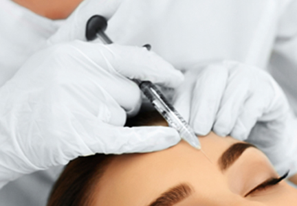
Topical shows promise for periorbital skin rejuvenation
A novel, topical multipotent growth factor was found to be safe, effective, and well tolerated for periorbital skin rejuvenation, according to results from an open-label pilot study.
The product, manufactured by Suneva Medical and known as Regenica Revitalizing Eye Creme, contains a novel multipotent resignaling complex (MRCx), in which human fibroblasts are harvested from a neonatal cell bank and cultured in bovine-free media. “The cultured cells are then anchored to dextran microbeads and suspended in a culture chamber within which the oxygen tension is decreased from 21%” to less than 5%, researchers led by Dr. Hema Sundaram explained in the December 2015 issue of the Journal of Drugs in Dermatology (2015;14[12]:1410-17). “During this process, the cells revert to an embryonic-like state exhibiting stem cell properties, releasing multipotent growth factors including significant quantities of vascular endothelial growth factor (VEGF), keratinocyte growth factor (KGF), and interleukin-8 (IL-8).”
Dr. Sundaram, a dermatologist who practices in Rockville, Md., and her associates at three other centers in the United States enrolled 39 women who had periorbital lines and wrinkles, uneven skin texture, puffiness, and a lack of skin firmness. The women were instructed to apply the formulation to the periorbital area twice a day for 60 days and the researchers evaluated efficacy and treatment-related adverse events at baseline, day 14, day 30, and day 60. The women completed a questionnaire that assessed their self-perception of treatment-related changes in the skin appearance at every visit. At the same time, investigators used 10-point scales to rate the periorbital areas and the Global Aesthetic Improvement Scale (GAIS) to assess the overall improvement on day 14, 30, and 60.
The mean age of the women was 57 years and 38 completed the study. The researchers reported that at day 60, all of the participants had improvements in infraorbital brightness, moistness, wrinkles, sallowness, crepiness, smooth texture, and skin tone. According to investigator-rated scoring on the GAIS, 68% of the women were “improved” or “much improved” at day 14 and 63% remained improved at day 60.
At day 60, 76% of women said they were “very pleased/pleased” (39.4%) or “mostly pleased” (36.8%) with the appearance of their infraorbital areas at day 60, while 79% indicated they were “very pleased/pleased” (44.8%) or “pleased” (34.2%) with the appearance of their lateral canthal areas. Adverse events were rare and consisted of one case of mild canthal erythema which lasted 2 days and one case of mild eye irritation which lasted 1 day.
The researchers acknowledged that the evidence level of the study findings “would be increased by larger, vehicle-controlled studies with blinded evaluation.”
The study was supported by research grants from Suneva Medical. Dr. Sundaram reported being a clinical investigator and/or consultant for numerous companies, including Suneva. Two of the study authors are Suneva employees and the rest reported having numerous ties to the pharmaceutical industry.
FROM JOURNAL OF DRUGS IN DERMATOLOGY
Key clinical point: A novel, topical multipotent growth factor formulation was found effective for periorbital skin rejuvenation.
Major finding: According to investigator-rated scoring on the Global Aesthetic Improvement Scale, 68% of the women were “improved” or “much improved” at day 14, and 63% remained improved at day 60.
Data source: An open-label study in 38 women who used Regenica Revitalizing Eye Creme, a topical agent that contains a novel multipotent resignaling complex.
Disclosures: The study was supported by research grants from Suneva Medical. Dr. Sundaram reported being a clinical investigator and/or consultant for numerous companies, including Suneva Medical. Two of the study authors are Suneva employees and the rest reported having numerous ties to the pharmaceutical industry.
The ‘easy’ but ‘not-so-easy’ brow lift
One of the most apparent and difficult-to-treat aspects of the aging eye is the descent of the eyebrow. The change in the orbital bone structure as well as fat loss and fat redistribution along the upper and lower eyelids and loss of skin elasticity contribute to the skeletonization of the periorbital area and a “drooping” of the eyebrow. Patients either have loss of volume across the entire brow, or primarily across the lateral and central brow creating what is known as an “a-frame” deformity.

Nonsurgical techniques that help lift the brow include a combination of relaxation of the orbicularis oculi muscle with neurotoxins, in addition to the injection of hyaluronic acid fillers along the brow margin and upper third of the face.
Small amounts of hyaluronic acid fillers injected with a 22- to 25-gauge cannula both above and below the eyebrow along the orbital rim provide an instantaneous lifting effect with long-lasting results. Hyaluronic acid and poly-L-lactic acid in dilute concentrations can also be injected with a cannula in the forehead, which creates a repletion of the volume in the upper face that is often lost with aging to create a lift of the eyebrows. Temple hollows can also be filled with calcium hydroxylapatite, poly-L-lactic acid and less often with hyaluronic acid to revolumize and create a lift of the lateral brow. Care should be taken as fillers used in these areas are off-label and need to be done by trained, expert injectors. The periorbital area is a danger zone with many vessels and nerves, and proper injection technique is crucial to avoid arterial blockage, nerve damage, and long-term complications.

Nonablative skin tightening with radiofrequency energy or ultrasound can be used for achieving a brow-lift. Although these techniques do provide collagen remodeling, multiple procedures are often necessary, and results are not always substantial. In a study of 36 patients undergoing ultrasound tightening of the face and neck, 86% showed a clinically significant brow-lift 90 days after treatment. The average brow elevation in this study was 1.7 mm.
In practice, however, patients are often more satisfied with the brow elevation they achieve with neurotoxins and fillers. Injectables provide a faster onset of results, fewer treatments, and minimal discomfort. Combination treatments provide the best overall results and although injectables in the periorbital area are technically difficult, patients are often very satisfied and return for repeat treatments.
References Aesthet Surg J. 2009;May-Jun;29(3):174-9.
J Am Acad Dermatol. 2010;Feb;62(2):262-9.
Dr. Wesley and Dr. Talakoub are cocontributors to this column. Dr. Talakoub is in private practice in McLean, Va. Dr. Wesley practices dermatology in Beverly Hills, Calif. This month’s column is by Dr. Talakoub.
One of the most apparent and difficult-to-treat aspects of the aging eye is the descent of the eyebrow. The change in the orbital bone structure as well as fat loss and fat redistribution along the upper and lower eyelids and loss of skin elasticity contribute to the skeletonization of the periorbital area and a “drooping” of the eyebrow. Patients either have loss of volume across the entire brow, or primarily across the lateral and central brow creating what is known as an “a-frame” deformity.
Nonsurgical techniques that help lift the brow include a combination of relaxation of the orbicularis oculi muscle with neurotoxins, in addition to the injection of hyaluronic acid fillers along the brow margin and upper third of the face.
Small amounts of hyaluronic acid fillers injected with a 22- to 25-gauge cannula both above and below the eyebrow along the orbital rim provide an instantaneous lifting effect with long-lasting results. Hyaluronic acid and poly-L-lactic acid in dilute concentrations can also be injected with a cannula in the forehead, which creates a repletion of the volume in the upper face that is often lost with aging to create a lift of the eyebrows. Temple hollows can also be filled with calcium hydroxylapatite, poly-L-lactic acid and less often with hyaluronic acid to revolumize and create a lift of the lateral brow. Care should be taken as fillers used in these areas are off-label and need to be done by trained, expert injectors. The periorbital area is a danger zone with many vessels and nerves, and proper injection technique is crucial to avoid arterial blockage, nerve damage, and long-term complications.
Nonablative skin tightening with radiofrequency energy or ultrasound can be used for achieving a brow-lift. Although these techniques do provide collagen remodeling, multiple procedures are often necessary, and results are not always substantial. In a study of 36 patients undergoing ultrasound tightening of the face and neck, 86% showed a clinically significant brow-lift 90 days after treatment. The average brow elevation in this study was 1.7 mm.
In practice, however, patients are often more satisfied with the brow elevation they achieve with neurotoxins and fillers. Injectables provide a faster onset of results, fewer treatments, and minimal discomfort. Combination treatments provide the best overall results and although injectables in the periorbital area are technically difficult, patients are often very satisfied and return for repeat treatments.
References Aesthet Surg J. 2009;May-Jun;29(3):174-9.
J Am Acad Dermatol. 2010;Feb;62(2):262-9.
Dr. Wesley and Dr. Talakoub are cocontributors to this column. Dr. Talakoub is in private practice in McLean, Va. Dr. Wesley practices dermatology in Beverly Hills, Calif. This month’s column is by Dr. Talakoub.

Keep canthopexy, canthoplasty in your sights for periorbital aging
CHICAGO – Canthopexy and canthoplasty are simple, minimally invasive procedures that are often overlooked when treating periorbital aging, Dr. Cameron D. Chesnut said at the annual meeting of the American Society for Dermatologic Surgery.

“In a simple procedure you can do in the office in literally 5 or 10 minutes under local anesthesia, you can restore the integrity of the lower lid, which in addition gives some improvement in the upper lids by lifting the globe back up,” he said.
Canthopexy and canthoplasty are very commonly performed with blepharoplasty, but can be useful on their own or in combination with fillers in mild cases of retinacular aging or lower-lid ectropion where you don’t need or want a true, full-blown blepharoplasty.
There is a labyrinth of connective tissues attached to Whitnall’s tubercle and as patients age, there is laxity of the septum, retinaculum, and orbicularis; rounding of the lateral canthal angle; and a negative intercanthal tilt that develops as the lateral canthus drops in relation to the medial canthus, said Dr. Chesnut, who is in private practice in Spokane, Wash.

In the canthopexy, the lateral canthal tendon is folded over and stabilized with a stitch, but not dispensed with. “Yes, this is surgical, but this is literally a stab incision with an 11 blade to access this,” he noted.
The key to a successful canthopexy is to release the tarsal strap, which is relatively resistant to aging laxity and tethers the lateral canthus inferiorly. “While everything else around it becomes lax, it tends to stay pretty strong and pulls down the whole lateral canthal complex,” he observed.
The canthoplasty creates more tension on the lower lid, but is more invasive. The lateral canthal tendon is cut, releasing the retinaculum. The eyelid opening is then shortened and the tendon reconstructed and reattached to the bone.

Particular attention should be paid in both procedures to globe position, particularly in patients with a negative vector in which the globe protrudes beyond their malar eminence and lower eyelid, Dr. Chesnut pointed out.
“You need to be careful with these patients because if you tighten the lower lid too much, the lower lid margin will take the path of least resistance and descend down the globe instead of up,” he said. “These are the patients that may need volume or face lifting to increase that malar support.” Complications can include overtightening, but this is nearly impossible to do with a canthopexy and usually relaxes with a canthoplasty, Dr. Chesnut said in an interview. “Avoiding it all boils down to proper preoperative planning of the amount of tightening needed.”
Likewise, proper planning can avoid undercorrection.
“This can occasionally be from failure of the reconstructed or plicated lateral canthal tendon, but this is a very robust structure, and when properly fixated to the periosteum of Whitnall’s tubercle, failure is unlikely,” Dr. Chesnut explained. “The tendon will continue to age normally, so a canthopexy patient may require or want more tightening with a canthoplasty years down the road.”
Lower-lid laxity should be assessed in all patients prior to either procedure and can be determined by performing the distraction test, in which the eyelid is grasped and pulled anteriorly. If the eyelid can be distracted by more than 6 mm to 8 mm, then laxity is present. The snapback test, where the lower lid is pulled toward the inferior orbital rim and then released, can also be used to identify poor lid tone.
Dr. Chesnut reported having no conflicts of interest.
CHICAGO – Canthopexy and canthoplasty are simple, minimally invasive procedures that are often overlooked when treating periorbital aging, Dr. Cameron D. Chesnut said at the annual meeting of the American Society for Dermatologic Surgery.
“In a simple procedure you can do in the office in literally 5 or 10 minutes under local anesthesia, you can restore the integrity of the lower lid, which in addition gives some improvement in the upper lids by lifting the globe back up,” he said.
Canthopexy and canthoplasty are very commonly performed with blepharoplasty, but can be useful on their own or in combination with fillers in mild cases of retinacular aging or lower-lid ectropion where you don’t need or want a true, full-blown blepharoplasty.
There is a labyrinth of connective tissues attached to Whitnall’s tubercle and as patients age, there is laxity of the septum, retinaculum, and orbicularis; rounding of the lateral canthal angle; and a negative intercanthal tilt that develops as the lateral canthus drops in relation to the medial canthus, said Dr. Chesnut, who is in private practice in Spokane, Wash.
In the canthopexy, the lateral canthal tendon is folded over and stabilized with a stitch, but not dispensed with. “Yes, this is surgical, but this is literally a stab incision with an 11 blade to access this,” he noted.
The key to a successful canthopexy is to release the tarsal strap, which is relatively resistant to aging laxity and tethers the lateral canthus inferiorly. “While everything else around it becomes lax, it tends to stay pretty strong and pulls down the whole lateral canthal complex,” he observed.
The canthoplasty creates more tension on the lower lid, but is more invasive. The lateral canthal tendon is cut, releasing the retinaculum. The eyelid opening is then shortened and the tendon reconstructed and reattached to the bone.
Particular attention should be paid in both procedures to globe position, particularly in patients with a negative vector in which the globe protrudes beyond their malar eminence and lower eyelid, Dr. Chesnut pointed out.
“You need to be careful with these patients because if you tighten the lower lid too much, the lower lid margin will take the path of least resistance and descend down the globe instead of up,” he said. “These are the patients that may need volume or face lifting to increase that malar support.” Complications can include overtightening, but this is nearly impossible to do with a canthopexy and usually relaxes with a canthoplasty, Dr. Chesnut said in an interview. “Avoiding it all boils down to proper preoperative planning of the amount of tightening needed.”
Likewise, proper planning can avoid undercorrection.
“This can occasionally be from failure of the reconstructed or plicated lateral canthal tendon, but this is a very robust structure, and when properly fixated to the periosteum of Whitnall’s tubercle, failure is unlikely,” Dr. Chesnut explained. “The tendon will continue to age normally, so a canthopexy patient may require or want more tightening with a canthoplasty years down the road.”
Lower-lid laxity should be assessed in all patients prior to either procedure and can be determined by performing the distraction test, in which the eyelid is grasped and pulled anteriorly. If the eyelid can be distracted by more than 6 mm to 8 mm, then laxity is present. The snapback test, where the lower lid is pulled toward the inferior orbital rim and then released, can also be used to identify poor lid tone.
Dr. Chesnut reported having no conflicts of interest.
EXPERT ANALYSIS FROM THE ASDS ANNUAL MEETING
