User login
Look Beyond Obvious When Treating Veins
On the heels of the Food and Drug Administration's recent approval of polidocanol injections for the treatment of spider and reticular veins, Dr. Margaret W. Mann is working to educate her colleagues on how to cure the more serious implications of venous disease.
An estimated 55% of American women and 45% of American men suffer from some form of vein problem. Varicose veins affect one out of two people aged 50 and older, according to the Department of Health and Human Services.
"So it is very much not a cosmetic issue," Dr. Mann said at a cosmetic dermatology seminar sponsored by Skin Disease Education Foundation in Santa Monica, Calif. "And it is important to treat these patients before they develop serious sequelae to their disease."
Varicose veins are often inherited. If both parents have varicose veins, a person has an 89% risk of developing the condition; if one parent is affected, the risk is 47%; and if neither parent has them, the risk decreases to 20% (J. Dermatol. Surg. Oncol. 1994;20:318-26).
"When I approach a patient with telangiectasia and varicose veins, I know that it can be just the tip of the iceberg," said Dr. Mann, codirector of the dermatologic surgery and laser center at the University of California, Irvine. Early treatment is optimal to prevent worsening of the disease.
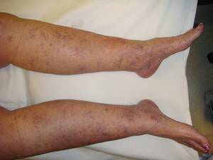
The path to curing venous disease lies in looking beyond the visible and treating the root of problem. Patients with severe spider veins along the ankle, prominent reticular veins greater than 5 mm, or palpable varicosities likely have underlying venous insufficiency, said Dr. Mann. These patients should undergo ultrasound examination to delineate their venous anatomy prior to treatment.
"In patients with outflow obstruction, varicosities must not be ablated because they are an important bypass pathway allowing blood to flow around the obstruction," explained Dr. Mann. "Specific diagnostic tests can distinguish between patients who will benefit from ablation of dilated superficial veins and those who will be harmed by the same procedure."
In patients diagnosed with great saphenous vein insufficiency, Dr. Mann recommends minimally invasive treatment with endovenous ablation and microphlebectomy. It is essential, according to Dr. Mann, to treat their underlying disease prior to treatment of their spider veins with the mind set that "everything is connected in a hierarchal way" and "therapy must start from a top down approach."
Dr. Mann disclosed she is a paid consultant with BioForm Medical (acquired by Merz Pharmaceuticas). SDEF and this news organization are owned by Elsevier.
On the heels of the Food and Drug Administration's recent approval of polidocanol injections for the treatment of spider and reticular veins, Dr. Margaret W. Mann is working to educate her colleagues on how to cure the more serious implications of venous disease.
An estimated 55% of American women and 45% of American men suffer from some form of vein problem. Varicose veins affect one out of two people aged 50 and older, according to the Department of Health and Human Services.
"So it is very much not a cosmetic issue," Dr. Mann said at a cosmetic dermatology seminar sponsored by Skin Disease Education Foundation in Santa Monica, Calif. "And it is important to treat these patients before they develop serious sequelae to their disease."
Varicose veins are often inherited. If both parents have varicose veins, a person has an 89% risk of developing the condition; if one parent is affected, the risk is 47%; and if neither parent has them, the risk decreases to 20% (J. Dermatol. Surg. Oncol. 1994;20:318-26).
"When I approach a patient with telangiectasia and varicose veins, I know that it can be just the tip of the iceberg," said Dr. Mann, codirector of the dermatologic surgery and laser center at the University of California, Irvine. Early treatment is optimal to prevent worsening of the disease.
The path to curing venous disease lies in looking beyond the visible and treating the root of problem. Patients with severe spider veins along the ankle, prominent reticular veins greater than 5 mm, or palpable varicosities likely have underlying venous insufficiency, said Dr. Mann. These patients should undergo ultrasound examination to delineate their venous anatomy prior to treatment.
"In patients with outflow obstruction, varicosities must not be ablated because they are an important bypass pathway allowing blood to flow around the obstruction," explained Dr. Mann. "Specific diagnostic tests can distinguish between patients who will benefit from ablation of dilated superficial veins and those who will be harmed by the same procedure."
In patients diagnosed with great saphenous vein insufficiency, Dr. Mann recommends minimally invasive treatment with endovenous ablation and microphlebectomy. It is essential, according to Dr. Mann, to treat their underlying disease prior to treatment of their spider veins with the mind set that "everything is connected in a hierarchal way" and "therapy must start from a top down approach."
Dr. Mann disclosed she is a paid consultant with BioForm Medical (acquired by Merz Pharmaceuticas). SDEF and this news organization are owned by Elsevier.
Stategies for Optimizing Laser Safety Outlined
Visible and infrared high-power lasers can cause permanent skin and organ damage if used inappropriately.
Patient protection measures, government and institutional guidelines, and familiarity with the physics behind laser technology are means by which dermatologists can optimize safety, Dr. E. Victor Ross said at a cosmetic dermatology seminar sponsored by Skin Disease Education Foundation in Santa Monica, Calif.

Mild to severe reddening, blisters, charring, depigmentation, ulceration, and scarring are among possible adverse effects when these devices are not employed safely, he said.
Even when just the skin is being treated, protective measures are warranted for the eyes, said Dr. Ross, director of the Scripps Clinic Laser and Cosmetic Dermatology Center in Carmel Valley, Calif.
Visible light and some infrared-wavelength light is focused as it enters the eye, making the eyes even more susceptible to laser damage. There have been reports of severe and sometimes permanent eye injuries with lasers. One patient, for example, reported seeing a white flash and hearing a click during treatment with an Nd:YAG 1064-nm laser. The result was a dark spot in the patient's visual field resulted, he said.
Protective eyewear is of paramount importance, Dr. Ross said. Select eyewear based on the laser and the anatomic region being treated. Look for eyewear marked with the wavelengths it protects against and the degree of light energy attenuation. Caution is advised when using goggles on pediatric patients because the fit may not be ideal.
In a very practical sense, Dr. Ross also advised that staff keep cups of water or coffee off the lasers. A spill or leak can get into the device power supply and cause an "explosion." In addition, never open the part of the laser where the high voltage energy is located, said Dr. Ross.
He recommended following the American National Standards Institute's (ANSI) series of laser safety standards. The recommendations are recognized by many hospitals as the standard for guiding a laser safety program, he said.
Examples of ANSI standards include physicians acting as the safety officer, routine laser maintenance, and having a set of standard operating procedures that is kept on file. Recommendations are not just for large practices, Dr. Ross said. "Even a small clinic should have a laser safety officer."
Government safety standards for laser use are available from the Occupational Safety and Health Administration and the Food and Drug Administration's Center for Devices and Radiological Health. Having a sign posted near each device warning of potential hazards specific to that class of laser is an example of a government safety standard, he said.
There are four general safety categories for lasers:
Class 1. Low-power device that is considered safe.
Class 2. The eye blink response generally provides enough eye protection during use.
Class 3. Direct exposure of the eye to these medium-power devices has potential to cause harm.
Class 4. These high-power devices require the most caution to avoid eye and skin hazards through direct and reflected exposure. These lasers also can pose a fire hazard if nearby materials are accidentally ignited from direct or scattered beams.
Credentialing is another safety measure. All clinicians operating a laser at the Scripps Clinic, for example, must be credentialed for that specific device and wavelength. Requirements include some didactic training, supervision during three or more laser treatments, and hands-on instruction by the device manufacturer, Dr. Ross said.
As with most government standards, there is official terminology regarding laser safety. Maximal permissible exposure (MPE) is an official term, describing the amount of irradiance to which a patient can be exposed without causing eye or skin damage. It is important to note that patient discomfort is possible within the MPE limitations. The MPE is calculated using wavelength and duration of laser exposure.
Dermatologists can optimize safety with knowledge of the basic physics behind the energy delivered in laser light and how it provides benefits to the skin when delivered properly, he suggested.
Power delivered per unit area is called the irradiance. This is typically expressed as the power in watts per square centimeter. Many dermatologists and device manufacturers compare lasers and outcomes in terms of joules, which is the energy delivered per square centimeter.
On the patient end, science determines the depth to which laser light penetrates and is absorbed. This absorption coefficient varies among hemoglobin, melanin, and water, for example. Each component interacts to a different extent with laser energy, and dermatologists utilize this selective photothermolysis to optimize patient outcomes from laser treatments, he said.
Dr. Ross disclosed that he is a researcher for and receives funding from multiple laser companies, including Candela, Cutera, Lumenis, Sciton, and Syneron.
SDEF and this news organization are owned by Elsevier.
Visible and infrared high-power lasers can cause permanent skin and organ damage if used inappropriately.
Patient protection measures, government and institutional guidelines, and familiarity with the physics behind laser technology are means by which dermatologists can optimize safety, Dr. E. Victor Ross said at a cosmetic dermatology seminar sponsored by Skin Disease Education Foundation in Santa Monica, Calif.
Mild to severe reddening, blisters, charring, depigmentation, ulceration, and scarring are among possible adverse effects when these devices are not employed safely, he said.
Even when just the skin is being treated, protective measures are warranted for the eyes, said Dr. Ross, director of the Scripps Clinic Laser and Cosmetic Dermatology Center in Carmel Valley, Calif.
Visible light and some infrared-wavelength light is focused as it enters the eye, making the eyes even more susceptible to laser damage. There have been reports of severe and sometimes permanent eye injuries with lasers. One patient, for example, reported seeing a white flash and hearing a click during treatment with an Nd:YAG 1064-nm laser. The result was a dark spot in the patient's visual field resulted, he said.
Protective eyewear is of paramount importance, Dr. Ross said. Select eyewear based on the laser and the anatomic region being treated. Look for eyewear marked with the wavelengths it protects against and the degree of light energy attenuation. Caution is advised when using goggles on pediatric patients because the fit may not be ideal.
In a very practical sense, Dr. Ross also advised that staff keep cups of water or coffee off the lasers. A spill or leak can get into the device power supply and cause an "explosion." In addition, never open the part of the laser where the high voltage energy is located, said Dr. Ross.
He recommended following the American National Standards Institute's (ANSI) series of laser safety standards. The recommendations are recognized by many hospitals as the standard for guiding a laser safety program, he said.
Examples of ANSI standards include physicians acting as the safety officer, routine laser maintenance, and having a set of standard operating procedures that is kept on file. Recommendations are not just for large practices, Dr. Ross said. "Even a small clinic should have a laser safety officer."
Government safety standards for laser use are available from the Occupational Safety and Health Administration and the Food and Drug Administration's Center for Devices and Radiological Health. Having a sign posted near each device warning of potential hazards specific to that class of laser is an example of a government safety standard, he said.
There are four general safety categories for lasers:
Class 1. Low-power device that is considered safe.
Class 2. The eye blink response generally provides enough eye protection during use.
Class 3. Direct exposure of the eye to these medium-power devices has potential to cause harm.
Class 4. These high-power devices require the most caution to avoid eye and skin hazards through direct and reflected exposure. These lasers also can pose a fire hazard if nearby materials are accidentally ignited from direct or scattered beams.
Credentialing is another safety measure. All clinicians operating a laser at the Scripps Clinic, for example, must be credentialed for that specific device and wavelength. Requirements include some didactic training, supervision during three or more laser treatments, and hands-on instruction by the device manufacturer, Dr. Ross said.
As with most government standards, there is official terminology regarding laser safety. Maximal permissible exposure (MPE) is an official term, describing the amount of irradiance to which a patient can be exposed without causing eye or skin damage. It is important to note that patient discomfort is possible within the MPE limitations. The MPE is calculated using wavelength and duration of laser exposure.
Dermatologists can optimize safety with knowledge of the basic physics behind the energy delivered in laser light and how it provides benefits to the skin when delivered properly, he suggested.
Power delivered per unit area is called the irradiance. This is typically expressed as the power in watts per square centimeter. Many dermatologists and device manufacturers compare lasers and outcomes in terms of joules, which is the energy delivered per square centimeter.
On the patient end, science determines the depth to which laser light penetrates and is absorbed. This absorption coefficient varies among hemoglobin, melanin, and water, for example. Each component interacts to a different extent with laser energy, and dermatologists utilize this selective photothermolysis to optimize patient outcomes from laser treatments, he said.
Dr. Ross disclosed that he is a researcher for and receives funding from multiple laser companies, including Candela, Cutera, Lumenis, Sciton, and Syneron.
SDEF and this news organization are owned by Elsevier.
Dermoscopy for Pigmented Lesions
Recent studies have highlighted the rising incidence of melanoma in non-white skin. In populations with darker complexions, melanomas may appear on areas of the skin not exposed to the sun, often of the acral lentiginous type.
The stage at diagnosis is also more advanced in non-white skin. Despite increased surveillance efforts, diagnosis of melanoma in darker skin is often difficult and delayed, which affects overall prognosis and likelihood of survival. When stratified by stage of diagnosis, melanoma in non-white skin has the same prognosis as melanoma in white skin.
Pigmented skin lesions are rarely noticed and are difficult to detect by clinical examination on dark skin because of reduced visibility of melanocytic lesions.
Using a dermatoscope to examine dark skin for melanomas could help improve the chance of correct diagnosis and earlier treatment if done properly. The use of dermoscopy is increasing in favor among dermatologists to improve diagnostic accuracy.
Few studies have reviewed the use of dermoscopy in darker skinned populations. In one study investigators evaluated the utility and efficacy of dermoscopy for pigmented lesions in black populations. The authors attempted to evaluate whether darker pigmentation influences dermatoscopic features in comparison with white populations (Brit. J. Dermatol. 2006:4;695-99).
In the study, 100 clinically doubtful or equivocal pigmented skin lesions in black patients were subjected to dermatoscopic examination. The lesions were observed using dermoscopy by two groups of dermatologists, one in Brazil (in vivo) and the other blinded evaluators in Italy (on slide images), both recording dermatoscopic features. The results showed that out of 100 cases, 79 were Clark nevi, 15 seborrheic keratoses, 4 blue nevi, 1 dermatofibroma, and 1 melanoma.
The agreement between observers was statistically evaluated and there was a high level of inter-observer agreement among dermatoscopic features. Only 3 out of the 100 suspicious lesions (Clark’s nevi) required surgical excision to confirm the diagnosis.
The melanomas presented dermatoscopic characteristics similar to the melanomas appearing in white populations with a blue-whitish veil, irregular globules and streaks at the periphery.
The increased incidence of melanoma, and decreased survival of ethnic patients due to a delay in diagnosis, is a sign of caution to dermatologists to increase surveillance of pigmented lesions in dark skin. Accurate classification of pigmented lesions is difficult; however, this study showed that dermoscopy can be a useful and even necessary tool in the identification of pigmented lesions in dark skin, as naked-eye examinations may be more difficult.
More accurate diagnoses can lead to, not only earlier treatment of clinically suspicious lesions, but also to a decrease in unnecessary biopsies in skin of color, which is more prone to hypertrophic and keloidal scarring.
ACMS: High Risk for Secondary Malignancies after Melanoma
NEW YORK - The risk of developing a primary malignancy of the salivary gland, bone, prostate, and other areas is significantly--and in some cases dramatically--increased following a cutaneous melanoma diagnosis.
That's according to research by Dr. Joshua Spanogle, a resident in the department of dermatology at the Mayo Clinic in Rochester, Minn., presented during an abstract session at the American College of Mohs Surgery annual meeting.
Dr. Spanogle looked at data from the Surveillance, Epidemiology and End Results database (SEER) from 1973 to 2003, which included "over 1.3 million person years of observation," he said.
A total of 151,996 patients were found to have a cutaneous melanoma diagnosis during that period; 16,591 (11%) of these patients had a second documented primary malignancy sometime in the next 120 months. That observed rate was 32% higher than what would be expected among a healthy population in that time period, he said.
Further analysis of the results revealed that particular cancers carried a significantly higher risk than others.
The most striking, perhaps not surprisingly, is for a second primary cutaneous melanoma: there were 3,923 through 120 months of follow-up, for a standardized incidence ratio of 8.99. "This is the 600-pound elephant in the room," said Dr. Spanogle.
But other cancers have high standardized incidence ratios (SIRs) as well. Salivary gland malignancies following carcinoma had a SIR of 2.18 overall. Prostate cancer had a SIR of 1.13 following melanoma. Breast cancer showed a SIR of 1.07. And soft tissue cancers, including malignancies of the heart, had a SIR of 2.80.
On the other hand, "Quite a few cancers had a decreased incidence following melanoma," said Dr. Spanogle, including cancer of the liver (SIR 0.77), lungs (0.83), cervix (0.57), and pharynx (0.61).
That could be because risk factors for melanoma are associated with higher socioeconomic status, like fair skin and intermittent high intensity UV exposure (tanning), said Dr. Spanogle. In contrast, risk factors for lung cancer and liver cancer are associated with comorbidities commonly found in lower socioeconomic patients, like smoking and hepatitis.
Moreover, according to Dr. Spanogle, the risks of secondary cancers of the prostate gland, bone, soft tissue, and salivary gland remained elevated throughout the study period, "implying no surveillance bias."
Instead, he speculated that the link could be genetic, and said that future research into the possibility is warranted.
Dr. Spanogle reported having no disclosures relevant to his presentation. He added that this study has been accepted for publication in the Journal of the American Academy of Dermatology.
Cryolipolysis Appears Safe for Fat Reduction
PHOENIX - Using noninvasive cryolipolysis for fat-layer reduction in the flanks and back fat pads caused no serious adverse events and no reports of skin damage or pigment changes up to 6 months after treatment, results from a large multicenter study showed.
Side effects included "mostly erythema and edema, numbness/tingling, and a little blanching," Dr. A. Jay Burns said at the annual meeting of the American Society for Laser Medicine and Surgery. "Occasionally there was bruising on the treatment site as well, but all side effects resolved by 6 months."
Invented by Dr. Dieter Manstein and Dr. R. Rox Anderson at Massachusetts General Hospital and Harvard Medical School in Boston, cryolipolysis is noninvasive way to cool fat cells to induce lipolysis without damage to other tissue. Pleasanton, Calif.-based Zeltiq currently owns exclusive rights to the technology and markets a cryolipolysis device known as the Zeltiq system.
The Zeltiq system is cleared in the European Union and Canada for noninvasive fat-layer reduction through cold-assisted lipolysis. It is also cleared by the Food and Drug Administration for various applications related to skin cooling during dermatologic treatments, with a pending application for noninvasive fat-layer reduction. The system is available for sale on a limited basis to physicians in the European Union, Canada, and the United States and other selected international markets.
At the meeting, Dr. Burns reported side effect data from two prospective, controlled clinical studies in which researchers at 16 centers used the Zeltiq system to treat 341 patients on their flanks and back fat pads. Efficacy was evaluated by ultrasound, comparison of pre- and post-treatment photographs, and physician assessment. Final follow-up occurred at 6 months or less.
"There were no reports of serious adverse effects, and all side effects were transient," said Dr. Burns, a plastic surgeon in Dallas who is also with the University of Texas Southwestern Medical Center. The most common post-treatment side effect was erythema, followed by hypoesthesia/tingling, edema, bruising, temperature sensation, blanching, and pressure at the treatment site.
ASLMS: Tips for Effective Laser Hair Removal in Darker Skin
PHOENIX - When performing laser hair removal in patients with Fitzpatrick skin types IV-VI, keep in mind that darker skin contains more melanin in the epidermis, which acts as a competing chromophore, "so the risk for epidermal injury is higher," Dr. Andrew F. Alexis said.
Other tips to remember when treating this patient population include the melanocytes' tendency "to be labile in response to injury and inflammation, so there is an increased risk of dyschromia," Dr. Alexis said at the annual meeting of the American Society for Laser Medicine and Surgery. "This can manifest as hyperpigmentation or hypopigmentation following laser hair removal procedures."
He went on to note that fibroblasts in the dermis "are also more reactive to injury, so there is an increased risk of hypertrophic scars and keloids when performing surgical or some cosmetic procedures. In addition, having curved follicles [which are primarily seen in people of African descent] is associated with an increased prevalence of a number of follicular disorders."
His guidelines for performing laser hair removal safely on skin of color include longer wavelengths, lower fluences, longer pulse durations, and increased epidermal cooling.
"You want longer wavelengths because we're trying to maximize the ratio of follicular bulb temperature to epidermal temperature," explained Dr. Alexis, director of the Skin of Color Center at St. Luke's & Roosevelt Hospitals, New York, and assistant professor of dermatology at Columbia University.
"Using wavelengths in the near infrared range, we are at a lower point on the melanin absorption curve and therefore are compromising some efficacy, but this is the range that is considered safest for darker skin types," he noted.
A review found that the long-pulsed 1064-nm laser had the lowest incidence of adverse events in dark-skinned patients, followed by the long-pulsed 800-nm or 810-nm diode laser (J. Drugs Dermatol. 2007;6:40-6).
"For patients with type IV or V skin, the diode laser is appropriate, as long as you are using long pulse durations," he said, "but for the darker skin types, particularly type VI, the 1064-nm Nd:YAG laser is the best choice for safety reasons."
In one study, researchers used a 1064-nm laser with contact cooling to treat pseudofolliculitis barbae in 37 patients with skin types IV, V, and VI. They found that the highest fluences tolerated by the epidermis were 50 J/cm2 for skin type VI and 100 J/cm2 for skin types IV and V (J. Am. Acad. Dermatol. 2002;47:263-70).
In another study, researchers used a 1064-nm laser with contact cooling for hair removal in 36 patients with skin types I-VI (Dermatol. Surg. 2004;30:13-7). Patients underwent three consecutive treatments at 4- to 6-week intervals. For skin types V-VI, investigators used a 30-millisecond pulse duration and a fluence of 30-45 J/cm2 on the face and 35-50 J/cm2 on nonfacial sites. Six months post treatment, the mean facial hair reduction ranged from 41% to 46%, while the mean hair reduction on the body ranged from 48% to 53%.
Based on the results of these and other studies, and from his own clinical experience, Dr. Alexis recommends a pulse duration of 100 milliseconds or 400 milliseconds when using a 810-nm diode laser and a duration of 20-30 milliseconds when using a 1064-nm Nd:YAG laser with contact cooling.
One recent study used a 1064-nm Nd:YAG laser with lower than traditional fluences for treating 22 patients with skin types IV-VI who had pseudofolliculitis barbae (Dermatol. Surg. 2009;35:98-107). Patients underwent five weekly treatments on the neck with a fluence of 12 J/cm2 and a pulse duration of 20 milliseconds and a spot size of 10 mm. At 4 weeks follow-up, the papule count had been reduced by a mean of 91% and dyspigmentation by a mean of 60%.
In another recent development, researchers studying a novel diode laser with a fluence of 5-10 J/cm2 used at a repetition rate of 10 Hz found that it resulted in less pain, faster treatment, and fewer adverse events compared with one pass of a high-fluence diode laser at 25-40 J/cm2 in Fitzpatrick skin types I-V (J. Drugs Dermatol. 2009;8:s14-7).
Options for epidermal cooling include cold gel, contact cryogen or forced air, and post treatment with ice packs for 10-15 minutes. Devices for contact cooling feature either a sapphire tip or chilled copper plate. Regardless of which type of contact cooling chosen, Dr. Alexis suggested "using a slower treatment speed in order to ensure adequate cooling before delivering pulse."
Dr. Alexis said that he had no relevant financial conflicts.
Fractional CO2 Laser Skin Resurfacing for the Treatment of Sun-Damaged Skin and Actinic Keratoses
Facial Rejuvenation
Maximizing Patient Satisfaction With Facial Soft Tissue Fillers: A Question of Balance
Amino Acid Analysis of Damaged Hair from Chemical "Relaxers"
As a follow-up to our blog about the relationship between ceramic flat iron use and trichorrhexis nodosa, we wanted to focus on the use of chemical relaxers and their association with hair damage.
Chemical relaxers have maintained their popularity over the years to make curly hair straight, especially in the African-American community. A relaxer is a chemical compound applied to curly hair to permanently break hydrogen disulfide bonds along the hair shaft to release the tight curl pattern. Typically, it is re-applied at varying intervals as new growth of unrelaxed hair occurs (Semin. Cutan. Med. Surg. 2009;28:103-8).
Relaxers have evolved from containing unusual ingredients such as lard, boiled eggs, and sodium hydroxide or lye (which is also found in many household cleaning products), to no-lye relaxers which contain guanidine hydroxide. These chemicals—whether made with or without lye—can result in hair breakage, particularly at points along the hair shaft where a new relaxer is applied to hair that has already been chemically treated.
In a recently published study, investigators performed a biochemical analysis on the hair of 30 women of African decent from Cape Town, South Africa. Of the women, 10 had natural hair (never used chemical treatments), 10 had asymptomatic relaxed hair, and 10 had symptomatic (brittle, breaking, or damaged) relaxed hair (J. Am. Acad. Dermatol. 2010; 62:402-8).
Cysteine levels were expected to be lower in chemically relaxed hair because it is estimated that 35% of cysteine changes to lanthionine during hydrolysis of the disulfide bonds (Clin. Dermatol. 1988;6:71-82).
The study found that cysteine levels were similar in proximal and distal natural hair. Levels were reduced in all relaxed hair, but were lowest in distal relaxed hair. Cysteine levels in distal and symptomatic relaxed hair were consistent with fragile, damaged hair such as that found in trichothiodystrophy.
In addition, arginine and citrulline levels were decreased, and glutamine levels were increased, in all relaxed hair when compared with natural hair. The authors noted that a decrease in arginine and citrulline has been associated with inflammation elsewhere, particularly in critically ill children, thus, relaxer use may also be associated with scalp inflammation.
However, Dr. Zoe Draelos wrote in an accompanying editorial that while cysteine levels were expected to be decreased, and this study validly confirms that, the association between decreased arginine and citrulline levels and scalp inflammation has not been as well elucidated.
Dr. Draelos, a dermatologist in High Point, N.C., also noted that prior amino acid analyses have failed to demonstrate that constituents of the body are accurately translated into the hair shaft composition in normal health. Further research is needed to elucidate whether or not relaxer use is also associated with amino acid deficiencies, which may be related to scalp inflammation, she concluded (J. Am. Acad. Dermatol. 2010;62:409-10).