User login
Pearls on Fillers and Combination Cosmetic Therapy
Soft Tissue Augmentation: A Review
Injectable Soft Tissue Fillers [editorial]
Topical Management of Postsurgical Scars
Subacute Cutaneous Lupus Erythematosus: A Case Report of Polypodium leucotomos as an Adjuvant Therapy
Skin of Color: Five Steps to Treating Earlobe Keloids
Earlobe keloids are a common problem in skin of color patients that can be frustrating to both patients and physicians.
An article in the March issue of Dermatologic Surgery (2012;38:406-12) described a new morphologic classification for earlobe keloids and a surgical approach for their management.
Although surgical management is important for resistant, recurrent keloids, I often use a five-step nonsurgical regimen for treating earlobe keloids.
Set expectations and counsel the patient. Keloids can recur.
Avoid triggers. Make sure there is no underlying contact dermatitis from nickel or other metal alloys that can cause inflammation contributing to the scar tissue formation.
Intralesional steroids. Consider starting with Kenalog 20 mg/cc and increase by 10 mg/cc at each 4 week visit for optimal benefit. Often times, I use very gentle subscision in the tough scar tissue with a 26 gauge ½ inch needle to open up channels for the Kenalog to penetrate.
Compression therapy. Clip on compression earrings or a medical compression device called a Zimmer splint, which has two discs that clip on the ear placing pressure on both sides of the lobe.
Use silicone based gels and instruct patients to massage the area several times daily.
Keloids can be a frustrating and aesthetically unacceptable problem. Surgical methods are useful, including keloidectomy, core extirpation, or a combination.
However, in-office and home-based remedies both decrease recurrence rates and improve the size and thickness of keloids so that surgery can be reserved as an adjunct treatment by removing redundant skin rather than thick keloidal scar tissue.
- Lily Talakoub, M.D.
Do you have questions about treating patients with darker skin? If so, send them to sknews@elsevier.com.
Skin of Color: Keloid Scarring Rates Lower Than Expected
A presentation at the Triological Society's 2012 Combined Sections Meeting held in January highlighted that, when compared with white patients, black patients have a sevenfold increased risk for developing keloid scarring following surgery in the head and neck area.
Dr. Lamont Jones and colleagues at Henry Ford Hospital, Detroit, conducted a retrospective chart review of 6,692 patients who had head and neck procedures requiring incisions between 2005 and 2009. Twenty patients developed keloid scarring as a result of their surgery. Keloid rates according to ethnicity were 0.8% in black patients, 0.1% in white patients, and 0.2% in other ethnic groups.
They explain, however, that the keloid rate in black patients is actually significantly lower than previous estimates, which have been as high as 16%.
There were no differences in keloid formation according to gender. Patients who developed keloid scars tended to be younger in age, but the age difference was not statistically significant.
While the decreased rates of keloid formation compared to previous estimates can be reassuring for many black patients who fear surgery, the increased risk over other ethnic groups is still real and should be discussed in preoperative discussions.
- Naissan Wesley, M.D.
Do you have questions about treating patients with darker skin? If so, send them to sknews@elsevier.com.
Clinical Tips Can Tailor Your Cannula Technique
MIAMI BEACH – As more U.S. dermatologists are using cannulas to deliver soft tissue fillers for rejuvenation, tips to refine the technique and optimize patient outcomes are emerging.
Cannula use "seems to be gaining a lot of momentum in this country, and some of that really stems from the European experience," Dr. Joel L. Cohen said in an interview.
The size of the cannula, as well as the gauge of the needle that creates the entry point, can determine outcomes in different anatomic areas, Dr. Cohen said at the South Beach Symposium. "Cannulas have very specific roles in very specific areas. Personally, I have found them useful in the cheek, the infraorbital area, in the décolleté, and the dorsal hands."
He also shared a practical tip that eases cannula insertion. To deliver filler product using a 27 G cannula, for example, Dr. Cohen first nicks the skin with a slightly larger 25 G needle where he wants his entry point. Then, he pinches and holds the skin around the needle while his assistant removes it. That way, he knows the precise point to insert the cannula "rather than [later] fumbling around in a little bit of bleeding to find it. It’s really a helpful trick we’ve incorporated into our office."
A 25 G cannula can also be used to deliver fillers to the décolleté or dorsal side of the hands, said Dr. Cohen, who is in private practice in Englewood, Colo. In contrast, a smaller 30 G cannula may be more appropriate for rejuvenation of the infraorbital area, he said. "Using a 30 G cannula below the eyes, I feel that I am seeing less bruising and less swelling in this area, but at the same time, I am able to precisely deliver the filler product deep into the muscle along the periosteum."
An ability to fan product out in multiple directions from one entry point is an advantage of the cannula. "It’s a useful technique, and cannulas have helped me get better results with less bruising in my practice," Dr. Cohen said. The lower bruising potential seems to come from the cannula’s blunt tip, which can tuck under, above, or around a blood vessel. In contrast, a sharp needle is more likely to puncture the same vessel and cause postprocedure bruising, he said. And, fanning a needle around under the skin can increase the risk of bruising even further (Derm. Surg. 2008;34:S105-9).
"Some physicians, including me, actually have found patients experience less pain" with a cannula than with a needle, he added.
Although dermatologic use of cannulas has grown over the past year, "I don’t think it’s going to completely take over. You definitely still need needles for many different areas. I continue top use needles in the nasolabial folds [and] the oral commissure. For fine lines and wrinkles and more shallow rhytids, you definitely still need needles," he said
More information on cannulas can be found in an article published by Dr. Cohen and his colleague Dr. Joshua A. Zeichner (J. Drugs. Dermatol. 2012;11:70-2).
Dr. Cohen reported that he had no relevant disclosures.
EXPERT ANALYSIS FROM THE SOUTH BEACH SYMPOSIUM
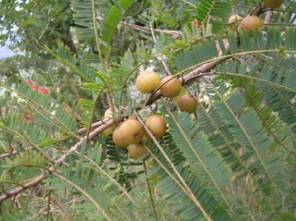
Emblica
Emblica officinalis (also known as Phyllanthus emblica, Indian gooseberry, or by its Hindi name amla) is a deciduous tree, the fruit of which is a rich dietary source of vitamin C as well as various minerals, amino acids, and phenolic compounds. Native to India, and found also in China, Indonesia, and the Malay Peninsula, amla has long been used in Ayurvedic medicine, and is highly regarded for its unique array of tannins (particularly emblicanin A and emblicanin B) and flavonoids, which exhibit antioxidant activity (Asian Pac. J. Cancer Prev. 2005;6:197-201).
In fact, the amla fruit is known to impart strong antioxidant activity and to protect human dermal fibroblasts against oxidative stress. Therefore, it is considered a useful adjuvant in natural skin care (J. Ethnopharmacol. 2008;119[1]:53-7). This column will focus on the recent research pertaining to modern medical usage and the potential dermatologic applications of this plant traditionally used in Indian medicine.
Antioxidant Properties
In 2002, a standardized extract of E. officinalis (trade name, Emblica) was found to exert a long-lasting and broad-spectrum antioxidant activity, appropriate for use in antiaging, sunscreen, and general purpose skin formulations given its demonstrated capacity to protect the skin from damage engendered by free radicals, nonradicals, and transition metal-induced oxidative stress (Skin Pharmacol. Appl. Skin Physiol. 2002;15:374-80). In addition, Emblica appears to be an ideal combination of the characteristics necessary for an effective skin-lightening ingredient. Specifically, it inhibits tyrosinase and/or tyrosinase-related proteins (TRP-1 and 2) and peroxidase/H2O2 at various points in the melanogenesis pathway (Skin Pharmacol. Appl. Skin Physiol. 2002;15:374-80), and acts as a broad-spectrum cascading antioxidant. (Antioxidants comprise one group of suitable hypopigmenting agents.) Emblica, which is photochemically and hydrolytically stable, therefore suitable for inclusion in skin care formulations, is thought to be comparably efficacious to hydroquinone and kojic acid as a skin-lightening compound, but without posing the same risks or likelihood of eliciting adverse effects.
Also in 2002, Morganti et al. assessed the in vitro and in vivo activity of various topical antioxidants and nutritional supplements in a randomized double-blind placebo-controlled study conducted over 8 weeks on 30 female volunteers (aged 48-59 years), with moderate dry skin and photodamage. All subjects twice daily applied a nanocolloidal gel and/or daily took two capsules of an oral diet supplement. The test formulations were antioxidant-enriched, containing ascorbic acid, tocopherol, alpha-lipoic acid, melatonin, and emblica. Oral and topical administration of the antioxidant compounds resulted in significant reduction in oxidative stress (ROS) and lipid peroxidation. Free radicals recovered in blood serum and on skin, in vivo, and ROS induced by in vitro irradiation of leucocytes with UVB, were also decreased in antioxidant-treated patients. The investigators concluded that all of the compounds, including emblica, showed therapeutic potential, in topical or systemic form, as photoprotectants and agents intended to lower the oxidative stress of photoaging-affected individuals (Int. J. Cosmet. Sci. 2002;24:331-9).
Oxidative Stress Reduction Properties
Yokozawa et al. studied the effects of amla on the lipid metabolism and protein expression in age-related oxidative stress. They administered a dose of SunAmla or ethyl acetate extract of amla, a polyphenol-rich fraction, to young rats aged 2 months and aged rats (10 months) and found that the lipid levels were significantly higher in aged control rats, but significantly reduced as a result of amla administration. Several additional metrics in the study led the team to conclude that amla, by diminishing oxidative stress, may prevent age-related hyperlipidemia (Br. J. Nutr. 2007;97:1187-95). Recently, Sharma et al. investigated in adult Swiss albino mice the protective role of the fruits of E. officinalis against arsenic-induced hepatopathy. Significant increases in serum transaminases and lipid peroxidation content in the liver were noted in the arsenic-treated group, along with significant decreases in hepatic superoxide dismutase, catalase, glutathione-S-transferase and serum alkaline phosphatase activity. Pre- and post-treatment with amla reversed these effects. In addition, liver histopathology revealed that amla diminished karyolysis, karyorrhexis, necrosis, and cytoplasmic vacuolization caused by the arsenic. The authors concluded that pre- and postsupplementation with E. officinalis fruit extract significantly reduces oxidative stress in the liver generated by arsenic (Chem. Biol. Interact. 2009;180:20-30).
Cutaneous Properties
Recent investigations with emblica also have dermatologic implications or suggestions of potential cutaneous benefits. In 2005, Sancheti et al. performed a two-stage skin carcinogenesis study in Swiss albino mice to ascertain the chemopreventive action of E. officinalis fruit extract. A single application of 7, 12-dimethyabenz(a)anthracene (DMBA) was used to induce skin cancer, followed 2 weeks later by thrice weekly applications of croton oil (through the end of the 16-week experiment) to promote tumors. A statistically significant difference in tumor incidence, yield, and burden as well as cumulative number of papillomas favored the mice treated with E. officinalis as opposed to the control group. The investigators concluded that E. officinalis fruit extract exhibited chemopreventive potential against DMBA-induced skin cancer in Swiss albino mice (Asian Pac. J. Cancer Prev. 2005:197-201).
Antimicrobial Properties
Based on evidence that Triphala (dried fruit of Terminalia chebula, Terminalia bellirica, and Phyllanthus emblica) has demonstrated in vitro antimicrobial activity against wound pathogens, including Staphylococcus aureus, Pseudomonas aeruginosa, and Streptococcus pyogenes, Kumar et al. recently prepared a Triphala ointment and evaluated its effects for in vivo wound healing on infected rats. The investigators identified significantly improved wound closure and bacterial count decreases in the treated group, along with significant hexosamine, uronic acid, and superoxide dismutase levels and diminished matrix metalloproteinase expression. They concluded that Triphala ointment exhibited antioxidant, antibacterial, and wound healing activities suitable for treating infected wounds and that the active constituents of the ointment warrant further consideration for wound therapy (J. Surg. Res. 2008;144:94-101).
In 2008, Fuji et al. studied the effects of amla extract on human skin fibroblasts, with a focus on in vitro production of procollagen and matrix metalloproteinases (MMPs). They employed the WST-8 assay to assess human skin fibroblast mitochondrial activity and an immunoassay to measure procollagen, MMPs, and Tissue inhibitor of metalloproteinase-1 (TIMP-1) released from the fibroblasts. The researchers found that amla extract controls collagen metabolism in therapeutic and cosmetic applications. Specifically, they found that in a concentration-dependent fashion, amla promoted fibroblast proliferation and in a concentration- and time-dependent manner stimulated procollagen synthesis. They also noted that amla significantly lowered MMP-1 production, with significant increases in TIMP-1, but had no effect on MMP-2. The authors concluded that their findings suggest the potential of amla to confer mollifying and therapeutic as well as cosmetic benefits (J. Ethnopharmacol. 2008;119:53-7).
Conclusion
The fruit, and other most other parts, of Emblica officinalis have long been used in Indian traditional medicine, particularly for antiaging purposes. Recent evidence suggests broad potential for the extract of the amla fruit in modern Western medical treatment and, in particular, as an ingredient in general purpose dermatologic topical formulations as well as antiaging, hypopigmenting, and sunscreen agents. Much more research is necessary to establish the effectiveness of such products, but current findings are encouraging.
Skin of Color: Brush Up at the AAD
It's always a good idea to expand your knowledge and expertise in regards to patients with skin of color. Here is a synopsis of the skin of color-themed presentations taking place at the annual American Academy of Dermatology Meeting in San Diego from March 16-20.
On Saturday, March 17, there will be a focus session on Medical & Aesthetic Dermatology in Skin of Color (U041) starting from 7:15 a.m. to 8:45 a.m., followed by a focus session entitled Practical Approach to Medical and Cosmetic Dermatology in Skin of Color Patients (U051) from 12:15 p.m. to 1:45 p.m. There will be a symposium on Disorders of Pigmentation (S019) from 2:00 p.m. to 5 p.m.
On Sunday, March 18, a focus session from 7 a.m. to 8 a.m. will cover Building Beauty: Understanding Facial Proportions, Phi, and Use of Volumizing Soft Tissue Fillers (U073).
Finally, on Monday, March 19, an early focus session from 7:15 a.m. to 8:45 a.m. will cover Hair Disease & the African American Patient (U108), and a second focus session from 2:30 p.m. to 4 p.m. will cover Melasma: Pathogenesis and Treatment (U129). There will also be a forum starting at 12 p.m. to 2 p.m. on Vitiligo (F063).
The online version of the scientific program contains all the information you'll need in regards to the meeting. Hope to see you there!
- Lily Talakoub, M.D.
Do you have questions about treating patients with darker skin? If so, send them to sknews@elsevier.com.