User login
Biologic may reduce glucocorticoid use
WASHINGTON – The biologic benralizumab cut glucocorticoid dosage by nearly 75% among patients with severe, uncontrolled asthma, compared with a 25% reduction in dosage among patients using a placebo, according to a study.
In this three-armed, double-blind study of 220 patients, those administered benralizumab every 4 and 8 weeks were 4.09 (95% confidence interval, 2.22-7.57) and 4.12 (95% CI, 2.22-7.57) times as likely to see a reduction in glucocorticoid dose, compared with those in the placebo group, according to investigators (NEJM. 2017 May 21. doi: 10.1056/NEJMoa1703501).The study was presented at an international conference of the American Thoracic Society and published simultaneously in the New England Journal of Medicine.
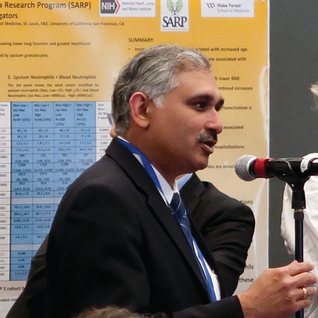
“Frequent or long-term use of systemic corticosteroids can lead to potentially life-threatening complications, including osteoporosis, diabetes, cardiovascular disease, and adrenal suppression,” Parameswaran Nair, MD, PhD, professor at McMaster University, Hamilton, Ont., said in a press release. “We need new, safe therapies that would replace the need for systemic corticosteroids for patients with severe asthma.”
To test benralizumab’s effectiveness, investigators measured a baseline level of glucocorticoid dosage of 220 patients with severe, uncontrolled asthma. Patients were then given one of three treatment options: one dose of benralizumab every 4 weeks, one dose of benralizumab every 8 weeks, or a placebo. All three treatments were decreased each time until minimal dosage was found while still maintaining asthma control. The average age of patients was around 50 years, with the majority of patients in both treatment groups and the placebo group having been female.
The researchers also analyzed patients’ accounts of any worsening asthma symptoms, which were recorded in an electronic asthma daily diary.
Along with the median 75% decrease in glucocorticoid dosage seen in both groups of patients receiving benralizumab, 24 patients (33%) in the 4-week group and 27 patients in the 8-week group (37%) showed a 90% reduction from their baseline glucocorticoid dosage. In contrast, only nine patients (12%) in the placebo group experienced a 90% drop in glucocorticoid use.
The researchers observed an additional finding suggesting benralizumab’s usefulness in a subgroup of patients with a baseline prednisone dose of less than 12.5 mg. These patients were more likely to stop taking their glucocorticoid dose if they were taking benralizumab instead of the placebo. Specifically, patients who took benralizumab every 4 weeks were 5.23 times more likely and those who took the biologic every 8 weeks were 4.19 times more likely to cease using glucocorticoids.
Similar to the current biologics used to treat severe eosinophilic asthma, mepolizumab and reslizumab, benralizumab is a form of a monoclonal antibody. Instead of targeting interleukin-5, benralizumab works against a subunit of the iterleukin-5 receptor. They investigators said this aspect of benralizumab may explain why it was successful in this study.
“Targeting of the alpha-subunit of the iterleukin-5 receptor with benralizumab has potential advantages over existing anti–interleukin-5 therapies,” according to Dr. Nier and fellow investigators. “By targeting the interleukin receptor rather than the cytokine, luminal depletion of eosinophils can occur, which may be related to greater clinical efficiency.”
The investigators noted that FEV1 levels seemed relatively unaffected by benralizumab.
This study was limited by the length of the trials, which lasted 28 weeks. Investigators also note that 20% of the original patients were not used in the final population.
This study was sponsored by, and organized in partnership with, AstraZeneca. All of the investigators reported having a financial or other type of relationship with AstraZeneca.
ezimmerman@fronlinemedcom.com
On Twitter @eaztweets
WASHINGTON – The biologic benralizumab cut glucocorticoid dosage by nearly 75% among patients with severe, uncontrolled asthma, compared with a 25% reduction in dosage among patients using a placebo, according to a study.
In this three-armed, double-blind study of 220 patients, those administered benralizumab every 4 and 8 weeks were 4.09 (95% confidence interval, 2.22-7.57) and 4.12 (95% CI, 2.22-7.57) times as likely to see a reduction in glucocorticoid dose, compared with those in the placebo group, according to investigators (NEJM. 2017 May 21. doi: 10.1056/NEJMoa1703501).The study was presented at an international conference of the American Thoracic Society and published simultaneously in the New England Journal of Medicine.
“Frequent or long-term use of systemic corticosteroids can lead to potentially life-threatening complications, including osteoporosis, diabetes, cardiovascular disease, and adrenal suppression,” Parameswaran Nair, MD, PhD, professor at McMaster University, Hamilton, Ont., said in a press release. “We need new, safe therapies that would replace the need for systemic corticosteroids for patients with severe asthma.”
To test benralizumab’s effectiveness, investigators measured a baseline level of glucocorticoid dosage of 220 patients with severe, uncontrolled asthma. Patients were then given one of three treatment options: one dose of benralizumab every 4 weeks, one dose of benralizumab every 8 weeks, or a placebo. All three treatments were decreased each time until minimal dosage was found while still maintaining asthma control. The average age of patients was around 50 years, with the majority of patients in both treatment groups and the placebo group having been female.
The researchers also analyzed patients’ accounts of any worsening asthma symptoms, which were recorded in an electronic asthma daily diary.
Along with the median 75% decrease in glucocorticoid dosage seen in both groups of patients receiving benralizumab, 24 patients (33%) in the 4-week group and 27 patients in the 8-week group (37%) showed a 90% reduction from their baseline glucocorticoid dosage. In contrast, only nine patients (12%) in the placebo group experienced a 90% drop in glucocorticoid use.
The researchers observed an additional finding suggesting benralizumab’s usefulness in a subgroup of patients with a baseline prednisone dose of less than 12.5 mg. These patients were more likely to stop taking their glucocorticoid dose if they were taking benralizumab instead of the placebo. Specifically, patients who took benralizumab every 4 weeks were 5.23 times more likely and those who took the biologic every 8 weeks were 4.19 times more likely to cease using glucocorticoids.
Similar to the current biologics used to treat severe eosinophilic asthma, mepolizumab and reslizumab, benralizumab is a form of a monoclonal antibody. Instead of targeting interleukin-5, benralizumab works against a subunit of the iterleukin-5 receptor. They investigators said this aspect of benralizumab may explain why it was successful in this study.
“Targeting of the alpha-subunit of the iterleukin-5 receptor with benralizumab has potential advantages over existing anti–interleukin-5 therapies,” according to Dr. Nier and fellow investigators. “By targeting the interleukin receptor rather than the cytokine, luminal depletion of eosinophils can occur, which may be related to greater clinical efficiency.”
The investigators noted that FEV1 levels seemed relatively unaffected by benralizumab.
This study was limited by the length of the trials, which lasted 28 weeks. Investigators also note that 20% of the original patients were not used in the final population.
This study was sponsored by, and organized in partnership with, AstraZeneca. All of the investigators reported having a financial or other type of relationship with AstraZeneca.
ezimmerman@fronlinemedcom.com
On Twitter @eaztweets
WASHINGTON – The biologic benralizumab cut glucocorticoid dosage by nearly 75% among patients with severe, uncontrolled asthma, compared with a 25% reduction in dosage among patients using a placebo, according to a study.
In this three-armed, double-blind study of 220 patients, those administered benralizumab every 4 and 8 weeks were 4.09 (95% confidence interval, 2.22-7.57) and 4.12 (95% CI, 2.22-7.57) times as likely to see a reduction in glucocorticoid dose, compared with those in the placebo group, according to investigators (NEJM. 2017 May 21. doi: 10.1056/NEJMoa1703501).The study was presented at an international conference of the American Thoracic Society and published simultaneously in the New England Journal of Medicine.
“Frequent or long-term use of systemic corticosteroids can lead to potentially life-threatening complications, including osteoporosis, diabetes, cardiovascular disease, and adrenal suppression,” Parameswaran Nair, MD, PhD, professor at McMaster University, Hamilton, Ont., said in a press release. “We need new, safe therapies that would replace the need for systemic corticosteroids for patients with severe asthma.”
To test benralizumab’s effectiveness, investigators measured a baseline level of glucocorticoid dosage of 220 patients with severe, uncontrolled asthma. Patients were then given one of three treatment options: one dose of benralizumab every 4 weeks, one dose of benralizumab every 8 weeks, or a placebo. All three treatments were decreased each time until minimal dosage was found while still maintaining asthma control. The average age of patients was around 50 years, with the majority of patients in both treatment groups and the placebo group having been female.
The researchers also analyzed patients’ accounts of any worsening asthma symptoms, which were recorded in an electronic asthma daily diary.
Along with the median 75% decrease in glucocorticoid dosage seen in both groups of patients receiving benralizumab, 24 patients (33%) in the 4-week group and 27 patients in the 8-week group (37%) showed a 90% reduction from their baseline glucocorticoid dosage. In contrast, only nine patients (12%) in the placebo group experienced a 90% drop in glucocorticoid use.
The researchers observed an additional finding suggesting benralizumab’s usefulness in a subgroup of patients with a baseline prednisone dose of less than 12.5 mg. These patients were more likely to stop taking their glucocorticoid dose if they were taking benralizumab instead of the placebo. Specifically, patients who took benralizumab every 4 weeks were 5.23 times more likely and those who took the biologic every 8 weeks were 4.19 times more likely to cease using glucocorticoids.
Similar to the current biologics used to treat severe eosinophilic asthma, mepolizumab and reslizumab, benralizumab is a form of a monoclonal antibody. Instead of targeting interleukin-5, benralizumab works against a subunit of the iterleukin-5 receptor. They investigators said this aspect of benralizumab may explain why it was successful in this study.
“Targeting of the alpha-subunit of the iterleukin-5 receptor with benralizumab has potential advantages over existing anti–interleukin-5 therapies,” according to Dr. Nier and fellow investigators. “By targeting the interleukin receptor rather than the cytokine, luminal depletion of eosinophils can occur, which may be related to greater clinical efficiency.”
The investigators noted that FEV1 levels seemed relatively unaffected by benralizumab.
This study was limited by the length of the trials, which lasted 28 weeks. Investigators also note that 20% of the original patients were not used in the final population.
This study was sponsored by, and organized in partnership with, AstraZeneca. All of the investigators reported having a financial or other type of relationship with AstraZeneca.
ezimmerman@fronlinemedcom.com
On Twitter @eaztweets
FROM ATS 2017
Key clinical point:
Major finding: Patients administered benralizumab averaged a 75% reduction rate in final glucocorticoid dosage, compared with 25% in the control group (P less than .001).
Data source: Randomized, double blind, placebo-controlled study of 220 patients with severe asthma given benralizumab every 4 weeks or every 8 weeks or a placebo between April 2014 and November 2015.
Disclosures: The study was sponsored by and designed in collaboration with AstraZeneca. All of the investigators reported having a financial or other type of relationship with AstraZeneca.
California Opens Treatment Center for Native Youth
The IHS announced the opening of another youth regional treatment center (YRTC) to provide culturally centered, evidence-based substance use disorder services to Native youth.
Desert Sage Youth Wellness Center in Hemet is the first federally owned and operated health care facility in California to serve American Indians and Alaska Natives (AI/AN). Although California has the largest population of AI/AN in the country, IHS says, teens were usually sent out of state to non-IHS or nontribal facilities that do not always meet their “unique cultural needs.”
Desert Sage is 1 of 11 YRTCs funded by IHS across the country. The YRTCs provide comprehensive, holistic care, including mental health assessments, individualized treatment plans, academic education, vocational and life-skills training, and activities to meet the spiritual and cultural needs of Native American youth.
The Desert Sage center, which includes 3 buildings with 32 beds and 5 family suites, will employ 70 full-time employees and treat about 100 tribal youth annually.
Sacred Oaks Healing Center in Davis, the next YRTC planned for California, is expected to be completed in 2019.
The IHS announced the opening of another youth regional treatment center (YRTC) to provide culturally centered, evidence-based substance use disorder services to Native youth.
Desert Sage Youth Wellness Center in Hemet is the first federally owned and operated health care facility in California to serve American Indians and Alaska Natives (AI/AN). Although California has the largest population of AI/AN in the country, IHS says, teens were usually sent out of state to non-IHS or nontribal facilities that do not always meet their “unique cultural needs.”
Desert Sage is 1 of 11 YRTCs funded by IHS across the country. The YRTCs provide comprehensive, holistic care, including mental health assessments, individualized treatment plans, academic education, vocational and life-skills training, and activities to meet the spiritual and cultural needs of Native American youth.
The Desert Sage center, which includes 3 buildings with 32 beds and 5 family suites, will employ 70 full-time employees and treat about 100 tribal youth annually.
Sacred Oaks Healing Center in Davis, the next YRTC planned for California, is expected to be completed in 2019.
The IHS announced the opening of another youth regional treatment center (YRTC) to provide culturally centered, evidence-based substance use disorder services to Native youth.
Desert Sage Youth Wellness Center in Hemet is the first federally owned and operated health care facility in California to serve American Indians and Alaska Natives (AI/AN). Although California has the largest population of AI/AN in the country, IHS says, teens were usually sent out of state to non-IHS or nontribal facilities that do not always meet their “unique cultural needs.”
Desert Sage is 1 of 11 YRTCs funded by IHS across the country. The YRTCs provide comprehensive, holistic care, including mental health assessments, individualized treatment plans, academic education, vocational and life-skills training, and activities to meet the spiritual and cultural needs of Native American youth.
The Desert Sage center, which includes 3 buildings with 32 beds and 5 family suites, will employ 70 full-time employees and treat about 100 tribal youth annually.
Sacred Oaks Healing Center in Davis, the next YRTC planned for California, is expected to be completed in 2019.
PCV10 vaccination in Brazil nearly eliminated serotypes
The effect of 10-valent pneumococcal conjugate vaccine (PCV10) after 4 years of routine childhood vaccination in Brazil was near elimination of PCV10 serotypes in a study of over 500 children, said Felipe P.G. Neves, PhD, of the University of California, Berkeley, and his associates.
The emergence of multidrug-resistant (MDR) serotype 6C isolates, however, is of concern, they said.
Of the 284 children who attended a public clinic and 238 children who went to two private clinics in the greater Rio de Janeiro metropolitan area between Sept. 29 and Dec. 5, 2014, 118 (23%) were pneumococcal carriers. Their median age was 2 years and all were under age 6 years. All 118 isolates were susceptible to levofloxacin, rifampicin, and vancomycin; 26 (22%) isolates were MDR, and 14 of the 18 serotype 6C isolates were MDR.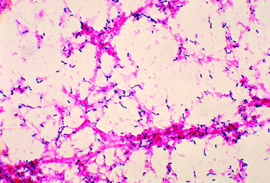
“Serotype 6C has already been reported as having caused meningitis in the pre-PCV10 era in northeastern Brazil. Additionally, following PCV7 implementation, carriage with serotype 6C emerged worldwide and reflected in an increase in IPD [invasive pneumococcal disease] cases,” Dr. Neves and his associates said. “Considering the apparent consistency of MDR 6C as the most common serotype associated with colonization in Brazil after PCV10 universal use, ongoing surveillance to monitor its increase in invasive diseases in Brazil is warranted.”
Read more in Vaccine (2017 May 15;35[21]:2794-800).
The effect of 10-valent pneumococcal conjugate vaccine (PCV10) after 4 years of routine childhood vaccination in Brazil was near elimination of PCV10 serotypes in a study of over 500 children, said Felipe P.G. Neves, PhD, of the University of California, Berkeley, and his associates.
The emergence of multidrug-resistant (MDR) serotype 6C isolates, however, is of concern, they said.
Of the 284 children who attended a public clinic and 238 children who went to two private clinics in the greater Rio de Janeiro metropolitan area between Sept. 29 and Dec. 5, 2014, 118 (23%) were pneumococcal carriers. Their median age was 2 years and all were under age 6 years. All 118 isolates were susceptible to levofloxacin, rifampicin, and vancomycin; 26 (22%) isolates were MDR, and 14 of the 18 serotype 6C isolates were MDR.
“Serotype 6C has already been reported as having caused meningitis in the pre-PCV10 era in northeastern Brazil. Additionally, following PCV7 implementation, carriage with serotype 6C emerged worldwide and reflected in an increase in IPD [invasive pneumococcal disease] cases,” Dr. Neves and his associates said. “Considering the apparent consistency of MDR 6C as the most common serotype associated with colonization in Brazil after PCV10 universal use, ongoing surveillance to monitor its increase in invasive diseases in Brazil is warranted.”
Read more in Vaccine (2017 May 15;35[21]:2794-800).
The effect of 10-valent pneumococcal conjugate vaccine (PCV10) after 4 years of routine childhood vaccination in Brazil was near elimination of PCV10 serotypes in a study of over 500 children, said Felipe P.G. Neves, PhD, of the University of California, Berkeley, and his associates.
The emergence of multidrug-resistant (MDR) serotype 6C isolates, however, is of concern, they said.
Of the 284 children who attended a public clinic and 238 children who went to two private clinics in the greater Rio de Janeiro metropolitan area between Sept. 29 and Dec. 5, 2014, 118 (23%) were pneumococcal carriers. Their median age was 2 years and all were under age 6 years. All 118 isolates were susceptible to levofloxacin, rifampicin, and vancomycin; 26 (22%) isolates were MDR, and 14 of the 18 serotype 6C isolates were MDR.
“Serotype 6C has already been reported as having caused meningitis in the pre-PCV10 era in northeastern Brazil. Additionally, following PCV7 implementation, carriage with serotype 6C emerged worldwide and reflected in an increase in IPD [invasive pneumococcal disease] cases,” Dr. Neves and his associates said. “Considering the apparent consistency of MDR 6C as the most common serotype associated with colonization in Brazil after PCV10 universal use, ongoing surveillance to monitor its increase in invasive diseases in Brazil is warranted.”
Read more in Vaccine (2017 May 15;35[21]:2794-800).
FROM VACCINE
PNSP rates did not increase after PCV13 introduction
Streptococcus pneumoniae did not grow more resistant to penicillin after the introduction of 13-valent pneumococcal conjugate vaccine, though geographic differences remained, according to Cheryl P. Andam, Ph.D., and her associates.
Data from the Active Bacterial Core surveillance system on 285 patients before introduction of PCV13 and from 339 patients after PCV13 introduction were used in the study. Patients were from California, Colorado, Connecticut, Georgia, Maryland, Minnesota, New Mexico, New York, Oregon, and Tennessee. New Mexico, Maryland, and Georgia saw the largest increases in penicillin-nonsusceptible pneumococcus (PNSP) rates, while Colorado, New York, and Connecticut saw decreases. No change was seen in the nationwide PNSP rate.
Preintroduction of PCV13, geographic heterogeneity in serotype distribution, and serotype-specific differences in penicillin resistance were equally responsible for geographic variation in PNSP rates. Although no significant change was seen after introduction, influence of serotype-specific differences did decrease slightly while geographic heterogeneity of PSNP serotypes increased.
“Further long-term nationwide surveillance of serotype dynamics is required to assess the multiple ecologic factors that influence antibiotic resistance in the pneumococcus in the conjugate vaccine era,” the investigators concluded.
Find the full research letter in Emerging Infectious Diseases (doi: 10.3201/eid2306.161331).
Streptococcus pneumoniae did not grow more resistant to penicillin after the introduction of 13-valent pneumococcal conjugate vaccine, though geographic differences remained, according to Cheryl P. Andam, Ph.D., and her associates.
Data from the Active Bacterial Core surveillance system on 285 patients before introduction of PCV13 and from 339 patients after PCV13 introduction were used in the study. Patients were from California, Colorado, Connecticut, Georgia, Maryland, Minnesota, New Mexico, New York, Oregon, and Tennessee. New Mexico, Maryland, and Georgia saw the largest increases in penicillin-nonsusceptible pneumococcus (PNSP) rates, while Colorado, New York, and Connecticut saw decreases. No change was seen in the nationwide PNSP rate.
Preintroduction of PCV13, geographic heterogeneity in serotype distribution, and serotype-specific differences in penicillin resistance were equally responsible for geographic variation in PNSP rates. Although no significant change was seen after introduction, influence of serotype-specific differences did decrease slightly while geographic heterogeneity of PSNP serotypes increased.
“Further long-term nationwide surveillance of serotype dynamics is required to assess the multiple ecologic factors that influence antibiotic resistance in the pneumococcus in the conjugate vaccine era,” the investigators concluded.
Find the full research letter in Emerging Infectious Diseases (doi: 10.3201/eid2306.161331).
Streptococcus pneumoniae did not grow more resistant to penicillin after the introduction of 13-valent pneumococcal conjugate vaccine, though geographic differences remained, according to Cheryl P. Andam, Ph.D., and her associates.
Data from the Active Bacterial Core surveillance system on 285 patients before introduction of PCV13 and from 339 patients after PCV13 introduction were used in the study. Patients were from California, Colorado, Connecticut, Georgia, Maryland, Minnesota, New Mexico, New York, Oregon, and Tennessee. New Mexico, Maryland, and Georgia saw the largest increases in penicillin-nonsusceptible pneumococcus (PNSP) rates, while Colorado, New York, and Connecticut saw decreases. No change was seen in the nationwide PNSP rate.
Preintroduction of PCV13, geographic heterogeneity in serotype distribution, and serotype-specific differences in penicillin resistance were equally responsible for geographic variation in PNSP rates. Although no significant change was seen after introduction, influence of serotype-specific differences did decrease slightly while geographic heterogeneity of PSNP serotypes increased.
“Further long-term nationwide surveillance of serotype dynamics is required to assess the multiple ecologic factors that influence antibiotic resistance in the pneumococcus in the conjugate vaccine era,” the investigators concluded.
Find the full research letter in Emerging Infectious Diseases (doi: 10.3201/eid2306.161331).
FROM EMERGING INFECTIOUS DISEASES
GOLD guidelines for the management of COPD – 2017 update
Chronic obstructive lung disease (COPD) is the third leading cause of death in the United States1 and a major cause of mortality and morbidity around the world. The Global Initiative for Chronic Obstructive Lung Disease (GOLD) released a new “2017 Report”2 with modified recommendations for the diagnosis, management, and prevention of COPD. The report contains several changes that are relevant to the primary care provider that will be outlined below.
Redefining COPD
GOLD’s definition of COPD was changed in its 2017 Report: “COPD is a common, preventable, and treatable disease that is characterized by persistent respiratory symptoms and airflow limitations that are due to airway and/or alveolar abnormalities usually caused by significant exposure to noxious particles or gases.” The report emphasizes that “COPD may be punctuated by periods of acute worsening of respiratory symptoms, called exacerbations.” Note that the terms “emphysema” and “chronic bronchitis” have been removed in favor of a more comprehensive description of the pathophysiology of COPD. Importantly, the report states that cough and sputum production for at least 3 months in each of 2 consecutive years, previously accepted as diagnostic criteria, are present in only a minority of patients. It is noted that chronic respiratory symptoms may exist without spirometric changes and many patients (usually smokers) have structural evidence of COPD without airflow limitation.

Changes to COPD initial assessment
The primary criterion for diagnosis is unchanged: post-bronchodilator forced expiratory volume in 1 second (FEV1)/forced vital capacity (FVC) less than 0.70. Spirometry remains important to confirm the diagnosis in those with classic symptoms of dyspnea, chronic cough, and/or sputum production with a history of exposure to noxious particles or gases.
The GOLD assessment system previously incorporated spirometry and included an “ABCD” system such that patients in group A are least severe. Spirometry has been progressively deemphasized in favor of symptom-based classification and the 2017 Report, for the first time, dissociates spirometric findings from severity classification.
The new system uses symptom severity and exacerbation risk to classify COPD. Two specific standardized COPD symptom measurement tools, The Modified British Medical Research Council (mMRC) questionnaire and COPD Assessment Test (CAT), are reported by GOLD as the most widely used. Low symptom severity is considered an mMRC less than or equal to 1 or CAT less than or equal to 9, high symptom severity is considered an mMRC greater than or equal to 2 or CAT greater than or equal to 10. Low risk of exacerbation is defined as no more than one exacerbation not resulting in hospital admission in the last 12 months; high risk of exacerbation is defined as at least two exacerbations or any exacerbations resulting in hospital admission in the last 12 months. Symptom severity and exacerbation risk is divided into four quadrants:
• GOLD group A: Low symptom severity, low exacerbation risk.
• GOLD group B: High symptom severity, low exacerbation risk.
• GOLD group C: Low symptom severity, high exacerbation risk.
• GOLD group D: High symptom severity, high exacerbation risk.
Changes to prevention and management of stable COPD
Smoking cessation remains important in the prevention of COPD. The 2017 Report reflects the U.S. Preventive Services Task Force’s guidelines for smoking cessation: Offer nicotine replacement, cessation counseling, and pharmacotherapy (varenicline, bupropion or nortriptyline). There is insufficient evidence to support the use of e-cigarettes. Influenza and pneumococcal vaccinations are recommended. Pulmonary rehabilitation remains important.
The 2017 Report includes an expanded discussion of COPD medications. The role of short-acting bronchodilators (SABD) in COPD remains prominent. Changes include a stronger recommendation to use combination short-acting beta-agonists and short-acting muscarinic antagonists (SABA/SAMA) as these seem to be superior to SABD monotherapy in improving symptoms and FEV1.
There were several changes to the pharmacologic treatment algorithm. For the first time, GOLD proposes escalation strategies. Preference is given to LABA/LAMA (long-acting beta-agonist/long-acting muscarinic antagonists) combinations over LABA/ICS (long-acting beta-agonist/inhaled corticosteroid) combinations as a mainstay of treatment. The rationale for this change is that LABA/LAMAs give greater bronchodilation compared with LABA/ICS, and one study showed a decreased rate of exacerbations compared to LABA/ICS in patients with a history of exacerbations. In addition, patients with COPD who receive ICS appear to have a higher risk of developing pneumonia. GOLD recommendations are:
• Group A: Start with single bronchodilator (short- or long-acting), escalate to alternative class of bronchodilator if necessary.
• Group B: Start with LABA or LAMA, escalate to LABA/LAMA if symptoms persist.
• Group C: Start with LAMA, escalate to LABA/LAMA (preferred) or LABA/ICS if exacerbations continue.
• Group D: Start with LABA/LAMA (preferred) or LAMA monotherapy, escalate to LABA/LAMA/ICS (preferred) or try LABA/ICS before escalating to LAMA/LABA/ICS if symptoms persist or exacerbations continue; roflumilast and/or a macrolide may be considered if further exacerbations occur with LABA/LAMA/ICS.
Bottom line
1. GOLD classification of COPD severity is now based on clinical criteria alone: symptom assessment and risk for exacerbation.
2. SABA/SAMA combination therapy seems to be superior to either SABA or SAMA alone.
3. Patients in group A (milder symptoms, low exacerbation risk) may be initiated on either short- or long-acting bronchodilator therapy.
4. Patients in group B (milder symptoms, increased exacerbation risk) should be initiated on LAMA monotherapy.
5. LABA/LAMA combination therapy seems to be superior to LABA/ICS combination therapy and should be used when long-acting bronchodilator monotherapy fails to control symptoms or reduce exacerbations.
References
1. CDC MMWR 11/23/12
2. Global Strategy for the Diagnosis, Management and Prevention of COPD, Global Initiative for Chronic Obstructive Lung Disease (GOLD) 2017 at http://goldcopd.org (accessed 3/10/2017)
Dr. Skolnik is associate director of the family medicine residency program at Abington (Pa.) Memorial Hospital. Dr. Lent is chief resident in the program.
Chronic obstructive lung disease (COPD) is the third leading cause of death in the United States1 and a major cause of mortality and morbidity around the world. The Global Initiative for Chronic Obstructive Lung Disease (GOLD) released a new “2017 Report”2 with modified recommendations for the diagnosis, management, and prevention of COPD. The report contains several changes that are relevant to the primary care provider that will be outlined below.
Redefining COPD
GOLD’s definition of COPD was changed in its 2017 Report: “COPD is a common, preventable, and treatable disease that is characterized by persistent respiratory symptoms and airflow limitations that are due to airway and/or alveolar abnormalities usually caused by significant exposure to noxious particles or gases.” The report emphasizes that “COPD may be punctuated by periods of acute worsening of respiratory symptoms, called exacerbations.” Note that the terms “emphysema” and “chronic bronchitis” have been removed in favor of a more comprehensive description of the pathophysiology of COPD. Importantly, the report states that cough and sputum production for at least 3 months in each of 2 consecutive years, previously accepted as diagnostic criteria, are present in only a minority of patients. It is noted that chronic respiratory symptoms may exist without spirometric changes and many patients (usually smokers) have structural evidence of COPD without airflow limitation.
Changes to COPD initial assessment
The primary criterion for diagnosis is unchanged: post-bronchodilator forced expiratory volume in 1 second (FEV1)/forced vital capacity (FVC) less than 0.70. Spirometry remains important to confirm the diagnosis in those with classic symptoms of dyspnea, chronic cough, and/or sputum production with a history of exposure to noxious particles or gases.
The GOLD assessment system previously incorporated spirometry and included an “ABCD” system such that patients in group A are least severe. Spirometry has been progressively deemphasized in favor of symptom-based classification and the 2017 Report, for the first time, dissociates spirometric findings from severity classification.
The new system uses symptom severity and exacerbation risk to classify COPD. Two specific standardized COPD symptom measurement tools, The Modified British Medical Research Council (mMRC) questionnaire and COPD Assessment Test (CAT), are reported by GOLD as the most widely used. Low symptom severity is considered an mMRC less than or equal to 1 or CAT less than or equal to 9, high symptom severity is considered an mMRC greater than or equal to 2 or CAT greater than or equal to 10. Low risk of exacerbation is defined as no more than one exacerbation not resulting in hospital admission in the last 12 months; high risk of exacerbation is defined as at least two exacerbations or any exacerbations resulting in hospital admission in the last 12 months. Symptom severity and exacerbation risk is divided into four quadrants:
• GOLD group A: Low symptom severity, low exacerbation risk.
• GOLD group B: High symptom severity, low exacerbation risk.
• GOLD group C: Low symptom severity, high exacerbation risk.
• GOLD group D: High symptom severity, high exacerbation risk.
Changes to prevention and management of stable COPD
Smoking cessation remains important in the prevention of COPD. The 2017 Report reflects the U.S. Preventive Services Task Force’s guidelines for smoking cessation: Offer nicotine replacement, cessation counseling, and pharmacotherapy (varenicline, bupropion or nortriptyline). There is insufficient evidence to support the use of e-cigarettes. Influenza and pneumococcal vaccinations are recommended. Pulmonary rehabilitation remains important.
The 2017 Report includes an expanded discussion of COPD medications. The role of short-acting bronchodilators (SABD) in COPD remains prominent. Changes include a stronger recommendation to use combination short-acting beta-agonists and short-acting muscarinic antagonists (SABA/SAMA) as these seem to be superior to SABD monotherapy in improving symptoms and FEV1.
There were several changes to the pharmacologic treatment algorithm. For the first time, GOLD proposes escalation strategies. Preference is given to LABA/LAMA (long-acting beta-agonist/long-acting muscarinic antagonists) combinations over LABA/ICS (long-acting beta-agonist/inhaled corticosteroid) combinations as a mainstay of treatment. The rationale for this change is that LABA/LAMAs give greater bronchodilation compared with LABA/ICS, and one study showed a decreased rate of exacerbations compared to LABA/ICS in patients with a history of exacerbations. In addition, patients with COPD who receive ICS appear to have a higher risk of developing pneumonia. GOLD recommendations are:
• Group A: Start with single bronchodilator (short- or long-acting), escalate to alternative class of bronchodilator if necessary.
• Group B: Start with LABA or LAMA, escalate to LABA/LAMA if symptoms persist.
• Group C: Start with LAMA, escalate to LABA/LAMA (preferred) or LABA/ICS if exacerbations continue.
• Group D: Start with LABA/LAMA (preferred) or LAMA monotherapy, escalate to LABA/LAMA/ICS (preferred) or try LABA/ICS before escalating to LAMA/LABA/ICS if symptoms persist or exacerbations continue; roflumilast and/or a macrolide may be considered if further exacerbations occur with LABA/LAMA/ICS.
Bottom line
1. GOLD classification of COPD severity is now based on clinical criteria alone: symptom assessment and risk for exacerbation.
2. SABA/SAMA combination therapy seems to be superior to either SABA or SAMA alone.
3. Patients in group A (milder symptoms, low exacerbation risk) may be initiated on either short- or long-acting bronchodilator therapy.
4. Patients in group B (milder symptoms, increased exacerbation risk) should be initiated on LAMA monotherapy.
5. LABA/LAMA combination therapy seems to be superior to LABA/ICS combination therapy and should be used when long-acting bronchodilator monotherapy fails to control symptoms or reduce exacerbations.
References
1. CDC MMWR 11/23/12
2. Global Strategy for the Diagnosis, Management and Prevention of COPD, Global Initiative for Chronic Obstructive Lung Disease (GOLD) 2017 at http://goldcopd.org (accessed 3/10/2017)
Dr. Skolnik is associate director of the family medicine residency program at Abington (Pa.) Memorial Hospital. Dr. Lent is chief resident in the program.
Chronic obstructive lung disease (COPD) is the third leading cause of death in the United States1 and a major cause of mortality and morbidity around the world. The Global Initiative for Chronic Obstructive Lung Disease (GOLD) released a new “2017 Report”2 with modified recommendations for the diagnosis, management, and prevention of COPD. The report contains several changes that are relevant to the primary care provider that will be outlined below.
Redefining COPD
GOLD’s definition of COPD was changed in its 2017 Report: “COPD is a common, preventable, and treatable disease that is characterized by persistent respiratory symptoms and airflow limitations that are due to airway and/or alveolar abnormalities usually caused by significant exposure to noxious particles or gases.” The report emphasizes that “COPD may be punctuated by periods of acute worsening of respiratory symptoms, called exacerbations.” Note that the terms “emphysema” and “chronic bronchitis” have been removed in favor of a more comprehensive description of the pathophysiology of COPD. Importantly, the report states that cough and sputum production for at least 3 months in each of 2 consecutive years, previously accepted as diagnostic criteria, are present in only a minority of patients. It is noted that chronic respiratory symptoms may exist without spirometric changes and many patients (usually smokers) have structural evidence of COPD without airflow limitation.
Changes to COPD initial assessment
The primary criterion for diagnosis is unchanged: post-bronchodilator forced expiratory volume in 1 second (FEV1)/forced vital capacity (FVC) less than 0.70. Spirometry remains important to confirm the diagnosis in those with classic symptoms of dyspnea, chronic cough, and/or sputum production with a history of exposure to noxious particles or gases.
The GOLD assessment system previously incorporated spirometry and included an “ABCD” system such that patients in group A are least severe. Spirometry has been progressively deemphasized in favor of symptom-based classification and the 2017 Report, for the first time, dissociates spirometric findings from severity classification.
The new system uses symptom severity and exacerbation risk to classify COPD. Two specific standardized COPD symptom measurement tools, The Modified British Medical Research Council (mMRC) questionnaire and COPD Assessment Test (CAT), are reported by GOLD as the most widely used. Low symptom severity is considered an mMRC less than or equal to 1 or CAT less than or equal to 9, high symptom severity is considered an mMRC greater than or equal to 2 or CAT greater than or equal to 10. Low risk of exacerbation is defined as no more than one exacerbation not resulting in hospital admission in the last 12 months; high risk of exacerbation is defined as at least two exacerbations or any exacerbations resulting in hospital admission in the last 12 months. Symptom severity and exacerbation risk is divided into four quadrants:
• GOLD group A: Low symptom severity, low exacerbation risk.
• GOLD group B: High symptom severity, low exacerbation risk.
• GOLD group C: Low symptom severity, high exacerbation risk.
• GOLD group D: High symptom severity, high exacerbation risk.
Changes to prevention and management of stable COPD
Smoking cessation remains important in the prevention of COPD. The 2017 Report reflects the U.S. Preventive Services Task Force’s guidelines for smoking cessation: Offer nicotine replacement, cessation counseling, and pharmacotherapy (varenicline, bupropion or nortriptyline). There is insufficient evidence to support the use of e-cigarettes. Influenza and pneumococcal vaccinations are recommended. Pulmonary rehabilitation remains important.
The 2017 Report includes an expanded discussion of COPD medications. The role of short-acting bronchodilators (SABD) in COPD remains prominent. Changes include a stronger recommendation to use combination short-acting beta-agonists and short-acting muscarinic antagonists (SABA/SAMA) as these seem to be superior to SABD monotherapy in improving symptoms and FEV1.
There were several changes to the pharmacologic treatment algorithm. For the first time, GOLD proposes escalation strategies. Preference is given to LABA/LAMA (long-acting beta-agonist/long-acting muscarinic antagonists) combinations over LABA/ICS (long-acting beta-agonist/inhaled corticosteroid) combinations as a mainstay of treatment. The rationale for this change is that LABA/LAMAs give greater bronchodilation compared with LABA/ICS, and one study showed a decreased rate of exacerbations compared to LABA/ICS in patients with a history of exacerbations. In addition, patients with COPD who receive ICS appear to have a higher risk of developing pneumonia. GOLD recommendations are:
• Group A: Start with single bronchodilator (short- or long-acting), escalate to alternative class of bronchodilator if necessary.
• Group B: Start with LABA or LAMA, escalate to LABA/LAMA if symptoms persist.
• Group C: Start with LAMA, escalate to LABA/LAMA (preferred) or LABA/ICS if exacerbations continue.
• Group D: Start with LABA/LAMA (preferred) or LAMA monotherapy, escalate to LABA/LAMA/ICS (preferred) or try LABA/ICS before escalating to LAMA/LABA/ICS if symptoms persist or exacerbations continue; roflumilast and/or a macrolide may be considered if further exacerbations occur with LABA/LAMA/ICS.
Bottom line
1. GOLD classification of COPD severity is now based on clinical criteria alone: symptom assessment and risk for exacerbation.
2. SABA/SAMA combination therapy seems to be superior to either SABA or SAMA alone.
3. Patients in group A (milder symptoms, low exacerbation risk) may be initiated on either short- or long-acting bronchodilator therapy.
4. Patients in group B (milder symptoms, increased exacerbation risk) should be initiated on LAMA monotherapy.
5. LABA/LAMA combination therapy seems to be superior to LABA/ICS combination therapy and should be used when long-acting bronchodilator monotherapy fails to control symptoms or reduce exacerbations.
References
1. CDC MMWR 11/23/12
2. Global Strategy for the Diagnosis, Management and Prevention of COPD, Global Initiative for Chronic Obstructive Lung Disease (GOLD) 2017 at http://goldcopd.org (accessed 3/10/2017)
Dr. Skolnik is associate director of the family medicine residency program at Abington (Pa.) Memorial Hospital. Dr. Lent is chief resident in the program.
Preterm infants at high risk for RSV morbidity without immunoprophylaxis
SAN FRANCISCO – Preterm infants born at 29-35 weeks’ gestation and hospitalized for respiratory syncytial virus (RSV) can experience particularly severe morbidity if they have not received immunoprophylaxis, according to new industry-funded research.
Across two RSV seasons at more than 40 study sites, nearly half of preterm infants admitted for lab-confirmed RSV required ICU admission, and one in five required invasive mechanical ventilation.
“Severity of illness and resource utilization are greatest in infants aged less than 3 months,” said John DeVincenzo, MD, professor in the pediatric infectious diseases division at the University of Tennessee, Memphis. “The results from the two seasons were consistent with one another and with previous and recent studies,” he said at the annual Pediatric Academic Societies meeting.

Previous research has shown that preterm infants born at 35 weeks or less gestation have a higher risk of RSV-related hospitalizations and subsequent morbidity, and that monthly immunoprophylaxis reduced RSV-related hospitalization in high-risk infants, including preterm infants.
Until 2014, the American Academy of Pediatrics recommended respiratory syncytial virus (RSV) immunoprophylaxis for all preterm infants under 32 weeks’ gestation and for infants between 32-35 weeks with additional risk factors, such as chronic lung disease or cyanotic heart disease (Pediatrics. 2003 Dec;112[6]:1442-6).
New recommendations in 2014 restricted immunoprophylaxis to preterm infants younger than 29 weeks’ gestational age unless they had additional risk factors such as chronic lung disease or hemodynamically significant heart disease (Pediatrics. 2014 Aug. doi: 10.1542/peds.2014-1665).
This study compared outcomes among all preterm infants born at 29-35 weeks’ gestation who were hospitalized during RSV season (October-April) for at least 24 hours with laboratory-confirmed RSV and who had not received RSV immunoprophylaxis within the 35 days before symptom onset. The 1,378 infants were younger than age 12 months when they were hospitalized at one of 43 sites during the 2014-2015 RSV season or one of 42 sites in the 2015-2016 season.
Of the 702 preterm infants hospitalized in 2014-2015, 42% were admitted to intensive care, and 20% needed invasive mechanical ventilation. Nearly half (48%) of the 676 infants admitted during the 2015-2016 season went to the ICU, and 19% required mechanical ventilation. One infant died of RSV in each season.
Throughout both seasons, more than three quarters (78%) of all RSV hospitalizations were infants younger than 6 months old. In 2014-2015, infants younger than 6 months accounted for 87% of all RSV admissions to the ICU and 92% of those needing mechanical ventilation. Similarly, young infants accounted for 81% of ICU admissions and 90% of RSV-related mechanical ventilation during the 2015-2016 season. Overall, preterm infants younger than 6 months old without immunoprophylaxis accounted for 84% of RSV-related ICU admissions and 91% of RSV-related mechanical ventilation.
The younger the infants were, the more likely they were to need ICU care and/or mechanical ventilation, the researchers found. Across both seasons, 56% of infants under 3 months old with RSV were admitted to the ICU, compared to 34% of those between 3 and 12 months old. Likewise, 29% of those under 3 months old and 10% of those between 3 and 12 months needed invasive mechanical ventilation.
Nearly half (46%) of all infants hospitalized for RSV had been discharged from their birth hospital within the previous 30 days, and 82% of all hospitalizations occurred within 2 months of birth discharge.
A cost analysis revealed that mean hospital charges for RSV-related hospitalizations of preterm infants ranged from $31,366 for 35-week gestation infants between ages 3-6 months to $122,301 for infants under 3 months old born between 29-32 weeks.
AstraZeneca/MedImmune funded the study. Dr. DeVincenzo and a number of his colleagues have received grants/research support from AstraZeneca/MedImmune, and some of his colleagues are or were AstraZeneca employees.
SAN FRANCISCO – Preterm infants born at 29-35 weeks’ gestation and hospitalized for respiratory syncytial virus (RSV) can experience particularly severe morbidity if they have not received immunoprophylaxis, according to new industry-funded research.
Across two RSV seasons at more than 40 study sites, nearly half of preterm infants admitted for lab-confirmed RSV required ICU admission, and one in five required invasive mechanical ventilation.
“Severity of illness and resource utilization are greatest in infants aged less than 3 months,” said John DeVincenzo, MD, professor in the pediatric infectious diseases division at the University of Tennessee, Memphis. “The results from the two seasons were consistent with one another and with previous and recent studies,” he said at the annual Pediatric Academic Societies meeting.
Previous research has shown that preterm infants born at 35 weeks or less gestation have a higher risk of RSV-related hospitalizations and subsequent morbidity, and that monthly immunoprophylaxis reduced RSV-related hospitalization in high-risk infants, including preterm infants.
Until 2014, the American Academy of Pediatrics recommended respiratory syncytial virus (RSV) immunoprophylaxis for all preterm infants under 32 weeks’ gestation and for infants between 32-35 weeks with additional risk factors, such as chronic lung disease or cyanotic heart disease (Pediatrics. 2003 Dec;112[6]:1442-6).
New recommendations in 2014 restricted immunoprophylaxis to preterm infants younger than 29 weeks’ gestational age unless they had additional risk factors such as chronic lung disease or hemodynamically significant heart disease (Pediatrics. 2014 Aug. doi: 10.1542/peds.2014-1665).
This study compared outcomes among all preterm infants born at 29-35 weeks’ gestation who were hospitalized during RSV season (October-April) for at least 24 hours with laboratory-confirmed RSV and who had not received RSV immunoprophylaxis within the 35 days before symptom onset. The 1,378 infants were younger than age 12 months when they were hospitalized at one of 43 sites during the 2014-2015 RSV season or one of 42 sites in the 2015-2016 season.
Of the 702 preterm infants hospitalized in 2014-2015, 42% were admitted to intensive care, and 20% needed invasive mechanical ventilation. Nearly half (48%) of the 676 infants admitted during the 2015-2016 season went to the ICU, and 19% required mechanical ventilation. One infant died of RSV in each season.
Throughout both seasons, more than three quarters (78%) of all RSV hospitalizations were infants younger than 6 months old. In 2014-2015, infants younger than 6 months accounted for 87% of all RSV admissions to the ICU and 92% of those needing mechanical ventilation. Similarly, young infants accounted for 81% of ICU admissions and 90% of RSV-related mechanical ventilation during the 2015-2016 season. Overall, preterm infants younger than 6 months old without immunoprophylaxis accounted for 84% of RSV-related ICU admissions and 91% of RSV-related mechanical ventilation.
The younger the infants were, the more likely they were to need ICU care and/or mechanical ventilation, the researchers found. Across both seasons, 56% of infants under 3 months old with RSV were admitted to the ICU, compared to 34% of those between 3 and 12 months old. Likewise, 29% of those under 3 months old and 10% of those between 3 and 12 months needed invasive mechanical ventilation.
Nearly half (46%) of all infants hospitalized for RSV had been discharged from their birth hospital within the previous 30 days, and 82% of all hospitalizations occurred within 2 months of birth discharge.
A cost analysis revealed that mean hospital charges for RSV-related hospitalizations of preterm infants ranged from $31,366 for 35-week gestation infants between ages 3-6 months to $122,301 for infants under 3 months old born between 29-32 weeks.
AstraZeneca/MedImmune funded the study. Dr. DeVincenzo and a number of his colleagues have received grants/research support from AstraZeneca/MedImmune, and some of his colleagues are or were AstraZeneca employees.
SAN FRANCISCO – Preterm infants born at 29-35 weeks’ gestation and hospitalized for respiratory syncytial virus (RSV) can experience particularly severe morbidity if they have not received immunoprophylaxis, according to new industry-funded research.
Across two RSV seasons at more than 40 study sites, nearly half of preterm infants admitted for lab-confirmed RSV required ICU admission, and one in five required invasive mechanical ventilation.
“Severity of illness and resource utilization are greatest in infants aged less than 3 months,” said John DeVincenzo, MD, professor in the pediatric infectious diseases division at the University of Tennessee, Memphis. “The results from the two seasons were consistent with one another and with previous and recent studies,” he said at the annual Pediatric Academic Societies meeting.
Previous research has shown that preterm infants born at 35 weeks or less gestation have a higher risk of RSV-related hospitalizations and subsequent morbidity, and that monthly immunoprophylaxis reduced RSV-related hospitalization in high-risk infants, including preterm infants.
Until 2014, the American Academy of Pediatrics recommended respiratory syncytial virus (RSV) immunoprophylaxis for all preterm infants under 32 weeks’ gestation and for infants between 32-35 weeks with additional risk factors, such as chronic lung disease or cyanotic heart disease (Pediatrics. 2003 Dec;112[6]:1442-6).
New recommendations in 2014 restricted immunoprophylaxis to preterm infants younger than 29 weeks’ gestational age unless they had additional risk factors such as chronic lung disease or hemodynamically significant heart disease (Pediatrics. 2014 Aug. doi: 10.1542/peds.2014-1665).
This study compared outcomes among all preterm infants born at 29-35 weeks’ gestation who were hospitalized during RSV season (October-April) for at least 24 hours with laboratory-confirmed RSV and who had not received RSV immunoprophylaxis within the 35 days before symptom onset. The 1,378 infants were younger than age 12 months when they were hospitalized at one of 43 sites during the 2014-2015 RSV season or one of 42 sites in the 2015-2016 season.
Of the 702 preterm infants hospitalized in 2014-2015, 42% were admitted to intensive care, and 20% needed invasive mechanical ventilation. Nearly half (48%) of the 676 infants admitted during the 2015-2016 season went to the ICU, and 19% required mechanical ventilation. One infant died of RSV in each season.
Throughout both seasons, more than three quarters (78%) of all RSV hospitalizations were infants younger than 6 months old. In 2014-2015, infants younger than 6 months accounted for 87% of all RSV admissions to the ICU and 92% of those needing mechanical ventilation. Similarly, young infants accounted for 81% of ICU admissions and 90% of RSV-related mechanical ventilation during the 2015-2016 season. Overall, preterm infants younger than 6 months old without immunoprophylaxis accounted for 84% of RSV-related ICU admissions and 91% of RSV-related mechanical ventilation.
The younger the infants were, the more likely they were to need ICU care and/or mechanical ventilation, the researchers found. Across both seasons, 56% of infants under 3 months old with RSV were admitted to the ICU, compared to 34% of those between 3 and 12 months old. Likewise, 29% of those under 3 months old and 10% of those between 3 and 12 months needed invasive mechanical ventilation.
Nearly half (46%) of all infants hospitalized for RSV had been discharged from their birth hospital within the previous 30 days, and 82% of all hospitalizations occurred within 2 months of birth discharge.
A cost analysis revealed that mean hospital charges for RSV-related hospitalizations of preterm infants ranged from $31,366 for 35-week gestation infants between ages 3-6 months to $122,301 for infants under 3 months old born between 29-32 weeks.
AstraZeneca/MedImmune funded the study. Dr. DeVincenzo and a number of his colleagues have received grants/research support from AstraZeneca/MedImmune, and some of his colleagues are or were AstraZeneca employees.
AT PAS 17
Key clinical point: Preterm infants, particularly those younger than 3 months, can experience severe respiratory syncytial virus (RSV) illness without immunoprophylaxis.
Major finding: Of the 702 preterm infants hospitalized during 2014-2015, 42% went to the ICU and 20% needed invasive mechanical ventilation. Nearly half (48%) of the 676 infants admitted during 2015-2016 went to the ICU and 19% required mechanical ventilation.
Data source: An analysis of 1,378 preterm infants born at 29-35 weeks’ gestation and hospitalized at under 1 year for lab-confirmed RSV during the 2014-2015 and 2015-2016 RSV seasons.
Disclosures: AstraZeneca/MedImmune funded the study. Dr. DeVincenzo and a number of his colleagues have received grants/research support from AstraZeneca/MedImmune, and some of his colleagues are or were AstraZeneca employees.
Children and Teens at Rising Risk for Diabetes
Diabetes is on the rise among children and teens in the U.S. Between 2002 and 2012, type 1 diabetes mellitus (T1DM) rose 1.8% annually and type 2 DM (T2DM) rose 4.8% annually, according to findings from the SEARCH for Diabetes in Youth study. The study involved 11,245 participants aged ≤ 19 years with T1 DM and 2,846 aged 10 to 19 years with T2DM.
Minority racial and ethnic groups saw the highest increases. The greatest rise (4.2%) in T1 DM was among Hispanics. The greatest increases of T2DM were in non-Hispanic blacks, Asians/Pacific Islanders, and Native Americans. The annual rate among Native American youth was 8.9%, followed by 8.5% among Asian Americans/Pacific Islanders, and 6.3% among non-Hispanic blacks. The researchers note that the results for Native Americans cannot be generalized to all Native American youth nationwide.
Across all ethnic/racial groups, T 1DM increased more each year in males (2.2%) than in females (1.4%). Type 2 DM, by contrast, increased twice as fast in girls as boys aged 10 to 19 years (6.2% vs 3.7%).
The study is the first to estimate trends in newly diagnosed cases of diabetes among people aged < 20 years from the 5 major racial and ethnic groups in the U.S.
Diabetes is on the rise among children and teens in the U.S. Between 2002 and 2012, type 1 diabetes mellitus (T1DM) rose 1.8% annually and type 2 DM (T2DM) rose 4.8% annually, according to findings from the SEARCH for Diabetes in Youth study. The study involved 11,245 participants aged ≤ 19 years with T1 DM and 2,846 aged 10 to 19 years with T2DM.
Minority racial and ethnic groups saw the highest increases. The greatest rise (4.2%) in T1 DM was among Hispanics. The greatest increases of T2DM were in non-Hispanic blacks, Asians/Pacific Islanders, and Native Americans. The annual rate among Native American youth was 8.9%, followed by 8.5% among Asian Americans/Pacific Islanders, and 6.3% among non-Hispanic blacks. The researchers note that the results for Native Americans cannot be generalized to all Native American youth nationwide.
Across all ethnic/racial groups, T 1DM increased more each year in males (2.2%) than in females (1.4%). Type 2 DM, by contrast, increased twice as fast in girls as boys aged 10 to 19 years (6.2% vs 3.7%).
The study is the first to estimate trends in newly diagnosed cases of diabetes among people aged < 20 years from the 5 major racial and ethnic groups in the U.S.
Diabetes is on the rise among children and teens in the U.S. Between 2002 and 2012, type 1 diabetes mellitus (T1DM) rose 1.8% annually and type 2 DM (T2DM) rose 4.8% annually, according to findings from the SEARCH for Diabetes in Youth study. The study involved 11,245 participants aged ≤ 19 years with T1 DM and 2,846 aged 10 to 19 years with T2DM.
Minority racial and ethnic groups saw the highest increases. The greatest rise (4.2%) in T1 DM was among Hispanics. The greatest increases of T2DM were in non-Hispanic blacks, Asians/Pacific Islanders, and Native Americans. The annual rate among Native American youth was 8.9%, followed by 8.5% among Asian Americans/Pacific Islanders, and 6.3% among non-Hispanic blacks. The researchers note that the results for Native Americans cannot be generalized to all Native American youth nationwide.
Across all ethnic/racial groups, T 1DM increased more each year in males (2.2%) than in females (1.4%). Type 2 DM, by contrast, increased twice as fast in girls as boys aged 10 to 19 years (6.2% vs 3.7%).
The study is the first to estimate trends in newly diagnosed cases of diabetes among people aged < 20 years from the 5 major racial and ethnic groups in the U.S.
Mepolizumab proves effective for eosinophilic granulomatosis with polyangiitis
Adding mepolizumab to standard-of-care glucocorticoids with or without immunosuppressive agents can induce remission in many patients who have eosinophilic granulomatosis with polyangiitis (EGPA), according to a report published online May 18 in the New England Journal of Medicine.
EGPA, a rare disorder characterized by asthma, sinusitis, pulmonary infiltrates, neuropathy, and eosinophilic vasculitis in at least one end-organ, frequently relapses despite glucocorticoid therapy or fails to respond adequately to the treatment. Patients have elevated levels of the cytokine interleukin-5, which regulates eosinophil maturation, differentiation, and proliferation. Neutralizing this cytokine is thought to be a potential therapeutic approach, said Michael E. Wechsler, MD, of National Jewish Health, Denver, and his associates.
Proof-of-concept studies have demonstrated the efficacy of subcutaneous mepolizumab, an anti–interleukin-5 monoclonal antibody, in EGPA, so Dr. Wechsler and his colleagues assessed the safety and efficacy of a 1-year course of mepolizumab (300 mg) as add-on therapy in a double-blind, randomized, phase III trial, which involved 136 adults treated at 31 academic medical centers in nine countries. The study was sponsored by GlaxoSmithKline and the National Institute of Allergy and Infectious Diseases.
The first of two primary efficacy endpoints was the total accrued weeks of remission. A total of 28% of the mepolizumab group achieved remission for at least 24 weeks, compared with only 3% of the placebo group, for an odds ratio of 5.91.
The second primary efficacy endpoint was the proportion of patients in remission at both week 36 and week 48. Again, significantly more patients in the mepolizumab group (32%) than in the placebo group (3%) met this end point (OR, 16.74).
Mepolizumab also proved superior to placebo regarding numerous secondary endpoints, the investigators said (N Engl J Med. 2017 May 18. doi: 10.1056/NEJMoa1702079). More patients who received active treatment achieved remission within the first 6 months of treatment and remained in remission for a full year (19% vs. 1%; OR, 19.65). The time to first relapse was significantly longer for mepolizumab, with only 56% of that group experiencing a relapse within 1 year, compared with 82% of the placebo group. The annualized relapse rate was half as high with mepolizumab (1.14) as with placebo (2.27).
In addition, patients in the mepolizumab group were more likely to reduce their doses of glucocorticoids (OR, 0.20) or discontinue the drugs altogether (18% vs. 3% taking placebo).
Mepolizumab was most effective among the 79 patients who had a high absolute eosinophil count (150 or more cells per cubic millimeter) at baseline. In this subgroup, 33% of patients taking mepolizumab achieved remission for 6 months or more, compared with none of the patients taking placebo (OR, 26.1).
Although the effectiveness of mepolizumab in this difficult-to-treat population was noteworthy, only about half of the patients given the active treatment achieved remission as defined by the study protocol. It is unclear why the drug was not effective in the other half of patients. One possible reason is that some manifestations of the disorder are not driven by eosinophils. Another is that nonresponsive patients may have sustained longstanding, irreversible vasculitic damage that is no longer amenable to anti–interleukin-5 therapy.
Alternatively, it’s possible that mepolizumab reduced eosinophils in the blood but not those in the body tissues of nonresponsive patients or that the patients who didn’t respond well simply required a higher dose of the drug, Dr. Wechsler and his associates said.
The NIAID is now supporting a study of blood, urine, sputum, and tissue samples from some of these participants “to address questions related to disease risk and pathological features, as well as response to treatment,” they added.
Many authors reported receiving payments from pharmaceutical companies, including several from GlaxoSmithKline. Four authors are employees of the company.
The study by Michael E. Wechsler, MD, and his associates can be considered proof of concept. Now, researchers must turn to identifying biomarkers that predict the success or failure of mepolizumab in patients.
Researchers must also elucidate the fate of eosinophils in the tissues, especially in vasculitic lesions, after treatment with mepolizumab. And they should address possible synergistic activity when the drug is given together with immunosuppressants such as azathioprine and cyclophosphamide.
In addition, future studies should include patients who have organ-threatening or life-threatening eosinophilic granulomatosis with polyangiitis, who were excluded from this trial but who are most in need of novel treatments.
Ratko Djukanovic, MD, is with the University of Southampton (England) and the National Institute for Health Research Southampton Biomedical Research Centre. Paul M. O’Byrne, MD, is with the Firestone Institute for Respiratory Health within St. Joseph’s Healthcare and McMaster University in Hamilton, Ont. Dr. Djukanovic and Dr. O’Byrne both reported financial relationships with pharmaceutical companies outside their editorial. They made these remarks in an editorial accompanying Dr. Wechsler and colleagues’ report (N Engl J Med. 2017 May 18. doi: 10.1056/NEJMe1704402).
The study by Michael E. Wechsler, MD, and his associates can be considered proof of concept. Now, researchers must turn to identifying biomarkers that predict the success or failure of mepolizumab in patients.
Researchers must also elucidate the fate of eosinophils in the tissues, especially in vasculitic lesions, after treatment with mepolizumab. And they should address possible synergistic activity when the drug is given together with immunosuppressants such as azathioprine and cyclophosphamide.
In addition, future studies should include patients who have organ-threatening or life-threatening eosinophilic granulomatosis with polyangiitis, who were excluded from this trial but who are most in need of novel treatments.
Ratko Djukanovic, MD, is with the University of Southampton (England) and the National Institute for Health Research Southampton Biomedical Research Centre. Paul M. O’Byrne, MD, is with the Firestone Institute for Respiratory Health within St. Joseph’s Healthcare and McMaster University in Hamilton, Ont. Dr. Djukanovic and Dr. O’Byrne both reported financial relationships with pharmaceutical companies outside their editorial. They made these remarks in an editorial accompanying Dr. Wechsler and colleagues’ report (N Engl J Med. 2017 May 18. doi: 10.1056/NEJMe1704402).
The study by Michael E. Wechsler, MD, and his associates can be considered proof of concept. Now, researchers must turn to identifying biomarkers that predict the success or failure of mepolizumab in patients.
Researchers must also elucidate the fate of eosinophils in the tissues, especially in vasculitic lesions, after treatment with mepolizumab. And they should address possible synergistic activity when the drug is given together with immunosuppressants such as azathioprine and cyclophosphamide.
In addition, future studies should include patients who have organ-threatening or life-threatening eosinophilic granulomatosis with polyangiitis, who were excluded from this trial but who are most in need of novel treatments.
Ratko Djukanovic, MD, is with the University of Southampton (England) and the National Institute for Health Research Southampton Biomedical Research Centre. Paul M. O’Byrne, MD, is with the Firestone Institute for Respiratory Health within St. Joseph’s Healthcare and McMaster University in Hamilton, Ont. Dr. Djukanovic and Dr. O’Byrne both reported financial relationships with pharmaceutical companies outside their editorial. They made these remarks in an editorial accompanying Dr. Wechsler and colleagues’ report (N Engl J Med. 2017 May 18. doi: 10.1056/NEJMe1704402).
Adding mepolizumab to standard-of-care glucocorticoids with or without immunosuppressive agents can induce remission in many patients who have eosinophilic granulomatosis with polyangiitis (EGPA), according to a report published online May 18 in the New England Journal of Medicine.
EGPA, a rare disorder characterized by asthma, sinusitis, pulmonary infiltrates, neuropathy, and eosinophilic vasculitis in at least one end-organ, frequently relapses despite glucocorticoid therapy or fails to respond adequately to the treatment. Patients have elevated levels of the cytokine interleukin-5, which regulates eosinophil maturation, differentiation, and proliferation. Neutralizing this cytokine is thought to be a potential therapeutic approach, said Michael E. Wechsler, MD, of National Jewish Health, Denver, and his associates.
Proof-of-concept studies have demonstrated the efficacy of subcutaneous mepolizumab, an anti–interleukin-5 monoclonal antibody, in EGPA, so Dr. Wechsler and his colleagues assessed the safety and efficacy of a 1-year course of mepolizumab (300 mg) as add-on therapy in a double-blind, randomized, phase III trial, which involved 136 adults treated at 31 academic medical centers in nine countries. The study was sponsored by GlaxoSmithKline and the National Institute of Allergy and Infectious Diseases.
The first of two primary efficacy endpoints was the total accrued weeks of remission. A total of 28% of the mepolizumab group achieved remission for at least 24 weeks, compared with only 3% of the placebo group, for an odds ratio of 5.91.
The second primary efficacy endpoint was the proportion of patients in remission at both week 36 and week 48. Again, significantly more patients in the mepolizumab group (32%) than in the placebo group (3%) met this end point (OR, 16.74).
Mepolizumab also proved superior to placebo regarding numerous secondary endpoints, the investigators said (N Engl J Med. 2017 May 18. doi: 10.1056/NEJMoa1702079). More patients who received active treatment achieved remission within the first 6 months of treatment and remained in remission for a full year (19% vs. 1%; OR, 19.65). The time to first relapse was significantly longer for mepolizumab, with only 56% of that group experiencing a relapse within 1 year, compared with 82% of the placebo group. The annualized relapse rate was half as high with mepolizumab (1.14) as with placebo (2.27).
In addition, patients in the mepolizumab group were more likely to reduce their doses of glucocorticoids (OR, 0.20) or discontinue the drugs altogether (18% vs. 3% taking placebo).
Mepolizumab was most effective among the 79 patients who had a high absolute eosinophil count (150 or more cells per cubic millimeter) at baseline. In this subgroup, 33% of patients taking mepolizumab achieved remission for 6 months or more, compared with none of the patients taking placebo (OR, 26.1).
Although the effectiveness of mepolizumab in this difficult-to-treat population was noteworthy, only about half of the patients given the active treatment achieved remission as defined by the study protocol. It is unclear why the drug was not effective in the other half of patients. One possible reason is that some manifestations of the disorder are not driven by eosinophils. Another is that nonresponsive patients may have sustained longstanding, irreversible vasculitic damage that is no longer amenable to anti–interleukin-5 therapy.
Alternatively, it’s possible that mepolizumab reduced eosinophils in the blood but not those in the body tissues of nonresponsive patients or that the patients who didn’t respond well simply required a higher dose of the drug, Dr. Wechsler and his associates said.
The NIAID is now supporting a study of blood, urine, sputum, and tissue samples from some of these participants “to address questions related to disease risk and pathological features, as well as response to treatment,” they added.
Many authors reported receiving payments from pharmaceutical companies, including several from GlaxoSmithKline. Four authors are employees of the company.
Adding mepolizumab to standard-of-care glucocorticoids with or without immunosuppressive agents can induce remission in many patients who have eosinophilic granulomatosis with polyangiitis (EGPA), according to a report published online May 18 in the New England Journal of Medicine.
EGPA, a rare disorder characterized by asthma, sinusitis, pulmonary infiltrates, neuropathy, and eosinophilic vasculitis in at least one end-organ, frequently relapses despite glucocorticoid therapy or fails to respond adequately to the treatment. Patients have elevated levels of the cytokine interleukin-5, which regulates eosinophil maturation, differentiation, and proliferation. Neutralizing this cytokine is thought to be a potential therapeutic approach, said Michael E. Wechsler, MD, of National Jewish Health, Denver, and his associates.
Proof-of-concept studies have demonstrated the efficacy of subcutaneous mepolizumab, an anti–interleukin-5 monoclonal antibody, in EGPA, so Dr. Wechsler and his colleagues assessed the safety and efficacy of a 1-year course of mepolizumab (300 mg) as add-on therapy in a double-blind, randomized, phase III trial, which involved 136 adults treated at 31 academic medical centers in nine countries. The study was sponsored by GlaxoSmithKline and the National Institute of Allergy and Infectious Diseases.
The first of two primary efficacy endpoints was the total accrued weeks of remission. A total of 28% of the mepolizumab group achieved remission for at least 24 weeks, compared with only 3% of the placebo group, for an odds ratio of 5.91.
The second primary efficacy endpoint was the proportion of patients in remission at both week 36 and week 48. Again, significantly more patients in the mepolizumab group (32%) than in the placebo group (3%) met this end point (OR, 16.74).
Mepolizumab also proved superior to placebo regarding numerous secondary endpoints, the investigators said (N Engl J Med. 2017 May 18. doi: 10.1056/NEJMoa1702079). More patients who received active treatment achieved remission within the first 6 months of treatment and remained in remission for a full year (19% vs. 1%; OR, 19.65). The time to first relapse was significantly longer for mepolizumab, with only 56% of that group experiencing a relapse within 1 year, compared with 82% of the placebo group. The annualized relapse rate was half as high with mepolizumab (1.14) as with placebo (2.27).
In addition, patients in the mepolizumab group were more likely to reduce their doses of glucocorticoids (OR, 0.20) or discontinue the drugs altogether (18% vs. 3% taking placebo).
Mepolizumab was most effective among the 79 patients who had a high absolute eosinophil count (150 or more cells per cubic millimeter) at baseline. In this subgroup, 33% of patients taking mepolizumab achieved remission for 6 months or more, compared with none of the patients taking placebo (OR, 26.1).
Although the effectiveness of mepolizumab in this difficult-to-treat population was noteworthy, only about half of the patients given the active treatment achieved remission as defined by the study protocol. It is unclear why the drug was not effective in the other half of patients. One possible reason is that some manifestations of the disorder are not driven by eosinophils. Another is that nonresponsive patients may have sustained longstanding, irreversible vasculitic damage that is no longer amenable to anti–interleukin-5 therapy.
Alternatively, it’s possible that mepolizumab reduced eosinophils in the blood but not those in the body tissues of nonresponsive patients or that the patients who didn’t respond well simply required a higher dose of the drug, Dr. Wechsler and his associates said.
The NIAID is now supporting a study of blood, urine, sputum, and tissue samples from some of these participants “to address questions related to disease risk and pathological features, as well as response to treatment,” they added.
Many authors reported receiving payments from pharmaceutical companies, including several from GlaxoSmithKline. Four authors are employees of the company.
FROM NEW ENGLAND JOURNAL OF MEDICINE
Key clinical point:
Major finding: Of the mepolizumab group, 28% achieved remission for at least 24 weeks, compared with only 3% of the placebo group (OR, 5.91).
Data source: An international double-blind randomized placebo-controlled phase III trial involving 136 adults treated for 1 year.
Disclosures: This study was supported by GlaxoSmithKline and the National Institute of Allergy and Infectious Diseases. Many authors reported receiving payments from pharmaceutical companies, including several from GlaxoSmithKline. Four authors are employees of the company.
Imatinib cuts mast cells in severe asthma
Imatinib decreased airway mast-cell counts and airway hyperresponsiveness in adults with asthma, who were not responding well to maximal therapy, according to a report published online May 17 in the New England Journal of Medicine.
Imatinib is an inhibitor of the stem-cell factor receptor KIT, which is essential for mast-cell development and survival in bodily tissues. This study’s findings suggest that KIT-dependent processes and mast cells contribute to the pathobiology of severe asthma.
The researchers undertook this study because imatinib is known to reduce bone-marrow mast cells and tryptase levels in chronic myeloid leukemia and to reduce serum tryptase in patients with pulmonary hypertension. Tryptase is a marker of mast-cell burden and activation when detected in extracellular fluids, and it is elevated in the bronchoalveolar lavage fluid from patients with uncontrolled asthma.
To examine whether imatinib would decrease mast-cell counts and activation in the airways of adults with severe, refractory asthma, the investigators performed the randomized double-blind proof-of-principle trial at seven academic centers across the United States over the course of 5 years. A total of 62 patients were assigned to 24 weeks of either oral imatinib (32 participants) or a matching placebo (30 participants). Fifty patients, 24 in the imatinib group and 26 in the placebo group, completed the trial.
The primary outcome measure was the change in airway hyperresponsiveness at 6 months, as measured by the increase in the concentration of methacholine that causes significant bronchoconstriction (PC20). Imatinib decreased airway hyperresponsiveness to a greater degree than did placebo. Imatinib increased PC20 by a mean of 1.20 doubling doses at 3 months and by a mean of 1.73 doubling doses at 6 months, compared with 0.03 and 1.07, respectively, for placebo.
The small improvement in the placebo group is consistent with a phenomenon reported in other studies, in which patients show a delayed improvement in airway hyperresponsiveness for several months after they started inhaled glucocorticoids, Dr. Cahill and her associates noted (N Engl J Med. 2017 May 18. doi: 10.1056/NEJMoa1613125).
Imatinib also reduced mast-cell activity as measured by serum and airway levels of tryptase. Serum tryptase decreased by 43% in the imatinib group, compared with a 12% decline in the placebo group. And tryptase levels in bronchoalveolar lavage fluid tended to decrease in the imatinib group but to increase in the placebo group.
Imatinib also increased mean forced expiratory volume in 1 second (FEV1).
“Although the increase in FEV1 may not seem substantial, it suggests that mast-cell–dependent processes contribute to airway obstruction in these patients despite high-dose, anti-inflammatory glucocorticoid therapy. The near–50-mL difference in the change in baseline FEV1 between the imatinib and placebo groups is small, but it is likely to be important in light of the population we studied,” Dr. Cahill and her associates wrote.
In addition, exploratory analyses showed that the reduction in airway hyperresponsiveness with imatinib “negatively correlated with baseline blood eosinophil counts, and baseline numbers of neutrophils in bronchoalveolar lavage fluid were strongly correlated with increases in FEV1. Together, these findings support a role for mast cells in noneosinophilic asthma. Since almost half of the patients with severe asthma have neutrophilic airway inflammation, we speculate that KIT inhibition might represent an important approach to treatment for this group,” they said.
This study was supported by the National Heart, Lung, and Blood Institute, the National Institute of Allergy and Infectious Diseases, the Vinik family, and the Kaye family; Novartis provided imatinib free of charge. The authors’ financial disclosures are available at www.nejm.org.
As Cahill et al. noted, these data suggest that the role of mast cells should be studied further, but cautiously.
It is not yet time to target mast cells in patients with asthma. Evolution has given us these cells for a reason. They appear to assist in host defense against parasites and play a role in other innate and adaptive immune responses. And they likely have other beneficial effects that haven’t been discovered yet.
However, in the unfortunate patients in whom mast cells can be strongly implicated as contributing to disease, either reducing their numbers or suppressing their function may confer more benefit than harm. This is particularly true in the case of asthma, where mast cells can be targeted locally, in the airways.
Stephen J. Galli, MD, is the Mary Hewitt Loveless, MD, Professor in the school of medicine, and is at the Sean N. Parker Center for Allergy and Asthma Research at Stanford (Calif.) University. His financial disclosures are available at www.nejm.org. Dr. Galli made these remarks in an editorial accompanying Dr. Cahill’s report (N Engl J Med. 2017 May 18. doi: 10.1056/NEJMe1702653).
As Cahill et al. noted, these data suggest that the role of mast cells should be studied further, but cautiously.
It is not yet time to target mast cells in patients with asthma. Evolution has given us these cells for a reason. They appear to assist in host defense against parasites and play a role in other innate and adaptive immune responses. And they likely have other beneficial effects that haven’t been discovered yet.
However, in the unfortunate patients in whom mast cells can be strongly implicated as contributing to disease, either reducing their numbers or suppressing their function may confer more benefit than harm. This is particularly true in the case of asthma, where mast cells can be targeted locally, in the airways.
Stephen J. Galli, MD, is the Mary Hewitt Loveless, MD, Professor in the school of medicine, and is at the Sean N. Parker Center for Allergy and Asthma Research at Stanford (Calif.) University. His financial disclosures are available at www.nejm.org. Dr. Galli made these remarks in an editorial accompanying Dr. Cahill’s report (N Engl J Med. 2017 May 18. doi: 10.1056/NEJMe1702653).
As Cahill et al. noted, these data suggest that the role of mast cells should be studied further, but cautiously.
It is not yet time to target mast cells in patients with asthma. Evolution has given us these cells for a reason. They appear to assist in host defense against parasites and play a role in other innate and adaptive immune responses. And they likely have other beneficial effects that haven’t been discovered yet.
However, in the unfortunate patients in whom mast cells can be strongly implicated as contributing to disease, either reducing their numbers or suppressing their function may confer more benefit than harm. This is particularly true in the case of asthma, where mast cells can be targeted locally, in the airways.
Stephen J. Galli, MD, is the Mary Hewitt Loveless, MD, Professor in the school of medicine, and is at the Sean N. Parker Center for Allergy and Asthma Research at Stanford (Calif.) University. His financial disclosures are available at www.nejm.org. Dr. Galli made these remarks in an editorial accompanying Dr. Cahill’s report (N Engl J Med. 2017 May 18. doi: 10.1056/NEJMe1702653).
Imatinib decreased airway mast-cell counts and airway hyperresponsiveness in adults with asthma, who were not responding well to maximal therapy, according to a report published online May 17 in the New England Journal of Medicine.
Imatinib is an inhibitor of the stem-cell factor receptor KIT, which is essential for mast-cell development and survival in bodily tissues. This study’s findings suggest that KIT-dependent processes and mast cells contribute to the pathobiology of severe asthma.
The researchers undertook this study because imatinib is known to reduce bone-marrow mast cells and tryptase levels in chronic myeloid leukemia and to reduce serum tryptase in patients with pulmonary hypertension. Tryptase is a marker of mast-cell burden and activation when detected in extracellular fluids, and it is elevated in the bronchoalveolar lavage fluid from patients with uncontrolled asthma.
To examine whether imatinib would decrease mast-cell counts and activation in the airways of adults with severe, refractory asthma, the investigators performed the randomized double-blind proof-of-principle trial at seven academic centers across the United States over the course of 5 years. A total of 62 patients were assigned to 24 weeks of either oral imatinib (32 participants) or a matching placebo (30 participants). Fifty patients, 24 in the imatinib group and 26 in the placebo group, completed the trial.
The primary outcome measure was the change in airway hyperresponsiveness at 6 months, as measured by the increase in the concentration of methacholine that causes significant bronchoconstriction (PC20). Imatinib decreased airway hyperresponsiveness to a greater degree than did placebo. Imatinib increased PC20 by a mean of 1.20 doubling doses at 3 months and by a mean of 1.73 doubling doses at 6 months, compared with 0.03 and 1.07, respectively, for placebo.
The small improvement in the placebo group is consistent with a phenomenon reported in other studies, in which patients show a delayed improvement in airway hyperresponsiveness for several months after they started inhaled glucocorticoids, Dr. Cahill and her associates noted (N Engl J Med. 2017 May 18. doi: 10.1056/NEJMoa1613125).
Imatinib also reduced mast-cell activity as measured by serum and airway levels of tryptase. Serum tryptase decreased by 43% in the imatinib group, compared with a 12% decline in the placebo group. And tryptase levels in bronchoalveolar lavage fluid tended to decrease in the imatinib group but to increase in the placebo group.
Imatinib also increased mean forced expiratory volume in 1 second (FEV1).
“Although the increase in FEV1 may not seem substantial, it suggests that mast-cell–dependent processes contribute to airway obstruction in these patients despite high-dose, anti-inflammatory glucocorticoid therapy. The near–50-mL difference in the change in baseline FEV1 between the imatinib and placebo groups is small, but it is likely to be important in light of the population we studied,” Dr. Cahill and her associates wrote.
In addition, exploratory analyses showed that the reduction in airway hyperresponsiveness with imatinib “negatively correlated with baseline blood eosinophil counts, and baseline numbers of neutrophils in bronchoalveolar lavage fluid were strongly correlated with increases in FEV1. Together, these findings support a role for mast cells in noneosinophilic asthma. Since almost half of the patients with severe asthma have neutrophilic airway inflammation, we speculate that KIT inhibition might represent an important approach to treatment for this group,” they said.
This study was supported by the National Heart, Lung, and Blood Institute, the National Institute of Allergy and Infectious Diseases, the Vinik family, and the Kaye family; Novartis provided imatinib free of charge. The authors’ financial disclosures are available at www.nejm.org.
Imatinib decreased airway mast-cell counts and airway hyperresponsiveness in adults with asthma, who were not responding well to maximal therapy, according to a report published online May 17 in the New England Journal of Medicine.
Imatinib is an inhibitor of the stem-cell factor receptor KIT, which is essential for mast-cell development and survival in bodily tissues. This study’s findings suggest that KIT-dependent processes and mast cells contribute to the pathobiology of severe asthma.
The researchers undertook this study because imatinib is known to reduce bone-marrow mast cells and tryptase levels in chronic myeloid leukemia and to reduce serum tryptase in patients with pulmonary hypertension. Tryptase is a marker of mast-cell burden and activation when detected in extracellular fluids, and it is elevated in the bronchoalveolar lavage fluid from patients with uncontrolled asthma.
To examine whether imatinib would decrease mast-cell counts and activation in the airways of adults with severe, refractory asthma, the investigators performed the randomized double-blind proof-of-principle trial at seven academic centers across the United States over the course of 5 years. A total of 62 patients were assigned to 24 weeks of either oral imatinib (32 participants) or a matching placebo (30 participants). Fifty patients, 24 in the imatinib group and 26 in the placebo group, completed the trial.
The primary outcome measure was the change in airway hyperresponsiveness at 6 months, as measured by the increase in the concentration of methacholine that causes significant bronchoconstriction (PC20). Imatinib decreased airway hyperresponsiveness to a greater degree than did placebo. Imatinib increased PC20 by a mean of 1.20 doubling doses at 3 months and by a mean of 1.73 doubling doses at 6 months, compared with 0.03 and 1.07, respectively, for placebo.
The small improvement in the placebo group is consistent with a phenomenon reported in other studies, in which patients show a delayed improvement in airway hyperresponsiveness for several months after they started inhaled glucocorticoids, Dr. Cahill and her associates noted (N Engl J Med. 2017 May 18. doi: 10.1056/NEJMoa1613125).
Imatinib also reduced mast-cell activity as measured by serum and airway levels of tryptase. Serum tryptase decreased by 43% in the imatinib group, compared with a 12% decline in the placebo group. And tryptase levels in bronchoalveolar lavage fluid tended to decrease in the imatinib group but to increase in the placebo group.
Imatinib also increased mean forced expiratory volume in 1 second (FEV1).
“Although the increase in FEV1 may not seem substantial, it suggests that mast-cell–dependent processes contribute to airway obstruction in these patients despite high-dose, anti-inflammatory glucocorticoid therapy. The near–50-mL difference in the change in baseline FEV1 between the imatinib and placebo groups is small, but it is likely to be important in light of the population we studied,” Dr. Cahill and her associates wrote.
In addition, exploratory analyses showed that the reduction in airway hyperresponsiveness with imatinib “negatively correlated with baseline blood eosinophil counts, and baseline numbers of neutrophils in bronchoalveolar lavage fluid were strongly correlated with increases in FEV1. Together, these findings support a role for mast cells in noneosinophilic asthma. Since almost half of the patients with severe asthma have neutrophilic airway inflammation, we speculate that KIT inhibition might represent an important approach to treatment for this group,” they said.
This study was supported by the National Heart, Lung, and Blood Institute, the National Institute of Allergy and Infectious Diseases, the Vinik family, and the Kaye family; Novartis provided imatinib free of charge. The authors’ financial disclosures are available at www.nejm.org.
Key clinical point: Imatinib, a KIT inhibitor, reduced mast cell counts and airway hyperresponsiveness in severe asthma.
Major finding: Imatinib increased PC20 by a mean of 1.20 doubling doses at 3 months and by a mean of 1.73 doubling doses at 6 months, compared with 0.03 and 1.07, respectively, for placebo.
Data source: A randomized, double-blind, placebo-controlled proof-of-principle trial involving 62 adults treated for 24 weeks.
Disclosures: This study was supported by the National Heart, Lung, and Blood Institute, the National Institute of Allergy and Infectious Diseases, the Vinik family, and the Kaye family; Novartis provided imatinib free of charge. The researchers’ financial disclosures are available at www.nejm.org.
Assessing the Status of Personal Protective Equipment
The National Institute of Occupational Safety and Health (NIOSH) has been gathering information about storage conditions and inventory for personal protective equipment (PPE) in federal, state, municipal, county, and hospital system stockpiles. Maintaining the stockpiles for public health emergencies, such as infectious disease outbreaks, is a “significant cost and time investment,” NIOSH says. That investment includes restocking and leasing or purchasing environmentally controlled storage space.
NIOSH asked for comments on how facilities monitor respirator and gown deterioration, how they rotate PPE, and the degree to which environmental controls are implemented and maintained. Even when resources exist to store PPE per the manufacturer’s recommendations, NIOHS says, “the influence of long-term storage time alone or PPE performance has been questioned.”
The period for comment is closed. Survey responses will be used to inform a research study in which N95 respirators and high-protection–level surgical gowns are sampled from stockpiles nationwide and tested against established performance standards. The study will assess the potential to extend manufacturer-recommended shelf life and the effect of “common, albeit sometimes non-ideal, stockpile conditions” on the protections provided by respirators and surgical gowns.
The information collected will be added to the PPE database, available at https://wwwn.cdc.gov/ppeinfo.
The National Institute of Occupational Safety and Health (NIOSH) has been gathering information about storage conditions and inventory for personal protective equipment (PPE) in federal, state, municipal, county, and hospital system stockpiles. Maintaining the stockpiles for public health emergencies, such as infectious disease outbreaks, is a “significant cost and time investment,” NIOSH says. That investment includes restocking and leasing or purchasing environmentally controlled storage space.
NIOSH asked for comments on how facilities monitor respirator and gown deterioration, how they rotate PPE, and the degree to which environmental controls are implemented and maintained. Even when resources exist to store PPE per the manufacturer’s recommendations, NIOHS says, “the influence of long-term storage time alone or PPE performance has been questioned.”
The period for comment is closed. Survey responses will be used to inform a research study in which N95 respirators and high-protection–level surgical gowns are sampled from stockpiles nationwide and tested against established performance standards. The study will assess the potential to extend manufacturer-recommended shelf life and the effect of “common, albeit sometimes non-ideal, stockpile conditions” on the protections provided by respirators and surgical gowns.
The information collected will be added to the PPE database, available at https://wwwn.cdc.gov/ppeinfo.
The National Institute of Occupational Safety and Health (NIOSH) has been gathering information about storage conditions and inventory for personal protective equipment (PPE) in federal, state, municipal, county, and hospital system stockpiles. Maintaining the stockpiles for public health emergencies, such as infectious disease outbreaks, is a “significant cost and time investment,” NIOSH says. That investment includes restocking and leasing or purchasing environmentally controlled storage space.
NIOSH asked for comments on how facilities monitor respirator and gown deterioration, how they rotate PPE, and the degree to which environmental controls are implemented and maintained. Even when resources exist to store PPE per the manufacturer’s recommendations, NIOHS says, “the influence of long-term storage time alone or PPE performance has been questioned.”
The period for comment is closed. Survey responses will be used to inform a research study in which N95 respirators and high-protection–level surgical gowns are sampled from stockpiles nationwide and tested against established performance standards. The study will assess the potential to extend manufacturer-recommended shelf life and the effect of “common, albeit sometimes non-ideal, stockpile conditions” on the protections provided by respirators and surgical gowns.
The information collected will be added to the PPE database, available at https://wwwn.cdc.gov/ppeinfo.