User login
A Quick, Effective Screen for Youth at Risk for Suicide
In 2016 > 6,000 young people in the US killed themselves, according to the CDC. Most visited a health care provider or medical setting in the month prior. That means health care settings are not ideal for suicide intervention efforts. Researchers from the National Institute of Mental Health (NIMH) offer advice on how hospitals can address the rising suicide rate in a way that “is flexible and mindful of limited resources”—that is, an easily administered screening survey that provides immediate actionable response.
The NIMH report presents a 3-tiered clinical pathway system, created by a subcommittee of the Pathways in Clinical Care workgroup of the Physically Ill Child committee of the American Academy of Child and Adolescent Psychiatry.
The first step is an initial screen of all patients aged 10 to 24 years (for those with mental health issues, aged > 10 years), using the Ask Suicide-Screening Questions (ASQ) tool.
The first screening tool developed specifically for pediatric patients, the ASQ is free and available in 14 languages. Four questions (for example, “In the past few weeks, have you wished you were dead?”) take about 20 seconds to administer. A “yes” to one or more identified 97% of youth at risk in an NIMH study.
The second step is the most critical, the researchers say: A brief suicide safety assessment, which takes about 10 to 15 minutes to administer. This classifies a patient’s risk of suicide based on survey responses and clinical judgment.
The third step, if deemed necessary, is a full comprehensive safety evaluation by a licensed mental health provider, with the goal of addressing safety issues and establishing an intervention plan.
One of the biggest barriers to screening, NIMH says, is how to effectively and efficiently manage the patients who screen positive. Therefore, NIMH recommends that each health care setting have a plan. The ASQ Toolkit is designed to help with such a plan and to provide tools for managing care. The toolkit is organized according to medical setting: emergency department, inpatient medical/surgical unit, and outpatient primary care and specialty clinics. The kit includes information sheets, the Brief Suicide Safety Assessment Guide, nursing scripts, and information for parents and guardians.
The ASQ Toolkit is available at https://www.nimh.nih.gov/labs-at-nimh/asq-toolkit-materials/index.shtml.
In 2016 > 6,000 young people in the US killed themselves, according to the CDC. Most visited a health care provider or medical setting in the month prior. That means health care settings are not ideal for suicide intervention efforts. Researchers from the National Institute of Mental Health (NIMH) offer advice on how hospitals can address the rising suicide rate in a way that “is flexible and mindful of limited resources”—that is, an easily administered screening survey that provides immediate actionable response.
The NIMH report presents a 3-tiered clinical pathway system, created by a subcommittee of the Pathways in Clinical Care workgroup of the Physically Ill Child committee of the American Academy of Child and Adolescent Psychiatry.
The first step is an initial screen of all patients aged 10 to 24 years (for those with mental health issues, aged > 10 years), using the Ask Suicide-Screening Questions (ASQ) tool.
The first screening tool developed specifically for pediatric patients, the ASQ is free and available in 14 languages. Four questions (for example, “In the past few weeks, have you wished you were dead?”) take about 20 seconds to administer. A “yes” to one or more identified 97% of youth at risk in an NIMH study.
The second step is the most critical, the researchers say: A brief suicide safety assessment, which takes about 10 to 15 minutes to administer. This classifies a patient’s risk of suicide based on survey responses and clinical judgment.
The third step, if deemed necessary, is a full comprehensive safety evaluation by a licensed mental health provider, with the goal of addressing safety issues and establishing an intervention plan.
One of the biggest barriers to screening, NIMH says, is how to effectively and efficiently manage the patients who screen positive. Therefore, NIMH recommends that each health care setting have a plan. The ASQ Toolkit is designed to help with such a plan and to provide tools for managing care. The toolkit is organized according to medical setting: emergency department, inpatient medical/surgical unit, and outpatient primary care and specialty clinics. The kit includes information sheets, the Brief Suicide Safety Assessment Guide, nursing scripts, and information for parents and guardians.
The ASQ Toolkit is available at https://www.nimh.nih.gov/labs-at-nimh/asq-toolkit-materials/index.shtml.
In 2016 > 6,000 young people in the US killed themselves, according to the CDC. Most visited a health care provider or medical setting in the month prior. That means health care settings are not ideal for suicide intervention efforts. Researchers from the National Institute of Mental Health (NIMH) offer advice on how hospitals can address the rising suicide rate in a way that “is flexible and mindful of limited resources”—that is, an easily administered screening survey that provides immediate actionable response.
The NIMH report presents a 3-tiered clinical pathway system, created by a subcommittee of the Pathways in Clinical Care workgroup of the Physically Ill Child committee of the American Academy of Child and Adolescent Psychiatry.
The first step is an initial screen of all patients aged 10 to 24 years (for those with mental health issues, aged > 10 years), using the Ask Suicide-Screening Questions (ASQ) tool.
The first screening tool developed specifically for pediatric patients, the ASQ is free and available in 14 languages. Four questions (for example, “In the past few weeks, have you wished you were dead?”) take about 20 seconds to administer. A “yes” to one or more identified 97% of youth at risk in an NIMH study.
The second step is the most critical, the researchers say: A brief suicide safety assessment, which takes about 10 to 15 minutes to administer. This classifies a patient’s risk of suicide based on survey responses and clinical judgment.
The third step, if deemed necessary, is a full comprehensive safety evaluation by a licensed mental health provider, with the goal of addressing safety issues and establishing an intervention plan.
One of the biggest barriers to screening, NIMH says, is how to effectively and efficiently manage the patients who screen positive. Therefore, NIMH recommends that each health care setting have a plan. The ASQ Toolkit is designed to help with such a plan and to provide tools for managing care. The toolkit is organized according to medical setting: emergency department, inpatient medical/surgical unit, and outpatient primary care and specialty clinics. The kit includes information sheets, the Brief Suicide Safety Assessment Guide, nursing scripts, and information for parents and guardians.
The ASQ Toolkit is available at https://www.nimh.nih.gov/labs-at-nimh/asq-toolkit-materials/index.shtml.
FDA approves Adacel for repeat Tdap vaccinations
The Food and Drug Administration has approved the expanded use of Adacel (Tetanus Toxoid, Reduced Diphtheria Toxoid and Acellular Pertussis (Tdap) Vaccine Adsorbed) to include repeat vaccinations 8 years or more after the first vaccination in people aged 10-64 years.

The expanded indication was based on results of a randomized, controlled trial, published in the Journal of the Pediatric Infectious Diseases Society, in which more than 1,300 adults aged 18-64 years received either Adacel or a Td (tetanus-diphtheria) vaccine 8-12 years after receiving a previous dose of Adacel.
Over the course of the study, no significant difference in adverse event incidence was observed between groups. Injection-site reaction was the most common adverse event during the study, occurring in 87.7% of those who received Adacel and 88.0% of those who received the Td vaccine. Other common adverse events associated with Adacel include headache, body ache or muscle weakness, tiredness, muscle aches, and general discomfort.
“While strong vaccination programs are in place for young adolescents, a single Tdap immunization does not offer lifetime protection against pertussis due to waning immunity. The licensure of Adacel as the first Tdap vaccine in the U.S. for repeat vaccination is an important step for eligible patients and offers flexibility for health care providers to help manage their immunization schedules,” said David P. Greenberg, MD, regional medical head North America at Sanofi Pasteur, in the press release.
Find the full press release on the Sanofi website.
The Food and Drug Administration has approved the expanded use of Adacel (Tetanus Toxoid, Reduced Diphtheria Toxoid and Acellular Pertussis (Tdap) Vaccine Adsorbed) to include repeat vaccinations 8 years or more after the first vaccination in people aged 10-64 years.
The expanded indication was based on results of a randomized, controlled trial, published in the Journal of the Pediatric Infectious Diseases Society, in which more than 1,300 adults aged 18-64 years received either Adacel or a Td (tetanus-diphtheria) vaccine 8-12 years after receiving a previous dose of Adacel.
Over the course of the study, no significant difference in adverse event incidence was observed between groups. Injection-site reaction was the most common adverse event during the study, occurring in 87.7% of those who received Adacel and 88.0% of those who received the Td vaccine. Other common adverse events associated with Adacel include headache, body ache or muscle weakness, tiredness, muscle aches, and general discomfort.
“While strong vaccination programs are in place for young adolescents, a single Tdap immunization does not offer lifetime protection against pertussis due to waning immunity. The licensure of Adacel as the first Tdap vaccine in the U.S. for repeat vaccination is an important step for eligible patients and offers flexibility for health care providers to help manage their immunization schedules,” said David P. Greenberg, MD, regional medical head North America at Sanofi Pasteur, in the press release.
Find the full press release on the Sanofi website.
The Food and Drug Administration has approved the expanded use of Adacel (Tetanus Toxoid, Reduced Diphtheria Toxoid and Acellular Pertussis (Tdap) Vaccine Adsorbed) to include repeat vaccinations 8 years or more after the first vaccination in people aged 10-64 years.
The expanded indication was based on results of a randomized, controlled trial, published in the Journal of the Pediatric Infectious Diseases Society, in which more than 1,300 adults aged 18-64 years received either Adacel or a Td (tetanus-diphtheria) vaccine 8-12 years after receiving a previous dose of Adacel.
Over the course of the study, no significant difference in adverse event incidence was observed between groups. Injection-site reaction was the most common adverse event during the study, occurring in 87.7% of those who received Adacel and 88.0% of those who received the Td vaccine. Other common adverse events associated with Adacel include headache, body ache or muscle weakness, tiredness, muscle aches, and general discomfort.
“While strong vaccination programs are in place for young adolescents, a single Tdap immunization does not offer lifetime protection against pertussis due to waning immunity. The licensure of Adacel as the first Tdap vaccine in the U.S. for repeat vaccination is an important step for eligible patients and offers flexibility for health care providers to help manage their immunization schedules,” said David P. Greenberg, MD, regional medical head North America at Sanofi Pasteur, in the press release.
Find the full press release on the Sanofi website.
Getting the Right Numbers for Native American Drug-Overdose Deaths
More Native Americans have died of a drug overdose than members of any other racial or ethnic group in the US—which as a whole has seen drug overdose deaths triple since 1999. But little is known about the regional impact of opioids in tribal and urban American Indian/Alaska Native (AI/AN) communities, according to Indian Health Service (IHS) researchers from Portland, Oregon. They examined death records from the Washington State Center for Health Statistics to identify trends and disparities in drug, opioid-involved, and heroin-involved overdose deaths for AI/AN and non-Hispanic whites in Washington.
AI/AN and whites had similar overdose-related death rates during 1999 and 2001, but then the deaths began to multiply faster among Native Americans: Between 2013 and 2015, Native Americans were dying at nearly 3 times the rate of drug and opioid-related overdose. Heroin-related deaths were 4 times higher.
During 2013-2015, 184 AI/AN people died of a drug overdose in Washington; 126 from opioids. Most of the deaths were in urban communities (both Native American and white). Men were nearly twice as likely as women to die of overdose. Adults aged 25 to 54 years had the highest rates. Age-specific drug overdose mortality rates among AI/AN were almost twice those among whites.
The death rates are disturbing enough, but the researchers also found that death certificates that were not corrected for misclassification of AI/AN race underestimated the mortality rates among AI/AN by about 40%. For example, the uncorrected data showed 28.7 drug-overdose deaths in Washington. The corrected number was 40.9. In contrast, the uncorrected number for whites was 15.7, vs 15.1.
Even before correction for misclassification, AI/AN in Washington had higher drug-opioid-involved, and heroin-involved overdose mortality rates than did whites in Washington and AI/AN in the US.
The researchers note that misclassification of AI/AN in public health data can obscure the prevalence of disease and result in suppression of health statistics because of small numbers. That, in turn, can affect the ability of state and federal programs to direct resources needed for a “robust public health response to this epidemic.”
More Native Americans have died of a drug overdose than members of any other racial or ethnic group in the US—which as a whole has seen drug overdose deaths triple since 1999. But little is known about the regional impact of opioids in tribal and urban American Indian/Alaska Native (AI/AN) communities, according to Indian Health Service (IHS) researchers from Portland, Oregon. They examined death records from the Washington State Center for Health Statistics to identify trends and disparities in drug, opioid-involved, and heroin-involved overdose deaths for AI/AN and non-Hispanic whites in Washington.
AI/AN and whites had similar overdose-related death rates during 1999 and 2001, but then the deaths began to multiply faster among Native Americans: Between 2013 and 2015, Native Americans were dying at nearly 3 times the rate of drug and opioid-related overdose. Heroin-related deaths were 4 times higher.
During 2013-2015, 184 AI/AN people died of a drug overdose in Washington; 126 from opioids. Most of the deaths were in urban communities (both Native American and white). Men were nearly twice as likely as women to die of overdose. Adults aged 25 to 54 years had the highest rates. Age-specific drug overdose mortality rates among AI/AN were almost twice those among whites.
The death rates are disturbing enough, but the researchers also found that death certificates that were not corrected for misclassification of AI/AN race underestimated the mortality rates among AI/AN by about 40%. For example, the uncorrected data showed 28.7 drug-overdose deaths in Washington. The corrected number was 40.9. In contrast, the uncorrected number for whites was 15.7, vs 15.1.
Even before correction for misclassification, AI/AN in Washington had higher drug-opioid-involved, and heroin-involved overdose mortality rates than did whites in Washington and AI/AN in the US.
The researchers note that misclassification of AI/AN in public health data can obscure the prevalence of disease and result in suppression of health statistics because of small numbers. That, in turn, can affect the ability of state and federal programs to direct resources needed for a “robust public health response to this epidemic.”
More Native Americans have died of a drug overdose than members of any other racial or ethnic group in the US—which as a whole has seen drug overdose deaths triple since 1999. But little is known about the regional impact of opioids in tribal and urban American Indian/Alaska Native (AI/AN) communities, according to Indian Health Service (IHS) researchers from Portland, Oregon. They examined death records from the Washington State Center for Health Statistics to identify trends and disparities in drug, opioid-involved, and heroin-involved overdose deaths for AI/AN and non-Hispanic whites in Washington.
AI/AN and whites had similar overdose-related death rates during 1999 and 2001, but then the deaths began to multiply faster among Native Americans: Between 2013 and 2015, Native Americans were dying at nearly 3 times the rate of drug and opioid-related overdose. Heroin-related deaths were 4 times higher.
During 2013-2015, 184 AI/AN people died of a drug overdose in Washington; 126 from opioids. Most of the deaths were in urban communities (both Native American and white). Men were nearly twice as likely as women to die of overdose. Adults aged 25 to 54 years had the highest rates. Age-specific drug overdose mortality rates among AI/AN were almost twice those among whites.
The death rates are disturbing enough, but the researchers also found that death certificates that were not corrected for misclassification of AI/AN race underestimated the mortality rates among AI/AN by about 40%. For example, the uncorrected data showed 28.7 drug-overdose deaths in Washington. The corrected number was 40.9. In contrast, the uncorrected number for whites was 15.7, vs 15.1.
Even before correction for misclassification, AI/AN in Washington had higher drug-opioid-involved, and heroin-involved overdose mortality rates than did whites in Washington and AI/AN in the US.
The researchers note that misclassification of AI/AN in public health data can obscure the prevalence of disease and result in suppression of health statistics because of small numbers. That, in turn, can affect the ability of state and federal programs to direct resources needed for a “robust public health response to this epidemic.”
INPULSIS-ON: Nintedanib shows manageable long-term safety for IPF
For patients with showed acceptable safety and tolerability and might have slowed disease progression, according to the results of the open-label INPULSIS-ON trial.
No new safety signals were identified among patients who continued nintedanib or switched from placebo to the medication after completing one of the two 52-week phase 3 INPULSIS trials, reported Bruno Crestani, MD, of H
Idiopathic pulmonary fibrosis has had a poor prognosis – before antifibrotic therapy became available in the United States, median survival after diagnosis was estimated at 3-5 years, the researchers noted. Patients often die or deteriorate because of acute declines in respiratory function, often from unknown causes. Nintedanib (Ofev) is an intracellular tyrosine kinase inhibitor first approved for idiopathic pulmonary fibrosis in the United States in 2014, based on the results of the replicate randomized, placebo-controlled, double-blind, phase 3 INPULSIS trials, in which nintedanib (150 mg twice daily) was usually tolerable, showed an acceptable overall toxicity profile, and significantly lessened the annual rate of decline in forced vital capacity (FVC), compared with placebo.
Because idiopathic pulmonary fibrosis has a chronic trajectory, data on long-term safety and efficacy were clearly desirable. “Results from the open-label extension of the [foundational] phase 2 TOMORROW trial [also] identified no new safety signals and suggested an effect of nintedanib on slowing the progression of idiopathic pulmonary fibrosis beyond 52 weeks; however, only 35 patients treated with nintedanib 150 mg twice daily entered the extension study,” Dr. Crestani and his associates noted.
The open-label INPULSIS-ON extension trial included 734 patients, which was 91% of the population that completed the INPULSIS trials. A total of 59% patients in the open-label trial continued nintedanib while the rest switched to nintedanib from placebo. When considering both cohorts, the median duration of exposure to nintedanib was 44.7 months (range, 1.9-68.3 months).
Rates of major adverse cardiovascular events were 2.4 per 100 person-years of drug exposure among treatment initiators and 3.6 per 100 person-years among continuers, the researchers reported. Rates of bleeding were 6.7 and 8.4 events per 100 person-years, respectively, while rates of myocardial infarction, using the broadest possible definition, were 0.7 and 1.3 events per 100 person-years, respectively. The most common adverse event was diarrhea, with 60.1 and 71.2 events per 100 person-years among treatment initiators and continuers, respectively. In all, 10% of treatment initiators and 5% of continuers stopped nintedanib because of diarrhea. A total of 14% of treatment initiators and 12% of continuers stopped treatment because they experienced progression of idiopathic pulmonary fibrosis, making this adverse event the most common reason to stop treatment.
The adjusted annual rate of decline in FVC was −135.1 mL overall, –145 mL in nintedanib continuers, and –119.7 mL in nintedanib initiators, which resembled the findings of the INPULSIS trials, the researcher said. They added that the difference in FVC decline between INPULSIS-ON nintedanib initiators and continuers does not seem clinically meaningful, especially given that the average FVC decline in the INPULSIS placebo group was −223.5 mL per year, and the minimal clinically important difference in FVC is thought to be 2%-6% predicted, a difference of at least 75 mL-80 mL.
Boehringer Ingelheim funded the study. Dr. Crestani disclosed grants and personal fees from Boehringer Ingelheim and Roche, grants from Apellis and MedImmune, and personal fees from AstraZeneca and Sanofi.
SOURCE: Crestani B et al. Lancet Respir Med. 2018 Sep 14. doi: 10.1016/S2213-2600(18)30339-4.
The study provides “invaluable safety data, including a very low incidence of cardiovascular events” among patients who received long-term nintedanib therapy for idiopathic pulmonary fibrosis, wrote Athol U. Wells, MD, in an editorial published alongside the study.
But the efficacy data were substantially more problematic, he said. “At first sight, the data seem to show that treatment benefits are sustained during long-term follow-up. However, this finding applied to patients completing 4 years of treatment. Approximately 70% of patients discontinued nintedanib [during the open-label extension trial].”
Death, probable treatment failure, or adverse events unrelated to idiopathic pulmonary fibrosis accounted for 62% of withdrawals from this study, and the investigators did not present FVC trends for these patients, he noted. This makes it difficult to know whether bias affected the efficacy results. Long-term stability or slow progression was seen in 30%-40% of patients, exceeding results from previous IPF cohorts, but “this finding, although encouraging, is clearly non-definitive.”
The mortality data also were problematic because the trial excluded patients with major comorbidities and severe disease, and the researchers only tracked vital status for 6 weeks after patients withdrew from INPULSIS-ON, he said. “One cannot help but feel that a major opportunity was lost in this study and, equally, in the pirfenidone extension study. An intention-to-treat study design would have provided invaluable long-term efficacy data and should be prioritized in future.”
Dr. Wells is with Royal Brompton Hospital in London. He disclosed personal fees from Boehringer Ingelheim, Intermune/Roche, Bayer, Actelion, and Raffo, outside the submitted work (Lancet Respir Med. 2018 Sep 14. doi: 10.1016/S2213-2600[18]30385-0).
The study provides “invaluable safety data, including a very low incidence of cardiovascular events” among patients who received long-term nintedanib therapy for idiopathic pulmonary fibrosis, wrote Athol U. Wells, MD, in an editorial published alongside the study.
But the efficacy data were substantially more problematic, he said. “At first sight, the data seem to show that treatment benefits are sustained during long-term follow-up. However, this finding applied to patients completing 4 years of treatment. Approximately 70% of patients discontinued nintedanib [during the open-label extension trial].”
Death, probable treatment failure, or adverse events unrelated to idiopathic pulmonary fibrosis accounted for 62% of withdrawals from this study, and the investigators did not present FVC trends for these patients, he noted. This makes it difficult to know whether bias affected the efficacy results. Long-term stability or slow progression was seen in 30%-40% of patients, exceeding results from previous IPF cohorts, but “this finding, although encouraging, is clearly non-definitive.”
The mortality data also were problematic because the trial excluded patients with major comorbidities and severe disease, and the researchers only tracked vital status for 6 weeks after patients withdrew from INPULSIS-ON, he said. “One cannot help but feel that a major opportunity was lost in this study and, equally, in the pirfenidone extension study. An intention-to-treat study design would have provided invaluable long-term efficacy data and should be prioritized in future.”
Dr. Wells is with Royal Brompton Hospital in London. He disclosed personal fees from Boehringer Ingelheim, Intermune/Roche, Bayer, Actelion, and Raffo, outside the submitted work (Lancet Respir Med. 2018 Sep 14. doi: 10.1016/S2213-2600[18]30385-0).
The study provides “invaluable safety data, including a very low incidence of cardiovascular events” among patients who received long-term nintedanib therapy for idiopathic pulmonary fibrosis, wrote Athol U. Wells, MD, in an editorial published alongside the study.
But the efficacy data were substantially more problematic, he said. “At first sight, the data seem to show that treatment benefits are sustained during long-term follow-up. However, this finding applied to patients completing 4 years of treatment. Approximately 70% of patients discontinued nintedanib [during the open-label extension trial].”
Death, probable treatment failure, or adverse events unrelated to idiopathic pulmonary fibrosis accounted for 62% of withdrawals from this study, and the investigators did not present FVC trends for these patients, he noted. This makes it difficult to know whether bias affected the efficacy results. Long-term stability or slow progression was seen in 30%-40% of patients, exceeding results from previous IPF cohorts, but “this finding, although encouraging, is clearly non-definitive.”
The mortality data also were problematic because the trial excluded patients with major comorbidities and severe disease, and the researchers only tracked vital status for 6 weeks after patients withdrew from INPULSIS-ON, he said. “One cannot help but feel that a major opportunity was lost in this study and, equally, in the pirfenidone extension study. An intention-to-treat study design would have provided invaluable long-term efficacy data and should be prioritized in future.”
Dr. Wells is with Royal Brompton Hospital in London. He disclosed personal fees from Boehringer Ingelheim, Intermune/Roche, Bayer, Actelion, and Raffo, outside the submitted work (Lancet Respir Med. 2018 Sep 14. doi: 10.1016/S2213-2600[18]30385-0).
For patients with showed acceptable safety and tolerability and might have slowed disease progression, according to the results of the open-label INPULSIS-ON trial.
No new safety signals were identified among patients who continued nintedanib or switched from placebo to the medication after completing one of the two 52-week phase 3 INPULSIS trials, reported Bruno Crestani, MD, of H
Idiopathic pulmonary fibrosis has had a poor prognosis – before antifibrotic therapy became available in the United States, median survival after diagnosis was estimated at 3-5 years, the researchers noted. Patients often die or deteriorate because of acute declines in respiratory function, often from unknown causes. Nintedanib (Ofev) is an intracellular tyrosine kinase inhibitor first approved for idiopathic pulmonary fibrosis in the United States in 2014, based on the results of the replicate randomized, placebo-controlled, double-blind, phase 3 INPULSIS trials, in which nintedanib (150 mg twice daily) was usually tolerable, showed an acceptable overall toxicity profile, and significantly lessened the annual rate of decline in forced vital capacity (FVC), compared with placebo.
Because idiopathic pulmonary fibrosis has a chronic trajectory, data on long-term safety and efficacy were clearly desirable. “Results from the open-label extension of the [foundational] phase 2 TOMORROW trial [also] identified no new safety signals and suggested an effect of nintedanib on slowing the progression of idiopathic pulmonary fibrosis beyond 52 weeks; however, only 35 patients treated with nintedanib 150 mg twice daily entered the extension study,” Dr. Crestani and his associates noted.
The open-label INPULSIS-ON extension trial included 734 patients, which was 91% of the population that completed the INPULSIS trials. A total of 59% patients in the open-label trial continued nintedanib while the rest switched to nintedanib from placebo. When considering both cohorts, the median duration of exposure to nintedanib was 44.7 months (range, 1.9-68.3 months).
Rates of major adverse cardiovascular events were 2.4 per 100 person-years of drug exposure among treatment initiators and 3.6 per 100 person-years among continuers, the researchers reported. Rates of bleeding were 6.7 and 8.4 events per 100 person-years, respectively, while rates of myocardial infarction, using the broadest possible definition, were 0.7 and 1.3 events per 100 person-years, respectively. The most common adverse event was diarrhea, with 60.1 and 71.2 events per 100 person-years among treatment initiators and continuers, respectively. In all, 10% of treatment initiators and 5% of continuers stopped nintedanib because of diarrhea. A total of 14% of treatment initiators and 12% of continuers stopped treatment because they experienced progression of idiopathic pulmonary fibrosis, making this adverse event the most common reason to stop treatment.
The adjusted annual rate of decline in FVC was −135.1 mL overall, –145 mL in nintedanib continuers, and –119.7 mL in nintedanib initiators, which resembled the findings of the INPULSIS trials, the researcher said. They added that the difference in FVC decline between INPULSIS-ON nintedanib initiators and continuers does not seem clinically meaningful, especially given that the average FVC decline in the INPULSIS placebo group was −223.5 mL per year, and the minimal clinically important difference in FVC is thought to be 2%-6% predicted, a difference of at least 75 mL-80 mL.
Boehringer Ingelheim funded the study. Dr. Crestani disclosed grants and personal fees from Boehringer Ingelheim and Roche, grants from Apellis and MedImmune, and personal fees from AstraZeneca and Sanofi.
SOURCE: Crestani B et al. Lancet Respir Med. 2018 Sep 14. doi: 10.1016/S2213-2600(18)30339-4.
For patients with showed acceptable safety and tolerability and might have slowed disease progression, according to the results of the open-label INPULSIS-ON trial.
No new safety signals were identified among patients who continued nintedanib or switched from placebo to the medication after completing one of the two 52-week phase 3 INPULSIS trials, reported Bruno Crestani, MD, of H
Idiopathic pulmonary fibrosis has had a poor prognosis – before antifibrotic therapy became available in the United States, median survival after diagnosis was estimated at 3-5 years, the researchers noted. Patients often die or deteriorate because of acute declines in respiratory function, often from unknown causes. Nintedanib (Ofev) is an intracellular tyrosine kinase inhibitor first approved for idiopathic pulmonary fibrosis in the United States in 2014, based on the results of the replicate randomized, placebo-controlled, double-blind, phase 3 INPULSIS trials, in which nintedanib (150 mg twice daily) was usually tolerable, showed an acceptable overall toxicity profile, and significantly lessened the annual rate of decline in forced vital capacity (FVC), compared with placebo.
Because idiopathic pulmonary fibrosis has a chronic trajectory, data on long-term safety and efficacy were clearly desirable. “Results from the open-label extension of the [foundational] phase 2 TOMORROW trial [also] identified no new safety signals and suggested an effect of nintedanib on slowing the progression of idiopathic pulmonary fibrosis beyond 52 weeks; however, only 35 patients treated with nintedanib 150 mg twice daily entered the extension study,” Dr. Crestani and his associates noted.
The open-label INPULSIS-ON extension trial included 734 patients, which was 91% of the population that completed the INPULSIS trials. A total of 59% patients in the open-label trial continued nintedanib while the rest switched to nintedanib from placebo. When considering both cohorts, the median duration of exposure to nintedanib was 44.7 months (range, 1.9-68.3 months).
Rates of major adverse cardiovascular events were 2.4 per 100 person-years of drug exposure among treatment initiators and 3.6 per 100 person-years among continuers, the researchers reported. Rates of bleeding were 6.7 and 8.4 events per 100 person-years, respectively, while rates of myocardial infarction, using the broadest possible definition, were 0.7 and 1.3 events per 100 person-years, respectively. The most common adverse event was diarrhea, with 60.1 and 71.2 events per 100 person-years among treatment initiators and continuers, respectively. In all, 10% of treatment initiators and 5% of continuers stopped nintedanib because of diarrhea. A total of 14% of treatment initiators and 12% of continuers stopped treatment because they experienced progression of idiopathic pulmonary fibrosis, making this adverse event the most common reason to stop treatment.
The adjusted annual rate of decline in FVC was −135.1 mL overall, –145 mL in nintedanib continuers, and –119.7 mL in nintedanib initiators, which resembled the findings of the INPULSIS trials, the researcher said. They added that the difference in FVC decline between INPULSIS-ON nintedanib initiators and continuers does not seem clinically meaningful, especially given that the average FVC decline in the INPULSIS placebo group was −223.5 mL per year, and the minimal clinically important difference in FVC is thought to be 2%-6% predicted, a difference of at least 75 mL-80 mL.
Boehringer Ingelheim funded the study. Dr. Crestani disclosed grants and personal fees from Boehringer Ingelheim and Roche, grants from Apellis and MedImmune, and personal fees from AstraZeneca and Sanofi.
SOURCE: Crestani B et al. Lancet Respir Med. 2018 Sep 14. doi: 10.1016/S2213-2600(18)30339-4.
FROM THE LANCET RESPIRATORY MEDICINE
Key clinical point: Nintedanib showed manageable safety and tolerability and might have helped conserve forced vital capacity during long-term use for the treatment of IPF.
Major finding: No new safety signals were found over up to 68 months of treatment.
Study details: Open-label extension study of nintedanib initiation or continuation in 734 patients with idiopathic pulmonary fibrosis (median drug exposure 44.7 months; range, 11.9-68.3 months).
Disclosures: Boehringer Ingelheim funded the study. Dr. Crestani disclosed grants and personal fees from Boehringer Ingelheim and Roche, grants from Apellis and MedImmune, and personal fees from AstraZeneca and Sanofi.
Source: Crestani B et al. Lancet Respir Med. 2018 Sep 14.
In pediatric asthma, jet nebulizers beat breath enhanced
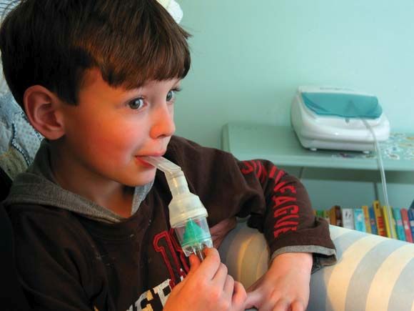
In children with moderate to severe acute asthma, albuterol delivered by a conventional jet nebulizer led to more improvement in forced expiratory volume in 1 second (FEV1) than delivery via a breath-enhanced nebulizer.
Only one previous study has compared the two types of nebulizers in children with acute asthma. It showed that the new technology is noninferior to the older device, but it had a small sample size and did not examine spirometry data.
Mike Gardiner, MD, of the department of pediatrics, University of California, San Diego, and Matthew H. Wilkinson, MD, of the department of pediatrics, University of Texas at Austin, conducted a randomized, observer-blind study to look at effectiveness of these two nebulizers in a larger population of pediatric users. The results were published in the Journal of Pediatrics.
At a large, urban pediatric emergency department, researchers randomized 107 children (aged 6-18 years) presenting with a moderate to severe asthma exacerbation to receive one or the other nebulizer.
Children treated with the conventional jet nebulizer had a greater improvement in FEV1 (+13.8% vs. +9.1% of predicted; P = .04). The improvements were similar in a subgroup analysis of 57 subjects who met ATS/ERS (American Thoracic Society/ European Respiratory Society) spirometry guidelines (+14.5% vs. +8.5% of predicted; P = .03).
The researchers found no significant differences in changes in Pediatric Asthma Score, Pediatric Asthma Severity Score, ED length of stay, or admission rate. There was no significant difference in side effects between the two groups.
Breath-enhanced nebulizers use a holding chamber to store continuously nebulized medication, and one-way valves that direct exhaled air away from the holding chamber. The system reduces medication loss during exhalation and delivers a bolus of medication.
In lung models and healthy adult controls, breath-enhanced nebulizers achieved more effective lung deposition of aerosol. The authors speculate that the reduced clinical effect of the breath-enhanced nebulizer could be because the design of the mouthpiece, which allows a nonaerosolized “dead space” volume to be inhaled first. This volume may have a greater clinical impact in children than in adults.
Children experiencing asthma also have a rapid and shallow breathing pattern, which could also lead to a larger contribution of “dead space” to the overall dose, thus reducing drug exposure.
The study, Comparison of Breath-Enhanced and T-Piece Nebulizers in Children With Acute Asthma (NCT02566902) was funded by the University of Texas Southwestern, Austin. The authors declared no conflicts of interest.
SOURCE: Gardiner M, Wilkinson M. J Pediatr. 2019;204:245-9.
In children with moderate to severe acute asthma, albuterol delivered by a conventional jet nebulizer led to more improvement in forced expiratory volume in 1 second (FEV1) than delivery via a breath-enhanced nebulizer.
Only one previous study has compared the two types of nebulizers in children with acute asthma. It showed that the new technology is noninferior to the older device, but it had a small sample size and did not examine spirometry data.
Mike Gardiner, MD, of the department of pediatrics, University of California, San Diego, and Matthew H. Wilkinson, MD, of the department of pediatrics, University of Texas at Austin, conducted a randomized, observer-blind study to look at effectiveness of these two nebulizers in a larger population of pediatric users. The results were published in the Journal of Pediatrics.
At a large, urban pediatric emergency department, researchers randomized 107 children (aged 6-18 years) presenting with a moderate to severe asthma exacerbation to receive one or the other nebulizer.
Children treated with the conventional jet nebulizer had a greater improvement in FEV1 (+13.8% vs. +9.1% of predicted; P = .04). The improvements were similar in a subgroup analysis of 57 subjects who met ATS/ERS (American Thoracic Society/ European Respiratory Society) spirometry guidelines (+14.5% vs. +8.5% of predicted; P = .03).
The researchers found no significant differences in changes in Pediatric Asthma Score, Pediatric Asthma Severity Score, ED length of stay, or admission rate. There was no significant difference in side effects between the two groups.
Breath-enhanced nebulizers use a holding chamber to store continuously nebulized medication, and one-way valves that direct exhaled air away from the holding chamber. The system reduces medication loss during exhalation and delivers a bolus of medication.
In lung models and healthy adult controls, breath-enhanced nebulizers achieved more effective lung deposition of aerosol. The authors speculate that the reduced clinical effect of the breath-enhanced nebulizer could be because the design of the mouthpiece, which allows a nonaerosolized “dead space” volume to be inhaled first. This volume may have a greater clinical impact in children than in adults.
Children experiencing asthma also have a rapid and shallow breathing pattern, which could also lead to a larger contribution of “dead space” to the overall dose, thus reducing drug exposure.
The study, Comparison of Breath-Enhanced and T-Piece Nebulizers in Children With Acute Asthma (NCT02566902) was funded by the University of Texas Southwestern, Austin. The authors declared no conflicts of interest.
SOURCE: Gardiner M, Wilkinson M. J Pediatr. 2019;204:245-9.
In children with moderate to severe acute asthma, albuterol delivered by a conventional jet nebulizer led to more improvement in forced expiratory volume in 1 second (FEV1) than delivery via a breath-enhanced nebulizer.
Only one previous study has compared the two types of nebulizers in children with acute asthma. It showed that the new technology is noninferior to the older device, but it had a small sample size and did not examine spirometry data.
Mike Gardiner, MD, of the department of pediatrics, University of California, San Diego, and Matthew H. Wilkinson, MD, of the department of pediatrics, University of Texas at Austin, conducted a randomized, observer-blind study to look at effectiveness of these two nebulizers in a larger population of pediatric users. The results were published in the Journal of Pediatrics.
At a large, urban pediatric emergency department, researchers randomized 107 children (aged 6-18 years) presenting with a moderate to severe asthma exacerbation to receive one or the other nebulizer.
Children treated with the conventional jet nebulizer had a greater improvement in FEV1 (+13.8% vs. +9.1% of predicted; P = .04). The improvements were similar in a subgroup analysis of 57 subjects who met ATS/ERS (American Thoracic Society/ European Respiratory Society) spirometry guidelines (+14.5% vs. +8.5% of predicted; P = .03).
The researchers found no significant differences in changes in Pediatric Asthma Score, Pediatric Asthma Severity Score, ED length of stay, or admission rate. There was no significant difference in side effects between the two groups.
Breath-enhanced nebulizers use a holding chamber to store continuously nebulized medication, and one-way valves that direct exhaled air away from the holding chamber. The system reduces medication loss during exhalation and delivers a bolus of medication.
In lung models and healthy adult controls, breath-enhanced nebulizers achieved more effective lung deposition of aerosol. The authors speculate that the reduced clinical effect of the breath-enhanced nebulizer could be because the design of the mouthpiece, which allows a nonaerosolized “dead space” volume to be inhaled first. This volume may have a greater clinical impact in children than in adults.
Children experiencing asthma also have a rapid and shallow breathing pattern, which could also lead to a larger contribution of “dead space” to the overall dose, thus reducing drug exposure.
The study, Comparison of Breath-Enhanced and T-Piece Nebulizers in Children With Acute Asthma (NCT02566902) was funded by the University of Texas Southwestern, Austin. The authors declared no conflicts of interest.
SOURCE: Gardiner M, Wilkinson M. J Pediatr. 2019;204:245-9.
FROM THE JOURNAL OF PEDIATRICS
Key clinical point: New nebulizers worked better in adults, but pediatric populations may pose a challenge.
Major finding: FEV1 improved +13.8% in standard inhalers vs. +9.1% in breath-enhanced analyzers.
Study details: Randomized, controlled trial (n = 107).
Disclosures: The study was funded by the University of Texas Southwestern, Austin. The authors declared no conflicts of interest.
Source: Gardiner M, Wilkinson M. J Pediatr. 2019;204:245-9.
Flu season showing signs of decline
The 2018-2019 flu season may have peaked as measures of influenza-like illness (ILI) activity dropped in the first week of the new year, according to the U.S. Centers for Disease Control and Prevention.

The proportion of outpatients visits for ILI dropped to 3.5% for the week ending Jan. 5, 2019, after reaching 4.0% the previous week. Outpatient ILI visits first topped the national baseline of 2.2% during the week ending Dec. 8, 2018, and have remained above that value for 5 consecutive weeks, the CDC’s influenza division said on Jan. 11.
Flu activity reported by the states reflects the national drop: 10 states came in at level 10 on the CDC’s 1-10 scale of activity for the week ending Jan. 5 – down from 12 the week before – and a total of 15 were in the high range from 8 to 10, compared with 19 the previous week, the CDC said. Two states, Mississippi and Texas, dropped from level 10 to level 7, which the CDC categorizes as moderate activity.
A total of 73 ILI-related deaths were reported during the week ending Dec. 29 (the latest with data available; reporting less than 68% complete), which already exceeds the 71 deaths reported for the week ending Dec. 22 (reporting 85% complete). Flu deaths totaled 437 through the first 13 weeks of the 2018-2019 season, compared with the 1,659 that occurred during weeks 1-13 of the very severe 2017-2018 season, CDC data show.
For the week ending Jan. 5, the CDC received reports of three flu-related pediatric deaths, all of which occurred the previous week. For the season so far, there have been 16 pediatric deaths, compared with 20 at this point in the 2017-2018 season.
Estimates released during the flu season for the first time show that between 6 and 7 million Americans have been infected since Oct. 1, 2018, and that 69,000-84,000 people have been hospitalized with the flu through Jan. 5, 2019. These cumulative totals have previously been available only at the end of the season, the CDC noted.
The 2018-2019 flu season may have peaked as measures of influenza-like illness (ILI) activity dropped in the first week of the new year, according to the U.S. Centers for Disease Control and Prevention.
The proportion of outpatients visits for ILI dropped to 3.5% for the week ending Jan. 5, 2019, after reaching 4.0% the previous week. Outpatient ILI visits first topped the national baseline of 2.2% during the week ending Dec. 8, 2018, and have remained above that value for 5 consecutive weeks, the CDC’s influenza division said on Jan. 11.
Flu activity reported by the states reflects the national drop: 10 states came in at level 10 on the CDC’s 1-10 scale of activity for the week ending Jan. 5 – down from 12 the week before – and a total of 15 were in the high range from 8 to 10, compared with 19 the previous week, the CDC said. Two states, Mississippi and Texas, dropped from level 10 to level 7, which the CDC categorizes as moderate activity.
A total of 73 ILI-related deaths were reported during the week ending Dec. 29 (the latest with data available; reporting less than 68% complete), which already exceeds the 71 deaths reported for the week ending Dec. 22 (reporting 85% complete). Flu deaths totaled 437 through the first 13 weeks of the 2018-2019 season, compared with the 1,659 that occurred during weeks 1-13 of the very severe 2017-2018 season, CDC data show.
For the week ending Jan. 5, the CDC received reports of three flu-related pediatric deaths, all of which occurred the previous week. For the season so far, there have been 16 pediatric deaths, compared with 20 at this point in the 2017-2018 season.
Estimates released during the flu season for the first time show that between 6 and 7 million Americans have been infected since Oct. 1, 2018, and that 69,000-84,000 people have been hospitalized with the flu through Jan. 5, 2019. These cumulative totals have previously been available only at the end of the season, the CDC noted.
The 2018-2019 flu season may have peaked as measures of influenza-like illness (ILI) activity dropped in the first week of the new year, according to the U.S. Centers for Disease Control and Prevention.
The proportion of outpatients visits for ILI dropped to 3.5% for the week ending Jan. 5, 2019, after reaching 4.0% the previous week. Outpatient ILI visits first topped the national baseline of 2.2% during the week ending Dec. 8, 2018, and have remained above that value for 5 consecutive weeks, the CDC’s influenza division said on Jan. 11.
Flu activity reported by the states reflects the national drop: 10 states came in at level 10 on the CDC’s 1-10 scale of activity for the week ending Jan. 5 – down from 12 the week before – and a total of 15 were in the high range from 8 to 10, compared with 19 the previous week, the CDC said. Two states, Mississippi and Texas, dropped from level 10 to level 7, which the CDC categorizes as moderate activity.
A total of 73 ILI-related deaths were reported during the week ending Dec. 29 (the latest with data available; reporting less than 68% complete), which already exceeds the 71 deaths reported for the week ending Dec. 22 (reporting 85% complete). Flu deaths totaled 437 through the first 13 weeks of the 2018-2019 season, compared with the 1,659 that occurred during weeks 1-13 of the very severe 2017-2018 season, CDC data show.
For the week ending Jan. 5, the CDC received reports of three flu-related pediatric deaths, all of which occurred the previous week. For the season so far, there have been 16 pediatric deaths, compared with 20 at this point in the 2017-2018 season.
Estimates released during the flu season for the first time show that between 6 and 7 million Americans have been infected since Oct. 1, 2018, and that 69,000-84,000 people have been hospitalized with the flu through Jan. 5, 2019. These cumulative totals have previously been available only at the end of the season, the CDC noted.
Benralizumab maintains effectiveness in severe asthma at 2 years
out to 2 years, according findings of the BORA trial, an extension study of the phase 3 SIROCCO and CALIMA trials. The study follows up and reinforces previously reported 1-year data and was reported by William W. Busse, MD, of University of Wisconsin, Madison, and his colleagues in the Lancet Respiratory Medicine.

Benralizumab is a monoclonal antibody that targets interleukin-5 receptor alpha. It causes rapid deletion of eosinophils through cell-mediated cytotoxicity. A 30-mg dose of benralizumab every 8 weeks is approved for severe asthma treatment in Canada, Europe, Japan, the United States, and other countries.
In the second year of treatment, there were no new adverse events associated with depleted eosinophils, and the frequency of opportunistic infections was similar to that observed in the first year.
Eosinophilic inflammation occurs in about half of asthma cases and is associated with greater severity.
The 48-week SIROCCO trial, the 56-week CALIMA trial, and the 28-week ZONDA trial tested the effect of benralizumab 30 mg given every 4 weeks or 8 weeks, combined with high-dosage inhaled steroids and long-acting beta2-agonists. The 8-week dose of the drug reduced annual exacerbations by 51%, compared with placebo in the SIROCCO trial and by 28% in the CALIMA trial. In the ZONDA trial, benralizumab reduced oral glucocorticoid use by 75%, compared with placebo, and by 25% from baseline.
The BORA extension trial included participants in the previous three trials. In the current report, researchers presented results from the analysis from BORA participants recruited from the SIROCCO and CALIMA trials. Data from participants from all three trials will be reported in the future.
The analysis included 1,576 patients who continued to receive benralizumab after being assigned to the treatment arm in SIROCCO or CALIMA, or who had received placebo were randomized to benralizumab on the 4-week (n = 783; 265 from placebo) or 8-week dose (n = 793; 281 from placebo) schedule.
A total of 166 patients, or about 10% in each group, discontinued treatment. The frequency of any serious adverse event (SAE) ranged between 10% and 11% in all groups. SAEs associated with infections ranged from 1% to 3%, indicating that there were no significant differences in SAE frequencies between those who were originally assigned to placebo and those who originally received benralizumab. That suggests no safety differences between receiving the drug for 1 year or 2 years.
A total of 1,046 subjects had blood eosinophil counts of 300 cells per mcL or greater at baseline; 72% of these patients had no asthma exacerbations during the BORA study. This was true for 74% of patients in the 8-week treatment arm.
The crude asthma exacerbation rate for patients who received benralizumab in SIROCCO or CALIMA was 0.48 in the 4-week arm, compared with placebo (95% confidence interval, 0.42-0.56) and 0.46 in the 8-week arm (95% CI, 0.39-0.53). For patients who started out on placebo, the crude exacerbation rate during BORA was 0.53 in the 4-week group (95% CI, 0.43-0.65) and 0.57 in the 8-week group (95% CI, 0.47-0.68).
Patients who started on benralizumab had similar exacerbation frequencies during year 1 and year 2.
AstraZeneca and Kyowa Hakko Kirin funded the studies. The authors have received fees from AstraZeneca and other pharmaceutical companies, and some are employees of AstraZeneca.
SOURCE: Busse WW et al. Lancet Respir Med. 2019 Jan 1;7(1):46-59.
out to 2 years, according findings of the BORA trial, an extension study of the phase 3 SIROCCO and CALIMA trials. The study follows up and reinforces previously reported 1-year data and was reported by William W. Busse, MD, of University of Wisconsin, Madison, and his colleagues in the Lancet Respiratory Medicine.
Benralizumab is a monoclonal antibody that targets interleukin-5 receptor alpha. It causes rapid deletion of eosinophils through cell-mediated cytotoxicity. A 30-mg dose of benralizumab every 8 weeks is approved for severe asthma treatment in Canada, Europe, Japan, the United States, and other countries.
In the second year of treatment, there were no new adverse events associated with depleted eosinophils, and the frequency of opportunistic infections was similar to that observed in the first year.
Eosinophilic inflammation occurs in about half of asthma cases and is associated with greater severity.
The 48-week SIROCCO trial, the 56-week CALIMA trial, and the 28-week ZONDA trial tested the effect of benralizumab 30 mg given every 4 weeks or 8 weeks, combined with high-dosage inhaled steroids and long-acting beta2-agonists. The 8-week dose of the drug reduced annual exacerbations by 51%, compared with placebo in the SIROCCO trial and by 28% in the CALIMA trial. In the ZONDA trial, benralizumab reduced oral glucocorticoid use by 75%, compared with placebo, and by 25% from baseline.
The BORA extension trial included participants in the previous three trials. In the current report, researchers presented results from the analysis from BORA participants recruited from the SIROCCO and CALIMA trials. Data from participants from all three trials will be reported in the future.
The analysis included 1,576 patients who continued to receive benralizumab after being assigned to the treatment arm in SIROCCO or CALIMA, or who had received placebo were randomized to benralizumab on the 4-week (n = 783; 265 from placebo) or 8-week dose (n = 793; 281 from placebo) schedule.
A total of 166 patients, or about 10% in each group, discontinued treatment. The frequency of any serious adverse event (SAE) ranged between 10% and 11% in all groups. SAEs associated with infections ranged from 1% to 3%, indicating that there were no significant differences in SAE frequencies between those who were originally assigned to placebo and those who originally received benralizumab. That suggests no safety differences between receiving the drug for 1 year or 2 years.
A total of 1,046 subjects had blood eosinophil counts of 300 cells per mcL or greater at baseline; 72% of these patients had no asthma exacerbations during the BORA study. This was true for 74% of patients in the 8-week treatment arm.
The crude asthma exacerbation rate for patients who received benralizumab in SIROCCO or CALIMA was 0.48 in the 4-week arm, compared with placebo (95% confidence interval, 0.42-0.56) and 0.46 in the 8-week arm (95% CI, 0.39-0.53). For patients who started out on placebo, the crude exacerbation rate during BORA was 0.53 in the 4-week group (95% CI, 0.43-0.65) and 0.57 in the 8-week group (95% CI, 0.47-0.68).
Patients who started on benralizumab had similar exacerbation frequencies during year 1 and year 2.
AstraZeneca and Kyowa Hakko Kirin funded the studies. The authors have received fees from AstraZeneca and other pharmaceutical companies, and some are employees of AstraZeneca.
SOURCE: Busse WW et al. Lancet Respir Med. 2019 Jan 1;7(1):46-59.
out to 2 years, according findings of the BORA trial, an extension study of the phase 3 SIROCCO and CALIMA trials. The study follows up and reinforces previously reported 1-year data and was reported by William W. Busse, MD, of University of Wisconsin, Madison, and his colleagues in the Lancet Respiratory Medicine.
Benralizumab is a monoclonal antibody that targets interleukin-5 receptor alpha. It causes rapid deletion of eosinophils through cell-mediated cytotoxicity. A 30-mg dose of benralizumab every 8 weeks is approved for severe asthma treatment in Canada, Europe, Japan, the United States, and other countries.
In the second year of treatment, there were no new adverse events associated with depleted eosinophils, and the frequency of opportunistic infections was similar to that observed in the first year.
Eosinophilic inflammation occurs in about half of asthma cases and is associated with greater severity.
The 48-week SIROCCO trial, the 56-week CALIMA trial, and the 28-week ZONDA trial tested the effect of benralizumab 30 mg given every 4 weeks or 8 weeks, combined with high-dosage inhaled steroids and long-acting beta2-agonists. The 8-week dose of the drug reduced annual exacerbations by 51%, compared with placebo in the SIROCCO trial and by 28% in the CALIMA trial. In the ZONDA trial, benralizumab reduced oral glucocorticoid use by 75%, compared with placebo, and by 25% from baseline.
The BORA extension trial included participants in the previous three trials. In the current report, researchers presented results from the analysis from BORA participants recruited from the SIROCCO and CALIMA trials. Data from participants from all three trials will be reported in the future.
The analysis included 1,576 patients who continued to receive benralizumab after being assigned to the treatment arm in SIROCCO or CALIMA, or who had received placebo were randomized to benralizumab on the 4-week (n = 783; 265 from placebo) or 8-week dose (n = 793; 281 from placebo) schedule.
A total of 166 patients, or about 10% in each group, discontinued treatment. The frequency of any serious adverse event (SAE) ranged between 10% and 11% in all groups. SAEs associated with infections ranged from 1% to 3%, indicating that there were no significant differences in SAE frequencies between those who were originally assigned to placebo and those who originally received benralizumab. That suggests no safety differences between receiving the drug for 1 year or 2 years.
A total of 1,046 subjects had blood eosinophil counts of 300 cells per mcL or greater at baseline; 72% of these patients had no asthma exacerbations during the BORA study. This was true for 74% of patients in the 8-week treatment arm.
The crude asthma exacerbation rate for patients who received benralizumab in SIROCCO or CALIMA was 0.48 in the 4-week arm, compared with placebo (95% confidence interval, 0.42-0.56) and 0.46 in the 8-week arm (95% CI, 0.39-0.53). For patients who started out on placebo, the crude exacerbation rate during BORA was 0.53 in the 4-week group (95% CI, 0.43-0.65) and 0.57 in the 8-week group (95% CI, 0.47-0.68).
Patients who started on benralizumab had similar exacerbation frequencies during year 1 and year 2.
AstraZeneca and Kyowa Hakko Kirin funded the studies. The authors have received fees from AstraZeneca and other pharmaceutical companies, and some are employees of AstraZeneca.
SOURCE: Busse WW et al. Lancet Respir Med. 2019 Jan 1;7(1):46-59.
FROM THE LANCET RESPIRATORY MEDICINE
Key clinical point: The antibody had similar safety, efficacy in year 2 as in year 1.
Major finding: The crude asthma exacerbation rate for patients who received benralizumab in SIROCCO or CALIMA was 0.48 in the 4-week arm and 0.46 in the 8-week arm; the crude exacerbation rate during BORA was 0.53 in the 4-week group and 0.57 in the 8-week group.
Study details: Extension of randomized, clinical trial (n = 1,576).
Disclosures: AstraZeneca and Kyowa Hakko Kirin funded the studies. The authors have received fees from AstraZeneca and other pharmaceutical companies, and some are employees of AstraZeneca.
Source: Busse WW et al. Lancet Respir Med. 2019 Jan 1;7(1):46-59.
Secondhand vaping aerosols linked to childhood asthma exacerbations
Just like exposure to secondhand smoke, , according to a review of the 11,830 kids with asthma in the 2016 Florida Youth Tobacco survey.

Every year, the Florida Department of Health surveys public school children aged 11-17 years about various tobacco issues. In 2016, almost 12% of the asthmatic children in the survey said they vaped. Almost half were exposed to secondhand smoke, and a third reported exposure to secondhand vaping aerosols within the past 30 days. Overall, 21% reported an asthma attack in the past 12 months.
Using data from the Florida survey, the investigators crunched the numbers and found that secondhand aerosol exposure increased the odds of an asthma attack by 27%, independent of exposure to secondhand smoke and whether children smoked or vaped themselves (adjusted odds ratio, 1.27; 95% confidence interval, 1.11-1.47).
“Health professionals may wish to counsel asthmatic youth and their families regarding the potential risks of ENDS [electronic nicotine delivery system] use and exposure to ENDS aerosols.” Providers “may also consider including ENDS aerosol exposure as a possible trigger in asthma self-management/action plans and updating asthma home environment assessments to include exposure to ENDS aerosols,” said investigators led by medical student Jennifer Bayly, a research fellow at the National Institute on Minority Health and Health Disparities in Bethesda, Md.
About 4% of adults in the United States and 11% of high school students vape, and almost 10% of U.S. adolescents reported living with an ENDS user in 2014. Given the data, “it is likely that a substantial number of asthmatic youth are exposed,” the investigators said.
The study adds to a growing body of evidence linking e-cigarettes to asthma. There’s moderate evidence for increased cough and wheezing in adolescents who use e-cigarettes, plus an association with e-cigarette use and increased asthma exacerbations. The new study, however, is likely the first to look specifically at secondhand exposure among asthmatic children. Ingredients in vaping aerosols, including flavorings, propylene glycol, and vegetable glycerin, are physiologically active in the lungs, and may be lung irritants.
Overall, about half of the respondents were female, and two-thirds were 11-13 years old. About a third identified as Hispanic, a third as white, and just over a fifth as black. Three-quarters of the sample lived in large or midsized metropolitan areas, and close to two-thirds in stand-alone homes. Participants were considered exposed to secondhand aerosols if they reported that in the past month they were in a room or car with someone who was vaping.
The work was funded by the National Institutes of Health. The investigators had no disclosures.
SOURCE: Bayly JE et al. CHEST®. 2018 Oct 22. doi: 10.1016/j.chest.2018.10.005.
Just like exposure to secondhand smoke, , according to a review of the 11,830 kids with asthma in the 2016 Florida Youth Tobacco survey.
Every year, the Florida Department of Health surveys public school children aged 11-17 years about various tobacco issues. In 2016, almost 12% of the asthmatic children in the survey said they vaped. Almost half were exposed to secondhand smoke, and a third reported exposure to secondhand vaping aerosols within the past 30 days. Overall, 21% reported an asthma attack in the past 12 months.
Using data from the Florida survey, the investigators crunched the numbers and found that secondhand aerosol exposure increased the odds of an asthma attack by 27%, independent of exposure to secondhand smoke and whether children smoked or vaped themselves (adjusted odds ratio, 1.27; 95% confidence interval, 1.11-1.47).
“Health professionals may wish to counsel asthmatic youth and their families regarding the potential risks of ENDS [electronic nicotine delivery system] use and exposure to ENDS aerosols.” Providers “may also consider including ENDS aerosol exposure as a possible trigger in asthma self-management/action plans and updating asthma home environment assessments to include exposure to ENDS aerosols,” said investigators led by medical student Jennifer Bayly, a research fellow at the National Institute on Minority Health and Health Disparities in Bethesda, Md.
About 4% of adults in the United States and 11% of high school students vape, and almost 10% of U.S. adolescents reported living with an ENDS user in 2014. Given the data, “it is likely that a substantial number of asthmatic youth are exposed,” the investigators said.
The study adds to a growing body of evidence linking e-cigarettes to asthma. There’s moderate evidence for increased cough and wheezing in adolescents who use e-cigarettes, plus an association with e-cigarette use and increased asthma exacerbations. The new study, however, is likely the first to look specifically at secondhand exposure among asthmatic children. Ingredients in vaping aerosols, including flavorings, propylene glycol, and vegetable glycerin, are physiologically active in the lungs, and may be lung irritants.
Overall, about half of the respondents were female, and two-thirds were 11-13 years old. About a third identified as Hispanic, a third as white, and just over a fifth as black. Three-quarters of the sample lived in large or midsized metropolitan areas, and close to two-thirds in stand-alone homes. Participants were considered exposed to secondhand aerosols if they reported that in the past month they were in a room or car with someone who was vaping.
The work was funded by the National Institutes of Health. The investigators had no disclosures.
SOURCE: Bayly JE et al. CHEST®. 2018 Oct 22. doi: 10.1016/j.chest.2018.10.005.
Just like exposure to secondhand smoke, , according to a review of the 11,830 kids with asthma in the 2016 Florida Youth Tobacco survey.
Every year, the Florida Department of Health surveys public school children aged 11-17 years about various tobacco issues. In 2016, almost 12% of the asthmatic children in the survey said they vaped. Almost half were exposed to secondhand smoke, and a third reported exposure to secondhand vaping aerosols within the past 30 days. Overall, 21% reported an asthma attack in the past 12 months.
Using data from the Florida survey, the investigators crunched the numbers and found that secondhand aerosol exposure increased the odds of an asthma attack by 27%, independent of exposure to secondhand smoke and whether children smoked or vaped themselves (adjusted odds ratio, 1.27; 95% confidence interval, 1.11-1.47).
“Health professionals may wish to counsel asthmatic youth and their families regarding the potential risks of ENDS [electronic nicotine delivery system] use and exposure to ENDS aerosols.” Providers “may also consider including ENDS aerosol exposure as a possible trigger in asthma self-management/action plans and updating asthma home environment assessments to include exposure to ENDS aerosols,” said investigators led by medical student Jennifer Bayly, a research fellow at the National Institute on Minority Health and Health Disparities in Bethesda, Md.
About 4% of adults in the United States and 11% of high school students vape, and almost 10% of U.S. adolescents reported living with an ENDS user in 2014. Given the data, “it is likely that a substantial number of asthmatic youth are exposed,” the investigators said.
The study adds to a growing body of evidence linking e-cigarettes to asthma. There’s moderate evidence for increased cough and wheezing in adolescents who use e-cigarettes, plus an association with e-cigarette use and increased asthma exacerbations. The new study, however, is likely the first to look specifically at secondhand exposure among asthmatic children. Ingredients in vaping aerosols, including flavorings, propylene glycol, and vegetable glycerin, are physiologically active in the lungs, and may be lung irritants.
Overall, about half of the respondents were female, and two-thirds were 11-13 years old. About a third identified as Hispanic, a third as white, and just over a fifth as black. Three-quarters of the sample lived in large or midsized metropolitan areas, and close to two-thirds in stand-alone homes. Participants were considered exposed to secondhand aerosols if they reported that in the past month they were in a room or car with someone who was vaping.
The work was funded by the National Institutes of Health. The investigators had no disclosures.
SOURCE: Bayly JE et al. CHEST®. 2018 Oct 22. doi: 10.1016/j.chest.2018.10.005.
FROM CHEST®
Key clinical point: It’s important to screen asthmatic children for exposure to secondhand vaping aerosols, and minimize exposure.
Major finding: Secondhand aerosols increased the odds of an asthma attack 27%, independent of exposure to secondhand smoke and whether children smoked or vaped themselves.
Study details: Analysis of 11,830 children with asthma in the 2016 Florida Youth Tobacco survey.
Disclosures: The work was funded by the National Institutes of Health. The investigators had no disclosures.
Source: Bayly JE et al. CHEST®. 2018 Oct 22. doi: 10.1016/j.chest.2018.10.005.
New CHEST expert panel advice on cough diagnosis
The CHEST Expert Cough Panel has released two new in adults and children.

Upper and lower respiratory tract infections are a common reason for primary care visits. A cough caused by influenza or pneumonia represents an opportunity to intervene for a significant benefit. The recommendations were published in CHEST®. The panel drafted recommendations based on available evidence and graded them using the CHEST grading system. The grading is based on the strength of the recommendation (either strong or weak) and a rating of the overall quality of the body of evidence. Where available evidence was weak, but guidance was still warranted, a weak suggestion was developed and graded 2C. Recommendations based on consensus in cases of insufficient clinical evidence are labeled “ungraded consensus-based statement.”
Suspected pneumonia or influenza
In adult outpatients with acute cough, the clinical signs of pneumonia include cough, dyspnea, pleural pain, sweating/fevers/shivers, aches and pains, temperature greater than or equal to 38°C, tachypnea, and new and localizing chest examination signs. When pneumonia is suspected to cause acute cough, C-reactive protein (CRP) should be measured. A CRP value higher than 30 mg/L bolsters the case for pneumonia, whereas a CRP value of lower than 10 mg/L, or between 10 mg/L and 50 mg/L in the absence of dyspnea and daily fever, makes pneumonia less likely.
The guidelines recommend against routine measurement of procalcitonin for outpatient adults suspected to have pneumonia. For adults with acute cough and abnormal vital signs believed to be secondary to pneumonia, the guidelines call for a chest x-ray.
Routine microbiological testing need not be performed in suspected pneumonia, but it should be considered if the results could guide or lead to a change in therapy.
When pneumonia is suspected but imaging is unavailable, empiric antibiotics should be used in concordance with local and national guidelines. If imaging turns up negative, antibiotics should not be used. However, if there is no clinical or radiographic evidence of pneumonia, antibiotics should not be used routinely.
Finally, adult patients with acute cough and suspected influenza should begin antiviral treatment within 48 hours of the start of symptoms.
Pertussis
Pertussis has significant morbidity and mortality, with infants being particularly vulnerable, and it is highly contagious. Although antibiotics will not affect the course of the disease, they should be administered as quickly as possible in order to prevent further spread. This puts pressure on the clinician to make a treatment decision before further testing is available.
A prespecified meta-analysis found high sensitivity and low specificity for paroxysmal cough (sensitivity, 93.2%; specificity, 20.6%) and absence of fever (sensitivity, 81.8%; specificity, 18.8%). The study found low sensitivity and high specificity for inspiratory whoop (sensitivity, 29.8%; specificity, 79.5%) and posttussive vomiting (sensitivity, 32.5%; specificity, 77.7%). In children, the review found that posttussive vomiting was moderately sensitive (60.0%) and specific (66.0%).
In adult patients with acute cough (less than 3 weeks’ duration) or subacute cough (3-8 weeks), the new guidelines recommend that physicians consider four key characteristics: the presence of recurrent, prolonged coughing episodes with an inability to breathe during the spell (paroxysmal); posttussive vomiting; inspiratory whooping; and presence of fever.
In acute or subacute cough, if the patient has a fever (body temperature greater than 98.6° F or 37.6° C) or does not have a paroxysmal cough, pertussis is unlikely. On the other hand, posttussive vomiting or an associated inspiratory whooping sound suggests pertussis.
Children with a cough lasting fewer than 4 weeks (acute) should be assessed for paroxysmal cough, posttussive vomiting, and inspiratory whooping. A cough associated with any of these characteristics may be caused by pertussis.
SOURCES: Moore A et al. CHEST. 2019 Jan;155:147-154; Hill A et al. CHEST. 2019 Jan;155:155-167.
The CHEST Expert Cough Panel has released two new in adults and children.
Upper and lower respiratory tract infections are a common reason for primary care visits. A cough caused by influenza or pneumonia represents an opportunity to intervene for a significant benefit. The recommendations were published in CHEST®. The panel drafted recommendations based on available evidence and graded them using the CHEST grading system. The grading is based on the strength of the recommendation (either strong or weak) and a rating of the overall quality of the body of evidence. Where available evidence was weak, but guidance was still warranted, a weak suggestion was developed and graded 2C. Recommendations based on consensus in cases of insufficient clinical evidence are labeled “ungraded consensus-based statement.”
Suspected pneumonia or influenza
In adult outpatients with acute cough, the clinical signs of pneumonia include cough, dyspnea, pleural pain, sweating/fevers/shivers, aches and pains, temperature greater than or equal to 38°C, tachypnea, and new and localizing chest examination signs. When pneumonia is suspected to cause acute cough, C-reactive protein (CRP) should be measured. A CRP value higher than 30 mg/L bolsters the case for pneumonia, whereas a CRP value of lower than 10 mg/L, or between 10 mg/L and 50 mg/L in the absence of dyspnea and daily fever, makes pneumonia less likely.
The guidelines recommend against routine measurement of procalcitonin for outpatient adults suspected to have pneumonia. For adults with acute cough and abnormal vital signs believed to be secondary to pneumonia, the guidelines call for a chest x-ray.
Routine microbiological testing need not be performed in suspected pneumonia, but it should be considered if the results could guide or lead to a change in therapy.
When pneumonia is suspected but imaging is unavailable, empiric antibiotics should be used in concordance with local and national guidelines. If imaging turns up negative, antibiotics should not be used. However, if there is no clinical or radiographic evidence of pneumonia, antibiotics should not be used routinely.
Finally, adult patients with acute cough and suspected influenza should begin antiviral treatment within 48 hours of the start of symptoms.
Pertussis
Pertussis has significant morbidity and mortality, with infants being particularly vulnerable, and it is highly contagious. Although antibiotics will not affect the course of the disease, they should be administered as quickly as possible in order to prevent further spread. This puts pressure on the clinician to make a treatment decision before further testing is available.
A prespecified meta-analysis found high sensitivity and low specificity for paroxysmal cough (sensitivity, 93.2%; specificity, 20.6%) and absence of fever (sensitivity, 81.8%; specificity, 18.8%). The study found low sensitivity and high specificity for inspiratory whoop (sensitivity, 29.8%; specificity, 79.5%) and posttussive vomiting (sensitivity, 32.5%; specificity, 77.7%). In children, the review found that posttussive vomiting was moderately sensitive (60.0%) and specific (66.0%).
In adult patients with acute cough (less than 3 weeks’ duration) or subacute cough (3-8 weeks), the new guidelines recommend that physicians consider four key characteristics: the presence of recurrent, prolonged coughing episodes with an inability to breathe during the spell (paroxysmal); posttussive vomiting; inspiratory whooping; and presence of fever.
In acute or subacute cough, if the patient has a fever (body temperature greater than 98.6° F or 37.6° C) or does not have a paroxysmal cough, pertussis is unlikely. On the other hand, posttussive vomiting or an associated inspiratory whooping sound suggests pertussis.
Children with a cough lasting fewer than 4 weeks (acute) should be assessed for paroxysmal cough, posttussive vomiting, and inspiratory whooping. A cough associated with any of these characteristics may be caused by pertussis.
SOURCES: Moore A et al. CHEST. 2019 Jan;155:147-154; Hill A et al. CHEST. 2019 Jan;155:155-167.
The CHEST Expert Cough Panel has released two new in adults and children.
Upper and lower respiratory tract infections are a common reason for primary care visits. A cough caused by influenza or pneumonia represents an opportunity to intervene for a significant benefit. The recommendations were published in CHEST®. The panel drafted recommendations based on available evidence and graded them using the CHEST grading system. The grading is based on the strength of the recommendation (either strong or weak) and a rating of the overall quality of the body of evidence. Where available evidence was weak, but guidance was still warranted, a weak suggestion was developed and graded 2C. Recommendations based on consensus in cases of insufficient clinical evidence are labeled “ungraded consensus-based statement.”
Suspected pneumonia or influenza
In adult outpatients with acute cough, the clinical signs of pneumonia include cough, dyspnea, pleural pain, sweating/fevers/shivers, aches and pains, temperature greater than or equal to 38°C, tachypnea, and new and localizing chest examination signs. When pneumonia is suspected to cause acute cough, C-reactive protein (CRP) should be measured. A CRP value higher than 30 mg/L bolsters the case for pneumonia, whereas a CRP value of lower than 10 mg/L, or between 10 mg/L and 50 mg/L in the absence of dyspnea and daily fever, makes pneumonia less likely.
The guidelines recommend against routine measurement of procalcitonin for outpatient adults suspected to have pneumonia. For adults with acute cough and abnormal vital signs believed to be secondary to pneumonia, the guidelines call for a chest x-ray.
Routine microbiological testing need not be performed in suspected pneumonia, but it should be considered if the results could guide or lead to a change in therapy.
When pneumonia is suspected but imaging is unavailable, empiric antibiotics should be used in concordance with local and national guidelines. If imaging turns up negative, antibiotics should not be used. However, if there is no clinical or radiographic evidence of pneumonia, antibiotics should not be used routinely.
Finally, adult patients with acute cough and suspected influenza should begin antiviral treatment within 48 hours of the start of symptoms.
Pertussis
Pertussis has significant morbidity and mortality, with infants being particularly vulnerable, and it is highly contagious. Although antibiotics will not affect the course of the disease, they should be administered as quickly as possible in order to prevent further spread. This puts pressure on the clinician to make a treatment decision before further testing is available.
A prespecified meta-analysis found high sensitivity and low specificity for paroxysmal cough (sensitivity, 93.2%; specificity, 20.6%) and absence of fever (sensitivity, 81.8%; specificity, 18.8%). The study found low sensitivity and high specificity for inspiratory whoop (sensitivity, 29.8%; specificity, 79.5%) and posttussive vomiting (sensitivity, 32.5%; specificity, 77.7%). In children, the review found that posttussive vomiting was moderately sensitive (60.0%) and specific (66.0%).
In adult patients with acute cough (less than 3 weeks’ duration) or subacute cough (3-8 weeks), the new guidelines recommend that physicians consider four key characteristics: the presence of recurrent, prolonged coughing episodes with an inability to breathe during the spell (paroxysmal); posttussive vomiting; inspiratory whooping; and presence of fever.
In acute or subacute cough, if the patient has a fever (body temperature greater than 98.6° F or 37.6° C) or does not have a paroxysmal cough, pertussis is unlikely. On the other hand, posttussive vomiting or an associated inspiratory whooping sound suggests pertussis.
Children with a cough lasting fewer than 4 weeks (acute) should be assessed for paroxysmal cough, posttussive vomiting, and inspiratory whooping. A cough associated with any of these characteristics may be caused by pertussis.
SOURCES: Moore A et al. CHEST. 2019 Jan;155:147-154; Hill A et al. CHEST. 2019 Jan;155:155-167.
FROM THE JOURNAL CHEST®
Veterans Learn the Healing Arts
Making art is not only life enriching—it can help reduce stress and anxiety, provide emotional release, relieve depression, and boost self-esteem. That is why art therapy has been a valuable part of VA mental health services since 1945. Plein air painting—painting outdoors “in the fresh air”—gives an added dimension to that. It can be both calming and energizing, offers challenges not found in a studio setting, teaches observation, and can lead to rewarding social interactions and connections. Not least, there is the benefit of being surrounded by nature.
The publishers and editor of Plein Air magazine wanted to introduce more veterans to the pleasures and benefits of outdoor painting, so they created the Plein Air Force Veterans Squad. Dennis Tyson, Commander of the Veterans Squad, says the goal is to “[s]how our gratitude to veterans by teaching them painting, allowing them to enjoy the benefits and stress reduction that come with painting outdoors.” The program seeks to enlist painters from around the nation to visit veterans’ groups, do demonstrations, and offer free lessons. The guiding principle, Tyson says, is that cost should not prevent any veteran from participating, so lessons and supplies are free or low-cost.
The program runs 2 websites. PleinAirForce.com gives existing plein air painters “missions” and tools to spread the word about plein air painting. That site provides scripts, posters, and guidance on promoting speaking engagements. A main purpose is to drive veterans and others interested in learning to the second website, PaintOutside.com, “so we can give them encouragement, free lessons, tips and tools to start painting.” PaintOutside.com also offers a directory of artists who teach, and a directory of “painting buddies.”
In addition to the websites, interested veterans can get more information at www.facebook.com/pleinairmagazine.
Making art is not only life enriching—it can help reduce stress and anxiety, provide emotional release, relieve depression, and boost self-esteem. That is why art therapy has been a valuable part of VA mental health services since 1945. Plein air painting—painting outdoors “in the fresh air”—gives an added dimension to that. It can be both calming and energizing, offers challenges not found in a studio setting, teaches observation, and can lead to rewarding social interactions and connections. Not least, there is the benefit of being surrounded by nature.
The publishers and editor of Plein Air magazine wanted to introduce more veterans to the pleasures and benefits of outdoor painting, so they created the Plein Air Force Veterans Squad. Dennis Tyson, Commander of the Veterans Squad, says the goal is to “[s]how our gratitude to veterans by teaching them painting, allowing them to enjoy the benefits and stress reduction that come with painting outdoors.” The program seeks to enlist painters from around the nation to visit veterans’ groups, do demonstrations, and offer free lessons. The guiding principle, Tyson says, is that cost should not prevent any veteran from participating, so lessons and supplies are free or low-cost.
The program runs 2 websites. PleinAirForce.com gives existing plein air painters “missions” and tools to spread the word about plein air painting. That site provides scripts, posters, and guidance on promoting speaking engagements. A main purpose is to drive veterans and others interested in learning to the second website, PaintOutside.com, “so we can give them encouragement, free lessons, tips and tools to start painting.” PaintOutside.com also offers a directory of artists who teach, and a directory of “painting buddies.”
In addition to the websites, interested veterans can get more information at www.facebook.com/pleinairmagazine.
Making art is not only life enriching—it can help reduce stress and anxiety, provide emotional release, relieve depression, and boost self-esteem. That is why art therapy has been a valuable part of VA mental health services since 1945. Plein air painting—painting outdoors “in the fresh air”—gives an added dimension to that. It can be both calming and energizing, offers challenges not found in a studio setting, teaches observation, and can lead to rewarding social interactions and connections. Not least, there is the benefit of being surrounded by nature.
The publishers and editor of Plein Air magazine wanted to introduce more veterans to the pleasures and benefits of outdoor painting, so they created the Plein Air Force Veterans Squad. Dennis Tyson, Commander of the Veterans Squad, says the goal is to “[s]how our gratitude to veterans by teaching them painting, allowing them to enjoy the benefits and stress reduction that come with painting outdoors.” The program seeks to enlist painters from around the nation to visit veterans’ groups, do demonstrations, and offer free lessons. The guiding principle, Tyson says, is that cost should not prevent any veteran from participating, so lessons and supplies are free or low-cost.
The program runs 2 websites. PleinAirForce.com gives existing plein air painters “missions” and tools to spread the word about plein air painting. That site provides scripts, posters, and guidance on promoting speaking engagements. A main purpose is to drive veterans and others interested in learning to the second website, PaintOutside.com, “so we can give them encouragement, free lessons, tips and tools to start painting.” PaintOutside.com also offers a directory of artists who teach, and a directory of “painting buddies.”
In addition to the websites, interested veterans can get more information at www.facebook.com/pleinairmagazine.