User login
MDedge latest news is breaking news from medical conferences, journals, guidelines, the FDA and CDC.
Spinal Cord Stimulation Promising for Chronic Back, Leg Pain
TOPLINE:
Spinal cord stimulation (SCS) therapies for chronic back and/or leg pain is superior to conventional medical management (CMM) for reduced pain intensity and functional disability, new research suggests.
METHODOLOGY:
- Researchers performed a systematic review and network meta-analysis of 13 randomized clinical trials that compared conventional and novel SCS therapies with CMM.
- More than 1500 adults with chronic back and/or leg pain and no past history of receiving SCS therapies were included.
- Novel therapies included high frequency, burst, differential target multiplexed, and closed-loop SCS; conventional therapies included tonic SCS wave forms.
- Study outcomes included pain intensity in the back and in the leg, proportion of patients achieving at least 50% pain reduction in the back and in the leg, quality of life as measured by the EuroQol-5 Dimensions (EQ-5D) index, and functional disability on the Oswestry Disability Index.
- The analysis included data from multiple follow-up points at 3, 6, 12, and 24 months, with 6-month data being those from the longest mutually reported timepoint across all outcomes.
TAKEAWAY:
- Both conventional and novel SCS therapies demonstrated superior efficacy vs CMM in pain reduction, but the novel SCS therapies were more likely to provide ≥ 50% reduction in back pain (odds ratio, 8.76; 95% credible interval [CrI], 3.84-22.31).
- Both SCS therapies showed a significant reduction in pain intensity, with novel SCS providing the greatest mean difference (MD) for back pain (–2.34; 95% CrI, –2.96 to –1.73) and lower leg pain (MD, –4.01; 95% CrI, –5.31 to –2.75).
- Quality of life improved with both types of SCS therapies, with novel SCS therapies yielding the highest MD (0.17; 95% CrI, 0.13-0.21) in EQ-5D index score.
- Conventional SCS showed greater improvement in functionality vs CMM, yielding the lowest MD (–7.10; 95% CrI, –10.91 to –3.36) in Oswestry Disability Index score.
IN PRACTICE:
“We found that SCS was associated with improved pain and QOL [quality of life] and reduced disability, compared with CMM, after 6 months of follow-up. These findings highlight the potential of SCS therapies as an effective and valuable option in chronic pain management,” the investigators wrote.
SOURCE:
The study was led by Frank J.P.M. Huygen, PhD, MD, Erasmus Medical Center, Rotterdam, the Netherlands. It was published online in JAMA Network Open.
LIMITATIONS:
The lack of randomized clinical trials with long-term follow-up data restricted the inclusion of extended outcome assessments. Most included studies showed a high risk for bias. Safety estimates could not be evaluated as adverse events were only reported as procedure-related outcomes, which are not applicable for CMM. Additionally, the network meta-analytical approach, which combined evidence from studies with varying patient eligibility criteria, may have introduced bias because of between-study heterogeneity.
DISCLOSURES:
This study was funded by Medtronic. Huygen reported receiving personal fees from Abbott, Saluda, and Grunenthal outside the submitted work. The four other authors reported receiving funding from Medtronic.
This article was created using several editorial tools, including AI, as part of the process. Human editors reviewed this content before publication. A version of this article appeared on Medscape.com.
Europe’s Lifeline: Science Weighs in on Suicide Prevention
Suicide and self-harm continue to be serious concerns in Europe, despite decreasing rates over the past two decades. In 2021 alone, 47,346 people died by suicide in the European Union, close to 1% of all deaths reported that year. Measures have been taken at population, subpopulation, and individual levels to prevent suicide and suicide attempts. But can more be done? Yes, according to experts.
Researchers are investigating factors that contribute to suicide at the individual level, as well as environmental and societal pressures that may increase risk. New predictive tools show promise in identifying individuals at high risk, and ongoing programs offer hope for early and ongoing interventions. Successful preventive strategies are multimodal, emphasizing the need for trained primary care and mental health professionals to work together to identify and support individuals at risk at every age and in all settings.
‘Radical Change’ Needed
The medical community’s approach to suicide prevention is all wrong, according to Igor Galynker, MD, PhD, clinical professor of psychiatry and director of the Mount Sinai Suicide Prevention Research Lab in New York City.
Galynker is collaborating with colleagues in various parts of the world, including Europe, to validate the use of suicide crisis syndrome (SCS) as a diagnosis to help imminent suicide risk evaluation and treatment.
SCS is a negative cognitive-affective state associated with imminent suicidal behavior in those who are already at high risk for suicide. Galynker and his colleagues want to see SCS recognized and accepted as a suicide-specific diagnosis in the Diagnostic and Statistical Manual of Mental Disorders and the World Health Organization’s International Classification of Diseases.
Currently, he explained to this news organization, clinicians depend on a person at risk for suicide telling them that this is what they are feeling. This is “absurd,” he said, because people in this situation are in acute pain and distress and cannot answer accurately.
“It is the most lethal psychiatric condition, because people die from it ... yet we rely on people at the worst moment of their lives to tell us accurately when and how they are going to kill themselves. We don’t ask people with serious mental illness to diagnose their own mental illness and rely on that diagnosis.”
Data show that most people who attempt or die by suicide deny suicidal thoughts when assessed by healthcare providers using current questionnaires and scales. Thus, there needs to be “a radical change” in how patients at acute risk are assessed and treated to help “prevent suicides and avoid lost opportunities to intervene,” he said.
Galynker explained that SCS is the final and most acute stage of the “ narrative crisis model” of suicide, which reflects the progression of suicidal risk from chronic risk factors to imminent suicidal risk. “The narrative crisis model has four distinct and successive stages, with specific guidance and applicable interventions that enable patients to receive a stage-specific treatment.”
“Suicide crisis syndrome is a very treatable syndrome that rapidly resolves” with appropriate interventions, he said. “Once it is treated, the patient can engage with psychotherapy and other treatments.”
Galynker said he and his colleagues have had encouraging results with their studies so far on the subjective and objective views of clinicians using the risk assessment tools they are developing to assess suicidal ideation. Further studies are ongoing.
Improving Prediction
There is definitely room for improvement in current approaches to suicide prevention, said Raffaella Calati, PhD, assistant professor of clinical psychology at the University of Milano-Bicocca, Italy, who has had research collaborations with Galynker.
Calati advocates for a more integrated approach across disciplines, institutions, and the community to provide an effective support network for those at risk.
Accurately predicting suicide risk is challenging, she told this news organization. She and colleagues are working to develop more precise predictive tools for identifying individuals at risk, often by leveraging artificial intelligence and data analytics. They have designed and implemented app-based interventions for psychiatric patients at risk for suicide and university students with psychological distress. The interventions are personalized and based on multiple approaches, such as cognitive-behavioral therapy (CBT) and third-wave CBT.
The results of current studies are preliminary, she acknowledged, “but even if apps are extremely complex, our projects received high interest from participants and the scientific community,” she said. The aim now is to integrate these tools into healthcare systems so that monitoring high-risk patients becomes part of regular care.
Another area of focus is the identification of specific subtypes of individuals at risk for suicide, particularly by examining factors such as pain, dissociation, and interoception — the ability to sense and interpret internal signals from the body.
“By understanding how these experiences intersect and contribute to suicide risk, I aim to identify distinct profiles within at-risk populations, which could ultimately enable more tailored and effective prevention efforts,” she said.
Her work also involves meta-research to build large, comprehensive datasets that increase statistical power for exploring suicide risk factors, such as physical health conditions and symptoms associated with borderline personality disorder. By creating these datasets, she aims to “improve understanding of how various factors contribute to suicide risk, ultimately supporting more effective prevention strategies.”
Country-Level Efforts
Preventive work is underway in other countries as well. In Nordic countries such as Denmark, Finland, and Sweden, large-scale national registries that track people’s medical histories, prescriptions, and demographic information are being used to develop predictive algorithms that identify those at high risk for suicide. The predictions are based on known risk factors like previous mental health diagnoses, substance abuse, and social determinants of health.
A recent Norwegian study found that a novel assessment tool used at admission to an acute inpatient unit was a powerful predictor of suicide within 3 years post-discharge.
Researchers in the Netherlands have also recently co-designed a digital integrated suicide prevention program, which has led to a significant reduction in suicide mortality.
SUPREMOCOL (suicide prevention by monitoring and collaborative care) was implemented in Noord-Brabant, a province in the Netherlands that historically had high suicide rates. It combines technology and personal care, allowing healthcare providers to track a person’s mental health, including by phone calls, text messages, and mobile apps that help people express their feelings and report any changes in their mental state. By staying connected, the program aims to identify warning signs early and provide timely interventions.
The results from the 5-year project showed that rates dropped by 21.5%, from 14.4 per 100,000 to 11.8 per 100,000, and remained low, with a rate of 11.3 per 100,000 by 2021.
Finland used to have one of the highest suicide rates in the world. Now it is implementing its suicide prevention program for 2020-2030, with 36 proposed measures to prevent suicide mortality.
The program includes measures such as increasing public awareness, early intervention, supporting at-risk groups, developing new treatment options, and enhancing research efforts. Earlier successful interventions included limiting access to firearms and poison, and increasing use of antidepressants and other targeted interventions.
“A key is to ensure that the individuals at risk of suicide have access to adequate, timely, and evidence-based care,” said Timo Partonen, MD, research professor at the Finnish Institute for Health and Welfare and associate professor of psychiatry at the University of Helsinki.
“Emergency and frontline professionals, as well as general practitioners and occupational health physicians, have a key role in identifying people at risk of suicide,” he noted. “High-quality competencies will be developed for healthcare professionals, including access to evidence-based suicide prevention models for addressing and assessing suicide risk.”
Global Strategies
Policymakers across Europe are increasingly recognizing the importance of enhanced public health approaches to suicide prevention.
The recently adopted EU Action Plan on Mental Health emphasizes the need for comprehensive suicide prevention strategies across Europe, including the promotion of mental health literacy and the provision of accessible mental health services.
The plan was informed by initiatives such as the European Alliance Against Depression (EAAD)-Best project, which ran from 2021 until March 2024. The collaborative project brought together researchers, healthcare providers, and community organizations to improve care for patients with depression and to prevent suicidal behavior in Europe.
The multimodal approach included community engagement and training for healthcare professionals, as well as promoting the international uptake of the iFightDepression tool, an internet-based self-management approach for patients with depression. It has shown promise in reducing suicide rates in participating regions, including Europe, Australia, South America, and Africa.
“What we now know is that multiple interventions produce a synergic effect with a tendency to reduce suicidal behavior,” said EAAD founding member Ricardo Gusmão, MD, PhD, professor of public mental health at the University of Porto, Portugal. Current approaches to suicide prevention globally vary widely, with “many, fragmentary, atomized interventions, and we know that none of them, in isolation, produces spectacular results.”
Gusmão explained that promising national suicide prevention strategies are based on multicomponent community interventions. On the clinical side, they encompass training primary health and specialized mental health professionals, and have a guaranteed chain of care and functioning pathways for access. They also involve educational programs in schools, universities, prisons, work settings, and geriatric care centers. Additionally, they have well-developed good standards for media communication and health marketing campaigns on well-being and mental health literacy.
Relevant and cohesive themes for successful strategies include the promotion of positive mental health, the identification and available treatments for depression and common mental disorders, and the management of suicidal crisis stigma.
“We are now focusing on workplace settings and vulnerable groups such as youth, the elderly, unemployed, migrants and, of course, people affected by mental disorders,” he said. “Suicide prevention is like a web that must be weaved by long-lasting efforts and intersectoral collaboration.”
“Even one suicide is one too many,” Brendan Kelly, MD, PhD, professor of psychiatry, Trinity College Dublin, and author of The Modern Psychiatrist’s Guide to Contemporary Practice, told this news organization. “Nobody is born wanting to die by suicide. And every suicide is an individual tragedy, not a statistic. We need to work ever more intensively to reduce rates of suicide. All contributions to research and fresh thinking are welcome.”
Galynker, Calati, Partonen, and Kelly have disclosed no relevant financial relationships. Gusmão has been involved in organizing Janssen-funded trainings for registrars on suicidal crisis management.
A version of this article first appeared on Medscape.com.
RA Assessment Via Automated Ultrasound Scanner With AI Saves Time, Performs as Well as Rheumatologists
WASHINGTON — A fully automated ultrasound scanning system combined with artificial intelligence–based disease activity scoring performed as well as expert rheumatologists in hand joint assessment of patients with rheumatoid arthritis (RA), new research found.
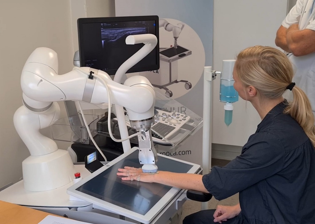
The system, made by a Danish company called ROPCA, comprises an ultrasound scanner called ARTHUR (RA Ultrasound Robot) that interacts directly with the patient and scans 11 joints per hand and a neural network–based software system, DIANA (Diagnosis Aid Network for RA), that evaluates the images and monitors RA activity.
The combined system classifies the degree of RA according to the joint European Alliance of Associations for Rheumatology (EULAR)–Outcome Measures in Rheumatology (OMERACT) standards for RA diagnosis. It received a CE Mark in Europe in 2022 and is currently in use in six rheumatology clinics in Denmark, Germany, Switzerland, and Austria, with more to come, ROPCA Co-founder and Chief Medical Officer Søren A. Just, MD, said in an interview.
“Automated systems could help rheumatologists in the early detection and monitoring of arthritis diseases. Systems can be placed or move in areas with insufficient rheumatological expertise,” Just said during a special late-breaker session presentation at the annual meeting of the American College of Rheumatology (ACR).
He said in an interview: “Currently, there are so many people referred and few and fewer rheumatologists. So we need to think differently. We need good automated assistants.” As a screening tool, the system can determine whether a person with hand pain has RA or just osteoarthritis “and also can give the patient an immediate answer, instead of waiting sometimes up to 6 months to get the information.”
Just, who is also a senior physician in the Department of Internal Medicine at Odense University Hospital in Denmark, said that his department is also using the system to assess flares in patients with established RA. “They can have a blood sample taken. They’re scanned by the robot, and you can see if there is any disease activity. But I think that screening of patients with joint pain is the beginning.”
Asked to comment, session moderator Gregory C. Gardner, MD, Emeritus Professor in the Division of Rheumatology at the University of Washington, Seattle, and a member of the ACR conference program committee, said in an interview “one of the reasons we chose to feature this abstract is because we’re interested in science at the convergence. We really thought this was a potential way to move the field forward for rheumatologists.”
Gardner said it’s an advantage that the patient could potentially have an ARTHUR scan with a DIANA report and get blood tests done prior to a visit with the rheumatologist. “It’s really time-consuming for a human to do these studies, so if you automate it, that’s a step forward in terms of having the data available for the rheumatologist to view and use sequentially to follow how patients are doing.”
When introducing Just’s presentation, Gardner called it “the coolest abstract of the meeting.”
Both DIANA and ARTHUR Performed At Least as Well as Human Rheumatologists
In the study, 30 patients with RA underwent two scans by ARTHUR, followed by a scan from a rheumatologist specialist in musculoskeletal ultrasound. The scans were sent to DIANA, who graded the images according to the Global OMERACT-EULAR Synovitis Score, as did the human rheumatologist.
A “ground truth” was established by another human expert who evaluated both ARTHUR’s and the other rheumatologist’s images, blinded to the scanning method. The image with the highest disease activity was deemed “ground truth,” and agreement with that was assessed for the two individual methods.

Just showed a video of a patient being scanned by ARTHUR. The machine verbally guided her through removing her jewelry, applying the gel, and placing her hand on the screen under the scanner. ARTHUR’s arm moved around on the patient’s hand, locating the best angles to take grayscale images and Doppler images and Doppler video. The scan takes 15-20 minutes, and the images are stored, Just said.
The study patients had a mean age of 65 years, and 23 of the 30 were men. Their average disease duration was 11 years, and mean Disease Activity Score in 28 joints using C-reactive protein was 3.86, indicating moderate disease. A majority (73%) of patients were taking disease-modifying antirheumatic drugs, and about one third were taking biologics. ARTHUR scanned a total of 660 joints, and 564 scans were successful.
For repeatability between the two ARTHUR scans, percent exact agreement was 63% for synovial hypertrophy, 75% for Doppler activity, and 60% combined. Percent close (within a point) agreements were 93%, 94%, and 92%, respectively. Binary agreements as to whether the joint was healthy vs diseased were 88%, 91%, and 85%, respectively.
At the joint level, ARTHUR and DIANA’s percent exact agreement with ground truth was 49% for synovial hypertrophy, 63% for Doppler activity, and 48% combined. Binary agreements with disease vs healthy were 80%, 88%, and 78%, respectively.
The human rheumatologists scored very similarly. Percent exact agreement with ground truth was 51% for synovial hypertrophy, 64% for Doppler activity, and 50% combined. Percent close agreements were 94%, 94%, and 92%, respectively. And binary agreements with diseased vs healthy were 83%, 91%, and 80%, respectively.
At the patient level (all joints combined), ARTHUR and DIANA’s binary disease assessment of healthy vs disease showed agreement with the ground truth of 87% for synovial hypertrophy, 83% for Doppler activity, and 87% combined. Here, the rheumatologists scored lower, at 53%, 67%, and 60%, respectively.
“In this study, we think the precision of ARTHUR and DIANA was comparable to that of an experienced rheumatologist, at both the joint and patient level,” Just said.
Gardner pointed out another advantage of the system. “DIANA doesn’t get fatigued. ... With human reading, the precision may change based on the time of day or stress level. ... But with DIANA, you’re going to get consistent information.”
Just said that the Arthritis Foundation in Germany recently put ARTHUR and DIANA on a bus and took it to cities that lacked a rheumatologist. Patients lined up, answered a questionnaire, had blood drawn, and received their scans. A rheumatologist on the bus then interpreted the data and consulted with the individuals about their RA risk. “In the last trip, we screened 800 patients in 6 days. So there are definitely possibilities here.”
Just is co-owner of ROPCA. Gardner had no disclosures.
A version of this article appeared on Medscape.com.
WASHINGTON — A fully automated ultrasound scanning system combined with artificial intelligence–based disease activity scoring performed as well as expert rheumatologists in hand joint assessment of patients with rheumatoid arthritis (RA), new research found.
The system, made by a Danish company called ROPCA, comprises an ultrasound scanner called ARTHUR (RA Ultrasound Robot) that interacts directly with the patient and scans 11 joints per hand and a neural network–based software system, DIANA (Diagnosis Aid Network for RA), that evaluates the images and monitors RA activity.
The combined system classifies the degree of RA according to the joint European Alliance of Associations for Rheumatology (EULAR)–Outcome Measures in Rheumatology (OMERACT) standards for RA diagnosis. It received a CE Mark in Europe in 2022 and is currently in use in six rheumatology clinics in Denmark, Germany, Switzerland, and Austria, with more to come, ROPCA Co-founder and Chief Medical Officer Søren A. Just, MD, said in an interview.
“Automated systems could help rheumatologists in the early detection and monitoring of arthritis diseases. Systems can be placed or move in areas with insufficient rheumatological expertise,” Just said during a special late-breaker session presentation at the annual meeting of the American College of Rheumatology (ACR).
He said in an interview: “Currently, there are so many people referred and few and fewer rheumatologists. So we need to think differently. We need good automated assistants.” As a screening tool, the system can determine whether a person with hand pain has RA or just osteoarthritis “and also can give the patient an immediate answer, instead of waiting sometimes up to 6 months to get the information.”
Just, who is also a senior physician in the Department of Internal Medicine at Odense University Hospital in Denmark, said that his department is also using the system to assess flares in patients with established RA. “They can have a blood sample taken. They’re scanned by the robot, and you can see if there is any disease activity. But I think that screening of patients with joint pain is the beginning.”
Asked to comment, session moderator Gregory C. Gardner, MD, Emeritus Professor in the Division of Rheumatology at the University of Washington, Seattle, and a member of the ACR conference program committee, said in an interview “one of the reasons we chose to feature this abstract is because we’re interested in science at the convergence. We really thought this was a potential way to move the field forward for rheumatologists.”
Gardner said it’s an advantage that the patient could potentially have an ARTHUR scan with a DIANA report and get blood tests done prior to a visit with the rheumatologist. “It’s really time-consuming for a human to do these studies, so if you automate it, that’s a step forward in terms of having the data available for the rheumatologist to view and use sequentially to follow how patients are doing.”
When introducing Just’s presentation, Gardner called it “the coolest abstract of the meeting.”
Both DIANA and ARTHUR Performed At Least as Well as Human Rheumatologists
In the study, 30 patients with RA underwent two scans by ARTHUR, followed by a scan from a rheumatologist specialist in musculoskeletal ultrasound. The scans were sent to DIANA, who graded the images according to the Global OMERACT-EULAR Synovitis Score, as did the human rheumatologist.
A “ground truth” was established by another human expert who evaluated both ARTHUR’s and the other rheumatologist’s images, blinded to the scanning method. The image with the highest disease activity was deemed “ground truth,” and agreement with that was assessed for the two individual methods.
Just showed a video of a patient being scanned by ARTHUR. The machine verbally guided her through removing her jewelry, applying the gel, and placing her hand on the screen under the scanner. ARTHUR’s arm moved around on the patient’s hand, locating the best angles to take grayscale images and Doppler images and Doppler video. The scan takes 15-20 minutes, and the images are stored, Just said.
The study patients had a mean age of 65 years, and 23 of the 30 were men. Their average disease duration was 11 years, and mean Disease Activity Score in 28 joints using C-reactive protein was 3.86, indicating moderate disease. A majority (73%) of patients were taking disease-modifying antirheumatic drugs, and about one third were taking biologics. ARTHUR scanned a total of 660 joints, and 564 scans were successful.
For repeatability between the two ARTHUR scans, percent exact agreement was 63% for synovial hypertrophy, 75% for Doppler activity, and 60% combined. Percent close (within a point) agreements were 93%, 94%, and 92%, respectively. Binary agreements as to whether the joint was healthy vs diseased were 88%, 91%, and 85%, respectively.
At the joint level, ARTHUR and DIANA’s percent exact agreement with ground truth was 49% for synovial hypertrophy, 63% for Doppler activity, and 48% combined. Binary agreements with disease vs healthy were 80%, 88%, and 78%, respectively.
The human rheumatologists scored very similarly. Percent exact agreement with ground truth was 51% for synovial hypertrophy, 64% for Doppler activity, and 50% combined. Percent close agreements were 94%, 94%, and 92%, respectively. And binary agreements with diseased vs healthy were 83%, 91%, and 80%, respectively.
At the patient level (all joints combined), ARTHUR and DIANA’s binary disease assessment of healthy vs disease showed agreement with the ground truth of 87% for synovial hypertrophy, 83% for Doppler activity, and 87% combined. Here, the rheumatologists scored lower, at 53%, 67%, and 60%, respectively.
“In this study, we think the precision of ARTHUR and DIANA was comparable to that of an experienced rheumatologist, at both the joint and patient level,” Just said.
Gardner pointed out another advantage of the system. “DIANA doesn’t get fatigued. ... With human reading, the precision may change based on the time of day or stress level. ... But with DIANA, you’re going to get consistent information.”
Just said that the Arthritis Foundation in Germany recently put ARTHUR and DIANA on a bus and took it to cities that lacked a rheumatologist. Patients lined up, answered a questionnaire, had blood drawn, and received their scans. A rheumatologist on the bus then interpreted the data and consulted with the individuals about their RA risk. “In the last trip, we screened 800 patients in 6 days. So there are definitely possibilities here.”
Just is co-owner of ROPCA. Gardner had no disclosures.
A version of this article appeared on Medscape.com.
FROM ACR 2024
Uric Acid Levels, Gout Symptoms Improved With Plant-Based Diet in Pilot Trial
A Mediterranean-inspired plant-based diet improved self-reported measures of gout as well as uric acid levels, a pilot study has found.
There hasn’t been a lot of research on diet in gout, according to Anna Kretova, RD, who presented the study at the annual research symposium of the Gout Hyperuricemia and Crystal-Associated Disease Network. She noted that a 2019 systematic review of low-calorie diets, low-purine diets, and Mediterranean diets found that uric acid levels below 0.6 mmol/L were achieved only in those on the Mediterranean diet (Nutrients. 2019 Dec 4;11[12]:2955). A 2020 study compared a low-fat, high-carbohydrate, plant-based diet vs an animal-based, ketogenic diet in healthy individuals. After 2 weeks, uric acid levels increased in those on the animal-based, low-carb diet and decreased in those on the plant-based diet.
Some foods are considered to be proinflammatory and generally come from animal origins, including saturated fats and animal protein in addition to ultraprocessed foods. Foods that have anti-inflammatory properties are mostly plant based and unprocessed and often rich in fiber. “From recent interventional studies, we also know that the whole-foods plant-based diet has shown to be effective as treatments of the main comorbidities of gout, such as obesity, cardiovascular disease, or [osteoarthritis],” said Kretova, who is a registered dietitian and a researcher at the Reade Rehabilitation and Rheumatology Center, Amsterdam, the Netherlands.
Those findings led the researchers to develop a whole-foods, plant-based diet and test its effect on serum uric acid in patients with gout, as well as gout disease activity and cardiovascular disease risk. Participants could not eat meat, fish, eggs, or dairy.
The trial included 33 individuals with gout who were randomized to a 16-week intervention with five consultations with a registered dietitian (n = 18) or a wait-list control group (n = 15) who received standard care. The mean age overall was 52 years, and 91% were men. The mean body mass index (BMI) was 32.6 kg/m2, and the median uric acid level was 0.50 mmol/L (8.4 mg/dL).
Among gout-related outcomes, the researchers noted improvements in gout severity as measured by visual analog scale (VAS; between group difference, –2.0; P =.01), pain as measured by VAS (between group difference, –2.0; P =.04), and uric acid levels after adjustment for age, sex, and BMI (between group difference, –0.05 mmol/L, P =.004). There were also improvements in the intervention group in weight loss (between group difference, –5.3 kg; P <.0001), BMI (between group difference, –1.7; P < .0001), waist circumference (between group difference, –3.9 cm; P = .004), and low-density lipoprotein (LDL) cholesterol (between group difference, –0.5; P = .007).
At 16 weeks, “we concluded that a Mediterranean-inspired whole-foods, plant-based diet significantly lowers serum uric acid in patients with gout and abdominal obesity, and additionally, the diet reduces gout-related pain and disease activity, promotes substantial weight loss, decreases weight circumference, and improves LDL cholesterol levels, and thus decreases [cardiovascular disease] risk in these patients,” Kretova said.
She added that some might question whether a uric acid reduction of –0.05 mmol/L is clinically relevant. “We would argue it is because of the strong decrease in disease activity and pain in the intervention group,” Kretova said.
The study is limited by its small size, the fact that it was not blinded, and the 4-month duration, which might be too short to capture potential indirect effects of diet on hyperuricemia and chronic inflammation, Kretova said. The group is planning to follow participants out to 12 months in an extension study.
During the Q&A session after the presentation, an audience member asked if the participants were vegetarians before they entered the study, and whether the dietary change could be sustained. “It’s a very good proof-of-concept study, but whether an intervention based entirely on plant-based therapy will be something that patients will be able to adhere to long term [is uncertain],” Kretova said.
She was optimistic, even though the participants generally enjoyed food and ate a lot of red meat. “I think there will be a gradation of people who can sustain and who cannot sustain [the diet]. From what we saw, people actually found it easier to follow than they expected, and a lot of participants changed their diet permanently for the better. Not everyone became [entirely] plant-based, but they became much more plant-based than they expected from themselves. So, it is definitely feasible,” she said.
Kretova reported no relevant financial relationships.
A version of this article appeared on Medscape.com.
FROM G-CAN 2024
Three Vascular Risk Factors May Up Severe Stroke Risk
TOPLINE:
, a global study shows.
METHODOLOGY:
- The INTERSTROKE case-control study included nearly 27,000 participants, half of whom had a first acute stroke (ischemic or hemorrhagic) and the other half acting as age- and sex-matched controls.
- Participants (mean age, 62 years; 40% women) were recruited across 142 centers in 32 countries between 2007 and 2015. Baseline demographics and lifestyle risk factors for stroke were gathered using standardized questionnaires
- Modified Rankin Scale (mRS) scores measured within 72 hours of hospital admission were used to classify stroke severity (0-3, nonsevere stroke; 4-6, severe stroke).
TAKEAWAY:
- Among the participants with acute stroke, 64% had nonsevere stroke and 36% had severe stroke, based on the mRS.
- Hypertension, atrial fibrillation, and smoking showed a significantly stronger association with severe stroke than with nonsevere stroke (odds ratios [ORs], 3.21 vs 2.87, 4.70 vs 3.61, and 1.87 vs 1.65, respectively; all P < .001).
- A high waist-to-hip ratio showed a stronger association with nonsevere stroke than with severe stroke (OR, 1.37 vs 1.11, respectively; P < .001).
- Diabetes, poor diet, physical inactivity, and stress were linked to increased odds of both severe and nonsevere stroke, whereas alcohol consumption and high apolipoprotein B levels were linked to higher odds of only nonsevere stroke. No significant differences in odds were observed between stroke severities in matched individuals.
IN PRACTICE:
“Our findings emphasize the importance of controlling high blood pressure, which is the most important modifiable risk factor for stroke globally,” lead author Catriona Reddin, MB BCh, BAO, MSc, School of Medicine, University of Galway, in Ireland, said in a press release.
SOURCE:
The study was published online in Neurology.
LIMITATIONS:
The study limitations included potential unmeasured confounders; reliance on the mRS score, which may have underestimated stroke severity; and challenges with recruiting patients with severe stroke in a case-control study. Smoking-related comorbidities and regional or sex-related variations in alcohol intake may also have influenced the results.
DISCLOSURES:
The study was funded by various organizations, including health research councils and foundations from Canada, Sweden, and Scotland, and pharmaceutical companies such as AstraZeneca, Boehringer Ingelheim, Pfizer, and MSD. One investigator reported receiving funding from the Irish Clinical Academic Training Programme, the Wellcome Trust and the Health Research Board, the Health Service Executive, National Doctors Training and Planning, and the Health and Social Care, Research and Development Division in Northern Ireland. No other conflicts of interest were reported.
This article was created using several editorial tools, including AI, as part of the process. Human editors reviewed this content before publication. A version of this article appeared on Medscape.com.
Phase 3 Lupus Trial Shows Promising Results for Dapirolizumab Pegol
WASHINGTON — The investigational anti-CD40 ligand agent dapirolizumab pegol (DZP) outperformed placebo in improving disease activity and reducing high-dose corticosteroid use in patients with systemic lupus erythematosus (SLE) in the phase 3 PHOENYCS GO trial.
“We really think that dapirolizumab pegol may represent a novel treatment for lupus, particularly given its broad immune modulatory effects,” said Megan Clowse, MD, MPH, associate professor of medicine and chief of the Division of Rheumatology and Immunology at Duke University School of Medicine in Durham, North Carolina. She presented the study in a late-breaking poster session at the American College of Rheumatology (ACR) 2024 Annual Meeting.
There is a “huge unmet need” for drugs for lupus, Clowse told this news organization. Patients with SLE continue to have high disease burden, including ongoing symptoms often driven by inflammation. Corticosteroids are often the best medications to control disease activity, she said, but they can result in long-term toxicity.
What Makes DZP Unique?
Through CD40 ligand signaling, DZP has been shown to reduce B- and T-cell activation and to downregulate interferon pathways. Previous antibodies targeting the CD40 ligand have been associated with an increased risk for thromboembolic events. However, DZP lacks the Fc portion of the antibody, which can bind to platelets and cause clotting. Data from phase 1, 2, and 3 trials thus far do not show an elevated risk for these events, Clowse explained. In fact, safety signals were strong enough that patients with antiphospholipid antibodies — a key driver for blood clots in patients with SLE — were included in the trial.
In PHOENYCS GO, investigators enrolled 321 patients with moderate to severe SLE with persistently active or frequently flaring/relapsing-remitting disease activity despite stable standard of care (SOC) medications such as antimalarials, corticosteroids, and immunosuppressants.
Patients were randomized 2:1 to receive intravenous DZP (24 mg/kg) plus SOC or intravenous placebo plus SOC every 4 weeks, with patients and investigators blinded to treatment assignments.
Patients taking a corticosteroid dose > 7.5 mg/day began a mandatory steroid taper by week 8 of the trial, with the goal of reducing that to < 7.5 mg/day. The tapering regimen was at the discretion of providers and was adapted to each patient’s individual disease activity.
The primary endpoint was British Isles Lupus Assessment Group–based Composite Lupus Assessment (BICLA) response at week 48.
Patients in the DZP and placebo groups were on average 43.5 and 41.5 years old, respectively. More than 90% of patients were women, all on concomitant SLE medications. About half of the participants took a daily corticosteroid dose > 7.5 mg.
At 48 weeks, half of the DZP group (49.5%) achieved BICLA response compared with 34.6% in the placebo group (P = .0110). A higher proportion of patients taking DZP achieved SLE Responder Index-4 response than those taking placebo (60.1% vs 41.1%, respectively; P = .0014), and the rate of severe British Isles Lupus Assessment Group flares in the DZP group was half that of the placebo group (11.6% vs 23.4%; P = .0257). In the subgroup of patients who underwent corticosteroid tapering, 72.4% receiving DZP and 52.9% taking placebo reduced their dose to < 7.5 mg/day by 48 weeks (P = .0404).
DZP was generally well tolerated. Over 48 weeks, 82.6% of the DZP group and 75% of the placebo group reported treatment-emergent adverse events, but serious occurrences were more common in the placebo group (14.8%) than in the DZP group (9.9%). Herpes viral infections were higher in the placebo group, although there were three ophthalmic herpes cases in the DZP group. There was one case of acute myocardial infarction and one death linked to gangrene-related sepsis in patients receiving DZP.
A ‘Mild to Moderate’ Response
Although these are definitely positive results, they show a “mild to moderate response” to DZP, commented Gregory Gardner, MD, an emeritus professor in the Division of Rheumatology at the University of Washington, Seattle, and chair of the American College of Rheumatology’s annual meeting planning committee. He moderated the session where the research was presented. Although DZP showed efficacy among some patients, he noted, “there were still 51% patients that it didn’t work for.”
The drug uses an alternative pathway to current lupus drugs, Gardner added, and more research is needed to understand how best to use this medication in practice.
Clowse noted that DZP could be particularly beneficial for patients with SLE who want to get pregnant. Many drugs used to treat the disease are teratogenic; however, “because of the lack of Fc portion on this drug, it very likely does not cross the placenta in any kind of significant amount,” she said. Although there are not yet any reproductive safety data on DZP, she added, “that is a great potential niche.”
Biogen and UCB, which are jointly developing DZP, aim to start a second phase 3 trial of DZP in patients with SLE, called PHOENYCS FLY, in 2024.
The trial was sponsored by UCB. Clowse is a consultant and has received grant/research support from GSK and UCB. Gardner had no relevant disclosures.
A version of this article appeared on Medscape.com.
FROM ACR 2024
New Gout Remission Criteria Approved
In a nearly unanimous vote at the annual research symposium of the Gout Hyperuricemia and Crystal-Associated Disease Network (G-CAN), members approved a revision to gout remission criteria first established in 2016. The new version simplifies the definition in response to patient comments that the earlier version was redundant in some areas.
The previous version was developed following deliberations by 49 clinicians and researchers with experience in gout. They settled on a definition of gout remission that included five criteria:
- Serum urate levels lower than 0.36 mmol/L measured at least twice over 12 months, with no intervening values of 0.36 mmol/L or higher
- No gout flares over 12 months
- No tophi
- Pain score due to gout < 2 at least twice over 12 months on a 10-point Likert scale or 10-cm visual analog scale, with no intervening values ≥ 2
- Patient global assessment of gout disease activity < 2 on a 10-point Likert scale or 10-cm visual analog scale, with no intervening values of ≥ 2.
Some participants reported that patients sometimes misattributed pain from other sources while using patient-reported outcomes (PROs). The argument for keeping PROs was that they are validated measures and endorsed by Outcome Measures in Rheumatology. Nevertheless, there was no direct patient involvement in the development of the 2016 criteria.
Researchers later interviewed 20 individuals with well-controlled gout to get their feedback on the 2016 criteria. Those individuals endorsed the existing criteria and did not suggest any new ones, but they suggested that the pain due to gout and the absence of gout flares were redundant measures. One said: “If you have no flare-ups, you’ve got no pain; it sort of answers itself.”
“That was a bit challenging for us because it wasn’t quite what we expected, but I think it did make us look again at the definition and think about whether we could simplify the definition further,” Nicola Dalbeth, MBChB, said during a presentation at G-CAN. Dalbeth is an academic rheumatologist at the University of Auckland, in New Zealand, who was also the lead author of the original criteria.
Simplified Version Created With Only Three Criteria
In response to these points, researchers produced a revised version with only three criteria, including the serum urate, absence of gout flares, and absence of subcutaneous tophi at the time of assessment.
To determine if the simplified criteria performed well, they compared the original and revised remission criteria in the context of the CARES trial, the Nottingham nurse-led trial, and randomized controlled trials in patients with gout that were conducted in New Zealand (here and here).
Dansoa Tabi-Amponsah, a PhD candidate at the University of Auckland, presented results of a study comparing the two versions in the Nottingham trial, which included 517 participants who received nurse-led or usual general practitioner care. The nurse-led care included education, regular follow-up and serum urate testing, individualized advice on gout flare management, and escalation of urate-lowering therapy with a treat-to-target strategy.
Both definitions demonstrated a link between the nurse-led strategy and increased rates of remission at year 1 and year 2, although the simplified definition found that more patients were in remission (17.6% vs 9.9% at year 1 and 42.7% vs 28.4% at year 2, both P < .001). “This is something we’ve seen across all of our analyses,” said Tabi-Amponsah.
Both criteria also found significant differences in remission rates between the nurse-led group in year 2 vs year 1 but not in the usual care group.
Participants who achieved remission had better gout impact scale scores in areas like worrying that a gout attack will occur, fears of worsening gout, and concerns about the impact of gout on future activities. “This is important because during that qualitative study, a key aspect of being in remission was no longer being worried about their gout, no longer feeling anxious about having constant gout flares, and having control over their gout. So, it’s important to note that despite the absence of PROs in that simplified definition, it’s still able to align with the patients’ perspectives of their disease state,” said Tabi-Amponsah.
During the Q&A period after her talk, an audience member asked whether the higher rate of remission found by the simplified criteria is actually a good thing. “If I compare that to rheumatoid arthritis, when you use DAS28 you have a lot more remission, but still progression. So, are we missing some people? Are we including people in remission that still have disease?” she asked.
Tabi-Amponsah responded that the pain and patient global assessment domains seem to be quite difficult to achieve. In a separate analysis, the researchers examined tender and swollen joint counts and found that those achieving remission no longer had tender or swollen joints. “So, we don’t think the simplified definition is heavily misclassifying anyone as being in remission,” she said.
During the Q&A following Dalbeth’s talk, an audience member asked about patients with what he described as “mountains of tophi,” despite responding well to uricase therapy. “They may take months or even a year to really resolve that burden. They may be doing very well, yet they’re not going to be in remission because they’ve still got visible tophi. So, are we underselling them, and do we need a different definition for them doing well that this doesn’t capture?” he asked.
Dalbeth suggested that patients with large amounts of tophi aren’t really in remission. “I think we do need to be thinking about the disease, not just in terms of just crystals or just inflammation, but actually trying to integrate both of those, and I think this is where these composite measures might work quite well. I think we need to be aiming for holistic disease control, which is essentially what this is,” she said.
Tabi-Amponsah and Dalbeth did not disclose any financial relationships.
A version of this article appeared on Medscape.com.
FROM G-CAN 2024
Higher Early-Onset CRC Mortality Seen in Racial, Ethnic Minorities
TOPLINE:
The largest racial and ethnic disparities in survival were linked to neighborhood socioeconomic status.
METHODOLOGY:
- US rates of EOCRC are increasing, with differences across racial and ethnic groups, but few studies have provided detailed risk estimates in the categories of Asian American and of Native Hawaiian or Other Pacific Islander, as well as the contribution of sociodemographic factors to these differences.
- A population-based cohort study analyzed California Cancer Registry data for 22,834 individuals aged 18-49 years diagnosed with EOCRC between January 2000 and December 2019.
- Researchers examined the association between mortality risk and racial and ethnic groups, including Asian American (15.5%, separated into seven subcategories), Hispanic (30.2%), Native Hawaiian or Other Pacific Islander (0.6%), non-Hispanic American Indian or Alaska Native (0.5%), non-Hispanic Black (7.3%), and non-Hispanic White (45.9%) individuals, with a median follow-up of 4.2 years.
- Statistical models measured baseline associations adjusting for clinical features and then tested for the contribution of socioeconomic factors together and separately, with adjustments for insurance status, neighborhood socioeconomic status, and more.
TAKEAWAY:
- Native Hawaiian or Other Pacific Islander individuals demonstrated the highest EOCRC mortality risk compared with non-Hispanic White individuals (socioeconomic status–adjusted HR [SES aHR], 1.34; 95% CI, 1.01-1.76).
- Non-Hispanic Black individuals showed a higher EOCRC mortality risk than non-Hispanic White individuals (SES aHR, 1.18; 95% CI, 1.07-1.29).
- Hispanic individuals’ higher EOCRC mortality (base aHR, 1.15; 95% CI, 1.08-1.22) disappeared after adjusting for neighborhood socioeconomic status (SES aHR, 0.98; 95% CI, 0.92-1.04).
- Southeast Asian individuals’ increased mortality risk (base aHR, 1.17; 95% CI, 1.03-1.34) was no longer significant after adjusting for insurance status (SES aHR, 1.10; 95% CI, 0.96-1.26).
IN PRACTICE:
“As clinicians and researchers, we should ask ourselves how to act on these findings,” wrote the authors of an invited commentary. “The effort cannot stop with data analysis alone, it must extend to actionable steps,” such as tailored efforts to deliver culturally competent care and patient navigation services to those with greatest need and at highest risk, they added.
SOURCE:
The study was led by Joshua Demb, PhD, University of California, San Diego. The study was published online on November 22 in JAMA Network Open (2024. doi: 10.1001/jamanetworkopen.2024.46820) with the invited commentary led by Clare E. Jacobson, MD, University of Michigan, Ann Arbor.
LIMITATIONS:
The study was limited by a relatively short follow-up time and small sample sizes in some racial and ethnic groups, potentially leading to imprecise aHR estimates. The generalizability of findings beyond California requires further investigation, and the ability to examine potential associations between neighborhood socioeconomic status and other factors was also constrained by small sample sizes.
DISCLOSURES:
The study received support from the National Cancer Institute at the National Institutes of Health. One study author reported receiving consulting fees from Guardant Health, InterVenn Biosciences, Geneoscopy, and Universal DX; research support from Freenome; and stock options from CellMax outside the submitted work. No other disclosures were reported by other authors of the study or the commentary.
This article was created using several editorial tools, including AI, as part of the process. Human editors reviewed this content before publication. A version of this article appeared on Medscape.com.
It’s Not Too Late for Influenza Vaccination: Q&A With CDC’s Dr. Lisa Grohskopf
Text has been edited for length.
Are there any updates to this season’s influenza vaccine or vaccine recommendations?
Yes, we have updates to both the vaccine and the vaccine recommendations this year. Typically we have some changes each year, and this year there are two main changes in the recommendations. One relates to the composition of the vaccine for this season, and the other is a new recommendation for adult solid organ transplant recipients.
We typically have changes in the vaccine composition each season. For most seasons, one or more parts of the vaccine will change, but this year is a little different in that all of the vaccines available in the US for the 2024-2025 season are going to be three-virus, or trivalent, vaccines. They are going to be formulated to protect against three viruses: an influenza A(H1N1) virus, an influenza A(H3N2) virus, and an influenza B/Victoria lineage virus.
The reason for this change is that since the 2013-2014 season through the 2023-2024 season, we had quadrivalent vaccines that were available in the US that contained four viruses. Those vaccines contained a second influenza B virus from the Yamagata lineage (B viruses come from two main lineages).
The reason for the change to trivalent vaccines this season is that influenza B/Yamagata viruses have not been detected in global surveillance since March 2020, and so their inclusion is no longer warranted. So this season, all of the vaccines available in the US are going to be trivalent.
In addition to that change, we have an update in the influenza A(H3N2) component of the vaccine compared with last season.
The second change concerning adult solid organ transplant recipients is that Advisory Committee on Immunization Practices (ACIP) now recommends that solid organ transplant recipients aged 18-64 years can receive as acceptable options either the high-dose inactivated vaccine or the adjuvanted inactivated vaccine without a preference over other age appropriate, inactivated, or recombinant vaccines.
Those vaccines are both formulated with features intended to make them more immunogenic — ie, promote a stronger immune response — and there are data for immunogenicity that suggest they could be more immunogenic in that population.
Who needs an influenza vaccine this season?
That recommendation is the same as it’s been for a number of years, which is that everybody aged 6 months or older is recommended to get a flu vaccine, with some rare exceptions, mainly concerning contraindications to vaccination.
Contraindications are detailed in the ACIP flu statement each year, and they’re relatively uncommon conditions overall, so most people are recommended, if they’re in that age group 6 months and up, to get an annual flu vaccine.
Are there groups for whom influenza vaccination is especially important?
Yes. While influenza vaccination is recommended for everybody in that age group 6 months and up — and in truth, we can never really predict who’s going to get severely ill — some people are more likely to be at risk of having serious illness or hospitalization. Those people include adults aged 65 years or older; young children; people with certain chronic health conditions such as heart disease, lung disease, and diabetes; and people from some racial and ethnic groups.
Are there any specific influenza vaccination recommendations for these groups or others?
Not for most people. In general, we have a number of different influenza vaccines each season; this year we have a total of nine brands. In general, there’s no preferential recommendation for one vaccine or type of vaccine for specific groups of people, with one exception: For people aged 65 years or older, there’s a preferential recommendation for three specific vaccines — the high-dose inactivated vaccine, Fluzone High-Dose; the recombinant vaccine, Flublok; and the adjuvanted inactivated vaccine, FLUAD.
Among those three, there’s no preference for any one of them over the other two; they’re all preferred vaccines for this age group, if available. If none of those three vaccines are available at the time that somebody aged 65 or older is there to get vaccinated, people in this age group should get any other age-appropriate influenza vaccine that is available.
When should people get vaccinated if they haven’t already?
CDC and ACIP recommend vaccination for most people, ideally by the end of October. But for those who missed the end of October, it is absolutely not too late. Providers should continue to encourage vaccination and people should get their vaccines as long as flu viruses are circulating.
The timing of the onset and the peak and the end of the flu season vary a bit from year to year. We often start to see generally activity begin to increase in the US in the fall, which is the reason for the end of October recommendation; however, flu activity doesn’t tend to peak in the US until after October. We’re talking December, January, or later, so getting vaccinated after October can still provide important protection during the peak of the season.
There does seem to be a tendency for people to think, OK, I haven’t gotten the vaccine yet, and there probably isn’t a lot of reason to do it now. But really, it’s definitely not too late, and that’s something we like to encourage people to think about, particularly as we move into December and January — it’s not too late if you missed October.
Influenza vaccination is also available in so many places. You don’t necessarily have to go to a healthcare provider’s office; there are many retail chains which offer influenza vaccines.
Is influenza spreading right now? Are activity levels increasing?
Overall influenza activity currently is low nationally, although there’s starting to be some slight increases in the pediatric age groups and, of course, we do anticipate that it will increase in the coming weeks and months.
When we get vaccinated, the protection isn’t instantaneous. The immune system needs a bit of time to react to the vaccine and to develop antibodies. That can take about 2 weeks. Even with that, now is still absolutely not too late to get a vaccine. Neither is December, for that matter. As long as the flu viruses are circulating where you are, it is still worth getting vaccinated.
What was influenza vaccination coverage like last season?
It’s a little bit early to tell for the current season, but one of the things that we do know is that since the COVID-19 pandemic, coverage has dropped compared with before the COVID-19 pandemic. Before COVID-19, influenza vaccination coverage had been slowly increasing in most groups, but it has decreased since then, and those downturns in coverage haven’t recovered to prepandemic levels. For example, during 2023-2024, about half of children and adults received a flu vaccine.
What can providers do to encourage influenza vaccination in their patients?
We know that a healthcare provider’s strong recommendation for flu vaccination is a really major factor in whether or not patients get a flu vaccine, and is more effective in increasing acceptance of vaccination than just about any other factor.
There’s a method from CDC called SHARE, which is a helpful way to help make a strong recommendation and provide information to help patients make an informed decision about whether or not they want to be vaccinated.
To implement SHARE, it’s an acronym with five parts. S is for Share the reasons why the flu vaccine is right for that patient. H is for Highlight positive experiences with flu vaccination, either personal or in practice. A is for Address patient concerns and questions about the flu vaccine, including things such as side effects, safety, and effectiveness. R is Remind patients that vaccination protects them and their loved ones from serious illness and related complications. E is Explain the potential complications and consequences of getting influenza, including serious health effects, time lost from family, work, and school, and potential financial costs.
Additional resources are accessible on CDC’s influenza resources page, including brochures, posters, and fact sheets that can help providers in encouraging and reminding people to get vaccinated.
A version of this article appeared on Medscape.com.
RSV Infections Take Toll on Adults
based on new data from more than 67,000 cases.
RSV remains a top cause of acute respiratory tract infections among adults in the United States, with an estimated 159,000 hospitalizations in those aged 65 years or older, wrote Suzanne N. Landi, MPH, PhD, of Pfizer in New York City, and colleagues in a study published in JAMA Network Open.
“Currently, limited estimates exist to determine the risk of hospitalization following outpatient RSV disease diagnoses in the United States,” said corresponding author Joshua T. Swan, PharmD, MPH, in an interview.
The current study was conducted to inform development of clinical trials, said Swan, senior director and category clinician in internal medicine and disease development at Pfizer, the sponsor of the study. These trials would assess the efficacy of an outpatient RSV antiviral treatment in preventing RSV-related hospitalization within 28 days among adults at a high risk for progression to severe illness, he said.
The authors reviewed data from 67,239 adults aged 18 years or older with medically attended RSV infections between October 1, 2016, and September 30, 2022. The data came from three databases: Optum (2771 patients), TriNetX (7442 patients), and Veradigm Network Electronic Health Record (VNEHR; 57,026 patients).
The primary outcome was all-cause hospitalization within 28 days of medically attended RSV.
Overall, the proportions of patients hospitalized within 28 days of infection were 6.2%, 6.0%, and 4.5% in Optum, TriNetX, and VNEHR databases, respectively.
Approximately two thirds of the patients (62%-67% across the three databases) were women, and 14.0%-54.5% were aged 65 years or older. The researchers also identified comorbidity prevalences of 20.0%-30.5% for chronic obstructive pulmonary disease (COPD), 14.6%-24.4% for heart failure (HF), and 14.6%-24.4% for asthma.
A majority of the patients (ranging from 74.5% to 90.6% across the three databases) fell into a high-risk subgroup, defined as age 65 years or older with asthma, COPD, and HF. In this high-risk group, the proportions of hospitalizations were 7.6%, 8.5%, and 6.5% for Optum, TriNetX, and VNEHR, respectively.
The findings were limited by several factors, including the use only of data from outpatient settings, which cannot be used to estimate the RSV burden in the general population, and the reliance only on diagnosis or procedure codes to identify comorbidities, the researchers noted.
However, “the absolute risk of hospitalization of 1 out of 20 patients observed in our study represents significant and meaningful risk for vulnerable adults, in a disease where much of the public’s attention has historically focused on risk of hospitalization for young children,” Swan said. “These results highlight the unmet medical need for outpatient interventions and preventive measures that can reduce hospitalizations.”
Don’t Underestimate RSV Impact
The current study highlights the fact that RSV is a major cause of respiratory viral illness, said David R. Manoff, MD, associate professor of clinical thoracic medicine and surgery at Temple University, Philadelphia, in an interview.
“Historically, influenza, and, more recently, COVID-19 infection have generally been thought of as more likely to cause harm and, thus, have been more emphasized in terms of both vaccination and treatment,” said Manoff, who was not involved in the study.
The current study provides new evidence that infection with RSV can be far more serious than often recognized and a major potential source of both hospitalization and morbidity, Manoff said. In fact, data published in Morbidity and Mortality Weekly Report in 2023 showed that the risks of needing oxygen, intensive care unit (ICU) admission, intubation, and death were actually higher in patients hospitalized with RSV infections than in those hospitalized with influenza or COVID-19. “
Understanding which population is hospitalized in the first place is vital to targeting prevention measures,” he added.
The new data are consistent with previous studies showing that most patients with RSV infection have primarily upper respiratory tract infection–type symptoms, but that a minority will develop lower respiratory tract disease, Manoff noted.
The findings add to the argument for implementation of RSV vaccination, especially in high-risk individuals, and support the need for RSV testing when patients present for care, he said.
However, more research is needed to reflect recent numbers, Manoff said. The study timeframe of 2016-2022 not only precedes commercially available RSV vaccines but also includes the period of increased isolation and masking seen during the COVID-19 pandemic years of 2020-2021. “We need to see if the same trends continue in the post-pandemic era.”
Additionally, the studies leading to approval of the RSV vaccine showed a reduction in hospitalization with RSV, and it is important to see how this reduction translates in real-world data and whether the RSV vaccines are reducing need for ICU admission, intubation, and death, Manoff said.
The study was funded by Pfizer, and Swan is a Pfizer employee. Manoff had no financial conflicts to disclose.
A version of this article appeared on Medscape.com.
FROM JAMA NETWORK OPEN