User login
MDedge latest news is breaking news from medical conferences, journals, guidelines, the FDA and CDC.
Nutrition, Drugs, or Bariatric Surgery: What’s the Best Approach for Sustained Weight Loss?
Given that more than 100 million US adults have obesity, including 22 million with severe obesity, physicians regularly see patients with the condition in their practices.
Fortunately, doctors have more tools than ever to help their patients. But the question remains: Which method is the safest and most effective? Is it diet and lifestyle changes, one of the recently approved anti-obesity medications (AOMs), bariatric surgery, or a combination approach?
There are no head-to-head trials comparing these three approaches, said Vanita Rahman, MD, clinic director of the Barnard Medical Center, Washington, DC, at the International Conference on Nutrition in Medicine, sponsored by the Physicians Committee for Responsible Medicine.
Instead, doctors must evaluate the merits and drawbacks of each intervention and decide with their patients which treatment is best for them, she told Medscape Medical News. When she sees patients, Rahman shares the pertinent research with them, so they are able to make an informed choice.
Looking at the Options
In her presentation at the conference, Rahman summarized the guidelines issued by the American Heart Association/American College of Cardiology/The Obesity Society for Management of Overweight and Obesity in Adults and the American Association of Clinical Endocrinologists and American College of Endocrinology Comprehensive Clinical Practice Guidelines For Medical Care of Patients with Obesity, including lifestyle changes, AOMs, and bariatric surgery (Table 1).
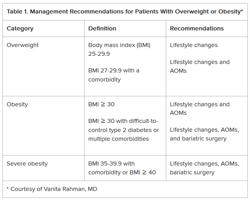
As shown, the current clinical guidelines offer recommendations that consider such factors as the patient’s BMI and presence of one or more comorbidities. Generally, they begin with lifestyle changes for people with overweight, the possibility of an AOM for those with obesity, and bariatric surgery as an option for those with severe obesity-related complications.
“In obesity, we traditionally thought the process was ‘either-or’ — either lifestyle or surgery or medication — and somehow lifestyle is better,” Sheethal Reddy, PhD, a psychologist at the Bariatric Center at Emory University Hospital Midtown, Atlanta, told Medscape Medical News.
Now physicians often use a combination of methods, but lifestyle is foundational to all of them, she said.
“If you don’t make lifestyle changes, none of the approaches will ultimately be effective,” said Reddy, who also is an assistant professor in the Division of General and GI Surgery at Emory School of Medicine, Atlanta.
Lifestyle changes don’t just involve diet and nutrition but include physical exercise.
“Being sedentary affects everything — sleep quality, appetite regulation, and metabolism. Without sufficient exercise, the body isn’t functioning well enough to have a healthy metabolism,” Reddy said.
How Durable Are the Interventions?
Although bariatric surgery has demonstrated effectiveness in helping patients lose weight, many of them regain some or most of it, Rahman said.
A systematic review and meta-analysis found weight regain in 49% of patients who underwent bariatric surgery patients, with the highest prevalence after Roux-en-Y gastric bypass.
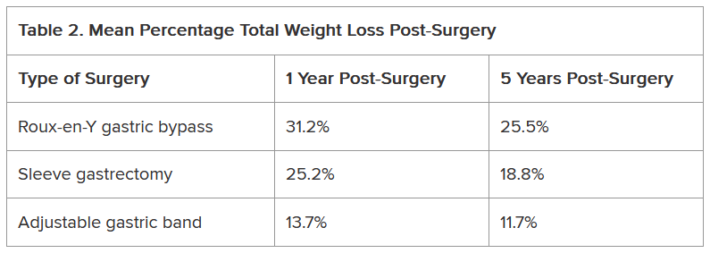
Another study of approximately 45,000 patients who underwent bariatric surgery found differences not only in the percentage of total weight loss among Roux-en-Y gastric bypass, sleeve gastrectomy, and adjustable gastric band procedures but also in how much of that weight stayed off between 1 and 5 years following the procedure (Table 2).
Weight regain also is a risk with AOMs, if they’re discontinued.
The STEP 1 trial tested the effectiveness of semaglutide — a glucagon-like peptide 1 (GLP-1) receptor agonist — as an adjunct to lifestyle intervention for weight loss in patients with obesity or with overweight and at least one comorbidity but not diabetes. Mean weight loss with semaglutide was 17.3% but that figure dropped 11.6 percentage points after treatment was discontinued.
Other studies also have found that patients regain weight after GLP-1 discontinuation.
Tirzepatide, a GLP-1 and glucose-dependent insulinotropic polypeptide (GIP) combination, has shown efficacy with weight reduction, but patients experienced some weight regain upon discontinuation. In one study, patients experienced a mean weight loss of 20.9% after 36 weeks of tirzepatide. In the study’s subsequent 52-week double-blind, placebo-controlled period, patients who stopped taking the medication experienced a weight regain of 14%, whereas those who remained on the medication lost an additional 5.5% of weight.
GLP-1 and GLP-1/GIP medications do not address the factors that contribute to overweight and obesity, Rahman said. “They simply suppress the appetite; therefore, weight gain occurs after stopping them.”
Patients may stop taking anti-obesity drugs for a variety of reasons, including side effects. Rahman noted that the common side effects include nausea, vomiting, and constipation, whereas rare side effects include gastroparesis, gallbladder and biliary disease, thyroid cancer, and suicidal thoughts. GLP-1 and GLP-1/GIP medications also carry a risk for non-arteritic anterior ischemic optic neuropathy, she said.
Moreover, health insurance does not always cover these medications, which likely affects patient access to the drugs and compliance rates.
“Given the side effects and frequent lack of insurance coverage, significant questions remain about long-term safety and feasibility of these agents,” Rahman said.
What About Nutritional Approaches?
The lifestyle interventions in the semaglutide and tirzepatide studies included 500 kcal/d deficit diets, which is difficult for people to maintain, noted Rahman, who is the author of the book Simply Plant Based: Fabulous Food for a Healthy Life.
Additionally, bariatric surgery has been associated with long-term micronutrient deficiencies, including deficiencies in vitamins A, D, E, K, B1, and B12, as well as folate, iron, zinc, copper, selenium, and calcium, she said.
The best approach to food from a patient compliance standpoint and to avoid nutrient deficiencies is a whole-food, plant-based diet, Rahman said. She advocates this nutritional approach, along with physical activity, for patients regardless of whether they’ve selected lifestyle intervention alone or combined with an AOM or bariatric surgery to address obesity.
Rahman cited a 5-year heart disease study comparing an intensive lifestyle program involving a vegetarian diet, aerobic exercise, stress management training, smoking cessation, and group psychosocial support to treatment as usual. Patients in the lifestyle group lost 10.9 kg at 1 year and sustained weight loss of 5.8 kg at 5 years, whereas weight in the control group remained relatively unchanged from baseline.
She also pointed to the findings of a study of patients with obesity or with overweight and at least one comorbidity that compared standard care with a low-fat, whole-food, plant-based diet with vitamin B12 supplementation. At 6 months, mean BMI reduction was greater in the intervention group than the standard care group (−4.4 vs −0.4).
In her practice, Rahman has seen the benefits of a whole-food, plant-based diet for patients with obesity.
If people are committed to this type of dietary approach and are given the tools and resources to do it effectively, “their thinking changes, their taste buds change, and they grow to enjoy this new way of eating,” she said. “They see results, and it’s a lifestyle that can be sustained long-term.”
Addressing Drivers of Weight Gain
Patients also need help addressing the various factors that may contribute to overweight and obesity, including overconsumption of ultra-processed foods, substandard nutritional quality of restaurant foods, increasing portion sizes, distraction during eating, emotional eating, late-night eating, and cultural/traditional values surrounding food, Rahman noted.
Supatra Tovar, PsyD, RD, a clinical psychologist with a practice in Pasadena, California, agreed that identifying the reasons for weight gain is critical for treatment.
“If you’re not addressing underlying issues, such as a person’s relationship with food, behaviors around food, the tendency to mindlessly eat or emotionally eat or eat to seek comfort, the person’s weight problems won’t ultimately be fully solved by any of the three approaches — dieting, medications, or bariatric surgery,” she said.
Some of her patients “engage in extreme dieting and deprivation, and many who use medications or have had bariatric surgery hardly eat and often develop nutritional deficiencies,” said Tovar, author of the book Deprogram Diet Culture: Rethink Your Relationship with Food, Heal Your Mind, and Live a Diet-Free Life.
The key to healthy and sustained weight loss is to “become attuned to the body’s signals, learn how to honor hunger, stop eating when satisfied, and eat more healthful foods, such as fruits and vegetables, whole grains, lean proteins — especially plant-based proteins — and the body gives signals that this is what it wants,” she said.
Tovar doesn’t give her clients a specific diet or set of portions.
“I teach them to listen to their bodies,” she said. “They’ve lost significant amounts of weight and continued to keep it off because they’ve done this kind of work.”
When Lifestyle Changes Aren’t Enough
For many patients, lifestyle interventions are insufficient to address the degree of overweight and obesity and common comorbidities, said W. Timothy Garvey, MD, associate director and professor, Department of Nutrition Sciences, School of Health Professions, University of Alabama at Birmingham.
“Of course, nutritional approaches are very important, not only for weight but also for general health-related reasons,” said Garvey, lead author of the 2016 American Association of Clinical Endocrinologists obesity guidelines. “We’ve seen that the Mediterranean and some plant-based diets can prevent progression from prediabetes to diabetes and improve other parameters that reflect metabolic health.”
However, it’s “not common that patients can follow these diets, lose weight, and keep it off,” Garvey cautioned. Up to 50% of weight that’s lost through lifestyle changes is typically regained by 1-year follow-up, with almost all remaining lost weight subsequently regained in the majority of individuals because the person “has to fight against pathophysiological process that drive weight regain,” he noted.
Weight-loss medications can address these pathophysiologic processes by “addressing interactions of satiety hormones with feeding centers in the brain, suppressing the appetite, and making it easier for patients to adhere to a reduced-calorie diet.”
Garvey views the weight-loss medications in the same light as drugs for diabetes and hypertension, in that people need to keep taking them to sustain the benefit.
There’s still a role for bariatric surgery because not everyone can tolerate the AOMs or achieve sufficient weight loss.
“Patients with very high BMI who have trouble ambulating might benefit from a combination of bariatric surgery and medication,” Garvey said.
While some side effects are associated with AOMs, being an “alarmist” about them can be detrimental to patients, he warned.
Rahman and Tovar are authors of books about weight loss. Reddy reported no relevant financial relationships. Garvey is a consultant on advisory boards for Boehringer Ingelheim, Eli Lilly, Novo Nordisk, Pfizer, Fractyl Health, Alnylam Pharmaceuticals, Inogen, Zealand, Allurion, Carmot/Roche, Terns Pharmaceuticals, Neurocrine, Keros Therapeutics, and Regeneron. He is the site principal investigator for multi-centered clinical trials sponsored by his university and funded by Novo Nordisk, Eli Lilly, Epitomee, Neurovalens, and Pfizer. He serves as a consultant on the advisory board for the nonprofit Milken Foundation and is a member of the Data Monitoring Committee for phase 3 clinical trials conducted by Boehringer-Ingelheim and Eli Lilly.
A version of this article first appeared on Medscape.com.
Given that more than 100 million US adults have obesity, including 22 million with severe obesity, physicians regularly see patients with the condition in their practices.
Fortunately, doctors have more tools than ever to help their patients. But the question remains: Which method is the safest and most effective? Is it diet and lifestyle changes, one of the recently approved anti-obesity medications (AOMs), bariatric surgery, or a combination approach?
There are no head-to-head trials comparing these three approaches, said Vanita Rahman, MD, clinic director of the Barnard Medical Center, Washington, DC, at the International Conference on Nutrition in Medicine, sponsored by the Physicians Committee for Responsible Medicine.
Instead, doctors must evaluate the merits and drawbacks of each intervention and decide with their patients which treatment is best for them, she told Medscape Medical News. When she sees patients, Rahman shares the pertinent research with them, so they are able to make an informed choice.
Looking at the Options
In her presentation at the conference, Rahman summarized the guidelines issued by the American Heart Association/American College of Cardiology/The Obesity Society for Management of Overweight and Obesity in Adults and the American Association of Clinical Endocrinologists and American College of Endocrinology Comprehensive Clinical Practice Guidelines For Medical Care of Patients with Obesity, including lifestyle changes, AOMs, and bariatric surgery (Table 1).
As shown, the current clinical guidelines offer recommendations that consider such factors as the patient’s BMI and presence of one or more comorbidities. Generally, they begin with lifestyle changes for people with overweight, the possibility of an AOM for those with obesity, and bariatric surgery as an option for those with severe obesity-related complications.
“In obesity, we traditionally thought the process was ‘either-or’ — either lifestyle or surgery or medication — and somehow lifestyle is better,” Sheethal Reddy, PhD, a psychologist at the Bariatric Center at Emory University Hospital Midtown, Atlanta, told Medscape Medical News.
Now physicians often use a combination of methods, but lifestyle is foundational to all of them, she said.
“If you don’t make lifestyle changes, none of the approaches will ultimately be effective,” said Reddy, who also is an assistant professor in the Division of General and GI Surgery at Emory School of Medicine, Atlanta.
Lifestyle changes don’t just involve diet and nutrition but include physical exercise.
“Being sedentary affects everything — sleep quality, appetite regulation, and metabolism. Without sufficient exercise, the body isn’t functioning well enough to have a healthy metabolism,” Reddy said.
How Durable Are the Interventions?
Although bariatric surgery has demonstrated effectiveness in helping patients lose weight, many of them regain some or most of it, Rahman said.
A systematic review and meta-analysis found weight regain in 49% of patients who underwent bariatric surgery patients, with the highest prevalence after Roux-en-Y gastric bypass.
Another study of approximately 45,000 patients who underwent bariatric surgery found differences not only in the percentage of total weight loss among Roux-en-Y gastric bypass, sleeve gastrectomy, and adjustable gastric band procedures but also in how much of that weight stayed off between 1 and 5 years following the procedure (Table 2).
Weight regain also is a risk with AOMs, if they’re discontinued.
The STEP 1 trial tested the effectiveness of semaglutide — a glucagon-like peptide 1 (GLP-1) receptor agonist — as an adjunct to lifestyle intervention for weight loss in patients with obesity or with overweight and at least one comorbidity but not diabetes. Mean weight loss with semaglutide was 17.3% but that figure dropped 11.6 percentage points after treatment was discontinued.
Other studies also have found that patients regain weight after GLP-1 discontinuation.
Tirzepatide, a GLP-1 and glucose-dependent insulinotropic polypeptide (GIP) combination, has shown efficacy with weight reduction, but patients experienced some weight regain upon discontinuation. In one study, patients experienced a mean weight loss of 20.9% after 36 weeks of tirzepatide. In the study’s subsequent 52-week double-blind, placebo-controlled period, patients who stopped taking the medication experienced a weight regain of 14%, whereas those who remained on the medication lost an additional 5.5% of weight.
GLP-1 and GLP-1/GIP medications do not address the factors that contribute to overweight and obesity, Rahman said. “They simply suppress the appetite; therefore, weight gain occurs after stopping them.”
Patients may stop taking anti-obesity drugs for a variety of reasons, including side effects. Rahman noted that the common side effects include nausea, vomiting, and constipation, whereas rare side effects include gastroparesis, gallbladder and biliary disease, thyroid cancer, and suicidal thoughts. GLP-1 and GLP-1/GIP medications also carry a risk for non-arteritic anterior ischemic optic neuropathy, she said.
Moreover, health insurance does not always cover these medications, which likely affects patient access to the drugs and compliance rates.
“Given the side effects and frequent lack of insurance coverage, significant questions remain about long-term safety and feasibility of these agents,” Rahman said.
What About Nutritional Approaches?
The lifestyle interventions in the semaglutide and tirzepatide studies included 500 kcal/d deficit diets, which is difficult for people to maintain, noted Rahman, who is the author of the book Simply Plant Based: Fabulous Food for a Healthy Life.
Additionally, bariatric surgery has been associated with long-term micronutrient deficiencies, including deficiencies in vitamins A, D, E, K, B1, and B12, as well as folate, iron, zinc, copper, selenium, and calcium, she said.
The best approach to food from a patient compliance standpoint and to avoid nutrient deficiencies is a whole-food, plant-based diet, Rahman said. She advocates this nutritional approach, along with physical activity, for patients regardless of whether they’ve selected lifestyle intervention alone or combined with an AOM or bariatric surgery to address obesity.
Rahman cited a 5-year heart disease study comparing an intensive lifestyle program involving a vegetarian diet, aerobic exercise, stress management training, smoking cessation, and group psychosocial support to treatment as usual. Patients in the lifestyle group lost 10.9 kg at 1 year and sustained weight loss of 5.8 kg at 5 years, whereas weight in the control group remained relatively unchanged from baseline.
She also pointed to the findings of a study of patients with obesity or with overweight and at least one comorbidity that compared standard care with a low-fat, whole-food, plant-based diet with vitamin B12 supplementation. At 6 months, mean BMI reduction was greater in the intervention group than the standard care group (−4.4 vs −0.4).
In her practice, Rahman has seen the benefits of a whole-food, plant-based diet for patients with obesity.
If people are committed to this type of dietary approach and are given the tools and resources to do it effectively, “their thinking changes, their taste buds change, and they grow to enjoy this new way of eating,” she said. “They see results, and it’s a lifestyle that can be sustained long-term.”
Addressing Drivers of Weight Gain
Patients also need help addressing the various factors that may contribute to overweight and obesity, including overconsumption of ultra-processed foods, substandard nutritional quality of restaurant foods, increasing portion sizes, distraction during eating, emotional eating, late-night eating, and cultural/traditional values surrounding food, Rahman noted.
Supatra Tovar, PsyD, RD, a clinical psychologist with a practice in Pasadena, California, agreed that identifying the reasons for weight gain is critical for treatment.
“If you’re not addressing underlying issues, such as a person’s relationship with food, behaviors around food, the tendency to mindlessly eat or emotionally eat or eat to seek comfort, the person’s weight problems won’t ultimately be fully solved by any of the three approaches — dieting, medications, or bariatric surgery,” she said.
Some of her patients “engage in extreme dieting and deprivation, and many who use medications or have had bariatric surgery hardly eat and often develop nutritional deficiencies,” said Tovar, author of the book Deprogram Diet Culture: Rethink Your Relationship with Food, Heal Your Mind, and Live a Diet-Free Life.
The key to healthy and sustained weight loss is to “become attuned to the body’s signals, learn how to honor hunger, stop eating when satisfied, and eat more healthful foods, such as fruits and vegetables, whole grains, lean proteins — especially plant-based proteins — and the body gives signals that this is what it wants,” she said.
Tovar doesn’t give her clients a specific diet or set of portions.
“I teach them to listen to their bodies,” she said. “They’ve lost significant amounts of weight and continued to keep it off because they’ve done this kind of work.”
When Lifestyle Changes Aren’t Enough
For many patients, lifestyle interventions are insufficient to address the degree of overweight and obesity and common comorbidities, said W. Timothy Garvey, MD, associate director and professor, Department of Nutrition Sciences, School of Health Professions, University of Alabama at Birmingham.
“Of course, nutritional approaches are very important, not only for weight but also for general health-related reasons,” said Garvey, lead author of the 2016 American Association of Clinical Endocrinologists obesity guidelines. “We’ve seen that the Mediterranean and some plant-based diets can prevent progression from prediabetes to diabetes and improve other parameters that reflect metabolic health.”
However, it’s “not common that patients can follow these diets, lose weight, and keep it off,” Garvey cautioned. Up to 50% of weight that’s lost through lifestyle changes is typically regained by 1-year follow-up, with almost all remaining lost weight subsequently regained in the majority of individuals because the person “has to fight against pathophysiological process that drive weight regain,” he noted.
Weight-loss medications can address these pathophysiologic processes by “addressing interactions of satiety hormones with feeding centers in the brain, suppressing the appetite, and making it easier for patients to adhere to a reduced-calorie diet.”
Garvey views the weight-loss medications in the same light as drugs for diabetes and hypertension, in that people need to keep taking them to sustain the benefit.
There’s still a role for bariatric surgery because not everyone can tolerate the AOMs or achieve sufficient weight loss.
“Patients with very high BMI who have trouble ambulating might benefit from a combination of bariatric surgery and medication,” Garvey said.
While some side effects are associated with AOMs, being an “alarmist” about them can be detrimental to patients, he warned.
Rahman and Tovar are authors of books about weight loss. Reddy reported no relevant financial relationships. Garvey is a consultant on advisory boards for Boehringer Ingelheim, Eli Lilly, Novo Nordisk, Pfizer, Fractyl Health, Alnylam Pharmaceuticals, Inogen, Zealand, Allurion, Carmot/Roche, Terns Pharmaceuticals, Neurocrine, Keros Therapeutics, and Regeneron. He is the site principal investigator for multi-centered clinical trials sponsored by his university and funded by Novo Nordisk, Eli Lilly, Epitomee, Neurovalens, and Pfizer. He serves as a consultant on the advisory board for the nonprofit Milken Foundation and is a member of the Data Monitoring Committee for phase 3 clinical trials conducted by Boehringer-Ingelheim and Eli Lilly.
A version of this article first appeared on Medscape.com.
Are Patients On GLP-1s Getting the Right Nutrients?
As the use of glucagon-like peptide 1 receptor agonists (GLP-1 RAs) continues to exponentially expand obesity treatment, concerns have arisen regarding their impact on nutrition in people who take them.
While the medications’ dampening effects on appetite result in an average weight reduction ≥ 15%, they also pose a risk for malnutrition.
“It’s important to eat a balanced diet when taking these medications,” Deena Adimoolam, MD, an endocrinologist based in New York City and a member of the national advisory committees for the Endocrine Society and the American Diabetes Association, said in an interview.
The decreased caloric intake resulting from the use of GLP-1 RAs makes it essential for patients to consume nutrient-dense foods. Clinicians can help patients achieve a healthy diet by anticipating nutrition problems, advising them on recommended target ranges of nutrient intake, and referring them for appropriate counseling.
Where to Begin
The task begins with “setting the right expectations before the patient starts treatment,” said Scott Isaacs, MD, president-elect of the American Association of Clinical Endocrinology.
To that end, it’s important to explain to patients how the medications affect appetite and how to adapt. GLP-1 RAs don’t completely turn off the appetite, and the effect at the beginning will likely be very mild, Isaacs said in an interview.
Some patients don’t notice a change for 2-3 months, although others see an effect sooner.
“Typically, people will notice that the main impact is on satiation, meaning they’ll fill up more quickly,” said Isaacs, who is an adjunct associate professor at Emory University School of Medicine, Atlanta, Georgia. “It’s important to tell them to stop eating when they feel full because eating when full can increase the side effects, such as nausea, vomiting, diarrhea, and constipation.”
A review article, written by lead author Jaime Almandoz, MD, University of Texas Southwestern Medical Center, Dallas, in Obesity offers a “5 A’s model” as a guide on how to begin discussing overweight or obesity with patients. This involves asking for permission to discuss weight and asking about food and vitamin/supplement intake; assessing the patient’s medical history and root causes of obesity, and conducting a physical examination; advising the patient regarding treatment options and reasonable expectations; agreeing on treatment and lifestyle goals; and assisting the patient to address challenges, referring them as needed to for additional support (eg, a dietitian), as well as arranging for follow-up.
Impact of GLP-1 RAs on Food Preferences
Besides reducing hunger and increasing satiety, GLP-1 RAs may affect food preferences, according to a research review published in The International Journal of Obesity. It cites a 2014 study that found that people taking GLP-1 RAs displayed decreased neuronal responses to images of food measured by functional magnetic resonance imaging in the areas of brain associated with appetite and reward. This might affect taste preferences and food intake.
Additionally, a 2023 study suggested that during the weight-loss phase of treatment (as opposed to the maintenance phase), patients may experience reduced cravings for dairy and starchy food, less desire to eat salty or spicy foods, and less difficulty controlling eating and resisting cravings.
“Altered food preferences, decreased food cravings, and reduced food intake may contribute to long-term weight loss,” according to the research review. Tailored treatments focusing on the weight maintenance phase are needed, the authors wrote.
Are Patients Vulnerable to Malnutrition?
A recent review found that total caloric intake was reduced by 16%-39% in patients taking a GLP-1 RA or dual glucose-dependent insulinotropic polypeptide (GIP)/GLP-1 RA, but few studies evaluated the composition of these patients’ diets. Research that examines the qualitative changes in macronutrient and micronutrient intake of patients on these medications is needed, the authors wrote.
They outlined several nutritional concerns, including whether GLP-1 RA or GIP/ GLP-1 RA use could result in protein intake insufficient for maintaining muscle strength, mass, and function or in inadequate dietary quality (ie, poor intake of micronutrients, fiber, and fluid).
“Although we don’t necessarily see ‘malnutrition’ in our practice, we do see patients who lose too much weight after months and months of treatment, patients who aren’t hungry and don’t eat all day and have one big meal at the end of the day because they don’t feel like eating, and people who continue to eat unhealthy foods,” Isaacs said.
Some patients, however, have medical histories placing them at a greater risk for malnutrition. “Identification of these individuals may help prevent more serious nutritional and medical complications that might occur with decreased food intake associated with AOMs [anti-obesity medications],” Almandoz and colleagues noted in their review.
What Should Patients Eat?
Nutritional needs vary based on the patient’s age, sex, body weight, physical activity, and other factors, Almandoz and colleagues wrote. For this reason, energy intake during weight loss should be “personalized.”
The authors also recommended specific sources of the various dietary components and noted red flags signaling potential deficiencies
Nutritional needs vary based on the degree of appetite suppression in the patient, Adimoolam said. “I recommend at least two servings of fruits and vegetables daily, and drinking plenty of water throughout the day,” she added.
Protein in particular is a “key macronutrient,” and insufficient intake can lead to a variety of adverse effects, including sarcopenia — which is already a concern in individuals being treated with GLP-1 RAs. Meal replacement products (eg, shakes or bars) can supplement diets to help meet protein needs, especially if appetite is significantly reduced.
“There are definitely concerns for sarcopenia, so we have our patients taking these drugs try to eat healthy lean proteins – 100 g/d — and exercise,” Isaacs said. Exercise, including resistance training, not only improves muscle mass but also potentiates the effects of the GLP-1 RAs in patients with obesity and type 2 diabetes.
Adequate hydration is essential for patients taking GLP-1 RAs. “One of the commonly described side effects is fatigue, but there’s no biological reason why these medications should cause fatigue. My opinion is that these patients are dehydrated, and that may be causing the fatigue,” Isaacs said.
Some patients taking GLP-1 RAs lose interest in food. Isaacs regarded this as an “adverse reaction to the medication, which necessitates either stopping it altogether, changing the dose, or adjusting the diet.” There are “many different solutions, and one size doesn’t fit all,” he said.
Dietary and Behavioral Counseling
The drugs don’t necessarily motivate a person to eat healthier food, only to eat less food, Isaacs noted.
“The person might be eating low-volume but high-calorie food, such as bag of chips or a cookie instead of an apple,” Isaacs said. Patients who are losing weight “may not realize that weight loss isn’t the only important outcome. Because they’re losing weight, they think it’s okay to eat junk food.”
Patients need education and guidance about how to eat while on these medications. Most patients find counseling about meal planning helpful, he said.
Isaacs gives nutritional guidance to his patients when he prescribes a weight loss medication. “But most physicians don’t have time to offer that type of specific counseling on an ongoing basis,” he said. Isaacs refers patients requiring more detailed and long-term guidance to a dietitian.
Patients with monotonous diets of poor quality are at increased risk for nutrition deficiencies, and counseling by a registered dietitian could help improve their dietary quality.
Registered dietitians can develop a multifaceted approach not only focusing on medication management but also on customizing the patient’s diet, assisting with lifestyle adjustments, and addressing the mental health issues surrounding obesity and its management.
People seeking obesity treatment often have psychiatric conditions, psychological distress, or disordered eating patterns, and questions and concerns have emerged about how GLP-1 RA use might affect existing mental health problems. For example, if the medication suppresses the feeling of gratification a person once got from eating high-energy dense foods, that individual may “seek rewards or pleasure elsewhere, and possibly from unhealthy sources.”
Psychological issues also may emerge as a result of weight loss, so it’s helpful to take a multidisciplinary approach that includes mental health practitioners to support patients who are being treated with GLP-1 RAs. Patients taking these agents should be monitored for the emergence or worsening of psychiatric conditions, such as depression and suicidal ideation.
Achieving significant weight loss may lead to “unexpected changes” in the dynamics of patients’ relationship with others, “which can be distressing.” Clinicians should be “sensitive to patients’ social and emotional needs” and provide support or refer patients for help with coping strategies.
GLP-1 RAs have enormous potential to improve health outcomes in patients with obesity. Careful patient selection, close monitoring, and support for patients with nutrition and other lifestyle issues can increase the chances that these agents will fulfill their potential.
Isaacs declared no relevant financial relationships.
A version of this article appeared on Medscape.com.
High-Dose Atropine Curbs Myopia in Kids Despite Side Effects
TOPLINE:
METHODOLOGY:
- Researchers conducted a secondary analysis of the 3-year results of the MOSAIC trial to investigate the efficacy and safety of different atropine regimens in treatment-naive children aged 6-16 years with a spherical equivalent ≤ −0.50 diopters (D).
- They analyzed data of 199 children in Europe with myopia (mean age, 13.9 years; 60.8% girls) who were randomly assigned to either group 1 (nightly placebo for 2 years followed by 0.05% atropine eye drops for 1 year; n = 66) or group 2 (nightly 0.01% atropine eye drops for 2 years followed by another random assignment to nightly placebo, tapering placebo, or tapering of 0.01% atropine eye drops for 1 year; n = 133).
- The nightly and tapered placebo groups were combined as a single treatment group for the sake of analysis.
- The primary outcome measures included observed changes in the progression of myopia, assessed using cycloplegic spherical equivalent refraction and axial length from month 24 to month 36.
TAKEAWAY:
- Children in the 0.01% atropine then placebo groups showed greater spherical equivalent progression (adjusted difference, –0.13 D; P = .01) and axial elongation (adjusted difference, 0.06 mm; P = .008) than those in the placebo then 0.05% atropine group.
- Children in the placebo then 0.05% atropine group also experienced less axial elongation (P = .04) than those in the 0.01% atropine then tapering 0.01% atropine group.
- Among participants using 0.05% atropine, 15% reported blurred near vision and 8% reported photophobia, whereas 3% reported blurred near vision and 0% reported photophobia in the 0.01% atropine then tapering 0.01% atropine group.
- Despite experiencing adverse events, no participants in the placebo then 0.05% atropine group discontinued treatment, with 92% completing the 36-month visit and 81% adhering to the treatment regimen.
IN PRACTICE:
“Recognizing a 2-year delay in treatment initiation in the group of children originally assigned to placebo, 0.05% atropine eyedrops slowed both myopia progression and axial eye growth over the course of a 1-year period,” the authors of the study wrote.
SOURCE:
This study was led by James Loughman, PhD, of the Centre for Eye Research Ireland, Dublin. It was published online in JAMA Ophthalmology.
LIMITATIONS:
Limitations included smaller sample sizes across treatment groups in year 3 and potential carry-over effects for participants transitioning from 0.01% atropine to placebo or tapered dosing. Because the study lacked an untreated control group, rebound myopia progression could be measured based only on the expected third-year results from the 0.01% atropine then placebo groups. The age of participants during the third year may have affected the ability to detect rebound progression.
DISCLOSURES:
This study was supported partly by a grant from the Health Research Board; Fighting Blindness, Ireland; and Vyluma. Some authors reported receiving grants, nonfinancial support, or consultant fees or having several other ties with Vyluma and other sources.
This article was created using several editorial tools, including artificial intelligence, as part of the process. Human editors reviewed this content before publication. A version of this article appeared on Medscape.com.
New Proposed Health Cybersecurity Rule: What Physicians Should Know
A new federal rule could force hospitals and doctors’ groups to boost health cybersecurity measures to better protect patients’ health information and prevent ransomware attacks.
The proposed rule, issued by the US Department of Health & Human Services (HHS) and published on January 6 in the Federal Register, marks the first time in a decade that the federal government has updated regulations governing the security of private health information (PHI) that’s kept or shared online. Comments on the rule are due on March 6.
Because the risks for cyberattacks have increased exponentially, “there is a greater need to invest than ever before in both people and technologies to secure patient information,” Adam Greene, an attorney at Davis Wright Tremaine in Washington, DC, who advises healthcare clients on cybersecurity, said in an interview.
Bad actors continue to evolve and are often far ahead of their targets, added Mark Fox, privacy and research compliance officer for the American College of Cardiology.
In the proposed rule, HHS noted that breaches have risen by more than 50% since 2020. Damages from health data breaches are more expensive than in any other sector, averaging $10 million per incident, said HHS.
The damage can continue for years, as much of the data — such as date of birth — in PHI are “immutable,” unlike a credit card number, the agency said. A review of breach reports made to HHS’ Office for Civil Rights shows near-daily data breaches affecting hundreds to tens of thousands of patients. Since December 1 alone, healthcare providers reported breaches affecting nearly 3 million US patients, according to federal data.
Debi Carr, a Florida-based cybersecurity consultant for small physician and dental practices, welcomed the new proposal. “Many practices are clinging to doing things the way they have always done it, and hackers are taking full advantage of that mindset,” she said in an interview. “We have to change our mindset.”
Among the proposal’s recommendations:
- A shift away from making security specifications “addressable” to required. Fox said that many interpreted addressable to mean optional. The clarification is important. The government will require greater accountability, including a requirement to annually revise the risk analysis, to review policies and procedures and implementation, and to perform penetration testing, said Greene.
- Requiring multifactor authentication (MFA) and encryption of PHI at rest and in transit. “A reasonable person who does security will tell you that should be a requirement,” said Fox. Carr added that the February 2024 Change Healthcare ransomware attack happened because workers at the payment processing company were not using MFA.
- Requiring all entities to verify at least once a year that “business associates” have put into place the required safeguards; the associates would need to provide a written analysis of relevant electronic information systems by a subject matter expert and a written certification that the analysis has been performed and is accurate. In the past, the rule “only required that you sign a business associate agreement” with the associate, which could be a payer, a pharmacy, or another physician practice, said Fox. The rule would require all entities to get certification that the controls are in place.
- Requiring a detailed map of an electronic network. For a physician practice, that means creating an inventory of all the technology assets, including devices, applications, and anything that would touch electronic PHI, and then creating a map of how it comes into the office, flows through it, and departs, said Greene.
- Having a plan of action in the case of a breach. The rule will require written procedures to restore certain relevant systems and data within 72 hours and written incident response plans.
Some physician practices — especially those still relying on passwords instead of more sophisticated MFA or encryption — may have to invest significantly to strengthen their information security, said Greene. Smaller organizations, for example, may need to upgrade systems to ensure that user access is terminated within an hour after someone’s employment ends.
Carr said practices should not view the investments as a burden. The regulation “will force practices to implement best cybersecurity practices,” she said.
Implementing those best practices serves as insurance, said Fox. He suggests that anyone in doubt “talk to someone who’s actually lived through a breach and had to recover.”
Tampa General Hospital in Florida, for instance, recently settled a class action suit, agreeing to pay $6.8 million to patients whose PHI was compromised.
It is not certain whether or when the health cybersecurity rule will be made final.
The incoming Trump administration could cancel or delay the rulemaking process.
Even if it continues, “I would not expect a final rule in 2025,” said Greene. He estimates that the rule would not take effect until at least 2026; healthcare entities would have 180 days to comply. Still, those 180 days can go by fast.
“I would say don’t panic, but don’t ignore it either,” he said.
A version of this article first appeared on Medscape.com.
Do Antibiotics Before Conception Affect Fertility?
Is there a connection between antibiotics taken before conception and adverse outcomes, such as reduced fertility, miscarriages, and congenital malformations?
A meta-analysis published in the journal eClinicalMedicine suggests a potential link between antibiotics taken before conception and negative outcomes, such as reduced fertility, miscarriages, and congenital malformations. However, a German expert in reproductive toxicology warned against drawing false conclusions.
“It would be fatal if women who want to have children refused necessary antibiotic treatment because they are afraid of infertility, miscarriages, and malformations,” said Wolfgang Paulus, MD, from the Reproductive Toxicology Advisory Center at the University Women’s Hospital in Ulm, Germany. In an interview, the expert criticized not only the authors’ conclusions but also the selection of studies included in the meta-analysis.
Confusion Over Use and Exposure
The meta-analysis, conducted by Bekalu Kassie Alemu, PhD, and colleagues from the Department of Obstetrics and Gynecology at The Chinese University of Hong Kong included 15 studies involving over 1.2 million women to examine how preconception antibiotic use affects fertility and pregnancy outcomes. In most studies (n = 11) that were included in the meta-analysis, fertility was examined as an endpoint, primarily in infertile women. One study involved Danish pharmacy employees who handled antibiotics at work.
“Not only was the therapeutic use of antibiotics not examined in this study, but the biological plausibility is completely lacking in this context,” Paulus noted.
The possible effects of preconception antibiotics on miscarriages were investigated in four studies, while two studies focused on congenital malformations as an endpoint.
Mixed Findings on Infertility
Regarding infertility, the authors reported abnormalities in macrolides and sulfonamides. Women who had received macrolide antibiotics, such as azithromycin, before conception showed a 35% reduction in fertility rates.
However, Paulus questioned whether this was solely because of macrolides. “Macrolide antibiotics are typically used for chlamydia, and chlamydia infection is a significant factor in women with unmet fertility desires,” he explained. Often, the chlamydia has already caused damage, such as inflammatory processes in the fallopian tubes, contributing to infertility that cannot be resolved by administering antibiotics.
The meta-analysis also showed that women who received sulfonamide before conception had a 2.35-fold increased likelihood of infertility. However, this association is not always one-sided. The results for tetracyclines were heterogeneous; while chlortetracycline appeared to increase the risk for infertility, exposure to oxytetracycline appeared to decrease it.
Treatment with oxytetracycline and beta-lactam antibiotics (except penicillin G) was associated with a 64% lower likelihood of infertility. The authors also found that fluoroquinolone antibiotics were associated with a 13% lower likelihood of infertility.
Miscarriage and Malformation Risks
Alemu and colleagues found a significant association between the use of antibiotics before conception and adverse pregnancy outcomes, showing a 34% increased risk for miscarriages and an 85% higher risk for congenital malformations with the use of trimethoprim during preconception. These findings highlight the need for caution regarding antibiotic use in women who are planning to conceive.
“Most antibiotics have half-lives of only a few hours. Therefore, antibiotics administered before conception can hardly have a direct effect on embryonic development,” Paulus noted. He pointed out that extensive data exist on most antibiotic classes included in this meta-analysis regarding childhood anomalies when used during the sensitive phase of organ development. These data do not indicate an increased risk for malformation. Therefore, the increased risk for malformations due to exposure before conception seems less plausible.
Alemu and colleagues assumed that antibiotics might negatively affect female reproductive health by disrupting the gut microbiome. The reasons for the reduced risk for infertility associated with beta-lactams and fluoroquinolones require further investigation. They reach a significant conclusion: “Preconception antibiotics exposure in females increases the risk of infertility, miscarriage, and congenital anomalies.” However, differences exist between the antibiotic classes. While the risk for infertility, spontaneous miscarriages, and congenital malformations increases with the use of macrolide antibiotics, sulfonamides, and trimethoprim, it decreases with the use of beta-lactams and fluoroquinolone antibiotics.
Expert Disagreement
“It is conceivable that the use of antibiotics damages the physiological environment, such as in the vaginal area. This may allow unwanted microbes to establish themselves, leading to more adverse outcomes such as infertility and miscarriages,” Paulus acknowledged.
Disruption of the microbiome due to antibiotic therapy could also result in a deficiency in relevant vitamins and trace elements (eg, folic acid), which could contribute to organogenesis disorders. Therefore, it may be beneficial to stabilize the gut and vaginal flora using probiotics after antibiotic treatment.
However, Paulus disagrees with the study conclusions. First, the studies included in the meta-analysis, which were largely observational, did not allow for the direct effect of antibiotics on the examined outcomes. Second, “quinolone antibiotics are highlighted as positive here, as if they were less problematic for patients trying to have children.”
Quinolone antibiotics are generally “frowned upon,” regardless of whether the patient wants to have children, as they can cause damage to the tendons, muscles, joints, and nervous system. They are currently used only as reserve medications.
“Quinolone antibiotics should not be administered during pregnancy, as they have already caused problems in animal studies, and they should not be used before pregnancy because of their side-effect profile,” Paulus stressed.
Serious Consequences
Paulus clarified:
In these cases, antibiotic treatment is appropriate, and there should be no fear of adverse effects on fertility or pregnancy outcomes. “If antibiotics are not given and the infection worsens, the patient will be even less likely to conceive successfully.”
This story was translated from Medscape’s German edition using several editorial tools, including AI, as part of the process. Human editors reviewed this content before publication. A version of this article appeared on Medscape.com.
FROM ECLINICALMEDICINE
Pruritus: Diagnosing and Treating Older Adults
Chronic pruritus is a common problem among older individuals. During a session at the Dermatology Days of Paris 2024 conference dedicated to general practitioners, Juliette Delaunay, MD, a dermatologist and venereologist at Angers University Hospital Center in Angers, France, and Gabrielle Lisembard, MD, a general practitioner in the French town Grand-Fort-Philippe, discussed diagnostic approaches and key principles for the therapeutic management of pruritus.
Identifying Causes
“Pruritus in older people is most often linked to physiological changes in the skin caused by aging, leading to significant xerosis. However, before attributing it to aging, we need to rule out several causes,” Delaunay noted.
Beyond simple aging, one must consider autoimmune bullous dermatoses (bullous pemphigoid), drug-related causes, metabolic disorders (can occur at any age), cutaneous T-cell lymphomas, scabies, lice, and HIV infection.
Senile Pruritus
Aging-related xerosis can cause senile pruritus, often presenting as itching with scratch marks and dry skin. “This is a diagnosis of exclusion,” Delaunay insisted.
In older individuals with pruritus, initial examinations should include complete blood cell count (CBC), liver function tests, and thyroid-stimulating hormone levels. Syphilis serology, HIV testing, and beta-2 microglobulin levels are secondary evaluations. Renal function analysis may also be performed, and imaging may be required to investigate neoplasia.
“Annual etiological reassessment is essential if the initial evaluation is negative, as patients may later develop or report a neoplasia or hematological disorder,” Delaunay emphasized.
Paraneoplastic pruritus can occur, particularly those linked to hematological disorders (lymphomas, polycythemia, or myeloma).
Bullous Pemphigoid
Bullous pemphigoid often begins with pruritus, which can be severe and lead to insomnia. General practitioners should consider bullous pemphigoids when there is a bullous rash (tense blisters with citrine content) or an urticarial or chronic eczematous rash that does not heal spontaneously within a few days. The first-line biologic test to confirm the diagnosis is the CBC, which may reveal significant hypereosinophilia.
The diagnosis is confirmed by a skin biopsy showing a subepidermal blister with a preserved roof, unlike intraepidermal dermatoses, where the roof ruptures.
Direct immunofluorescence revealed deposits of immunoglobulin G antibodies along the dermoepidermal junction.
Approximately 40% of cases of bullous pemphigoid are associated with neurodegenerative diseases, such as stroke, parkinsonism, or dementia syndromes — occurring at a rate two to three times higher than in the general population.
It’s important to identify drugs that induce bullous pemphigoid, such as gliptins, anti-programmed cell death protein 1-programmed death-ligand 1 agents, loop diuretics (furosemide and bumetanide), anti-aldosterones (spironolactone), antiarrhythmics (amiodarone), and neuroleptics (phenothiazines).
“Stopping the medication is not mandatory if the bullous pemphigoid is well controlled by local or systemic treatments and the medication is essential. The decision to stop should be made on a case-by-case basis in consultation with the treating specialist,” Delaunay emphasized.
Treatment consists of very strong local corticosteroid therapy as the first-line treatment. If ineffective, systemic treatments based on methotrexate, oral corticosteroids, or immunomodulatory agents may be considered. Hospitalization is sometimes required.
Drug-Induced Pruritus
Drug-induced pruritus is common because older individuals often take multiple medications (antihypertensives, statins, oral hypoglycemics, psychotropic drugs, antiarrhythmics, etc.). “Sometimes, drug-induced pruritus can occur even if the medication was started several months or years ago,” Delaunay emphasized.
The lesions are generally nonspecific and scratching.
“This is a diagnosis of exclusion for other causes of pruritus. In the absence of specific lesions pointing to a dermatosis, eviction/reintroduction tests with treatments should be conducted one by one, which can be quite lengthy,” she explained.
Awareness for Scabies
Delaunay reminded attendees to consider scabies in older individuals when classic signs of pruritus flare up at night, with a rash affecting the face, scabs, or vesicles in the interdigital spaces of the hands, wrists, scrotal area, or the peri-mammary region.
“The incidence is increasing, particularly in nursing homes, where outbreaks pose a significant risk of rapid spread. Treatment involves three courses of topical and oral treatments administered on days 0, 7, and 14. All contact cases must also be treated. Sometimes, these thick lesions are stripped with 10% salicylated petroleum jelly. Environmental treatment with acaricides is essential, along with strict isolation measures,” Delaunay emphasized.
Adherent nits on the scalp or other hairy areas should raise suspicion of pediculosis.
Neurogenic and Psychogenic Origins
Neurogenic pruritus can occur during a stroke, presenting as contralateral pruritus, or in the presence of a brain tumor or following neurosurgery. Opioid-containing medications may also induce neurogenic pruritus.
The presence of unilateral painful or itchy sensations should prompt the investigation of shingles in older individuals.
Psychogenic pruritus is also common and can arise in the context of psychosis with parasitophobia or as part of anxiety-depression syndromes.
Supportive Measures
For managing pruritus, it is essential to:
- Keep nails trimmed short
- Wash with cold or lukewarm water
- Use lipid-rice soaps and syndets
- Avoid irritants, including antiseptics, cologne, no-rinse cleansers, and steroidal or nonsteroidal anti-inflammatory drugs
- Limit bathing frequency
- Avoid wearing nylon, wool, or tight clothing
- Minimize exposure to heat and excessive heating
“Alternatives to scratching, such as applying a moisturizing emollient, can be beneficial and may have a placebo effect,” explained the dermatologist. She further emphasized that local corticosteroids are effective only in the presence of inflammatory dermatosis and should not be applied to healthy skin. Similarly, antihistamines should only be prescribed if the pruritus is histamine-mediated.
Capsaicin may be useful in the treatment of localized neuropathic pruritus.
In cases of neurogenic pruritus, gabapentin and pregabalin may be prescribed, but tolerance can be problematic at this age. Other measures include acupuncture, cryotherapy, relaxation, hypnosis, psychotherapy, and music therapy. In cases of repeated therapeutic failure, patients may be treated with biotherapy (dupilumab) by a dermatologist.
This story was translated from Medscape’s French edition using several editorial tools, including AI, as part of the process. Human editors reviewed this content before publication. A version of this article appeared on Medscape.com.
AGA Clinical Practice Update: P-CABs Can Help When PPI Therapy Fails
, according to a recent clinical practice update from the American Gastroenterological Association (AGA).
However, P-CABs are recommended in place of proton pump inhibitors (PPIs) for most patients with Helicobacter pylori and other conditions where patients haven’t responded to PPIs.
“P-CABs are a newer medication class now available in the US, associated with more rapid, potent, and prolonged gastric acid inhibition than PPI formulations,” said lead author Amit Patel, MD, a gastroenterologist at the Duke University School of Medicine and Durham Veterans Affairs Medical Center, Durham, North Carolina.

“P-CABs have potentially significant clinical benefits in the management of Helicobacter pylori infection and GERD, particularly more severe erosive esophagitis,” he said. “Emerging data are affording additional insights into the clinical benefits of P-CABs in settings such as on-demand therapy for reflux-associated symptoms, bleeding gastroduodenal ulcers, and endoscopic eradication therapy for Barrett’s esophagus.”
The update was published in Gastroenterology .
P-CAB Developments
For most patients, PPIs and histamine-2 receptor antagonists remain the primary way to inhibit gastric acid secretion for common upper gastrointestinal conditions, the authors wrote. However, P-CABs such as vonoprazan and tegoprazan may provide relief when PPIs have limitations.
Unlike PPIs, P-CABs are considered acid-stable, don’t require premeal dosing, aren’t prodrugs, and don’t require conversion to an active form to provide pharmacologic effects. They tend to have longer half-lives and more rapid onset. Serum gastrin levels typically remain higher with P-CABs.
In terms of safety, randomized trial data indicate that P-CABs are generally well tolerated and have short-term and medium-term safety similar to PPIs. Due to potent acid suppression, enteric infection risks remain higher, though long-term safety data is needed, the authors wrote.
Overall, P-CABs appear to be equally as potent or more potent than PPIs, though more potent acid inhibition isn’t necessarily associated with better outcomes, the authors wrote. For most foregut acid-related disorders — such as heartburn and prevention of nonsteroidal anti-inflammatory drug–associated ulcers — P-CABs can help when patients fail PPI therapy.
In general, though, nonclinical factors related to cost, barriers to obtaining medication, and limited long-term safety data may outweigh the advantages of P-CABs, especially if clinical superiority isn’t yet known, the authors wrote.
For GERD, clinicians generally shouldn’t use P-CABs as first-line therapy for patients with uninvestigated heartburn symptoms or nonerosive reflux disease. However, P-CABs should be used for those with documented acid-related reflux who fail therapy with twice-daily PPIs. They may also be appropriate for on-demand heartburn therapy, although more evidence is needed.
For erosive esophagitis, P-CABs generally shouldn’t be used for milder cases but can be considered for patients with more severe cases that haven’t responded to PPIs, including refractory esophagitis.
For H pylori, P-CABs should be used in place of PPIs for eradication regimens, including among patients with clarithromycin-resistant strains. In contrast with most of the other indications in the update, the short-term duration of H pylori treatment reduced the authors’ concerns about P-CAB costs and safety.
For peptic ulcer disease, P-CABs generally shouldn’t be used as first-line treatment or prophylaxis. However, the rapid onset and potent acid inhibition could be useful for patients with bleeding gastroduodenal ulcers and high-risk stigmata.
“Emerging data will allow refinements in the populations and clinical settings for which P-CABs at various doses may be considered and advised — and may reveal more clinical scenarios in which they can provide meaningful benefit,” Patel said. “Further investigations, including additional populations and novel indicators, as well as evaluating long-term safety data and cost-effectiveness, are warranted, as P-CABs are incorporated more broadly into clinical practice worldwide.”
The authors received no specific funding for this update. Patel reported no relevant disclosures.
A version of this article appeared on Medscape.com.
, according to a recent clinical practice update from the American Gastroenterological Association (AGA).
However, P-CABs are recommended in place of proton pump inhibitors (PPIs) for most patients with Helicobacter pylori and other conditions where patients haven’t responded to PPIs.
“P-CABs are a newer medication class now available in the US, associated with more rapid, potent, and prolonged gastric acid inhibition than PPI formulations,” said lead author Amit Patel, MD, a gastroenterologist at the Duke University School of Medicine and Durham Veterans Affairs Medical Center, Durham, North Carolina.
“P-CABs have potentially significant clinical benefits in the management of Helicobacter pylori infection and GERD, particularly more severe erosive esophagitis,” he said. “Emerging data are affording additional insights into the clinical benefits of P-CABs in settings such as on-demand therapy for reflux-associated symptoms, bleeding gastroduodenal ulcers, and endoscopic eradication therapy for Barrett’s esophagus.”
The update was published in Gastroenterology .
P-CAB Developments
For most patients, PPIs and histamine-2 receptor antagonists remain the primary way to inhibit gastric acid secretion for common upper gastrointestinal conditions, the authors wrote. However, P-CABs such as vonoprazan and tegoprazan may provide relief when PPIs have limitations.
Unlike PPIs, P-CABs are considered acid-stable, don’t require premeal dosing, aren’t prodrugs, and don’t require conversion to an active form to provide pharmacologic effects. They tend to have longer half-lives and more rapid onset. Serum gastrin levels typically remain higher with P-CABs.
In terms of safety, randomized trial data indicate that P-CABs are generally well tolerated and have short-term and medium-term safety similar to PPIs. Due to potent acid suppression, enteric infection risks remain higher, though long-term safety data is needed, the authors wrote.
Overall, P-CABs appear to be equally as potent or more potent than PPIs, though more potent acid inhibition isn’t necessarily associated with better outcomes, the authors wrote. For most foregut acid-related disorders — such as heartburn and prevention of nonsteroidal anti-inflammatory drug–associated ulcers — P-CABs can help when patients fail PPI therapy.
In general, though, nonclinical factors related to cost, barriers to obtaining medication, and limited long-term safety data may outweigh the advantages of P-CABs, especially if clinical superiority isn’t yet known, the authors wrote.
For GERD, clinicians generally shouldn’t use P-CABs as first-line therapy for patients with uninvestigated heartburn symptoms or nonerosive reflux disease. However, P-CABs should be used for those with documented acid-related reflux who fail therapy with twice-daily PPIs. They may also be appropriate for on-demand heartburn therapy, although more evidence is needed.
For erosive esophagitis, P-CABs generally shouldn’t be used for milder cases but can be considered for patients with more severe cases that haven’t responded to PPIs, including refractory esophagitis.
For H pylori, P-CABs should be used in place of PPIs for eradication regimens, including among patients with clarithromycin-resistant strains. In contrast with most of the other indications in the update, the short-term duration of H pylori treatment reduced the authors’ concerns about P-CAB costs and safety.
For peptic ulcer disease, P-CABs generally shouldn’t be used as first-line treatment or prophylaxis. However, the rapid onset and potent acid inhibition could be useful for patients with bleeding gastroduodenal ulcers and high-risk stigmata.
“Emerging data will allow refinements in the populations and clinical settings for which P-CABs at various doses may be considered and advised — and may reveal more clinical scenarios in which they can provide meaningful benefit,” Patel said. “Further investigations, including additional populations and novel indicators, as well as evaluating long-term safety data and cost-effectiveness, are warranted, as P-CABs are incorporated more broadly into clinical practice worldwide.”
The authors received no specific funding for this update. Patel reported no relevant disclosures.
A version of this article appeared on Medscape.com.
FROM GASTROENTEROLOGY
Leaving ED Without Being Seen Entails Increasing Risks
Higher rates of leaving the emergency department (ED) without being seen are linked to increased short-term mortality or hospitalization, according to a cohort study in Ontario, Canada.
“We found that after 2020, there was a 14% higher risk for death or hospitalization within 7 days” among patients who left without being seen (LWBS), Candace McNaughton, MD, PhD, associate professor of medicine at the University of Toronto and scientist at Sunnybrook Research Institute, both in Toronto, Ontario, Canada, told this news organization.
“When we looked at death by itself, there was a 46% higher risk after 2020,” she said. “Even 30 days after a LWBS ED visit, there was still a 5% increased risk for death/hospitalization and a 24% increased risk for death.”
The study was published in the Journal of the American College of Emergency Physicians Open.
LWBS Rates Increased
Researchers used linked administrative data to analyze temporal trends in monthly rates of ED and LWBS visits for adults in Ontario from 2014 to 2023.
They compared the composite outcome of 7-day all-cause mortality or hospitalization following an LWBS ED visit in April 2022‒March 2023 (recent period) with that following an LWBS ED visit in April 2014‒March 2020 (baseline period), after adjustment for age, sex, and Charlson Comorbidity Index (CCI).
In the two periods, patient characteristics were similar across age, sex, neighborhood-level income quartile, history of being unhoused, rurality, CCI, day, time, and mode of arrival. The median age was 40 years for the baseline period and 42 years for the recent period.
Temporal trends showed sustained increases in monthly LWBS rates after 2020, despite fewer monthly ED visits. The rate of LWBS ED visits after April 1, 2020, exceeded the baseline period’s single-month LWBS maximum of 4% in 15 of 36 months.
The rate of 7-day all-cause mortality or hospitalization was 3.4% in the recent period vs 2.9% in the baseline period (adjusted risk ratio [aRR], 1.14), despite similar rates of post-ED outpatient visits (7-day recent and baseline, 38.9% and 39.7%, respectively).
Similar trends were seen at 30 days for all-cause mortality or hospitalization (6.2% in the recent period vs 5.8% at baseline; aRR, 1.05) despite similar rates of post-ED outpatient visits (59.4% and 59.7%, respectively).
After April 1, 2020, monthly ED visits and the proportion of patients who LWBS varied widely.
The proportion of LWBS visits categorized as emergent on the Canadian Triage and Acuity Scale was higher during the recent period (12.9% vs 9.2% in the baseline period), and fewer visits were categorized as semiurgent (22.6% vs 31.9%, respectively). This finding suggested a higher acuity of illness among patients who LWBS in the recent period.
LWBS Visits ‘Not Benign’
Results of a preplanned subgroup analysis examining the risk for all-cause mortality after an LWBS visit were “particularly notable,” the authors wrote, with a 46% higher adjusted risk for death at 7 days and 24% higher adjusted risk at 30 days.
The observational study had several limitations, however. The authors could not draw conclusions regarding direct causes of the increased risk for severe short-term adverse health outcomes after an LWBS ED visit, and residual confounding is possible. Cause-of-death information was not available to generate hypotheses for future studies of potential causes. Furthermore, the findings may not be generalizable to systems without universal access to healthcare.
Nevertheless, the findings are a “concerning signal [and] should prompt interventions to address system- and population-level causes,” the authors wrote.
“Unfortunately, because of politics, since 2020, ED closures in Ontario have become more and more common and seem to be affecting more and more Ontarians,” said McNaughton. “It would be surprising if ED closure didn’t play some role in our findings.”
She added, “It is important to note that people in our study were relatively young, with a median age in their 40s; this makes our findings all the more concerning. Clinicians should be aware that LWBS ED visits are not necessarily benign, particularly when rates of LWBS ED visits are high.”
Unanswered Questions
The study raised the following questions that the authors are or will be investigating, according to McNaughton:
- Which patients are at greatest risk for bad outcomes if they leave the ED without being seen, and why?
- How much of the findings might be related to recent ED closures, longer ED wait times, or other factors? Are there geographic variations in risk?
- What can be done in the ED to prevent LWBS ED visits, and what can be changed outside the ED to prevent LWBS ED visits? For example, what can hospitals do to reduce boarding in the ED? If patients leave without being seen, should they be contacted to try to meet their health needs in other ways?
- What worked in terms of maintaining access to outpatient medical care, despite the considerable disruptions starting in 2020, and how can continued success be ensured?
To address the current situation, McNaughton said, “We need consistent, predictable, and sustained investment in our public healthcare system. We need long-term, consistent funding for primary care, ED care, as well as hospital and long-term care.”
“It takes years to recruit and train the teams of people necessary to provide the high-quality medical care that Canadians have a right to. There are no shortcuts,” she concluded.
‘Tragic Situation’
American College of Emergency Physicians (ACEP) spokesperson Jesse Pines, MD, chief of clinical innovation at US Acute Care Solutions; clinical professor of emergency medicine at George Washington University in Washington, DC; and professor of emergency medicine at Drexel University in Philadelphia, commented on the study for this news organization.
“Similar to what the authors found in their report, LWBS and other metrics — specifically boarding — have progressively increased in the United States, in particular, since the early part of 2021,” he said. “The primary factor in the US driving this, and one that ACEP is trying to address on a national scale, is the boarding of admitted patients.”
When the number of boarded patients increases, there is less space in the ED for new patients, and waits increase, Pines explained. Some patients leave without being seen, and a subset of those patients experience poor outcomes. “It’s a tragic situation that is worsening.”
“Emergency physicians like me always worry when patients leave without being seen,” he said. While some of those patients have self-limited conditions that will improve on their own, “some have critical life-threatening conditions that require care and hospitalization. The worry is that these patients experience poorer outcomes,” Pines said. “The authors showed that this is increasingly the case in Canada. The same is likely true in the US.”
The study was funded by the Canadian Institutes of Health Research. McNaughton and Pines declared no relevant financial relationships.
A version of this article first appeared on Medscape.com.
Scientific Publications Face Credibility Crisis
The quality and credibility of scientific publications have received increasing scrutiny. Findings from studies by Maria Ángeles Oviedo-García, PhD, from the Department of Business and Marketing at the University of Seville in Spain, highlight growing concerns about the integrity of published research. Insights from the journal Science and the US blog Retraction Watch reveal similar concerns regarding research integrity.
Artificial Intelligence (AI) Spurs Low-Quality Submissions
According to a report in Science, journals are inundated with low-quality contributions such as letters and comments generated by AI. Daniel Prevedello, MD, editor in chief of Neurosurgical Review, announced that the journal would temporarily stop accepting these submissions because of their poor quality.
Neurosurgical Review is not the only journal to experience low-quality submissions. In the journal Oral Oncology Reports (Elsevier), comments comprised 70% of the content, whereas in the International Journal of Surgery Open (Wolters Kluwer), they accounted for nearly half. In Neurosurgical Review, letters, comments, and editorials made up 58% of the total content from January to October 2024, compared with only 9% in the previous year.
This trend benefits authors by allowing them to inflate their publication lists with quickly produced contributions that bypass peer review. Publishers may also profit, as many charge fees to publish comments. Additionally, universities and research institutions find this type of content generation useful as more publications can enhance their reputation.
Concerns Over Peer Reviews
The troubling behavior described by Oviedo-García in the journal Scientometrics raises further doubts. An analysis of 263 peer reviews from 37 journals revealed that reviewers often used identical or very similar phrases in their evaluations, regardless of the content. In one case, the reviewer used the same wording in 52 reviews. This suggests that some reviewers read the studies that they are supposed to evaluate only superficially. Such practices can lead to valueless reviews and jeopardize the integrity of scientific literature. “Some other researchers will probably base their future research on these fake reports, which is frightening, especially when it comes to health and medicine,” Oviedo-García stated.
She suspects that the reviewers may have relied on templates to produce their reports quickly. This allowed them to list this work on their resumes for potential career advantages. Some reviewers have reportedly even “requested” the authors of the studies they reviewed to cite their own scientific work.
AI Complicates Peer Review
The process of research and publication has become increasingly challenging in recent years, and more standard and predatory journals allow anyone to publish their work for a fee. Roger W. Byard, MD, PhD, from the University of Adelaide in Australia, explained this trend in the journal Forensic Science, Medicine and Pathology. AI is increasingly being used to generate articles. At international conferences, experts have highlighted claims that AI can complete papers in just a few weeks and dissertations in less than a year. According to the authors of a letter in Critical Care, generative AI is infiltrating the peer review process.
Moreover, the peer review process can be bypassed by publishing research findings on online platforms (eg, preprint servers). Another issue is that some publications have hundreds of authors who can extend their publication list in this manner, even if their contribution to the publication is ambiguous or not substantial.
In a guest article for the Laborjournal, Ulrich Dirnagl, MD, PhD, from the Charité — Universitätsmedizin Berlin in Germany, emphasized that the scientific papers have become so complex that two or three experts often cannot thoroughly assess everything presented. The review process is time-consuming and can take several days for reviewers. Currently, very few people have time, especially because it is an unpaid and anonymous task. Dirnagl stated, “the self-correction of science no longer works as it claims.”
The old Russian saying ‘Dowjerjaj, no prowjerjaj: Trust, but verify’ remains a timeless recommendation that is likely to stay relevant for years to come.
This story was translated from Univadis Germany using several editorial tools, including AI, as part of the process. Human editors reviewed this content before publication.
A version of this article appeared on Medscape.com.
Reducing Risk, One Mask at a Time: What the Science Says
A few items bring back unpleasant memories of COVID-19, such as masks. However, they are among the simplest and most effective ways to prevent the spread of severe acute respiratory syndrome coronavirus 2 (SARS-CoV-2). If everyone had worn them correctly, the transmission could have been reduced as much as ninefold, according to a theoretical study published in Physical Review E by Richard P. Sear, PhD, from the University of Surrey, Guildford, England.
Study Overcomes Limitations
This study aimed to address the limitations of epidemiological investigations of masks, which can be complex and error-prone. Sear used data obtained from the UK’s COVID-19 app, totaling 7 million contacts, to create a mathematical model of virus transmission, focusing on the correlation between contact duration and infection. The model estimates that if all UK residents had worn masks during every potential exposure, virus transmission would have been approximately nine times lower.
Although this is a mathematical model, it adds to the growing evidence that supports the benefits of masks. Masks are among the best strategies for treating SARS-CoV-2. This conclusion has been supported by several systematic reviews and additional statistical studies. Conversely, the decision to relax and eliminate mask regulations has had consequences that have received little attention.
As expected, removing the mask mandate leads to increased virus transmission, resulting in more hospitalizations and deaths. A 2024 study estimated that in Japan, where cultural factors lead to much higher mask use in public than in Europe, the decline in mask use from 97% of the population in 2022 to 63% in October 2023 may have caused an additional 3500 deaths.
Impact Beyond SARS-CoV-2
One remarkable effect of non-pharmaceutical interventions during the pandemic was the probable extinction of an entire influenza strain (B/Yamagata), which could improve future influenza vaccines and significantly reduce the spread of respiratory syncytial virus. While this was not solely caused by masks, it was also influenced by emergency measures such as lockdowns and social distancing. These behavioral changes can positively alter the landscape of infectious diseases.
Masks play a role in reducing influenza transmission during pandemics. Their effectiveness has been supported by several studies and systematic reviews on a wide range of respiratory viruses. A randomized clinical trial involving 4647 Norwegian participants from February to April 2023, published in May 2024 by the British Medical Journal, suggested that wearing a mask reduces the incidence of respiratory symptoms. Specifically, 8.9% of those who wore masks reported respiratory symptoms during the study period compared with 12.2% of those who did not, representing a relative risk reduction of 27%.
Widespread mask use could also protect against other factors such as fine particulate matter, indirectly reducing the risk for various health conditions. A retrospective study involving 7.8 million residents in the Chinese city of Weifang, published in December 2024 by BMC Public Health, suggested that mask use during the pandemic may have also protected the population from pollution, reducing the number of stroke cases by 38.6% over 33 months of follow-up.
Although there are still voices in bioethics calling for the reintroduction of mask mandates in public places, it is unlikely that, barring emergencies, mask mandates are politically and socially acceptable today. Mask use is also considered a politically polarizing topic in several Western countries. Nevertheless, it is worth considering whether, as we move away from the acute phase of the COVID-19 pandemic, we can more objectively promote the use of masks in public places.
Communicating the importance of public health initiatives and persuading people to support them is a well-known challenge. However, scientific literature offers valuable insights. These include encouraging people to rely on rational thinking rather than emotions and providing information on how masks protect those around them. The fact that East Asian cultures tend to have a more positive relationship with the use of masks shows that, in principle, it is possible to make them acceptable. Data from studies suggest that, as we prepare for potential future pandemics, it may be time to move past polarization and reintroduce masks — not as a universal mandate but as an individual choice for many.
This story was translated from Univadis Italy using several editorial tools, including AI, as part of the process. Human editors reviewed this content before publication.
A version of this article appeared on Medscape.com.