User login
MDedge conference coverage features onsite reporting of the latest study results and expert perspectives from leading researchers.
Why dermatologists should support artificial intelligence efforts
“AI is meant to be an enhancement strategy, a support tool to improve our diagnostic abilities,” Dr. Patel, a Mohs surgeon who is director of cutaneous oncology at the George Washington University Cancer Center, Washington, said during the ODAC Dermatology, Aesthetic & Surgical Conference. “Dermatologists should embrace AI and drive how it is utilized – be the captain of the plane (technology) and the passenger (patient). If we’re not in the forefront of the plane, we’re not to be able to dictate which way we are going with this.”

In 2019, a group of German researchers found that AI can improve accuracy and efficiency of specialists in classifying skin cancer based on dermoscopic images. “I really do believe this is going to be the future,” said Dr. Patel, who was not involved with the study. “Current research involves using supervised learning on known outcomes to determine inputs to predict them. In dermatology, think of identifying melanoma from clinical or dermoscopic images or predicting metastasis risk from digitized pathology slides.”
However, there are currently no universal guidelines on how large an AI dataset needs to be to yield accurate results. In the dermatology literature, most AI datasets range between 600 and 14,000 examples, Dr. Patel said, with a large study-specific variation in performance. “Misleading results can result from unanticipated training errors,” he said.
“The AI network may learn its intended task or an unrelated situational cue. For example, you can use great images to predict melanoma, but you may have an unintended poor outcome related to images that have, say, a ruler inside of them clustered within the melanoma diagnoses.” And unbeknown to the system’s developer, “the algorithm picks up that the ruler is predictive of an image being a melanoma and not the pigmented lesion itself.” In other words, the algorithm is only as good as the dataset being used, he said. “This is the key element, to ask what the dataset is that’s training the tool that you may one day use.”
Convolutional neural network
In 2017, a seminal study published in Nature showed that for classification of melanoma and epidermal lesions, a type of AI used in image processing known as a convolutional neural network (CNN) was on par with dermatologists and outperformed the average. For epidermal lesions, the network was one standard deviation higher above the average for dermatologists, while for melanocytic lesions, the network was just below one standard deviation above the average of the dermatologists. A CNN “clearly can perform well because it works on a different level than how our brains work,” Dr. Patel said.
In a separate study, a CNN trained to recognize melanoma in dermoscopic images was compared to 58 international dermatologists with varying levels of dermoscopy experience; 29% were “beginners,” with less than 2 years of experience; 19% were “skilled,” with 2-5 years of experience; and 52% were “experts,” with at least 5 years of experience. The analysis consisted of two experiments: In level I, dermatologists classified lesions based on dermoscopy only. In level II, dermatologists were provided dermoscopy, clinical images, and additional clinical information, while the CNN was trained on images only. The researchers found that most dermatologists were outperformed by the CNN. “Physicians of all different levels of training and experience may benefit from assistance by a CNN’s image classification,” they concluded.
Gene expression profiling
Another aspect of AI is gene expression profiling (GEP), which Dr. Patel defined as the evaluation of frequency and intensity of genetic activity at once to create a global picture of cellular function. “It’s AI that uses machine learning to evaluate genetic expression to assess lesion behavior,” he explained.
One GEP test on the market is the Pigmented Lesion Assay (PLA) from DermTech, a noninvasive test that looks at the expression of two genes to predict if a lesion is malignant or not. “Based on their validation set, they have shown some impressive numbers,” with sensitivities above 90%, and published registry data that have shown higher sensitivities “and even specificities above 90%,” he said.
“On the surface, it looks like this would be a useful test,” Dr. Patel said. A study published in 2021 looked at the evidence of applying real-world evidence with this test to see if results held up. Based on the authors’ analysis, he noted, “you would need a sensitivity and specificity of 95% to yield a positivity rate of 9.5% for the PLA test, which is what has been reported in real-world use. So, there’s a disconnect somewhere and we are not quite there yet.” That may be a result of the dataset itself not being as uniform between the validation and the training datasets, he continued. Also, the expression of certain genes is different “if you don’t have a clean input variable” of what the test is being used for, he added.
“If you’re not mirroring the dataset, you’re not going to get clean data,” he said. “So, if you’re using this on younger patients or for sun-damaged lesional skin or nonmelanocytic lesions around sun-damaged areas, there are variable expressions that may not be accurately captured by that algorithm. This might help explain the real-world variation that we’re seeing.”
Another GEP test in use is the 31-Gene Expression Profile Test for Melanoma, which evaluates gene expressions in melanoma tumors and what the behavior of that tumor may be. The test has been available for more than a decade “and there is a lot of speculation about its use,” Dr. Patel said. “A recent paper attempted to come up with an algorithm of how to use this, but there’s a lot of concern about the endpoints of what changes in management might result from this test. That is what we need to be thinking about. There’s a lot of back and forth about this.”
In 2020, authors of a consensus statement on prognostic GEP in cutaneous melanoma concluded that before GEP testing is routinely used, the clinical benefit in the management of patients with melanoma should be established through further clinical investigation. Dr. Patel recommended the accompanying editorial on GEP in melanoma, written by Hensin Tsao, MD, PhD, and Warren H. Chan, MS, in JAMA Dermatology.
In Dr. Patel’s opinion, T1a melanomas (0.8 mm, nonulcerated) do not need routine GEP, but the GEP test may be useful in cases that are in the “gray zone,” such as those with T1b or some borderline T2a melanomas (> 0.8 mm, < 1.2mm, nonulcerated, but with high mitosis, etc.); patients with unique coexisting conditions such as pregnancy, and patients who may not tolerate sentinel lymph node biopsy (SLNB) or adjuvant therapy.
Echoing sentiments expressed in the JAMA Dermatology editorial, he advised dermatologists to “remember your training and know the data. GEP predicting survival is not the same as SLNB positive rate. GEP should not replace standard guidelines in T2a and higher melanomas. Nodal sampling remains part of all major guidelines and determines adjuvant therapy.”
He cited the characterization of GEP in the editorial as “a powerful technology” that heralds the age of personalized medicine, but it is not ready for ubiquitous use. Prospective studies and time will lead to highly accurate tools.”
Dr. Patel disclosed that he is chief medical officer for Lazarus AI.
“AI is meant to be an enhancement strategy, a support tool to improve our diagnostic abilities,” Dr. Patel, a Mohs surgeon who is director of cutaneous oncology at the George Washington University Cancer Center, Washington, said during the ODAC Dermatology, Aesthetic & Surgical Conference. “Dermatologists should embrace AI and drive how it is utilized – be the captain of the plane (technology) and the passenger (patient). If we’re not in the forefront of the plane, we’re not to be able to dictate which way we are going with this.”
In 2019, a group of German researchers found that AI can improve accuracy and efficiency of specialists in classifying skin cancer based on dermoscopic images. “I really do believe this is going to be the future,” said Dr. Patel, who was not involved with the study. “Current research involves using supervised learning on known outcomes to determine inputs to predict them. In dermatology, think of identifying melanoma from clinical or dermoscopic images or predicting metastasis risk from digitized pathology slides.”
However, there are currently no universal guidelines on how large an AI dataset needs to be to yield accurate results. In the dermatology literature, most AI datasets range between 600 and 14,000 examples, Dr. Patel said, with a large study-specific variation in performance. “Misleading results can result from unanticipated training errors,” he said.
“The AI network may learn its intended task or an unrelated situational cue. For example, you can use great images to predict melanoma, but you may have an unintended poor outcome related to images that have, say, a ruler inside of them clustered within the melanoma diagnoses.” And unbeknown to the system’s developer, “the algorithm picks up that the ruler is predictive of an image being a melanoma and not the pigmented lesion itself.” In other words, the algorithm is only as good as the dataset being used, he said. “This is the key element, to ask what the dataset is that’s training the tool that you may one day use.”
Convolutional neural network
In 2017, a seminal study published in Nature showed that for classification of melanoma and epidermal lesions, a type of AI used in image processing known as a convolutional neural network (CNN) was on par with dermatologists and outperformed the average. For epidermal lesions, the network was one standard deviation higher above the average for dermatologists, while for melanocytic lesions, the network was just below one standard deviation above the average of the dermatologists. A CNN “clearly can perform well because it works on a different level than how our brains work,” Dr. Patel said.
In a separate study, a CNN trained to recognize melanoma in dermoscopic images was compared to 58 international dermatologists with varying levels of dermoscopy experience; 29% were “beginners,” with less than 2 years of experience; 19% were “skilled,” with 2-5 years of experience; and 52% were “experts,” with at least 5 years of experience. The analysis consisted of two experiments: In level I, dermatologists classified lesions based on dermoscopy only. In level II, dermatologists were provided dermoscopy, clinical images, and additional clinical information, while the CNN was trained on images only. The researchers found that most dermatologists were outperformed by the CNN. “Physicians of all different levels of training and experience may benefit from assistance by a CNN’s image classification,” they concluded.
Gene expression profiling
Another aspect of AI is gene expression profiling (GEP), which Dr. Patel defined as the evaluation of frequency and intensity of genetic activity at once to create a global picture of cellular function. “It’s AI that uses machine learning to evaluate genetic expression to assess lesion behavior,” he explained.
One GEP test on the market is the Pigmented Lesion Assay (PLA) from DermTech, a noninvasive test that looks at the expression of two genes to predict if a lesion is malignant or not. “Based on their validation set, they have shown some impressive numbers,” with sensitivities above 90%, and published registry data that have shown higher sensitivities “and even specificities above 90%,” he said.
“On the surface, it looks like this would be a useful test,” Dr. Patel said. A study published in 2021 looked at the evidence of applying real-world evidence with this test to see if results held up. Based on the authors’ analysis, he noted, “you would need a sensitivity and specificity of 95% to yield a positivity rate of 9.5% for the PLA test, which is what has been reported in real-world use. So, there’s a disconnect somewhere and we are not quite there yet.” That may be a result of the dataset itself not being as uniform between the validation and the training datasets, he continued. Also, the expression of certain genes is different “if you don’t have a clean input variable” of what the test is being used for, he added.
“If you’re not mirroring the dataset, you’re not going to get clean data,” he said. “So, if you’re using this on younger patients or for sun-damaged lesional skin or nonmelanocytic lesions around sun-damaged areas, there are variable expressions that may not be accurately captured by that algorithm. This might help explain the real-world variation that we’re seeing.”
Another GEP test in use is the 31-Gene Expression Profile Test for Melanoma, which evaluates gene expressions in melanoma tumors and what the behavior of that tumor may be. The test has been available for more than a decade “and there is a lot of speculation about its use,” Dr. Patel said. “A recent paper attempted to come up with an algorithm of how to use this, but there’s a lot of concern about the endpoints of what changes in management might result from this test. That is what we need to be thinking about. There’s a lot of back and forth about this.”
In 2020, authors of a consensus statement on prognostic GEP in cutaneous melanoma concluded that before GEP testing is routinely used, the clinical benefit in the management of patients with melanoma should be established through further clinical investigation. Dr. Patel recommended the accompanying editorial on GEP in melanoma, written by Hensin Tsao, MD, PhD, and Warren H. Chan, MS, in JAMA Dermatology.
In Dr. Patel’s opinion, T1a melanomas (0.8 mm, nonulcerated) do not need routine GEP, but the GEP test may be useful in cases that are in the “gray zone,” such as those with T1b or some borderline T2a melanomas (> 0.8 mm, < 1.2mm, nonulcerated, but with high mitosis, etc.); patients with unique coexisting conditions such as pregnancy, and patients who may not tolerate sentinel lymph node biopsy (SLNB) or adjuvant therapy.
Echoing sentiments expressed in the JAMA Dermatology editorial, he advised dermatologists to “remember your training and know the data. GEP predicting survival is not the same as SLNB positive rate. GEP should not replace standard guidelines in T2a and higher melanomas. Nodal sampling remains part of all major guidelines and determines adjuvant therapy.”
He cited the characterization of GEP in the editorial as “a powerful technology” that heralds the age of personalized medicine, but it is not ready for ubiquitous use. Prospective studies and time will lead to highly accurate tools.”
Dr. Patel disclosed that he is chief medical officer for Lazarus AI.
FROM ODAC 2022
Prep shortages, coverage restrictions create new colonoscopy barriers
In April 2021, Express Scripts stopped covering low-volume bowel preparations in its National Preferred Formulary, a move that had the potential to affect many of the 75 million Americans covered by that pharmacy benefit manager’s programs, according to the American Society for Gastrointestinal Endoscopy (ASGE).
For gastroenterologists and their patients, it was an action that added insult to injury. The COVID-19 pandemic had already led to shortages of various preps, causing many thousands of Americans to forego colonoscopies. One study estimated that there was a 95% decline in weekly colorectal cancer screenings in the first half of 2020.
Just as screenings were returning to prepandemic levels, the Express Scripts coverage change threatened to create a new barrier for those already hesitant, especially since the prep is what patients most loathe about colonoscopy, said the gastroenterologists interviewed for this story.
Almost a year later, not much has changed. or instituting higher copays.
Gastroenterologists are having to delay procedures and patients are canceling appointments; some never return.
“I and many of my colleagues are very concerned that we are going to see an increase in advanced colon polyps and colon cancer,” said Jennifer A. Christie, MD, professor of medicine in the digestive diseases division at Emory University School of Medicine, Atlanta, and vice president of the ASGE.
Obstacles to getting the right prep “not only [delay] the care, but the negative outcomes could be horrible,” agreed Tauseef Ali, MD, clinical assistant professor at the University of Oklahoma and a member of the American College of Gastroenterology’s board of governors.
For the majority of patients, a wait might not be an issue, said Christian Stevoff, MD, assistant professor of medicine at Northwestern University Feinberg School of Medicine, Chicago. But a delayed diagnosis would be significant for those with larger polyps or cancer in the colon, he said.
“It’s a major problem for those people that it does affect,” Dr. Stevoff told this news organization.
He noted that his practice had to delay around 3,000 procedures in 2020, and while they have since caught up, approximately 25% of cases are being delayed right now for a variety of reasons. Most of those are in patients deemed to be low risk, though, he said.
PBMs: ‘a parasitic infection to our health care system’
Shortages of preps have been a persistent headache, but restrictions such as those instituted by Express Scripts have become a bigger problem, said some gastroenterologists.
Express Scripts did have several exceptions to its prohibition on coverage of low-volume preps. First, it could be approved if the patient had failed with a polyethylene glycol (PEG)–based prep like GoLYTELY. It could also be approved if the patient had tried MoviPrep and failed, if MoviPrep was unavailable, or if the patient has phenylketonuria or glucose-6-phosphate dehydrogenase deficiency.
Cigna-owned Express Scripts is one of three PBMs – along with CVS Caremark and OptumRX (owned by UnitedHealth Group) – that control 85% of prescription drug benefits in the United States, according to a 2019 investigation of the industry by the New York State Senate.
Express Scripts did not return requests for comment on its bowel prep coverage, and CVS Caremark declined to participate. A spokesperson for OptumRX told this news organization that the PBM provides bowel preps at “$0 cost-share” but only for health plan sponsors that are subject to Affordable Care Act regulations that require providing colonoscopies under such a payment structure. The company did not provide further information.
For some gastroenterologists, the anger toward PBMs is palpable. Dr. Ali calls PBMs “a parasitic infection to our health care system.”
“Keeping track of these bowel prep coverages has become a nightmare,” he said, noting that every payer seems to have its own preferred prep. “We have a dedicated nurse whose only job is to keep tabs on this, and she’s unable to because it’s just getting out of control.”
Some preps are contraindicated for patients, Dr. Ali said. Yet even in those cases, it’s difficult to get the alternatives covered. It often comes down to a joint effort by a pharmacist, the patient, and Dr. Ali’s office staff to get coverage for a medically necessary prep.
If it’s an emergency, Dr. Ali said, “either our patients bite the bullet and pay the price, or we have to come up with alternative solutions that may not lead to an optimal bowel preparation. It defeats the whole purpose of having a good bowel preparation and giving them a good outcome.”
He added that “there are a lot of patients who cancel their colonoscopies out of frustration,” because the bowel prep is not available or too expensive and that some patients choose to simply not reschedule.
Dr. Christie’s experience is similar. Sometimes patients must be rescheduled because “we could not get the prep in a timely fashion for them to be ready for their procedure.” Bringing back patients can be hard: They are busy, or they can’t get a ride, or time off work, or coverage for caregiving, she said. Ultimately, “some patients do decide to either defer or decline screening.”
The hassles also have the potential to exacerbate existing health disparities, she added.
Dr. Stevoff, the gastroenterologist at Northwestern, said cancellations are a concern, but to his knowledge, none of his patients have quit in frustration.
He said that because “most of the [preps] are equivalent to each other,” he often gives preference to what’s available.
He does tell patients that they may have a higher copay. For some that may be fine for what is only a once every 5- or 10-year payment. For others who cannot afford the cost, it may mean spending time trying to convince the insurer to pay for a prep that is not normally covered, he said.
A step backwards
Dr. Stevoff understands why payers might have preferred preps.
“As long as the outcomes are equivalent, I don’t think they’re going to be willing to pay for a prep that’s three or four times more expensive for a night of inconvenience for the patient,” he said.
David Johnson, MD, professor of medicine and chief of gastroenterology at Eastern Virginia Medical School, Norfolk, said he’s mystified by moves to limit bowel preps.
“Having to do an end-run to figure out what preps we can get for a patient is a step backwards,” he said.
Douglas Rex, MD, distinguished professor emeritus of medicine at Indiana University School of Medicine, Indianapolis, said it was dangerous to limit options.
“Trying to save relatively small amounts of money by restricting access to specific preps is seriously wrong-headed and a mistake,” he said. “If you keep the big picture in mind, we’re trying to keep people from getting colon cancer.”
Dr. Johnson also noted that a poor-quality prep might lead to a poor-quality exam, which is associated with not only reduced adenoma detection rates but also a shortened interval for a repeat exam, which just adds costs to the system.
“The whole impact of colon cancer screening is to discover polyps and remove them to prevent cancer,” Dr. Johnson said.
No rhyme or reason to ongoing shortages
Pandemic-related supply chain headaches have trickled down to bowel preps, which then lead to occasional delays in procedures.
“We have definitely seen shortages throughout most of the pandemic,” Dr. Rex said. At various times, he added, low-volume or high-volume prescription preps have not been available in all pharmacies or not available in certain pharmacies.
The spotty supplies have created a hassle and added costs because his office staff spends time making calls to find an available prep, he said.
Dr. Christie described similar issues at her practice, where the shortages have been “a significant challenge and issue.”
As of late January, some polyethylene glycol 3,350-based preps were still in short supply or had been discontinued, according to the American Society of Health-System Pharmacists (ASHP). Two companies – Teva and Lupin – did not provide a reason for the lack of product. However, ASHP said the companies anticipated being able to provide supplies in February.
Many PEG-based prescription products have been on back order for some time, said Dr. Johnson, who tries to avoid the higher-volume PEG-based preps in favor of low-volume preps that are more tolerable to patients.
The lack of information about a reason for the shortages has led to speculation.
At the beginning of the COVID-19 pandemic, GoLYTELY, one of the more commonly used PEG-based preps, was not available at all, Dr. Ali said. It was his understanding that PEG was being used as an ingredient in COVID vaccines, which helped explain the shortage, at least initially.
Dr. Stevoff also heard this explanation but said he had come to believe it was an “urban myth,” and added that he “never got confirmation from any of the companies” that they couldn’t get PEG because it was being used for vaccines. He noted that shortages of some PEG-based preps have continued even though vaccine production has stabilized.
Over-the-counter alternatives can be ‘hit or miss’
With the price for all bowel preps – and co-pays – increasing in the last 2 years, some practices are directing patients on how to mix up their own using over-the-counter (OTC) ingredients, which is likely to be less expensive.
The out-of-pocket cost for a four-liter, high-volume PEG-based prep might be $35-$50, according to GoodRx. Alternatives such as Suprep (sodium/potassium/magnesium) run $110-$120, according to the website, and a sodium phosphate-based prep, such as OsmoPrep, runs close to $300.
The OTC prep uses MiraLAX (PEG-based but without additional electrolytes mixed in) and bisacodyl (Dulcolax). Johns Hopkins University, Cleveland Clinic, and Memorial Sloan Kettering, among many other institutions, have advised patients to use the OTC do-it-yourself preps. It is a split prep, using 238 grams of MiraLAX mixed with 64 ounces of water or a sports drink (for example, Gatorade). The day before the procedure, patients take 2 bisacodyl (5 mg) tablets, followed by four 8-ounce glasses of a MiraLAX/water mixture. The same regimen is followed the day of the procedure.
There are mixed results on how adequately the regimen cleans the colon. A 2014 meta-analysis found that the MiraLAX-based prep was inferior in terms of bowel cleansing to PEG-based formulations premixed with electrolyte solutions (PEG-ELS). There was no statistically significant difference in polyp detection between the two. In a 2011 analysis, researchers concluded that GoLYTELY was superior to MiraLAX in colon prep and adenoma detection.
The 2014 U.S. Multi-Society Task Force on Colorectal Cancer Guidelines reported that the MiraLAX-based prep had less effective bowel preparation than 4-liter PEG-ELS solutions in at least one study but that it appeared to be more tolerable for patients and associated with few adverse events. More study of its safety is “warranted and desirable,” write the authors.
Even before the pandemic, Dr. Rex said his practice used the MiraLAX-based prep because it was less expensive, and “anecdotally it tends to be very well tolerated.”
However, he noted that the regimen is not approved by the Food and Drug Administration.
“A lot of people don’t like to use non–FDA approved preps because they’re afraid of some liability if there is a complication,” Dr. Rex said.
Dr. Ali added that he has advised patients to use the OTC preps, but the results can be “hit or miss.”
Some patients can easily comply with the instructions and will have good results, but others may not have the education or understanding or may have underlying medical conditions that lessen the OTC formulation’s effectiveness in getting a good cleanout, said Dr. Ali.
Dr. Stevoff has occasionally used the OTC prep but agrees that not all patients will be able to follow the directions.
“The more complicated a process is, the more likely it is that somebody will make a mistake,” he said.
A version of this article first appeared on Medscape.com.
MRI far safer than CT for guiding radiotherapy in prostate cancer
shows a study from the University of California, Los Angeles.
Among the first 100 men in the phase 3 MIRAGE trial (Magnetic Resonance Imaging–Guided Versus Computed Tomography–Guided Stereotactic Body Radiotherapy for Prostate Cancer), MRI guidance more than halved the incidence of grade 2 or higher physician-reported genitourinary toxicity within 90 days of the procedure, which fell from 47.1% with CT to 22.4% with MRI.
While 13.7% of men had gastrointestinal complications with CT guidance, there wasn’t a single case in the MRI arm. The findings were presented Feb. 17 at the American Society of Clinical Oncology Genitourinary Cancers Symposium.
The investigators thought they’d need 300 men to detect a safety difference, but the results are so strong that they’ve scaled back enrollment to 154. In the meantime, MRI-guided SBRT is now offered routinely to men with localized prostate cancer at UCLA.
“Our final results are expected later this year, but we are extremely optimistic by what we’re seeing, and hope this technology will soon begin to offer men undergoing radiotherapy for prostate cancer better outcomes,” said lead investigator Amar Upadhyaya Kishan, MD, a genitourinary oncology radiologist, in a UCLA press release.
The better outcomes are caused by the enhanced imaging capabilities of MRI, including real time tracking and automatic beam shutoff when the prostate moves too far outside of the treatment boundary, Dr. Kishan explained on Twitter.
Because of the extra precision, “we felt we could safely reduce the planning margins to only 2 mm” with MRI, down from 4 mm with CT. It translated to smaller treatment volumes and less collateral tissue damage, he said.
Across the first 100 subjects, 49 men were randomized to MRI-guided SBRT and 51 to SBRT with CT guidance. Their prostates and proximal seminal vesicles were dosed with 40 Gy of radiation in five fractions. Rectal spacing and nodal irradiation were at physician discretion.
Patients in the MRI arm also reported significantly fewer urinary symptoms, including urgency, incontinence, burning sensations, and bowel dysfunction, such as pain, diarrhea, and obstruction, among others, at 1 month with MRI guidance. The differences diminished at 3 months with adverse event management in the CT arm.
Lymph nodes were irradiated in 29% of men in the CT group versus 20% in the MRI arm, and 37% of the CT group versus 27% with MRI had rectal spacing.
Baseline gland size was a median of 39 mL in both groups. Baseline International Prostate Symptom Scores were a median of 8 points in the MRI group but 5 points in the CT arm.
The work was funded by UCLA, among others. Dr. Kishan has ownership interests in ViewRay, the company that makes the MRI-guiding technology used in the trial, and reported honoraria and research funding from the company.
FROM ASCO GU 2022
Robotic transcranial Doppler improves PFO detection after stroke
in a new study.
Being far easier to perform than regular transcranial Doppler ultrasound, it’s hoped that use of the robotic device will enable many more patients to undergo the more sensitive transcranial screening modality and increase the number of shunts identified.
“I believe robot-assisted transcranial Doppler ultrasound can fill the gap between the gold standard transcranial Doppler and transthoracic echocardiography, which is the current standard of care,” said lead author Mark Rubin, MD.
Dr. Rubin, who is assistant professor of neurology at University of Tennessee Health Science Center, Memphis, presented results of the BUBL study at the International Stroke Conference (ISC) 2022, where they were greeted with applause from the floor.
An improvement in the current standard of care
Dr. Rubin explained that patients with suspected embolic stroke are routinely screened for shunts in the heart, such as patent foramen ovale (PFO), that allow blood to flow from the right chamber to the left chamber and can lead to clots from the venous system, accessing the arterial system, then traveling to the brain and causing a stroke.
The current standard of care in screening for such shunts is the use of transthoracic echocardiography (TTE), a widely available and easy to perform, non-invasive procedure. “But we have known for decades that TTE does not pick up these shunts very well. With a sensitivity of only around 45%, it identifies less than half of the patients affected,” Dr. Rubin noted.
The more sensitive transesophageal echocardiography (TEE) gives much better results, but it is an invasive and unpleasant procedure with the ultrasound probe being passed down the throat, and the patient needing to be sedated, so it’s not appropriate for everyone, he noted.
“Transcranial Doppler ultrasound (TCD) also gives excellent results, with a sensitivity of about 96% for detecting PFO, but this procedure is difficult to perform and requires a great deal of skill in placing the probes in the right position and interpreting the signal,” Dr. Rubin said. “TCD has been around for decades, but it hasn’t caught on, as it is too difficult to do. It takes a lot of time to learn the technique.”
“With the robotic-assisted transcranial Doppler device, we can achieve the sensitivity of TCD without needing expert operators. This should vastly improve accessibility to this technology,” he said. “With such technology we can make significant strides into more accurate diagnoses on the cause of stroke, which should lead to better preventive treatments in those found to have right-to-left shunts.”
Robotic detection of shunts
For the BUBL study, the robotic TCD technique was compared with the standard TTE in 129 patients who had a diagnosis of presumed embolic stroke or transient ischemic attack (TIA), with all patients undergoing both procedures.
The robotic TCD device resembles a giant pair of headphones containing the ultrasound probes, which are attached to a frame. In the study, it was operated by a health care professional without TCD skills. Each ultrasound probe independently scans the temporal area autonomously – with angling and positive pressure against the scalp akin to a sonographer – to find and optimize bilateral middle cerebral artery signals, Dr. Rubin explained.
The primary endpoint was the detection of a right-to-left shunt. This occurred in 82 of the 129 patients (63.6%) with the robotic TCD device but in only 27 patients (20.9%) when TTE was used. This gives an absolute difference of 42.6% (95% confidence interval, 28.6%-56.7%; P < .001), which Dr. Rubin described as “astounding.”
However, he said he was not surprised by these results.
“In my experience with transcranial Doppler, I find shunts in patients every day that have not been seen with transthoracic echo,” he commented.
He noted that a previous meta-analysis has suggested a similar difference between TCD and transthoracic echo, but the current study provides prospectively collected data produced in a clinical trial setting and is therefore more reliable.
“What I hope comes from this is that more patients will be able to undergo transcranial Doppler, which is a far superior screening technique for identifying right-to-left shunts. There is so much evidence to support the use of transcranial Doppler, but with this new artificial-intelligence robotic device, we don’t need an expert to use it,” Dr. Rubin said.
He explained that finding a right-to-left shunt in stroke patients is particularly important, as it can direct treatment strategies to reduce future risk of recurrent strokes.
“If a patient has a large shunt, then they have a high risk of having another stroke, and the PFO should be closed.”
In this study, the robotic-assisted TCD detected three times as many large shunts that were considered “intervenable,” compared with transthoracic echo, identifying these shunts in 35 patients (27%) compared to just 13 (10%) with TTE.
“Of the 35 patients with intervenable shunts detected with robotic transcranial Doppler, TTE was completely negative in 18 of them and only suggested a small shunt in the others. So, the standard of care (TTE) missed half the patients with intervenable PFOs,” Dr. Rubin reported.
Study should ‘dramatically change’ practice
Commenting on the study, Patrick Lyden, MD, professor of physiology and neuroscience and of neurology, University of Southern California, Los Angeles, said: “Most clinicians hesitate to use transcranial Doppler given the need for specialized technical expertise to obtain a reliable result. This study showed that a robotic transcranial Doppler device – which can be applied by any cardiac non-invasive lab technician – provides reliable and rigorous data.”
He added: “This result will dramatically change the typical evaluation of patients with suspected PFO: In place of an invasive transesophageal echo that requires anesthesia and a cardiologist, most patients can have a non-invasive, robotic-guided transcranial Doppler and get the same diagnostic benefit.”
Dr. Lyden also pointed out that the cost of TCD is typically one-tenth that of TEE, although he said the cost of the robotic guided TCD “is not clear.”
A representative of the company that makes the robotic assisted device, NovaSignal, says the cost of the equipment is approximately $250,000, but “understanding the importance of the technology, we work with each hospital to meet their unique needs.”
The company adds that it currently has “over 45 commercial solutions deployed across 25 centers with 3-4 times growth expected year over year.”
The study was supported by NovaSignal, the company which makes the robotic device. Dr. Rubin reports acting as a consultant for the NovaSignal.
A version of this article first appeared on Medscape.com.
FROM ISC 2022
Treat-to-target in RA: Questions remain about adoption, measurement
The roots of treat-to-target (T2T) in rheumatology extend back over 30 years to the development of the 28-joint Disease Activity Score. Although it has been shown to be effective in clinical trials and has been included in guidelines, it has yet to be widely adopted in practice. The question remains: What is the role of T2T in rheumatology?
That’s what Jack Cush, MD, and Martin Bergman, MD, sought to answer in a point-counterpoint session at the 2022 Rheumatology Winter Clinical Symposium.

“I do think that this is a concept we need to keep in mind, and it is a concept whose time is long overdue,” Dr. Bergman said in his presentation arguing in favor of T2T. “As good as you think you are – your ability to see the patient and determine how they’re doing – you’re not.”
But metrics alone are not enough to make clinical decisions, said Dr. Bergman, clinical professor of medicine at Drexel University, Philadelphia, citing a recommendation from 2014 recommendations on T2T published in Annals of the Rheumatic Diseases. “You don’t just follow a number. You have to take into consideration structural changes, functional impairment, comorbidities – and that’s going to adjust how you approach your patient and what you do with them.”
However, implementation of T2T to make changes in clinical practice in RA has been inconsistent. Referencing an abstract from the 2021 American College of Rheumatology annual meeting on 15-year follow-up for changing therapy in RA, Dr. Bergman argued it is not the patient who is unwilling to switch treatments. Between 2006 and 2021, patient unwillingness to change therapies decreased from 64% to 51%.
“What’s driving it mostly here [is] the doctor’s recommendation,” Dr. Bergman said. “And we know this is true because we’ve seen it in other studies.”
Many rheumatologists are “asleep at the wheel” when it comes to administering T2T, he said. “What we need to do is, after we wake up from this nap, we need to get back on the highway and drive to where we should be, which is treat-to-target.”
Dr. Bergman also shared his paradigm for administering T2T, which he noted does not typically take more than a few minutes to administer regardless of the measure chosen. “Pick a measure. I don’t care which measure you take. I personally use two: I like the RAPID3 and the CDAI,” he said. “But then, after you have the measure, evaluate the entire patient. Don’t just look at the number. Look at the patient, what’s going on, solid history, solid physical. And most importantly: Be a doctor, don’t be a computer screen.”
Problems in measurement of RA remission and adoption of T2T
Dr. Cush, who admitted early in his presentation that he is in favor of T2T, delivered his counterpoint somewhat tongue-in-cheek. However, he pointed out that there are several concerns about the goals in measuring outcomes in RA with T2T.
The goal in RA is disease remission, but how you define remission can vary, especially since sometimes there is evidence of synovitis or other disease activity visible through an exam or imaging, said Dr. Cush, a rheumatologist based in Dallas, and executive editor of RheumNow.com. Most of the evidence for T2T is in clinical trials, but adoption is inconsistent in clinical practice, and patients in general appear to be improving without widespread adoption, he argued.
“Are clinical trials the same as clinical practice?” he asked. “I think that this boils down to: Is this a quest for remissions, or the best-you-can-get, low disease activity state? Or is this a quest for physician change, which is actually the path you have to go through to get to remissions?”
“In the end, evidence that the metrics should drive prescribing, especially in private practice is, I think, lacking,” he said.
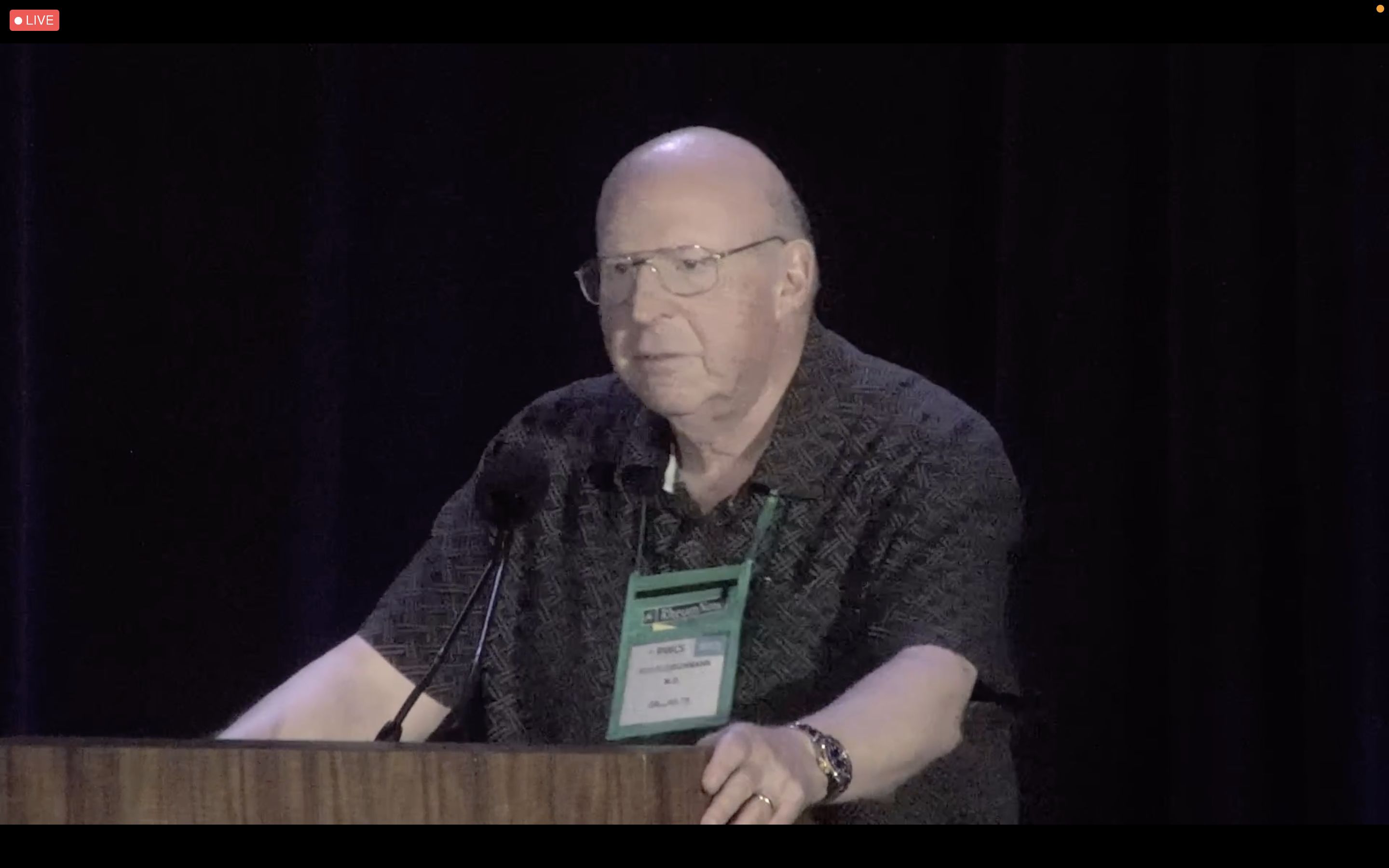
Roy Fleischmann, MD, agreed with Dr. Cush’s point of how one defines disease remission, as well as Dr. Bergman’s paradigm for T2T. “If you’re a good rheumatologist, you really do examine joints,” but you also “take a look at patient function, patient global [assessment], your global [assessment], joint count,” he said. “You can put a number to it, but you have to take a look at all of that. Really, treat-to-target – it is all of that. It isn’t just looking at a number, it’s looking at everything. And the better that the patient can do, assuming comorbidities and everything else, the better it is.”
“The problem isn’t the patient. The problem isn’t the metric. The problem is the rheumatologist, because the rheumatologist isn’t putting in enough effort in order to reach that goal,” added Dr. Fleischmann, clinical professor of medicine at the University of Texas, Dallas, and codirector of the Metroplex Clinical Research Center, also in in Dallas.
Eric Ruderman, MD, professor of rheumatology at Northwestern University, Chicago, commented that T2T is “great for somebody who’s been doing it for a while, and seeing a lot of patients and has that comfort zone,” but he questioned whether new rheumatologists without a lot of clinical experience could apply the approach. “What information do they use to integrate, and how do they get to that point?” he asked. “I don’t have the answer to that.”
During a rebuttal, Dr. Bergman pushed back on the idea that clinical experience alone was enough. “You need something. You need a benchmark. You need something more than, ‘I say so.’ ”
“The problem is, we still haven’t convinced people to adopt them,” Dr. Bergman said. “And I think it’s failure of training because, in my opinion, I don’t know how you can do a modern current fellowship program and not teach metrics.”
Dr. Cush and Dr. Bergman used the same trials to argue for their side. “I think that us using the same slides, but maybe having different points, speaks to the problem,” Dr. Cush said. “And I choose not to make it the problem of the rheumatologists.”
“You’re in the field of pattern recognition,” Dr. Cush argued. “It’s a visual art. You can have all the numbers you want. You make the most of your decisions based on pattern recognition, which is not rooted in metrics, and that’s why you’re successful at what you do.
“I am a big believer in T2T, but I think you have to measure something, and you have to use it,” Dr. Cush closed. “And the problem is, we can’t be forced into this.”
Dr. Bergman and Dr. Cush reported having financial relationships with numerous pharmaceutical companies.
The roots of treat-to-target (T2T) in rheumatology extend back over 30 years to the development of the 28-joint Disease Activity Score. Although it has been shown to be effective in clinical trials and has been included in guidelines, it has yet to be widely adopted in practice. The question remains: What is the role of T2T in rheumatology?
That’s what Jack Cush, MD, and Martin Bergman, MD, sought to answer in a point-counterpoint session at the 2022 Rheumatology Winter Clinical Symposium.
“I do think that this is a concept we need to keep in mind, and it is a concept whose time is long overdue,” Dr. Bergman said in his presentation arguing in favor of T2T. “As good as you think you are – your ability to see the patient and determine how they’re doing – you’re not.”
But metrics alone are not enough to make clinical decisions, said Dr. Bergman, clinical professor of medicine at Drexel University, Philadelphia, citing a recommendation from 2014 recommendations on T2T published in Annals of the Rheumatic Diseases. “You don’t just follow a number. You have to take into consideration structural changes, functional impairment, comorbidities – and that’s going to adjust how you approach your patient and what you do with them.”
However, implementation of T2T to make changes in clinical practice in RA has been inconsistent. Referencing an abstract from the 2021 American College of Rheumatology annual meeting on 15-year follow-up for changing therapy in RA, Dr. Bergman argued it is not the patient who is unwilling to switch treatments. Between 2006 and 2021, patient unwillingness to change therapies decreased from 64% to 51%.
“What’s driving it mostly here [is] the doctor’s recommendation,” Dr. Bergman said. “And we know this is true because we’ve seen it in other studies.”
Many rheumatologists are “asleep at the wheel” when it comes to administering T2T, he said. “What we need to do is, after we wake up from this nap, we need to get back on the highway and drive to where we should be, which is treat-to-target.”
Dr. Bergman also shared his paradigm for administering T2T, which he noted does not typically take more than a few minutes to administer regardless of the measure chosen. “Pick a measure. I don’t care which measure you take. I personally use two: I like the RAPID3 and the CDAI,” he said. “But then, after you have the measure, evaluate the entire patient. Don’t just look at the number. Look at the patient, what’s going on, solid history, solid physical. And most importantly: Be a doctor, don’t be a computer screen.”
Problems in measurement of RA remission and adoption of T2T
Dr. Cush, who admitted early in his presentation that he is in favor of T2T, delivered his counterpoint somewhat tongue-in-cheek. However, he pointed out that there are several concerns about the goals in measuring outcomes in RA with T2T.
The goal in RA is disease remission, but how you define remission can vary, especially since sometimes there is evidence of synovitis or other disease activity visible through an exam or imaging, said Dr. Cush, a rheumatologist based in Dallas, and executive editor of RheumNow.com. Most of the evidence for T2T is in clinical trials, but adoption is inconsistent in clinical practice, and patients in general appear to be improving without widespread adoption, he argued.
“Are clinical trials the same as clinical practice?” he asked. “I think that this boils down to: Is this a quest for remissions, or the best-you-can-get, low disease activity state? Or is this a quest for physician change, which is actually the path you have to go through to get to remissions?”
“In the end, evidence that the metrics should drive prescribing, especially in private practice is, I think, lacking,” he said.
Roy Fleischmann, MD, agreed with Dr. Cush’s point of how one defines disease remission, as well as Dr. Bergman’s paradigm for T2T. “If you’re a good rheumatologist, you really do examine joints,” but you also “take a look at patient function, patient global [assessment], your global [assessment], joint count,” he said. “You can put a number to it, but you have to take a look at all of that. Really, treat-to-target – it is all of that. It isn’t just looking at a number, it’s looking at everything. And the better that the patient can do, assuming comorbidities and everything else, the better it is.”
“The problem isn’t the patient. The problem isn’t the metric. The problem is the rheumatologist, because the rheumatologist isn’t putting in enough effort in order to reach that goal,” added Dr. Fleischmann, clinical professor of medicine at the University of Texas, Dallas, and codirector of the Metroplex Clinical Research Center, also in in Dallas.
Eric Ruderman, MD, professor of rheumatology at Northwestern University, Chicago, commented that T2T is “great for somebody who’s been doing it for a while, and seeing a lot of patients and has that comfort zone,” but he questioned whether new rheumatologists without a lot of clinical experience could apply the approach. “What information do they use to integrate, and how do they get to that point?” he asked. “I don’t have the answer to that.”
During a rebuttal, Dr. Bergman pushed back on the idea that clinical experience alone was enough. “You need something. You need a benchmark. You need something more than, ‘I say so.’ ”
“The problem is, we still haven’t convinced people to adopt them,” Dr. Bergman said. “And I think it’s failure of training because, in my opinion, I don’t know how you can do a modern current fellowship program and not teach metrics.”
Dr. Cush and Dr. Bergman used the same trials to argue for their side. “I think that us using the same slides, but maybe having different points, speaks to the problem,” Dr. Cush said. “And I choose not to make it the problem of the rheumatologists.”
“You’re in the field of pattern recognition,” Dr. Cush argued. “It’s a visual art. You can have all the numbers you want. You make the most of your decisions based on pattern recognition, which is not rooted in metrics, and that’s why you’re successful at what you do.
“I am a big believer in T2T, but I think you have to measure something, and you have to use it,” Dr. Cush closed. “And the problem is, we can’t be forced into this.”
Dr. Bergman and Dr. Cush reported having financial relationships with numerous pharmaceutical companies.
FROM RWCS 2022
Liquid embolism of AVM tied to high cure rate
new observational data suggest. In a prospective, real-world study of more than 100 patients, use of the Onyx system was associated with a cure rate of 86% for cAVMs smaller than 3 cm.
“Endovascular treatment using Onyx is able to achieve, on its own, a very efficient cure rate with a low morbidity and mortality rate,” said investigator Laurent Spelle, MD, PhD, professor of neuroradiology at Paris-Saclay University and chair of NEURI, the Brain Vascular Center, Bicêtre Hospital, also in Paris.
Dr. Spelle presented the findings at the International Stroke Conference sponsored by the American Heart Association.
Prospective, multicenter study
Currently, the main treatment options for cAVM are embolization, neurosurgery, and radiosurgery. The Onyx liquid system, one method of providing embolization, uses a biocompatible ethylene vinyl alcohol copolymer.
It has been used in Europe for 22 years as a curative treatment and as a treatment before radiosurgery or neurosurgery. In the United States, Onyx is indicated for presurgical and preradiotherapy treatment only.
For this analysis, the researchers conducted a prospective, multicenter study to evaluate the long-term safety and efficacy of Onyx for the embolization of cAVM as curative treatment or preoperative preparation.
They enrolled 165 patients in the nonrandomized, observational study, which was conducted at 15 hospitals in France. Eligible participants had an untreated cAVM.
Patients were assigned to one of three groups, according to the hospital’s standard of care. One group underwent embolization with Onyx as curative treatment, one received Onyx as preparation for neurosurgery, and one underwent embolization with Onyx as preparation for radiosurgery.
The study’s safety endpoints were device- and procedure-related serious adverse events at 1 month after each embolization. The efficacy endpoints were recovery at 12 months after the last embolization or neurosurgery, or at a minimum of 36 months after radiosurgery.
The researchers defined morbidity as a worsening of modified Rankin Scale score of 2 or more points for patients presenting with baseline mRS of 0 or 1, or a worsening of 1 or more points for patients with an mRS of 2 or greater at baseline. An independent clinical events committee and core laboratory adjudicated the results.
‘A fantastic result’
In all, 140 patients were prospectively included, and 212 embolization procedures were performed. The population’s mean age was 41.4 years, and 60% of participants were men. About 61% of patients presented with symptoms, the most common of which were progressive neurologic deficit (41.2%) and headache (36.5%).
Approximately 64% of the cAVMs were ruptured. Most (75.7%) were smaller than 3 cm, and the remainder were between 3 and 6 cm. Most patients (59.3%) did not have an aneurysm.
Eight (3.8%) adverse events were associated with the use of Onyx. The rate of procedure-related neurologic serious adverse events was 7.1% within 1 month post embolization. Three deaths occurred (2.1%), one of which was considered device or procedure related.
A total of 87 patients underwent embolization alone, 14 of whom did not complete the study (2 died, 5 were lost to follow-up, and 7 withdrew). Of the 73 who completed the study, 58 (79.5%) had complete occlusion and full recovery at last follow-up. An additional 6.8% had 99% occlusion.
In addition, 3.4% of the population had significant morbidity, and 18.4% presented at baseline with mRS scores of 3-5. Of the latter group, 81.3% had mRS scores of 0-2 at last visit.
Of 21 patients who underwent subsequent neurosurgery, 18 completed follow-up. Of this group, 94.4% had complete occlusion. Of 32 patients receiving subsequent radiosurgery, 54.8% had complete occlusion, which was “a little bit disappointing,” said Dr. Spelle.
Overall, most patients (92.9%) had improved or stable mRS score. The overall mortality rate was 2.9%, and the rate of significant morbidity was 4.3%.
The rate of improved or stable mRS score was 94.3% for patients who underwent embolization alone, 85.7% for patients who also underwent neurosurgery, and 93.75% for patients who also underwent radiosurgery.
The mortality rate was 3.4% for patients who underwent embolization alone, 4.8% for patients who also underwent neurosurgery, and 0% for patients who also underwent radiosurgery.
The rate of significant morbidity was 2.3% for patients who underwent embolization alone, 9.5% for those who also underwent neurosurgery, and 6.25% for those who also underwent radiosurgery.
“We knew that this treatment was very effective, but this effectiveness was only known in a limited number of centers with a very high level of expertise,” said Dr. Spelle. “We were very pleasantly surprised that a larger-scale, multicenter study conducted in 15 different hospitals in France could achieve such a fantastic result.”
The study sites, however, were all departments in university hospitals with great experience in endovascular treatment of cAVM, he added.
Effective in unruptured AVMs?
Commenting on the findings, Mitchell Elkind, MD, professor of neurology and epidemiology, Columbia University, New York, said: “Arteriovenous malformations remain a relatively uncommon but serious cerebrovascular disorder. Any additional tool in the armamentarium to treat these lesions is welcome.”
The study results are encouraging, said Dr. Elkind, who was not involved in the study. They suggest that Onyx embolization can play an important role in the care of these patients. The treatment is associated with “low morbidity and excellent efficacy, particularly in combination with other surgical and radiographic approaches.”
The lack of a direct comparison with alternative embolization materials is a limitation of the study, however. “It is hard to compare Onyx to other agents based on these results,” said Dr. Elkind.
“It is also notable that one-third of the patients in the study had unruptured AVMs, which at least in one randomized trial, ARUBA, were not clearly shown to benefit from an intervention at all,” he continued.
It would have been valuable for the researchers to stratify the study results by ruptured versus unruptured AVMs, Dr. Elkind said.
The study was funded by Medtronic. Dr. Spelle reported receiving honoraria from the company. Dr. Elkind disclosed no relevant financial relationships.
A version of this article first appeared on Medscape.com.
FROM ISC 2022
Twitter storm over ‘reprehensible behavior’ at conference podium
, held in San Francisco and also online. One of the panelists was accused of being less than professional in handling questions from the floor.
It began with a tweet from Sumanta Pal, MD, of the City of Hope, when he called out the behavior, although he did not identify the perpetrator.
“Yesterday we saw some reprehensible behavior emanating from the @ASCO #GU22 podium, with a well known investigator insulting (in a manner deeply imbued w microaggressions) an esteemed colleague at the mic. It’s a teachable moment & the lesson is simple: be kind to ur colleagues.”
It was not immediately clear what had occurred, and several people asked him to identify the people involved. One physician responded, “If it was bad enough to infer and share on Twitter, name the name.”
But even without details, the post provoked a reaction.
Erika Hamilton, MD, wrote, “I am not aware of this incident, but I always wonder ... if someone would do that from the podium, what would they/have they done and said when they didn’t think everyone was watching?”
Another tweet questioned why there were no codes of conduct in place, so that situations like this one wouldn’t happen.
“[It is] about time professional societies had codes of conduct in place for invited speakers, moderators of sessions etc,” wrote Deborah Verran, MD. “Then if it is breached the individual concerned is not invited back. This kind of incident also needs to be mentioned as part of the conference feedback process.”
As the number of clinicians chiming in increased, it soon became apparent that others were aware of the incident, and one posted a video clip, putting the mystery to rest.
Moderator quips, ‘Behave ... children’
The exchange occurred immediately following the oral abstract presentations on prostate cancer on Feb. 17. Two of these abstracts featured new data from large clinical trials investigating the use of PARP inhibitors in metastatic prostate cancer, which had slightly different results, leading to some debate. The PROpel trial with olaparib showed a benefit in all patients, irrespective of gene mutation status, but the MAGNITUDE trial with niraparib showed a benefit only in patients with gene alterations, and especially in those with BRCA1/2 mutations.
The invited discussant, Celestia Higano, MD, from the University of British Columbia, Vancouver, compared and contrasted the two trials and the differing results.
During the question-and-answer session that followed, Neeraj Agarwal, MD, from the Huntsman Cancer Institute, University of Utah, directed a question to Fred Saad, MD, who had presented the results of the PROpel trial.*
“As a practicing oncologist, I am very intrigued by the different results of the MAGNITUDE and PROpel trials. I am trying to figure out what do I do in my practice,” Dr. Agarwal began.
He continued on, explaining that he didn’t think that olaparib and niraparib were that different from one another for the two studies to have such differing results, when both of the drugs were given in combination with abiraterone.
The inclusion criteria for PROpel stipulated that patients undergo testing by ctDNA to determine if they were biomarker positive or not, but he pointed out that it is possible for testing to miss patients who might otherwise be positive for homologous recombination repair (HRR) gene alterations. He posed the question, could some of the patients deemed biomarker negative in fact have been positive but whose status was not detected by ctDNA testing?
At that point, Dr. Higano interrupted him before he had completed his question and asked, “Can I make a comment? Were you listening to my discussion?”
She then continued, pointing out that “you can’t talk about comparing olaparib and niraparib – these two trials had two very different populations.”
She emphasized that this was about the combinations in the populations being studied and not about olaparib and niraparib. “I clearly wasn’t very clear,” she said.
Dr. Agarwal then repeated that he wanted to know what to do with his patients and asked again about the accuracy of ctDNA testing.
“That’s a good question,” Dr. Higano said, “But the other comments you made weren’t.”
At that point, the moderator chimed in. “Let’s all calm down ... children.”
After a brief applause following the moderator’s comment, Dr. Saad then addressed the question.
When the video clip of this exchange was posted, a flood of tweets poured in, supporting Dr. Pal’s initial summation of the situation.
Alison Birtle, MD, tweeted, “I’m even more appalled having seen this exchange. Unacceptable. You don’t humiliate either the speaker or the delegate and the questions were entirely valid in my opinion. Basically what do I do tomorrow with these data so why ask were you listening. That’s just rude.”
Jason Kovac, MD, tweeted: “It’s absolutely not a proper way to behave. Whether it’s behind the scenes or to your face. You can hear the arrogance from everyone including the moderator with his children comment. This is the true face of medicine.”
Jarey Wang, MD, PhD, however, liked the moderator’s input. “Love it. ‘Let’s all calm down children.’ Good for the moderator.”
Several of the physicians who commented thought that the issue should have been addressed at the time it happened. Don S. Dizon, MD, wrote, “Calling out microagressions as they occur is so important. And very difficult. Agree, the teachable moment must be met with bravery. But it should happen in real time too.”
After 2 days of bantering on Twitter, with the thread growing increasingly longer and with the vast majority of posts supporting Dr. Pal’s initial assessment, Dr. Higano finally entered the discussion and defended her comments: “What you do not know is that the questioner and I are good colleagues,” she tweeted. “I have been involved in two of his academic promotions. My main concern was his comment re: how the two trials could have ‘so different results with the same combination.’ I sought to rectify wrong message.”
There were no direct replies to Dr. Higano’s tweet.
Perhaps the line to draw under this affair is the tweet from Simon Kim, MD, MPH, who wrote: “We can do better!”
A version of this article first appeared on Medscape.com.
Correction, 2/24/22: Dr. Fred Saad's name was misstated in an earlier version of this article.
FROM ASCO GU 2022
More evidence links MI to cognitive decline over time
new research suggests.
Although cognition in the acute phase after MI was not different than those without an MI in large observational cohorts, cognitive decline became significantly different over a median 6.5 years of follow-up.
The results reinforce the idea that heart health is closely tied to brain health, lead study author Michelle C. Johansen, MD, PhD, assistant professor of neurology cerebrovascular division, Johns Hopkins University, Baltimore, said in an interview. “From a clinical standpoint, heart health affects brain health and there may be effective interventions to prevent heart attack from happening that could reduce the rate of cognitive decline.”
The study was presented during the International Stroke Conference sponsored by the American Heart Association.
Researchers are increasingly recognizing the vascular contribution to cognitive impairment, said Dr. Johansen. This could involve “silent” or subclinical strokes that go unrecognized until seen on imaging.
The study included 31,377 adults free of MI and dementia from six large, well-known cohort studies: the Atherosclerosis Risk in Communities Study, the Coronary Artery Risk Development in Young Adults Study, the Cardiovascular Health Study, the Framingham Offspring Study, the Multi-Ethnic Study of Atherosclerosis, and the Northern Manhattan Study
About 56% of study participants were women, 23% were Black, 8% were Hispanic, and 69% were White.
They were followed from 1971 to 2017 with investigators repeatedly measuring vascular risk factors. The median study follow-up was 6.5 years, but some were followed for up to 20 years. During that time, there were 1,047 incident MIs.
The researchers performed a pooled analysis from these studies “using some fancy statistical techniques,” said Dr. Johansen. “The unique thing about this study was we were able to harmonize the cognitive measures.”
This allowed the researchers to determine if incident MI affected cognitive decline soon after the event and then long-term after the event. The primary outcome was change in global cognition. Additional outcomes were memory and executive function.
The median time between the first MI and the cognitive assessment was about 1.8 years but ranged from about 6 months to 4 years, said Dr. Johansen. Participants were a median age of 60 years at the time of the first cognitive assessment.
The researchers adjusted results for demographic factors, heart disease risk factors, and cognitive test results prior to the MI. Participants who had a stroke during the follow-up period were excluded from the analysis as stroke can affect cognition.
The study showed incident MI was associated with significant decline in global cognition (–0.71; 95% confidence interval, –1.02 to 0.42; P < .0001) and executive function (–0.68; 95% CI, –0.97 to 0.39; P < .004), but not memory, after the MI.
As cognition naturally declines with age, the researchers took that into consideration. “We anticipated cognition over time was going to go down, which it did, but the question we asked was: ‘How did the slope, which we knew was going to decline over time, compare in people who did not have a MI versus those that did?’ ” said Dr. Johansen.
After adjusting the model accordingly, the effect estimates indicating declines in global cognition and executive function were not significant.
However, another model that looked at the effect of incident MI on decline in cognitive function over the years following the event found significant differences.
Compared with participants without MI, those with incident MI had significantly faster declines in global cognition (–0.15 points/year faster, 95% CI, –0.21 to –0.10; P < .002), memory (–0.13 points/year faster, 95% CI, –0.23 to –0.04; P = .004), and executive function (–0.14 points/year faster, 95% CI, –0.20 to –0.08; P < .0001).
Dr. Johansen surmised that MI may result in subclinical infarcts or inflammation, or that MI and cognitive decline have shared vascular risk factors.
She said she can only speculate about why there was not more of a cognitive decline surrounding the MI. “It may be that right after the event, subjects are kind of sick from other things so it’s hard to see exactly what’s going on. Sometimes people can have other problems just from being in the hospital and having a heart attack may make cognition difficult to assess.”
The researchers also looked at those who had a second MI. “We asked whether the decline we saw after the first heart attack among those who had two heart attacks was explained by the fact they had more than one heart attack, and the answer to that question is no,” Dr. Johansen said.
The next research steps for Dr. Johansen and associates are to look at differences in race and sex.
Karen L. Furie, MD, chair, department of neurology, Brown University, and chief of neurology at Rhode Island Hospital, the Miriam Hospital, and the Bradley Hospital, all in Providence, provided a comment on the research.
MI and cognitive decline have a number of common risk factors, including hypertension, diabetes, high cholesterol, smoking, physical inactivity, and poor diet that can lead to obesity, said Dr. Furie.
“It’s critically important to identify these risk factors as early as possible,” she said. “People in early and middle life may not be receiving optimal medical management or engaging in ideal lifestyle choices and these contribute to the development and progression of atherosclerotic disease over the subsequent decades.”
In theory, she said, if these risk factors were eliminated or adequately treated earlier in life, “both the heart and brain could age naturally and in a healthy manner, enabling a higher functioning and better quality of life.”
The study was funded by the National Institute of Neurological Disorders and Stroke and the National Institute of Aging of the National Institutes of Health. Dr. Johansen receives research funding from NINDS.
A version of this article first appeared on Medscape.com.
FROM ISC 2022
Two factors linked to higher risk of long COVID in IBD
Two features are significantly associated with a higher risk for developing long COVID symptoms among people with inflammatory bowel disease (IBD), according to a large Danish population study.
People with Crohn’s disease (CD) who experienced adverse acute COVID-19, defined as requiring hospitalization, were nearly three times more likely to report persistent symptoms 12 weeks after acute infection.
“Long-term, persisting symptoms following COVID-19 is a frequently occurring problem, which is probably underappreciated. IBD specialists should therefore be aware of any of these symptoms and actively ask patients whether they have these problems,” lead author Mohamed Attauabi, MD, PhD, said in an interview.
Dr. Attauabi and colleagues also found that people with ulcerative colitis (UC) who discontinued immunosuppressive agents because of COVID-19 were 1.5 times more likely to experience long COVID symptoms, a result that surprised the researchers.
“This has not been shown before and remains to be confirmed,” said Dr. Attauabi, a fellow in the department of gastroenterology at Herlev Hospital at the University of Copenhagen.
Attauabi presented the results as a digital oral presentation at the 17th congress of the European Crohn’s and Colitis Organisation.
A closer look at IBD and COVID-19
Large, hospital-based studies of symptoms consistent with long COVID reveal a high prevalence of fatigue, sleep difficulties, and anxiety at 12 weeks or more post acute infection. However, these were not specific to people with CD or UC, Dr. Attauabi said.
“In patients with IBD, the risk of long-term sequelae of COVID-19 remains to be investigated,” he said.
Dr. Attauabi and colleagues studied 197 people with CD and 319 with UC, all of whom had polymerase chain reaction–confirmed COVID-19. Participants were prospectively enrolled in the population-based Danish IBD-COVID registry from January 28, 2020 to April 1, 2021. At a median of 5.1 months, a subset of 85 people with CD and 137 with UC agreed to report any post-COVID symptoms.
Older age, smoking, IBD disease activity, and presence of comorbidities were not associated with a significantly elevated risk of long COVID.
In a multivariate analysis, hospitalization for COVID-19 among people with CD was significantly associated with long COVID (odds ratio, 2.76; 95% confidence interval, 1.05-3.90; P = .04).
Furthermore, people with UC who stopped taking immunosuppressive agents also had a significantly higher risk (OR, 1.50; 95% CI, 1.07-10.22; P = .01).
“However, IBD medications such as systemic steroids were not associated with this outcome,” Dr. Attauabi said.
Fatigue most common long COVID symptom
Fatigue was the most common long COVID symptom, reported by 37% of patients with CD and 36% with UC.
Anosmia and ageusia were also common, reported by 29% and 28% of patients with CD, and 27% and 19% of those with UC, respectively.
“In our cohort of patients with UC or CD who developed COVID-19, the long-term health effects of COVID-19 did not appear to differ among patients with UC or CD nor according to IBD medications,” Dr. Attauabi said.
That is a “great study,” said session cochair Torsten Kucharzik, MD, PhD, head of internal medicine and gastroenterology at Lueneburg (Germany) Hospital.
When Dr. Kucharzik asked about smoking, Dr. Attauabi responded that they collected information on current and previous smoking, but they chose not to include the data because it was not statistically significant.
Dr. Attauabi has reported no relevant financial relationships. Dr. Kucharzik has reported receiving grants from Takeda and personal fees from companies including MSD/Essex, AbbVie, Falk Foundation, Biogen, Bristol-Myers Squibb, Arena, Celgene, Celltrion, Ferring, Janssen, Galapagos, Olympus, Mundipharma, Takeda, Amgen, Pfizer, Roche, and Vifor Pharma.
A version of this article first appeared on Medscape.com.
FROM ECCO 2022
HIV: Dual therapy with twice-yearly injections on the horizon
The findings, presented at the Conference on Retroviruses and Opportunistic Infections, also showed the drug was well tolerated, with 2 of 182 people developing drug-resistant mutations to lenacapavir and one person developing a nodule at the injection site.
Laura Waters, MD, consulting physician in HIV and sexual health at Central and Northwest London NHS Trust, who was not involved in the trial, called the findings “hugely exciting,” especially given its unique mode of administration. Right now it’s formulated as a subcutaneous injection, not an intramuscular injection like cabotegravir and rilpivirine (Cabenuva, ViiV Healthcare).
“Clearly it’s incredibly exciting to have the option for a subcutaneous drug that could be given at home every 6 months,” Dr. Waters said in an interview. With phase 3 trials and long-term follow-up still to come, she said she’s looking forward to how the treatment evolves.
CALIBRATE is a phase 2, four-arm, open-label, active-control study. Of the 182 participants, 25 were randomized to the active control arm, in which participants took daily oral bictegravir, emtricitabine, and tenofovir alafenamide (Biktarvy, Gilead Sciences) as standard of care. The rest of the 182 participants were divided evenly between three arms. In one of those, treatment group three, participants took a daily oral version of 50-mg LEN with emtricitabine and tenofovir alafenamide (F/TAF, Descovy, Gilead Sciences).
The other two arms, treatment groups one and two, were the arms in which Samir K. Gupta, MD, of Indiana University, Indianapolis, and colleagues tried the twice-yearly subcutaneous shots of LEN (LEN SC). Participants in those arms underwent a 2-week lead-in period in which they took 600 mg of LEN orally on days 1 and 2 and then a 300-mg pill on day 8 before starting the shots on day 15. As currently formulated, LEN SC shots can be given into the stomach and are designed to be simple enough to administer at home.
“It’s like doing an insulin shot,” Dr. Gupta said in an interview.
In addition to LEN SC, participants in treatment group one took a daily Descovy lead-in and then engaged in dual therapy of LEN plus TAF. In group two, they took a daily Descovy lead-in and then switched to LEN SC plus bictegravir (BIC) daily at 28 weeks.
Study results presented at the meeting were the results at 54 weeks. Researchers had previously presented 28-week results for CALIBRATE at the International AIDS Society Conference on HIV Science 2021.
Of the participants, 7% were cisgender women, 52% were Black, and 45% were Latinx. A total of 15% of participants had baseline viral loads of more than 100,000 copies, and median CD4 counts were 437. None of the participants in either the active-control arm or the LEN SC plus Descovy group had a history of AIDS, defined as fewer than 200 cells/mcL. In the LEN SC plus Biktarvy group, two did, and there were six people with a history of AIDS in the oral LEN plus Descovy group.
At 54 weeks, 88% of participants in groups one and two – the LEN SC arms – had undetectable viral loads (viral loads below 50 copies/mL). Specifically, 90% of those in the LEN SC plus TAF arm had viral loads below 50 copies. Those taking LEN SC plus BIC had an 85% viral suppression rate. The best performing of all was the control arm, in which 92% had undetectable viral loads. The protocol didn’t allow for analysis of statistical significance between the arms, so it’s unclear if any of the treatments really surpassed the others, Dr. Gupta said.
When they looked only at people whose viral loads became undetectable early, by week 28, the results were slightly better, with 93% of people between the two subcutaneous arms having undetectable viral loads at week 54. Again, the results were slightly higher in the LEN SC plus TAF arm (94%) than the LEN SC plus BIC (92%). People taking daily oral LEN plus Descovy had the lowest rate of viral suppression, but it was still at 90% if they were virally suppressed early. Again, it’s unclear whether those differences were statistically significant.
Well tolerated, with a chance of a nodule
Regarding safety, LEN was well tolerated across treatment arms, though the oral LEN group had as many people develop resistance mutations as the other three groups combined. Still, that was only three people, with one each in the two subcutaneous arms and one in the control arm.
Two people developed resistance to LEN – one in the LEN SC plus BIC arm and one in the oral LEN plus Descovy arm. In the LEN SC plus BIC arm, the mutations Q67H and K70R emerged at week 10, following a mutation that’s common when people don’t take integrase inhibitors as they’re supposed to (M184M/I). This led Dr. Gupta to conclude that lack of adherence to the oral part of the regimen may have contributed to the development of resistance, rather than an issue with LEN SC itself.
In the oral LEN plus Descovy arm, mutation Q67H didn’t emerge until week 54, when pill counts and drug levels revealed that that participant hadn’t been taking his Descovy as prescribed. In both cases, viral loads in those participants returned to undetectable after switching to an integrase inhibitor–based three-drug regimen.
Most safety concerns were mild and included nausea, diarrhea, and vomiting. But that was for non–injection-site reactions. There were more side effects of that sort, with most of them being pain, hardening of the site, and swelling. But 11% of participants developed more serious side effects, including nodules. Only one nodule was considered a grade 3 reaction; that person didn’t leave the trial because of it.
“These phase 2 data from the ongoing CALIBRATE trial support the further evaluation of lenacapavir for treatment and prevention of HIV-1,” he said.
Finding the right partner
In short, the findings are promising, Dr. Gupta told this news organization . But the question now, as Gilead prepares to begin a phase 3 efficacy trial, is what would be a good combination with LEN SC?
“Lenacapavir in a two-drug regimen should work,” he said. “The question now, though, is you have to find a suitable, potent, second agent to pair it with.”
Indeed, the long-acting HIV treatment pipeline sustained a blow in December 2021 when the U.S. Food and Drug Administration halted all trials for Merck’s investigational, long-acting drug islatravir. This led Dr. Waters to tweet: “Is it just me or are the islatravir abstracts at #CROI2022 quite sad to read?” She ended the tweet with, “I just hope it recovers!”
Indeed, Merck and Gilead have entered into an agreement to codevelop lenacapavir and islatravir as a weekly oral tablet. Islatravir seemed like the obvious choice as a partner to lenacapavir if phase 3 trials are successful, said Dr. Gupta. But now, it’s anyone’s guess as to what will happen. And while it’s too soon to say that lenacapavir is a success, it does leave the field wondering about how to use LEN SC without another, equally long-acting agent.
“When I talk about LEN, I show a picture of a man standing solo at a party, leaning against the wall looking a bit lonely, with ‘lenacapavir’ written over his head,” she said in an interview. “Right now, lenacapavir is the only drug at the 6-monthly party. It’s going to need some other guests if that party is going to rock.”
The study was funded by Gilead Sciences. Dr. Gupta has received research funding from ViiV Healthcare and advisory board fees from Gilead Sciences and ViiV Healthcare. Dr. Waters has received speaker or advisory fees from Gilead Sciences, ViiV Healthcare, Merck, Janssen, Theratech, Sipla, and Mylan.
A version of this article first appeared on Medscape.com.
FROM CROI 22