User login
A Rare Case of Chondromyxoid Fibroma of the Scapula
Simultaneous Solitary Glomus Tumors in Nonadjacent Digits
Bilateral Fractures of the Medial Malleoli Without a History of Trauma
Sacral Stress Fractures in Children
Stage 2 Sarcoidosis
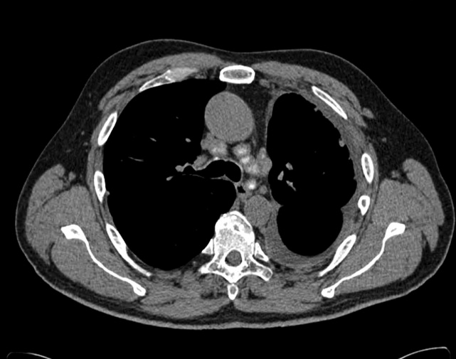
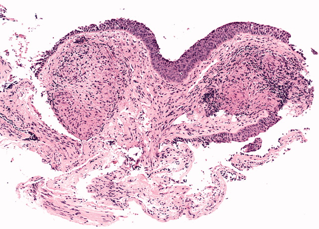
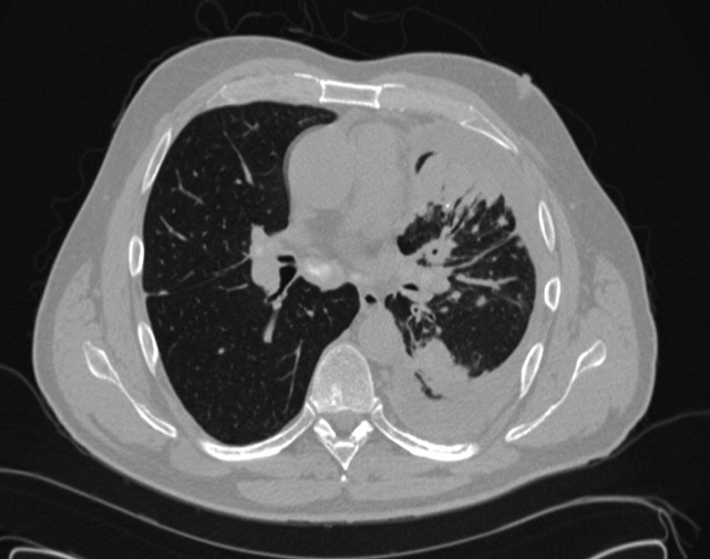
A 50‐year‐old man presented to the emergency department with progressive shortness of breath for 6 months. He described a dry cough, left‐sided chest pain, malaise, night sweats, and a 15‐pound weight loss. The patient had never smoked cigarettes, but he had been exposed to asbestos and wood dust when working at a sawmill. His physical examination was remarkable for decreased breath sounds at the left lung base. The admission blood tests were within normal limits. Chest radiography and a computed tomography (CT) scan of the chest were performed (the CT scan is shown in Figures 1 and 2). The CT scan showed a left pleural effusion with subpleural and peribronchovascular nodules. Also demonstrated on the CT scan were bilateral hilar and mediastinal lymphadenopathies with faint central calcification. As the left‐sided pleural effusion was initially suspected to be malignant, a thoracentesis was performed, and it revealed an exudative effusion. The total white cell count in fluid was 2100/L (lymphocytes, 76%), and cultures for aerobic and anaerobic bacteria, acid fasting bacilli, and fungi were negative. Cytology was negative for malignant cells. On the basis of the findings in the lung parenchyma and the presence of mediastinal lymphadenopathies, fiber‐optic bronchoscopy with bronchoalveolar lavage, protected specimen brushing, transbronchial needle aspiration, and transbronchial biopsies were performed. Mediastinal lymph node cytology was negative for malignant cells, whereas transbronchial biopsies showed noncaseating granulomas (Figure 3). At that time, our differential diagnoses of noncaseating granulomas included mycobacterium infection (although this usually presents caseating granulomas), berylliosis, histoplasmosis, and sarcoidosis. The tuberculin skin test (purified protein derivative) and serology for human immunodeficiency virus were negative. Bronchoalveolar lavage and cultures of lung tissue biopsies as well as needle aspiration from mediastinal lymph nodes were negative for mycobacterial, fungal, and bacterial organisms. The beryllium lymphocyte proliferation test was normal. Serologic antibodies for Aspergillus, Blastomyces, Coccidioides, and Histoplasma were negative. The urinary Histoplasma antigen was negative as well. The Department of Infectious Diseases was consulted, and an empirical treatment for histoplasmosis with itraconazole was started on the basis of the residence of the patient and the presence of noncaseating granulomas. After 1 month of antifungal treatment, there was no significant improvement. Video‐assisted thoracoscopic surgery with pleural biopsy was performed because of persistent pleural effusion and concern about an underlying infectious or malignant process. Pleural biopsies showed noncaseating granulomas (Figure 4). Pleural fluid was sent for adenosine deaminase (17 U/L) and flow cytometry (CD4/CD8 2.71). Cultures and cytology remained negative. A diagnosis of stage 2 sarcoidosis with pleural involvement was made, and treatment with prednisone was started.


Discussion
The overall prevalence of pleural involvement in sarcoidosis is about 3%. Patients with pleural sarcoidosis tend to be between 30 and 50 years of age, in contrast to the usual presentation of sarcoidosis between 20 and 30 years of age. The most common forms of pleural involvement are pleural effusions, pneumothorax, pleural thickening, and pleural nodules.1 Most effusions are usually small or modest in size, with few reports describing massive effusions.2 Recurrent pleural and pericardial effusions due to sarcoidosis have been reported as well.3 The fluid is typically a lymphocytic exudate, and almost all cases describe a CD4 predominant lymphocytic effusion with CD4/CD8 ratios ranging from 2.35 to 8.6.1 The presence of bloody pleural effusions in sarcoidosis most likely represents the rupture of small vessels that are compressed or infiltrated by granulomas.4
The majority of patients with reported sarcoid pleural effusions have stage 2 disease. With the progression of parenchymal disease, the prevalence of pleural effusions decreases, whereas pleural thickening and pneumothorax increase.5 It is important to emphasize that 40% of pleural effusions in sarcoidosis may be due to other causes, such as tuberculosis and mesothelioma. Our patient was initially treated with itraconazole as histoplasmosis is most prevalent in the Central and Eastern United States, especially in Ohio River valleys, where this patient lived.
The prevalence of a pneumothorax in sarcoidosis is up to 4%.1 Pleural thickening can be demonstrated in 11% to 71% of patients with pleural sarcoidosis, and 10% to 20% of these cases have thickening without effusion. Detection of subpleural nodules and cysts has been possible since the introduction of high‐resolution CT scans. Their prevalence in sarcoidosis ranges from 22% to 76%, and they are often described as masses that correspond to nodules seen in both parietal and visceral surfaces. Hilar or mediastinal lymphadenopathy is present on CT in 47% to 94% of patients with sarcoidosis. Lymph node enlargement is usually bilateral, most commonly with right‐sided predominance. The most involved stations are the right lower paratracheal, right hilar, subcarinal, aortopulmonary window, and right interlobar stations. Nodal calcification is noted in 53% with eggshell calcification present in 9%. The enlargement of internal mammary and pericardial lymph nodes requires the exclusion of lymphoma.6
The management of pleural sarcoidosis should be individualized because a majority of these effusions resolve spontaneously in 1 to 3 months.5 There have been reports of resolution in 2 weeks with steroid therapy. Incomplete resolution of the pleural effusions with progression to chronic pleural thickening or a trapped lung has been reported. There is agreement that oral corticosteroid treatment should be considered in patients with severe persistent or progressively worsening respiratory symptoms or declining lung function. Severe symptoms can be considered as those that interfere with essential aspects of the patient's daily life.7 The initial dosage of oral prednisone recommended by the American Thoracic Society, the European Respiratory Society, and the World Association of Sarcoidosis and Other Granulomatous Disorders guidelines is 20 to 40 mg/day.8 Further evaluation is recommended after 1 to 3 months. If the patient responds, the dose should be reduced gradually to a maintenance dose, such as 5 to 15 mg/day of prednisolone. American Thoracic Society/European Respiratory Society/World Association of Sarcoidosis and Other Granulomatous Disorders guidelines advise treatment for at least 1 year. Immunosuppressive, cytotoxic, and immunomodulatory agents have been used to treat patients failing or experiencing adverse effects of steroids. Favorable responses have been reported with methotrexate, leflunomide, azathioprine, cyclophosphamide, chlorambucil, cyclosporine A, antimalarials, tumor necrosis factor inhibitors, and thalidomide. Because of potential serious toxicities associated with cyclophosphamide and chlorambucil, these agents are not recommended.9
Our patient presented with pleural sarcoidosis with a pleural effusion and nodules. Treatment with 20 mg of prednisone daily was started initially. Four weeks after discharge, he was still dyspneic and had persistent left pleural effusion. He also had gained a significant amount of weight and developed bilateral lower extremity edema; these were thought to be secondary to prednisone treatment. Steroids were subsequently tapered, and leflunomide was started. His symptoms improved dramatically after 1 month of treatment with leflunomide and steroids, and 3 months later, his pleural effusion had completely resolved.
- ,.Pleural involvement in sarcoidosis.Curr Opin Pulm Med.2000;6(5):455–468.
- ,.Pleural sarcoidosis with massive effusion and lung entrapment.Kans Med.1990;91(4):103–105.
- ,,,,.Recurrent pleural and pericardial effusions due to sarcoidosis.PLoS Med.2005;2(3):e63.
- ,,,,.Pulmonary sarcoidosis with associated bloody pleurisy.Intern Med.2002;41(11):1021–1023.
- ,,,,.Pleural effusions in a series of 181 outpatients with sarcoidosis.Chest.2006;129(6):1599–1604.
- ,,,,.Imaging in sarcoidosis.Semin Respir Crit Care Med.2007;28(1):102–120.
- .Guidelines for the use of corticosteroids in the treatment of pulmonary sarcoidosis.Drugs.2007;67(8):1139–1147.
- ,,, et al.ATS/ERS/WASOG statement on sarcoidosis.Sarcoidosis Vasc Diffuse Lung Dis.1999;16(2):149–173.
- ,,,.Pulmonary sarcoidosis.Semin Respir Crit Care Med.2007;28(1):53–74.
A 50‐year‐old man presented to the emergency department with progressive shortness of breath for 6 months. He described a dry cough, left‐sided chest pain, malaise, night sweats, and a 15‐pound weight loss. The patient had never smoked cigarettes, but he had been exposed to asbestos and wood dust when working at a sawmill. His physical examination was remarkable for decreased breath sounds at the left lung base. The admission blood tests were within normal limits. Chest radiography and a computed tomography (CT) scan of the chest were performed (the CT scan is shown in Figures 1 and 2). The CT scan showed a left pleural effusion with subpleural and peribronchovascular nodules. Also demonstrated on the CT scan were bilateral hilar and mediastinal lymphadenopathies with faint central calcification. As the left‐sided pleural effusion was initially suspected to be malignant, a thoracentesis was performed, and it revealed an exudative effusion. The total white cell count in fluid was 2100/L (lymphocytes, 76%), and cultures for aerobic and anaerobic bacteria, acid fasting bacilli, and fungi were negative. Cytology was negative for malignant cells. On the basis of the findings in the lung parenchyma and the presence of mediastinal lymphadenopathies, fiber‐optic bronchoscopy with bronchoalveolar lavage, protected specimen brushing, transbronchial needle aspiration, and transbronchial biopsies were performed. Mediastinal lymph node cytology was negative for malignant cells, whereas transbronchial biopsies showed noncaseating granulomas (Figure 3). At that time, our differential diagnoses of noncaseating granulomas included mycobacterium infection (although this usually presents caseating granulomas), berylliosis, histoplasmosis, and sarcoidosis. The tuberculin skin test (purified protein derivative) and serology for human immunodeficiency virus were negative. Bronchoalveolar lavage and cultures of lung tissue biopsies as well as needle aspiration from mediastinal lymph nodes were negative for mycobacterial, fungal, and bacterial organisms. The beryllium lymphocyte proliferation test was normal. Serologic antibodies for Aspergillus, Blastomyces, Coccidioides, and Histoplasma were negative. The urinary Histoplasma antigen was negative as well. The Department of Infectious Diseases was consulted, and an empirical treatment for histoplasmosis with itraconazole was started on the basis of the residence of the patient and the presence of noncaseating granulomas. After 1 month of antifungal treatment, there was no significant improvement. Video‐assisted thoracoscopic surgery with pleural biopsy was performed because of persistent pleural effusion and concern about an underlying infectious or malignant process. Pleural biopsies showed noncaseating granulomas (Figure 4). Pleural fluid was sent for adenosine deaminase (17 U/L) and flow cytometry (CD4/CD8 2.71). Cultures and cytology remained negative. A diagnosis of stage 2 sarcoidosis with pleural involvement was made, and treatment with prednisone was started.
Discussion
The overall prevalence of pleural involvement in sarcoidosis is about 3%. Patients with pleural sarcoidosis tend to be between 30 and 50 years of age, in contrast to the usual presentation of sarcoidosis between 20 and 30 years of age. The most common forms of pleural involvement are pleural effusions, pneumothorax, pleural thickening, and pleural nodules.1 Most effusions are usually small or modest in size, with few reports describing massive effusions.2 Recurrent pleural and pericardial effusions due to sarcoidosis have been reported as well.3 The fluid is typically a lymphocytic exudate, and almost all cases describe a CD4 predominant lymphocytic effusion with CD4/CD8 ratios ranging from 2.35 to 8.6.1 The presence of bloody pleural effusions in sarcoidosis most likely represents the rupture of small vessels that are compressed or infiltrated by granulomas.4
The majority of patients with reported sarcoid pleural effusions have stage 2 disease. With the progression of parenchymal disease, the prevalence of pleural effusions decreases, whereas pleural thickening and pneumothorax increase.5 It is important to emphasize that 40% of pleural effusions in sarcoidosis may be due to other causes, such as tuberculosis and mesothelioma. Our patient was initially treated with itraconazole as histoplasmosis is most prevalent in the Central and Eastern United States, especially in Ohio River valleys, where this patient lived.
The prevalence of a pneumothorax in sarcoidosis is up to 4%.1 Pleural thickening can be demonstrated in 11% to 71% of patients with pleural sarcoidosis, and 10% to 20% of these cases have thickening without effusion. Detection of subpleural nodules and cysts has been possible since the introduction of high‐resolution CT scans. Their prevalence in sarcoidosis ranges from 22% to 76%, and they are often described as masses that correspond to nodules seen in both parietal and visceral surfaces. Hilar or mediastinal lymphadenopathy is present on CT in 47% to 94% of patients with sarcoidosis. Lymph node enlargement is usually bilateral, most commonly with right‐sided predominance. The most involved stations are the right lower paratracheal, right hilar, subcarinal, aortopulmonary window, and right interlobar stations. Nodal calcification is noted in 53% with eggshell calcification present in 9%. The enlargement of internal mammary and pericardial lymph nodes requires the exclusion of lymphoma.6
The management of pleural sarcoidosis should be individualized because a majority of these effusions resolve spontaneously in 1 to 3 months.5 There have been reports of resolution in 2 weeks with steroid therapy. Incomplete resolution of the pleural effusions with progression to chronic pleural thickening or a trapped lung has been reported. There is agreement that oral corticosteroid treatment should be considered in patients with severe persistent or progressively worsening respiratory symptoms or declining lung function. Severe symptoms can be considered as those that interfere with essential aspects of the patient's daily life.7 The initial dosage of oral prednisone recommended by the American Thoracic Society, the European Respiratory Society, and the World Association of Sarcoidosis and Other Granulomatous Disorders guidelines is 20 to 40 mg/day.8 Further evaluation is recommended after 1 to 3 months. If the patient responds, the dose should be reduced gradually to a maintenance dose, such as 5 to 15 mg/day of prednisolone. American Thoracic Society/European Respiratory Society/World Association of Sarcoidosis and Other Granulomatous Disorders guidelines advise treatment for at least 1 year. Immunosuppressive, cytotoxic, and immunomodulatory agents have been used to treat patients failing or experiencing adverse effects of steroids. Favorable responses have been reported with methotrexate, leflunomide, azathioprine, cyclophosphamide, chlorambucil, cyclosporine A, antimalarials, tumor necrosis factor inhibitors, and thalidomide. Because of potential serious toxicities associated with cyclophosphamide and chlorambucil, these agents are not recommended.9
Our patient presented with pleural sarcoidosis with a pleural effusion and nodules. Treatment with 20 mg of prednisone daily was started initially. Four weeks after discharge, he was still dyspneic and had persistent left pleural effusion. He also had gained a significant amount of weight and developed bilateral lower extremity edema; these were thought to be secondary to prednisone treatment. Steroids were subsequently tapered, and leflunomide was started. His symptoms improved dramatically after 1 month of treatment with leflunomide and steroids, and 3 months later, his pleural effusion had completely resolved.
- ,.Pleural involvement in sarcoidosis.Curr Opin Pulm Med.2000;6(5):455–468.
- ,.Pleural sarcoidosis with massive effusion and lung entrapment.Kans Med.1990;91(4):103–105.
- ,,,,.Recurrent pleural and pericardial effusions due to sarcoidosis.PLoS Med.2005;2(3):e63.
- ,,,,.Pulmonary sarcoidosis with associated bloody pleurisy.Intern Med.2002;41(11):1021–1023.
- ,,,,.Pleural effusions in a series of 181 outpatients with sarcoidosis.Chest.2006;129(6):1599–1604.
- ,,,,.Imaging in sarcoidosis.Semin Respir Crit Care Med.2007;28(1):102–120.
- .Guidelines for the use of corticosteroids in the treatment of pulmonary sarcoidosis.Drugs.2007;67(8):1139–1147.
- ,,, et al.ATS/ERS/WASOG statement on sarcoidosis.Sarcoidosis Vasc Diffuse Lung Dis.1999;16(2):149–173.
- ,,,.Pulmonary sarcoidosis.Semin Respir Crit Care Med.2007;28(1):53–74.
Rapid Response: A QI Conundrum
Many in‐hospital cardiac arrests and other adverse events are heralded by warning signs that are evident in the preceding 6 to 8 hours.1 By promptly intervening before further deterioration occurs, rapid response teams (RRTs) are designed to decrease unexpected intensive care unit (ICU) transfers, cardiac arrests, and inpatient mortality. While implementing RRTs is 1 of the 6 initiatives recommended by the Institute for Healthcare Improvement,2 data supporting their effectiveness is equivocal.3, 4
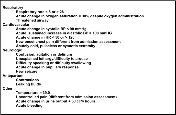
In October 2006, at Denver Health Medical Center, an academic, safety net hospital, we initiated a rapid response systemclinical triggers program (RRS‐CTP).5 In our RRS‐CTP, an abrupt change in patient status (Figure 1) triggers a mandatory call by the patient's nurse to the primary team, which is then required to perform an immediate bedside evaluation. By incorporating the primary team into the RRT‐CTP, we sought to preserve as much continuity of care as possible. Also, since the same house staff compose our cardiopulmonary arrest or cor team, and staff the ICUs and non‐ICU hospital wards, we did not feel that creating a separate RRT was an efficient use of resources. Our nurses have undergone extensive education about the necessity of a prompt bedside evaluation and have been instructed and empowered to escalate concerns to senior physicians if needed. We present a case that illustrates challenges to both implementing an RRS and measuring its potential benefits.

Case
A 59‐year‐old woman with a history of bipolar mood disorder was admitted for altered mental status. At presentation, she had signs of acute mania with normal vital signs. After initial laboratory workup, her altered mental status was felt to be multifactorial due to urinary tract infection, hypernatremia (attributed to lithium‐induced nephrogenic diabetes insipidus), and acute mania (attributed to medication discontinuation). Because she was slow to recover from the acute mania, her hospital stay was prolonged. From admission, the patient was treated with heparin 5000 units subcutaneously twice daily for venous thromboembolism prophylaxis.
On hospital day 7, at 21:32, the patient was noted to have asymptomatic tachycardia at 149 beats per minute and a new oxygen requirement of 3 L/minute. The cross‐cover team was called; however, although criteria were met, the RRS‐CTP was not activated and a bedside evaluation was not performed. A chest X‐ray was found to be normal and, with the exception of the oxygen requirement, her vital signs normalized by 23:45. No further diagnostic testing was performed at the time.
The next morning, at 11:58, the patient was found to have a blood pressure of 60/40 mmHg and heart rate of 42 beats per minute. The RRS‐CTP was activated. The primary team arrived at the bedside at 12:00 and found the patient to be alert, oriented, and without complaints. Her respiratory rate was 30/minute, and her oxygen saturation was 86% on 3 L/minute. An arterial blood gas analysis demonstrated acute respiratory alkalosis with hypoxemia and an electrocardiogram showed sinus tachycardia with a new S1Q3T3 pattern. A computed tomography angiogram revealed a large, nearly occlusive pulmonary embolus (PE) filling an enlarged right pulmonary artery, as well as thrombus within the left main pulmonary artery. She was transferred to the medical ICU and alteplase was administered. The patient survived and was discharged in good clinical condition.
Discussion
Despite the strong theoretical benefit of the RRT concept, a recent review by Ranji et al.4 concluded that RRTs had not yet been shown to improve patient outcomes. In contrast to dedicated RRTs, this case illustrates a different type of RRS that was designed to address abrupt changes in patient status, while maintaining continuity of care and efficiently utilizing resources.
If one considers an RRS to have both afferent (criteria recognition) and efferent (RRT or primary team response) limbs, the afferent limb must be consistently activated in order to obtain the efferent limb's response.6 The greatest opportunities to improve RRSs are thought to lie in the afferent limb.3 Our RRS‐CTP was not triggered in 1 of 2 instances in which criteria for mandatory initiation of the system were met. This is consistent with the findings of the Medical Early Response Intervention and Therapy (MERIT) trial, in which RRTs were called in only 41% of the patients meeting criteria and subsequently having adverse events,7 and with the ongoing monitoring of the use of the system at our hospital. Had the cross‐covering team seen the patient at the bedside initially, the PE might have been diagnosed while the patient was hemodynamically stable, giving the patient nearly a 3‐fold lower relative mortality.8 When the RRS‐CTP was activated, a prompt bedside evaluation occurred, allowing for lytic therapy to be administered before cardiopulmonary arrest (attendant mortality of 90%).9
While rapid response criteria were originally based upon published sensitivity analyses, more recent studies suggest that these criteria lack diagnostic accuracy. As demonstrated by Cretikos et al,10 to reach a sensitivity of 70%, the corresponding specificity would be only 86%. Given that the prevalence of adverse events in the MERIT trial was only 0.6%, the resulting positive predictive value (PPV) of rapid response call criteria is 3%. Accordingly, 33 calls would be needed to prevent 1 unplanned ICU transfer, cardiac arrest, or death. Nurses' attempts to minimize false‐positive calls may help explain the low call rates for patients meeting RRT criteria. The 2 avenues to increase the PPV of criteria are:
-
Increase the prevalence of disease in the population screened by risk factor stratification.
-
Increase the specificity of the call criteria, which has been limited by the associated decrease in sensitivity.10
Regarding the efferent response limb of RRS, our case demonstrates that the primary team (rather than a separate group of caregivers), when alerted appropriately, can effectively respond to critical changes in patient status. Accordingly, our data show that since the inception of the program, cardiopulmonary arrests have decreased from a mean of 4.1 per month to 2.3 per month (P = 0.03).
Many clinical trials of RRTs would not capture the success demonstrated in this case. For example, due to the low prevalence of events, the MERIT trial used a composite endpoint that included unplanned ICU transfers, cardiac arrests, and mortality. Because our patient still required an unplanned ICU transfer after being evaluated by the responding team, she would have been counted as a system failure.
Conclusion
While local needs should inform the type of RRS implemented, this case illustrates one of the major obstacles ubiquitous to RRS implementation: failure of system activation. With appropriate activation, an RRS‐CTP can meet RRS goals while maintaining continuity of care and maximizing existing resources. This case also illustrates the difficulty of achieving a statistically relevant outcome, while demonstrating the potential benefits of evolving RRSs.
- ,,,,.Rapid response teams—do they make a difference.Dimens Crit Care Nurs.2007;26(6):253–260.
- Institute for Healthcare Improvement. 5 Million Lives Campaign. Available at: http://www.ihi.org/IHI/Programs/Campaign/Campaign.htm?TabId=1IHI. Accessed February2009.
- .The rapid response team paradox: why doesn't anyone call for help?Crit Care Med.2008;36(2):634–636.
- ,,,,.Effects of rapid response systems on clinical outcomes: review and meta‐analyses.J Hosp Med.2007;2:422–432.
- ,,.Clinical triggers and rapid response escalation criteria.Patient Saf Qual Healthc.2007;4(2):12–13. Available at: http://www.psqh.com/archives.html. Accessed February 2009.
- ,,, et al.Use of medical emergency team responses to reduce hospital cardiopulmonary arrest.Qual Saf Health Care.2004;13:251–254.
- MERIT Study Investigators.Introduction of the medical emergency team (MET) system: a cluster‐randomised controlled trial.Lancet.2005;365:2091–2097.
- ,,.Acute pulmonary embolism: clinical outcomes in the international cooperative pulmonary embolism registry (ICOPER).Lancet.1999;353(9162):1386–1389.
- ,,,.Early predictors of mortality for hospitalized patients suffering cardiopulmonary arrest.Chest.1990;97(2):413–419.
- ,,,,,.The objective medical emergency team activation criteria: a case–control study.Resuscitation.2007;73:62–72.
Many in‐hospital cardiac arrests and other adverse events are heralded by warning signs that are evident in the preceding 6 to 8 hours.1 By promptly intervening before further deterioration occurs, rapid response teams (RRTs) are designed to decrease unexpected intensive care unit (ICU) transfers, cardiac arrests, and inpatient mortality. While implementing RRTs is 1 of the 6 initiatives recommended by the Institute for Healthcare Improvement,2 data supporting their effectiveness is equivocal.3, 4
In October 2006, at Denver Health Medical Center, an academic, safety net hospital, we initiated a rapid response systemclinical triggers program (RRS‐CTP).5 In our RRS‐CTP, an abrupt change in patient status (Figure 1) triggers a mandatory call by the patient's nurse to the primary team, which is then required to perform an immediate bedside evaluation. By incorporating the primary team into the RRT‐CTP, we sought to preserve as much continuity of care as possible. Also, since the same house staff compose our cardiopulmonary arrest or cor team, and staff the ICUs and non‐ICU hospital wards, we did not feel that creating a separate RRT was an efficient use of resources. Our nurses have undergone extensive education about the necessity of a prompt bedside evaluation and have been instructed and empowered to escalate concerns to senior physicians if needed. We present a case that illustrates challenges to both implementing an RRS and measuring its potential benefits.
Case
A 59‐year‐old woman with a history of bipolar mood disorder was admitted for altered mental status. At presentation, she had signs of acute mania with normal vital signs. After initial laboratory workup, her altered mental status was felt to be multifactorial due to urinary tract infection, hypernatremia (attributed to lithium‐induced nephrogenic diabetes insipidus), and acute mania (attributed to medication discontinuation). Because she was slow to recover from the acute mania, her hospital stay was prolonged. From admission, the patient was treated with heparin 5000 units subcutaneously twice daily for venous thromboembolism prophylaxis.
On hospital day 7, at 21:32, the patient was noted to have asymptomatic tachycardia at 149 beats per minute and a new oxygen requirement of 3 L/minute. The cross‐cover team was called; however, although criteria were met, the RRS‐CTP was not activated and a bedside evaluation was not performed. A chest X‐ray was found to be normal and, with the exception of the oxygen requirement, her vital signs normalized by 23:45. No further diagnostic testing was performed at the time.
The next morning, at 11:58, the patient was found to have a blood pressure of 60/40 mmHg and heart rate of 42 beats per minute. The RRS‐CTP was activated. The primary team arrived at the bedside at 12:00 and found the patient to be alert, oriented, and without complaints. Her respiratory rate was 30/minute, and her oxygen saturation was 86% on 3 L/minute. An arterial blood gas analysis demonstrated acute respiratory alkalosis with hypoxemia and an electrocardiogram showed sinus tachycardia with a new S1Q3T3 pattern. A computed tomography angiogram revealed a large, nearly occlusive pulmonary embolus (PE) filling an enlarged right pulmonary artery, as well as thrombus within the left main pulmonary artery. She was transferred to the medical ICU and alteplase was administered. The patient survived and was discharged in good clinical condition.
Discussion
Despite the strong theoretical benefit of the RRT concept, a recent review by Ranji et al.4 concluded that RRTs had not yet been shown to improve patient outcomes. In contrast to dedicated RRTs, this case illustrates a different type of RRS that was designed to address abrupt changes in patient status, while maintaining continuity of care and efficiently utilizing resources.
If one considers an RRS to have both afferent (criteria recognition) and efferent (RRT or primary team response) limbs, the afferent limb must be consistently activated in order to obtain the efferent limb's response.6 The greatest opportunities to improve RRSs are thought to lie in the afferent limb.3 Our RRS‐CTP was not triggered in 1 of 2 instances in which criteria for mandatory initiation of the system were met. This is consistent with the findings of the Medical Early Response Intervention and Therapy (MERIT) trial, in which RRTs were called in only 41% of the patients meeting criteria and subsequently having adverse events,7 and with the ongoing monitoring of the use of the system at our hospital. Had the cross‐covering team seen the patient at the bedside initially, the PE might have been diagnosed while the patient was hemodynamically stable, giving the patient nearly a 3‐fold lower relative mortality.8 When the RRS‐CTP was activated, a prompt bedside evaluation occurred, allowing for lytic therapy to be administered before cardiopulmonary arrest (attendant mortality of 90%).9
While rapid response criteria were originally based upon published sensitivity analyses, more recent studies suggest that these criteria lack diagnostic accuracy. As demonstrated by Cretikos et al,10 to reach a sensitivity of 70%, the corresponding specificity would be only 86%. Given that the prevalence of adverse events in the MERIT trial was only 0.6%, the resulting positive predictive value (PPV) of rapid response call criteria is 3%. Accordingly, 33 calls would be needed to prevent 1 unplanned ICU transfer, cardiac arrest, or death. Nurses' attempts to minimize false‐positive calls may help explain the low call rates for patients meeting RRT criteria. The 2 avenues to increase the PPV of criteria are:
-
Increase the prevalence of disease in the population screened by risk factor stratification.
-
Increase the specificity of the call criteria, which has been limited by the associated decrease in sensitivity.10
Regarding the efferent response limb of RRS, our case demonstrates that the primary team (rather than a separate group of caregivers), when alerted appropriately, can effectively respond to critical changes in patient status. Accordingly, our data show that since the inception of the program, cardiopulmonary arrests have decreased from a mean of 4.1 per month to 2.3 per month (P = 0.03).
Many clinical trials of RRTs would not capture the success demonstrated in this case. For example, due to the low prevalence of events, the MERIT trial used a composite endpoint that included unplanned ICU transfers, cardiac arrests, and mortality. Because our patient still required an unplanned ICU transfer after being evaluated by the responding team, she would have been counted as a system failure.
Conclusion
While local needs should inform the type of RRS implemented, this case illustrates one of the major obstacles ubiquitous to RRS implementation: failure of system activation. With appropriate activation, an RRS‐CTP can meet RRS goals while maintaining continuity of care and maximizing existing resources. This case also illustrates the difficulty of achieving a statistically relevant outcome, while demonstrating the potential benefits of evolving RRSs.
- ,,,,.Rapid response teams—do they make a difference.Dimens Crit Care Nurs.2007;26(6):253–260.
- Institute for Healthcare Improvement. 5 Million Lives Campaign. Available at: http://www.ihi.org/IHI/Programs/Campaign/Campaign.htm?TabId=1IHI. Accessed February2009.
- .The rapid response team paradox: why doesn't anyone call for help?Crit Care Med.2008;36(2):634–636.
- ,,,,.Effects of rapid response systems on clinical outcomes: review and meta‐analyses.J Hosp Med.2007;2:422–432.
- ,,.Clinical triggers and rapid response escalation criteria.Patient Saf Qual Healthc.2007;4(2):12–13. Available at: http://www.psqh.com/archives.html. Accessed February 2009.
- ,,, et al.Use of medical emergency team responses to reduce hospital cardiopulmonary arrest.Qual Saf Health Care.2004;13:251–254.
- MERIT Study Investigators.Introduction of the medical emergency team (MET) system: a cluster‐randomised controlled trial.Lancet.2005;365:2091–2097.
- ,,.Acute pulmonary embolism: clinical outcomes in the international cooperative pulmonary embolism registry (ICOPER).Lancet.1999;353(9162):1386–1389.
- ,,,.Early predictors of mortality for hospitalized patients suffering cardiopulmonary arrest.Chest.1990;97(2):413–419.
- ,,,,,.The objective medical emergency team activation criteria: a case–control study.Resuscitation.2007;73:62–72.