User login
Bringing you the latest news, research and reviews, exclusive interviews, podcasts, quizzes, and more.
Powered by CHEST Physician, Clinician Reviews, MDedge Family Medicine, Internal Medicine News, and The Journal of Clinical Outcomes Management.
Choice of Surgical Treatment for Mesothelioma Remains Complex
Surgery for malignant pleural mesothelioma remains a complicated and controversial issue. Thus far, the benefits of surgery vs. nonsurgical treatment have yet to be demonstrated.
Complete resection with surgery alone (R0) appears theoretically unattainable since it is impossible to eradicate residual microscopic disease, regardless of the surgical method used. Hence, most surgical treatment today is coupled to various adjuvant treatments, primarily a trimodality mode with radiotherapy and chemotherapy, according to Dr. Raja M. Flores, professor and chief of thoracic surgery, Mount Sinai School of Medicine, New York.
A “curative” surgical procedure remains an elusive goal, and thus the focus of lung surgery for malignant pleural mesothelioma (MPM) has shifted to R1 surgical resection for cytoreduction in the hope of prolonging life, relieving symptoms, and enhancing the effectiveness of adjuvant therapies. This approach has often meant a shift from the more radical extrapleural pneumonectomy (EPP), when possible, to the more lung-sparing pleurectomy/decortication (PD) procedure, according to Dr. Flores (Sem. Thorac. Cardiovasc. Surg.2009;21:149-53).
EPP involves a radical en bloc resection of the lung, pleura, diaphragm, and pericardium. PD involves resection of the parietal and visceral pleurae, pericardium, and – when necessary – the diaphragm, but it spares the entire lung. Both operations are technically complex and require extensive surgical expertise.
The operative mortality rate of EPP in the literature ranges from 4% to 15%, vs. and 1% to 5% for PD. In addition, PD has lower morbidity than does EPP. But the two techniques are not interchangeable, according to Dr. Flores. The choice of surgical technique depends on multiple factors, and the decision is often made at the time of surgery because the preoperative imaging may have underestimated the amount of disease present.
Staging is critical in determining the appropriate procedure, and the merits of each surgical approach have been debated in several recent clinical and registry trials examining individual mortality and morbidity of these procedures at different stages, coupled with the use of a variety of adjuvant therapies. However, many decisions are based on surgical conjecture and bias rather than scientific data.
Evidence indicates that PD provides a survival advantage for patients with stage I MPM, which may be accounted for by “lower mortality, lower postoperative adverse events, and greater lung capacity when relapse occurs,” according to Dr. Flores. However, he explained, most patients with mesothelioma will present at a stage that requires EPP to eradicate all gross disease. PD can provide an R1 resection in early-stage disease, but as the tumor enlarges and invades the lung, fissures, and costophrenic sulcus, a PD is suboptimal regardless of resection of the pericardium and diaphragm.
There is, however, a critical balance between optimal cytoreduction and morbidity that varies across stages for these two procedures. For stage II disease, there is a “trend toward improved survival for EPP, despite an inherently higher tumor stage than PD,” Dr. Flores said.
Stage III disease proved more complex, with similar survival data seen for both EPP and PD. Ultimately, “one should focus on obtaining a complete macroscopic resection based on the extent of tumor” for this stage of disease, choosing the best procedure accordingly, he advised.
For more advanced (stage IV) disease characterized by diffuse chest wall invasion and extensions through the diaphragm to the underlying peritoneum, the situation is much different.
“The tumor may be amenable to EPP, but there will be gross residual tumor left behind in the hemithorax. Because one of the most likely sites of recurrence is the contralateral pleura, the patient is better served by preserving lung function,” Dr. Flores explained.
In stage IV disease, PD trended toward better survival, presumably because “when disease spreads to the contralateral lung, PD or debulked patients will be less symptomatic and better functionally able to tolerate systemic therapy because of their greater pulmonary reserve,” he said.
“The goal is to remove all gross tumor while preserving as much of the lung as possible. Every patient and clinical situation is unique; therefore, treatment is difficult to generalize. Find an experienced mesothelioma surgeon you trust and leave it in their hands,” Dr. Flores said in an interview.
Ultimately, the situation remains complex. Dr. Heyman Luckraz of the New Cross Hospital, Wolverhampton, United Kingdom, and his colleagues recently reported results with 139 patients. EPP was chosen for clinically fit patients with stage I disease, while patients with advanced disease or who were unfit for EPP underwent PD. “EPP may only have a limited role in diffuse MPM, particularly as neither operative procedure is curative. Ultimately, the place of EPP will only be determined by randomized trial in comparison to PD in stage I disease with both groups receiving adjuvant therapy,” the investigators concluded (Eur. J. Cardio-Thorac. Surg. 2010;37:552-6).
Whether such trials will ever be performed is an open question. Despite the recent Mesothelioma and Radical Surgery (MARS) trial, which demonstrated the possibility of randomizing patients to surgical vs. nonsurgical treatment, there will likely never be a randomized clinical trial powered enough to completely solve the puzzle, according to Dr. Tom Treasure of the University College of London (Eur. J. Cardio-Thorac. Surg. 2010;37:509-10).
Efforts continue to develop surgical alternatives with less mortality and morbidity than those of the standard EPP and PD procedures. For example, Dr. M.D. Kluger and colleagues at Columbia University, New York, reported the phase I and II results of a recent clinical trial on a two-stage operative cytoreduction procedure coupled with intraperitoneal chemotherapy (Eur. J. Surg. Oncol. 2010; doi:10.1016/j.ejso.2010.07.001). They found that their protocol offered median survival comparable to that of one-stage protocols; rates of morbidity, mortality, visceral resections were relatively low and length of stay was relatively short despite the need for two operations.
Ultimately, surgery might be totally immaterial in some cases. In two recent papers, the type of surgery was not found to be predictive of survival. The poor prognosis of sarcomatoid MPM was independent of the extent of surgery, unlike other cell types (Ann. Thorac. Surg. 2010;89:907-11), and the combination of several immunohistochemical markers was found to be the only valid prognostic indicator of survival, including type of surgery (Eur. J. Cardio-Thorac. Surg. 2010;38:245-53).
None of the authors mentioned in this article had disclosures deemed relevant to their reported research.
Dr. Flores (left) transects the main pulmonary artery of a mesothelioma patient with the assistance of a thoracic fellow. (Photos Courtesy of Dr. Raja M. Flores)
In this photo, the tumor has been completely resected. It takes highly trained suregons to perform an en bloc resection of the lung, pleura, lining of the heart (pericardium), diaphragm, and tumor. The chest is free of all gross disease.
Surgery for malignant pleural mesothelioma remains a complicated and controversial issue. Thus far, the benefits of surgery vs. nonsurgical treatment have yet to be demonstrated.
Complete resection with surgery alone (R0) appears theoretically unattainable since it is impossible to eradicate residual microscopic disease, regardless of the surgical method used. Hence, most surgical treatment today is coupled to various adjuvant treatments, primarily a trimodality mode with radiotherapy and chemotherapy, according to Dr. Raja M. Flores, professor and chief of thoracic surgery, Mount Sinai School of Medicine, New York.
A “curative” surgical procedure remains an elusive goal, and thus the focus of lung surgery for malignant pleural mesothelioma (MPM) has shifted to R1 surgical resection for cytoreduction in the hope of prolonging life, relieving symptoms, and enhancing the effectiveness of adjuvant therapies. This approach has often meant a shift from the more radical extrapleural pneumonectomy (EPP), when possible, to the more lung-sparing pleurectomy/decortication (PD) procedure, according to Dr. Flores (Sem. Thorac. Cardiovasc. Surg.2009;21:149-53).
EPP involves a radical en bloc resection of the lung, pleura, diaphragm, and pericardium. PD involves resection of the parietal and visceral pleurae, pericardium, and – when necessary – the diaphragm, but it spares the entire lung. Both operations are technically complex and require extensive surgical expertise.
The operative mortality rate of EPP in the literature ranges from 4% to 15%, vs. and 1% to 5% for PD. In addition, PD has lower morbidity than does EPP. But the two techniques are not interchangeable, according to Dr. Flores. The choice of surgical technique depends on multiple factors, and the decision is often made at the time of surgery because the preoperative imaging may have underestimated the amount of disease present.
Staging is critical in determining the appropriate procedure, and the merits of each surgical approach have been debated in several recent clinical and registry trials examining individual mortality and morbidity of these procedures at different stages, coupled with the use of a variety of adjuvant therapies. However, many decisions are based on surgical conjecture and bias rather than scientific data.
Evidence indicates that PD provides a survival advantage for patients with stage I MPM, which may be accounted for by “lower mortality, lower postoperative adverse events, and greater lung capacity when relapse occurs,” according to Dr. Flores. However, he explained, most patients with mesothelioma will present at a stage that requires EPP to eradicate all gross disease. PD can provide an R1 resection in early-stage disease, but as the tumor enlarges and invades the lung, fissures, and costophrenic sulcus, a PD is suboptimal regardless of resection of the pericardium and diaphragm.
There is, however, a critical balance between optimal cytoreduction and morbidity that varies across stages for these two procedures. For stage II disease, there is a “trend toward improved survival for EPP, despite an inherently higher tumor stage than PD,” Dr. Flores said.
Stage III disease proved more complex, with similar survival data seen for both EPP and PD. Ultimately, “one should focus on obtaining a complete macroscopic resection based on the extent of tumor” for this stage of disease, choosing the best procedure accordingly, he advised.
For more advanced (stage IV) disease characterized by diffuse chest wall invasion and extensions through the diaphragm to the underlying peritoneum, the situation is much different.
“The tumor may be amenable to EPP, but there will be gross residual tumor left behind in the hemithorax. Because one of the most likely sites of recurrence is the contralateral pleura, the patient is better served by preserving lung function,” Dr. Flores explained.
In stage IV disease, PD trended toward better survival, presumably because “when disease spreads to the contralateral lung, PD or debulked patients will be less symptomatic and better functionally able to tolerate systemic therapy because of their greater pulmonary reserve,” he said.
“The goal is to remove all gross tumor while preserving as much of the lung as possible. Every patient and clinical situation is unique; therefore, treatment is difficult to generalize. Find an experienced mesothelioma surgeon you trust and leave it in their hands,” Dr. Flores said in an interview.
Ultimately, the situation remains complex. Dr. Heyman Luckraz of the New Cross Hospital, Wolverhampton, United Kingdom, and his colleagues recently reported results with 139 patients. EPP was chosen for clinically fit patients with stage I disease, while patients with advanced disease or who were unfit for EPP underwent PD. “EPP may only have a limited role in diffuse MPM, particularly as neither operative procedure is curative. Ultimately, the place of EPP will only be determined by randomized trial in comparison to PD in stage I disease with both groups receiving adjuvant therapy,” the investigators concluded (Eur. J. Cardio-Thorac. Surg. 2010;37:552-6).
Whether such trials will ever be performed is an open question. Despite the recent Mesothelioma and Radical Surgery (MARS) trial, which demonstrated the possibility of randomizing patients to surgical vs. nonsurgical treatment, there will likely never be a randomized clinical trial powered enough to completely solve the puzzle, according to Dr. Tom Treasure of the University College of London (Eur. J. Cardio-Thorac. Surg. 2010;37:509-10).
Efforts continue to develop surgical alternatives with less mortality and morbidity than those of the standard EPP and PD procedures. For example, Dr. M.D. Kluger and colleagues at Columbia University, New York, reported the phase I and II results of a recent clinical trial on a two-stage operative cytoreduction procedure coupled with intraperitoneal chemotherapy (Eur. J. Surg. Oncol. 2010; doi:10.1016/j.ejso.2010.07.001). They found that their protocol offered median survival comparable to that of one-stage protocols; rates of morbidity, mortality, visceral resections were relatively low and length of stay was relatively short despite the need for two operations.
Ultimately, surgery might be totally immaterial in some cases. In two recent papers, the type of surgery was not found to be predictive of survival. The poor prognosis of sarcomatoid MPM was independent of the extent of surgery, unlike other cell types (Ann. Thorac. Surg. 2010;89:907-11), and the combination of several immunohistochemical markers was found to be the only valid prognostic indicator of survival, including type of surgery (Eur. J. Cardio-Thorac. Surg. 2010;38:245-53).
None of the authors mentioned in this article had disclosures deemed relevant to their reported research.
Dr. Flores (left) transects the main pulmonary artery of a mesothelioma patient with the assistance of a thoracic fellow. (Photos Courtesy of Dr. Raja M. Flores)
In this photo, the tumor has been completely resected. It takes highly trained suregons to perform an en bloc resection of the lung, pleura, lining of the heart (pericardium), diaphragm, and tumor. The chest is free of all gross disease.
Surgery for malignant pleural mesothelioma remains a complicated and controversial issue. Thus far, the benefits of surgery vs. nonsurgical treatment have yet to be demonstrated.
Complete resection with surgery alone (R0) appears theoretically unattainable since it is impossible to eradicate residual microscopic disease, regardless of the surgical method used. Hence, most surgical treatment today is coupled to various adjuvant treatments, primarily a trimodality mode with radiotherapy and chemotherapy, according to Dr. Raja M. Flores, professor and chief of thoracic surgery, Mount Sinai School of Medicine, New York.
A “curative” surgical procedure remains an elusive goal, and thus the focus of lung surgery for malignant pleural mesothelioma (MPM) has shifted to R1 surgical resection for cytoreduction in the hope of prolonging life, relieving symptoms, and enhancing the effectiveness of adjuvant therapies. This approach has often meant a shift from the more radical extrapleural pneumonectomy (EPP), when possible, to the more lung-sparing pleurectomy/decortication (PD) procedure, according to Dr. Flores (Sem. Thorac. Cardiovasc. Surg.2009;21:149-53).
EPP involves a radical en bloc resection of the lung, pleura, diaphragm, and pericardium. PD involves resection of the parietal and visceral pleurae, pericardium, and – when necessary – the diaphragm, but it spares the entire lung. Both operations are technically complex and require extensive surgical expertise.
The operative mortality rate of EPP in the literature ranges from 4% to 15%, vs. and 1% to 5% for PD. In addition, PD has lower morbidity than does EPP. But the two techniques are not interchangeable, according to Dr. Flores. The choice of surgical technique depends on multiple factors, and the decision is often made at the time of surgery because the preoperative imaging may have underestimated the amount of disease present.
Staging is critical in determining the appropriate procedure, and the merits of each surgical approach have been debated in several recent clinical and registry trials examining individual mortality and morbidity of these procedures at different stages, coupled with the use of a variety of adjuvant therapies. However, many decisions are based on surgical conjecture and bias rather than scientific data.
Evidence indicates that PD provides a survival advantage for patients with stage I MPM, which may be accounted for by “lower mortality, lower postoperative adverse events, and greater lung capacity when relapse occurs,” according to Dr. Flores. However, he explained, most patients with mesothelioma will present at a stage that requires EPP to eradicate all gross disease. PD can provide an R1 resection in early-stage disease, but as the tumor enlarges and invades the lung, fissures, and costophrenic sulcus, a PD is suboptimal regardless of resection of the pericardium and diaphragm.
There is, however, a critical balance between optimal cytoreduction and morbidity that varies across stages for these two procedures. For stage II disease, there is a “trend toward improved survival for EPP, despite an inherently higher tumor stage than PD,” Dr. Flores said.
Stage III disease proved more complex, with similar survival data seen for both EPP and PD. Ultimately, “one should focus on obtaining a complete macroscopic resection based on the extent of tumor” for this stage of disease, choosing the best procedure accordingly, he advised.
For more advanced (stage IV) disease characterized by diffuse chest wall invasion and extensions through the diaphragm to the underlying peritoneum, the situation is much different.
“The tumor may be amenable to EPP, but there will be gross residual tumor left behind in the hemithorax. Because one of the most likely sites of recurrence is the contralateral pleura, the patient is better served by preserving lung function,” Dr. Flores explained.
In stage IV disease, PD trended toward better survival, presumably because “when disease spreads to the contralateral lung, PD or debulked patients will be less symptomatic and better functionally able to tolerate systemic therapy because of their greater pulmonary reserve,” he said.
“The goal is to remove all gross tumor while preserving as much of the lung as possible. Every patient and clinical situation is unique; therefore, treatment is difficult to generalize. Find an experienced mesothelioma surgeon you trust and leave it in their hands,” Dr. Flores said in an interview.
Ultimately, the situation remains complex. Dr. Heyman Luckraz of the New Cross Hospital, Wolverhampton, United Kingdom, and his colleagues recently reported results with 139 patients. EPP was chosen for clinically fit patients with stage I disease, while patients with advanced disease or who were unfit for EPP underwent PD. “EPP may only have a limited role in diffuse MPM, particularly as neither operative procedure is curative. Ultimately, the place of EPP will only be determined by randomized trial in comparison to PD in stage I disease with both groups receiving adjuvant therapy,” the investigators concluded (Eur. J. Cardio-Thorac. Surg. 2010;37:552-6).
Whether such trials will ever be performed is an open question. Despite the recent Mesothelioma and Radical Surgery (MARS) trial, which demonstrated the possibility of randomizing patients to surgical vs. nonsurgical treatment, there will likely never be a randomized clinical trial powered enough to completely solve the puzzle, according to Dr. Tom Treasure of the University College of London (Eur. J. Cardio-Thorac. Surg. 2010;37:509-10).
Efforts continue to develop surgical alternatives with less mortality and morbidity than those of the standard EPP and PD procedures. For example, Dr. M.D. Kluger and colleagues at Columbia University, New York, reported the phase I and II results of a recent clinical trial on a two-stage operative cytoreduction procedure coupled with intraperitoneal chemotherapy (Eur. J. Surg. Oncol. 2010; doi:10.1016/j.ejso.2010.07.001). They found that their protocol offered median survival comparable to that of one-stage protocols; rates of morbidity, mortality, visceral resections were relatively low and length of stay was relatively short despite the need for two operations.
Ultimately, surgery might be totally immaterial in some cases. In two recent papers, the type of surgery was not found to be predictive of survival. The poor prognosis of sarcomatoid MPM was independent of the extent of surgery, unlike other cell types (Ann. Thorac. Surg. 2010;89:907-11), and the combination of several immunohistochemical markers was found to be the only valid prognostic indicator of survival, including type of surgery (Eur. J. Cardio-Thorac. Surg. 2010;38:245-53).
None of the authors mentioned in this article had disclosures deemed relevant to their reported research.
Dr. Flores (left) transects the main pulmonary artery of a mesothelioma patient with the assistance of a thoracic fellow. (Photos Courtesy of Dr. Raja M. Flores)
In this photo, the tumor has been completely resected. It takes highly trained suregons to perform an en bloc resection of the lung, pleura, lining of the heart (pericardium), diaphragm, and tumor. The chest is free of all gross disease.
Excessive opioids blamed for respiratory arrest…A rising PSA, but no evaluation…A hemorrhoid…or something else?
Excessive opioids blamed for respiratory arrest
A MIDNIGHT VISIT TO THE HOSPITAL prompted by abdominal pain, nausea, and vomiting led to a diagnosis of acute pancreatitis and secondary conditions in a 67-year-old woman. She was admitted to the intensive care unit (ICU) and given pain medication, including Demerol, morphine, and a fentanyl transdermal patch, despite the fact that she was opioid naïve, with no tolerance to strong opioid-based medications. A black box warning for fentanyl specifies that it should not be administered to opioid-naïve patients for acute or short-term pain.
During her stay in the ICU, the patient received increasing amounts of pain medication. On the third day, a physician prescribed almost 10 times the dose given on the previous day. The patient subsequently suffered respiratory arrest, resulting in brain damage that left her with no short-term memory and in need of full-time care.
PLAINTIFF’S CLAIM Excessive administration of opioids caused respiratory arrest and brain damage.
THE DEFENSE Respiratory arrest resulted from the patient’s underlying illnesses, not opioid overdose. The patient did not show typical signs of overdose, such as slowed heart rate and decreased breathing, and was, in fact, agitated up to the time she went into respiratory arrest.
VERDICT Confidential Missouri settlement.
COMMENT I’m seeing many malpractice suits involving the prescription of opioids. Caution and due diligence are essential.
A rising PSA, but no evaluation
A 59-YEAR-OLD MAN received a prostate-specific antigen (PSA) score of 2.0 in 2003. In 2006, his score was 5.26. His primary care physician didn’t evaluate him for prostate cancer.
A year later, the patient complained of frequent, slow urination. A digital rectal examination revealed a hardened, nodular prostate. The patient’s PSA was 209. A biopsy showed stage 4 terminal prostate cancer. Computed tomography and bone scans of the abdomen and pelvis indicated metastasis to lymph nodes and bones. The patient wasn’t a candidate for surgery or radiation.
PLAINTIFF’S CLAIM The patient had been diagnosed with benign prostatic hypertrophy in 2005 and 2006, but had received no further evaluation. A biopsy should have been performed in 2003, at the time of the initial PSA test. If the cancer had been diagnosed and treated with radiation then, the patient’s condition wouldn’t have become terminal.
THE DEFENSE No information about the defense is available.
VERDICT $500,000 California settlement.
COMMENT We may disagree with the assessment that more aggressive evaluation would have been lifesaving. Nonetheless, the lack of follow-up and discussion with the patient makes for a very unfortunate situation.
A hemorrhoid…or something else?
WHILE GIVING BIRTH TO HER SECOND CHILD, a 35-year-old woman sustained a second-degree vaginal tear that required repair. The physician who performed the repair noticed a large hemorrhoid and told a nurse midwife to have it evaluated with a possible gastroenterological consult to rule out a mass. The next day, another doctor and midwife examined the patient. They agreed with the patient to defer a gastroenterology consult and have the patient follow up with her primary care physician in a few weeks.
When the patient saw her primary care physician 3 weeks after delivery, her exam revealed no hemorrhoids; she was instructed to call back if the hemorrhoids recurred. The hemorrhoids didn’t recur, and the patient didn’t follow up with her primary care physician.
During the next 4 years, the patient received care from her gynecologist that didn’t include rectal examinations. Five years after delivery, the patient went to her primary care physician complaining of rectal bleeding with bowel movements. The physician found no external hemorrhoids but noted a rectal mass.
He referred the patient for a gastroenterology consult and biopsy, which revealed intramucosal adenocarcinoma. A computed tomography (CT) scan of the chest showed a nodule in the lower lobe of the right lung, which was suspected to be a metastasis. An abdominal CT scan and a positron-emission tomography scan indicated likely liver metastasis. A liver biopsy confi rmed adenocarcinoma.
The patient underwent chemotherapy and chemoradiation followed several months later by abdominal perineal resection, left lateral segmentectomy of the liver, cholecystectomy, and appendectomy. At the time of the settlement, she was doing well and receiving no cancer treatment.
PLAINTIFF’S CLAIM The primary care physician should have followed up on the rectal finding, which would have led to earlier diagnosis and treatment of the cancer.
THE DEFENSE The finding made at the time of the delivery was a simple hemorrhoid, which went away after delivery. The absence of symptoms for 4½ years indicated that the cancer couldn’t have been present at the time of delivery. The diagnosed cancer was in a different place than the original hemorrhoid.
VERDICT $1 million Massachusetts settlement.
COMMENT The folly of the failed hand off. One of the most common root causes of litigation is poor communication that results in a bad outcome. How many lives could be saved simply by phone calls between physicians?
Excessive opioids blamed for respiratory arrest
A MIDNIGHT VISIT TO THE HOSPITAL prompted by abdominal pain, nausea, and vomiting led to a diagnosis of acute pancreatitis and secondary conditions in a 67-year-old woman. She was admitted to the intensive care unit (ICU) and given pain medication, including Demerol, morphine, and a fentanyl transdermal patch, despite the fact that she was opioid naïve, with no tolerance to strong opioid-based medications. A black box warning for fentanyl specifies that it should not be administered to opioid-naïve patients for acute or short-term pain.
During her stay in the ICU, the patient received increasing amounts of pain medication. On the third day, a physician prescribed almost 10 times the dose given on the previous day. The patient subsequently suffered respiratory arrest, resulting in brain damage that left her with no short-term memory and in need of full-time care.
PLAINTIFF’S CLAIM Excessive administration of opioids caused respiratory arrest and brain damage.
THE DEFENSE Respiratory arrest resulted from the patient’s underlying illnesses, not opioid overdose. The patient did not show typical signs of overdose, such as slowed heart rate and decreased breathing, and was, in fact, agitated up to the time she went into respiratory arrest.
VERDICT Confidential Missouri settlement.
COMMENT I’m seeing many malpractice suits involving the prescription of opioids. Caution and due diligence are essential.
A rising PSA, but no evaluation
A 59-YEAR-OLD MAN received a prostate-specific antigen (PSA) score of 2.0 in 2003. In 2006, his score was 5.26. His primary care physician didn’t evaluate him for prostate cancer.
A year later, the patient complained of frequent, slow urination. A digital rectal examination revealed a hardened, nodular prostate. The patient’s PSA was 209. A biopsy showed stage 4 terminal prostate cancer. Computed tomography and bone scans of the abdomen and pelvis indicated metastasis to lymph nodes and bones. The patient wasn’t a candidate for surgery or radiation.
PLAINTIFF’S CLAIM The patient had been diagnosed with benign prostatic hypertrophy in 2005 and 2006, but had received no further evaluation. A biopsy should have been performed in 2003, at the time of the initial PSA test. If the cancer had been diagnosed and treated with radiation then, the patient’s condition wouldn’t have become terminal.
THE DEFENSE No information about the defense is available.
VERDICT $500,000 California settlement.
COMMENT We may disagree with the assessment that more aggressive evaluation would have been lifesaving. Nonetheless, the lack of follow-up and discussion with the patient makes for a very unfortunate situation.
A hemorrhoid…or something else?
WHILE GIVING BIRTH TO HER SECOND CHILD, a 35-year-old woman sustained a second-degree vaginal tear that required repair. The physician who performed the repair noticed a large hemorrhoid and told a nurse midwife to have it evaluated with a possible gastroenterological consult to rule out a mass. The next day, another doctor and midwife examined the patient. They agreed with the patient to defer a gastroenterology consult and have the patient follow up with her primary care physician in a few weeks.
When the patient saw her primary care physician 3 weeks after delivery, her exam revealed no hemorrhoids; she was instructed to call back if the hemorrhoids recurred. The hemorrhoids didn’t recur, and the patient didn’t follow up with her primary care physician.
During the next 4 years, the patient received care from her gynecologist that didn’t include rectal examinations. Five years after delivery, the patient went to her primary care physician complaining of rectal bleeding with bowel movements. The physician found no external hemorrhoids but noted a rectal mass.
He referred the patient for a gastroenterology consult and biopsy, which revealed intramucosal adenocarcinoma. A computed tomography (CT) scan of the chest showed a nodule in the lower lobe of the right lung, which was suspected to be a metastasis. An abdominal CT scan and a positron-emission tomography scan indicated likely liver metastasis. A liver biopsy confi rmed adenocarcinoma.
The patient underwent chemotherapy and chemoradiation followed several months later by abdominal perineal resection, left lateral segmentectomy of the liver, cholecystectomy, and appendectomy. At the time of the settlement, she was doing well and receiving no cancer treatment.
PLAINTIFF’S CLAIM The primary care physician should have followed up on the rectal finding, which would have led to earlier diagnosis and treatment of the cancer.
THE DEFENSE The finding made at the time of the delivery was a simple hemorrhoid, which went away after delivery. The absence of symptoms for 4½ years indicated that the cancer couldn’t have been present at the time of delivery. The diagnosed cancer was in a different place than the original hemorrhoid.
VERDICT $1 million Massachusetts settlement.
COMMENT The folly of the failed hand off. One of the most common root causes of litigation is poor communication that results in a bad outcome. How many lives could be saved simply by phone calls between physicians?
Excessive opioids blamed for respiratory arrest
A MIDNIGHT VISIT TO THE HOSPITAL prompted by abdominal pain, nausea, and vomiting led to a diagnosis of acute pancreatitis and secondary conditions in a 67-year-old woman. She was admitted to the intensive care unit (ICU) and given pain medication, including Demerol, morphine, and a fentanyl transdermal patch, despite the fact that she was opioid naïve, with no tolerance to strong opioid-based medications. A black box warning for fentanyl specifies that it should not be administered to opioid-naïve patients for acute or short-term pain.
During her stay in the ICU, the patient received increasing amounts of pain medication. On the third day, a physician prescribed almost 10 times the dose given on the previous day. The patient subsequently suffered respiratory arrest, resulting in brain damage that left her with no short-term memory and in need of full-time care.
PLAINTIFF’S CLAIM Excessive administration of opioids caused respiratory arrest and brain damage.
THE DEFENSE Respiratory arrest resulted from the patient’s underlying illnesses, not opioid overdose. The patient did not show typical signs of overdose, such as slowed heart rate and decreased breathing, and was, in fact, agitated up to the time she went into respiratory arrest.
VERDICT Confidential Missouri settlement.
COMMENT I’m seeing many malpractice suits involving the prescription of opioids. Caution and due diligence are essential.
A rising PSA, but no evaluation
A 59-YEAR-OLD MAN received a prostate-specific antigen (PSA) score of 2.0 in 2003. In 2006, his score was 5.26. His primary care physician didn’t evaluate him for prostate cancer.
A year later, the patient complained of frequent, slow urination. A digital rectal examination revealed a hardened, nodular prostate. The patient’s PSA was 209. A biopsy showed stage 4 terminal prostate cancer. Computed tomography and bone scans of the abdomen and pelvis indicated metastasis to lymph nodes and bones. The patient wasn’t a candidate for surgery or radiation.
PLAINTIFF’S CLAIM The patient had been diagnosed with benign prostatic hypertrophy in 2005 and 2006, but had received no further evaluation. A biopsy should have been performed in 2003, at the time of the initial PSA test. If the cancer had been diagnosed and treated with radiation then, the patient’s condition wouldn’t have become terminal.
THE DEFENSE No information about the defense is available.
VERDICT $500,000 California settlement.
COMMENT We may disagree with the assessment that more aggressive evaluation would have been lifesaving. Nonetheless, the lack of follow-up and discussion with the patient makes for a very unfortunate situation.
A hemorrhoid…or something else?
WHILE GIVING BIRTH TO HER SECOND CHILD, a 35-year-old woman sustained a second-degree vaginal tear that required repair. The physician who performed the repair noticed a large hemorrhoid and told a nurse midwife to have it evaluated with a possible gastroenterological consult to rule out a mass. The next day, another doctor and midwife examined the patient. They agreed with the patient to defer a gastroenterology consult and have the patient follow up with her primary care physician in a few weeks.
When the patient saw her primary care physician 3 weeks after delivery, her exam revealed no hemorrhoids; she was instructed to call back if the hemorrhoids recurred. The hemorrhoids didn’t recur, and the patient didn’t follow up with her primary care physician.
During the next 4 years, the patient received care from her gynecologist that didn’t include rectal examinations. Five years after delivery, the patient went to her primary care physician complaining of rectal bleeding with bowel movements. The physician found no external hemorrhoids but noted a rectal mass.
He referred the patient for a gastroenterology consult and biopsy, which revealed intramucosal adenocarcinoma. A computed tomography (CT) scan of the chest showed a nodule in the lower lobe of the right lung, which was suspected to be a metastasis. An abdominal CT scan and a positron-emission tomography scan indicated likely liver metastasis. A liver biopsy confi rmed adenocarcinoma.
The patient underwent chemotherapy and chemoradiation followed several months later by abdominal perineal resection, left lateral segmentectomy of the liver, cholecystectomy, and appendectomy. At the time of the settlement, she was doing well and receiving no cancer treatment.
PLAINTIFF’S CLAIM The primary care physician should have followed up on the rectal finding, which would have led to earlier diagnosis and treatment of the cancer.
THE DEFENSE The finding made at the time of the delivery was a simple hemorrhoid, which went away after delivery. The absence of symptoms for 4½ years indicated that the cancer couldn’t have been present at the time of delivery. The diagnosed cancer was in a different place than the original hemorrhoid.
VERDICT $1 million Massachusetts settlement.
COMMENT The folly of the failed hand off. One of the most common root causes of litigation is poor communication that results in a bad outcome. How many lives could be saved simply by phone calls between physicians?
When to suspect atypical cystic fibrosis
• Don’t dismiss a cystic fibrosis diagnosis just because a patient’s sweat chloride levels are <60 mmol/L. A
• Suspect atypical cystic fibrosis in adults with single organ involvement, including mild lung disease, nasal polyposis, recurrent pancreatitis, biliary cirrhosis, portal hypertension, or obstructive azoospermia. A
• Consider respiratory therapies such as tobramycin, hypertonic saline, and recombinant human DNase in cystic fibrosis patients with relatively mild or atypical disease. A
Strength of recommendation (SOR)
A Good-quality patient-oriented evidence
B Inconsistent or limited-quality patient-oriented evidence
C Consensus, usual practice, opinion, disease-oriented evidence, case series
CASE 1: Lauren W*
Two years ago, Lauren W, a 57-year-old Caucasian woman, sought care at our medical center after learning that her pregnant daughter tested positive during a prenatal cystic fibrosis mutation genetic screen. Lauren had clinical symptoms of malodorous and greasy bowel movements, dyspepsia, early satiety, and a history of recurrent bronchitis since childhood.
According to her history, she did not suffer from failure to thrive as a child. She’d had 5 episodes of adult-onset acute pancreatitis and had 2 surgeries for sinusitis.
On physical exam, we heard no crackles during lung auscultation. Lauren also had mild digital clubbing.
Testing: We ordered a chest x-ray, which revealed left upper lobe atelectasis, but there was no bronchiectasis.
Pulmonary function tests indicated mild obstructive lung disease with forced vital capacity (FVC) 2.39 L or 84% predicted; forced expiratory volume in 1 second (FEV1) 1.59 L or 68% predicted; and an FEV1/FVC of 0.66.
A sweat chloride test was positive on both arms: 77 and 83 mmol/L. Genetic testing revealed compound heterozygosity for cystic fibrosis transmembrane conductance regulator (CFTR) gene mutations Δ F508 and R117H with a 5T allele. To test for pancreatic insufficiency, we performed a 72-hour fecal fat testing while she was on a low-fat diet; it revealed 5.5 g of fat per 24-hour period, suggesting fat malabsorption. Based on positive quantitative sweat test ≥60 mmol/L and the presence of 2 cystic fibrosis–causing mutations, we made the diagnosis of cystic fibrosis.
Treatment: We put Lauren on albuterol and recombinant human DNase respiratory treatments, pancreatic enzymes, and multivitamins with extra lipid-soluble vitamins and calcium supplements. We also continued her low-fat diet of 1500 to 1800 calories per day due to a diagnosis of coronary artery disease.
*Patients’ names have been changed to protect their privacy.
CASE 2: Zack P*
Zack P, a 6-year-old Caucasian boy, was admitted to the hospital with what we suspected was acute gastroenteritis. Serum testing revealed elevated pancreatic enzymes. He had recently sustained an injury to the mid-abdomen during a soccer game and also had a history of chronic sinusitis. One month after his release from the hospital, his symptoms resolved, but his pancreatic enzymes remained elevated (amylase 130 U/L, lipase 177 U/L).
Testing: He underwent 2 sweat chloride tests, which were borderline elevated at 49 mmol/L on the right arm and 40 mmol/L on the left arm; repeat testing was 49 mmol/L on the left arm, 43 mmol/L on the right arm. Genetic testing revealed heterozygosity for CFTR gene mutation Δ F508 with the presence of 7T and 9T allele variants. Over an 8-month period, Zack remained asymptomatic, but his pancreatic enzymes were persistently elevated.
Zack’s physical exam showed that his weight had dropped from the 75th to the 50th percentile. A magnetic resonance cholangiopancreatography indicated homogeneous parenchyma and normal enhancement throughout his pancreas. Similarly, we could not find evidence of acute pancreatitis or biliary or pancreatic duct dilation, and a 72-hour fecal fat study was normal. Given Zack’s borderline sweat chloride, compound heterozygosity of cystic fibrosis mutations, and his phenotype of recurrent pancreatitis, he was given a diagnosis of atypical cystic fibrosis.
Treatment: Based on our diagnostic workup, pancreatic enzyme therapy was not warranted.
*Patients’ names have been changed to protect their privacy.
These 2 cases illustrate how clinically diverse cystic fibrosis can be. The cystic fibrosis phenotype can range from a patient with 2 disease-causing cystic fibrosis mutations with significant sweat gland dysfunction and childhood onset of mild cystic fibrosis symptomatology with normal growth—Lauren—to a patient who is CFTR heterozygous with pancreatitis and a borderline sweat chloride concentration—Zack. Both cases emphasize the need to think “outside the box” and not expect all patients with cystic fibrosis to come in with typical signs and symptoms.
What does the “nonclassic” cystic fibrosis patient look like?
Patients with cystic fibrosis are usually diagnosed during childhood with pulmonary disease, pancreatic insufficiency, malabsorption, malnutrition, elevated sweat chloride, and male infertility. But more recently, patients have been diagnosed in adulthood because either they lacked significant clinical symptoms in childhood or they came in with atypical signs or symptoms (pancreatic sufficiency and sweat chloride <60 mmol/L).
In the past, cystic fibrosis patients rarely lived past their second decade, but those with atypical cystic fibrosis tend to have milder disease, including less severe respiratory signs and symptoms. The good news is that often translates into a long lifespan.1 As recently as 2005, the Cystic Fibrosis Foundation had listed a median survival age of 37.2 Advances in respiratory, gastrointestinal, and nutritional therapies have significantly contributed to the increased survival of these patients. Unfortunately, such milder cases can easily go undetected.
Sweat chloride testing remains a gold standard for the diagnosis of cystic fibrosis. As previously mentioned, classic cystic fibrosis patients are typically diagnosed during childhood and have a sweat chloride concentration ≥60 mmol/L and more severe multiorgan involvement (sinopulmonary and gastrointestinal disease with failure to thrive).
There are 5 classes of CFTR mutations that result in compromised CFTR function: The presence of 2 severe CFTR mutations (classes I-III) completely abolishes CFTR function. It is diagnosed in childhood and usually results in the classic clinical features of pancreatic insufficiency and failure to thrive. However, patients who are compound heterozygous for the milder CFTR mutations (classes IV and V) experience partial CFTR function, which in turn results in atypical features such as pancreatic sufficiency and normal or borderline sweat chloride concentrations. That makes the diagnosis more elusive during early childhood.1,3
The severity of sweat gland and exocrine pancreatic dysfunction produced by a cystic fibrosis mutation depends on the class of CFTR gene mutation and the level of CFTR gene and protein expression.4,5 In certain genetic backgrounds, the 5T allele associated with a high number of TG (thymine/guanine) repeats found in compound heterozygosity with a disease-causing CFTR mutation acts as a “mild CFTR mutation,” resulting in the nonclassic cystic fibrosis phenotype.3 Individuals with atypical cystic fibrosis diagnosed later in life may have single organ involvement, such as mild lung disease, nasal polyposis, recurrent pancreatitis, biliary cirrhosis, portal hypertension, or obstructive azoospermia.1
Lauren had several classic cystic fibrosis features, including recurrent lung disease, pancreatic insufficiency, and sweat gland dysfunction, but it’s likely that her relatively mild pulmonary presentation, normal body mass index, and lack of failure to thrive led to a delay in her diagnosis.
Some patients with atypical cystic fibrosis seek care for idiopathic chronic pancreatitis (ICP), and researchers have found a link between ICP and CFTR gene mutations. For instance, recent studies of ICP patients compared with geographically and ethnically matched controls revealed a higher frequency of abnormal CFTR alleles in the ICP population.6,7 Milder CFTR mutations resulting in partial CFTR function have also been associated with ICP.7,8
Zack was heterozygous for Δ F508, a common CFTR mutation in the Caucasian population. Cohn et al found a higher frequency of this mutation in ICP patients from Europe (mostly English, Italian, and Czech).9 Poly T allele variants such as 5T, 7T, and 9T have not been associated with a higher frequency of ICP.6,7
Early detection may translate into better treatment
Although patients diagnosed with cystic fibrosis as adults are less likely to present with classic clinical features, they may develop bronchiectasis and advanced lung disease.10,11 Those who are identified early on—including those who are asymptomatic and have normal lung function—may benefit from respiratory therapy to prevent or delay lung disease.12 Several studies have shown that patients with mild cystic fibrosis disease and stable spirometry results have evidence of bronchiectasis on their x-rays and advanced lung disease that appears on high-resolution CT.13,14
Judge et al have suggested that mucus plugging occurs early in cystic fibrosis lung disease and at a milder stage of lung function impairment, and that bronchiectasis may be an end result of such abnormalities.14 Nonclassic cystic fibrosis patients often have episodes of “bronchitis,” and once the practitioner becomes concerned, the radiographic image may already show evidence of bronchiectasis. Once again, this emphasizes the importance of early detection and prompt treatment.15
CASE 1: Lauren
Unfortunately, Lauren was unable to benefit from early use of respiratory therapies like tobramycin, hypertonic saline, and recombinant human DNase. She began these treatments after developing advanced lung disease. Studies have shown that tobramycin, long-term inhaled hypertonic saline, and recombinant human DNase can reduce the number of pulmonary exacerbations and increase both FVC and FEV1 values in previously stable cystic fibrosis patients.16-18
Similarly, because Lauren’s pancreatitis was due to pancreatic insufficiency, early recognition of pancreatic insufficiency and enzyme therapy may have greatly reduced the number and severity of her pancreatic episodes.19
Unfortunately, over the last few years, Lauren’s lung function has declined and she has been hospitalized for cystic fibrosis exacerbations and sinusitis; she has had 3 additional episodes of acute pancreatitis. Although her FEV1 is lower than on initial evaluation, she is clinically stable.
CASE 2: Zack
Clinically, Zack is stable and his recent amylase and lipase are elevated at 92 U/L and 71 U/L, respectively. He has had no acute exacerbations.
Patients like Lauren and Zack serve to remind us of the need to recognize and closely monitor patients with nonclassic cystic fibrosis. These patients may come to the office with “asthma-like” symptoms, bronchitis, polyps, pancreatitis, cholelithiasis, constipation, abdominal bloating/flatus, and infertility. Because their symptoms may not be severe enough to be referred to a subspecialist, family physicians play a critical role in recognizing these overlooked cases early on.
CORRESPONDENCE Anupama Chawla, MD, Director, Division of Pediatric Gastroenterology and Nutrition, Stony Brook University Hospital, Stony Brook, NY 11794; anchawla@notes.cc.sunysb.edu
1. Kerem E. Atypical CF and CF related diseases. Paediatr Respir Rev. 2006;7(suppl 1):S144-S146.
2. Cystic Fibrosis Foundation. Cystic Fibrosis Foundation Annual Patient Registry Data Report 2005. Bethesda, Md; 2006.
3. Castellani C, Cuppens H, Macek M, Jr, et al. Consensus on the use and interpretation of cystic fibrosis mutation analysis in clinical practice. J Cyst Fibros. 2008;7:179-196.
4. Wilchanski M, Zielenski J, Markiewicz D. Correlation of sweat chloride concentration with classes of the cystic fibrosis trans-membrane conductance regulator gene mutation, J Pediatr. 1995;127:705-710.
5. Mickle JE, Cutting GR. Genotype-phenotype relationships in cystic fibrosis. Med Clin North Am. 2000;84:597-607.
6. Weiss FU, Simon P, Bogdanova MJ, et al. Complete cystic fibrosis transmembrane conductance regulator gene sequencing in patients with idiopathic pancreatitis and controls. Gut. 2005;54:1456-1460.
7. Chang MC, Chang YT, Wei S, et al. Spectrum of mutations and variants/haplotypes of CFTR and genotype-phenotype correlation in idiopathic chronic pancreatitis and controls in Chinese by complete analysis. Clin Genet. 2007;71:530-539.
8. Noone PG, Knowles MR. “CFTR-opathies”: disease phenotypes associated with cystic fibrosis transmembrane regulator gene mutations. Respir Res. 2001;2:328-332.
9. Cohn JA, Neoptolemos JP, Feng J, et al. Increased risk of idiopathic pancreatitis in cystic fibrosis carriers. Hum Mutat. 2005;26:303-307.
10. Nick JA, Rodman DM. Manifestations of cystic fibrosis diagnosed in adulthood. Curr Opin Pulm Med 2005;11:513-518.
11. Poller W, Farber JP, Scholz S, et al. Sequence analysis of the cystic fibrosis gene in patients with disseminated bronchiectatic lung disease. Application in the identification of a cystic fibrosis patient with atypical clinical course. Klin Wochenschr. 1991;69:657-663.
12. Quan JM, Tiddens HA, Sy JP, et al. A two-year randomized, placebo-controlled trial of dornase alfa in young patients with cystic fibrosis with mild lung function abnormalities. J Pediatr. 2001;139:813-820.
13. Tiddens HA. Detecting early structural lung damage in cystic fibrosis. Pediatr Pulmonol. 2002;34:228-231.
14. Judge EP, Dodd JD, Masterson JB, et al. Pulmonary abnormalities on high-resolution CT demonstrate more rapid decline than FEV1 in adults with cystic fibrosis. Chest. 2006;130:1424-1432.
15. Robinson TE, Goris ML, Zhu HJ, et al. Dornase alfa reduces air trapping in children with mild cystic fibrosis lung disease. Chest. 2005;128:2327-2335.
16. Fuchs HJ, Borowitz DS, Christiansen DH, et al. The Pulmozyme Study Group. Effect of aerosolized recombinant human DNase on exacerbations of respiratory symptoms and on pulmonary function in patients with cystic fibrosis. N Engl J Med. 1994;331:637-642.
17. Ramsey BW, Pepe MS, Quan JM, et al. Cystic Fibrosis Inhaled Tobramycin Study Group. Intermittent administration of inhaled tobramycin in patients with cystic fibrosis. N Engl J Med. 1999;340:23-30.
18. Elkins MR, Robinson M, Rose BR, et al. The National Hyper-tonic Saline in Cystic Fibrosis Study Group. A controlled trial of long-term inhaled hypertonic saline in patients with cystic fibrosis. N Engl J Med. 2006;354:229-240.
19. Witt H. Chronic pancreatitis and cystic fibrosis. Gut. 2003;52(suppl 2):ii31-ii41.
• Don’t dismiss a cystic fibrosis diagnosis just because a patient’s sweat chloride levels are <60 mmol/L. A
• Suspect atypical cystic fibrosis in adults with single organ involvement, including mild lung disease, nasal polyposis, recurrent pancreatitis, biliary cirrhosis, portal hypertension, or obstructive azoospermia. A
• Consider respiratory therapies such as tobramycin, hypertonic saline, and recombinant human DNase in cystic fibrosis patients with relatively mild or atypical disease. A
Strength of recommendation (SOR)
A Good-quality patient-oriented evidence
B Inconsistent or limited-quality patient-oriented evidence
C Consensus, usual practice, opinion, disease-oriented evidence, case series
CASE 1: Lauren W*
Two years ago, Lauren W, a 57-year-old Caucasian woman, sought care at our medical center after learning that her pregnant daughter tested positive during a prenatal cystic fibrosis mutation genetic screen. Lauren had clinical symptoms of malodorous and greasy bowel movements, dyspepsia, early satiety, and a history of recurrent bronchitis since childhood.
According to her history, she did not suffer from failure to thrive as a child. She’d had 5 episodes of adult-onset acute pancreatitis and had 2 surgeries for sinusitis.
On physical exam, we heard no crackles during lung auscultation. Lauren also had mild digital clubbing.
Testing: We ordered a chest x-ray, which revealed left upper lobe atelectasis, but there was no bronchiectasis.
Pulmonary function tests indicated mild obstructive lung disease with forced vital capacity (FVC) 2.39 L or 84% predicted; forced expiratory volume in 1 second (FEV1) 1.59 L or 68% predicted; and an FEV1/FVC of 0.66.
A sweat chloride test was positive on both arms: 77 and 83 mmol/L. Genetic testing revealed compound heterozygosity for cystic fibrosis transmembrane conductance regulator (CFTR) gene mutations Δ F508 and R117H with a 5T allele. To test for pancreatic insufficiency, we performed a 72-hour fecal fat testing while she was on a low-fat diet; it revealed 5.5 g of fat per 24-hour period, suggesting fat malabsorption. Based on positive quantitative sweat test ≥60 mmol/L and the presence of 2 cystic fibrosis–causing mutations, we made the diagnosis of cystic fibrosis.
Treatment: We put Lauren on albuterol and recombinant human DNase respiratory treatments, pancreatic enzymes, and multivitamins with extra lipid-soluble vitamins and calcium supplements. We also continued her low-fat diet of 1500 to 1800 calories per day due to a diagnosis of coronary artery disease.
*Patients’ names have been changed to protect their privacy.
CASE 2: Zack P*
Zack P, a 6-year-old Caucasian boy, was admitted to the hospital with what we suspected was acute gastroenteritis. Serum testing revealed elevated pancreatic enzymes. He had recently sustained an injury to the mid-abdomen during a soccer game and also had a history of chronic sinusitis. One month after his release from the hospital, his symptoms resolved, but his pancreatic enzymes remained elevated (amylase 130 U/L, lipase 177 U/L).
Testing: He underwent 2 sweat chloride tests, which were borderline elevated at 49 mmol/L on the right arm and 40 mmol/L on the left arm; repeat testing was 49 mmol/L on the left arm, 43 mmol/L on the right arm. Genetic testing revealed heterozygosity for CFTR gene mutation Δ F508 with the presence of 7T and 9T allele variants. Over an 8-month period, Zack remained asymptomatic, but his pancreatic enzymes were persistently elevated.
Zack’s physical exam showed that his weight had dropped from the 75th to the 50th percentile. A magnetic resonance cholangiopancreatography indicated homogeneous parenchyma and normal enhancement throughout his pancreas. Similarly, we could not find evidence of acute pancreatitis or biliary or pancreatic duct dilation, and a 72-hour fecal fat study was normal. Given Zack’s borderline sweat chloride, compound heterozygosity of cystic fibrosis mutations, and his phenotype of recurrent pancreatitis, he was given a diagnosis of atypical cystic fibrosis.
Treatment: Based on our diagnostic workup, pancreatic enzyme therapy was not warranted.
*Patients’ names have been changed to protect their privacy.
These 2 cases illustrate how clinically diverse cystic fibrosis can be. The cystic fibrosis phenotype can range from a patient with 2 disease-causing cystic fibrosis mutations with significant sweat gland dysfunction and childhood onset of mild cystic fibrosis symptomatology with normal growth—Lauren—to a patient who is CFTR heterozygous with pancreatitis and a borderline sweat chloride concentration—Zack. Both cases emphasize the need to think “outside the box” and not expect all patients with cystic fibrosis to come in with typical signs and symptoms.
What does the “nonclassic” cystic fibrosis patient look like?
Patients with cystic fibrosis are usually diagnosed during childhood with pulmonary disease, pancreatic insufficiency, malabsorption, malnutrition, elevated sweat chloride, and male infertility. But more recently, patients have been diagnosed in adulthood because either they lacked significant clinical symptoms in childhood or they came in with atypical signs or symptoms (pancreatic sufficiency and sweat chloride <60 mmol/L).
In the past, cystic fibrosis patients rarely lived past their second decade, but those with atypical cystic fibrosis tend to have milder disease, including less severe respiratory signs and symptoms. The good news is that often translates into a long lifespan.1 As recently as 2005, the Cystic Fibrosis Foundation had listed a median survival age of 37.2 Advances in respiratory, gastrointestinal, and nutritional therapies have significantly contributed to the increased survival of these patients. Unfortunately, such milder cases can easily go undetected.
Sweat chloride testing remains a gold standard for the diagnosis of cystic fibrosis. As previously mentioned, classic cystic fibrosis patients are typically diagnosed during childhood and have a sweat chloride concentration ≥60 mmol/L and more severe multiorgan involvement (sinopulmonary and gastrointestinal disease with failure to thrive).
There are 5 classes of CFTR mutations that result in compromised CFTR function: The presence of 2 severe CFTR mutations (classes I-III) completely abolishes CFTR function. It is diagnosed in childhood and usually results in the classic clinical features of pancreatic insufficiency and failure to thrive. However, patients who are compound heterozygous for the milder CFTR mutations (classes IV and V) experience partial CFTR function, which in turn results in atypical features such as pancreatic sufficiency and normal or borderline sweat chloride concentrations. That makes the diagnosis more elusive during early childhood.1,3
The severity of sweat gland and exocrine pancreatic dysfunction produced by a cystic fibrosis mutation depends on the class of CFTR gene mutation and the level of CFTR gene and protein expression.4,5 In certain genetic backgrounds, the 5T allele associated with a high number of TG (thymine/guanine) repeats found in compound heterozygosity with a disease-causing CFTR mutation acts as a “mild CFTR mutation,” resulting in the nonclassic cystic fibrosis phenotype.3 Individuals with atypical cystic fibrosis diagnosed later in life may have single organ involvement, such as mild lung disease, nasal polyposis, recurrent pancreatitis, biliary cirrhosis, portal hypertension, or obstructive azoospermia.1
Lauren had several classic cystic fibrosis features, including recurrent lung disease, pancreatic insufficiency, and sweat gland dysfunction, but it’s likely that her relatively mild pulmonary presentation, normal body mass index, and lack of failure to thrive led to a delay in her diagnosis.
Some patients with atypical cystic fibrosis seek care for idiopathic chronic pancreatitis (ICP), and researchers have found a link between ICP and CFTR gene mutations. For instance, recent studies of ICP patients compared with geographically and ethnically matched controls revealed a higher frequency of abnormal CFTR alleles in the ICP population.6,7 Milder CFTR mutations resulting in partial CFTR function have also been associated with ICP.7,8
Zack was heterozygous for Δ F508, a common CFTR mutation in the Caucasian population. Cohn et al found a higher frequency of this mutation in ICP patients from Europe (mostly English, Italian, and Czech).9 Poly T allele variants such as 5T, 7T, and 9T have not been associated with a higher frequency of ICP.6,7
Early detection may translate into better treatment
Although patients diagnosed with cystic fibrosis as adults are less likely to present with classic clinical features, they may develop bronchiectasis and advanced lung disease.10,11 Those who are identified early on—including those who are asymptomatic and have normal lung function—may benefit from respiratory therapy to prevent or delay lung disease.12 Several studies have shown that patients with mild cystic fibrosis disease and stable spirometry results have evidence of bronchiectasis on their x-rays and advanced lung disease that appears on high-resolution CT.13,14
Judge et al have suggested that mucus plugging occurs early in cystic fibrosis lung disease and at a milder stage of lung function impairment, and that bronchiectasis may be an end result of such abnormalities.14 Nonclassic cystic fibrosis patients often have episodes of “bronchitis,” and once the practitioner becomes concerned, the radiographic image may already show evidence of bronchiectasis. Once again, this emphasizes the importance of early detection and prompt treatment.15
CASE 1: Lauren
Unfortunately, Lauren was unable to benefit from early use of respiratory therapies like tobramycin, hypertonic saline, and recombinant human DNase. She began these treatments after developing advanced lung disease. Studies have shown that tobramycin, long-term inhaled hypertonic saline, and recombinant human DNase can reduce the number of pulmonary exacerbations and increase both FVC and FEV1 values in previously stable cystic fibrosis patients.16-18
Similarly, because Lauren’s pancreatitis was due to pancreatic insufficiency, early recognition of pancreatic insufficiency and enzyme therapy may have greatly reduced the number and severity of her pancreatic episodes.19
Unfortunately, over the last few years, Lauren’s lung function has declined and she has been hospitalized for cystic fibrosis exacerbations and sinusitis; she has had 3 additional episodes of acute pancreatitis. Although her FEV1 is lower than on initial evaluation, she is clinically stable.
CASE 2: Zack
Clinically, Zack is stable and his recent amylase and lipase are elevated at 92 U/L and 71 U/L, respectively. He has had no acute exacerbations.
Patients like Lauren and Zack serve to remind us of the need to recognize and closely monitor patients with nonclassic cystic fibrosis. These patients may come to the office with “asthma-like” symptoms, bronchitis, polyps, pancreatitis, cholelithiasis, constipation, abdominal bloating/flatus, and infertility. Because their symptoms may not be severe enough to be referred to a subspecialist, family physicians play a critical role in recognizing these overlooked cases early on.
CORRESPONDENCE Anupama Chawla, MD, Director, Division of Pediatric Gastroenterology and Nutrition, Stony Brook University Hospital, Stony Brook, NY 11794; anchawla@notes.cc.sunysb.edu
• Don’t dismiss a cystic fibrosis diagnosis just because a patient’s sweat chloride levels are <60 mmol/L. A
• Suspect atypical cystic fibrosis in adults with single organ involvement, including mild lung disease, nasal polyposis, recurrent pancreatitis, biliary cirrhosis, portal hypertension, or obstructive azoospermia. A
• Consider respiratory therapies such as tobramycin, hypertonic saline, and recombinant human DNase in cystic fibrosis patients with relatively mild or atypical disease. A
Strength of recommendation (SOR)
A Good-quality patient-oriented evidence
B Inconsistent or limited-quality patient-oriented evidence
C Consensus, usual practice, opinion, disease-oriented evidence, case series
CASE 1: Lauren W*
Two years ago, Lauren W, a 57-year-old Caucasian woman, sought care at our medical center after learning that her pregnant daughter tested positive during a prenatal cystic fibrosis mutation genetic screen. Lauren had clinical symptoms of malodorous and greasy bowel movements, dyspepsia, early satiety, and a history of recurrent bronchitis since childhood.
According to her history, she did not suffer from failure to thrive as a child. She’d had 5 episodes of adult-onset acute pancreatitis and had 2 surgeries for sinusitis.
On physical exam, we heard no crackles during lung auscultation. Lauren also had mild digital clubbing.
Testing: We ordered a chest x-ray, which revealed left upper lobe atelectasis, but there was no bronchiectasis.
Pulmonary function tests indicated mild obstructive lung disease with forced vital capacity (FVC) 2.39 L or 84% predicted; forced expiratory volume in 1 second (FEV1) 1.59 L or 68% predicted; and an FEV1/FVC of 0.66.
A sweat chloride test was positive on both arms: 77 and 83 mmol/L. Genetic testing revealed compound heterozygosity for cystic fibrosis transmembrane conductance regulator (CFTR) gene mutations Δ F508 and R117H with a 5T allele. To test for pancreatic insufficiency, we performed a 72-hour fecal fat testing while she was on a low-fat diet; it revealed 5.5 g of fat per 24-hour period, suggesting fat malabsorption. Based on positive quantitative sweat test ≥60 mmol/L and the presence of 2 cystic fibrosis–causing mutations, we made the diagnosis of cystic fibrosis.
Treatment: We put Lauren on albuterol and recombinant human DNase respiratory treatments, pancreatic enzymes, and multivitamins with extra lipid-soluble vitamins and calcium supplements. We also continued her low-fat diet of 1500 to 1800 calories per day due to a diagnosis of coronary artery disease.
*Patients’ names have been changed to protect their privacy.
CASE 2: Zack P*
Zack P, a 6-year-old Caucasian boy, was admitted to the hospital with what we suspected was acute gastroenteritis. Serum testing revealed elevated pancreatic enzymes. He had recently sustained an injury to the mid-abdomen during a soccer game and also had a history of chronic sinusitis. One month after his release from the hospital, his symptoms resolved, but his pancreatic enzymes remained elevated (amylase 130 U/L, lipase 177 U/L).
Testing: He underwent 2 sweat chloride tests, which were borderline elevated at 49 mmol/L on the right arm and 40 mmol/L on the left arm; repeat testing was 49 mmol/L on the left arm, 43 mmol/L on the right arm. Genetic testing revealed heterozygosity for CFTR gene mutation Δ F508 with the presence of 7T and 9T allele variants. Over an 8-month period, Zack remained asymptomatic, but his pancreatic enzymes were persistently elevated.
Zack’s physical exam showed that his weight had dropped from the 75th to the 50th percentile. A magnetic resonance cholangiopancreatography indicated homogeneous parenchyma and normal enhancement throughout his pancreas. Similarly, we could not find evidence of acute pancreatitis or biliary or pancreatic duct dilation, and a 72-hour fecal fat study was normal. Given Zack’s borderline sweat chloride, compound heterozygosity of cystic fibrosis mutations, and his phenotype of recurrent pancreatitis, he was given a diagnosis of atypical cystic fibrosis.
Treatment: Based on our diagnostic workup, pancreatic enzyme therapy was not warranted.
*Patients’ names have been changed to protect their privacy.
These 2 cases illustrate how clinically diverse cystic fibrosis can be. The cystic fibrosis phenotype can range from a patient with 2 disease-causing cystic fibrosis mutations with significant sweat gland dysfunction and childhood onset of mild cystic fibrosis symptomatology with normal growth—Lauren—to a patient who is CFTR heterozygous with pancreatitis and a borderline sweat chloride concentration—Zack. Both cases emphasize the need to think “outside the box” and not expect all patients with cystic fibrosis to come in with typical signs and symptoms.
What does the “nonclassic” cystic fibrosis patient look like?
Patients with cystic fibrosis are usually diagnosed during childhood with pulmonary disease, pancreatic insufficiency, malabsorption, malnutrition, elevated sweat chloride, and male infertility. But more recently, patients have been diagnosed in adulthood because either they lacked significant clinical symptoms in childhood or they came in with atypical signs or symptoms (pancreatic sufficiency and sweat chloride <60 mmol/L).
In the past, cystic fibrosis patients rarely lived past their second decade, but those with atypical cystic fibrosis tend to have milder disease, including less severe respiratory signs and symptoms. The good news is that often translates into a long lifespan.1 As recently as 2005, the Cystic Fibrosis Foundation had listed a median survival age of 37.2 Advances in respiratory, gastrointestinal, and nutritional therapies have significantly contributed to the increased survival of these patients. Unfortunately, such milder cases can easily go undetected.
Sweat chloride testing remains a gold standard for the diagnosis of cystic fibrosis. As previously mentioned, classic cystic fibrosis patients are typically diagnosed during childhood and have a sweat chloride concentration ≥60 mmol/L and more severe multiorgan involvement (sinopulmonary and gastrointestinal disease with failure to thrive).
There are 5 classes of CFTR mutations that result in compromised CFTR function: The presence of 2 severe CFTR mutations (classes I-III) completely abolishes CFTR function. It is diagnosed in childhood and usually results in the classic clinical features of pancreatic insufficiency and failure to thrive. However, patients who are compound heterozygous for the milder CFTR mutations (classes IV and V) experience partial CFTR function, which in turn results in atypical features such as pancreatic sufficiency and normal or borderline sweat chloride concentrations. That makes the diagnosis more elusive during early childhood.1,3
The severity of sweat gland and exocrine pancreatic dysfunction produced by a cystic fibrosis mutation depends on the class of CFTR gene mutation and the level of CFTR gene and protein expression.4,5 In certain genetic backgrounds, the 5T allele associated with a high number of TG (thymine/guanine) repeats found in compound heterozygosity with a disease-causing CFTR mutation acts as a “mild CFTR mutation,” resulting in the nonclassic cystic fibrosis phenotype.3 Individuals with atypical cystic fibrosis diagnosed later in life may have single organ involvement, such as mild lung disease, nasal polyposis, recurrent pancreatitis, biliary cirrhosis, portal hypertension, or obstructive azoospermia.1
Lauren had several classic cystic fibrosis features, including recurrent lung disease, pancreatic insufficiency, and sweat gland dysfunction, but it’s likely that her relatively mild pulmonary presentation, normal body mass index, and lack of failure to thrive led to a delay in her diagnosis.
Some patients with atypical cystic fibrosis seek care for idiopathic chronic pancreatitis (ICP), and researchers have found a link between ICP and CFTR gene mutations. For instance, recent studies of ICP patients compared with geographically and ethnically matched controls revealed a higher frequency of abnormal CFTR alleles in the ICP population.6,7 Milder CFTR mutations resulting in partial CFTR function have also been associated with ICP.7,8
Zack was heterozygous for Δ F508, a common CFTR mutation in the Caucasian population. Cohn et al found a higher frequency of this mutation in ICP patients from Europe (mostly English, Italian, and Czech).9 Poly T allele variants such as 5T, 7T, and 9T have not been associated with a higher frequency of ICP.6,7
Early detection may translate into better treatment
Although patients diagnosed with cystic fibrosis as adults are less likely to present with classic clinical features, they may develop bronchiectasis and advanced lung disease.10,11 Those who are identified early on—including those who are asymptomatic and have normal lung function—may benefit from respiratory therapy to prevent or delay lung disease.12 Several studies have shown that patients with mild cystic fibrosis disease and stable spirometry results have evidence of bronchiectasis on their x-rays and advanced lung disease that appears on high-resolution CT.13,14
Judge et al have suggested that mucus plugging occurs early in cystic fibrosis lung disease and at a milder stage of lung function impairment, and that bronchiectasis may be an end result of such abnormalities.14 Nonclassic cystic fibrosis patients often have episodes of “bronchitis,” and once the practitioner becomes concerned, the radiographic image may already show evidence of bronchiectasis. Once again, this emphasizes the importance of early detection and prompt treatment.15
CASE 1: Lauren
Unfortunately, Lauren was unable to benefit from early use of respiratory therapies like tobramycin, hypertonic saline, and recombinant human DNase. She began these treatments after developing advanced lung disease. Studies have shown that tobramycin, long-term inhaled hypertonic saline, and recombinant human DNase can reduce the number of pulmonary exacerbations and increase both FVC and FEV1 values in previously stable cystic fibrosis patients.16-18
Similarly, because Lauren’s pancreatitis was due to pancreatic insufficiency, early recognition of pancreatic insufficiency and enzyme therapy may have greatly reduced the number and severity of her pancreatic episodes.19
Unfortunately, over the last few years, Lauren’s lung function has declined and she has been hospitalized for cystic fibrosis exacerbations and sinusitis; she has had 3 additional episodes of acute pancreatitis. Although her FEV1 is lower than on initial evaluation, she is clinically stable.
CASE 2: Zack
Clinically, Zack is stable and his recent amylase and lipase are elevated at 92 U/L and 71 U/L, respectively. He has had no acute exacerbations.
Patients like Lauren and Zack serve to remind us of the need to recognize and closely monitor patients with nonclassic cystic fibrosis. These patients may come to the office with “asthma-like” symptoms, bronchitis, polyps, pancreatitis, cholelithiasis, constipation, abdominal bloating/flatus, and infertility. Because their symptoms may not be severe enough to be referred to a subspecialist, family physicians play a critical role in recognizing these overlooked cases early on.
CORRESPONDENCE Anupama Chawla, MD, Director, Division of Pediatric Gastroenterology and Nutrition, Stony Brook University Hospital, Stony Brook, NY 11794; anchawla@notes.cc.sunysb.edu
1. Kerem E. Atypical CF and CF related diseases. Paediatr Respir Rev. 2006;7(suppl 1):S144-S146.
2. Cystic Fibrosis Foundation. Cystic Fibrosis Foundation Annual Patient Registry Data Report 2005. Bethesda, Md; 2006.
3. Castellani C, Cuppens H, Macek M, Jr, et al. Consensus on the use and interpretation of cystic fibrosis mutation analysis in clinical practice. J Cyst Fibros. 2008;7:179-196.
4. Wilchanski M, Zielenski J, Markiewicz D. Correlation of sweat chloride concentration with classes of the cystic fibrosis trans-membrane conductance regulator gene mutation, J Pediatr. 1995;127:705-710.
5. Mickle JE, Cutting GR. Genotype-phenotype relationships in cystic fibrosis. Med Clin North Am. 2000;84:597-607.
6. Weiss FU, Simon P, Bogdanova MJ, et al. Complete cystic fibrosis transmembrane conductance regulator gene sequencing in patients with idiopathic pancreatitis and controls. Gut. 2005;54:1456-1460.
7. Chang MC, Chang YT, Wei S, et al. Spectrum of mutations and variants/haplotypes of CFTR and genotype-phenotype correlation in idiopathic chronic pancreatitis and controls in Chinese by complete analysis. Clin Genet. 2007;71:530-539.
8. Noone PG, Knowles MR. “CFTR-opathies”: disease phenotypes associated with cystic fibrosis transmembrane regulator gene mutations. Respir Res. 2001;2:328-332.
9. Cohn JA, Neoptolemos JP, Feng J, et al. Increased risk of idiopathic pancreatitis in cystic fibrosis carriers. Hum Mutat. 2005;26:303-307.
10. Nick JA, Rodman DM. Manifestations of cystic fibrosis diagnosed in adulthood. Curr Opin Pulm Med 2005;11:513-518.
11. Poller W, Farber JP, Scholz S, et al. Sequence analysis of the cystic fibrosis gene in patients with disseminated bronchiectatic lung disease. Application in the identification of a cystic fibrosis patient with atypical clinical course. Klin Wochenschr. 1991;69:657-663.
12. Quan JM, Tiddens HA, Sy JP, et al. A two-year randomized, placebo-controlled trial of dornase alfa in young patients with cystic fibrosis with mild lung function abnormalities. J Pediatr. 2001;139:813-820.
13. Tiddens HA. Detecting early structural lung damage in cystic fibrosis. Pediatr Pulmonol. 2002;34:228-231.
14. Judge EP, Dodd JD, Masterson JB, et al. Pulmonary abnormalities on high-resolution CT demonstrate more rapid decline than FEV1 in adults with cystic fibrosis. Chest. 2006;130:1424-1432.
15. Robinson TE, Goris ML, Zhu HJ, et al. Dornase alfa reduces air trapping in children with mild cystic fibrosis lung disease. Chest. 2005;128:2327-2335.
16. Fuchs HJ, Borowitz DS, Christiansen DH, et al. The Pulmozyme Study Group. Effect of aerosolized recombinant human DNase on exacerbations of respiratory symptoms and on pulmonary function in patients with cystic fibrosis. N Engl J Med. 1994;331:637-642.
17. Ramsey BW, Pepe MS, Quan JM, et al. Cystic Fibrosis Inhaled Tobramycin Study Group. Intermittent administration of inhaled tobramycin in patients with cystic fibrosis. N Engl J Med. 1999;340:23-30.
18. Elkins MR, Robinson M, Rose BR, et al. The National Hyper-tonic Saline in Cystic Fibrosis Study Group. A controlled trial of long-term inhaled hypertonic saline in patients with cystic fibrosis. N Engl J Med. 2006;354:229-240.
19. Witt H. Chronic pancreatitis and cystic fibrosis. Gut. 2003;52(suppl 2):ii31-ii41.
1. Kerem E. Atypical CF and CF related diseases. Paediatr Respir Rev. 2006;7(suppl 1):S144-S146.
2. Cystic Fibrosis Foundation. Cystic Fibrosis Foundation Annual Patient Registry Data Report 2005. Bethesda, Md; 2006.
3. Castellani C, Cuppens H, Macek M, Jr, et al. Consensus on the use and interpretation of cystic fibrosis mutation analysis in clinical practice. J Cyst Fibros. 2008;7:179-196.
4. Wilchanski M, Zielenski J, Markiewicz D. Correlation of sweat chloride concentration with classes of the cystic fibrosis trans-membrane conductance regulator gene mutation, J Pediatr. 1995;127:705-710.
5. Mickle JE, Cutting GR. Genotype-phenotype relationships in cystic fibrosis. Med Clin North Am. 2000;84:597-607.
6. Weiss FU, Simon P, Bogdanova MJ, et al. Complete cystic fibrosis transmembrane conductance regulator gene sequencing in patients with idiopathic pancreatitis and controls. Gut. 2005;54:1456-1460.
7. Chang MC, Chang YT, Wei S, et al. Spectrum of mutations and variants/haplotypes of CFTR and genotype-phenotype correlation in idiopathic chronic pancreatitis and controls in Chinese by complete analysis. Clin Genet. 2007;71:530-539.
8. Noone PG, Knowles MR. “CFTR-opathies”: disease phenotypes associated with cystic fibrosis transmembrane regulator gene mutations. Respir Res. 2001;2:328-332.
9. Cohn JA, Neoptolemos JP, Feng J, et al. Increased risk of idiopathic pancreatitis in cystic fibrosis carriers. Hum Mutat. 2005;26:303-307.
10. Nick JA, Rodman DM. Manifestations of cystic fibrosis diagnosed in adulthood. Curr Opin Pulm Med 2005;11:513-518.
11. Poller W, Farber JP, Scholz S, et al. Sequence analysis of the cystic fibrosis gene in patients with disseminated bronchiectatic lung disease. Application in the identification of a cystic fibrosis patient with atypical clinical course. Klin Wochenschr. 1991;69:657-663.
12. Quan JM, Tiddens HA, Sy JP, et al. A two-year randomized, placebo-controlled trial of dornase alfa in young patients with cystic fibrosis with mild lung function abnormalities. J Pediatr. 2001;139:813-820.
13. Tiddens HA. Detecting early structural lung damage in cystic fibrosis. Pediatr Pulmonol. 2002;34:228-231.
14. Judge EP, Dodd JD, Masterson JB, et al. Pulmonary abnormalities on high-resolution CT demonstrate more rapid decline than FEV1 in adults with cystic fibrosis. Chest. 2006;130:1424-1432.
15. Robinson TE, Goris ML, Zhu HJ, et al. Dornase alfa reduces air trapping in children with mild cystic fibrosis lung disease. Chest. 2005;128:2327-2335.
16. Fuchs HJ, Borowitz DS, Christiansen DH, et al. The Pulmozyme Study Group. Effect of aerosolized recombinant human DNase on exacerbations of respiratory symptoms and on pulmonary function in patients with cystic fibrosis. N Engl J Med. 1994;331:637-642.
17. Ramsey BW, Pepe MS, Quan JM, et al. Cystic Fibrosis Inhaled Tobramycin Study Group. Intermittent administration of inhaled tobramycin in patients with cystic fibrosis. N Engl J Med. 1999;340:23-30.
18. Elkins MR, Robinson M, Rose BR, et al. The National Hyper-tonic Saline in Cystic Fibrosis Study Group. A controlled trial of long-term inhaled hypertonic saline in patients with cystic fibrosis. N Engl J Med. 2006;354:229-240.
19. Witt H. Chronic pancreatitis and cystic fibrosis. Gut. 2003;52(suppl 2):ii31-ii41.
Recurrent pleural effusions and a normal cardiac CT
CASE An 84-year-old man was admitted to our family medicine inpatient service with increasing weakness, fatigue, nausea, jaundice, and anorexia after undergoing thoracentesis for recurrent large right-sided pleural effusion twice within the past 6 weeks. The patient had no fever, chills, night sweats, chest pain, or chronic cough.
His past medical history included coronary artery disease and hyperlipidemia, and he had undergone coronary artery bypass grafting 6 years prior to this admission.
Initial vital signs included a blood pressure of 116/68 mm hg and a pulse rate of 78 beats per minute. physical examination revealed a thin but otherwise active and functional man, with the presence of scleral icterus and a 2/6 systolic murmur that was loudest at the left upper sternal border, with no pericardial knocks or rubs. There was no jugular venous distension. The patient had decreased breath sounds at the right base, but no appreciable rales, rhonchi, or wheezes, and an enlarged liver with an irregular edge approximately 7 cm below the costal margin. he had bilateral trace pitting edema of the lower extremities and an erythematous chronic venous stasis ulcer on his left lower leg that was being treated with sulfamethoxazole and trimethoprim and rifampin.
Laboratory findings included a sodium level of 130 mEq/L; creatinine, 1.6 mg/dL; total bilirubin, 4.2 mg/dL; and an international normalized ratio (INR) of 1.6, with an otherwise normal liver profile. pleural fluid was transudative by light’s criteria, with negative pleural fluid cultures.
A chest x-ray showed right-sided airspace disease, with an associated small effusion (FIGURE 1), and an electrocardiogram (EKG) revealed a right axis deviation with flipped T waves in V3-V6 (FIGURE 2). A computed tomography (CT) scan of the chest performed 2 months prior to admission revealed a small right pleural effusion, moderate emphysema, and pleural plaques. A transthoracic echocardiogram performed a week earlier was significant for a normal ejection fraction without pericardial thickening, and mild dilation of the left and right atria and the right ventricle.
Our patient had recurrent transudative pleural effusions with no history of congestive heart failure, a normal thyroid-stimulating hormone, no signs of nephrotic syndrome, no risk factors for lung cancer, and a presentation that was not consistent with an acute pulmonary embolus. he had jaundice, but no risk factors for cirrhosis. A comprehensive hepatology consult suggested that the jaundice was associated with recent rifampin use, along with hepatic congestion likely due to cardiac etiology.
The patient also had a documented history of constrictive pericarditis. Although a cardiac CT scan 15 months prior to his hospital admission showed normal pericardial thickness, constrictive pericarditis remained high on our differential.
To further evaluate the patient’s cardiac function, a cardiologist performed a right-sided cardiac catheterization. in constrictive pericarditis, equalization of end-diastolic pressures occurs due to limitation of the total volume of the ventricles by a rigid pericardium. in our patient, these pressures did not equalize. his right and left ventricular pressure tracings did, however, have the classic square root sign often seen in constrictive pericarditis (FIGURE 3). This “dip and plateau” is the result of rapid filling of the ventricles during early diastole, with the inflexible pericardium causing an abrupt halt in ventricular filling.1
We considered restrictive cardiomyopathy in the differential, too, but the signs and symptoms weren’t a perfect fit there, either. equalization of left and right ventricular end-diastolic pressures in cardiac catheterization is usually not seen in restrictive cardiomyopathy, but neither is the square-root sign evident on ventricular pressure monitors.1
FIGURE 1
X-ray shows right-sided airspace disease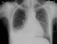
FIGURE 2
Right axis deviation with flipped T waves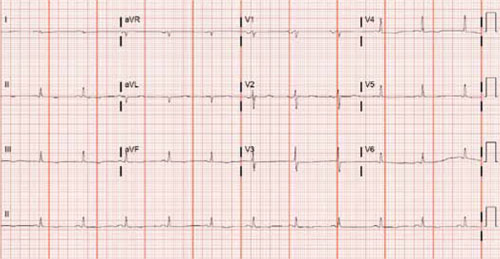
FIGURE 3
Classic square root sign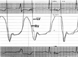
WHAT IS THE MOST LIKELY EXPLANATION FOR HIS CONDITION?
Constrictive pericarditis
On the advice of a cardiologist who specializes in advanced cardiac imaging, our patient underwent tissue Doppler velocity echocardiography—a diagnostic test that provides evidence of a diseased myocardium (as in restrictive cardiomyopathy), as well as changes in diastolic blood flow that differentiate constrictive pericarditis from restrictive cardiomyopathy. Our patient’s tissue Doppler velocity echocardiogram revealed a normal myocardium with an abrupt cessation of early left ventricular filling, consistent with constrictive pericarditis. Along with his clinical presentation and history, this test was conclusive enough to diagnose constrictive pericarditis.
Making the diagnosis in more “typical” cases
Symptoms of constrictive pericarditis include fluid overload (eg, recurrent pleural effusions2-4) hepatic dysfunction, and peripheral edema. Studies show that 44% to 54% of patients with constrictive pericarditis present with pleural effusions;5 chest pain and dyspnea are other common symptoms.6 Past medical history is also important, considering the 3 most common risks for constrictive pericarditis: cardiac surgery, pericarditis, and irradiation of the mediastinum (TABLE).7
Typical physical exam findings in patients with constrictive pericarditis include an elevated jugular venous pressure, third spacing of fluids, a pericardial knock, and Kussmaul’s sign. Our patient had third spacing of fluids, including mild peripheral edema and the recurrent effusions as noted, as well as signs of hepatic dysfunction that were initially attributed to his rifampin use. While these symptoms raise the suspicion of constrictive pericarditis, none is specific to that condition alone.
Studies that help in the diagnosis of constrictive pericarditis include chest x-ray, cardiac CT, echocardiography, cardiac magnetic resonance imaging (MRI), and cardiac catheterization. EKG has no specific findings, but cardiac arrhythmias (eg, atrial fibrillation) are common.8 Pericardial thickening on cardiac CT scan is a definitive but not universal finding; in a Mayo Clinic study, such thickening was not evident in 26 of 143 patients with confirmed constrictive pericarditis.8 Cardiac MRI has been shown to have 88% sensitivity and 100% specificity,9 but will miss up to 18% of patients with constrictive pericarditis.8
Differentiating between restriction and constriction. It can be difficult to distinguish between constrictive pericarditis and restrictive cardiomyopathy. Although these conditions can present in a similar manner, they require different modes of treatment. Laboratory testing, cardiac catheterization, and tissue Doppler velocity echocardiography (which we relied on) can help to distinguish between them.
TABLE
Causes of constrictive pericarditis7
| More common |
|---|
| Idiopathic Postcardiac surgery (coronary artery bypass grafting, valve replacement) Postradiation therapy (eg, for breast cancer, lymphoma) Viral (pericarditis) |
| Less common |
| Asbestosis Cancer and myeloproliferative disorders Connective tissue disorders (rheumatoid arthritis, systemic lupus erythematosus) Adverse drug reaction Infection (tuberculosis, fungal) Sarcoidosis Trauma Uremia |
Pericardiectomy is the treatment of choice
Pericardiectomy, the optimal treatment for most patients with constrictive pericarditis, carries a 30-day perioperative mortality of approximately 6%.5,7 Patients with minimal symptoms can be monitored for up to 2 months, but only 17% of cases are self-limited.6 Patients with end-stage disease or those who have radiation-induced constrictive pericarditis experience poor surgical outcomes, and may be better served by medical management.7
In light of our patient’s excellent baseline functional status, clinical presentation, and Doppler test results, a cardiothoracic surgeon performed a pericardiectomy. His symptoms improved postoperatively, and he has had no further pleural effusions. The patient’s fatigue, anorexia, weight loss, and dyspnea fully resolved, as well.
- Include constrictive pericarditis in the differential diagnosis of patients with recurrent pleural effusions, an important presenting symptom in 44% to 54% of patients with this condition.
- Consider multiple testing modalities to arrive at a diagnosis of constrictive pericarditis, including cardiac CT or MRI, tissue Doppler echocardiography, and cardiac catheterization.
- Do not rule out constrictive pericarditis if pericardial thickening is not found on cardiac CT scan; in 1 study, this finding was not present in 18% of patients with a confirmed diagnosis.
CORRESPONDENCE Jeffrey S. Morgeson, MD; 2123 Auburn Avenue, 340 MOB, Cincinnati, OH 45219; morgesjs@gmail.com
1. Maisch B, Seferovic PM, Ristic AD, et al. Guidelines on the diagnosis and management of pericardial diseases executive summary; The Task force on the diagnosis and management of pericardial diseases of the European Society of Cardiology. Eur Heart J. 2004;25:587-610.
2. Akhter MW, Nuño IN, Rahimtoola SH. Constrictive pericarditis masquerading as chronic idiopathic pleural effusion: importance of physical examination. Am J Med. 2006;119:e1-e4.
3. Ramar K, Daniels CA. Constrictive pericarditis presenting as unexplained dyspnea with recurrent pleural effusion. Respir Care. 2008;53:912-915.
4. Sadikot RT, Fredi JL, Light RW. A 43-year-old man with a large recurrent right-sided pleural effusion. Diagnosis: constrictive pericarditis. Chest. 2000;117:1191-1194.
5. Bertog SC, Thambidorai SK, Parakh K, et al. Constrictive pericarditis: etiology and cause-specific survival after pericardiectomy. J Am Coll Cardiol. 2004;43:1445-1452.
6. Haley JH, Tajik AJ, Danielson GK, et al. Transient constrictive pericarditis: causes and natural history. J Am Coll Cardiol. 2004;43:271-275.
7. Ling LH, Oh JK, Schaff HV, et al. Constrictive pericarditis in the modern era: evolving clinical spectrum and impact on outcome after pericardiectomy. Circulation. 1999;100:1380-1386.
8. Talreja DP, Edwards WD, Danielson GK, et al. Constrictive pericarditis in 26 patients with histologically normal pericardial thickness. Circulation. 2003;108:1852-1857.
9. Masui T, Finck S, Higgins CB. Constrictive pericarditis and restrictive cardiomyopathy: evaluation with MR imaging. Radiology. 1992;182:369-373.
CASE An 84-year-old man was admitted to our family medicine inpatient service with increasing weakness, fatigue, nausea, jaundice, and anorexia after undergoing thoracentesis for recurrent large right-sided pleural effusion twice within the past 6 weeks. The patient had no fever, chills, night sweats, chest pain, or chronic cough.
His past medical history included coronary artery disease and hyperlipidemia, and he had undergone coronary artery bypass grafting 6 years prior to this admission.
Initial vital signs included a blood pressure of 116/68 mm hg and a pulse rate of 78 beats per minute. physical examination revealed a thin but otherwise active and functional man, with the presence of scleral icterus and a 2/6 systolic murmur that was loudest at the left upper sternal border, with no pericardial knocks or rubs. There was no jugular venous distension. The patient had decreased breath sounds at the right base, but no appreciable rales, rhonchi, or wheezes, and an enlarged liver with an irregular edge approximately 7 cm below the costal margin. he had bilateral trace pitting edema of the lower extremities and an erythematous chronic venous stasis ulcer on his left lower leg that was being treated with sulfamethoxazole and trimethoprim and rifampin.
Laboratory findings included a sodium level of 130 mEq/L; creatinine, 1.6 mg/dL; total bilirubin, 4.2 mg/dL; and an international normalized ratio (INR) of 1.6, with an otherwise normal liver profile. pleural fluid was transudative by light’s criteria, with negative pleural fluid cultures.
A chest x-ray showed right-sided airspace disease, with an associated small effusion (FIGURE 1), and an electrocardiogram (EKG) revealed a right axis deviation with flipped T waves in V3-V6 (FIGURE 2). A computed tomography (CT) scan of the chest performed 2 months prior to admission revealed a small right pleural effusion, moderate emphysema, and pleural plaques. A transthoracic echocardiogram performed a week earlier was significant for a normal ejection fraction without pericardial thickening, and mild dilation of the left and right atria and the right ventricle.
Our patient had recurrent transudative pleural effusions with no history of congestive heart failure, a normal thyroid-stimulating hormone, no signs of nephrotic syndrome, no risk factors for lung cancer, and a presentation that was not consistent with an acute pulmonary embolus. he had jaundice, but no risk factors for cirrhosis. A comprehensive hepatology consult suggested that the jaundice was associated with recent rifampin use, along with hepatic congestion likely due to cardiac etiology.
The patient also had a documented history of constrictive pericarditis. Although a cardiac CT scan 15 months prior to his hospital admission showed normal pericardial thickness, constrictive pericarditis remained high on our differential.
To further evaluate the patient’s cardiac function, a cardiologist performed a right-sided cardiac catheterization. in constrictive pericarditis, equalization of end-diastolic pressures occurs due to limitation of the total volume of the ventricles by a rigid pericardium. in our patient, these pressures did not equalize. his right and left ventricular pressure tracings did, however, have the classic square root sign often seen in constrictive pericarditis (FIGURE 3). This “dip and plateau” is the result of rapid filling of the ventricles during early diastole, with the inflexible pericardium causing an abrupt halt in ventricular filling.1
We considered restrictive cardiomyopathy in the differential, too, but the signs and symptoms weren’t a perfect fit there, either. equalization of left and right ventricular end-diastolic pressures in cardiac catheterization is usually not seen in restrictive cardiomyopathy, but neither is the square-root sign evident on ventricular pressure monitors.1
FIGURE 1
X-ray shows right-sided airspace disease
FIGURE 2
Right axis deviation with flipped T waves
FIGURE 3
Classic square root sign
WHAT IS THE MOST LIKELY EXPLANATION FOR HIS CONDITION?
Constrictive pericarditis
On the advice of a cardiologist who specializes in advanced cardiac imaging, our patient underwent tissue Doppler velocity echocardiography—a diagnostic test that provides evidence of a diseased myocardium (as in restrictive cardiomyopathy), as well as changes in diastolic blood flow that differentiate constrictive pericarditis from restrictive cardiomyopathy. Our patient’s tissue Doppler velocity echocardiogram revealed a normal myocardium with an abrupt cessation of early left ventricular filling, consistent with constrictive pericarditis. Along with his clinical presentation and history, this test was conclusive enough to diagnose constrictive pericarditis.
Making the diagnosis in more “typical” cases
Symptoms of constrictive pericarditis include fluid overload (eg, recurrent pleural effusions2-4) hepatic dysfunction, and peripheral edema. Studies show that 44% to 54% of patients with constrictive pericarditis present with pleural effusions;5 chest pain and dyspnea are other common symptoms.6 Past medical history is also important, considering the 3 most common risks for constrictive pericarditis: cardiac surgery, pericarditis, and irradiation of the mediastinum (TABLE).7
Typical physical exam findings in patients with constrictive pericarditis include an elevated jugular venous pressure, third spacing of fluids, a pericardial knock, and Kussmaul’s sign. Our patient had third spacing of fluids, including mild peripheral edema and the recurrent effusions as noted, as well as signs of hepatic dysfunction that were initially attributed to his rifampin use. While these symptoms raise the suspicion of constrictive pericarditis, none is specific to that condition alone.
Studies that help in the diagnosis of constrictive pericarditis include chest x-ray, cardiac CT, echocardiography, cardiac magnetic resonance imaging (MRI), and cardiac catheterization. EKG has no specific findings, but cardiac arrhythmias (eg, atrial fibrillation) are common.8 Pericardial thickening on cardiac CT scan is a definitive but not universal finding; in a Mayo Clinic study, such thickening was not evident in 26 of 143 patients with confirmed constrictive pericarditis.8 Cardiac MRI has been shown to have 88% sensitivity and 100% specificity,9 but will miss up to 18% of patients with constrictive pericarditis.8
Differentiating between restriction and constriction. It can be difficult to distinguish between constrictive pericarditis and restrictive cardiomyopathy. Although these conditions can present in a similar manner, they require different modes of treatment. Laboratory testing, cardiac catheterization, and tissue Doppler velocity echocardiography (which we relied on) can help to distinguish between them.
TABLE
Causes of constrictive pericarditis7
| More common |
|---|
| Idiopathic Postcardiac surgery (coronary artery bypass grafting, valve replacement) Postradiation therapy (eg, for breast cancer, lymphoma) Viral (pericarditis) |
| Less common |
| Asbestosis Cancer and myeloproliferative disorders Connective tissue disorders (rheumatoid arthritis, systemic lupus erythematosus) Adverse drug reaction Infection (tuberculosis, fungal) Sarcoidosis Trauma Uremia |
Pericardiectomy is the treatment of choice
Pericardiectomy, the optimal treatment for most patients with constrictive pericarditis, carries a 30-day perioperative mortality of approximately 6%.5,7 Patients with minimal symptoms can be monitored for up to 2 months, but only 17% of cases are self-limited.6 Patients with end-stage disease or those who have radiation-induced constrictive pericarditis experience poor surgical outcomes, and may be better served by medical management.7
In light of our patient’s excellent baseline functional status, clinical presentation, and Doppler test results, a cardiothoracic surgeon performed a pericardiectomy. His symptoms improved postoperatively, and he has had no further pleural effusions. The patient’s fatigue, anorexia, weight loss, and dyspnea fully resolved, as well.
- Include constrictive pericarditis in the differential diagnosis of patients with recurrent pleural effusions, an important presenting symptom in 44% to 54% of patients with this condition.
- Consider multiple testing modalities to arrive at a diagnosis of constrictive pericarditis, including cardiac CT or MRI, tissue Doppler echocardiography, and cardiac catheterization.
- Do not rule out constrictive pericarditis if pericardial thickening is not found on cardiac CT scan; in 1 study, this finding was not present in 18% of patients with a confirmed diagnosis.
CORRESPONDENCE Jeffrey S. Morgeson, MD; 2123 Auburn Avenue, 340 MOB, Cincinnati, OH 45219; morgesjs@gmail.com
CASE An 84-year-old man was admitted to our family medicine inpatient service with increasing weakness, fatigue, nausea, jaundice, and anorexia after undergoing thoracentesis for recurrent large right-sided pleural effusion twice within the past 6 weeks. The patient had no fever, chills, night sweats, chest pain, or chronic cough.
His past medical history included coronary artery disease and hyperlipidemia, and he had undergone coronary artery bypass grafting 6 years prior to this admission.
Initial vital signs included a blood pressure of 116/68 mm hg and a pulse rate of 78 beats per minute. physical examination revealed a thin but otherwise active and functional man, with the presence of scleral icterus and a 2/6 systolic murmur that was loudest at the left upper sternal border, with no pericardial knocks or rubs. There was no jugular venous distension. The patient had decreased breath sounds at the right base, but no appreciable rales, rhonchi, or wheezes, and an enlarged liver with an irregular edge approximately 7 cm below the costal margin. he had bilateral trace pitting edema of the lower extremities and an erythematous chronic venous stasis ulcer on his left lower leg that was being treated with sulfamethoxazole and trimethoprim and rifampin.
Laboratory findings included a sodium level of 130 mEq/L; creatinine, 1.6 mg/dL; total bilirubin, 4.2 mg/dL; and an international normalized ratio (INR) of 1.6, with an otherwise normal liver profile. pleural fluid was transudative by light’s criteria, with negative pleural fluid cultures.
A chest x-ray showed right-sided airspace disease, with an associated small effusion (FIGURE 1), and an electrocardiogram (EKG) revealed a right axis deviation with flipped T waves in V3-V6 (FIGURE 2). A computed tomography (CT) scan of the chest performed 2 months prior to admission revealed a small right pleural effusion, moderate emphysema, and pleural plaques. A transthoracic echocardiogram performed a week earlier was significant for a normal ejection fraction without pericardial thickening, and mild dilation of the left and right atria and the right ventricle.
Our patient had recurrent transudative pleural effusions with no history of congestive heart failure, a normal thyroid-stimulating hormone, no signs of nephrotic syndrome, no risk factors for lung cancer, and a presentation that was not consistent with an acute pulmonary embolus. he had jaundice, but no risk factors for cirrhosis. A comprehensive hepatology consult suggested that the jaundice was associated with recent rifampin use, along with hepatic congestion likely due to cardiac etiology.
The patient also had a documented history of constrictive pericarditis. Although a cardiac CT scan 15 months prior to his hospital admission showed normal pericardial thickness, constrictive pericarditis remained high on our differential.
To further evaluate the patient’s cardiac function, a cardiologist performed a right-sided cardiac catheterization. in constrictive pericarditis, equalization of end-diastolic pressures occurs due to limitation of the total volume of the ventricles by a rigid pericardium. in our patient, these pressures did not equalize. his right and left ventricular pressure tracings did, however, have the classic square root sign often seen in constrictive pericarditis (FIGURE 3). This “dip and plateau” is the result of rapid filling of the ventricles during early diastole, with the inflexible pericardium causing an abrupt halt in ventricular filling.1
We considered restrictive cardiomyopathy in the differential, too, but the signs and symptoms weren’t a perfect fit there, either. equalization of left and right ventricular end-diastolic pressures in cardiac catheterization is usually not seen in restrictive cardiomyopathy, but neither is the square-root sign evident on ventricular pressure monitors.1
FIGURE 1
X-ray shows right-sided airspace disease
FIGURE 2
Right axis deviation with flipped T waves
FIGURE 3
Classic square root sign
WHAT IS THE MOST LIKELY EXPLANATION FOR HIS CONDITION?
Constrictive pericarditis
On the advice of a cardiologist who specializes in advanced cardiac imaging, our patient underwent tissue Doppler velocity echocardiography—a diagnostic test that provides evidence of a diseased myocardium (as in restrictive cardiomyopathy), as well as changes in diastolic blood flow that differentiate constrictive pericarditis from restrictive cardiomyopathy. Our patient’s tissue Doppler velocity echocardiogram revealed a normal myocardium with an abrupt cessation of early left ventricular filling, consistent with constrictive pericarditis. Along with his clinical presentation and history, this test was conclusive enough to diagnose constrictive pericarditis.
Making the diagnosis in more “typical” cases
Symptoms of constrictive pericarditis include fluid overload (eg, recurrent pleural effusions2-4) hepatic dysfunction, and peripheral edema. Studies show that 44% to 54% of patients with constrictive pericarditis present with pleural effusions;5 chest pain and dyspnea are other common symptoms.6 Past medical history is also important, considering the 3 most common risks for constrictive pericarditis: cardiac surgery, pericarditis, and irradiation of the mediastinum (TABLE).7
Typical physical exam findings in patients with constrictive pericarditis include an elevated jugular venous pressure, third spacing of fluids, a pericardial knock, and Kussmaul’s sign. Our patient had third spacing of fluids, including mild peripheral edema and the recurrent effusions as noted, as well as signs of hepatic dysfunction that were initially attributed to his rifampin use. While these symptoms raise the suspicion of constrictive pericarditis, none is specific to that condition alone.
Studies that help in the diagnosis of constrictive pericarditis include chest x-ray, cardiac CT, echocardiography, cardiac magnetic resonance imaging (MRI), and cardiac catheterization. EKG has no specific findings, but cardiac arrhythmias (eg, atrial fibrillation) are common.8 Pericardial thickening on cardiac CT scan is a definitive but not universal finding; in a Mayo Clinic study, such thickening was not evident in 26 of 143 patients with confirmed constrictive pericarditis.8 Cardiac MRI has been shown to have 88% sensitivity and 100% specificity,9 but will miss up to 18% of patients with constrictive pericarditis.8
Differentiating between restriction and constriction. It can be difficult to distinguish between constrictive pericarditis and restrictive cardiomyopathy. Although these conditions can present in a similar manner, they require different modes of treatment. Laboratory testing, cardiac catheterization, and tissue Doppler velocity echocardiography (which we relied on) can help to distinguish between them.
TABLE
Causes of constrictive pericarditis7
| More common |
|---|
| Idiopathic Postcardiac surgery (coronary artery bypass grafting, valve replacement) Postradiation therapy (eg, for breast cancer, lymphoma) Viral (pericarditis) |
| Less common |
| Asbestosis Cancer and myeloproliferative disorders Connective tissue disorders (rheumatoid arthritis, systemic lupus erythematosus) Adverse drug reaction Infection (tuberculosis, fungal) Sarcoidosis Trauma Uremia |
Pericardiectomy is the treatment of choice
Pericardiectomy, the optimal treatment for most patients with constrictive pericarditis, carries a 30-day perioperative mortality of approximately 6%.5,7 Patients with minimal symptoms can be monitored for up to 2 months, but only 17% of cases are self-limited.6 Patients with end-stage disease or those who have radiation-induced constrictive pericarditis experience poor surgical outcomes, and may be better served by medical management.7
In light of our patient’s excellent baseline functional status, clinical presentation, and Doppler test results, a cardiothoracic surgeon performed a pericardiectomy. His symptoms improved postoperatively, and he has had no further pleural effusions. The patient’s fatigue, anorexia, weight loss, and dyspnea fully resolved, as well.
- Include constrictive pericarditis in the differential diagnosis of patients with recurrent pleural effusions, an important presenting symptom in 44% to 54% of patients with this condition.
- Consider multiple testing modalities to arrive at a diagnosis of constrictive pericarditis, including cardiac CT or MRI, tissue Doppler echocardiography, and cardiac catheterization.
- Do not rule out constrictive pericarditis if pericardial thickening is not found on cardiac CT scan; in 1 study, this finding was not present in 18% of patients with a confirmed diagnosis.
CORRESPONDENCE Jeffrey S. Morgeson, MD; 2123 Auburn Avenue, 340 MOB, Cincinnati, OH 45219; morgesjs@gmail.com
1. Maisch B, Seferovic PM, Ristic AD, et al. Guidelines on the diagnosis and management of pericardial diseases executive summary; The Task force on the diagnosis and management of pericardial diseases of the European Society of Cardiology. Eur Heart J. 2004;25:587-610.
2. Akhter MW, Nuño IN, Rahimtoola SH. Constrictive pericarditis masquerading as chronic idiopathic pleural effusion: importance of physical examination. Am J Med. 2006;119:e1-e4.
3. Ramar K, Daniels CA. Constrictive pericarditis presenting as unexplained dyspnea with recurrent pleural effusion. Respir Care. 2008;53:912-915.
4. Sadikot RT, Fredi JL, Light RW. A 43-year-old man with a large recurrent right-sided pleural effusion. Diagnosis: constrictive pericarditis. Chest. 2000;117:1191-1194.
5. Bertog SC, Thambidorai SK, Parakh K, et al. Constrictive pericarditis: etiology and cause-specific survival after pericardiectomy. J Am Coll Cardiol. 2004;43:1445-1452.
6. Haley JH, Tajik AJ, Danielson GK, et al. Transient constrictive pericarditis: causes and natural history. J Am Coll Cardiol. 2004;43:271-275.
7. Ling LH, Oh JK, Schaff HV, et al. Constrictive pericarditis in the modern era: evolving clinical spectrum and impact on outcome after pericardiectomy. Circulation. 1999;100:1380-1386.
8. Talreja DP, Edwards WD, Danielson GK, et al. Constrictive pericarditis in 26 patients with histologically normal pericardial thickness. Circulation. 2003;108:1852-1857.
9. Masui T, Finck S, Higgins CB. Constrictive pericarditis and restrictive cardiomyopathy: evaluation with MR imaging. Radiology. 1992;182:369-373.
1. Maisch B, Seferovic PM, Ristic AD, et al. Guidelines on the diagnosis and management of pericardial diseases executive summary; The Task force on the diagnosis and management of pericardial diseases of the European Society of Cardiology. Eur Heart J. 2004;25:587-610.
2. Akhter MW, Nuño IN, Rahimtoola SH. Constrictive pericarditis masquerading as chronic idiopathic pleural effusion: importance of physical examination. Am J Med. 2006;119:e1-e4.
3. Ramar K, Daniels CA. Constrictive pericarditis presenting as unexplained dyspnea with recurrent pleural effusion. Respir Care. 2008;53:912-915.
4. Sadikot RT, Fredi JL, Light RW. A 43-year-old man with a large recurrent right-sided pleural effusion. Diagnosis: constrictive pericarditis. Chest. 2000;117:1191-1194.
5. Bertog SC, Thambidorai SK, Parakh K, et al. Constrictive pericarditis: etiology and cause-specific survival after pericardiectomy. J Am Coll Cardiol. 2004;43:1445-1452.
6. Haley JH, Tajik AJ, Danielson GK, et al. Transient constrictive pericarditis: causes and natural history. J Am Coll Cardiol. 2004;43:271-275.
7. Ling LH, Oh JK, Schaff HV, et al. Constrictive pericarditis in the modern era: evolving clinical spectrum and impact on outcome after pericardiectomy. Circulation. 1999;100:1380-1386.
8. Talreja DP, Edwards WD, Danielson GK, et al. Constrictive pericarditis in 26 patients with histologically normal pericardial thickness. Circulation. 2003;108:1852-1857.
9. Masui T, Finck S, Higgins CB. Constrictive pericarditis and restrictive cardiomyopathy: evaluation with MR imaging. Radiology. 1992;182:369-373.
Asthma Initiative Helps Disadvantaged Children
VANCOUVER, B.C. — An initiative that promotes improved asthma education and care at the family and community levels has reduced health care use and morbidity among disadvantaged children with asthma in Boston, according to Dr. Elizabeth R. Woods.
Four years into the Community Asthma Initiative, there was an 81% reduction in the percentage of participating children with asthma-related admissions, a 65% cut in the percentage of children making emergency department visits, a 39% reduction in the percentage missing school because of asthma, and a 37% reduction in the percentage having limitations in physical activity because of the disease.
“The program demonstrates a successful model that can be developed and is starting to be replicated in Massachusetts as well as nationally,” Dr. Woods said at the meeting. “It also promotes policy changes that substantially improve asthma education and care, including access to case management, home visits, and affordable medications.”
The initiative targeted children from the four Boston neighborhoods with the highest asthma rates and the greatest health disparities. The children were identified through asthma-related ED visits or hospital admissions, or were referred by primary care providers.
They and their families received case management and home visits by providers who helped them develop individualized management plans, performed environmental assessments, and supplied products such as vacuum cleaners with high-efficiency particulate air (HEPA) filters and bedding casings. Providers also instructed families in pest control techniques and connected them to community resources.
The initiative also targeted the community (families, providers, and teachers) through an educational campaign. Advocacy efforts were launched to encourage payers to address prohibitively high copayments for asthma medications.
Dr. Woods and her colleagues evaluated the effects of the initiative by analyzing parental reports obtained at 6-month intervals and administrative data.
Results were based on 441 children who had received case management through the initiative, the majority of whom had also received home visits. They were 7.8 years old on average. Most were African American (48%) or Latino/Hispanic (45%), and had public health insurance (70%).
From baseline to 12 months, the proportion of children making asthma-related ED visits fell from 63% to 22%, hospital admissions due to asthma fell from 51% to 10%, and the proportion of children who missed school because of asthma dropped from 93% to 56%. The proportion of children who had physical activity limitations due to asthma dropped from 55% to 35%.
The proportion of children with an up-to-date asthma action plan increased by 71% (from 49% to 84%).
In logistic regression analyses that controlled for potential confounders, the children had significant 90%–100% reductions in the odds of each adverse outcome, noted Dr. Woods, a pediatrician at Children's Hospital Boston.
In the initiative's first year, the cost of care per child was similar to that in a control neighborhood ($1,335 vs. $1,340). In the second year, it was approximately half as expensive in the initiative group ($750 vs. $1,322).
“There was clearly a cost saving, giving a return on investment of 1.46. … That's a return on investment not to hospitals, but to insurance companies and society at large,” Dr. Woods said.
The initiative is helping families in two main ways. “No. 1, it is helping them understand their medications,” she said, as many families are found to have bags and boxes of medications and a poor grasp of which ones to use in which circumstances. “The other big improvement relates to the environmental issues. Very few of these families had even a vacuum cleaner, let alone ones with HEPA bags and filters. These are incredibly helpful and much less costly than additional medication.”
VANCOUVER, B.C. — An initiative that promotes improved asthma education and care at the family and community levels has reduced health care use and morbidity among disadvantaged children with asthma in Boston, according to Dr. Elizabeth R. Woods.
Four years into the Community Asthma Initiative, there was an 81% reduction in the percentage of participating children with asthma-related admissions, a 65% cut in the percentage of children making emergency department visits, a 39% reduction in the percentage missing school because of asthma, and a 37% reduction in the percentage having limitations in physical activity because of the disease.
“The program demonstrates a successful model that can be developed and is starting to be replicated in Massachusetts as well as nationally,” Dr. Woods said at the meeting. “It also promotes policy changes that substantially improve asthma education and care, including access to case management, home visits, and affordable medications.”
The initiative targeted children from the four Boston neighborhoods with the highest asthma rates and the greatest health disparities. The children were identified through asthma-related ED visits or hospital admissions, or were referred by primary care providers.
They and their families received case management and home visits by providers who helped them develop individualized management plans, performed environmental assessments, and supplied products such as vacuum cleaners with high-efficiency particulate air (HEPA) filters and bedding casings. Providers also instructed families in pest control techniques and connected them to community resources.
The initiative also targeted the community (families, providers, and teachers) through an educational campaign. Advocacy efforts were launched to encourage payers to address prohibitively high copayments for asthma medications.
Dr. Woods and her colleagues evaluated the effects of the initiative by analyzing parental reports obtained at 6-month intervals and administrative data.
Results were based on 441 children who had received case management through the initiative, the majority of whom had also received home visits. They were 7.8 years old on average. Most were African American (48%) or Latino/Hispanic (45%), and had public health insurance (70%).
From baseline to 12 months, the proportion of children making asthma-related ED visits fell from 63% to 22%, hospital admissions due to asthma fell from 51% to 10%, and the proportion of children who missed school because of asthma dropped from 93% to 56%. The proportion of children who had physical activity limitations due to asthma dropped from 55% to 35%.
The proportion of children with an up-to-date asthma action plan increased by 71% (from 49% to 84%).
In logistic regression analyses that controlled for potential confounders, the children had significant 90%–100% reductions in the odds of each adverse outcome, noted Dr. Woods, a pediatrician at Children's Hospital Boston.
In the initiative's first year, the cost of care per child was similar to that in a control neighborhood ($1,335 vs. $1,340). In the second year, it was approximately half as expensive in the initiative group ($750 vs. $1,322).
“There was clearly a cost saving, giving a return on investment of 1.46. … That's a return on investment not to hospitals, but to insurance companies and society at large,” Dr. Woods said.
The initiative is helping families in two main ways. “No. 1, it is helping them understand their medications,” she said, as many families are found to have bags and boxes of medications and a poor grasp of which ones to use in which circumstances. “The other big improvement relates to the environmental issues. Very few of these families had even a vacuum cleaner, let alone ones with HEPA bags and filters. These are incredibly helpful and much less costly than additional medication.”
VANCOUVER, B.C. — An initiative that promotes improved asthma education and care at the family and community levels has reduced health care use and morbidity among disadvantaged children with asthma in Boston, according to Dr. Elizabeth R. Woods.
Four years into the Community Asthma Initiative, there was an 81% reduction in the percentage of participating children with asthma-related admissions, a 65% cut in the percentage of children making emergency department visits, a 39% reduction in the percentage missing school because of asthma, and a 37% reduction in the percentage having limitations in physical activity because of the disease.
“The program demonstrates a successful model that can be developed and is starting to be replicated in Massachusetts as well as nationally,” Dr. Woods said at the meeting. “It also promotes policy changes that substantially improve asthma education and care, including access to case management, home visits, and affordable medications.”
The initiative targeted children from the four Boston neighborhoods with the highest asthma rates and the greatest health disparities. The children were identified through asthma-related ED visits or hospital admissions, or were referred by primary care providers.
They and their families received case management and home visits by providers who helped them develop individualized management plans, performed environmental assessments, and supplied products such as vacuum cleaners with high-efficiency particulate air (HEPA) filters and bedding casings. Providers also instructed families in pest control techniques and connected them to community resources.
The initiative also targeted the community (families, providers, and teachers) through an educational campaign. Advocacy efforts were launched to encourage payers to address prohibitively high copayments for asthma medications.
Dr. Woods and her colleagues evaluated the effects of the initiative by analyzing parental reports obtained at 6-month intervals and administrative data.
Results were based on 441 children who had received case management through the initiative, the majority of whom had also received home visits. They were 7.8 years old on average. Most were African American (48%) or Latino/Hispanic (45%), and had public health insurance (70%).
From baseline to 12 months, the proportion of children making asthma-related ED visits fell from 63% to 22%, hospital admissions due to asthma fell from 51% to 10%, and the proportion of children who missed school because of asthma dropped from 93% to 56%. The proportion of children who had physical activity limitations due to asthma dropped from 55% to 35%.
The proportion of children with an up-to-date asthma action plan increased by 71% (from 49% to 84%).
In logistic regression analyses that controlled for potential confounders, the children had significant 90%–100% reductions in the odds of each adverse outcome, noted Dr. Woods, a pediatrician at Children's Hospital Boston.
In the initiative's first year, the cost of care per child was similar to that in a control neighborhood ($1,335 vs. $1,340). In the second year, it was approximately half as expensive in the initiative group ($750 vs. $1,322).
“There was clearly a cost saving, giving a return on investment of 1.46. … That's a return on investment not to hospitals, but to insurance companies and society at large,” Dr. Woods said.
The initiative is helping families in two main ways. “No. 1, it is helping them understand their medications,” she said, as many families are found to have bags and boxes of medications and a poor grasp of which ones to use in which circumstances. “The other big improvement relates to the environmental issues. Very few of these families had even a vacuum cleaner, let alone ones with HEPA bags and filters. These are incredibly helpful and much less costly than additional medication.”
Top Asthma Studies Will Reshape Treatment : Recent papers address topics ranging from theophylline to thermoplasty to nitric oxide.
KEYSTONE, COLO. — Quadrupling the dose of inhaled corticosteroid was an effective strategy for prevention of asthma exacerbations, and low-dose theophylline enhanced steroids' anti-inflammatory benefits, Dr. Harold S. Nelson noted in a review of new asthma studies.
In the 403-patient study of inhaled corticosteroid dosages (Am. J. Respir. Crit. Care Med. 2009;180:598–602), patients who quadrupled their inhaled corticosteroid dose in response to early evidence of an exacerbation based upon morning pulmonary function testing had a 57% reduction in the relative risk of requiring oral steroids, compared with patients who made no change in their low-dose inhaled steroid regimen, Dr. Nelson said at a meeting on allergy and respiratory disease sponsored by National Jewish Health, Denver.
Although the results didn't achieve significance, he rated this trial as among the past year's highlights in the asthma literature becausetit answers an important, previously unresolved clinical question.
“The long-time teaching has been to double the dose of inhaled corticosteroid when a patient notices the onset of an asthma exacerbation. That strategy has been shown to be totally ineffective in two large, well-done studies,” noted Dr. Nelson of National Jewish Health and professor of medicine at the University of Colorado, Denver.
Among the other highlights he identified in the recent asthma literature are:
▸ Low-dose theophylline enhances the anti-inflammatory benefits of steroids: In a study of 68 asthmatic smokers, 4 weeks of theophylline at 400 mg/day plus beclomethasone at 200 mcg/day resulted in significantly greater improvements both in lung function and in asthma symptoms than either drug alone (Eur. Respir. J. 2009;33:1010-17).
The rationale for using low-dose theophylline in this setting is that cigarette smoke inhibits histone deacetylase, an enzyme that mediates the therapeutic response to corticosteroids. Low-dose theophylline increases histone deacetylase activity.
The notion that low-dose theophylline has a place in the treatment of smokers with chronic respiratory disease was reinforced in another recent study, which involved 35 patients hospitalized for acute exacerbations of COPD.
They were randomized to standard therapy—bronchodilators and systemic steroids while hospitalized, long-acting beta-agonists and inhaled corticosteroids after discharge—or to standard therapy plus 100 mg of theophylline twice daily.
At follow-up 3 months later, the theophylline group had significantly greater improvement in forced expiratory volume in 1 second than those on standard therapy. They also had more than a threefold greater increase in macrophage histone deacetylase activity, compared with baseline, and much greater reductions in inflammatory cytokine levels in their sputum (Thorax 2009;64:424-9).
▸ Tumor necrosis factor–alpha inhibition for treatment of severe persistent asthma: This double-blind trial randomized 309 patients to one of three doses of golimumab (Simponi) or placebo. The study was scheduled to run for a year but stopped early after eight golimumab-treated patients developed cancers, including five patients in the highest-dose arm. No cancers occurred in the placebo group.
There were no significant differences between the golimumab and placebo groups in the number of severe asthma exacerbations or forced expiratory volume in 1 second at 24 weeks, the two coprimary end points (Am. J. Respir. Crit. Care Med. 2009;179:549-58).
▸ Esomeprazole for poorly controlled asthma: In a study carried out by the American Lung Association Asthma Clinical Research Centers, 412 patients were randomized to 40 mg of esomeprazole twice daily or placebo for 24 weeks. There were no differences in outcomes between the two study arms in terms of number of episodes of poor asthma control, nocturnal awakening, quality of life, airway reactivity, or pulmonary function. Nor did the 40% of participants with silent gastroesophageal reflux disease benefit from esomeprazole in terms of the study end points (N. Engl. J. Med. 2009;360:1487-99). It is clear that silent or minimally symptomatic GERD is not a likely cause of poorly controlled asthma, Dr. Nelson said.
▸ Monitoring adherence to inhaled corticosteroid therapy in asthmatic children and teens: Four methods of monitoring treatment adherence were evaluated in a 1-year study of 102 asthmatic 3- to 14-year-olds. Adherence deteriorated progressively over the course of the year. Parent and self-reports gave a wildly inflated picture of adherence. So did pharmacy dispensing records. Tracking canister weight proved to be the most practical and accurate method (Allergy 2009;64:1458-62).
▸ Thermoplasty for severe asthma: Thermoplasty reduces airway smooth muscle mass. The regimen entails three treatment sessions at 2-week intervals.
In Dr. Nelson's view, the verdict remains out regarding this procedure, despite a 288-patient multicenter, randomized, double-blind, sham-controlled trial. The primary study end point was clinically meaningful improvement in the Asthma Quality of Life Questionnaire score at 52 weeks. This occurred in 79% of patients who underwent thermoplasty and 64% in the sham-procedure arm.
There were 19 hospitalizations for respiratory symptoms in the thermoplasty arm, vs. 2 in the sham-therapy arm. There were no differences between the two groups in pulmonary function tests, medication use, or asthma-free days (Am. J. Respir. Crit. Care Med. 2010;181:116-24). Dr. Nelson said he'd like to see longer follow-up to better assess the procedure's long-term benefits.
▸ Serum vitamin D correlates inversely with childhood asthma severity: A study in 616 asthmatic children ages 6–14 years showed that 28% had an insufficient serum vitamin D level of 30 ng/mL or less. Serum vitamin D was inversely correlated with hospitalizations for asthma in the previous year, bronchial responsiveness to methacholine, total IgE, and circulating eosinophil counts (Am. J. Respir. Crit. Care Med. 2009;179:765-71).
▸ Daily telemonitoring of exhaled nitric oxide in the treatment of childhood asthma: Dutch investigators randomized 151 children with atopic asthma to management directed by daily symptom monitoring alone or in conjunction with daily telemonitoring of exhaled nitric oxide. Patients improved equally in both groups, meaning monitoring exhaled nitric oxide added nothing (Am. J. Respir. Crit. Care Med. 2009;179:93-7).
“There are now six studies in which exhaled nitric oxide was used to guide asthma management. None has shown a significant improvement with addition of exhaled nitric oxide,” Dr. Nelson said.
Dr. Nelson disclosed serving as a consultant to Abbott, AstraZeneca, Boehringer-Ingelheim, Dey, Dynavax Technologies, Dyson, Genentech, GlaxoSmithKline, Johnson & Johnson, MediciNova, Novartis, Schering-Plough, Sepracor, and Teva, and has received grant and research support from several of these companies.
KEYSTONE, COLO. — Quadrupling the dose of inhaled corticosteroid was an effective strategy for prevention of asthma exacerbations, and low-dose theophylline enhanced steroids' anti-inflammatory benefits, Dr. Harold S. Nelson noted in a review of new asthma studies.
In the 403-patient study of inhaled corticosteroid dosages (Am. J. Respir. Crit. Care Med. 2009;180:598–602), patients who quadrupled their inhaled corticosteroid dose in response to early evidence of an exacerbation based upon morning pulmonary function testing had a 57% reduction in the relative risk of requiring oral steroids, compared with patients who made no change in their low-dose inhaled steroid regimen, Dr. Nelson said at a meeting on allergy and respiratory disease sponsored by National Jewish Health, Denver.
Although the results didn't achieve significance, he rated this trial as among the past year's highlights in the asthma literature becausetit answers an important, previously unresolved clinical question.
“The long-time teaching has been to double the dose of inhaled corticosteroid when a patient notices the onset of an asthma exacerbation. That strategy has been shown to be totally ineffective in two large, well-done studies,” noted Dr. Nelson of National Jewish Health and professor of medicine at the University of Colorado, Denver.
Among the other highlights he identified in the recent asthma literature are:
▸ Low-dose theophylline enhances the anti-inflammatory benefits of steroids: In a study of 68 asthmatic smokers, 4 weeks of theophylline at 400 mg/day plus beclomethasone at 200 mcg/day resulted in significantly greater improvements both in lung function and in asthma symptoms than either drug alone (Eur. Respir. J. 2009;33:1010-17).
The rationale for using low-dose theophylline in this setting is that cigarette smoke inhibits histone deacetylase, an enzyme that mediates the therapeutic response to corticosteroids. Low-dose theophylline increases histone deacetylase activity.
The notion that low-dose theophylline has a place in the treatment of smokers with chronic respiratory disease was reinforced in another recent study, which involved 35 patients hospitalized for acute exacerbations of COPD.
They were randomized to standard therapy—bronchodilators and systemic steroids while hospitalized, long-acting beta-agonists and inhaled corticosteroids after discharge—or to standard therapy plus 100 mg of theophylline twice daily.
At follow-up 3 months later, the theophylline group had significantly greater improvement in forced expiratory volume in 1 second than those on standard therapy. They also had more than a threefold greater increase in macrophage histone deacetylase activity, compared with baseline, and much greater reductions in inflammatory cytokine levels in their sputum (Thorax 2009;64:424-9).
▸ Tumor necrosis factor–alpha inhibition for treatment of severe persistent asthma: This double-blind trial randomized 309 patients to one of three doses of golimumab (Simponi) or placebo. The study was scheduled to run for a year but stopped early after eight golimumab-treated patients developed cancers, including five patients in the highest-dose arm. No cancers occurred in the placebo group.
There were no significant differences between the golimumab and placebo groups in the number of severe asthma exacerbations or forced expiratory volume in 1 second at 24 weeks, the two coprimary end points (Am. J. Respir. Crit. Care Med. 2009;179:549-58).
▸ Esomeprazole for poorly controlled asthma: In a study carried out by the American Lung Association Asthma Clinical Research Centers, 412 patients were randomized to 40 mg of esomeprazole twice daily or placebo for 24 weeks. There were no differences in outcomes between the two study arms in terms of number of episodes of poor asthma control, nocturnal awakening, quality of life, airway reactivity, or pulmonary function. Nor did the 40% of participants with silent gastroesophageal reflux disease benefit from esomeprazole in terms of the study end points (N. Engl. J. Med. 2009;360:1487-99). It is clear that silent or minimally symptomatic GERD is not a likely cause of poorly controlled asthma, Dr. Nelson said.
▸ Monitoring adherence to inhaled corticosteroid therapy in asthmatic children and teens: Four methods of monitoring treatment adherence were evaluated in a 1-year study of 102 asthmatic 3- to 14-year-olds. Adherence deteriorated progressively over the course of the year. Parent and self-reports gave a wildly inflated picture of adherence. So did pharmacy dispensing records. Tracking canister weight proved to be the most practical and accurate method (Allergy 2009;64:1458-62).
▸ Thermoplasty for severe asthma: Thermoplasty reduces airway smooth muscle mass. The regimen entails three treatment sessions at 2-week intervals.
In Dr. Nelson's view, the verdict remains out regarding this procedure, despite a 288-patient multicenter, randomized, double-blind, sham-controlled trial. The primary study end point was clinically meaningful improvement in the Asthma Quality of Life Questionnaire score at 52 weeks. This occurred in 79% of patients who underwent thermoplasty and 64% in the sham-procedure arm.
There were 19 hospitalizations for respiratory symptoms in the thermoplasty arm, vs. 2 in the sham-therapy arm. There were no differences between the two groups in pulmonary function tests, medication use, or asthma-free days (Am. J. Respir. Crit. Care Med. 2010;181:116-24). Dr. Nelson said he'd like to see longer follow-up to better assess the procedure's long-term benefits.
▸ Serum vitamin D correlates inversely with childhood asthma severity: A study in 616 asthmatic children ages 6–14 years showed that 28% had an insufficient serum vitamin D level of 30 ng/mL or less. Serum vitamin D was inversely correlated with hospitalizations for asthma in the previous year, bronchial responsiveness to methacholine, total IgE, and circulating eosinophil counts (Am. J. Respir. Crit. Care Med. 2009;179:765-71).
▸ Daily telemonitoring of exhaled nitric oxide in the treatment of childhood asthma: Dutch investigators randomized 151 children with atopic asthma to management directed by daily symptom monitoring alone or in conjunction with daily telemonitoring of exhaled nitric oxide. Patients improved equally in both groups, meaning monitoring exhaled nitric oxide added nothing (Am. J. Respir. Crit. Care Med. 2009;179:93-7).
“There are now six studies in which exhaled nitric oxide was used to guide asthma management. None has shown a significant improvement with addition of exhaled nitric oxide,” Dr. Nelson said.
Dr. Nelson disclosed serving as a consultant to Abbott, AstraZeneca, Boehringer-Ingelheim, Dey, Dynavax Technologies, Dyson, Genentech, GlaxoSmithKline, Johnson & Johnson, MediciNova, Novartis, Schering-Plough, Sepracor, and Teva, and has received grant and research support from several of these companies.
KEYSTONE, COLO. — Quadrupling the dose of inhaled corticosteroid was an effective strategy for prevention of asthma exacerbations, and low-dose theophylline enhanced steroids' anti-inflammatory benefits, Dr. Harold S. Nelson noted in a review of new asthma studies.
In the 403-patient study of inhaled corticosteroid dosages (Am. J. Respir. Crit. Care Med. 2009;180:598–602), patients who quadrupled their inhaled corticosteroid dose in response to early evidence of an exacerbation based upon morning pulmonary function testing had a 57% reduction in the relative risk of requiring oral steroids, compared with patients who made no change in their low-dose inhaled steroid regimen, Dr. Nelson said at a meeting on allergy and respiratory disease sponsored by National Jewish Health, Denver.
Although the results didn't achieve significance, he rated this trial as among the past year's highlights in the asthma literature becausetit answers an important, previously unresolved clinical question.
“The long-time teaching has been to double the dose of inhaled corticosteroid when a patient notices the onset of an asthma exacerbation. That strategy has been shown to be totally ineffective in two large, well-done studies,” noted Dr. Nelson of National Jewish Health and professor of medicine at the University of Colorado, Denver.
Among the other highlights he identified in the recent asthma literature are:
▸ Low-dose theophylline enhances the anti-inflammatory benefits of steroids: In a study of 68 asthmatic smokers, 4 weeks of theophylline at 400 mg/day plus beclomethasone at 200 mcg/day resulted in significantly greater improvements both in lung function and in asthma symptoms than either drug alone (Eur. Respir. J. 2009;33:1010-17).
The rationale for using low-dose theophylline in this setting is that cigarette smoke inhibits histone deacetylase, an enzyme that mediates the therapeutic response to corticosteroids. Low-dose theophylline increases histone deacetylase activity.
The notion that low-dose theophylline has a place in the treatment of smokers with chronic respiratory disease was reinforced in another recent study, which involved 35 patients hospitalized for acute exacerbations of COPD.
They were randomized to standard therapy—bronchodilators and systemic steroids while hospitalized, long-acting beta-agonists and inhaled corticosteroids after discharge—or to standard therapy plus 100 mg of theophylline twice daily.
At follow-up 3 months later, the theophylline group had significantly greater improvement in forced expiratory volume in 1 second than those on standard therapy. They also had more than a threefold greater increase in macrophage histone deacetylase activity, compared with baseline, and much greater reductions in inflammatory cytokine levels in their sputum (Thorax 2009;64:424-9).
▸ Tumor necrosis factor–alpha inhibition for treatment of severe persistent asthma: This double-blind trial randomized 309 patients to one of three doses of golimumab (Simponi) or placebo. The study was scheduled to run for a year but stopped early after eight golimumab-treated patients developed cancers, including five patients in the highest-dose arm. No cancers occurred in the placebo group.
There were no significant differences between the golimumab and placebo groups in the number of severe asthma exacerbations or forced expiratory volume in 1 second at 24 weeks, the two coprimary end points (Am. J. Respir. Crit. Care Med. 2009;179:549-58).
▸ Esomeprazole for poorly controlled asthma: In a study carried out by the American Lung Association Asthma Clinical Research Centers, 412 patients were randomized to 40 mg of esomeprazole twice daily or placebo for 24 weeks. There were no differences in outcomes between the two study arms in terms of number of episodes of poor asthma control, nocturnal awakening, quality of life, airway reactivity, or pulmonary function. Nor did the 40% of participants with silent gastroesophageal reflux disease benefit from esomeprazole in terms of the study end points (N. Engl. J. Med. 2009;360:1487-99). It is clear that silent or minimally symptomatic GERD is not a likely cause of poorly controlled asthma, Dr. Nelson said.
▸ Monitoring adherence to inhaled corticosteroid therapy in asthmatic children and teens: Four methods of monitoring treatment adherence were evaluated in a 1-year study of 102 asthmatic 3- to 14-year-olds. Adherence deteriorated progressively over the course of the year. Parent and self-reports gave a wildly inflated picture of adherence. So did pharmacy dispensing records. Tracking canister weight proved to be the most practical and accurate method (Allergy 2009;64:1458-62).
▸ Thermoplasty for severe asthma: Thermoplasty reduces airway smooth muscle mass. The regimen entails three treatment sessions at 2-week intervals.
In Dr. Nelson's view, the verdict remains out regarding this procedure, despite a 288-patient multicenter, randomized, double-blind, sham-controlled trial. The primary study end point was clinically meaningful improvement in the Asthma Quality of Life Questionnaire score at 52 weeks. This occurred in 79% of patients who underwent thermoplasty and 64% in the sham-procedure arm.
There were 19 hospitalizations for respiratory symptoms in the thermoplasty arm, vs. 2 in the sham-therapy arm. There were no differences between the two groups in pulmonary function tests, medication use, or asthma-free days (Am. J. Respir. Crit. Care Med. 2010;181:116-24). Dr. Nelson said he'd like to see longer follow-up to better assess the procedure's long-term benefits.
▸ Serum vitamin D correlates inversely with childhood asthma severity: A study in 616 asthmatic children ages 6–14 years showed that 28% had an insufficient serum vitamin D level of 30 ng/mL or less. Serum vitamin D was inversely correlated with hospitalizations for asthma in the previous year, bronchial responsiveness to methacholine, total IgE, and circulating eosinophil counts (Am. J. Respir. Crit. Care Med. 2009;179:765-71).
▸ Daily telemonitoring of exhaled nitric oxide in the treatment of childhood asthma: Dutch investigators randomized 151 children with atopic asthma to management directed by daily symptom monitoring alone or in conjunction with daily telemonitoring of exhaled nitric oxide. Patients improved equally in both groups, meaning monitoring exhaled nitric oxide added nothing (Am. J. Respir. Crit. Care Med. 2009;179:93-7).
“There are now six studies in which exhaled nitric oxide was used to guide asthma management. None has shown a significant improvement with addition of exhaled nitric oxide,” Dr. Nelson said.
Dr. Nelson disclosed serving as a consultant to Abbott, AstraZeneca, Boehringer-Ingelheim, Dey, Dynavax Technologies, Dyson, Genentech, GlaxoSmithKline, Johnson & Johnson, MediciNova, Novartis, Schering-Plough, Sepracor, and Teva, and has received grant and research support from several of these companies.
Gabapentin Enacarbil Promising for Restless Legs Syndrome
SAN ANTONIO — Long-term use of gabapentin enacarbil for treatment of restless leg syndrome does not lead to the symptom augmentation that commonly occurs with dopaminergic agents, a study shows.
The symptom augmentation study was but one of a flurry of clinical trials of the investigational drug for restless legs syndrome (RLS) presented at the meeting.
Gabapentin enacarbil is a once-daily oral prodrug of gabapentin with pharmacokinetics superior to the parent drug.
The RLS augmentation that occurs as an unwanted side effect of dopaminergic therapy consists of increased symptom severity, extension of symptoms to previously unaffected parts of the body, or onset of symptoms at least 2 hours earlier in the evening than before treatment.
Dr. Richard K. Bogan reported on 427 patients with a mean 14-year history of RLS who were randomized double-blind to 12 weeks of gabapentin enacarbil at 1,200 mg or placebo once daily at 5 p.m. taken with food.
Mean scores on the International Restless Legs Scale (IRLS) in the gabapentin enacarbil group improved by 13.1 points from a baseline of 23, which was significantly better than the 9.3-point drop with placebo.
The average duration of RLS symptoms per evening in the gabapentin enacarbil group decreased by 52 minutes from a baseline of 102 minutes, and by 37 minutes from a baseline of 112 minutes in placebo-treated patients.
There was no suggestion of RLS symptom augmentation with the investigational drug based on time to onset or duration of symptoms, according to Dr. Bogan, chairman and chief medical officer of SleepMed Inc. in Columbia, S.C.
Dr. Aaron L. Ellenbogen presented an open-label, 52-week extension study of 386 patients who were treated with gabapentin enacarbil.
Mean IRLS scores improved from 23.2 at baseline to 8. The investigators rated 85% of the subjects as “responders” on the Clinical Global Impression-Improvement scale.
Gabapentin enacarbil was not associated with any increase in daytime sleepiness, a common side effect with pramipexole (Mirapex) and ropinirole (Requip), the dopamine agonists approved for treatment of RLS.
Mean scores on the Epworth Sleepiness Scale in the open-label study went from 6.7 at enrollment to 5.7 after a year.
Eleven percent of patients withdrew because of adverse effects, the most common of which were somnolence and dizziness, reported Dr. Ellenbogen of the Michigan Institute for Neurological Disorders, Farmington Hills, Mich.
Roughly 60% of patients report pain in association with their RLS, and one-third of them describe this as their most troublesome symptom.
Gabapentin enacarbil effectively relieves this pain, according to Dr. Daniel O. Lee.
He reported on 321 patients with moderate to severe RLS who were randomized to gabapentin enacarbil at 1,200 or 600 mg or placebo once daily for 12 weeks.
Sixty-nine percent of those on gabapentin enacarbil at 1,200 mg and 68% on 600 mg reported at least a 30% reduction from baseline in pain scores, compared with 52% on placebo.
Moreover, 60% of patients on 1,200 mg/day and 56% on 600 mg/day reported at least a 50% reduction in pain, compared with 44% on placebo, said Dr. Lee of East Carolina Neurology in Greenville, N.C.
Disclosures: XenoPort Inc., which is collaborating with GlaxoSmithKline Inc. in developing gabapentin enacarbil, supported the studies. Dr. Lee, Dr. Ellenbogen, and Dr. Bogan disclosed serving as consultants to numerous pharmaceutical companies, including GlaxoSmithKline.
SAN ANTONIO — Long-term use of gabapentin enacarbil for treatment of restless leg syndrome does not lead to the symptom augmentation that commonly occurs with dopaminergic agents, a study shows.
The symptom augmentation study was but one of a flurry of clinical trials of the investigational drug for restless legs syndrome (RLS) presented at the meeting.
Gabapentin enacarbil is a once-daily oral prodrug of gabapentin with pharmacokinetics superior to the parent drug.
The RLS augmentation that occurs as an unwanted side effect of dopaminergic therapy consists of increased symptom severity, extension of symptoms to previously unaffected parts of the body, or onset of symptoms at least 2 hours earlier in the evening than before treatment.
Dr. Richard K. Bogan reported on 427 patients with a mean 14-year history of RLS who were randomized double-blind to 12 weeks of gabapentin enacarbil at 1,200 mg or placebo once daily at 5 p.m. taken with food.
Mean scores on the International Restless Legs Scale (IRLS) in the gabapentin enacarbil group improved by 13.1 points from a baseline of 23, which was significantly better than the 9.3-point drop with placebo.
The average duration of RLS symptoms per evening in the gabapentin enacarbil group decreased by 52 minutes from a baseline of 102 minutes, and by 37 minutes from a baseline of 112 minutes in placebo-treated patients.
There was no suggestion of RLS symptom augmentation with the investigational drug based on time to onset or duration of symptoms, according to Dr. Bogan, chairman and chief medical officer of SleepMed Inc. in Columbia, S.C.
Dr. Aaron L. Ellenbogen presented an open-label, 52-week extension study of 386 patients who were treated with gabapentin enacarbil.
Mean IRLS scores improved from 23.2 at baseline to 8. The investigators rated 85% of the subjects as “responders” on the Clinical Global Impression-Improvement scale.
Gabapentin enacarbil was not associated with any increase in daytime sleepiness, a common side effect with pramipexole (Mirapex) and ropinirole (Requip), the dopamine agonists approved for treatment of RLS.
Mean scores on the Epworth Sleepiness Scale in the open-label study went from 6.7 at enrollment to 5.7 after a year.
Eleven percent of patients withdrew because of adverse effects, the most common of which were somnolence and dizziness, reported Dr. Ellenbogen of the Michigan Institute for Neurological Disorders, Farmington Hills, Mich.
Roughly 60% of patients report pain in association with their RLS, and one-third of them describe this as their most troublesome symptom.
Gabapentin enacarbil effectively relieves this pain, according to Dr. Daniel O. Lee.
He reported on 321 patients with moderate to severe RLS who were randomized to gabapentin enacarbil at 1,200 or 600 mg or placebo once daily for 12 weeks.
Sixty-nine percent of those on gabapentin enacarbil at 1,200 mg and 68% on 600 mg reported at least a 30% reduction from baseline in pain scores, compared with 52% on placebo.
Moreover, 60% of patients on 1,200 mg/day and 56% on 600 mg/day reported at least a 50% reduction in pain, compared with 44% on placebo, said Dr. Lee of East Carolina Neurology in Greenville, N.C.
Disclosures: XenoPort Inc., which is collaborating with GlaxoSmithKline Inc. in developing gabapentin enacarbil, supported the studies. Dr. Lee, Dr. Ellenbogen, and Dr. Bogan disclosed serving as consultants to numerous pharmaceutical companies, including GlaxoSmithKline.
SAN ANTONIO — Long-term use of gabapentin enacarbil for treatment of restless leg syndrome does not lead to the symptom augmentation that commonly occurs with dopaminergic agents, a study shows.
The symptom augmentation study was but one of a flurry of clinical trials of the investigational drug for restless legs syndrome (RLS) presented at the meeting.
Gabapentin enacarbil is a once-daily oral prodrug of gabapentin with pharmacokinetics superior to the parent drug.
The RLS augmentation that occurs as an unwanted side effect of dopaminergic therapy consists of increased symptom severity, extension of symptoms to previously unaffected parts of the body, or onset of symptoms at least 2 hours earlier in the evening than before treatment.
Dr. Richard K. Bogan reported on 427 patients with a mean 14-year history of RLS who were randomized double-blind to 12 weeks of gabapentin enacarbil at 1,200 mg or placebo once daily at 5 p.m. taken with food.
Mean scores on the International Restless Legs Scale (IRLS) in the gabapentin enacarbil group improved by 13.1 points from a baseline of 23, which was significantly better than the 9.3-point drop with placebo.
The average duration of RLS symptoms per evening in the gabapentin enacarbil group decreased by 52 minutes from a baseline of 102 minutes, and by 37 minutes from a baseline of 112 minutes in placebo-treated patients.
There was no suggestion of RLS symptom augmentation with the investigational drug based on time to onset or duration of symptoms, according to Dr. Bogan, chairman and chief medical officer of SleepMed Inc. in Columbia, S.C.
Dr. Aaron L. Ellenbogen presented an open-label, 52-week extension study of 386 patients who were treated with gabapentin enacarbil.
Mean IRLS scores improved from 23.2 at baseline to 8. The investigators rated 85% of the subjects as “responders” on the Clinical Global Impression-Improvement scale.
Gabapentin enacarbil was not associated with any increase in daytime sleepiness, a common side effect with pramipexole (Mirapex) and ropinirole (Requip), the dopamine agonists approved for treatment of RLS.
Mean scores on the Epworth Sleepiness Scale in the open-label study went from 6.7 at enrollment to 5.7 after a year.
Eleven percent of patients withdrew because of adverse effects, the most common of which were somnolence and dizziness, reported Dr. Ellenbogen of the Michigan Institute for Neurological Disorders, Farmington Hills, Mich.
Roughly 60% of patients report pain in association with their RLS, and one-third of them describe this as their most troublesome symptom.
Gabapentin enacarbil effectively relieves this pain, according to Dr. Daniel O. Lee.
He reported on 321 patients with moderate to severe RLS who were randomized to gabapentin enacarbil at 1,200 or 600 mg or placebo once daily for 12 weeks.
Sixty-nine percent of those on gabapentin enacarbil at 1,200 mg and 68% on 600 mg reported at least a 30% reduction from baseline in pain scores, compared with 52% on placebo.
Moreover, 60% of patients on 1,200 mg/day and 56% on 600 mg/day reported at least a 50% reduction in pain, compared with 44% on placebo, said Dr. Lee of East Carolina Neurology in Greenville, N.C.
Disclosures: XenoPort Inc., which is collaborating with GlaxoSmithKline Inc. in developing gabapentin enacarbil, supported the studies. Dr. Lee, Dr. Ellenbogen, and Dr. Bogan disclosed serving as consultants to numerous pharmaceutical companies, including GlaxoSmithKline.
OSA Drugs No Longer Aim Only at Weight Loss
SAN ANTONIO — While obstructive sleep apnea is closely associated with obesity, not all the drugs being developed for the treatment of OSA are based upon weight loss as their mechanism of benefit.
For example, acetazolamide addresses ventilatory instability, which has emerged as a potential novel therapeutic target in OSA.
Another early study suggests the sedative eszopiclone (Lunesta) reduces sleep apnea severity and increases sleep duration by raising the respiratory arousal threshold, investigators reported at the meeting.
Still, weight loss is the classic source of pharmacologic improvement in OSA. The first drug shown to be of benefit in patients with OSA was sibutramine (Meridia), a serotonin and noradrenaline reuptake inhibitor, noted Dr. Ronald R. Grunstein, professor of sleep medicine at the University of Sydney.
He was lead investigator in a study that showed 6 months of sibutramine plus a 600-kcal-deficit diet and exercise not only resulted in significant weight loss, it also brought marked improvement in OSA, reduced insulin resistance, a rise in high-density lipoprotein cholesterol, and decreased visceral, subcutaneous, and hepatic fat, with no change in blood pressure (J. Clin. Sleep Med. 2009;5:416-21).
At the sleep disorders meeting, audiences learned of another weight-loss drug with evidence of efficacy for OSA: Qnexa, an investigational once-daily proprietary combination of phentermine and controlled-release topiramate.
Dr. David H. Winslow presented a double-blind, single-center trial in which 45 obese patients with OSA were randomized to once-daily Qnexa at 15-mg phentermine/92-mg topiramate CR or to placebo for 28 weeks.
All participants were either noncompliant with or disinterested in continuous positive airway pressure (CPAP) therapy, and all were provided with a structured lifestyle modification program.
At week 8, the mean apnea-hypopnea index (AHI) in the Qnexa group had dropped from a baseline of 45.5 to 19.1 events per hour.
By week 28, their mean AHI had fallen to 13.5, as compared with 27.2 in the placebo arm, reported Dr. Winslow, a chest physician and president of the Kentucky Research Group, Lexington.
The Qnexa group experienced a mean 11% reduction in body weight over the 28 weeks, twice that of the placebo group.
Other statistically significant and clinically meaningful changes in the Qnexa group included a mean 15–mm Hg drop in systolic blood pressure from a baseline of 138 mm Hg, as compared with a 7.3–mm Hg drop in controls, along with polysomnographic improvements in arousal index and mean and minimum overnight oxygen saturation.
The most common adverse events were mild to moderate dry mouth and altered taste. There were no serious adverse events in the study.
“I think we may be looking at a new paradigm in the treatment of OSA,” Dr. Winslow said in an interview.
Qnexa is under Food and Drug Administration review for a proposed indication as a treatment for obesity; a regulatory decision is expected later this year. While the results in the 45-patient OSA study are quite encouraging, getting an additional indication as a therapy for OSA will require much larger clinical trials, he noted.
Danny J. Eckert, Ph.D., of Brigham and Women's Hospital, Boston, presented a double-blind, randomized, crossover trial in which 17 untreated OSA patients received 3 mg of eszopiclone or placebo immediately prior to going to sleep during overnight polysomnography on two occasions in the sleep lab.
The patients' mean AHI was 24 events per hour on eszopiclone, compared with 31 per hour with placebo.
The seven patients with a low baseline respiratory arousal threshold, defined as less than 15 cm H2O, had a mean 42% improvement in AHI on active therapy, and all seven of them had at least a 20% improvement.
Patients on eszopiclone also had a marked increase in total sleep time, from 5.3 hours on placebo to 6.8 hours, along with fewer arousals per hour and improved sleep quality, he reported.
Dr. Bradley A. Edwards, also of Brigham and Women's Hospital, presented a preliminary physiologic study in which six CPAP-treated patients with OSA underwent 2 nights of baseline polysomnography, and then took acetazolamide SR 500 mg twice daily for a week.
This was followed by another 2 nights of polysomnography in which CPAP was intermittently turned down to subtherapeutic levels in order to see whether acetazolamide reduced ventilatory control instability. This indeed proved to be the case in all six patients.
Moreover, five of the six patients experienced an associated reduction in AHI.
Dr. Grunstein said other drugs being explored as possible OSA therapies include lorcaserin, now under FDA review as a potential antiobesity drug, and testosterone.
Disclosures: Dr. Winslow disclosed that he serves as a consultant to Vivus Inc., which is developing Qnexa. Dr. Eckert's study was partially funded by a research grant from Sepracor Inc. Dr. Grunstein's sibutramine study was supported by Abbott Laboratories. Dr. Edwards reported no financial conflicts.

'I think we may be looking at a new paradigm in the treatment of OSA.'
Source DR. WINSLOW
SAN ANTONIO — While obstructive sleep apnea is closely associated with obesity, not all the drugs being developed for the treatment of OSA are based upon weight loss as their mechanism of benefit.
For example, acetazolamide addresses ventilatory instability, which has emerged as a potential novel therapeutic target in OSA.
Another early study suggests the sedative eszopiclone (Lunesta) reduces sleep apnea severity and increases sleep duration by raising the respiratory arousal threshold, investigators reported at the meeting.
Still, weight loss is the classic source of pharmacologic improvement in OSA. The first drug shown to be of benefit in patients with OSA was sibutramine (Meridia), a serotonin and noradrenaline reuptake inhibitor, noted Dr. Ronald R. Grunstein, professor of sleep medicine at the University of Sydney.
He was lead investigator in a study that showed 6 months of sibutramine plus a 600-kcal-deficit diet and exercise not only resulted in significant weight loss, it also brought marked improvement in OSA, reduced insulin resistance, a rise in high-density lipoprotein cholesterol, and decreased visceral, subcutaneous, and hepatic fat, with no change in blood pressure (J. Clin. Sleep Med. 2009;5:416-21).
At the sleep disorders meeting, audiences learned of another weight-loss drug with evidence of efficacy for OSA: Qnexa, an investigational once-daily proprietary combination of phentermine and controlled-release topiramate.
Dr. David H. Winslow presented a double-blind, single-center trial in which 45 obese patients with OSA were randomized to once-daily Qnexa at 15-mg phentermine/92-mg topiramate CR or to placebo for 28 weeks.
All participants were either noncompliant with or disinterested in continuous positive airway pressure (CPAP) therapy, and all were provided with a structured lifestyle modification program.
At week 8, the mean apnea-hypopnea index (AHI) in the Qnexa group had dropped from a baseline of 45.5 to 19.1 events per hour.
By week 28, their mean AHI had fallen to 13.5, as compared with 27.2 in the placebo arm, reported Dr. Winslow, a chest physician and president of the Kentucky Research Group, Lexington.
The Qnexa group experienced a mean 11% reduction in body weight over the 28 weeks, twice that of the placebo group.
Other statistically significant and clinically meaningful changes in the Qnexa group included a mean 15–mm Hg drop in systolic blood pressure from a baseline of 138 mm Hg, as compared with a 7.3–mm Hg drop in controls, along with polysomnographic improvements in arousal index and mean and minimum overnight oxygen saturation.
The most common adverse events were mild to moderate dry mouth and altered taste. There were no serious adverse events in the study.
“I think we may be looking at a new paradigm in the treatment of OSA,” Dr. Winslow said in an interview.
Qnexa is under Food and Drug Administration review for a proposed indication as a treatment for obesity; a regulatory decision is expected later this year. While the results in the 45-patient OSA study are quite encouraging, getting an additional indication as a therapy for OSA will require much larger clinical trials, he noted.
Danny J. Eckert, Ph.D., of Brigham and Women's Hospital, Boston, presented a double-blind, randomized, crossover trial in which 17 untreated OSA patients received 3 mg of eszopiclone or placebo immediately prior to going to sleep during overnight polysomnography on two occasions in the sleep lab.
The patients' mean AHI was 24 events per hour on eszopiclone, compared with 31 per hour with placebo.
The seven patients with a low baseline respiratory arousal threshold, defined as less than 15 cm H2O, had a mean 42% improvement in AHI on active therapy, and all seven of them had at least a 20% improvement.
Patients on eszopiclone also had a marked increase in total sleep time, from 5.3 hours on placebo to 6.8 hours, along with fewer arousals per hour and improved sleep quality, he reported.
Dr. Bradley A. Edwards, also of Brigham and Women's Hospital, presented a preliminary physiologic study in which six CPAP-treated patients with OSA underwent 2 nights of baseline polysomnography, and then took acetazolamide SR 500 mg twice daily for a week.
This was followed by another 2 nights of polysomnography in which CPAP was intermittently turned down to subtherapeutic levels in order to see whether acetazolamide reduced ventilatory control instability. This indeed proved to be the case in all six patients.
Moreover, five of the six patients experienced an associated reduction in AHI.
Dr. Grunstein said other drugs being explored as possible OSA therapies include lorcaserin, now under FDA review as a potential antiobesity drug, and testosterone.
Disclosures: Dr. Winslow disclosed that he serves as a consultant to Vivus Inc., which is developing Qnexa. Dr. Eckert's study was partially funded by a research grant from Sepracor Inc. Dr. Grunstein's sibutramine study was supported by Abbott Laboratories. Dr. Edwards reported no financial conflicts.
'I think we may be looking at a new paradigm in the treatment of OSA.'
Source DR. WINSLOW
SAN ANTONIO — While obstructive sleep apnea is closely associated with obesity, not all the drugs being developed for the treatment of OSA are based upon weight loss as their mechanism of benefit.
For example, acetazolamide addresses ventilatory instability, which has emerged as a potential novel therapeutic target in OSA.
Another early study suggests the sedative eszopiclone (Lunesta) reduces sleep apnea severity and increases sleep duration by raising the respiratory arousal threshold, investigators reported at the meeting.
Still, weight loss is the classic source of pharmacologic improvement in OSA. The first drug shown to be of benefit in patients with OSA was sibutramine (Meridia), a serotonin and noradrenaline reuptake inhibitor, noted Dr. Ronald R. Grunstein, professor of sleep medicine at the University of Sydney.
He was lead investigator in a study that showed 6 months of sibutramine plus a 600-kcal-deficit diet and exercise not only resulted in significant weight loss, it also brought marked improvement in OSA, reduced insulin resistance, a rise in high-density lipoprotein cholesterol, and decreased visceral, subcutaneous, and hepatic fat, with no change in blood pressure (J. Clin. Sleep Med. 2009;5:416-21).
At the sleep disorders meeting, audiences learned of another weight-loss drug with evidence of efficacy for OSA: Qnexa, an investigational once-daily proprietary combination of phentermine and controlled-release topiramate.
Dr. David H. Winslow presented a double-blind, single-center trial in which 45 obese patients with OSA were randomized to once-daily Qnexa at 15-mg phentermine/92-mg topiramate CR or to placebo for 28 weeks.
All participants were either noncompliant with or disinterested in continuous positive airway pressure (CPAP) therapy, and all were provided with a structured lifestyle modification program.
At week 8, the mean apnea-hypopnea index (AHI) in the Qnexa group had dropped from a baseline of 45.5 to 19.1 events per hour.
By week 28, their mean AHI had fallen to 13.5, as compared with 27.2 in the placebo arm, reported Dr. Winslow, a chest physician and president of the Kentucky Research Group, Lexington.
The Qnexa group experienced a mean 11% reduction in body weight over the 28 weeks, twice that of the placebo group.
Other statistically significant and clinically meaningful changes in the Qnexa group included a mean 15–mm Hg drop in systolic blood pressure from a baseline of 138 mm Hg, as compared with a 7.3–mm Hg drop in controls, along with polysomnographic improvements in arousal index and mean and minimum overnight oxygen saturation.
The most common adverse events were mild to moderate dry mouth and altered taste. There were no serious adverse events in the study.
“I think we may be looking at a new paradigm in the treatment of OSA,” Dr. Winslow said in an interview.
Qnexa is under Food and Drug Administration review for a proposed indication as a treatment for obesity; a regulatory decision is expected later this year. While the results in the 45-patient OSA study are quite encouraging, getting an additional indication as a therapy for OSA will require much larger clinical trials, he noted.
Danny J. Eckert, Ph.D., of Brigham and Women's Hospital, Boston, presented a double-blind, randomized, crossover trial in which 17 untreated OSA patients received 3 mg of eszopiclone or placebo immediately prior to going to sleep during overnight polysomnography on two occasions in the sleep lab.
The patients' mean AHI was 24 events per hour on eszopiclone, compared with 31 per hour with placebo.
The seven patients with a low baseline respiratory arousal threshold, defined as less than 15 cm H2O, had a mean 42% improvement in AHI on active therapy, and all seven of them had at least a 20% improvement.
Patients on eszopiclone also had a marked increase in total sleep time, from 5.3 hours on placebo to 6.8 hours, along with fewer arousals per hour and improved sleep quality, he reported.
Dr. Bradley A. Edwards, also of Brigham and Women's Hospital, presented a preliminary physiologic study in which six CPAP-treated patients with OSA underwent 2 nights of baseline polysomnography, and then took acetazolamide SR 500 mg twice daily for a week.
This was followed by another 2 nights of polysomnography in which CPAP was intermittently turned down to subtherapeutic levels in order to see whether acetazolamide reduced ventilatory control instability. This indeed proved to be the case in all six patients.
Moreover, five of the six patients experienced an associated reduction in AHI.
Dr. Grunstein said other drugs being explored as possible OSA therapies include lorcaserin, now under FDA review as a potential antiobesity drug, and testosterone.
Disclosures: Dr. Winslow disclosed that he serves as a consultant to Vivus Inc., which is developing Qnexa. Dr. Eckert's study was partially funded by a research grant from Sepracor Inc. Dr. Grunstein's sibutramine study was supported by Abbott Laboratories. Dr. Edwards reported no financial conflicts.
'I think we may be looking at a new paradigm in the treatment of OSA.'
Source DR. WINSLOW
CPAP Alternatives Gaining Steam for Sleep Apnea : Suboptimal CPAP compliance is spurring greater use of oral appliances, maxillofacial surgery.
SAN ANTONIO — The days when continuous positive airway pressure was the only arrow in the quiver for physicians targeting obstructive sleep apnea are long gone.
The single most popular session at the meeting—the one whose overflow crowds brought out the fire marshals in full force—was devoted to alternatives to CPAP that have come of age: oral appliances; maxillofacial surgery; and weight loss through diet and exercise, bariatric surgery, or drugs.
Session chair James K. Walsh, Ph.D., set the stage, citing studies showing that typically 50% of patients discontinue CPAP within 1 year. Moreover, the percentage of nights patients use their CPAP drops after a couple of months from 50% to 40% and even 30%. An average of about 3 hours of use per night is the norm in clinical practice.
“The goal is to treat sleep apnea every night throughout the night. I'm not at all trying to suggest this therapy is totally ineffective, but I would term it highly suboptimal,” declared Dr. Walsh, executive director of the sleep medicine and research center at St. Luke's Hospital in St. Louis.
Although CPAP remains the guideline-recommended standard therapy for apnea, many patients dislike sleeping while wearing a mask, and often their sleep partners aren't crazy about the CPAP devices, either. Speakers at the session addressed the best-established alternatives.
Oral Appliances
This field has experienced phenomenal growth in recent years as a consequence of American Academy of Sleep Medicine guidelines declaring the devices are indicated for mild to moderate obstructive sleep apnea (OSA).
“For physicians, this is a particularly confusing field. There are more than 100 oral appliances on the market, and I've seen another 4 new ones introduced at this meeting. There's a lot of heavy marketing going on,” said Dr. Alan A. Lowe, professor of oral health sciences and chair of the division of orthodontics at the University of British Columbia, Vancouver.
Not all of the devices have been approved by the Food and Drug Administration, and only seven are backed by clinical trials data. No single device is right for all patients. But as a general rule, the best results are achieved with devices that are adjustable in all planes in space, he stressed.
“The titration of an oral appliance is essential, and it takes weeks to months,” Dr. Lowe said. “You don't just send patients home with a 'boil and bite' device and say, 'Okay, off you go.' You need to go through the titration phase. So physicians who are prescribing oral appliances and just giving them to their patients might as well give them CPAP with a pressure of 7 mm Hg and send them home and tell them to wear it. It's absolutely useless to do that.”
Oral appliances that have been subjected to formal trials typically show roughly an 80% success rate in patients with a baseline apnea-hypopnea index (AHI) below 30 episodes per hour, with the success rate dropping off to 60% in those with more severe OSA. Responders experience less daytime sleepiness, improved cognition, better results on simulated driving performance tests, and reductions in nighttime blood pressure and serum lipids.
When Dr. Lowe and coinvestigators gave patients who were adherent to CPAP a trial period on an oral appliance, 55% subsequently switched over, while 30% maintained a clear preference for CPAP.
“Oxygenation improvement is always greater with CPAP because it forces air into the lungs. Oral appliances simply make the tube bigger and take away the obstruction,” he explained.
Device titration needs to be done by a skilled dentist. The American Academy of Dental Sleep Medicine (
The main side effect associated with oral appliances is that they cause subtle tooth movement. In a series of 70 patients with full polysomnograms and dental records, Dr. Lowe found that only 10 had no change in dentition over time. Of the other 60 patients, 29 had favorable changes in the fit and function of their teeth, whereas 31 had unfavorable changes.
“The issue is how we manage it. I have yet to stop a patient from wearing an oral appliance because of tooth movement that we couldn't manage somehow. It's not an issue of having to cease wear. When we weigh tooth movement against adequate oxygen to the heart, tooth movement loses. I'm trying to train the profession to think that way—panic less about tooth movement and think more about what the treatment is doing for the sleep-disordered breathing,” Dr. Lowe continued.
Besides, his 3-year study of patients using classic CPAP masks showed that they, too, cause quantifiable changes in tooth position over time, he added.
Maxillofacial Surgery
Maxillomandibular advancement is a big operation, and it yields big results, said Dr. Kasey Li of Stanford (Calif.) University.
Dr. Li cited a recent meta-analysis that included a total of 627 patients who underwent maxillomandibular advancement (MMA).
Their mean AHI dropped from 63.9 to 9.5 events/hour. Treatment success, defined as an AHI below 20, occurred in 86% of patients. A surgical cure, meaning an AHI below 5, was obtained in 43% of patients (Sleep Med. Rev. 2010 [doi:10.1016/j.smrv.2009.11.003]).
This parallels Dr. Li's personal experience, which includes 302 patients with pre- and post-MMA sleep data. The operation typically takes about 3 hours, with a 2- to 3-day hospital stay and return to work in 4-5 weeks.
As in the meta-analysis, there have been no postoperative deaths in Dr. Li's own series. The most common side effect is cranial nerve paresthesia, which typically resolves within 6-12 months. Four of Dr. Li's patients had severe malocclusion requiring revision surgery. Ninety percent of patients report being satisfied with their results.
A multivariate regression analysis done as part of the meta-analysis identified four significant predictors of increased likelihood of MMA success: younger age, lower body mass index, less severe sleep apnea, and greater degree of maxillary advancement. This mirrors Dr. Li's experience as well.
“My enthusiasm for offering surgery to patients over age 60 goes down quite a bit. I'm fairly reluctant to offer surgery to patients with a BMI of 32-33 kg/m
Patients who have failed on oral appliances remain reasonable candidates for surgery.
“The average advancement with an oral appliance is 4-7 mm, in comparison to 15-16 mm of maxillary advancement measured at the teeth level with surgery, in my experience,” Dr. Li noted.
When asked if it makes sense to perform a less morbid soft tissue surgical procedure such as tonsillectomy or uvulopalatopharyngoplasty as a first-line operation for patients with moderate to severe OSA, reserving MMA for the nonresponders, Dr. Li's answer was emphatically no.
“In the majority of patients, the entire airway is involved; the obstruction is at multiple levels. Surgical procedures that focus on one site often will not be successful,” he said.
“The data over the past 10 years are very clear that patients with severe sleep apnea are not going to respond very well to soft tissue surgery, period. I tell patients that unless they're going to have MMA, they shouldn't bother with surgery. That's my personal bias, and I think it's supported by the data,” he added
A prospective study that will compare MMA with CPAP is in the planning stages at Stanford.
Weight Loss
Too many physicians are jaded about this well-established but seriously underused treatment for OSA, according to Dr. Ronald R. Grunstein, professor of sleep medicine at the University of Sydney.
“I think we need to have a less nihilistic view about weight loss. We in sleep medicine are often still thinking very much in silos,” he said.
Dr. Grunstein was first author of a large study with a 2-year follow-up period that demonstrated bariatric surgery to be a highly effective treatment for OSA in obese patients (Sleep 2007;30:703-10).
In addition, recent studies conducted in Finland (Am. J. Respir. Crit. Care Med. 2009;179:320-7) and Sweden (BMJ 2009;339:b4609) have shown substantial improvement in OSA with weight loss achieved through a very-low-calorie diet plus exercise followed by a maintenance diet.
The bigger the weight loss, the greater the improvement in OSA as reflected in the reduction in AHI.
Promising pharmacologic alternatives to CPAP are also in development, and not all are weight-loss drugs.
Disclosures: Dr. Lowe is the inventor of the Klearway oral appliance, the royalties for which are assigned to the University of British Columbia, where they pay for much of his research. Dr. Walsh is a consultant to Ventus Medical Inc., which markets the Provent sleep apnea therapy device, which uses nasal expiratory positive airway pressure. Dr. Li and Dr. Grunstein reported no financial conflicts.

'I'm not at all trying to suggest [CPAP] therapy is totally ineffective, but I would term it highly suboptimal.'
Source DR. WALSH

Oral appliances typically show an 80% success rate if the baseline apnea-hypopnea index is below 30 episodes/hour.
Source DR. LOWE

The mean apnea-hypopnea index dropped from 63.9 to 9.5 events/hour after maxillomandibular advancement.
Source DR. LI
SAN ANTONIO — The days when continuous positive airway pressure was the only arrow in the quiver for physicians targeting obstructive sleep apnea are long gone.
The single most popular session at the meeting—the one whose overflow crowds brought out the fire marshals in full force—was devoted to alternatives to CPAP that have come of age: oral appliances; maxillofacial surgery; and weight loss through diet and exercise, bariatric surgery, or drugs.
Session chair James K. Walsh, Ph.D., set the stage, citing studies showing that typically 50% of patients discontinue CPAP within 1 year. Moreover, the percentage of nights patients use their CPAP drops after a couple of months from 50% to 40% and even 30%. An average of about 3 hours of use per night is the norm in clinical practice.
“The goal is to treat sleep apnea every night throughout the night. I'm not at all trying to suggest this therapy is totally ineffective, but I would term it highly suboptimal,” declared Dr. Walsh, executive director of the sleep medicine and research center at St. Luke's Hospital in St. Louis.
Although CPAP remains the guideline-recommended standard therapy for apnea, many patients dislike sleeping while wearing a mask, and often their sleep partners aren't crazy about the CPAP devices, either. Speakers at the session addressed the best-established alternatives.
Oral Appliances
This field has experienced phenomenal growth in recent years as a consequence of American Academy of Sleep Medicine guidelines declaring the devices are indicated for mild to moderate obstructive sleep apnea (OSA).
“For physicians, this is a particularly confusing field. There are more than 100 oral appliances on the market, and I've seen another 4 new ones introduced at this meeting. There's a lot of heavy marketing going on,” said Dr. Alan A. Lowe, professor of oral health sciences and chair of the division of orthodontics at the University of British Columbia, Vancouver.
Not all of the devices have been approved by the Food and Drug Administration, and only seven are backed by clinical trials data. No single device is right for all patients. But as a general rule, the best results are achieved with devices that are adjustable in all planes in space, he stressed.
“The titration of an oral appliance is essential, and it takes weeks to months,” Dr. Lowe said. “You don't just send patients home with a 'boil and bite' device and say, 'Okay, off you go.' You need to go through the titration phase. So physicians who are prescribing oral appliances and just giving them to their patients might as well give them CPAP with a pressure of 7 mm Hg and send them home and tell them to wear it. It's absolutely useless to do that.”
Oral appliances that have been subjected to formal trials typically show roughly an 80% success rate in patients with a baseline apnea-hypopnea index (AHI) below 30 episodes per hour, with the success rate dropping off to 60% in those with more severe OSA. Responders experience less daytime sleepiness, improved cognition, better results on simulated driving performance tests, and reductions in nighttime blood pressure and serum lipids.
When Dr. Lowe and coinvestigators gave patients who were adherent to CPAP a trial period on an oral appliance, 55% subsequently switched over, while 30% maintained a clear preference for CPAP.
“Oxygenation improvement is always greater with CPAP because it forces air into the lungs. Oral appliances simply make the tube bigger and take away the obstruction,” he explained.
Device titration needs to be done by a skilled dentist. The American Academy of Dental Sleep Medicine (
The main side effect associated with oral appliances is that they cause subtle tooth movement. In a series of 70 patients with full polysomnograms and dental records, Dr. Lowe found that only 10 had no change in dentition over time. Of the other 60 patients, 29 had favorable changes in the fit and function of their teeth, whereas 31 had unfavorable changes.
“The issue is how we manage it. I have yet to stop a patient from wearing an oral appliance because of tooth movement that we couldn't manage somehow. It's not an issue of having to cease wear. When we weigh tooth movement against adequate oxygen to the heart, tooth movement loses. I'm trying to train the profession to think that way—panic less about tooth movement and think more about what the treatment is doing for the sleep-disordered breathing,” Dr. Lowe continued.
Besides, his 3-year study of patients using classic CPAP masks showed that they, too, cause quantifiable changes in tooth position over time, he added.
Maxillofacial Surgery
Maxillomandibular advancement is a big operation, and it yields big results, said Dr. Kasey Li of Stanford (Calif.) University.
Dr. Li cited a recent meta-analysis that included a total of 627 patients who underwent maxillomandibular advancement (MMA).
Their mean AHI dropped from 63.9 to 9.5 events/hour. Treatment success, defined as an AHI below 20, occurred in 86% of patients. A surgical cure, meaning an AHI below 5, was obtained in 43% of patients (Sleep Med. Rev. 2010 [doi:10.1016/j.smrv.2009.11.003]).
This parallels Dr. Li's personal experience, which includes 302 patients with pre- and post-MMA sleep data. The operation typically takes about 3 hours, with a 2- to 3-day hospital stay and return to work in 4-5 weeks.
As in the meta-analysis, there have been no postoperative deaths in Dr. Li's own series. The most common side effect is cranial nerve paresthesia, which typically resolves within 6-12 months. Four of Dr. Li's patients had severe malocclusion requiring revision surgery. Ninety percent of patients report being satisfied with their results.
A multivariate regression analysis done as part of the meta-analysis identified four significant predictors of increased likelihood of MMA success: younger age, lower body mass index, less severe sleep apnea, and greater degree of maxillary advancement. This mirrors Dr. Li's experience as well.
“My enthusiasm for offering surgery to patients over age 60 goes down quite a bit. I'm fairly reluctant to offer surgery to patients with a BMI of 32-33 kg/m
Patients who have failed on oral appliances remain reasonable candidates for surgery.
“The average advancement with an oral appliance is 4-7 mm, in comparison to 15-16 mm of maxillary advancement measured at the teeth level with surgery, in my experience,” Dr. Li noted.
When asked if it makes sense to perform a less morbid soft tissue surgical procedure such as tonsillectomy or uvulopalatopharyngoplasty as a first-line operation for patients with moderate to severe OSA, reserving MMA for the nonresponders, Dr. Li's answer was emphatically no.
“In the majority of patients, the entire airway is involved; the obstruction is at multiple levels. Surgical procedures that focus on one site often will not be successful,” he said.
“The data over the past 10 years are very clear that patients with severe sleep apnea are not going to respond very well to soft tissue surgery, period. I tell patients that unless they're going to have MMA, they shouldn't bother with surgery. That's my personal bias, and I think it's supported by the data,” he added
A prospective study that will compare MMA with CPAP is in the planning stages at Stanford.
Weight Loss
Too many physicians are jaded about this well-established but seriously underused treatment for OSA, according to Dr. Ronald R. Grunstein, professor of sleep medicine at the University of Sydney.
“I think we need to have a less nihilistic view about weight loss. We in sleep medicine are often still thinking very much in silos,” he said.
Dr. Grunstein was first author of a large study with a 2-year follow-up period that demonstrated bariatric surgery to be a highly effective treatment for OSA in obese patients (Sleep 2007;30:703-10).
In addition, recent studies conducted in Finland (Am. J. Respir. Crit. Care Med. 2009;179:320-7) and Sweden (BMJ 2009;339:b4609) have shown substantial improvement in OSA with weight loss achieved through a very-low-calorie diet plus exercise followed by a maintenance diet.
The bigger the weight loss, the greater the improvement in OSA as reflected in the reduction in AHI.
Promising pharmacologic alternatives to CPAP are also in development, and not all are weight-loss drugs.
Disclosures: Dr. Lowe is the inventor of the Klearway oral appliance, the royalties for which are assigned to the University of British Columbia, where they pay for much of his research. Dr. Walsh is a consultant to Ventus Medical Inc., which markets the Provent sleep apnea therapy device, which uses nasal expiratory positive airway pressure. Dr. Li and Dr. Grunstein reported no financial conflicts.
'I'm not at all trying to suggest [CPAP] therapy is totally ineffective, but I would term it highly suboptimal.'
Source DR. WALSH
Oral appliances typically show an 80% success rate if the baseline apnea-hypopnea index is below 30 episodes/hour.
Source DR. LOWE
The mean apnea-hypopnea index dropped from 63.9 to 9.5 events/hour after maxillomandibular advancement.
Source DR. LI
SAN ANTONIO — The days when continuous positive airway pressure was the only arrow in the quiver for physicians targeting obstructive sleep apnea are long gone.
The single most popular session at the meeting—the one whose overflow crowds brought out the fire marshals in full force—was devoted to alternatives to CPAP that have come of age: oral appliances; maxillofacial surgery; and weight loss through diet and exercise, bariatric surgery, or drugs.
Session chair James K. Walsh, Ph.D., set the stage, citing studies showing that typically 50% of patients discontinue CPAP within 1 year. Moreover, the percentage of nights patients use their CPAP drops after a couple of months from 50% to 40% and even 30%. An average of about 3 hours of use per night is the norm in clinical practice.
“The goal is to treat sleep apnea every night throughout the night. I'm not at all trying to suggest this therapy is totally ineffective, but I would term it highly suboptimal,” declared Dr. Walsh, executive director of the sleep medicine and research center at St. Luke's Hospital in St. Louis.
Although CPAP remains the guideline-recommended standard therapy for apnea, many patients dislike sleeping while wearing a mask, and often their sleep partners aren't crazy about the CPAP devices, either. Speakers at the session addressed the best-established alternatives.
Oral Appliances
This field has experienced phenomenal growth in recent years as a consequence of American Academy of Sleep Medicine guidelines declaring the devices are indicated for mild to moderate obstructive sleep apnea (OSA).
“For physicians, this is a particularly confusing field. There are more than 100 oral appliances on the market, and I've seen another 4 new ones introduced at this meeting. There's a lot of heavy marketing going on,” said Dr. Alan A. Lowe, professor of oral health sciences and chair of the division of orthodontics at the University of British Columbia, Vancouver.
Not all of the devices have been approved by the Food and Drug Administration, and only seven are backed by clinical trials data. No single device is right for all patients. But as a general rule, the best results are achieved with devices that are adjustable in all planes in space, he stressed.
“The titration of an oral appliance is essential, and it takes weeks to months,” Dr. Lowe said. “You don't just send patients home with a 'boil and bite' device and say, 'Okay, off you go.' You need to go through the titration phase. So physicians who are prescribing oral appliances and just giving them to their patients might as well give them CPAP with a pressure of 7 mm Hg and send them home and tell them to wear it. It's absolutely useless to do that.”
Oral appliances that have been subjected to formal trials typically show roughly an 80% success rate in patients with a baseline apnea-hypopnea index (AHI) below 30 episodes per hour, with the success rate dropping off to 60% in those with more severe OSA. Responders experience less daytime sleepiness, improved cognition, better results on simulated driving performance tests, and reductions in nighttime blood pressure and serum lipids.
When Dr. Lowe and coinvestigators gave patients who were adherent to CPAP a trial period on an oral appliance, 55% subsequently switched over, while 30% maintained a clear preference for CPAP.
“Oxygenation improvement is always greater with CPAP because it forces air into the lungs. Oral appliances simply make the tube bigger and take away the obstruction,” he explained.
Device titration needs to be done by a skilled dentist. The American Academy of Dental Sleep Medicine (
The main side effect associated with oral appliances is that they cause subtle tooth movement. In a series of 70 patients with full polysomnograms and dental records, Dr. Lowe found that only 10 had no change in dentition over time. Of the other 60 patients, 29 had favorable changes in the fit and function of their teeth, whereas 31 had unfavorable changes.
“The issue is how we manage it. I have yet to stop a patient from wearing an oral appliance because of tooth movement that we couldn't manage somehow. It's not an issue of having to cease wear. When we weigh tooth movement against adequate oxygen to the heart, tooth movement loses. I'm trying to train the profession to think that way—panic less about tooth movement and think more about what the treatment is doing for the sleep-disordered breathing,” Dr. Lowe continued.
Besides, his 3-year study of patients using classic CPAP masks showed that they, too, cause quantifiable changes in tooth position over time, he added.
Maxillofacial Surgery
Maxillomandibular advancement is a big operation, and it yields big results, said Dr. Kasey Li of Stanford (Calif.) University.
Dr. Li cited a recent meta-analysis that included a total of 627 patients who underwent maxillomandibular advancement (MMA).
Their mean AHI dropped from 63.9 to 9.5 events/hour. Treatment success, defined as an AHI below 20, occurred in 86% of patients. A surgical cure, meaning an AHI below 5, was obtained in 43% of patients (Sleep Med. Rev. 2010 [doi:10.1016/j.smrv.2009.11.003]).
This parallels Dr. Li's personal experience, which includes 302 patients with pre- and post-MMA sleep data. The operation typically takes about 3 hours, with a 2- to 3-day hospital stay and return to work in 4-5 weeks.
As in the meta-analysis, there have been no postoperative deaths in Dr. Li's own series. The most common side effect is cranial nerve paresthesia, which typically resolves within 6-12 months. Four of Dr. Li's patients had severe malocclusion requiring revision surgery. Ninety percent of patients report being satisfied with their results.
A multivariate regression analysis done as part of the meta-analysis identified four significant predictors of increased likelihood of MMA success: younger age, lower body mass index, less severe sleep apnea, and greater degree of maxillary advancement. This mirrors Dr. Li's experience as well.
“My enthusiasm for offering surgery to patients over age 60 goes down quite a bit. I'm fairly reluctant to offer surgery to patients with a BMI of 32-33 kg/m
Patients who have failed on oral appliances remain reasonable candidates for surgery.
“The average advancement with an oral appliance is 4-7 mm, in comparison to 15-16 mm of maxillary advancement measured at the teeth level with surgery, in my experience,” Dr. Li noted.
When asked if it makes sense to perform a less morbid soft tissue surgical procedure such as tonsillectomy or uvulopalatopharyngoplasty as a first-line operation for patients with moderate to severe OSA, reserving MMA for the nonresponders, Dr. Li's answer was emphatically no.
“In the majority of patients, the entire airway is involved; the obstruction is at multiple levels. Surgical procedures that focus on one site often will not be successful,” he said.
“The data over the past 10 years are very clear that patients with severe sleep apnea are not going to respond very well to soft tissue surgery, period. I tell patients that unless they're going to have MMA, they shouldn't bother with surgery. That's my personal bias, and I think it's supported by the data,” he added
A prospective study that will compare MMA with CPAP is in the planning stages at Stanford.
Weight Loss
Too many physicians are jaded about this well-established but seriously underused treatment for OSA, according to Dr. Ronald R. Grunstein, professor of sleep medicine at the University of Sydney.
“I think we need to have a less nihilistic view about weight loss. We in sleep medicine are often still thinking very much in silos,” he said.
Dr. Grunstein was first author of a large study with a 2-year follow-up period that demonstrated bariatric surgery to be a highly effective treatment for OSA in obese patients (Sleep 2007;30:703-10).
In addition, recent studies conducted in Finland (Am. J. Respir. Crit. Care Med. 2009;179:320-7) and Sweden (BMJ 2009;339:b4609) have shown substantial improvement in OSA with weight loss achieved through a very-low-calorie diet plus exercise followed by a maintenance diet.
The bigger the weight loss, the greater the improvement in OSA as reflected in the reduction in AHI.
Promising pharmacologic alternatives to CPAP are also in development, and not all are weight-loss drugs.
Disclosures: Dr. Lowe is the inventor of the Klearway oral appliance, the royalties for which are assigned to the University of British Columbia, where they pay for much of his research. Dr. Walsh is a consultant to Ventus Medical Inc., which markets the Provent sleep apnea therapy device, which uses nasal expiratory positive airway pressure. Dr. Li and Dr. Grunstein reported no financial conflicts.
'I'm not at all trying to suggest [CPAP] therapy is totally ineffective, but I would term it highly suboptimal.'
Source DR. WALSH
Oral appliances typically show an 80% success rate if the baseline apnea-hypopnea index is below 30 episodes/hour.
Source DR. LOWE
The mean apnea-hypopnea index dropped from 63.9 to 9.5 events/hour after maxillomandibular advancement.
Source DR. LI
Beta-Blockers May Boost COPD Survival Rates
Beta-blockers appeared to improve survival in chronic obstructive pulmonary disease and decreased the risk of exacerbations by nearly 30%, according to a recent report.
Beta-blockers are known to improve survival in patients with a wide spectrum of cardiovascular diseases. But the benefits shown in an observational cohort study were surprising, the study investigators noted, because the drugs often are withheld in COPD patients because of fear they will promote bronchospasm and induce respiratory failure.
Even more surprising was the finding that beta-blockers benefited COPD patients who had no known cardiovascular disease, said Dr. Frans H. Rutten of the University Medical Center Utrecht, the Netherlands, and his associates.
“Traditional dogma … states that beta-blockers are contraindicated in COPD because of their presumed bronchoconstrictive properties and 'competition' with beta-2 agonists,” the researchers said. In theory, however, those drugs could benefit COPD patients “by tempering the sympathetic nervous system or by reducing the ischemic burden,” they added (Arch. Intern. Med. 2010;170:880-7).
The researchers assessed 2,230 patients aged 45 years and older (mean age 65 years) who attended 23 general practices in the vicinity of Utrecht from 1995 through 2005. Those patients either had COPD at the start of the study period (560 patients) or developed the disorder during the study (1,670 patients).
A total of 665 patients used beta-blockers, while 1,565 did not.
Overall, 686 patients in the study died. All-cause mortality was 27% among those who used beta-blockers, a significantly smaller proportion than the 32% among subjects who did not use the drugs.
Similarly, 1,055 of the study's patients had at least one COPD exacerbation during follow-up. That included 43% of those who used beta-blockers, a significantly smaller proportion than the 49% rate in patients who did not use the drugs.
“To our knowledge, this is the first observational study that shows that long-term treatment with beta-blockers may improve survival and reduce the risk of an exacerbation of COPD in the broad spectrum of patients” with COPD, Dr. Rutten and his colleagues said.
“Cardioselective beta-blockers had larger beneficial effects on mortality than nonselective ones, but similar effects on risk of exacerbation of COPD,” they said.
“Interestingly, the association of beta-blocker use with all-cause mortality and risk of exacerbation of COPD also remained in patients who were taking two or more pulmonary drugs or who were using inhaled beta-2 sympathicomimetics or anticholinergic agents,” the investigators noted. “Therefore, inhaled pulmonary medication seems not to interfere with the results of beta-blocker use.”
A recent meta-analysis of randomized trials has already shown that beta-blockers are well tolerated by COPD patients. With the results of the observational study added to those findings, it seems clear that “the time has come to confirm these results in a randomized controlled trial,” Dr. Rutten and his associates said.
The study findings “provide a rationale for the practicing clinicians to use beta-blockers (even noncardioselective ones such as carvedilol) cautiously in their patients with COPD who also have a coexisting cardiovascular condition for which a beta-blocker is required, noted Dr. Don D. Sin and Dr. S.F. Paul Man, both of the University of British Columbia and Providence Heart and Lung Institute, Vancouver, in an editorial comment accompanying the report (Arch. Intern. Med. 2010;170:849-50).
“These data may be of great clinical relevance in COPD because cardiovascular diseases (and not respiratory failure) are the leading causes of hospitalization,” Dr. Sin and Dr. Man noted, “accounting for nearly 50% of all hospital admissions, as well as being the second-leading cause of mortality, responsible for 25% of all deaths, in patients with mild to moderate COPD.
Disclosures: No financial conflicts of interest were reported.
Beta-blockers appeared to improve survival in chronic obstructive pulmonary disease and decreased the risk of exacerbations by nearly 30%, according to a recent report.
Beta-blockers are known to improve survival in patients with a wide spectrum of cardiovascular diseases. But the benefits shown in an observational cohort study were surprising, the study investigators noted, because the drugs often are withheld in COPD patients because of fear they will promote bronchospasm and induce respiratory failure.
Even more surprising was the finding that beta-blockers benefited COPD patients who had no known cardiovascular disease, said Dr. Frans H. Rutten of the University Medical Center Utrecht, the Netherlands, and his associates.
“Traditional dogma … states that beta-blockers are contraindicated in COPD because of their presumed bronchoconstrictive properties and 'competition' with beta-2 agonists,” the researchers said. In theory, however, those drugs could benefit COPD patients “by tempering the sympathetic nervous system or by reducing the ischemic burden,” they added (Arch. Intern. Med. 2010;170:880-7).
The researchers assessed 2,230 patients aged 45 years and older (mean age 65 years) who attended 23 general practices in the vicinity of Utrecht from 1995 through 2005. Those patients either had COPD at the start of the study period (560 patients) or developed the disorder during the study (1,670 patients).
A total of 665 patients used beta-blockers, while 1,565 did not.
Overall, 686 patients in the study died. All-cause mortality was 27% among those who used beta-blockers, a significantly smaller proportion than the 32% among subjects who did not use the drugs.
Similarly, 1,055 of the study's patients had at least one COPD exacerbation during follow-up. That included 43% of those who used beta-blockers, a significantly smaller proportion than the 49% rate in patients who did not use the drugs.
“To our knowledge, this is the first observational study that shows that long-term treatment with beta-blockers may improve survival and reduce the risk of an exacerbation of COPD in the broad spectrum of patients” with COPD, Dr. Rutten and his colleagues said.
“Cardioselective beta-blockers had larger beneficial effects on mortality than nonselective ones, but similar effects on risk of exacerbation of COPD,” they said.
“Interestingly, the association of beta-blocker use with all-cause mortality and risk of exacerbation of COPD also remained in patients who were taking two or more pulmonary drugs or who were using inhaled beta-2 sympathicomimetics or anticholinergic agents,” the investigators noted. “Therefore, inhaled pulmonary medication seems not to interfere with the results of beta-blocker use.”
A recent meta-analysis of randomized trials has already shown that beta-blockers are well tolerated by COPD patients. With the results of the observational study added to those findings, it seems clear that “the time has come to confirm these results in a randomized controlled trial,” Dr. Rutten and his associates said.
The study findings “provide a rationale for the practicing clinicians to use beta-blockers (even noncardioselective ones such as carvedilol) cautiously in their patients with COPD who also have a coexisting cardiovascular condition for which a beta-blocker is required, noted Dr. Don D. Sin and Dr. S.F. Paul Man, both of the University of British Columbia and Providence Heart and Lung Institute, Vancouver, in an editorial comment accompanying the report (Arch. Intern. Med. 2010;170:849-50).
“These data may be of great clinical relevance in COPD because cardiovascular diseases (and not respiratory failure) are the leading causes of hospitalization,” Dr. Sin and Dr. Man noted, “accounting for nearly 50% of all hospital admissions, as well as being the second-leading cause of mortality, responsible for 25% of all deaths, in patients with mild to moderate COPD.
Disclosures: No financial conflicts of interest were reported.
Beta-blockers appeared to improve survival in chronic obstructive pulmonary disease and decreased the risk of exacerbations by nearly 30%, according to a recent report.
Beta-blockers are known to improve survival in patients with a wide spectrum of cardiovascular diseases. But the benefits shown in an observational cohort study were surprising, the study investigators noted, because the drugs often are withheld in COPD patients because of fear they will promote bronchospasm and induce respiratory failure.
Even more surprising was the finding that beta-blockers benefited COPD patients who had no known cardiovascular disease, said Dr. Frans H. Rutten of the University Medical Center Utrecht, the Netherlands, and his associates.
“Traditional dogma … states that beta-blockers are contraindicated in COPD because of their presumed bronchoconstrictive properties and 'competition' with beta-2 agonists,” the researchers said. In theory, however, those drugs could benefit COPD patients “by tempering the sympathetic nervous system or by reducing the ischemic burden,” they added (Arch. Intern. Med. 2010;170:880-7).
The researchers assessed 2,230 patients aged 45 years and older (mean age 65 years) who attended 23 general practices in the vicinity of Utrecht from 1995 through 2005. Those patients either had COPD at the start of the study period (560 patients) or developed the disorder during the study (1,670 patients).
A total of 665 patients used beta-blockers, while 1,565 did not.
Overall, 686 patients in the study died. All-cause mortality was 27% among those who used beta-blockers, a significantly smaller proportion than the 32% among subjects who did not use the drugs.
Similarly, 1,055 of the study's patients had at least one COPD exacerbation during follow-up. That included 43% of those who used beta-blockers, a significantly smaller proportion than the 49% rate in patients who did not use the drugs.
“To our knowledge, this is the first observational study that shows that long-term treatment with beta-blockers may improve survival and reduce the risk of an exacerbation of COPD in the broad spectrum of patients” with COPD, Dr. Rutten and his colleagues said.
“Cardioselective beta-blockers had larger beneficial effects on mortality than nonselective ones, but similar effects on risk of exacerbation of COPD,” they said.
“Interestingly, the association of beta-blocker use with all-cause mortality and risk of exacerbation of COPD also remained in patients who were taking two or more pulmonary drugs or who were using inhaled beta-2 sympathicomimetics or anticholinergic agents,” the investigators noted. “Therefore, inhaled pulmonary medication seems not to interfere with the results of beta-blocker use.”
A recent meta-analysis of randomized trials has already shown that beta-blockers are well tolerated by COPD patients. With the results of the observational study added to those findings, it seems clear that “the time has come to confirm these results in a randomized controlled trial,” Dr. Rutten and his associates said.
The study findings “provide a rationale for the practicing clinicians to use beta-blockers (even noncardioselective ones such as carvedilol) cautiously in their patients with COPD who also have a coexisting cardiovascular condition for which a beta-blocker is required, noted Dr. Don D. Sin and Dr. S.F. Paul Man, both of the University of British Columbia and Providence Heart and Lung Institute, Vancouver, in an editorial comment accompanying the report (Arch. Intern. Med. 2010;170:849-50).
“These data may be of great clinical relevance in COPD because cardiovascular diseases (and not respiratory failure) are the leading causes of hospitalization,” Dr. Sin and Dr. Man noted, “accounting for nearly 50% of all hospital admissions, as well as being the second-leading cause of mortality, responsible for 25% of all deaths, in patients with mild to moderate COPD.
Disclosures: No financial conflicts of interest were reported.