User login
Bringing you the latest news, research and reviews, exclusive interviews, podcasts, quizzes, and more.
Powered by CHEST Physician, Clinician Reviews, MDedge Family Medicine, Internal Medicine News, and The Journal of Clinical Outcomes Management.
Low-Dose Oral Steroids Work for Acute COPD
Major Finding: Treatment failure occurred in 10.9% of patients given high-dose IV steroids and 10.3% of those given low-dose oral steroids, a nonsignificant difference.
Data Source: An epidemiologic cohort study of nearly 80,000 adults hospitalized at 414 U.S. medical centers for acute exacerbations of COPD.
Disclosures: Premier Healthcare Informatics of Charlotte, N.C., provided the data used in this study. The authors reported no financial conflicts of interest.
Low-dose oral corticosteroids are as effective as high-dose intravenous corticosteroids in the initial treatment of acute exacerbations of COPD, according to findings from a retrospective cohort study of nearly 80,000 COPD hospitalizations.
In the study, 92% of the patients were initially given high-dose IV corticosteroids instead of less-risky low-dose oral steroids. This contrasts sharply with recommendations favoring a low-dose regimen included in clinical guidelines published by leading professional societies in the United States, the United Kingdom, and other European nations, said Dr. Peter K. Lindenauer of the Center for Quality of Care Research at Baystate Medical Center, Springfield, Mass., and his associates.
Dr. Lindenauer and his colleagues compared outcomes with these two treatment approaches using a database designed to measure health care quality and utilization. They reviewed the records of 79,985 hospitalizations for acute exacerbation of COPD at 414 U.S. medical centers over a 2-year period.
“Participating hospitals are geographically diverse and similar to the composition of acute care hospitals nationwide and are predominantly small to midsize nonteaching facilities that serve a largely urban patient population,” they noted.
The study participants had a median age of 69 years and had COPD that was uncomplicated by pneumonia or pulmonary embolism. The primary outcome was a composite measure of treatment failure, defined as the need for mechanical ventilation after the second day of hospitalization; death during hospitalization; or readmission for COPD within 30 days of discharge.
Overall, 11% of patients had this primary outcome, with approximately 1% requiring mechanical ventilation, 1% dying during hospitalization, and 9% being readmitted.
A total of 92% of patients were initially treated with high-dose IV steroids, and 8% were started on low-dose oral steroids. The composite outcome of treatment failure occurred in 10.9% of patients given high-dose IV steroids and 10.3% of those given low-dose oral steroids, a nonsignificant difference.
Similarly, the individual outcome of in-hospital mortality was approximately 1% in both groups, the investigators said (JAMA 2010;303:2359-67).
Further analysis showed that patients given oral steroids as recommended had lower hospital costs and shorter lengths of stay. Previous studies of the issue have shown that the oral route decreases patient pain and immobility, they added.
The findings clearly show that not complying with treatment recommendations and instead giving high-dose IV steroids to patients with acute exacerbations of COPD “does not appear to be associated with any measurable clinical benefit and at the same time exposes patients to the risks and inconvenience of an intravenous line, potentially unnecessarily high doses of steroids, greater hospital costs, and longer lengths of stay,” Dr. Lindenauer and his associates said.
“Because high-dose IV therapy is so common and because patients with COPD are hospitalized frequently for exacerbations, our findings have a significant potential to alter practice,” they added.
This study was not designed to determine why so many clinicians in real-world practice don't comply with recommendations.
Major Finding: Treatment failure occurred in 10.9% of patients given high-dose IV steroids and 10.3% of those given low-dose oral steroids, a nonsignificant difference.
Data Source: An epidemiologic cohort study of nearly 80,000 adults hospitalized at 414 U.S. medical centers for acute exacerbations of COPD.
Disclosures: Premier Healthcare Informatics of Charlotte, N.C., provided the data used in this study. The authors reported no financial conflicts of interest.
Low-dose oral corticosteroids are as effective as high-dose intravenous corticosteroids in the initial treatment of acute exacerbations of COPD, according to findings from a retrospective cohort study of nearly 80,000 COPD hospitalizations.
In the study, 92% of the patients were initially given high-dose IV corticosteroids instead of less-risky low-dose oral steroids. This contrasts sharply with recommendations favoring a low-dose regimen included in clinical guidelines published by leading professional societies in the United States, the United Kingdom, and other European nations, said Dr. Peter K. Lindenauer of the Center for Quality of Care Research at Baystate Medical Center, Springfield, Mass., and his associates.
Dr. Lindenauer and his colleagues compared outcomes with these two treatment approaches using a database designed to measure health care quality and utilization. They reviewed the records of 79,985 hospitalizations for acute exacerbation of COPD at 414 U.S. medical centers over a 2-year period.
“Participating hospitals are geographically diverse and similar to the composition of acute care hospitals nationwide and are predominantly small to midsize nonteaching facilities that serve a largely urban patient population,” they noted.
The study participants had a median age of 69 years and had COPD that was uncomplicated by pneumonia or pulmonary embolism. The primary outcome was a composite measure of treatment failure, defined as the need for mechanical ventilation after the second day of hospitalization; death during hospitalization; or readmission for COPD within 30 days of discharge.
Overall, 11% of patients had this primary outcome, with approximately 1% requiring mechanical ventilation, 1% dying during hospitalization, and 9% being readmitted.
A total of 92% of patients were initially treated with high-dose IV steroids, and 8% were started on low-dose oral steroids. The composite outcome of treatment failure occurred in 10.9% of patients given high-dose IV steroids and 10.3% of those given low-dose oral steroids, a nonsignificant difference.
Similarly, the individual outcome of in-hospital mortality was approximately 1% in both groups, the investigators said (JAMA 2010;303:2359-67).
Further analysis showed that patients given oral steroids as recommended had lower hospital costs and shorter lengths of stay. Previous studies of the issue have shown that the oral route decreases patient pain and immobility, they added.
The findings clearly show that not complying with treatment recommendations and instead giving high-dose IV steroids to patients with acute exacerbations of COPD “does not appear to be associated with any measurable clinical benefit and at the same time exposes patients to the risks and inconvenience of an intravenous line, potentially unnecessarily high doses of steroids, greater hospital costs, and longer lengths of stay,” Dr. Lindenauer and his associates said.
“Because high-dose IV therapy is so common and because patients with COPD are hospitalized frequently for exacerbations, our findings have a significant potential to alter practice,” they added.
This study was not designed to determine why so many clinicians in real-world practice don't comply with recommendations.
Major Finding: Treatment failure occurred in 10.9% of patients given high-dose IV steroids and 10.3% of those given low-dose oral steroids, a nonsignificant difference.
Data Source: An epidemiologic cohort study of nearly 80,000 adults hospitalized at 414 U.S. medical centers for acute exacerbations of COPD.
Disclosures: Premier Healthcare Informatics of Charlotte, N.C., provided the data used in this study. The authors reported no financial conflicts of interest.
Low-dose oral corticosteroids are as effective as high-dose intravenous corticosteroids in the initial treatment of acute exacerbations of COPD, according to findings from a retrospective cohort study of nearly 80,000 COPD hospitalizations.
In the study, 92% of the patients were initially given high-dose IV corticosteroids instead of less-risky low-dose oral steroids. This contrasts sharply with recommendations favoring a low-dose regimen included in clinical guidelines published by leading professional societies in the United States, the United Kingdom, and other European nations, said Dr. Peter K. Lindenauer of the Center for Quality of Care Research at Baystate Medical Center, Springfield, Mass., and his associates.
Dr. Lindenauer and his colleagues compared outcomes with these two treatment approaches using a database designed to measure health care quality and utilization. They reviewed the records of 79,985 hospitalizations for acute exacerbation of COPD at 414 U.S. medical centers over a 2-year period.
“Participating hospitals are geographically diverse and similar to the composition of acute care hospitals nationwide and are predominantly small to midsize nonteaching facilities that serve a largely urban patient population,” they noted.
The study participants had a median age of 69 years and had COPD that was uncomplicated by pneumonia or pulmonary embolism. The primary outcome was a composite measure of treatment failure, defined as the need for mechanical ventilation after the second day of hospitalization; death during hospitalization; or readmission for COPD within 30 days of discharge.
Overall, 11% of patients had this primary outcome, with approximately 1% requiring mechanical ventilation, 1% dying during hospitalization, and 9% being readmitted.
A total of 92% of patients were initially treated with high-dose IV steroids, and 8% were started on low-dose oral steroids. The composite outcome of treatment failure occurred in 10.9% of patients given high-dose IV steroids and 10.3% of those given low-dose oral steroids, a nonsignificant difference.
Similarly, the individual outcome of in-hospital mortality was approximately 1% in both groups, the investigators said (JAMA 2010;303:2359-67).
Further analysis showed that patients given oral steroids as recommended had lower hospital costs and shorter lengths of stay. Previous studies of the issue have shown that the oral route decreases patient pain and immobility, they added.
The findings clearly show that not complying with treatment recommendations and instead giving high-dose IV steroids to patients with acute exacerbations of COPD “does not appear to be associated with any measurable clinical benefit and at the same time exposes patients to the risks and inconvenience of an intravenous line, potentially unnecessarily high doses of steroids, greater hospital costs, and longer lengths of stay,” Dr. Lindenauer and his associates said.
“Because high-dose IV therapy is so common and because patients with COPD are hospitalized frequently for exacerbations, our findings have a significant potential to alter practice,” they added.
This study was not designed to determine why so many clinicians in real-world practice don't comply with recommendations.
Chronic Insomnia Tripled the Risk of Death
SAN ANTONIO — Chronic insomnia is an independent risk factor for all-cause mortality, conferring a threefold increased risk, according to data from the landmark Wisconsin Sleep Cohort Study.
This surprisingly robust increased risk was seen across all the major subtypes of chronic insomnia, including frequent difficulty in falling asleep, repeated awakening during the night, and waking up too early, Laurel A. Finn reported. Higher mortality also was seen for sleep maintenance insomnia marked by difficulty in getting back to sleep after awakening, she said.
These findings boost the priority level for treatment of chronic insomnia. The data provide added impetus for physicians to prescribe effective treatments for patients who complain of insomnia, even in the absence of comorbid medical or psychiatric conditions, added Ms. Finn of the University of Wisconsin, Madison.
She reported on 2,242 Wisconsin state employees, mean age 44 years, who completed at least two of the detailed sleep questionnaires mailed by investigators in 1989, 1994, and 2000. If on two or more surveys they reported insomnia symptoms more than five times in the prior month, they were classified as having chronic insomnia.
By this definition, 46% of the survey participants had chronic insomnia. Chronic repeated awakening was reported by 26% of participants, while each of the other three subtypes of chronic insomnia occurred in 15%-18%.
A total of 128 participants died during a mean follow-up of 19 years. The all-cause mortality rate was 8.6% in participants with chronic insomnia and 2.6% in those with no insomnia.
In a multivariate analysis adjusted for potential confounders, including age, gender, smoking, sleep-disordered breathing, alcohol use, asthma, cardiovascular disease, diabetes, depression, chronic obstructive pulmonary disease, and history of stroke, chronic insomnia remained independently associated with a threefold increased risk of mortality. Each of the subtypes of chronic insomnia was associated with a 2.5- to 3.3-fold increased risk.
Possible explanations for the increased mortality among individuals with chronic insomnia even after controlling for medical and psychiatric comorbidities include the well-documented increased accident rates associated with chronic insomnia, as well as decreased quality of life.
Chronic sleeplessness also could hamper recovery from major illness or injury, she observed.
The Wisconsin Sleep Cohort Study is funded by the National Institutes of Health. Ms. Finn reported having no financial conflicts.
SAN ANTONIO — Chronic insomnia is an independent risk factor for all-cause mortality, conferring a threefold increased risk, according to data from the landmark Wisconsin Sleep Cohort Study.
This surprisingly robust increased risk was seen across all the major subtypes of chronic insomnia, including frequent difficulty in falling asleep, repeated awakening during the night, and waking up too early, Laurel A. Finn reported. Higher mortality also was seen for sleep maintenance insomnia marked by difficulty in getting back to sleep after awakening, she said.
These findings boost the priority level for treatment of chronic insomnia. The data provide added impetus for physicians to prescribe effective treatments for patients who complain of insomnia, even in the absence of comorbid medical or psychiatric conditions, added Ms. Finn of the University of Wisconsin, Madison.
She reported on 2,242 Wisconsin state employees, mean age 44 years, who completed at least two of the detailed sleep questionnaires mailed by investigators in 1989, 1994, and 2000. If on two or more surveys they reported insomnia symptoms more than five times in the prior month, they were classified as having chronic insomnia.
By this definition, 46% of the survey participants had chronic insomnia. Chronic repeated awakening was reported by 26% of participants, while each of the other three subtypes of chronic insomnia occurred in 15%-18%.
A total of 128 participants died during a mean follow-up of 19 years. The all-cause mortality rate was 8.6% in participants with chronic insomnia and 2.6% in those with no insomnia.
In a multivariate analysis adjusted for potential confounders, including age, gender, smoking, sleep-disordered breathing, alcohol use, asthma, cardiovascular disease, diabetes, depression, chronic obstructive pulmonary disease, and history of stroke, chronic insomnia remained independently associated with a threefold increased risk of mortality. Each of the subtypes of chronic insomnia was associated with a 2.5- to 3.3-fold increased risk.
Possible explanations for the increased mortality among individuals with chronic insomnia even after controlling for medical and psychiatric comorbidities include the well-documented increased accident rates associated with chronic insomnia, as well as decreased quality of life.
Chronic sleeplessness also could hamper recovery from major illness or injury, she observed.
The Wisconsin Sleep Cohort Study is funded by the National Institutes of Health. Ms. Finn reported having no financial conflicts.
SAN ANTONIO — Chronic insomnia is an independent risk factor for all-cause mortality, conferring a threefold increased risk, according to data from the landmark Wisconsin Sleep Cohort Study.
This surprisingly robust increased risk was seen across all the major subtypes of chronic insomnia, including frequent difficulty in falling asleep, repeated awakening during the night, and waking up too early, Laurel A. Finn reported. Higher mortality also was seen for sleep maintenance insomnia marked by difficulty in getting back to sleep after awakening, she said.
These findings boost the priority level for treatment of chronic insomnia. The data provide added impetus for physicians to prescribe effective treatments for patients who complain of insomnia, even in the absence of comorbid medical or psychiatric conditions, added Ms. Finn of the University of Wisconsin, Madison.
She reported on 2,242 Wisconsin state employees, mean age 44 years, who completed at least two of the detailed sleep questionnaires mailed by investigators in 1989, 1994, and 2000. If on two or more surveys they reported insomnia symptoms more than five times in the prior month, they were classified as having chronic insomnia.
By this definition, 46% of the survey participants had chronic insomnia. Chronic repeated awakening was reported by 26% of participants, while each of the other three subtypes of chronic insomnia occurred in 15%-18%.
A total of 128 participants died during a mean follow-up of 19 years. The all-cause mortality rate was 8.6% in participants with chronic insomnia and 2.6% in those with no insomnia.
In a multivariate analysis adjusted for potential confounders, including age, gender, smoking, sleep-disordered breathing, alcohol use, asthma, cardiovascular disease, diabetes, depression, chronic obstructive pulmonary disease, and history of stroke, chronic insomnia remained independently associated with a threefold increased risk of mortality. Each of the subtypes of chronic insomnia was associated with a 2.5- to 3.3-fold increased risk.
Possible explanations for the increased mortality among individuals with chronic insomnia even after controlling for medical and psychiatric comorbidities include the well-documented increased accident rates associated with chronic insomnia, as well as decreased quality of life.
Chronic sleeplessness also could hamper recovery from major illness or injury, she observed.
The Wisconsin Sleep Cohort Study is funded by the National Institutes of Health. Ms. Finn reported having no financial conflicts.
More Hospital COPD Deaths Seen in Rural Vets
MINNEAPOLIS — Geographic isolation was associated with increased 30-day mortality in patients hospitalized for chronic obstructive pulmonary disease, according to the findings of a Veterans Health Administration study.
The results are consistent with those of studies that indicate rural location and hospital volume are associated with outcomes in patients hospitalized for cardiovascular-related illnesses, reported Dr. Thad Abrams of the University of Iowa, Iowa City.
“Hospital-level factors may be important drivers in the urban/rural outcome differences” seen for COPD patients, Dr. Abrams said. “Rural veterans were more likely to be admitted to VHA hospitals with lower volume and higher mortality.” Hospital volume and “rurality” were associated with 30-day mortality after adjusting for various other risk factors such as demographics, medical comorbidities, laboratory abnormalities at admission, and receipt of mechanical ventilation, he said.
Despite some of the study's limitations—particularly that the analysis does not account for veterans who were ultimately admitted to non-VHA hospitals—the findings suggest that residing in isolated rural regions “may serve as an independent risk factor for hospital mortality in COPD patients, and that the risk may be due to differences in hospital quality,” Dr. Abrams said.
“While we did not specifically examine underlying factors for the risk of isolated rural veterans, it is possible that the increased risk reflects delayed access or broader social and cultural factors among those living in isolated rural areas,” which will be analyzed in a future investigation, he said.
The study population included 26,938 consecutive patients with COPD admitted to 126 VHA hospitals in 2006-2008. Based on the Rural Urban Commuting Area classification scheme, about 10% of the patients were identified as residing in isolated rural areas, Dr. Abrams reported.
Patients were included in the analysis if they were admitted to one of the VHA hospitals with a primary diagnosis of COPD based on ICM-9-CM codes, Dr. Abrams said. Patients who were readmitted for COPD within 30 days of their initial admission were excluded from the analysis.
The only significant difference between patients living in urban, rural, and isolated rural areas was ethnicity, with nonwhite patients being significantly more likely to reside in urban areas, Dr. Abrams said. “Otherwise, patients' mean age, sex, and comorbidities were all relatively similar.”
The impact of hospital-level factors on 30-day mortality also was examined. Hospitals with 32-250 COPD admissions during the study period were categorized as low-volume centers. Those with 251-400 COPD admissions were medium-volume centers, and those with more than 400 COPD admissions were high-volume centers, Dr. Abrams said.
Hospital rurality was based on the percentage of rural and isolated rural patients admitted to each hospital. Low rurality was defined as 1%-15% of patients, medium as 16%-37%, and high as more than 37%.
The unadjusted 30-day mortality rate was 4.7% in low-volume hospitals and 3.7% in high-volume hospitals, Dr. Abrams reported. Also, mortality rose with hospital rurality, going from 3.5% in hospitals with a low percentage of rural admissions to 4.4% in hospitals with a high percentage.
Dr. Abrams disclosed no financial conflicts of interest with respect to his presentation.
MINNEAPOLIS — Geographic isolation was associated with increased 30-day mortality in patients hospitalized for chronic obstructive pulmonary disease, according to the findings of a Veterans Health Administration study.
The results are consistent with those of studies that indicate rural location and hospital volume are associated with outcomes in patients hospitalized for cardiovascular-related illnesses, reported Dr. Thad Abrams of the University of Iowa, Iowa City.
“Hospital-level factors may be important drivers in the urban/rural outcome differences” seen for COPD patients, Dr. Abrams said. “Rural veterans were more likely to be admitted to VHA hospitals with lower volume and higher mortality.” Hospital volume and “rurality” were associated with 30-day mortality after adjusting for various other risk factors such as demographics, medical comorbidities, laboratory abnormalities at admission, and receipt of mechanical ventilation, he said.
Despite some of the study's limitations—particularly that the analysis does not account for veterans who were ultimately admitted to non-VHA hospitals—the findings suggest that residing in isolated rural regions “may serve as an independent risk factor for hospital mortality in COPD patients, and that the risk may be due to differences in hospital quality,” Dr. Abrams said.
“While we did not specifically examine underlying factors for the risk of isolated rural veterans, it is possible that the increased risk reflects delayed access or broader social and cultural factors among those living in isolated rural areas,” which will be analyzed in a future investigation, he said.
The study population included 26,938 consecutive patients with COPD admitted to 126 VHA hospitals in 2006-2008. Based on the Rural Urban Commuting Area classification scheme, about 10% of the patients were identified as residing in isolated rural areas, Dr. Abrams reported.
Patients were included in the analysis if they were admitted to one of the VHA hospitals with a primary diagnosis of COPD based on ICM-9-CM codes, Dr. Abrams said. Patients who were readmitted for COPD within 30 days of their initial admission were excluded from the analysis.
The only significant difference between patients living in urban, rural, and isolated rural areas was ethnicity, with nonwhite patients being significantly more likely to reside in urban areas, Dr. Abrams said. “Otherwise, patients' mean age, sex, and comorbidities were all relatively similar.”
The impact of hospital-level factors on 30-day mortality also was examined. Hospitals with 32-250 COPD admissions during the study period were categorized as low-volume centers. Those with 251-400 COPD admissions were medium-volume centers, and those with more than 400 COPD admissions were high-volume centers, Dr. Abrams said.
Hospital rurality was based on the percentage of rural and isolated rural patients admitted to each hospital. Low rurality was defined as 1%-15% of patients, medium as 16%-37%, and high as more than 37%.
The unadjusted 30-day mortality rate was 4.7% in low-volume hospitals and 3.7% in high-volume hospitals, Dr. Abrams reported. Also, mortality rose with hospital rurality, going from 3.5% in hospitals with a low percentage of rural admissions to 4.4% in hospitals with a high percentage.
Dr. Abrams disclosed no financial conflicts of interest with respect to his presentation.
MINNEAPOLIS — Geographic isolation was associated with increased 30-day mortality in patients hospitalized for chronic obstructive pulmonary disease, according to the findings of a Veterans Health Administration study.
The results are consistent with those of studies that indicate rural location and hospital volume are associated with outcomes in patients hospitalized for cardiovascular-related illnesses, reported Dr. Thad Abrams of the University of Iowa, Iowa City.
“Hospital-level factors may be important drivers in the urban/rural outcome differences” seen for COPD patients, Dr. Abrams said. “Rural veterans were more likely to be admitted to VHA hospitals with lower volume and higher mortality.” Hospital volume and “rurality” were associated with 30-day mortality after adjusting for various other risk factors such as demographics, medical comorbidities, laboratory abnormalities at admission, and receipt of mechanical ventilation, he said.
Despite some of the study's limitations—particularly that the analysis does not account for veterans who were ultimately admitted to non-VHA hospitals—the findings suggest that residing in isolated rural regions “may serve as an independent risk factor for hospital mortality in COPD patients, and that the risk may be due to differences in hospital quality,” Dr. Abrams said.
“While we did not specifically examine underlying factors for the risk of isolated rural veterans, it is possible that the increased risk reflects delayed access or broader social and cultural factors among those living in isolated rural areas,” which will be analyzed in a future investigation, he said.
The study population included 26,938 consecutive patients with COPD admitted to 126 VHA hospitals in 2006-2008. Based on the Rural Urban Commuting Area classification scheme, about 10% of the patients were identified as residing in isolated rural areas, Dr. Abrams reported.
Patients were included in the analysis if they were admitted to one of the VHA hospitals with a primary diagnosis of COPD based on ICM-9-CM codes, Dr. Abrams said. Patients who were readmitted for COPD within 30 days of their initial admission were excluded from the analysis.
The only significant difference between patients living in urban, rural, and isolated rural areas was ethnicity, with nonwhite patients being significantly more likely to reside in urban areas, Dr. Abrams said. “Otherwise, patients' mean age, sex, and comorbidities were all relatively similar.”
The impact of hospital-level factors on 30-day mortality also was examined. Hospitals with 32-250 COPD admissions during the study period were categorized as low-volume centers. Those with 251-400 COPD admissions were medium-volume centers, and those with more than 400 COPD admissions were high-volume centers, Dr. Abrams said.
Hospital rurality was based on the percentage of rural and isolated rural patients admitted to each hospital. Low rurality was defined as 1%-15% of patients, medium as 16%-37%, and high as more than 37%.
The unadjusted 30-day mortality rate was 4.7% in low-volume hospitals and 3.7% in high-volume hospitals, Dr. Abrams reported. Also, mortality rose with hospital rurality, going from 3.5% in hospitals with a low percentage of rural admissions to 4.4% in hospitals with a high percentage.
Dr. Abrams disclosed no financial conflicts of interest with respect to his presentation.
CPAP Alternatives Gaining Steam for Sleep Apnea: Suboptimal CPAP compliance has led to growing use of oral appliances, maxillofacial surgery.
SAN ANTONIO — The days when continuous positive airway pressure was the only arrow in the quiver for physicians targeting obstructive sleep apnea are long gone.
The single most popular session at the annual meeting of the Associated Professional Sleep Societies—the one whose overflow crowds brought out the fire marshals in full force—was devoted to alternatives to CPAP that have come of age: oral appliances; maxillofacial surgery; and weight loss through diet and exercise, bariatric surgery, or drugs.
Session chair James K. Walsh, Ph.D., set the stage, citing studies showing that typically 50% of patients discontinue CPAP within 1 year.
Moreover, the percentage of nights patients use their CPAP drops after a couple of months from 50% to 40% and even 30%. An average of about 3 hours of use per night is the norm in clinical practice.
“The goal is to treat sleep apnea every night throughout the night. I'm not at all trying to suggest this therapy is totally ineffective, but I would term it highly suboptimal,” declared Dr. Walsh, executive director of the sleep medicine and research center at St. Luke's Hospital in St. Louis.
While CPAP remains the guideline-recommended gold standard therapy, many patients dislike sleeping while wearing a mask, and often their sleep partners aren't crazy about CPAP, either. Speakers at the session addressed the best-established alternatives.
Oral Appliances
This field has experienced phenomenal growth in recent years as a consequence of American Academy of Sleep Medicine guidelines declaring the devices are indicated for mild to moderate obstructive sleep apnea (OSA).
“For physicians, this is a particularly confusing field. There are more than 100 oral appliances on the market, and I've seen another four new ones introduced at this meeting. There's a lot of heavy marketing going on,” said Dr. Alan A. Lowe, professor of oral health sciences and chair of the division of orthodontics at the University of British Columbia, Vancouver.
Not all of the devices have been approved by the Food and Drug Administration, and only seven are backed by clinical trials data. No single device is right for all patients. But as a general rule, the best results are achieved with devices that are adjustable in all planes in space, he stressed.
“The titration of an oral appliance is essential, and it takes weeks to months,” Dr. Lowe said. “You don't just send patients home with a 'boil and bite' device and say, 'Okay, off you go.' You need to go through the titration phase. So physicians who are prescribing oral appliances and just giving them to their patients might as well give them CPAP with a pressure of 7 mm Hg and send them home and tell them to wear it. It's absolutely useless to do that.”
Oral appliances that have been subjected to formal trials typically show roughly an 80% success rate in patients with a baseline apnea-hypopnea index (AHI) below 30 episodes/hour, with the success rate dropping off to 60% in those with more severe OSA. Responders experience less daytime sleepiness, improved cognition, better results on simulated driving performance tests, and reductions in nighttime blood pressure and serum lipids.
When Dr. Lowe and his coinvestigators gave patients who were adherent to CPAP a trial period on an oral appliance, 55% subsequently switched over, while 30% maintained a clear preference for CPAP.
“Oxygenation improvement is always greater with CPAP because it forces air into the lungs. Oral appliances simply make the tube bigger and take away the obstruction,” he explained.
Device titration needs to be done by a skilled dentist. The American Academy of Dental Sleep Medicine (
The main side effect associated with oral appliances is that they cause subtle tooth movement. In a series of 70 patients with full polysomnograms and dental records, Dr. Lowe found that only 10 had no change in dentition over time.
Of the other 60 patients, 29 had favorable changes in the fit and function of their teeth, whereas 31 had unfavorable changes.
“The issue is how we manage it. I have yet to stop a patient from wearing an oral appliance because of tooth movement that we couldn't manage somehow. It's not an issue of having to cease wear. When we weigh tooth movement against adequate oxygen to the heart, tooth movement loses.
“I'm trying to train the profession to think that way—panic less about tooth movement and think more about what the treatment is doing for the sleep-disordered breathing,” continued Dr. Lowe.
Besides, his 3-year study of patients using classic CPAP masks showed that they, too, cause quantifiable changes in tooth position over time, he added.
Maxillofacial Surgery
Maxillomandibular advancement is a big operation, and it yields big results, said Dr. Kasey Li of Stanford (Calif.) University.
He cited a recent meta-analysis involving 627 patients who underwent maxillomandibular advancement (MMA). Their mean AHI dropped from 63.9 to 9.5 events/hour. Treatment success, defined as an AHI below 20, occurred in 86% of patients. A surgical cure, meaning an AHI below 5, was obtained in 43% of patients (Sleep Med. Rev. 2010 [doi:10.1016/j.smrv.2009.11.003]).
This parallels Dr. Li's own experience, which includes 302 patients with pre- and post-MMA sleep data. The operation typically takes about 3 hours, with a 2- to 3-day hospital stay and return to work in 4-5 weeks.
As in the meta-analysis, there have been no postoperative deaths in Dr. Li's own series.
The most common side effect is cranial nerve paresthesia, which typically resolves within 6-12 months. Four of Dr. Li's patients had severe malocclusion requiring revision surgery. Ninety percent of patients report being satisfied with their results.
A multivariate regression analysis performed as part of the meta-analysis identified four significant predictors of increased likelihood of MMA success: younger age, lower body mass index, less severe sleep apnea, and greater degree of maxillary advancement. This mirrors Dr. Li's experience as well.
“My enthusiasm for offering surgery to patients over age 60 goes down quite a bit. I'm fairly reluctant to offer surgery to patients with a BMI of 32-33 kg/m
Patients who have failed on oral appliances remain reasonable surgical candidates.
“The average advancement with an oral appliance is 4-7 mm, in comparison to 15-16 mm of maxillary advancement measured at the teeth level with surgery, in my experience,” Dr. Li noted.
When asked if it makes sense to perform a less morbid soft tissue surgical procedure such as tonsillectomy or uvulopalatopharyngoplasty as a first-line operation for moderate to severe OSA, reserving MMA for the nonresponders, Dr. Li's answer was emphatically no.
“In the majority of patients, the entire airway is involved; the obstruction is at multiple levels. Surgical procedures that focus on one site often will not be successful,” he said.
“The data over the past 10 years are very clear that patients with severe sleep apnea are not going to respond very well to soft tissue surgery, period. I tell patients that unless they're going to have MMA, they shouldn't bother with surgery. That's my personal bias, and I think it's supported by the data.”
A prospective study comparing MMA to CPAP is in the planning stages at Stanford.
Weight Loss
Too many physicians are jaded about this well-established but seriously underused treatment for OSA, according to Dr. Ronald R. Grunstein, professor of sleep medicine at the University of Sydney.
“I think we need to have a less nihilistic view about weight loss. We in sleep medicine are often still thinking very much in silos,” he said.
Dr. Grunstein was first author of a large study with a 2-year follow-up period that demonstrated bariatric surgery to be a highly effective treatment for OSA in obese patients (Sleep 2007;30:703-10).
In addition, recent studies conducted in Finland (Am. J. Respir. Crit. Care Med. 2009;179:320-7) and Sweden (BMJ 2009;339:b4609) have shown substantial improvement in OSA with weight loss achieved through a very-low-calorie diet plus exercise followed by a maintenance diet.
The bigger the weight loss, the greater the improvement in OSA as reflected in the reduction in AHI.
Promising pharmacologic alternatives to CPAP are also in development, and not all are weight-loss drugs.
Dr. Lowe disclosed that he is the inventor of the Klearway oral appliance, the royalties for which are assigned to the University of British Columbia, where they pay for much of his research. Dr. Walsh is a consultant to Ventus Medical Inc., which markets the Provent sleep apnea therapy device, which uses nasal expiratory positive airway pressure. Dr. Li and Dr. Grunstein reported no financial conflicts.

'I'm not at all trying to suggest [CPAP] therapy is totally ineffective, but I would term it highly suboptimal.'
Source DR. WALSH

Oral appliances typically show an 80% success rate if the baseline apnea-hypopnea index is below 30 episodes/hour.
Source DR. LOWE

The mean apnea-hypopnea index dropped from 63.9 to 9.5 events/hour after maxillomandibular advancement.
Source DR. LI
SAN ANTONIO — The days when continuous positive airway pressure was the only arrow in the quiver for physicians targeting obstructive sleep apnea are long gone.
The single most popular session at the annual meeting of the Associated Professional Sleep Societies—the one whose overflow crowds brought out the fire marshals in full force—was devoted to alternatives to CPAP that have come of age: oral appliances; maxillofacial surgery; and weight loss through diet and exercise, bariatric surgery, or drugs.
Session chair James K. Walsh, Ph.D., set the stage, citing studies showing that typically 50% of patients discontinue CPAP within 1 year.
Moreover, the percentage of nights patients use their CPAP drops after a couple of months from 50% to 40% and even 30%. An average of about 3 hours of use per night is the norm in clinical practice.
“The goal is to treat sleep apnea every night throughout the night. I'm not at all trying to suggest this therapy is totally ineffective, but I would term it highly suboptimal,” declared Dr. Walsh, executive director of the sleep medicine and research center at St. Luke's Hospital in St. Louis.
While CPAP remains the guideline-recommended gold standard therapy, many patients dislike sleeping while wearing a mask, and often their sleep partners aren't crazy about CPAP, either. Speakers at the session addressed the best-established alternatives.
Oral Appliances
This field has experienced phenomenal growth in recent years as a consequence of American Academy of Sleep Medicine guidelines declaring the devices are indicated for mild to moderate obstructive sleep apnea (OSA).
“For physicians, this is a particularly confusing field. There are more than 100 oral appliances on the market, and I've seen another four new ones introduced at this meeting. There's a lot of heavy marketing going on,” said Dr. Alan A. Lowe, professor of oral health sciences and chair of the division of orthodontics at the University of British Columbia, Vancouver.
Not all of the devices have been approved by the Food and Drug Administration, and only seven are backed by clinical trials data. No single device is right for all patients. But as a general rule, the best results are achieved with devices that are adjustable in all planes in space, he stressed.
“The titration of an oral appliance is essential, and it takes weeks to months,” Dr. Lowe said. “You don't just send patients home with a 'boil and bite' device and say, 'Okay, off you go.' You need to go through the titration phase. So physicians who are prescribing oral appliances and just giving them to their patients might as well give them CPAP with a pressure of 7 mm Hg and send them home and tell them to wear it. It's absolutely useless to do that.”
Oral appliances that have been subjected to formal trials typically show roughly an 80% success rate in patients with a baseline apnea-hypopnea index (AHI) below 30 episodes/hour, with the success rate dropping off to 60% in those with more severe OSA. Responders experience less daytime sleepiness, improved cognition, better results on simulated driving performance tests, and reductions in nighttime blood pressure and serum lipids.
When Dr. Lowe and his coinvestigators gave patients who were adherent to CPAP a trial period on an oral appliance, 55% subsequently switched over, while 30% maintained a clear preference for CPAP.
“Oxygenation improvement is always greater with CPAP because it forces air into the lungs. Oral appliances simply make the tube bigger and take away the obstruction,” he explained.
Device titration needs to be done by a skilled dentist. The American Academy of Dental Sleep Medicine (
The main side effect associated with oral appliances is that they cause subtle tooth movement. In a series of 70 patients with full polysomnograms and dental records, Dr. Lowe found that only 10 had no change in dentition over time.
Of the other 60 patients, 29 had favorable changes in the fit and function of their teeth, whereas 31 had unfavorable changes.
“The issue is how we manage it. I have yet to stop a patient from wearing an oral appliance because of tooth movement that we couldn't manage somehow. It's not an issue of having to cease wear. When we weigh tooth movement against adequate oxygen to the heart, tooth movement loses.
“I'm trying to train the profession to think that way—panic less about tooth movement and think more about what the treatment is doing for the sleep-disordered breathing,” continued Dr. Lowe.
Besides, his 3-year study of patients using classic CPAP masks showed that they, too, cause quantifiable changes in tooth position over time, he added.
Maxillofacial Surgery
Maxillomandibular advancement is a big operation, and it yields big results, said Dr. Kasey Li of Stanford (Calif.) University.
He cited a recent meta-analysis involving 627 patients who underwent maxillomandibular advancement (MMA). Their mean AHI dropped from 63.9 to 9.5 events/hour. Treatment success, defined as an AHI below 20, occurred in 86% of patients. A surgical cure, meaning an AHI below 5, was obtained in 43% of patients (Sleep Med. Rev. 2010 [doi:10.1016/j.smrv.2009.11.003]).
This parallels Dr. Li's own experience, which includes 302 patients with pre- and post-MMA sleep data. The operation typically takes about 3 hours, with a 2- to 3-day hospital stay and return to work in 4-5 weeks.
As in the meta-analysis, there have been no postoperative deaths in Dr. Li's own series.
The most common side effect is cranial nerve paresthesia, which typically resolves within 6-12 months. Four of Dr. Li's patients had severe malocclusion requiring revision surgery. Ninety percent of patients report being satisfied with their results.
A multivariate regression analysis performed as part of the meta-analysis identified four significant predictors of increased likelihood of MMA success: younger age, lower body mass index, less severe sleep apnea, and greater degree of maxillary advancement. This mirrors Dr. Li's experience as well.
“My enthusiasm for offering surgery to patients over age 60 goes down quite a bit. I'm fairly reluctant to offer surgery to patients with a BMI of 32-33 kg/m
Patients who have failed on oral appliances remain reasonable surgical candidates.
“The average advancement with an oral appliance is 4-7 mm, in comparison to 15-16 mm of maxillary advancement measured at the teeth level with surgery, in my experience,” Dr. Li noted.
When asked if it makes sense to perform a less morbid soft tissue surgical procedure such as tonsillectomy or uvulopalatopharyngoplasty as a first-line operation for moderate to severe OSA, reserving MMA for the nonresponders, Dr. Li's answer was emphatically no.
“In the majority of patients, the entire airway is involved; the obstruction is at multiple levels. Surgical procedures that focus on one site often will not be successful,” he said.
“The data over the past 10 years are very clear that patients with severe sleep apnea are not going to respond very well to soft tissue surgery, period. I tell patients that unless they're going to have MMA, they shouldn't bother with surgery. That's my personal bias, and I think it's supported by the data.”
A prospective study comparing MMA to CPAP is in the planning stages at Stanford.
Weight Loss
Too many physicians are jaded about this well-established but seriously underused treatment for OSA, according to Dr. Ronald R. Grunstein, professor of sleep medicine at the University of Sydney.
“I think we need to have a less nihilistic view about weight loss. We in sleep medicine are often still thinking very much in silos,” he said.
Dr. Grunstein was first author of a large study with a 2-year follow-up period that demonstrated bariatric surgery to be a highly effective treatment for OSA in obese patients (Sleep 2007;30:703-10).
In addition, recent studies conducted in Finland (Am. J. Respir. Crit. Care Med. 2009;179:320-7) and Sweden (BMJ 2009;339:b4609) have shown substantial improvement in OSA with weight loss achieved through a very-low-calorie diet plus exercise followed by a maintenance diet.
The bigger the weight loss, the greater the improvement in OSA as reflected in the reduction in AHI.
Promising pharmacologic alternatives to CPAP are also in development, and not all are weight-loss drugs.
Dr. Lowe disclosed that he is the inventor of the Klearway oral appliance, the royalties for which are assigned to the University of British Columbia, where they pay for much of his research. Dr. Walsh is a consultant to Ventus Medical Inc., which markets the Provent sleep apnea therapy device, which uses nasal expiratory positive airway pressure. Dr. Li and Dr. Grunstein reported no financial conflicts.
'I'm not at all trying to suggest [CPAP] therapy is totally ineffective, but I would term it highly suboptimal.'
Source DR. WALSH
Oral appliances typically show an 80% success rate if the baseline apnea-hypopnea index is below 30 episodes/hour.
Source DR. LOWE
The mean apnea-hypopnea index dropped from 63.9 to 9.5 events/hour after maxillomandibular advancement.
Source DR. LI
SAN ANTONIO — The days when continuous positive airway pressure was the only arrow in the quiver for physicians targeting obstructive sleep apnea are long gone.
The single most popular session at the annual meeting of the Associated Professional Sleep Societies—the one whose overflow crowds brought out the fire marshals in full force—was devoted to alternatives to CPAP that have come of age: oral appliances; maxillofacial surgery; and weight loss through diet and exercise, bariatric surgery, or drugs.
Session chair James K. Walsh, Ph.D., set the stage, citing studies showing that typically 50% of patients discontinue CPAP within 1 year.
Moreover, the percentage of nights patients use their CPAP drops after a couple of months from 50% to 40% and even 30%. An average of about 3 hours of use per night is the norm in clinical practice.
“The goal is to treat sleep apnea every night throughout the night. I'm not at all trying to suggest this therapy is totally ineffective, but I would term it highly suboptimal,” declared Dr. Walsh, executive director of the sleep medicine and research center at St. Luke's Hospital in St. Louis.
While CPAP remains the guideline-recommended gold standard therapy, many patients dislike sleeping while wearing a mask, and often their sleep partners aren't crazy about CPAP, either. Speakers at the session addressed the best-established alternatives.
Oral Appliances
This field has experienced phenomenal growth in recent years as a consequence of American Academy of Sleep Medicine guidelines declaring the devices are indicated for mild to moderate obstructive sleep apnea (OSA).
“For physicians, this is a particularly confusing field. There are more than 100 oral appliances on the market, and I've seen another four new ones introduced at this meeting. There's a lot of heavy marketing going on,” said Dr. Alan A. Lowe, professor of oral health sciences and chair of the division of orthodontics at the University of British Columbia, Vancouver.
Not all of the devices have been approved by the Food and Drug Administration, and only seven are backed by clinical trials data. No single device is right for all patients. But as a general rule, the best results are achieved with devices that are adjustable in all planes in space, he stressed.
“The titration of an oral appliance is essential, and it takes weeks to months,” Dr. Lowe said. “You don't just send patients home with a 'boil and bite' device and say, 'Okay, off you go.' You need to go through the titration phase. So physicians who are prescribing oral appliances and just giving them to their patients might as well give them CPAP with a pressure of 7 mm Hg and send them home and tell them to wear it. It's absolutely useless to do that.”
Oral appliances that have been subjected to formal trials typically show roughly an 80% success rate in patients with a baseline apnea-hypopnea index (AHI) below 30 episodes/hour, with the success rate dropping off to 60% in those with more severe OSA. Responders experience less daytime sleepiness, improved cognition, better results on simulated driving performance tests, and reductions in nighttime blood pressure and serum lipids.
When Dr. Lowe and his coinvestigators gave patients who were adherent to CPAP a trial period on an oral appliance, 55% subsequently switched over, while 30% maintained a clear preference for CPAP.
“Oxygenation improvement is always greater with CPAP because it forces air into the lungs. Oral appliances simply make the tube bigger and take away the obstruction,” he explained.
Device titration needs to be done by a skilled dentist. The American Academy of Dental Sleep Medicine (
The main side effect associated with oral appliances is that they cause subtle tooth movement. In a series of 70 patients with full polysomnograms and dental records, Dr. Lowe found that only 10 had no change in dentition over time.
Of the other 60 patients, 29 had favorable changes in the fit and function of their teeth, whereas 31 had unfavorable changes.
“The issue is how we manage it. I have yet to stop a patient from wearing an oral appliance because of tooth movement that we couldn't manage somehow. It's not an issue of having to cease wear. When we weigh tooth movement against adequate oxygen to the heart, tooth movement loses.
“I'm trying to train the profession to think that way—panic less about tooth movement and think more about what the treatment is doing for the sleep-disordered breathing,” continued Dr. Lowe.
Besides, his 3-year study of patients using classic CPAP masks showed that they, too, cause quantifiable changes in tooth position over time, he added.
Maxillofacial Surgery
Maxillomandibular advancement is a big operation, and it yields big results, said Dr. Kasey Li of Stanford (Calif.) University.
He cited a recent meta-analysis involving 627 patients who underwent maxillomandibular advancement (MMA). Their mean AHI dropped from 63.9 to 9.5 events/hour. Treatment success, defined as an AHI below 20, occurred in 86% of patients. A surgical cure, meaning an AHI below 5, was obtained in 43% of patients (Sleep Med. Rev. 2010 [doi:10.1016/j.smrv.2009.11.003]).
This parallels Dr. Li's own experience, which includes 302 patients with pre- and post-MMA sleep data. The operation typically takes about 3 hours, with a 2- to 3-day hospital stay and return to work in 4-5 weeks.
As in the meta-analysis, there have been no postoperative deaths in Dr. Li's own series.
The most common side effect is cranial nerve paresthesia, which typically resolves within 6-12 months. Four of Dr. Li's patients had severe malocclusion requiring revision surgery. Ninety percent of patients report being satisfied with their results.
A multivariate regression analysis performed as part of the meta-analysis identified four significant predictors of increased likelihood of MMA success: younger age, lower body mass index, less severe sleep apnea, and greater degree of maxillary advancement. This mirrors Dr. Li's experience as well.
“My enthusiasm for offering surgery to patients over age 60 goes down quite a bit. I'm fairly reluctant to offer surgery to patients with a BMI of 32-33 kg/m
Patients who have failed on oral appliances remain reasonable surgical candidates.
“The average advancement with an oral appliance is 4-7 mm, in comparison to 15-16 mm of maxillary advancement measured at the teeth level with surgery, in my experience,” Dr. Li noted.
When asked if it makes sense to perform a less morbid soft tissue surgical procedure such as tonsillectomy or uvulopalatopharyngoplasty as a first-line operation for moderate to severe OSA, reserving MMA for the nonresponders, Dr. Li's answer was emphatically no.
“In the majority of patients, the entire airway is involved; the obstruction is at multiple levels. Surgical procedures that focus on one site often will not be successful,” he said.
“The data over the past 10 years are very clear that patients with severe sleep apnea are not going to respond very well to soft tissue surgery, period. I tell patients that unless they're going to have MMA, they shouldn't bother with surgery. That's my personal bias, and I think it's supported by the data.”
A prospective study comparing MMA to CPAP is in the planning stages at Stanford.
Weight Loss
Too many physicians are jaded about this well-established but seriously underused treatment for OSA, according to Dr. Ronald R. Grunstein, professor of sleep medicine at the University of Sydney.
“I think we need to have a less nihilistic view about weight loss. We in sleep medicine are often still thinking very much in silos,” he said.
Dr. Grunstein was first author of a large study with a 2-year follow-up period that demonstrated bariatric surgery to be a highly effective treatment for OSA in obese patients (Sleep 2007;30:703-10).
In addition, recent studies conducted in Finland (Am. J. Respir. Crit. Care Med. 2009;179:320-7) and Sweden (BMJ 2009;339:b4609) have shown substantial improvement in OSA with weight loss achieved through a very-low-calorie diet plus exercise followed by a maintenance diet.
The bigger the weight loss, the greater the improvement in OSA as reflected in the reduction in AHI.
Promising pharmacologic alternatives to CPAP are also in development, and not all are weight-loss drugs.
Dr. Lowe disclosed that he is the inventor of the Klearway oral appliance, the royalties for which are assigned to the University of British Columbia, where they pay for much of his research. Dr. Walsh is a consultant to Ventus Medical Inc., which markets the Provent sleep apnea therapy device, which uses nasal expiratory positive airway pressure. Dr. Li and Dr. Grunstein reported no financial conflicts.
'I'm not at all trying to suggest [CPAP] therapy is totally ineffective, but I would term it highly suboptimal.'
Source DR. WALSH
Oral appliances typically show an 80% success rate if the baseline apnea-hypopnea index is below 30 episodes/hour.
Source DR. LOWE
The mean apnea-hypopnea index dropped from 63.9 to 9.5 events/hour after maxillomandibular advancement.
Source DR. LI
Home O2 Protocol Cuts Bronchiolitis Admissions
VANCOUVER, B.C. — Selected children with bronchiolitis who are seen in the emergency department can be safely managed with home oxygen therapy and thereby avoid hospital admission, according to Dr. Sarah M. Halstead.
In a retrospective study of more than 5,000 pediatric cases of bronchiolitis with hypoxia seen in the emergency department (ED), only 6% of children sent home on oxygen had to be admitted to the hospital at a later time, with none having adverse outcomes or requiring intensive care or placement of an advanced airway.
Moreover, the ED's overall rate of hospital admission for children with bronchiolitis fell by about a third from historical levels before the home oxygen protocol was used, based on results reported at the meeting.
“To improve clinical care, we hope that [these] data, which [do] support the safety of a home oxygen program for patients with bronchiolitis seen in the ED, will encourage other institutions to consider similar home oxygen protocols,” Dr. Halstead, the lead investigator, said in a poster.
“Increasing ED overcrowding and boarding of inpatients makes the development and analysis of this and other novel outpatient care strategies imperative,” she added.
The investigators used electronic medical records to assess outcomes among children aged 1–18 months seen in the ED with bronchiolitis during the 2005 through 2009 bronchiolitis seasons, a period when the emergency department had a home oxygen protocol in place. Children with cardiopulmonary conditions who required oxygen at baseline were excluded.
“Prior to discharge on home oxygen, we observed patients in the ED for 8 hours,” explained Dr. Halstead, a pediatrician at the Children's Hospital in Aurora, Colo.
“If they had oxygen saturations of greater than 90% on half a liter or less of nasal cannula oxygen, they were able to maintain adequate hydration without frequent deep suctioning, they had no signs of respiratory deterioration, and both the caregiver and the attending were comfortable with discharge home, then a follow-up appointment was arranged and … home oxygen was supplied for the family,” she said.
Study results were based on 5,065 cases of bronchiolitis seen in the ED, 13% of whom were discharged on home oxygen therapy. Within this group, only 6% had to be admitted at a later time'a value that did not differ significantly from the 4% seen among children discharged on room air.
The leading reason for admission after a discharge on home oxygen was an increased oxygen requirement (51%), followed by increased work of breathing (46%), parental concern or compliance issues (24%), a need for intravenous fluids (8%), and difficulties with home oxygen therapy (5%).
“There were no adverse outcomes, ICU admissions, or need for advanced airways in any of these patients,” Dr. Halstead reported.
The emergency department's overall hospital admission rate for bronchiolitis (which captured both children initially admitted and children admitted after initially being sent home) was 28% during the study period—substantially lower than the 39%–40% seen historically before implementation of the home oxygen protocol.
Because some children sent home on oxygen may have been admitted later to outside institutions, the admission rate found in the study may be an underestimate, Dr. Halstead said.
VANCOUVER, B.C. — Selected children with bronchiolitis who are seen in the emergency department can be safely managed with home oxygen therapy and thereby avoid hospital admission, according to Dr. Sarah M. Halstead.
In a retrospective study of more than 5,000 pediatric cases of bronchiolitis with hypoxia seen in the emergency department (ED), only 6% of children sent home on oxygen had to be admitted to the hospital at a later time, with none having adverse outcomes or requiring intensive care or placement of an advanced airway.
Moreover, the ED's overall rate of hospital admission for children with bronchiolitis fell by about a third from historical levels before the home oxygen protocol was used, based on results reported at the meeting.
“To improve clinical care, we hope that [these] data, which [do] support the safety of a home oxygen program for patients with bronchiolitis seen in the ED, will encourage other institutions to consider similar home oxygen protocols,” Dr. Halstead, the lead investigator, said in a poster.
“Increasing ED overcrowding and boarding of inpatients makes the development and analysis of this and other novel outpatient care strategies imperative,” she added.
The investigators used electronic medical records to assess outcomes among children aged 1–18 months seen in the ED with bronchiolitis during the 2005 through 2009 bronchiolitis seasons, a period when the emergency department had a home oxygen protocol in place. Children with cardiopulmonary conditions who required oxygen at baseline were excluded.
“Prior to discharge on home oxygen, we observed patients in the ED for 8 hours,” explained Dr. Halstead, a pediatrician at the Children's Hospital in Aurora, Colo.
“If they had oxygen saturations of greater than 90% on half a liter or less of nasal cannula oxygen, they were able to maintain adequate hydration without frequent deep suctioning, they had no signs of respiratory deterioration, and both the caregiver and the attending were comfortable with discharge home, then a follow-up appointment was arranged and … home oxygen was supplied for the family,” she said.
Study results were based on 5,065 cases of bronchiolitis seen in the ED, 13% of whom were discharged on home oxygen therapy. Within this group, only 6% had to be admitted at a later time'a value that did not differ significantly from the 4% seen among children discharged on room air.
The leading reason for admission after a discharge on home oxygen was an increased oxygen requirement (51%), followed by increased work of breathing (46%), parental concern or compliance issues (24%), a need for intravenous fluids (8%), and difficulties with home oxygen therapy (5%).
“There were no adverse outcomes, ICU admissions, or need for advanced airways in any of these patients,” Dr. Halstead reported.
The emergency department's overall hospital admission rate for bronchiolitis (which captured both children initially admitted and children admitted after initially being sent home) was 28% during the study period—substantially lower than the 39%–40% seen historically before implementation of the home oxygen protocol.
Because some children sent home on oxygen may have been admitted later to outside institutions, the admission rate found in the study may be an underestimate, Dr. Halstead said.
VANCOUVER, B.C. — Selected children with bronchiolitis who are seen in the emergency department can be safely managed with home oxygen therapy and thereby avoid hospital admission, according to Dr. Sarah M. Halstead.
In a retrospective study of more than 5,000 pediatric cases of bronchiolitis with hypoxia seen in the emergency department (ED), only 6% of children sent home on oxygen had to be admitted to the hospital at a later time, with none having adverse outcomes or requiring intensive care or placement of an advanced airway.
Moreover, the ED's overall rate of hospital admission for children with bronchiolitis fell by about a third from historical levels before the home oxygen protocol was used, based on results reported at the meeting.
“To improve clinical care, we hope that [these] data, which [do] support the safety of a home oxygen program for patients with bronchiolitis seen in the ED, will encourage other institutions to consider similar home oxygen protocols,” Dr. Halstead, the lead investigator, said in a poster.
“Increasing ED overcrowding and boarding of inpatients makes the development and analysis of this and other novel outpatient care strategies imperative,” she added.
The investigators used electronic medical records to assess outcomes among children aged 1–18 months seen in the ED with bronchiolitis during the 2005 through 2009 bronchiolitis seasons, a period when the emergency department had a home oxygen protocol in place. Children with cardiopulmonary conditions who required oxygen at baseline were excluded.
“Prior to discharge on home oxygen, we observed patients in the ED for 8 hours,” explained Dr. Halstead, a pediatrician at the Children's Hospital in Aurora, Colo.
“If they had oxygen saturations of greater than 90% on half a liter or less of nasal cannula oxygen, they were able to maintain adequate hydration without frequent deep suctioning, they had no signs of respiratory deterioration, and both the caregiver and the attending were comfortable with discharge home, then a follow-up appointment was arranged and … home oxygen was supplied for the family,” she said.
Study results were based on 5,065 cases of bronchiolitis seen in the ED, 13% of whom were discharged on home oxygen therapy. Within this group, only 6% had to be admitted at a later time'a value that did not differ significantly from the 4% seen among children discharged on room air.
The leading reason for admission after a discharge on home oxygen was an increased oxygen requirement (51%), followed by increased work of breathing (46%), parental concern or compliance issues (24%), a need for intravenous fluids (8%), and difficulties with home oxygen therapy (5%).
“There were no adverse outcomes, ICU admissions, or need for advanced airways in any of these patients,” Dr. Halstead reported.
The emergency department's overall hospital admission rate for bronchiolitis (which captured both children initially admitted and children admitted after initially being sent home) was 28% during the study period—substantially lower than the 39%–40% seen historically before implementation of the home oxygen protocol.
Because some children sent home on oxygen may have been admitted later to outside institutions, the admission rate found in the study may be an underestimate, Dr. Halstead said.
Delay in diagnosing blastomycosis cuts a young life short...A drug overdose, with plenty of blame to go around...more
Delay in diagnosing blastomycosis cuts a young life short
COUGH, FEVER, AND FLU-LIKE SYMPTOMS for a week prompted a 25-year-old man to visit his physician, who prescribed an antibiotic. When the symptoms didn‘t improve after 3 days, the patient went to a local health care group, where a physician assistant continued the antibiotic, performed a tuberculosis test, and instructed the young man to return in 3 days.
At the return visit, the patient still had the cough and a fever of 101°F, as well as decreased breath sounds and bilateral pain in his lower lungs when reclining. Another physician assistant diagnosed pneumonia and prescribed a different antibiotic, but didn’t order chest radiographs or blood work—or measure oxygen saturation. He wrote the patient a 5-day excuse from work and told him to return if his condition worsened.
A few days later, the patient went to the emergency department, where he was diagnosed with a pulmonary blastomycosis infection. The infection was too far advanced to treat effectively, and the man died shortly thereafter.
PLAINTIFF’S CLAIM The physician assistants were negligent for not having radiographs or blood work done and not consulting the supervising physician. The supervising physician didn’t review the examination and treatment notes.
THE DEFENSE No negligence occurred; an earlier diagnosis wouldn’t have changed the outcome.
VERDICT $3.7 million Wisconsin verdict.
COMMENT This case sends shivers down my spine. I really get worried when huge verdicts are returned for failure to diagnose rare conditions. How many times a week do we treat patients for “bronchitis” or community-acquired pneumonia without getting a radiograph or oxygen saturation measurement—especially in a 25-year-old!
A drug overdose, with plenty of blame to go around
AN 85-YEAR-OLD WOMAN was admitted to a nursing home for a temporary stay after she broke her arm shoveling snow in her driveway. Her physician prescribed a medication, to be given once a week, for the woman’s rheumatoid arthritis. But because a nurse transcribed the order incorrectly, the patient was given the medication every day. After 17 days, she died of an overdose.
PLAINTIFF’S CLAIM The nurse was negligent in transcribing the order incorrectly, the doctor was negligent for signing the order without reading the nurse’s note, and the pharmacy was negligent for failing to discover the dosage error.
THE DEFENSE No information about the defense is available.
VERDICT $1 million Ohio settlement.
COMMENT The moral of this story: Don’t sign those nursing home orders on autopilot!
Unexamined mass isn’t benign after all
A PEA-SIZED MASS on a 34-year-old woman’s head was diagnosed as a sebaceous cyst. A physician assistant removed the mass, which was thrown away without being sent for pathologic examination. A year later, the mass reappeared and was identified as a sarcoma. The woman died a year later.
PLAINTIFF’S CLAIM The doctor and physician assistant were negligent in failing to diagnose the mass accurately and failing to send it for pathologic analysis.
THE DEFENSE The mass appeared normal and didn’t require examination.
VERDICT $1.5 million Texas settlement.
COMMENT I make it a policy to send all skin specimens—no matter how innocuous—for pathologic determination. I recently testified for a defendant in a case similar to this one (fortunately the physician won).
Delay in diagnosing blastomycosis cuts a young life short
COUGH, FEVER, AND FLU-LIKE SYMPTOMS for a week prompted a 25-year-old man to visit his physician, who prescribed an antibiotic. When the symptoms didn‘t improve after 3 days, the patient went to a local health care group, where a physician assistant continued the antibiotic, performed a tuberculosis test, and instructed the young man to return in 3 days.
At the return visit, the patient still had the cough and a fever of 101°F, as well as decreased breath sounds and bilateral pain in his lower lungs when reclining. Another physician assistant diagnosed pneumonia and prescribed a different antibiotic, but didn’t order chest radiographs or blood work—or measure oxygen saturation. He wrote the patient a 5-day excuse from work and told him to return if his condition worsened.
A few days later, the patient went to the emergency department, where he was diagnosed with a pulmonary blastomycosis infection. The infection was too far advanced to treat effectively, and the man died shortly thereafter.
PLAINTIFF’S CLAIM The physician assistants were negligent for not having radiographs or blood work done and not consulting the supervising physician. The supervising physician didn’t review the examination and treatment notes.
THE DEFENSE No negligence occurred; an earlier diagnosis wouldn’t have changed the outcome.
VERDICT $3.7 million Wisconsin verdict.
COMMENT This case sends shivers down my spine. I really get worried when huge verdicts are returned for failure to diagnose rare conditions. How many times a week do we treat patients for “bronchitis” or community-acquired pneumonia without getting a radiograph or oxygen saturation measurement—especially in a 25-year-old!
A drug overdose, with plenty of blame to go around
AN 85-YEAR-OLD WOMAN was admitted to a nursing home for a temporary stay after she broke her arm shoveling snow in her driveway. Her physician prescribed a medication, to be given once a week, for the woman’s rheumatoid arthritis. But because a nurse transcribed the order incorrectly, the patient was given the medication every day. After 17 days, she died of an overdose.
PLAINTIFF’S CLAIM The nurse was negligent in transcribing the order incorrectly, the doctor was negligent for signing the order without reading the nurse’s note, and the pharmacy was negligent for failing to discover the dosage error.
THE DEFENSE No information about the defense is available.
VERDICT $1 million Ohio settlement.
COMMENT The moral of this story: Don’t sign those nursing home orders on autopilot!
Unexamined mass isn’t benign after all
A PEA-SIZED MASS on a 34-year-old woman’s head was diagnosed as a sebaceous cyst. A physician assistant removed the mass, which was thrown away without being sent for pathologic examination. A year later, the mass reappeared and was identified as a sarcoma. The woman died a year later.
PLAINTIFF’S CLAIM The doctor and physician assistant were negligent in failing to diagnose the mass accurately and failing to send it for pathologic analysis.
THE DEFENSE The mass appeared normal and didn’t require examination.
VERDICT $1.5 million Texas settlement.
COMMENT I make it a policy to send all skin specimens—no matter how innocuous—for pathologic determination. I recently testified for a defendant in a case similar to this one (fortunately the physician won).
Delay in diagnosing blastomycosis cuts a young life short
COUGH, FEVER, AND FLU-LIKE SYMPTOMS for a week prompted a 25-year-old man to visit his physician, who prescribed an antibiotic. When the symptoms didn‘t improve after 3 days, the patient went to a local health care group, where a physician assistant continued the antibiotic, performed a tuberculosis test, and instructed the young man to return in 3 days.
At the return visit, the patient still had the cough and a fever of 101°F, as well as decreased breath sounds and bilateral pain in his lower lungs when reclining. Another physician assistant diagnosed pneumonia and prescribed a different antibiotic, but didn’t order chest radiographs or blood work—or measure oxygen saturation. He wrote the patient a 5-day excuse from work and told him to return if his condition worsened.
A few days later, the patient went to the emergency department, where he was diagnosed with a pulmonary blastomycosis infection. The infection was too far advanced to treat effectively, and the man died shortly thereafter.
PLAINTIFF’S CLAIM The physician assistants were negligent for not having radiographs or blood work done and not consulting the supervising physician. The supervising physician didn’t review the examination and treatment notes.
THE DEFENSE No negligence occurred; an earlier diagnosis wouldn’t have changed the outcome.
VERDICT $3.7 million Wisconsin verdict.
COMMENT This case sends shivers down my spine. I really get worried when huge verdicts are returned for failure to diagnose rare conditions. How many times a week do we treat patients for “bronchitis” or community-acquired pneumonia without getting a radiograph or oxygen saturation measurement—especially in a 25-year-old!
A drug overdose, with plenty of blame to go around
AN 85-YEAR-OLD WOMAN was admitted to a nursing home for a temporary stay after she broke her arm shoveling snow in her driveway. Her physician prescribed a medication, to be given once a week, for the woman’s rheumatoid arthritis. But because a nurse transcribed the order incorrectly, the patient was given the medication every day. After 17 days, she died of an overdose.
PLAINTIFF’S CLAIM The nurse was negligent in transcribing the order incorrectly, the doctor was negligent for signing the order without reading the nurse’s note, and the pharmacy was negligent for failing to discover the dosage error.
THE DEFENSE No information about the defense is available.
VERDICT $1 million Ohio settlement.
COMMENT The moral of this story: Don’t sign those nursing home orders on autopilot!
Unexamined mass isn’t benign after all
A PEA-SIZED MASS on a 34-year-old woman’s head was diagnosed as a sebaceous cyst. A physician assistant removed the mass, which was thrown away without being sent for pathologic examination. A year later, the mass reappeared and was identified as a sarcoma. The woman died a year later.
PLAINTIFF’S CLAIM The doctor and physician assistant were negligent in failing to diagnose the mass accurately and failing to send it for pathologic analysis.
THE DEFENSE The mass appeared normal and didn’t require examination.
VERDICT $1.5 million Texas settlement.
COMMENT I make it a policy to send all skin specimens—no matter how innocuous—for pathologic determination. I recently testified for a defendant in a case similar to this one (fortunately the physician won).
Thermal Device Approved for Severe Asthma
The Food and Drug Administration approved a thermal device that ablates airway smooth muscle to treat severe, persistent asthma that is not well controlled with medication alone.
The device uses a radiofrequency (RF) generator and a single-use catheter with an electrode basket at the tip to deliver RF energy to the airway wall to reduce smooth muscle. The procedure is performed as outpatient bronchoscopy.
Asthmatx Inc. will market the device as the Alair Bronchial Thermoplasty System. The thermoplasty system is the first medical device to use RF energy to treat severe and persistent asthma “in certain adults,” according to the FDA statement announcing the April 27 approval.
The RF energy “heats the lung tissue in a controlled manner, reducing the thickness of smooth muscle in the airways and improving a patient's ability to breathe,” the FDA statement noted, adding that multiple treatment sessions to target different parts of the lungs are required for patients to benefit from treatment.
The FDA based its approval decision on a randomized, double-blind, controlled trial of 297 patients with severe, persistent asthma who experienced symptoms despite treatment with inhaled corticosteroids and long-acting beta agonists (Am. J. Respir. Crit. Care Med. 2010;181:116–24).
In that study, patients treated with the Alair system had improvements in asthma-specific quality of life and a reduction in severe exacerbations, as well as improvements in asthma-related quality of life.
Possible side effects during treatment include chest tightness or pain, atelectasis, hemoptysis, anxiety, headaches, and nausea. Other risks associated with treatment include acute asthma attacks and wheezing, according to the FDA statement.
The FDA also noted that the device is designed to reduce the number of severe asthma attacks on a long-term basis.
As a condition of approval, the FDA will require Asthmatx to conduct a 5-year postmarketing study to evaluate the long-term safety and effectiveness of the device.
That requirement reflects concerns by an FDA advisory panel that reviewed the device in October 2009. The panel agreed that there was reasonable evidence that the device was safe and effective, and it recommended approval. However, panel members recommended a postmarketing study to assess the device's long-term safety and efficacy.
For that postmarketing study, Asthmatx will enroll many of the patients who were enrolled in the clinical trial, as well as 300 new patients in the United States, according to the FDA.
Patients with asthma who have an implantable electronic device, such as a pacemaker, and those who are known to be sensitive to lidocaine, atropine, or benzodiazepines should not be treated with the thermal device, according to the FDA.
In addition, the procedure should not be performed in asthma patients who have an active respiratory infection or coagulopathy, or in those who are having an asthma exacerbation and those who have had changes to their corticosteroid regimen within 14 days prior to treatment.
In addition, areas of the lung that have been treated with the device should not be retreated, according to the FDA.
The Food and Drug Administration approved a thermal device that ablates airway smooth muscle to treat severe, persistent asthma that is not well controlled with medication alone.
The device uses a radiofrequency (RF) generator and a single-use catheter with an electrode basket at the tip to deliver RF energy to the airway wall to reduce smooth muscle. The procedure is performed as outpatient bronchoscopy.
Asthmatx Inc. will market the device as the Alair Bronchial Thermoplasty System. The thermoplasty system is the first medical device to use RF energy to treat severe and persistent asthma “in certain adults,” according to the FDA statement announcing the April 27 approval.
The RF energy “heats the lung tissue in a controlled manner, reducing the thickness of smooth muscle in the airways and improving a patient's ability to breathe,” the FDA statement noted, adding that multiple treatment sessions to target different parts of the lungs are required for patients to benefit from treatment.
The FDA based its approval decision on a randomized, double-blind, controlled trial of 297 patients with severe, persistent asthma who experienced symptoms despite treatment with inhaled corticosteroids and long-acting beta agonists (Am. J. Respir. Crit. Care Med. 2010;181:116–24).
In that study, patients treated with the Alair system had improvements in asthma-specific quality of life and a reduction in severe exacerbations, as well as improvements in asthma-related quality of life.
Possible side effects during treatment include chest tightness or pain, atelectasis, hemoptysis, anxiety, headaches, and nausea. Other risks associated with treatment include acute asthma attacks and wheezing, according to the FDA statement.
The FDA also noted that the device is designed to reduce the number of severe asthma attacks on a long-term basis.
As a condition of approval, the FDA will require Asthmatx to conduct a 5-year postmarketing study to evaluate the long-term safety and effectiveness of the device.
That requirement reflects concerns by an FDA advisory panel that reviewed the device in October 2009. The panel agreed that there was reasonable evidence that the device was safe and effective, and it recommended approval. However, panel members recommended a postmarketing study to assess the device's long-term safety and efficacy.
For that postmarketing study, Asthmatx will enroll many of the patients who were enrolled in the clinical trial, as well as 300 new patients in the United States, according to the FDA.
Patients with asthma who have an implantable electronic device, such as a pacemaker, and those who are known to be sensitive to lidocaine, atropine, or benzodiazepines should not be treated with the thermal device, according to the FDA.
In addition, the procedure should not be performed in asthma patients who have an active respiratory infection or coagulopathy, or in those who are having an asthma exacerbation and those who have had changes to their corticosteroid regimen within 14 days prior to treatment.
In addition, areas of the lung that have been treated with the device should not be retreated, according to the FDA.
The Food and Drug Administration approved a thermal device that ablates airway smooth muscle to treat severe, persistent asthma that is not well controlled with medication alone.
The device uses a radiofrequency (RF) generator and a single-use catheter with an electrode basket at the tip to deliver RF energy to the airway wall to reduce smooth muscle. The procedure is performed as outpatient bronchoscopy.
Asthmatx Inc. will market the device as the Alair Bronchial Thermoplasty System. The thermoplasty system is the first medical device to use RF energy to treat severe and persistent asthma “in certain adults,” according to the FDA statement announcing the April 27 approval.
The RF energy “heats the lung tissue in a controlled manner, reducing the thickness of smooth muscle in the airways and improving a patient's ability to breathe,” the FDA statement noted, adding that multiple treatment sessions to target different parts of the lungs are required for patients to benefit from treatment.
The FDA based its approval decision on a randomized, double-blind, controlled trial of 297 patients with severe, persistent asthma who experienced symptoms despite treatment with inhaled corticosteroids and long-acting beta agonists (Am. J. Respir. Crit. Care Med. 2010;181:116–24).
In that study, patients treated with the Alair system had improvements in asthma-specific quality of life and a reduction in severe exacerbations, as well as improvements in asthma-related quality of life.
Possible side effects during treatment include chest tightness or pain, atelectasis, hemoptysis, anxiety, headaches, and nausea. Other risks associated with treatment include acute asthma attacks and wheezing, according to the FDA statement.
The FDA also noted that the device is designed to reduce the number of severe asthma attacks on a long-term basis.
As a condition of approval, the FDA will require Asthmatx to conduct a 5-year postmarketing study to evaluate the long-term safety and effectiveness of the device.
That requirement reflects concerns by an FDA advisory panel that reviewed the device in October 2009. The panel agreed that there was reasonable evidence that the device was safe and effective, and it recommended approval. However, panel members recommended a postmarketing study to assess the device's long-term safety and efficacy.
For that postmarketing study, Asthmatx will enroll many of the patients who were enrolled in the clinical trial, as well as 300 new patients in the United States, according to the FDA.
Patients with asthma who have an implantable electronic device, such as a pacemaker, and those who are known to be sensitive to lidocaine, atropine, or benzodiazepines should not be treated with the thermal device, according to the FDA.
In addition, the procedure should not be performed in asthma patients who have an active respiratory infection or coagulopathy, or in those who are having an asthma exacerbation and those who have had changes to their corticosteroid regimen within 14 days prior to treatment.
In addition, areas of the lung that have been treated with the device should not be retreated, according to the FDA.
Diffuse rash and cough in elderly woman with a UTI
A 66-YEAR-OLD WOMAN came into the emergency department with a diffuse rash and a cough. She had a rash on the palms of her hands, which had developed the day before, but had improved a bit. She also had a rash on her feet, legs, and lower abdomen, which had developed that morning.
She said that over the previous 2 days she’d had a fever, dry cough, and some difficulty breathing. Her past medical history was significant for asthma, diabetes, hypertension, and osteoarthritis. Her medications included atenolol, celecoxib, metformin, pioglitazone, and an albuterol inhaler, as needed. In addition, she was on the ninth day of a 10-day course of nitrofurantoin for acute cystitis. She was allergic to ampicillin and erythromycin.
On physical exam, she had a fever of 101.5°F. On lung examination, she had diffuse wheezes and mild bibasilar crackles. Examination of her skin revealed a nonpainful, nonpruritic, erythematous, maculopapular rash located on the palms and legs (FIGURES 1A AND 1B), as well as on her lower abdomen. Chest radiograph showed mild opacification in the bases of the lungs (FIGURES 2A AND 2B). Her labs were significant for a white blood cell (WBC) count of 11.3 ×10³/mm3.
What is your diagnosis?
FIGURE 1
Rash on hands, feet, legs, and lower abdomen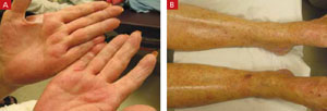
The patient had generalized palmar erythema with 1- to 2-mm papules (A). She also had an erythematous maculopapular rash that extended from the medial and dorsal aspects of her feet cranially to her lower abdomen (B).
FIGURE 2
Mild opacification in lung bases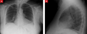
A posterior-anterior chest radiograph revealed bilateral lower lung opacities that were greater on the left side than on the right (A). A lateral chest radiograph revealed a positive spine sign: failure of the vertebral bodies to become more radiolucent as one looks down the spinal column, suggesting a posterior-inferior lung infiltrate that opacifies the normally radiolucent vertebral bodies of the lower chest (B).
Diagnosis: nitrofurantoin-induced lung disease
Nitrofurantoin (Macrodantin, Macrobid) is frequently used as first-line treatment for uncomplicated urinary tract infections (UTIs) and as prophylaxis for recurrent UTIs. Although generally well tolerated, it has a rare but serious side effect profile, including aplastic anemia, peripheral neuropathy, liver toxicity, and—as in the case of this patient—pulmonary toxicity.1
Nitrofurantoin-induced lung disease has an incidence of 3 cases per 1000 patients.2 Not surprisingly, elderly women are often affected, as this population is particularly susceptible to UTIs (and their recurrence), and thus are prescribed nitrofurantoin.3 The pathophysiology of the acute and chronic form of this drug reaction is unknown, but may be either an immunologic hypersensitivity reaction or a direct cytotoxic effect by nitrofurantoin or its metabolites on the lung parenchyma.4
The acute presentation begins within 3 weeks of initiation of nitrofurantoin. The most common symptoms include fever, dry cough, and dyspnea. Less common symptoms include an erythematous maculopapular rash, fatigue, arthralgias/myalgias, and anorexia. Patients may experience bronchospasm and have audible wheezing on exam. Eight percent of patients will have an abnormal chest radiograph, most often showing diffuse interstitial infiltrates or pleural effusions. An elevated WBC count appears in 40% of patients, many having an eosinophilia, and an elevated erythrocyte sedimentation rate may be present.5 Pulmonary function tests (PFTs), if performed, will show a restrictive pattern and a V/Q scan will show a ventilation-perfusion mismatch.2
The chronic presentation is much less common than the acute form. It occurs in patients on daily nitrofurantoin and its onset is delayed months to years after initiation of therapy. Symptoms are insidious and include cough and gradually worsening dyspnea on exertion. Physical exam may reveal wheezing or crackles and chest radiograph can show interstitial or alveolar infiltrates, as well as pleural effusions. Laboratory evaluation is similar to the acute form, and PFT findings also reveal a restrictive pattern.
Computed tomography has variable findings, such as ground-glass opacities, interstitial fibrosis, consolidation, peribronchial thickening, and centrilobular nodules. Lung biopsies have shown varying pathologic features: nonspecific interstitial pneumonia, pulmonary fibrosis, bronchiolitis obliterans organizing pneumonia, and hypersensitivity pneumonitis. 6,7
Differential Dx includes anaphylaxis, pneumonia
The differential diagnosis for nitrofurantoin-induced lung disease includes anaphylaxis, asthma, bronchitis, and pneumonia. Drug anaphylaxis typically occurs within hours of administration and often has skin findings of urticaria, pruritus, and angioedema of the oral mucosa. Patients may present with dyspnea, wheezing, stridor, hypoxemia, and respiratory distress.8
Although asthma, bronchitis, and pneumonia can present with a fever, cough, and radiograph findings, they are not usually associated with a rash. Identifying and removing the offending agent—a medication—is key in differentiating this disease from other common lung ailments.
Stop the offending agent, consider corticosteroids
Immediately discontinuing nitrofurantoin is usually sufficient to resolve the symptoms of acute nitrofurantoin-induced lung disease, often within 24 to 48 hours. Chest radiograph and PFT changes resolve within weeks of discontinuation. (Reintroducing nitrofurantoin to the patient will produce similar symptoms with a more rapid onset.) Corticosteroids are often used to treat this lung injury, although their effectiveness remains unproven.
With the chronic reaction, nitrofurantoin therapy is stopped and most patients are started on corticosteroids, typically dosed at 20 to 40 mg per day with a prolonged taper over several months. With treatment, many patients recover from their chronic pulmonary reaction, although it may take months to years.3 Some patients may never fully recover and continue to have symptoms, radiographic findings, and PFT changes characteristic of pulmonary fibrosis.
More seriously, nitrofurantoin-induced lung disease may require hospitalization. Rare cases have required lung transplantation and others have resulted in death due to respiratory failure.1
A speedy recovery for our patient
We told our patient to discontinue the nitrofurantoin, and we opted not to start her on corticosteroids. Her symptoms resolved within 3 days of discontinuing the medication, and she had no return of her UTI symptoms. Her rash also resolved within 5 days.
CORRESPONDENCE: Drew C. Baird, MD, Department of Family and Community Medicine, Carl R. Darnall Army Medical Center, 36000 Darnall Loop, Fort Hood, TX 76544; drew.baird@us.army.mil
1. Goemaere N, Grijm K, van Hal P, et al. Nitrofurantoin-induced pulmonary fibrosis: a case report. J Med Case Reports. 2008;2:169.-
2. Hainer BL, White AA. Nitrofurantoin pulmonary toxicity. J Fam Pract. 1981;13:817-823.
3. Bhullar S, Lele SM, Kraman S. Severe nitrofurantoin lung disease resolving without the use of steroids. J Postgrad Med. 2007;53:111-113.
4. Peall AF, Hodges A. Concomitant pulmonary and hepatic toxicity secondary to nitrofurantoin: a case report. J Med Case Reports. 2007;1:59.-
5. Holmberg L, Boman G, Bottiger LE, et al. Adverse reactions to nitrofurantoin. Analysis of 921 reports. Am J Med. 1980;69:733-738.
6. Mendez JL, Nadrous HF, Hartman TE, et al. Chronic nitrofurantoin-induced lung disease. Mayo Clin Proc. 2005;80:1298-1302.
7. Cameron RJ, Kolbe J, Wilsher ML, et al. Bronchiolitis obliterans organizing pneumonia associated with the use of nitrofurantoin. Thorax. 2000;55:249-251.
8. Tang AW. A practical guide to anaphylaxis. Am Fam Physician. 2003;68:1325-1332.
A 66-YEAR-OLD WOMAN came into the emergency department with a diffuse rash and a cough. She had a rash on the palms of her hands, which had developed the day before, but had improved a bit. She also had a rash on her feet, legs, and lower abdomen, which had developed that morning.
She said that over the previous 2 days she’d had a fever, dry cough, and some difficulty breathing. Her past medical history was significant for asthma, diabetes, hypertension, and osteoarthritis. Her medications included atenolol, celecoxib, metformin, pioglitazone, and an albuterol inhaler, as needed. In addition, she was on the ninth day of a 10-day course of nitrofurantoin for acute cystitis. She was allergic to ampicillin and erythromycin.
On physical exam, she had a fever of 101.5°F. On lung examination, she had diffuse wheezes and mild bibasilar crackles. Examination of her skin revealed a nonpainful, nonpruritic, erythematous, maculopapular rash located on the palms and legs (FIGURES 1A AND 1B), as well as on her lower abdomen. Chest radiograph showed mild opacification in the bases of the lungs (FIGURES 2A AND 2B). Her labs were significant for a white blood cell (WBC) count of 11.3 ×10³/mm3.
What is your diagnosis?
FIGURE 1
Rash on hands, feet, legs, and lower abdomen
The patient had generalized palmar erythema with 1- to 2-mm papules (A). She also had an erythematous maculopapular rash that extended from the medial and dorsal aspects of her feet cranially to her lower abdomen (B).
FIGURE 2
Mild opacification in lung bases
A posterior-anterior chest radiograph revealed bilateral lower lung opacities that were greater on the left side than on the right (A). A lateral chest radiograph revealed a positive spine sign: failure of the vertebral bodies to become more radiolucent as one looks down the spinal column, suggesting a posterior-inferior lung infiltrate that opacifies the normally radiolucent vertebral bodies of the lower chest (B).
Diagnosis: nitrofurantoin-induced lung disease
Nitrofurantoin (Macrodantin, Macrobid) is frequently used as first-line treatment for uncomplicated urinary tract infections (UTIs) and as prophylaxis for recurrent UTIs. Although generally well tolerated, it has a rare but serious side effect profile, including aplastic anemia, peripheral neuropathy, liver toxicity, and—as in the case of this patient—pulmonary toxicity.1
Nitrofurantoin-induced lung disease has an incidence of 3 cases per 1000 patients.2 Not surprisingly, elderly women are often affected, as this population is particularly susceptible to UTIs (and their recurrence), and thus are prescribed nitrofurantoin.3 The pathophysiology of the acute and chronic form of this drug reaction is unknown, but may be either an immunologic hypersensitivity reaction or a direct cytotoxic effect by nitrofurantoin or its metabolites on the lung parenchyma.4
The acute presentation begins within 3 weeks of initiation of nitrofurantoin. The most common symptoms include fever, dry cough, and dyspnea. Less common symptoms include an erythematous maculopapular rash, fatigue, arthralgias/myalgias, and anorexia. Patients may experience bronchospasm and have audible wheezing on exam. Eight percent of patients will have an abnormal chest radiograph, most often showing diffuse interstitial infiltrates or pleural effusions. An elevated WBC count appears in 40% of patients, many having an eosinophilia, and an elevated erythrocyte sedimentation rate may be present.5 Pulmonary function tests (PFTs), if performed, will show a restrictive pattern and a V/Q scan will show a ventilation-perfusion mismatch.2
The chronic presentation is much less common than the acute form. It occurs in patients on daily nitrofurantoin and its onset is delayed months to years after initiation of therapy. Symptoms are insidious and include cough and gradually worsening dyspnea on exertion. Physical exam may reveal wheezing or crackles and chest radiograph can show interstitial or alveolar infiltrates, as well as pleural effusions. Laboratory evaluation is similar to the acute form, and PFT findings also reveal a restrictive pattern.
Computed tomography has variable findings, such as ground-glass opacities, interstitial fibrosis, consolidation, peribronchial thickening, and centrilobular nodules. Lung biopsies have shown varying pathologic features: nonspecific interstitial pneumonia, pulmonary fibrosis, bronchiolitis obliterans organizing pneumonia, and hypersensitivity pneumonitis. 6,7
Differential Dx includes anaphylaxis, pneumonia
The differential diagnosis for nitrofurantoin-induced lung disease includes anaphylaxis, asthma, bronchitis, and pneumonia. Drug anaphylaxis typically occurs within hours of administration and often has skin findings of urticaria, pruritus, and angioedema of the oral mucosa. Patients may present with dyspnea, wheezing, stridor, hypoxemia, and respiratory distress.8
Although asthma, bronchitis, and pneumonia can present with a fever, cough, and radiograph findings, they are not usually associated with a rash. Identifying and removing the offending agent—a medication—is key in differentiating this disease from other common lung ailments.
Stop the offending agent, consider corticosteroids
Immediately discontinuing nitrofurantoin is usually sufficient to resolve the symptoms of acute nitrofurantoin-induced lung disease, often within 24 to 48 hours. Chest radiograph and PFT changes resolve within weeks of discontinuation. (Reintroducing nitrofurantoin to the patient will produce similar symptoms with a more rapid onset.) Corticosteroids are often used to treat this lung injury, although their effectiveness remains unproven.
With the chronic reaction, nitrofurantoin therapy is stopped and most patients are started on corticosteroids, typically dosed at 20 to 40 mg per day with a prolonged taper over several months. With treatment, many patients recover from their chronic pulmonary reaction, although it may take months to years.3 Some patients may never fully recover and continue to have symptoms, radiographic findings, and PFT changes characteristic of pulmonary fibrosis.
More seriously, nitrofurantoin-induced lung disease may require hospitalization. Rare cases have required lung transplantation and others have resulted in death due to respiratory failure.1
A speedy recovery for our patient
We told our patient to discontinue the nitrofurantoin, and we opted not to start her on corticosteroids. Her symptoms resolved within 3 days of discontinuing the medication, and she had no return of her UTI symptoms. Her rash also resolved within 5 days.
CORRESPONDENCE: Drew C. Baird, MD, Department of Family and Community Medicine, Carl R. Darnall Army Medical Center, 36000 Darnall Loop, Fort Hood, TX 76544; drew.baird@us.army.mil
A 66-YEAR-OLD WOMAN came into the emergency department with a diffuse rash and a cough. She had a rash on the palms of her hands, which had developed the day before, but had improved a bit. She also had a rash on her feet, legs, and lower abdomen, which had developed that morning.
She said that over the previous 2 days she’d had a fever, dry cough, and some difficulty breathing. Her past medical history was significant for asthma, diabetes, hypertension, and osteoarthritis. Her medications included atenolol, celecoxib, metformin, pioglitazone, and an albuterol inhaler, as needed. In addition, she was on the ninth day of a 10-day course of nitrofurantoin for acute cystitis. She was allergic to ampicillin and erythromycin.
On physical exam, she had a fever of 101.5°F. On lung examination, she had diffuse wheezes and mild bibasilar crackles. Examination of her skin revealed a nonpainful, nonpruritic, erythematous, maculopapular rash located on the palms and legs (FIGURES 1A AND 1B), as well as on her lower abdomen. Chest radiograph showed mild opacification in the bases of the lungs (FIGURES 2A AND 2B). Her labs were significant for a white blood cell (WBC) count of 11.3 ×10³/mm3.
What is your diagnosis?
FIGURE 1
Rash on hands, feet, legs, and lower abdomen
The patient had generalized palmar erythema with 1- to 2-mm papules (A). She also had an erythematous maculopapular rash that extended from the medial and dorsal aspects of her feet cranially to her lower abdomen (B).
FIGURE 2
Mild opacification in lung bases
A posterior-anterior chest radiograph revealed bilateral lower lung opacities that were greater on the left side than on the right (A). A lateral chest radiograph revealed a positive spine sign: failure of the vertebral bodies to become more radiolucent as one looks down the spinal column, suggesting a posterior-inferior lung infiltrate that opacifies the normally radiolucent vertebral bodies of the lower chest (B).
Diagnosis: nitrofurantoin-induced lung disease
Nitrofurantoin (Macrodantin, Macrobid) is frequently used as first-line treatment for uncomplicated urinary tract infections (UTIs) and as prophylaxis for recurrent UTIs. Although generally well tolerated, it has a rare but serious side effect profile, including aplastic anemia, peripheral neuropathy, liver toxicity, and—as in the case of this patient—pulmonary toxicity.1
Nitrofurantoin-induced lung disease has an incidence of 3 cases per 1000 patients.2 Not surprisingly, elderly women are often affected, as this population is particularly susceptible to UTIs (and their recurrence), and thus are prescribed nitrofurantoin.3 The pathophysiology of the acute and chronic form of this drug reaction is unknown, but may be either an immunologic hypersensitivity reaction or a direct cytotoxic effect by nitrofurantoin or its metabolites on the lung parenchyma.4
The acute presentation begins within 3 weeks of initiation of nitrofurantoin. The most common symptoms include fever, dry cough, and dyspnea. Less common symptoms include an erythematous maculopapular rash, fatigue, arthralgias/myalgias, and anorexia. Patients may experience bronchospasm and have audible wheezing on exam. Eight percent of patients will have an abnormal chest radiograph, most often showing diffuse interstitial infiltrates or pleural effusions. An elevated WBC count appears in 40% of patients, many having an eosinophilia, and an elevated erythrocyte sedimentation rate may be present.5 Pulmonary function tests (PFTs), if performed, will show a restrictive pattern and a V/Q scan will show a ventilation-perfusion mismatch.2
The chronic presentation is much less common than the acute form. It occurs in patients on daily nitrofurantoin and its onset is delayed months to years after initiation of therapy. Symptoms are insidious and include cough and gradually worsening dyspnea on exertion. Physical exam may reveal wheezing or crackles and chest radiograph can show interstitial or alveolar infiltrates, as well as pleural effusions. Laboratory evaluation is similar to the acute form, and PFT findings also reveal a restrictive pattern.
Computed tomography has variable findings, such as ground-glass opacities, interstitial fibrosis, consolidation, peribronchial thickening, and centrilobular nodules. Lung biopsies have shown varying pathologic features: nonspecific interstitial pneumonia, pulmonary fibrosis, bronchiolitis obliterans organizing pneumonia, and hypersensitivity pneumonitis. 6,7
Differential Dx includes anaphylaxis, pneumonia
The differential diagnosis for nitrofurantoin-induced lung disease includes anaphylaxis, asthma, bronchitis, and pneumonia. Drug anaphylaxis typically occurs within hours of administration and often has skin findings of urticaria, pruritus, and angioedema of the oral mucosa. Patients may present with dyspnea, wheezing, stridor, hypoxemia, and respiratory distress.8
Although asthma, bronchitis, and pneumonia can present with a fever, cough, and radiograph findings, they are not usually associated with a rash. Identifying and removing the offending agent—a medication—is key in differentiating this disease from other common lung ailments.
Stop the offending agent, consider corticosteroids
Immediately discontinuing nitrofurantoin is usually sufficient to resolve the symptoms of acute nitrofurantoin-induced lung disease, often within 24 to 48 hours. Chest radiograph and PFT changes resolve within weeks of discontinuation. (Reintroducing nitrofurantoin to the patient will produce similar symptoms with a more rapid onset.) Corticosteroids are often used to treat this lung injury, although their effectiveness remains unproven.
With the chronic reaction, nitrofurantoin therapy is stopped and most patients are started on corticosteroids, typically dosed at 20 to 40 mg per day with a prolonged taper over several months. With treatment, many patients recover from their chronic pulmonary reaction, although it may take months to years.3 Some patients may never fully recover and continue to have symptoms, radiographic findings, and PFT changes characteristic of pulmonary fibrosis.
More seriously, nitrofurantoin-induced lung disease may require hospitalization. Rare cases have required lung transplantation and others have resulted in death due to respiratory failure.1
A speedy recovery for our patient
We told our patient to discontinue the nitrofurantoin, and we opted not to start her on corticosteroids. Her symptoms resolved within 3 days of discontinuing the medication, and she had no return of her UTI symptoms. Her rash also resolved within 5 days.
CORRESPONDENCE: Drew C. Baird, MD, Department of Family and Community Medicine, Carl R. Darnall Army Medical Center, 36000 Darnall Loop, Fort Hood, TX 76544; drew.baird@us.army.mil
1. Goemaere N, Grijm K, van Hal P, et al. Nitrofurantoin-induced pulmonary fibrosis: a case report. J Med Case Reports. 2008;2:169.-
2. Hainer BL, White AA. Nitrofurantoin pulmonary toxicity. J Fam Pract. 1981;13:817-823.
3. Bhullar S, Lele SM, Kraman S. Severe nitrofurantoin lung disease resolving without the use of steroids. J Postgrad Med. 2007;53:111-113.
4. Peall AF, Hodges A. Concomitant pulmonary and hepatic toxicity secondary to nitrofurantoin: a case report. J Med Case Reports. 2007;1:59.-
5. Holmberg L, Boman G, Bottiger LE, et al. Adverse reactions to nitrofurantoin. Analysis of 921 reports. Am J Med. 1980;69:733-738.
6. Mendez JL, Nadrous HF, Hartman TE, et al. Chronic nitrofurantoin-induced lung disease. Mayo Clin Proc. 2005;80:1298-1302.
7. Cameron RJ, Kolbe J, Wilsher ML, et al. Bronchiolitis obliterans organizing pneumonia associated with the use of nitrofurantoin. Thorax. 2000;55:249-251.
8. Tang AW. A practical guide to anaphylaxis. Am Fam Physician. 2003;68:1325-1332.
1. Goemaere N, Grijm K, van Hal P, et al. Nitrofurantoin-induced pulmonary fibrosis: a case report. J Med Case Reports. 2008;2:169.-
2. Hainer BL, White AA. Nitrofurantoin pulmonary toxicity. J Fam Pract. 1981;13:817-823.
3. Bhullar S, Lele SM, Kraman S. Severe nitrofurantoin lung disease resolving without the use of steroids. J Postgrad Med. 2007;53:111-113.
4. Peall AF, Hodges A. Concomitant pulmonary and hepatic toxicity secondary to nitrofurantoin: a case report. J Med Case Reports. 2007;1:59.-
5. Holmberg L, Boman G, Bottiger LE, et al. Adverse reactions to nitrofurantoin. Analysis of 921 reports. Am J Med. 1980;69:733-738.
6. Mendez JL, Nadrous HF, Hartman TE, et al. Chronic nitrofurantoin-induced lung disease. Mayo Clin Proc. 2005;80:1298-1302.
7. Cameron RJ, Kolbe J, Wilsher ML, et al. Bronchiolitis obliterans organizing pneumonia associated with the use of nitrofurantoin. Thorax. 2000;55:249-251.
8. Tang AW. A practical guide to anaphylaxis. Am Fam Physician. 2003;68:1325-1332.
Optimize your use of stress tests: A Q&A guide
Order exercise stress testing without imaging for patients with a low to intermediate probability of coronary artery disease (CAD), unless preexisting electrocardiographic (EKG) changes would render such a test nondiagnostic. C
Order stress testing with imaging for patients with preexisting EKG changes and/or a high probability of CAD. C
Do not use stress testing to screen asymptomatic patients for CAD. C
Consider pharmacologic testing for patients who are unable to exercise to an appropriate cardiac workload; it has the same predictive value as a nuclear exercise stress test. B
Strength of recommendation (SOR)
A Good-quality patient-oriented evidence
B Inconsistent or limited-quality patient-oriented evidence
C Consensus, usual practice, opinion, disease-oriented evidence, case series
Exercise has been used for cardiac stress testing for decades. But testing and imaging techniques and knowledge of the efficacy of this common diagnostic tool continue to evolve. Optimizing your use of stress testing requires that you familiarize yourself with the latest evidence. The evidence-based answers to these 6 questions will help you do just that.
1. How reliable are exercise stress tests?
That depends, of course, on any number of variables, including the protocol utilized, the number of stenotic vessels, the degree of stenosis, and even the sex of the patient.
False-negative and false-positive results are frequent in treadmill testing without imaging. (For more on the different protocols, see “Standard and nuclear exercise stress tests: A look at protocols”) Sensitivity is related to the number of stenotic vessels and the degree of stenosis. For a man with single-vessel disease and ≥70% stenosis, the likelihood of an abnormal test is only 50% to 60%. Even in a man with left main artery disease, the sensitivity is only about 85%.1
In some cases, failure to reach a cardiac workload sufficient to produce ischemia can lead to a false-negative test, and it is up to the physician performing the test to label it as nondiagnostic. Other reasons for false-positive or false-negative results include preexisting ST segment abnormalities, which can cause false-positive elevation of the ST segment during exercise; the use of digitalis, which affects the ST segment; and the presence of ventricular hypertrophy or cardiomyopathy.1 Patients with any of these conditions should undergo stress testing with imaging instead.
Nuclear stress testing is indicated for patients who have baseline EKG abnormalities, suspected false-positive or false-negative results from a stress test without imaging, known CAD or previous revascularization, a pacemaker, or a moderate to high likelihood of a CAD diagnosis. The addition of a tracer isotope and imaging boosts the test’s predictive value.1
The positive predictive value of nuclear stress testing is difficult to calculate because an abnormal test should lead to initiation of therapy designed to reduce the risk of cardiac death or myocardial infarction (MI). Numerous studies have found the rate of cardiac events after a negative radionuclide stress test to be less than 1% per year.2 The event rate after a negative test is lower in women than in men; after a positive test, however, the event rate in women is 2 to 3 times higher.2,3 Overall, stress testing is less sensitive in women than in men, at least in part because of their lower likelihood of CAD associated with any given symptom set.4
Exercise stress testing can be done with a number of treadmill protocols. The most widely used are:18,19
- the Bruce Protocol (the most common),1 which increases the slope of the treadmill and the speed of the belt in 3-minute intervals;
- the modified Bruce Protocol, a less aggressive format in which slope and speed are alternatively increased; and
- the naughton Protocol (typically reserved for patients whose ability to walk is limited), which starts with a very slow belt speed and a nearly flat slope and increases both elements slowly.
During the test, heart rate and BP are measured, along with continuous EKG monitoring, but the frequency of BP measurement and 12-lead EKG printouts varies among testing facilities.
Patients must attain a heart rate of 85% of their age-predicted maximum for the test to be considered diagnostic; they typically exercise until they’re unable to continue or they develop symptoms that prompt the clinician performing the test to stop it. Monitoring continues for some time after the patient stops exercising—usually 4 to 5 minutes in an asymptomatic patient, or until any symptoms and EKG changes that developed during the test resolve. If chest pain or EKG changes persist, the patient may need to be admitted to the hospital.
The procedure for nuclear stress testing is similar, except that the patient must estimate when he or she can only walk for 1 more minute. A tracer isotope is injected at that time.
For years, thallium was used for this purpose. However, thallium is taken up by the perfused myocardium and has the drawback of rapid redistribution with resolution of ischemia, which can lead to false-negative tests.20
Technetium (99mTc-labeled sestamibi), which is commonly used for other nuclide scans, is now the preferred isotope for nuclear stress tests.21 It is taken up by mitochondria in the perfused myocardium and does not redistribute, which results in fewer false-negative scans. Additionally, the energy emitted by 99mTc-labeled sestamibi is higher and produces cleaner pictures.21
Single photon emission computed tomography (SPECT) scans are taken in 3 planes as part of the nuclear stress test. A set of resting scans is taken before the exercise test. The isotope is then allowed to wash out and another dose is injected at peak cardiac workload so a second set of scans can be taken and compared with the resting images.
Perfusion defects that are present both at rest and with stress indicate an area of infarction, whereas defects that appear with stress but not at rest indicate ischemia. The probable location of the coronary artery lesions responsible for the ischemia can be inferred from the area in which the defects appear.
Gated imaging—serial images that are coupled with EKG changes, then reassembled to produce a moving image of the heart—is now usually part of the process. The result can be examined for areas of wall motion abnormalities and used to calculate an ejection fraction.22
2. When should you rule out stress testing?
Stress testing is unnecessary in asymptomatic patients. Numerous studies have documented the lack of benefit from screening asymptomatic people for CAD using exercise stress testing.5,6 The US Preventive Services Task Force gives this a Grade D recommendation—recommending against routine stress testing.7
There are also numerous contraindications, both absolute and relative (TABLE).8 Relative contraindications, which include severe hypertension, left main coronary stenosis, moderate stenotic valvular disease, electrolyte abnormalities, cardiomyopathy, serious mental or physical impairment, and atrioventricular block are conditions that are likely to interfere with test performance or reliability.
Absolute contraindications, generally related to unstable cardiopulmonary disease, pose a far more serious threat. Indeed, administering a treadmill stress test to a patient with 1 or more absolute contraindications greatly increases the risk of death associated with the test.8
Even if a patient does not have any relative or absolute contraindications, there is still some risk of moving forward with the test. There is about a 1 in 2500 risk of MI or death during, or related to, exercise stress testing.8 The greater the likelihood that a patient has CAD, the higher the risk.
There is also a risk of hospitalization after the test, usually related to persistent chest pain or arrhythmias. (I generally admit patients whose chest pain is unresponsive to 3 doses of nitroglycerine or who develop EKG changes that persist after 20 minutes at rest.) The test also raises the possibility of injury from the equipment, such as sprains or fractures caused by falling from the treadmill.
Nuclear stress testing also has a small risk of an allergic reaction to the isotope used as a tracer. The radiation dose is 8 to 9 mSV, comparable to a computed tomography (CT) scan of the chest and generally less than that of a coronary angiogram.9
TABLE
Stress testing: Absolute and relative contraindications8
| Absolute contraindications |
|---|
| Recent MI (<2 days) |
| Unstable angina |
| Uncontrolled ventricular arrhythmia |
| Uncontrolled atrial arrhythmia that compromises cardiac function |
| Symptomatic HF (uncontrolled) |
| Severe aortic stenosis (uncontrolled) |
| Dissecting aneurysm (suspected or confirmed) |
| Myocarditis (active) |
| Pulmonary or systemic embolus (recent) |
| Acute pericarditis |
| Relative contraindications* |
| Severe hypertension |
| Left main coronary stenosis |
| Moderate stenotic valvular disease |
| Electrolyte abnormalities |
| Cardiomyopathy, including hypertrophic cardiomyopathy |
| Mental or physical impairment that results in an inability to exercise adequately |
| high-degree atrioventricular block |
| HF, heart failure; MI, myocardial infarction. |
| *Relative contraindications are conditions that are likely to interfere with test performance or reliability. |
3. Does the evidence support the use of stress tests for asymptomatic patients with diabetes? Are preop stress tests advisable?
The jury is still out on both questions.
The question of asymptomatic testing for patients with diabetes mellitus, who are more likely than those without the disease to develop CAD, frequently arises. Although individuals with diabetes have higher rates of silent ischemia than the general population, however, estimates of this prevalence vary widely.10 There are no clear guidelines for evaluation of asymptomatic diabetic patients with exercise stress testing. (See “Test your skills with these 3 cases”)
The addition of nuclide imaging adds diagnostic value to the test, but it is still not clear that this should be the preferred test for patients with diabetes who have normal resting EKGs.10,11 A recent randomized controlled trial investigating screening with pharmacologic stress testing in asymptomatic patients with type 2 diabetes did not show a reduction in cardiac event rates in patients who were screened compared with those who were not screened.12
Similarly, preoperative stress testing is subject to debate.13 Many studies have been done to evaluate the utility of preoperative stress testing, with revascularization procedures done before the planned surgery when significant CAD is found. (See “Before surgery: Have you done enough to mitigate risk?” J Fam Pract. 2010;59:202-211.) And, while many demonstrate the predictive power of various parameters that stress tests measure, literature reviews show that—with the exception of patients with unstable CAD—postop event rates are about the same for patients who underwent stress testing and subsequent revascularization vs those who were treated medically instead.13,14
CASE 1 Daniel G, a 68-year-old whom you’ve been treating for hypertension for more than 10 years, is about 25 pounds overweight. He has decided to begin an exercise regimen, and the trainer he hired to work with him at the gym has asked for medical clearance.
CASE 2 Marge H, age 73, has peripheral neuropathy and spinal stenosis. She sees a neurologist regularly, but has come to see you today to report that for the last several nights, her heart has been racing and she’s felt an uncomfortable sensation in her chest.
CASE 3 Ed W, a trim 56-year-old, has been swimming 5 days a week for years. Last week, he experienced a tightening in his chest in the middle of his swim. The pain subsided shortly after he stopped swimming, but it returned as soon as he got up to full speed again. He asks whether you think it’s a pulled muscle or angina.
Should any—or all—of these patients undergo cardiac stress testing?
CASE 1 Daniel’s case highlights the discrepancy between commonly held beliefs and medical evidence. For decades, people have been told to get a medical evaluation before starting an exercise program, and a stress test has commonly been part of that evaluation. However, numerous studies have failed to show a benefit of stress testing in asymptomatic people. The US Preventive Services Task force recommends against routine stress testing in asymptomatic people.7 And, while the american heart association/american college of cardiology guidelines suggest that stress testing in men over the age of 45 with 1 or more risk factors may occasionally yield useful information, the organizations acknowledge that this opinion is based on weak information.23
You tell Daniel that moderate exercise is unlikely to provoke a serious cardiac event and that if symptoms arise during exercise, he should report them promptly so that appropriate testing can be ordered.
CASE 2 Marge’s primary complaint sounds more like an arrhythmia than angina. however, coronary ischemia cannot be excluded; ischemia could be caused by decreased cardiac output from an arrhythmia, or it could be the cause of an arrhythmia. A holter monitor would be a good initial test for this patient, followed by stress testing to determine if angina is the cause of her symptoms. Because of marge’s peripheral neuropathy and spinal stenosis, she may be a candidate for a pharmacologic stress test.
Given that stress testing is less sensitive in women than in men, there is a widespread belief that women should not be tested with exercise stress testing alone. however, the available literature suggests that this test has appropriate predictive value for women with an intermediate CAD risk.4
CASE 3 Ed presents with typical symptoms of angina pectoris. While some noncardiac diseases—esophageal spasm, for example—can cause nearly identical symptoms, the likelihood that this patient has symptomatic CAD is high. Thus, he should undergo stress testing with nuclide imaging. This patient is physically fit and therefore can take an exercise test, which will provide information—most notably, functional capacity and the level of exertion needed to cause symptoms—that a pharmacologic stress test would not.
4. If your patient requires a pharmacologic stress test, what are your options besides adenosine?
While adenosine is the agent of choice, dipyridamole and dobutamine are other options. When any of these agents are used, it’s important to consider the side effects of each, and which drugs your patient will need to avoid prior to the stress test.
Adenosine is a mediator of coronary vasodilation. The drug dilates normal coronary arteries preferentially to stenotic vessels and causes redistribution of blood flow away from areas of the myocardium with compromised circulation.
Dipyridamole, a mediator of adenosine release, is sometimes used instead. Both drugs are given as a 4-minute infusion, with injection of the tracer late in the infusion.
The adverse effects of adenosine occur early in the infusion, and include dyspnea, bronchospasm, chest pain, nausea, and headache. Bradycardia can be marked, and brief periods of complete heart block and long sinus pauses may occur. Hypotension can likewise be profound. Many of these effects are extremely disturbing to the patient under-going the test, but they disappear within 30 seconds of stopping the infusion.
Dipyridamole has similar adverse effects, although heart block is not part of its adverse effect profile. In addition, the drug’s adverse effects occur later in the infusion than those associated with adenosine and last well after it is finished. However, dipyridamole’s side effects can be reversed with intravenous aminophylline without compromising the accuracy of the test.
Drugs to avoid that day. Methylxanthines antagonize adenosine and dipyridamole, and thus must be avoided on the day of the test. Caffeine and theophylline are among the substances to be avoided, although the degree to which they affect test results has been questioned recently.15
Severe COPD and asthma—especially in patients with uncontrolled wheezing—are relative contraindications to the use of adenosine and dipyridamole.
Interestingly, the cardiovascular effects (and EKG changes) associated with these drugs are not necessarily indicative of CAD. Thus, the entire EKG portion of a pharmacologic stress test is not useful in interpreting the finding. One small study suggests that, unlike exercise stress testing, adenosine stress testing may be safe in patients with severe aortic stenosis.16
Dobutamine is another alternative for pharmacologic stress testing, for patients who cannot take adenosine or are unable to stop theophylline or similar medications. An infusion of dobutamine with an escalating dose, sometimes including atropine, is used to accelerate the heart rate to 85% of the patient’s age-predicted maximum. The stress is primarily due to the chronotropic effect of the drug, but dobutamine has some coronary vasodilatory activity and may also induce some redistribution of coronary blood flow, similar to the effect of adenosine.
The positive and negative predictive values of pharmacologic stress testing are the same as for nuclear stress testing. Unlike exercise testing, however, functional capacity cannot be inferred from a pharmacologic stress test.
About 10% of patients undergoing pharmacologic stress testing will have a nondiagnostic test. The sensitivity of the test varies among studies, but it is approximately 84%, 95%, and 100% for single-, double-, and triplevessel disease, respectively. Patients with negative tests have an event rate of less than 1% per year.17
5. Is stress echocardiography comparable to stress testing?
Yes. Stress echocardiography, which involves echocardiographic studies taken before and after stress, can substitute for either exercise or pharmacologic stress testing (the stress can be achieved either with exercise or an infusion of dobutamine), and it has certain advantages: Stress echocardiography is cheaper than nuclear stress testing, and there is no radiation involved. In addition, stress echocardiography yields positive and negative predictive values similar to those seen with nuclear stress testing.2,3 The presence of ischemia is inferred from localized wall motion abnormalities.
The primary disadvantage of stress echocardiography is that it can be administered only by a cardiologist who has been specially trained in this procedure. In contrast, any community hospital nuclear medicine department has the capacity to perform nuclear imaging, and most radiologists are able to interpret the nuclide scans. In my experience, decisions about whether to order nuclear cardiac stress testing or stress echocardiography are influenced not only by the availability of these modalities, but also by the skill of the physicians who will interpret the tests.
6. Which exercise-induced EKG changes are related to ischemia?
The only changes that correlate with myocardial ischemia are ST depression and ST elevation. J-point depression is almost universally seen with exercise. For this reason, the ST level is measured 80 milliseconds after the J point.
ST depression—the most common abnormal finding—indicates subendocardial ischemia. ST changes are most commonly seen in the inferior and lateral leads, but do not correlate with the location of ischemia. ST depression can be downsloping, horizontal, or upsloping. The first 2 are the most significant patterns, and 1 mm of ST depression is the minimum significant level. Upsloping ST depression is less significant, and 1.5 mm of depression is the minimum significant change.1 The greater the degree of ST depression, the higher the likelihood that significant occlusion will be seen on coronary angiography. ST depression that develops in the recovery period is a rare occurrence but of equal significance to ST depression that occurs with exercise, and is probably due to ischemia caused by shunting of blood into skeletal muscle and away from the heart.1
ST elevation is less common, but more ominous than ST depression, as it indicates transmural ischemia.1 This finding most often indicates high-grade left anterior descending (LAD) or left main CAD. It is most often seen in the anterior leads, and the location of ST changes correlates with the area of ischemia. Bear in mind, however, that the correlation between ST elevation and transmural ischemia is true only if the patient has no history of MI. ST elevation in leads in which Q waves are present at rest usually indicates ventricular dyskinesia or aneurysm and not ischemia.1
Premenopausal women and women who are taking estrogen supplements, in particular, are more likely than men to have false-positive ST changes, most likely because of a poorly understood effect of estrogen. The molecules of estrogen and of digitalis glycosides have some regions of structural similarity, and it is thought that both molecules can cause ST changes.10
And what about arrhythmias? Arrhythmias are often seen at rest and with exertion. Supraventricular arrhythmias, including supraventricular tachycardia, are not associated with CAD. Premature ventricular contractions (PVCs) are common at peak exertion. PVCs are probably related to catecholamine release and do not indicate ischemia. (See “A look at the stress test report”)
Ventricular tachycardia, however—defined as 3 or more consecutive PVCs—has a 90% correlation with significant coronary artery stenosis, as shown on angiography.1
Rate-dependent conduction disturbances, including 2-to-1 atrioventricular block and bundle-branch blocks, may also be seen. These may be associated with ischemia, but are not highly predictive of coronary artery stenosis. Further testing may be indicated to determine whether stenosis is present.1
The report from the physician who performs or reads the stress test should contain the following elements:
Heart rate achieved, including both the rate itself and the percentage of the patient’s age-predicted maximum that the heart rate represents. Failure to reach 85% of the maximum may be related to underlying cardiac or pulmonary disease, the use of beta-blockers, musculoskeletal disorders, or general deconditioning. However, it is obviously noteworthy if the patient develops chest pain or significant ST changes at a lower heart rate.
BP at peak exertion. There are no established levels for systolic BP at various ages. But failure of the systolic pressure to rise, or a drop in systolic pressure with exercise, indicates a lack of ventricular reserve and is a poor prognostic sign.
Functional capacity (METS). In addition to documenting the METS level itself, the report should compare it to the expected functional capacity based on the patient’s age and sex.
Chest pain (or its absence). In addition to noting whether or not chest pain developed, the report should detail the character and intensity of any pain that the patient experienced, the time into the test and the heart rate at which it developed, and the response to rest or nitroglycerine.
ST changes. Unless something in the patient’s condition changes, the workload required to produce symptoms or ST changes should be reproducible from test to test. The workload at which angina or ST changes occur is key to assessing disease severity.
Arrhythmias. Whether they’re seen at rest or develop with exertion, arrhythmias should be noted, as well.
The final report should also indicate whether the test is negative, positive, or nondiagnostic for findings consistent with CAD. Whenever possible, it should include a validated treadmill score, as well.
CORRESPONDENCE Mark A. Knox, MD, UPMC Shadyside Family Medicine Residency Program, 5230 Centre Avenue, Pittsburgh, PA 15232; knoxma@upmc.edu
1. Ellestad MH. Stress Testing: Principles and Practice. 3rd ed. Philadelphia: F.A. Davis Company; 1986.
2. Metz LD, Beattie M, Hom R, et al. The prognostic value of normal exercise myocardial perfusion imaging and exercise echocardiography. J Am Coll Cardiol. 2007;49:227-237.
3. Gibbons RJ. Noninvasive diagnosis and prognosis assessment in chronic coronary artery disease: stress testing with and without imaging perspective. Circ Cardiovasc Imaging. 2008;1:257-269
4. Mieres JH, Shaw LJ, Arai A, et al. Role of noninvasive testing in the clinical evaluation of women with suspected coronary artery disease: Consensus Statement From the Cardiac Imaging Committee, Council on Clinical Cardiology, and the Cardiovascular Imaging and Intervention Committee, Council on Cardiovascular Radiology and Intervention, American Heart Association. Circulation. 2005;111:682-696.
5. Scott IA. Evaluating cardiovascular risk assessment for asymptomatic people. BMJ. 2009;338:164-168.
6. Livschitz S, Sharabi Y, Yushin J, et al. Limited clinical value of exercise stress test for the screening of coronary artery disease in young, asymptomatic adult men. Am J Cardiol. 2000;86:462-464.
7. US Preventive Services Task Force. Screening for coronary heart disease: recommendation statement. Ann Intern Med. 2004;140:569-572.
8. Gibbons RJ, Balady GJ, Beasley JW, et al. ACC/AHA Guidelines for Exercise Testing: Executive Summary: A Report of the American College of Cardiology/American Heart Association Task Force on Practice Guidelines (Committee on Exercise Testing). Circulation. 1997;96:345-354.
9. Health Physics Society. Doses from medical radiation sources. Available at: http://hps.org/hpspublications/articles/dosesfrommedicalradiation.html. Accessed April 9, 2010.
10. Albers AR, Krichavsky MZ, Balady GJ. Stress testing in patients with diabetes mellitus: diagnostic and prognostic value. Circulation. 2006;113:583-592.
11. Harris GD, White RD. Exercise stress testing in patients with type 2 diabetes: when are asymptomatic patients screened? Clin Diab. 2007;25:126-130.
12. Young LH, Wackers FJT, Chyun DA, et al. Cardiac outcomes after screening for asymptomatic coronary artery disease in patients with type 2 diabetes. The DIAD Study: a randomized controlled trial. JAMA. 2009;301:1547-1555.
13. Makaryus AN, Diamond JA. Nuclear stress testing in elderly persons: a review of its use in the assessment of cardiac risk, particularly in patients undergoing preoperative risk assessment. Drugs Aging. 2007;24:467-479.
14. McFalls EO, Ward HB, Moritz TE, et al. Coronary-artery revascularization before elective major vascular surgery. N Engl J Med 2004;351:2795-2804.
15. Kovacs D, Pivonka R, Khosla PG, et al. Effect of caffeine on myocardial perfusion imaging using single photon emission computed tomography during adenosine pharmacologic stress. Am J Ther 2008;15:431-434.
16. Samuels B, Kiat H, Friedman JD, et al. Adenosine pharmacologic stress myocardial perfusion tomographic imaging in patients with significant aortic stenosis: diagnostic effcacy and comparison of clinical, hemodynamic, and electrocardiographic variables with 100 age-matched control subjects. J Am Coll Cardiol. 1995;25:99-106.
17. Geleijnse ML, Elhendy A, Fioretti PM, et al. Dobutamine stress myocardial perfusion imaging. J Am Coll Cardiol. 2000;36:2017-2027.
18. Starling MR, Crawford MH, O’Rourke RA. Superiority of selected treadmill exercise protocols predischarge and six weeks postinfarction for detecting ischemic abnormalities. Am Heart J 1982;104:1054-1060.
19. Handler CE, Sowton E. A comparison of the Naughton and modified Bruce treadmill exercise protocols in their ability to detect ischaemic abnormalities six weeks after myocardial infarction. Eur Heart J. 1984;5:752-755.
20. Kahn JK, McGhie I, Akers MS, et al. Quantitative rotational tomography with 201T1 and 99mTc 2-methoxy-isobutyl-isonitrile. A direct comparison in normal individuals and patients with coronary artery disease. Circulation. 1989;79:1282-1293.
21. Vesely MR, Dilsizian V. Nuclear cardiac stress testing in the era of molecular medicine. J Nucl Med. 2008;49:399-413.
22. Avery P, Hudson N, Hubner P. Evaluation of changes in myocardial perfusion and function on exercise in patients with coronary artery disease by gated M1B1 scintigraphy. Br Heart J. 1993;70:22-26.
23. Gibbons RJ, Balady GJ, Bricker JT. ACC/AHA Guideline Update for Exercise Testing summary article: a report of the American College of Cardiology/American Heart Association Task Force on Practice Guidelines (Committee to Update the 1997 Exercise Testing Guidelines). Circulation. 2002;106:1883-1892.
Order exercise stress testing without imaging for patients with a low to intermediate probability of coronary artery disease (CAD), unless preexisting electrocardiographic (EKG) changes would render such a test nondiagnostic. C
Order stress testing with imaging for patients with preexisting EKG changes and/or a high probability of CAD. C
Do not use stress testing to screen asymptomatic patients for CAD. C
Consider pharmacologic testing for patients who are unable to exercise to an appropriate cardiac workload; it has the same predictive value as a nuclear exercise stress test. B
Strength of recommendation (SOR)
A Good-quality patient-oriented evidence
B Inconsistent or limited-quality patient-oriented evidence
C Consensus, usual practice, opinion, disease-oriented evidence, case series
Exercise has been used for cardiac stress testing for decades. But testing and imaging techniques and knowledge of the efficacy of this common diagnostic tool continue to evolve. Optimizing your use of stress testing requires that you familiarize yourself with the latest evidence. The evidence-based answers to these 6 questions will help you do just that.
1. How reliable are exercise stress tests?
That depends, of course, on any number of variables, including the protocol utilized, the number of stenotic vessels, the degree of stenosis, and even the sex of the patient.
False-negative and false-positive results are frequent in treadmill testing without imaging. (For more on the different protocols, see “Standard and nuclear exercise stress tests: A look at protocols”) Sensitivity is related to the number of stenotic vessels and the degree of stenosis. For a man with single-vessel disease and ≥70% stenosis, the likelihood of an abnormal test is only 50% to 60%. Even in a man with left main artery disease, the sensitivity is only about 85%.1
In some cases, failure to reach a cardiac workload sufficient to produce ischemia can lead to a false-negative test, and it is up to the physician performing the test to label it as nondiagnostic. Other reasons for false-positive or false-negative results include preexisting ST segment abnormalities, which can cause false-positive elevation of the ST segment during exercise; the use of digitalis, which affects the ST segment; and the presence of ventricular hypertrophy or cardiomyopathy.1 Patients with any of these conditions should undergo stress testing with imaging instead.
Nuclear stress testing is indicated for patients who have baseline EKG abnormalities, suspected false-positive or false-negative results from a stress test without imaging, known CAD or previous revascularization, a pacemaker, or a moderate to high likelihood of a CAD diagnosis. The addition of a tracer isotope and imaging boosts the test’s predictive value.1
The positive predictive value of nuclear stress testing is difficult to calculate because an abnormal test should lead to initiation of therapy designed to reduce the risk of cardiac death or myocardial infarction (MI). Numerous studies have found the rate of cardiac events after a negative radionuclide stress test to be less than 1% per year.2 The event rate after a negative test is lower in women than in men; after a positive test, however, the event rate in women is 2 to 3 times higher.2,3 Overall, stress testing is less sensitive in women than in men, at least in part because of their lower likelihood of CAD associated with any given symptom set.4
Exercise stress testing can be done with a number of treadmill protocols. The most widely used are:18,19
- the Bruce Protocol (the most common),1 which increases the slope of the treadmill and the speed of the belt in 3-minute intervals;
- the modified Bruce Protocol, a less aggressive format in which slope and speed are alternatively increased; and
- the naughton Protocol (typically reserved for patients whose ability to walk is limited), which starts with a very slow belt speed and a nearly flat slope and increases both elements slowly.
During the test, heart rate and BP are measured, along with continuous EKG monitoring, but the frequency of BP measurement and 12-lead EKG printouts varies among testing facilities.
Patients must attain a heart rate of 85% of their age-predicted maximum for the test to be considered diagnostic; they typically exercise until they’re unable to continue or they develop symptoms that prompt the clinician performing the test to stop it. Monitoring continues for some time after the patient stops exercising—usually 4 to 5 minutes in an asymptomatic patient, or until any symptoms and EKG changes that developed during the test resolve. If chest pain or EKG changes persist, the patient may need to be admitted to the hospital.
The procedure for nuclear stress testing is similar, except that the patient must estimate when he or she can only walk for 1 more minute. A tracer isotope is injected at that time.
For years, thallium was used for this purpose. However, thallium is taken up by the perfused myocardium and has the drawback of rapid redistribution with resolution of ischemia, which can lead to false-negative tests.20
Technetium (99mTc-labeled sestamibi), which is commonly used for other nuclide scans, is now the preferred isotope for nuclear stress tests.21 It is taken up by mitochondria in the perfused myocardium and does not redistribute, which results in fewer false-negative scans. Additionally, the energy emitted by 99mTc-labeled sestamibi is higher and produces cleaner pictures.21
Single photon emission computed tomography (SPECT) scans are taken in 3 planes as part of the nuclear stress test. A set of resting scans is taken before the exercise test. The isotope is then allowed to wash out and another dose is injected at peak cardiac workload so a second set of scans can be taken and compared with the resting images.
Perfusion defects that are present both at rest and with stress indicate an area of infarction, whereas defects that appear with stress but not at rest indicate ischemia. The probable location of the coronary artery lesions responsible for the ischemia can be inferred from the area in which the defects appear.
Gated imaging—serial images that are coupled with EKG changes, then reassembled to produce a moving image of the heart—is now usually part of the process. The result can be examined for areas of wall motion abnormalities and used to calculate an ejection fraction.22
2. When should you rule out stress testing?
Stress testing is unnecessary in asymptomatic patients. Numerous studies have documented the lack of benefit from screening asymptomatic people for CAD using exercise stress testing.5,6 The US Preventive Services Task Force gives this a Grade D recommendation—recommending against routine stress testing.7
There are also numerous contraindications, both absolute and relative (TABLE).8 Relative contraindications, which include severe hypertension, left main coronary stenosis, moderate stenotic valvular disease, electrolyte abnormalities, cardiomyopathy, serious mental or physical impairment, and atrioventricular block are conditions that are likely to interfere with test performance or reliability.
Absolute contraindications, generally related to unstable cardiopulmonary disease, pose a far more serious threat. Indeed, administering a treadmill stress test to a patient with 1 or more absolute contraindications greatly increases the risk of death associated with the test.8
Even if a patient does not have any relative or absolute contraindications, there is still some risk of moving forward with the test. There is about a 1 in 2500 risk of MI or death during, or related to, exercise stress testing.8 The greater the likelihood that a patient has CAD, the higher the risk.
There is also a risk of hospitalization after the test, usually related to persistent chest pain or arrhythmias. (I generally admit patients whose chest pain is unresponsive to 3 doses of nitroglycerine or who develop EKG changes that persist after 20 minutes at rest.) The test also raises the possibility of injury from the equipment, such as sprains or fractures caused by falling from the treadmill.
Nuclear stress testing also has a small risk of an allergic reaction to the isotope used as a tracer. The radiation dose is 8 to 9 mSV, comparable to a computed tomography (CT) scan of the chest and generally less than that of a coronary angiogram.9
TABLE
Stress testing: Absolute and relative contraindications8
| Absolute contraindications |
|---|
| Recent MI (<2 days) |
| Unstable angina |
| Uncontrolled ventricular arrhythmia |
| Uncontrolled atrial arrhythmia that compromises cardiac function |
| Symptomatic HF (uncontrolled) |
| Severe aortic stenosis (uncontrolled) |
| Dissecting aneurysm (suspected or confirmed) |
| Myocarditis (active) |
| Pulmonary or systemic embolus (recent) |
| Acute pericarditis |
| Relative contraindications* |
| Severe hypertension |
| Left main coronary stenosis |
| Moderate stenotic valvular disease |
| Electrolyte abnormalities |
| Cardiomyopathy, including hypertrophic cardiomyopathy |
| Mental or physical impairment that results in an inability to exercise adequately |
| high-degree atrioventricular block |
| HF, heart failure; MI, myocardial infarction. |
| *Relative contraindications are conditions that are likely to interfere with test performance or reliability. |
3. Does the evidence support the use of stress tests for asymptomatic patients with diabetes? Are preop stress tests advisable?
The jury is still out on both questions.
The question of asymptomatic testing for patients with diabetes mellitus, who are more likely than those without the disease to develop CAD, frequently arises. Although individuals with diabetes have higher rates of silent ischemia than the general population, however, estimates of this prevalence vary widely.10 There are no clear guidelines for evaluation of asymptomatic diabetic patients with exercise stress testing. (See “Test your skills with these 3 cases”)
The addition of nuclide imaging adds diagnostic value to the test, but it is still not clear that this should be the preferred test for patients with diabetes who have normal resting EKGs.10,11 A recent randomized controlled trial investigating screening with pharmacologic stress testing in asymptomatic patients with type 2 diabetes did not show a reduction in cardiac event rates in patients who were screened compared with those who were not screened.12
Similarly, preoperative stress testing is subject to debate.13 Many studies have been done to evaluate the utility of preoperative stress testing, with revascularization procedures done before the planned surgery when significant CAD is found. (See “Before surgery: Have you done enough to mitigate risk?” J Fam Pract. 2010;59:202-211.) And, while many demonstrate the predictive power of various parameters that stress tests measure, literature reviews show that—with the exception of patients with unstable CAD—postop event rates are about the same for patients who underwent stress testing and subsequent revascularization vs those who were treated medically instead.13,14
CASE 1 Daniel G, a 68-year-old whom you’ve been treating for hypertension for more than 10 years, is about 25 pounds overweight. He has decided to begin an exercise regimen, and the trainer he hired to work with him at the gym has asked for medical clearance.
CASE 2 Marge H, age 73, has peripheral neuropathy and spinal stenosis. She sees a neurologist regularly, but has come to see you today to report that for the last several nights, her heart has been racing and she’s felt an uncomfortable sensation in her chest.
CASE 3 Ed W, a trim 56-year-old, has been swimming 5 days a week for years. Last week, he experienced a tightening in his chest in the middle of his swim. The pain subsided shortly after he stopped swimming, but it returned as soon as he got up to full speed again. He asks whether you think it’s a pulled muscle or angina.
Should any—or all—of these patients undergo cardiac stress testing?
CASE 1 Daniel’s case highlights the discrepancy between commonly held beliefs and medical evidence. For decades, people have been told to get a medical evaluation before starting an exercise program, and a stress test has commonly been part of that evaluation. However, numerous studies have failed to show a benefit of stress testing in asymptomatic people. The US Preventive Services Task force recommends against routine stress testing in asymptomatic people.7 And, while the american heart association/american college of cardiology guidelines suggest that stress testing in men over the age of 45 with 1 or more risk factors may occasionally yield useful information, the organizations acknowledge that this opinion is based on weak information.23
You tell Daniel that moderate exercise is unlikely to provoke a serious cardiac event and that if symptoms arise during exercise, he should report them promptly so that appropriate testing can be ordered.
CASE 2 Marge’s primary complaint sounds more like an arrhythmia than angina. however, coronary ischemia cannot be excluded; ischemia could be caused by decreased cardiac output from an arrhythmia, or it could be the cause of an arrhythmia. A holter monitor would be a good initial test for this patient, followed by stress testing to determine if angina is the cause of her symptoms. Because of marge’s peripheral neuropathy and spinal stenosis, she may be a candidate for a pharmacologic stress test.
Given that stress testing is less sensitive in women than in men, there is a widespread belief that women should not be tested with exercise stress testing alone. however, the available literature suggests that this test has appropriate predictive value for women with an intermediate CAD risk.4
CASE 3 Ed presents with typical symptoms of angina pectoris. While some noncardiac diseases—esophageal spasm, for example—can cause nearly identical symptoms, the likelihood that this patient has symptomatic CAD is high. Thus, he should undergo stress testing with nuclide imaging. This patient is physically fit and therefore can take an exercise test, which will provide information—most notably, functional capacity and the level of exertion needed to cause symptoms—that a pharmacologic stress test would not.
4. If your patient requires a pharmacologic stress test, what are your options besides adenosine?
While adenosine is the agent of choice, dipyridamole and dobutamine are other options. When any of these agents are used, it’s important to consider the side effects of each, and which drugs your patient will need to avoid prior to the stress test.
Adenosine is a mediator of coronary vasodilation. The drug dilates normal coronary arteries preferentially to stenotic vessels and causes redistribution of blood flow away from areas of the myocardium with compromised circulation.
Dipyridamole, a mediator of adenosine release, is sometimes used instead. Both drugs are given as a 4-minute infusion, with injection of the tracer late in the infusion.
The adverse effects of adenosine occur early in the infusion, and include dyspnea, bronchospasm, chest pain, nausea, and headache. Bradycardia can be marked, and brief periods of complete heart block and long sinus pauses may occur. Hypotension can likewise be profound. Many of these effects are extremely disturbing to the patient under-going the test, but they disappear within 30 seconds of stopping the infusion.
Dipyridamole has similar adverse effects, although heart block is not part of its adverse effect profile. In addition, the drug’s adverse effects occur later in the infusion than those associated with adenosine and last well after it is finished. However, dipyridamole’s side effects can be reversed with intravenous aminophylline without compromising the accuracy of the test.
Drugs to avoid that day. Methylxanthines antagonize adenosine and dipyridamole, and thus must be avoided on the day of the test. Caffeine and theophylline are among the substances to be avoided, although the degree to which they affect test results has been questioned recently.15
Severe COPD and asthma—especially in patients with uncontrolled wheezing—are relative contraindications to the use of adenosine and dipyridamole.
Interestingly, the cardiovascular effects (and EKG changes) associated with these drugs are not necessarily indicative of CAD. Thus, the entire EKG portion of a pharmacologic stress test is not useful in interpreting the finding. One small study suggests that, unlike exercise stress testing, adenosine stress testing may be safe in patients with severe aortic stenosis.16
Dobutamine is another alternative for pharmacologic stress testing, for patients who cannot take adenosine or are unable to stop theophylline or similar medications. An infusion of dobutamine with an escalating dose, sometimes including atropine, is used to accelerate the heart rate to 85% of the patient’s age-predicted maximum. The stress is primarily due to the chronotropic effect of the drug, but dobutamine has some coronary vasodilatory activity and may also induce some redistribution of coronary blood flow, similar to the effect of adenosine.
The positive and negative predictive values of pharmacologic stress testing are the same as for nuclear stress testing. Unlike exercise testing, however, functional capacity cannot be inferred from a pharmacologic stress test.
About 10% of patients undergoing pharmacologic stress testing will have a nondiagnostic test. The sensitivity of the test varies among studies, but it is approximately 84%, 95%, and 100% for single-, double-, and triplevessel disease, respectively. Patients with negative tests have an event rate of less than 1% per year.17
5. Is stress echocardiography comparable to stress testing?
Yes. Stress echocardiography, which involves echocardiographic studies taken before and after stress, can substitute for either exercise or pharmacologic stress testing (the stress can be achieved either with exercise or an infusion of dobutamine), and it has certain advantages: Stress echocardiography is cheaper than nuclear stress testing, and there is no radiation involved. In addition, stress echocardiography yields positive and negative predictive values similar to those seen with nuclear stress testing.2,3 The presence of ischemia is inferred from localized wall motion abnormalities.
The primary disadvantage of stress echocardiography is that it can be administered only by a cardiologist who has been specially trained in this procedure. In contrast, any community hospital nuclear medicine department has the capacity to perform nuclear imaging, and most radiologists are able to interpret the nuclide scans. In my experience, decisions about whether to order nuclear cardiac stress testing or stress echocardiography are influenced not only by the availability of these modalities, but also by the skill of the physicians who will interpret the tests.
6. Which exercise-induced EKG changes are related to ischemia?
The only changes that correlate with myocardial ischemia are ST depression and ST elevation. J-point depression is almost universally seen with exercise. For this reason, the ST level is measured 80 milliseconds after the J point.
ST depression—the most common abnormal finding—indicates subendocardial ischemia. ST changes are most commonly seen in the inferior and lateral leads, but do not correlate with the location of ischemia. ST depression can be downsloping, horizontal, or upsloping. The first 2 are the most significant patterns, and 1 mm of ST depression is the minimum significant level. Upsloping ST depression is less significant, and 1.5 mm of depression is the minimum significant change.1 The greater the degree of ST depression, the higher the likelihood that significant occlusion will be seen on coronary angiography. ST depression that develops in the recovery period is a rare occurrence but of equal significance to ST depression that occurs with exercise, and is probably due to ischemia caused by shunting of blood into skeletal muscle and away from the heart.1
ST elevation is less common, but more ominous than ST depression, as it indicates transmural ischemia.1 This finding most often indicates high-grade left anterior descending (LAD) or left main CAD. It is most often seen in the anterior leads, and the location of ST changes correlates with the area of ischemia. Bear in mind, however, that the correlation between ST elevation and transmural ischemia is true only if the patient has no history of MI. ST elevation in leads in which Q waves are present at rest usually indicates ventricular dyskinesia or aneurysm and not ischemia.1
Premenopausal women and women who are taking estrogen supplements, in particular, are more likely than men to have false-positive ST changes, most likely because of a poorly understood effect of estrogen. The molecules of estrogen and of digitalis glycosides have some regions of structural similarity, and it is thought that both molecules can cause ST changes.10
And what about arrhythmias? Arrhythmias are often seen at rest and with exertion. Supraventricular arrhythmias, including supraventricular tachycardia, are not associated with CAD. Premature ventricular contractions (PVCs) are common at peak exertion. PVCs are probably related to catecholamine release and do not indicate ischemia. (See “A look at the stress test report”)
Ventricular tachycardia, however—defined as 3 or more consecutive PVCs—has a 90% correlation with significant coronary artery stenosis, as shown on angiography.1
Rate-dependent conduction disturbances, including 2-to-1 atrioventricular block and bundle-branch blocks, may also be seen. These may be associated with ischemia, but are not highly predictive of coronary artery stenosis. Further testing may be indicated to determine whether stenosis is present.1
The report from the physician who performs or reads the stress test should contain the following elements:
Heart rate achieved, including both the rate itself and the percentage of the patient’s age-predicted maximum that the heart rate represents. Failure to reach 85% of the maximum may be related to underlying cardiac or pulmonary disease, the use of beta-blockers, musculoskeletal disorders, or general deconditioning. However, it is obviously noteworthy if the patient develops chest pain or significant ST changes at a lower heart rate.
BP at peak exertion. There are no established levels for systolic BP at various ages. But failure of the systolic pressure to rise, or a drop in systolic pressure with exercise, indicates a lack of ventricular reserve and is a poor prognostic sign.
Functional capacity (METS). In addition to documenting the METS level itself, the report should compare it to the expected functional capacity based on the patient’s age and sex.
Chest pain (or its absence). In addition to noting whether or not chest pain developed, the report should detail the character and intensity of any pain that the patient experienced, the time into the test and the heart rate at which it developed, and the response to rest or nitroglycerine.
ST changes. Unless something in the patient’s condition changes, the workload required to produce symptoms or ST changes should be reproducible from test to test. The workload at which angina or ST changes occur is key to assessing disease severity.
Arrhythmias. Whether they’re seen at rest or develop with exertion, arrhythmias should be noted, as well.
The final report should also indicate whether the test is negative, positive, or nondiagnostic for findings consistent with CAD. Whenever possible, it should include a validated treadmill score, as well.
CORRESPONDENCE Mark A. Knox, MD, UPMC Shadyside Family Medicine Residency Program, 5230 Centre Avenue, Pittsburgh, PA 15232; knoxma@upmc.edu
Order exercise stress testing without imaging for patients with a low to intermediate probability of coronary artery disease (CAD), unless preexisting electrocardiographic (EKG) changes would render such a test nondiagnostic. C
Order stress testing with imaging for patients with preexisting EKG changes and/or a high probability of CAD. C
Do not use stress testing to screen asymptomatic patients for CAD. C
Consider pharmacologic testing for patients who are unable to exercise to an appropriate cardiac workload; it has the same predictive value as a nuclear exercise stress test. B
Strength of recommendation (SOR)
A Good-quality patient-oriented evidence
B Inconsistent or limited-quality patient-oriented evidence
C Consensus, usual practice, opinion, disease-oriented evidence, case series
Exercise has been used for cardiac stress testing for decades. But testing and imaging techniques and knowledge of the efficacy of this common diagnostic tool continue to evolve. Optimizing your use of stress testing requires that you familiarize yourself with the latest evidence. The evidence-based answers to these 6 questions will help you do just that.
1. How reliable are exercise stress tests?
That depends, of course, on any number of variables, including the protocol utilized, the number of stenotic vessels, the degree of stenosis, and even the sex of the patient.
False-negative and false-positive results are frequent in treadmill testing without imaging. (For more on the different protocols, see “Standard and nuclear exercise stress tests: A look at protocols”) Sensitivity is related to the number of stenotic vessels and the degree of stenosis. For a man with single-vessel disease and ≥70% stenosis, the likelihood of an abnormal test is only 50% to 60%. Even in a man with left main artery disease, the sensitivity is only about 85%.1
In some cases, failure to reach a cardiac workload sufficient to produce ischemia can lead to a false-negative test, and it is up to the physician performing the test to label it as nondiagnostic. Other reasons for false-positive or false-negative results include preexisting ST segment abnormalities, which can cause false-positive elevation of the ST segment during exercise; the use of digitalis, which affects the ST segment; and the presence of ventricular hypertrophy or cardiomyopathy.1 Patients with any of these conditions should undergo stress testing with imaging instead.
Nuclear stress testing is indicated for patients who have baseline EKG abnormalities, suspected false-positive or false-negative results from a stress test without imaging, known CAD or previous revascularization, a pacemaker, or a moderate to high likelihood of a CAD diagnosis. The addition of a tracer isotope and imaging boosts the test’s predictive value.1
The positive predictive value of nuclear stress testing is difficult to calculate because an abnormal test should lead to initiation of therapy designed to reduce the risk of cardiac death or myocardial infarction (MI). Numerous studies have found the rate of cardiac events after a negative radionuclide stress test to be less than 1% per year.2 The event rate after a negative test is lower in women than in men; after a positive test, however, the event rate in women is 2 to 3 times higher.2,3 Overall, stress testing is less sensitive in women than in men, at least in part because of their lower likelihood of CAD associated with any given symptom set.4
Exercise stress testing can be done with a number of treadmill protocols. The most widely used are:18,19
- the Bruce Protocol (the most common),1 which increases the slope of the treadmill and the speed of the belt in 3-minute intervals;
- the modified Bruce Protocol, a less aggressive format in which slope and speed are alternatively increased; and
- the naughton Protocol (typically reserved for patients whose ability to walk is limited), which starts with a very slow belt speed and a nearly flat slope and increases both elements slowly.
During the test, heart rate and BP are measured, along with continuous EKG monitoring, but the frequency of BP measurement and 12-lead EKG printouts varies among testing facilities.
Patients must attain a heart rate of 85% of their age-predicted maximum for the test to be considered diagnostic; they typically exercise until they’re unable to continue or they develop symptoms that prompt the clinician performing the test to stop it. Monitoring continues for some time after the patient stops exercising—usually 4 to 5 minutes in an asymptomatic patient, or until any symptoms and EKG changes that developed during the test resolve. If chest pain or EKG changes persist, the patient may need to be admitted to the hospital.
The procedure for nuclear stress testing is similar, except that the patient must estimate when he or she can only walk for 1 more minute. A tracer isotope is injected at that time.
For years, thallium was used for this purpose. However, thallium is taken up by the perfused myocardium and has the drawback of rapid redistribution with resolution of ischemia, which can lead to false-negative tests.20
Technetium (99mTc-labeled sestamibi), which is commonly used for other nuclide scans, is now the preferred isotope for nuclear stress tests.21 It is taken up by mitochondria in the perfused myocardium and does not redistribute, which results in fewer false-negative scans. Additionally, the energy emitted by 99mTc-labeled sestamibi is higher and produces cleaner pictures.21
Single photon emission computed tomography (SPECT) scans are taken in 3 planes as part of the nuclear stress test. A set of resting scans is taken before the exercise test. The isotope is then allowed to wash out and another dose is injected at peak cardiac workload so a second set of scans can be taken and compared with the resting images.
Perfusion defects that are present both at rest and with stress indicate an area of infarction, whereas defects that appear with stress but not at rest indicate ischemia. The probable location of the coronary artery lesions responsible for the ischemia can be inferred from the area in which the defects appear.
Gated imaging—serial images that are coupled with EKG changes, then reassembled to produce a moving image of the heart—is now usually part of the process. The result can be examined for areas of wall motion abnormalities and used to calculate an ejection fraction.22
2. When should you rule out stress testing?
Stress testing is unnecessary in asymptomatic patients. Numerous studies have documented the lack of benefit from screening asymptomatic people for CAD using exercise stress testing.5,6 The US Preventive Services Task Force gives this a Grade D recommendation—recommending against routine stress testing.7
There are also numerous contraindications, both absolute and relative (TABLE).8 Relative contraindications, which include severe hypertension, left main coronary stenosis, moderate stenotic valvular disease, electrolyte abnormalities, cardiomyopathy, serious mental or physical impairment, and atrioventricular block are conditions that are likely to interfere with test performance or reliability.
Absolute contraindications, generally related to unstable cardiopulmonary disease, pose a far more serious threat. Indeed, administering a treadmill stress test to a patient with 1 or more absolute contraindications greatly increases the risk of death associated with the test.8
Even if a patient does not have any relative or absolute contraindications, there is still some risk of moving forward with the test. There is about a 1 in 2500 risk of MI or death during, or related to, exercise stress testing.8 The greater the likelihood that a patient has CAD, the higher the risk.
There is also a risk of hospitalization after the test, usually related to persistent chest pain or arrhythmias. (I generally admit patients whose chest pain is unresponsive to 3 doses of nitroglycerine or who develop EKG changes that persist after 20 minutes at rest.) The test also raises the possibility of injury from the equipment, such as sprains or fractures caused by falling from the treadmill.
Nuclear stress testing also has a small risk of an allergic reaction to the isotope used as a tracer. The radiation dose is 8 to 9 mSV, comparable to a computed tomography (CT) scan of the chest and generally less than that of a coronary angiogram.9
TABLE
Stress testing: Absolute and relative contraindications8
| Absolute contraindications |
|---|
| Recent MI (<2 days) |
| Unstable angina |
| Uncontrolled ventricular arrhythmia |
| Uncontrolled atrial arrhythmia that compromises cardiac function |
| Symptomatic HF (uncontrolled) |
| Severe aortic stenosis (uncontrolled) |
| Dissecting aneurysm (suspected or confirmed) |
| Myocarditis (active) |
| Pulmonary or systemic embolus (recent) |
| Acute pericarditis |
| Relative contraindications* |
| Severe hypertension |
| Left main coronary stenosis |
| Moderate stenotic valvular disease |
| Electrolyte abnormalities |
| Cardiomyopathy, including hypertrophic cardiomyopathy |
| Mental or physical impairment that results in an inability to exercise adequately |
| high-degree atrioventricular block |
| HF, heart failure; MI, myocardial infarction. |
| *Relative contraindications are conditions that are likely to interfere with test performance or reliability. |
3. Does the evidence support the use of stress tests for asymptomatic patients with diabetes? Are preop stress tests advisable?
The jury is still out on both questions.
The question of asymptomatic testing for patients with diabetes mellitus, who are more likely than those without the disease to develop CAD, frequently arises. Although individuals with diabetes have higher rates of silent ischemia than the general population, however, estimates of this prevalence vary widely.10 There are no clear guidelines for evaluation of asymptomatic diabetic patients with exercise stress testing. (See “Test your skills with these 3 cases”)
The addition of nuclide imaging adds diagnostic value to the test, but it is still not clear that this should be the preferred test for patients with diabetes who have normal resting EKGs.10,11 A recent randomized controlled trial investigating screening with pharmacologic stress testing in asymptomatic patients with type 2 diabetes did not show a reduction in cardiac event rates in patients who were screened compared with those who were not screened.12
Similarly, preoperative stress testing is subject to debate.13 Many studies have been done to evaluate the utility of preoperative stress testing, with revascularization procedures done before the planned surgery when significant CAD is found. (See “Before surgery: Have you done enough to mitigate risk?” J Fam Pract. 2010;59:202-211.) And, while many demonstrate the predictive power of various parameters that stress tests measure, literature reviews show that—with the exception of patients with unstable CAD—postop event rates are about the same for patients who underwent stress testing and subsequent revascularization vs those who were treated medically instead.13,14
CASE 1 Daniel G, a 68-year-old whom you’ve been treating for hypertension for more than 10 years, is about 25 pounds overweight. He has decided to begin an exercise regimen, and the trainer he hired to work with him at the gym has asked for medical clearance.
CASE 2 Marge H, age 73, has peripheral neuropathy and spinal stenosis. She sees a neurologist regularly, but has come to see you today to report that for the last several nights, her heart has been racing and she’s felt an uncomfortable sensation in her chest.
CASE 3 Ed W, a trim 56-year-old, has been swimming 5 days a week for years. Last week, he experienced a tightening in his chest in the middle of his swim. The pain subsided shortly after he stopped swimming, but it returned as soon as he got up to full speed again. He asks whether you think it’s a pulled muscle or angina.
Should any—or all—of these patients undergo cardiac stress testing?
CASE 1 Daniel’s case highlights the discrepancy between commonly held beliefs and medical evidence. For decades, people have been told to get a medical evaluation before starting an exercise program, and a stress test has commonly been part of that evaluation. However, numerous studies have failed to show a benefit of stress testing in asymptomatic people. The US Preventive Services Task force recommends against routine stress testing in asymptomatic people.7 And, while the american heart association/american college of cardiology guidelines suggest that stress testing in men over the age of 45 with 1 or more risk factors may occasionally yield useful information, the organizations acknowledge that this opinion is based on weak information.23
You tell Daniel that moderate exercise is unlikely to provoke a serious cardiac event and that if symptoms arise during exercise, he should report them promptly so that appropriate testing can be ordered.
CASE 2 Marge’s primary complaint sounds more like an arrhythmia than angina. however, coronary ischemia cannot be excluded; ischemia could be caused by decreased cardiac output from an arrhythmia, or it could be the cause of an arrhythmia. A holter monitor would be a good initial test for this patient, followed by stress testing to determine if angina is the cause of her symptoms. Because of marge’s peripheral neuropathy and spinal stenosis, she may be a candidate for a pharmacologic stress test.
Given that stress testing is less sensitive in women than in men, there is a widespread belief that women should not be tested with exercise stress testing alone. however, the available literature suggests that this test has appropriate predictive value for women with an intermediate CAD risk.4
CASE 3 Ed presents with typical symptoms of angina pectoris. While some noncardiac diseases—esophageal spasm, for example—can cause nearly identical symptoms, the likelihood that this patient has symptomatic CAD is high. Thus, he should undergo stress testing with nuclide imaging. This patient is physically fit and therefore can take an exercise test, which will provide information—most notably, functional capacity and the level of exertion needed to cause symptoms—that a pharmacologic stress test would not.
4. If your patient requires a pharmacologic stress test, what are your options besides adenosine?
While adenosine is the agent of choice, dipyridamole and dobutamine are other options. When any of these agents are used, it’s important to consider the side effects of each, and which drugs your patient will need to avoid prior to the stress test.
Adenosine is a mediator of coronary vasodilation. The drug dilates normal coronary arteries preferentially to stenotic vessels and causes redistribution of blood flow away from areas of the myocardium with compromised circulation.
Dipyridamole, a mediator of adenosine release, is sometimes used instead. Both drugs are given as a 4-minute infusion, with injection of the tracer late in the infusion.
The adverse effects of adenosine occur early in the infusion, and include dyspnea, bronchospasm, chest pain, nausea, and headache. Bradycardia can be marked, and brief periods of complete heart block and long sinus pauses may occur. Hypotension can likewise be profound. Many of these effects are extremely disturbing to the patient under-going the test, but they disappear within 30 seconds of stopping the infusion.
Dipyridamole has similar adverse effects, although heart block is not part of its adverse effect profile. In addition, the drug’s adverse effects occur later in the infusion than those associated with adenosine and last well after it is finished. However, dipyridamole’s side effects can be reversed with intravenous aminophylline without compromising the accuracy of the test.
Drugs to avoid that day. Methylxanthines antagonize adenosine and dipyridamole, and thus must be avoided on the day of the test. Caffeine and theophylline are among the substances to be avoided, although the degree to which they affect test results has been questioned recently.15
Severe COPD and asthma—especially in patients with uncontrolled wheezing—are relative contraindications to the use of adenosine and dipyridamole.
Interestingly, the cardiovascular effects (and EKG changes) associated with these drugs are not necessarily indicative of CAD. Thus, the entire EKG portion of a pharmacologic stress test is not useful in interpreting the finding. One small study suggests that, unlike exercise stress testing, adenosine stress testing may be safe in patients with severe aortic stenosis.16
Dobutamine is another alternative for pharmacologic stress testing, for patients who cannot take adenosine or are unable to stop theophylline or similar medications. An infusion of dobutamine with an escalating dose, sometimes including atropine, is used to accelerate the heart rate to 85% of the patient’s age-predicted maximum. The stress is primarily due to the chronotropic effect of the drug, but dobutamine has some coronary vasodilatory activity and may also induce some redistribution of coronary blood flow, similar to the effect of adenosine.
The positive and negative predictive values of pharmacologic stress testing are the same as for nuclear stress testing. Unlike exercise testing, however, functional capacity cannot be inferred from a pharmacologic stress test.
About 10% of patients undergoing pharmacologic stress testing will have a nondiagnostic test. The sensitivity of the test varies among studies, but it is approximately 84%, 95%, and 100% for single-, double-, and triplevessel disease, respectively. Patients with negative tests have an event rate of less than 1% per year.17
5. Is stress echocardiography comparable to stress testing?
Yes. Stress echocardiography, which involves echocardiographic studies taken before and after stress, can substitute for either exercise or pharmacologic stress testing (the stress can be achieved either with exercise or an infusion of dobutamine), and it has certain advantages: Stress echocardiography is cheaper than nuclear stress testing, and there is no radiation involved. In addition, stress echocardiography yields positive and negative predictive values similar to those seen with nuclear stress testing.2,3 The presence of ischemia is inferred from localized wall motion abnormalities.
The primary disadvantage of stress echocardiography is that it can be administered only by a cardiologist who has been specially trained in this procedure. In contrast, any community hospital nuclear medicine department has the capacity to perform nuclear imaging, and most radiologists are able to interpret the nuclide scans. In my experience, decisions about whether to order nuclear cardiac stress testing or stress echocardiography are influenced not only by the availability of these modalities, but also by the skill of the physicians who will interpret the tests.
6. Which exercise-induced EKG changes are related to ischemia?
The only changes that correlate with myocardial ischemia are ST depression and ST elevation. J-point depression is almost universally seen with exercise. For this reason, the ST level is measured 80 milliseconds after the J point.
ST depression—the most common abnormal finding—indicates subendocardial ischemia. ST changes are most commonly seen in the inferior and lateral leads, but do not correlate with the location of ischemia. ST depression can be downsloping, horizontal, or upsloping. The first 2 are the most significant patterns, and 1 mm of ST depression is the minimum significant level. Upsloping ST depression is less significant, and 1.5 mm of depression is the minimum significant change.1 The greater the degree of ST depression, the higher the likelihood that significant occlusion will be seen on coronary angiography. ST depression that develops in the recovery period is a rare occurrence but of equal significance to ST depression that occurs with exercise, and is probably due to ischemia caused by shunting of blood into skeletal muscle and away from the heart.1
ST elevation is less common, but more ominous than ST depression, as it indicates transmural ischemia.1 This finding most often indicates high-grade left anterior descending (LAD) or left main CAD. It is most often seen in the anterior leads, and the location of ST changes correlates with the area of ischemia. Bear in mind, however, that the correlation between ST elevation and transmural ischemia is true only if the patient has no history of MI. ST elevation in leads in which Q waves are present at rest usually indicates ventricular dyskinesia or aneurysm and not ischemia.1
Premenopausal women and women who are taking estrogen supplements, in particular, are more likely than men to have false-positive ST changes, most likely because of a poorly understood effect of estrogen. The molecules of estrogen and of digitalis glycosides have some regions of structural similarity, and it is thought that both molecules can cause ST changes.10
And what about arrhythmias? Arrhythmias are often seen at rest and with exertion. Supraventricular arrhythmias, including supraventricular tachycardia, are not associated with CAD. Premature ventricular contractions (PVCs) are common at peak exertion. PVCs are probably related to catecholamine release and do not indicate ischemia. (See “A look at the stress test report”)
Ventricular tachycardia, however—defined as 3 or more consecutive PVCs—has a 90% correlation with significant coronary artery stenosis, as shown on angiography.1
Rate-dependent conduction disturbances, including 2-to-1 atrioventricular block and bundle-branch blocks, may also be seen. These may be associated with ischemia, but are not highly predictive of coronary artery stenosis. Further testing may be indicated to determine whether stenosis is present.1
The report from the physician who performs or reads the stress test should contain the following elements:
Heart rate achieved, including both the rate itself and the percentage of the patient’s age-predicted maximum that the heart rate represents. Failure to reach 85% of the maximum may be related to underlying cardiac or pulmonary disease, the use of beta-blockers, musculoskeletal disorders, or general deconditioning. However, it is obviously noteworthy if the patient develops chest pain or significant ST changes at a lower heart rate.
BP at peak exertion. There are no established levels for systolic BP at various ages. But failure of the systolic pressure to rise, or a drop in systolic pressure with exercise, indicates a lack of ventricular reserve and is a poor prognostic sign.
Functional capacity (METS). In addition to documenting the METS level itself, the report should compare it to the expected functional capacity based on the patient’s age and sex.
Chest pain (or its absence). In addition to noting whether or not chest pain developed, the report should detail the character and intensity of any pain that the patient experienced, the time into the test and the heart rate at which it developed, and the response to rest or nitroglycerine.
ST changes. Unless something in the patient’s condition changes, the workload required to produce symptoms or ST changes should be reproducible from test to test. The workload at which angina or ST changes occur is key to assessing disease severity.
Arrhythmias. Whether they’re seen at rest or develop with exertion, arrhythmias should be noted, as well.
The final report should also indicate whether the test is negative, positive, or nondiagnostic for findings consistent with CAD. Whenever possible, it should include a validated treadmill score, as well.
CORRESPONDENCE Mark A. Knox, MD, UPMC Shadyside Family Medicine Residency Program, 5230 Centre Avenue, Pittsburgh, PA 15232; knoxma@upmc.edu
1. Ellestad MH. Stress Testing: Principles and Practice. 3rd ed. Philadelphia: F.A. Davis Company; 1986.
2. Metz LD, Beattie M, Hom R, et al. The prognostic value of normal exercise myocardial perfusion imaging and exercise echocardiography. J Am Coll Cardiol. 2007;49:227-237.
3. Gibbons RJ. Noninvasive diagnosis and prognosis assessment in chronic coronary artery disease: stress testing with and without imaging perspective. Circ Cardiovasc Imaging. 2008;1:257-269
4. Mieres JH, Shaw LJ, Arai A, et al. Role of noninvasive testing in the clinical evaluation of women with suspected coronary artery disease: Consensus Statement From the Cardiac Imaging Committee, Council on Clinical Cardiology, and the Cardiovascular Imaging and Intervention Committee, Council on Cardiovascular Radiology and Intervention, American Heart Association. Circulation. 2005;111:682-696.
5. Scott IA. Evaluating cardiovascular risk assessment for asymptomatic people. BMJ. 2009;338:164-168.
6. Livschitz S, Sharabi Y, Yushin J, et al. Limited clinical value of exercise stress test for the screening of coronary artery disease in young, asymptomatic adult men. Am J Cardiol. 2000;86:462-464.
7. US Preventive Services Task Force. Screening for coronary heart disease: recommendation statement. Ann Intern Med. 2004;140:569-572.
8. Gibbons RJ, Balady GJ, Beasley JW, et al. ACC/AHA Guidelines for Exercise Testing: Executive Summary: A Report of the American College of Cardiology/American Heart Association Task Force on Practice Guidelines (Committee on Exercise Testing). Circulation. 1997;96:345-354.
9. Health Physics Society. Doses from medical radiation sources. Available at: http://hps.org/hpspublications/articles/dosesfrommedicalradiation.html. Accessed April 9, 2010.
10. Albers AR, Krichavsky MZ, Balady GJ. Stress testing in patients with diabetes mellitus: diagnostic and prognostic value. Circulation. 2006;113:583-592.
11. Harris GD, White RD. Exercise stress testing in patients with type 2 diabetes: when are asymptomatic patients screened? Clin Diab. 2007;25:126-130.
12. Young LH, Wackers FJT, Chyun DA, et al. Cardiac outcomes after screening for asymptomatic coronary artery disease in patients with type 2 diabetes. The DIAD Study: a randomized controlled trial. JAMA. 2009;301:1547-1555.
13. Makaryus AN, Diamond JA. Nuclear stress testing in elderly persons: a review of its use in the assessment of cardiac risk, particularly in patients undergoing preoperative risk assessment. Drugs Aging. 2007;24:467-479.
14. McFalls EO, Ward HB, Moritz TE, et al. Coronary-artery revascularization before elective major vascular surgery. N Engl J Med 2004;351:2795-2804.
15. Kovacs D, Pivonka R, Khosla PG, et al. Effect of caffeine on myocardial perfusion imaging using single photon emission computed tomography during adenosine pharmacologic stress. Am J Ther 2008;15:431-434.
16. Samuels B, Kiat H, Friedman JD, et al. Adenosine pharmacologic stress myocardial perfusion tomographic imaging in patients with significant aortic stenosis: diagnostic effcacy and comparison of clinical, hemodynamic, and electrocardiographic variables with 100 age-matched control subjects. J Am Coll Cardiol. 1995;25:99-106.
17. Geleijnse ML, Elhendy A, Fioretti PM, et al. Dobutamine stress myocardial perfusion imaging. J Am Coll Cardiol. 2000;36:2017-2027.
18. Starling MR, Crawford MH, O’Rourke RA. Superiority of selected treadmill exercise protocols predischarge and six weeks postinfarction for detecting ischemic abnormalities. Am Heart J 1982;104:1054-1060.
19. Handler CE, Sowton E. A comparison of the Naughton and modified Bruce treadmill exercise protocols in their ability to detect ischaemic abnormalities six weeks after myocardial infarction. Eur Heart J. 1984;5:752-755.
20. Kahn JK, McGhie I, Akers MS, et al. Quantitative rotational tomography with 201T1 and 99mTc 2-methoxy-isobutyl-isonitrile. A direct comparison in normal individuals and patients with coronary artery disease. Circulation. 1989;79:1282-1293.
21. Vesely MR, Dilsizian V. Nuclear cardiac stress testing in the era of molecular medicine. J Nucl Med. 2008;49:399-413.
22. Avery P, Hudson N, Hubner P. Evaluation of changes in myocardial perfusion and function on exercise in patients with coronary artery disease by gated M1B1 scintigraphy. Br Heart J. 1993;70:22-26.
23. Gibbons RJ, Balady GJ, Bricker JT. ACC/AHA Guideline Update for Exercise Testing summary article: a report of the American College of Cardiology/American Heart Association Task Force on Practice Guidelines (Committee to Update the 1997 Exercise Testing Guidelines). Circulation. 2002;106:1883-1892.
1. Ellestad MH. Stress Testing: Principles and Practice. 3rd ed. Philadelphia: F.A. Davis Company; 1986.
2. Metz LD, Beattie M, Hom R, et al. The prognostic value of normal exercise myocardial perfusion imaging and exercise echocardiography. J Am Coll Cardiol. 2007;49:227-237.
3. Gibbons RJ. Noninvasive diagnosis and prognosis assessment in chronic coronary artery disease: stress testing with and without imaging perspective. Circ Cardiovasc Imaging. 2008;1:257-269
4. Mieres JH, Shaw LJ, Arai A, et al. Role of noninvasive testing in the clinical evaluation of women with suspected coronary artery disease: Consensus Statement From the Cardiac Imaging Committee, Council on Clinical Cardiology, and the Cardiovascular Imaging and Intervention Committee, Council on Cardiovascular Radiology and Intervention, American Heart Association. Circulation. 2005;111:682-696.
5. Scott IA. Evaluating cardiovascular risk assessment for asymptomatic people. BMJ. 2009;338:164-168.
6. Livschitz S, Sharabi Y, Yushin J, et al. Limited clinical value of exercise stress test for the screening of coronary artery disease in young, asymptomatic adult men. Am J Cardiol. 2000;86:462-464.
7. US Preventive Services Task Force. Screening for coronary heart disease: recommendation statement. Ann Intern Med. 2004;140:569-572.
8. Gibbons RJ, Balady GJ, Beasley JW, et al. ACC/AHA Guidelines for Exercise Testing: Executive Summary: A Report of the American College of Cardiology/American Heart Association Task Force on Practice Guidelines (Committee on Exercise Testing). Circulation. 1997;96:345-354.
9. Health Physics Society. Doses from medical radiation sources. Available at: http://hps.org/hpspublications/articles/dosesfrommedicalradiation.html. Accessed April 9, 2010.
10. Albers AR, Krichavsky MZ, Balady GJ. Stress testing in patients with diabetes mellitus: diagnostic and prognostic value. Circulation. 2006;113:583-592.
11. Harris GD, White RD. Exercise stress testing in patients with type 2 diabetes: when are asymptomatic patients screened? Clin Diab. 2007;25:126-130.
12. Young LH, Wackers FJT, Chyun DA, et al. Cardiac outcomes after screening for asymptomatic coronary artery disease in patients with type 2 diabetes. The DIAD Study: a randomized controlled trial. JAMA. 2009;301:1547-1555.
13. Makaryus AN, Diamond JA. Nuclear stress testing in elderly persons: a review of its use in the assessment of cardiac risk, particularly in patients undergoing preoperative risk assessment. Drugs Aging. 2007;24:467-479.
14. McFalls EO, Ward HB, Moritz TE, et al. Coronary-artery revascularization before elective major vascular surgery. N Engl J Med 2004;351:2795-2804.
15. Kovacs D, Pivonka R, Khosla PG, et al. Effect of caffeine on myocardial perfusion imaging using single photon emission computed tomography during adenosine pharmacologic stress. Am J Ther 2008;15:431-434.
16. Samuels B, Kiat H, Friedman JD, et al. Adenosine pharmacologic stress myocardial perfusion tomographic imaging in patients with significant aortic stenosis: diagnostic effcacy and comparison of clinical, hemodynamic, and electrocardiographic variables with 100 age-matched control subjects. J Am Coll Cardiol. 1995;25:99-106.
17. Geleijnse ML, Elhendy A, Fioretti PM, et al. Dobutamine stress myocardial perfusion imaging. J Am Coll Cardiol. 2000;36:2017-2027.
18. Starling MR, Crawford MH, O’Rourke RA. Superiority of selected treadmill exercise protocols predischarge and six weeks postinfarction for detecting ischemic abnormalities. Am Heart J 1982;104:1054-1060.
19. Handler CE, Sowton E. A comparison of the Naughton and modified Bruce treadmill exercise protocols in their ability to detect ischaemic abnormalities six weeks after myocardial infarction. Eur Heart J. 1984;5:752-755.
20. Kahn JK, McGhie I, Akers MS, et al. Quantitative rotational tomography with 201T1 and 99mTc 2-methoxy-isobutyl-isonitrile. A direct comparison in normal individuals and patients with coronary artery disease. Circulation. 1989;79:1282-1293.
21. Vesely MR, Dilsizian V. Nuclear cardiac stress testing in the era of molecular medicine. J Nucl Med. 2008;49:399-413.
22. Avery P, Hudson N, Hubner P. Evaluation of changes in myocardial perfusion and function on exercise in patients with coronary artery disease by gated M1B1 scintigraphy. Br Heart J. 1993;70:22-26.
23. Gibbons RJ, Balady GJ, Bricker JT. ACC/AHA Guideline Update for Exercise Testing summary article: a report of the American College of Cardiology/American Heart Association Task Force on Practice Guidelines (Committee to Update the 1997 Exercise Testing Guidelines). Circulation. 2002;106:1883-1892.
Biomarkers Suggest Asthma Differs in Children, Adults
NEW ORLEANS — Children with severe asthma have significantly higher levels of serum IgE and exhaled nitric oxide than adults, based on data from 47 consecutive patients with severe asthma.
Severe asthma affects fewer than 10% of all asthma patients, but it accounts for a disproportionate number of all asthma-related hospitalizations and emergency department visits, said Dr. Jonathan Malka of National Jewish Health in Denver.
Few studies have examined the phenotypic differences in severe asthma based on age, and recognizing the differences and similarities could help clinicians identify severe asthmatics, Dr. Malka said.
To compare levels of impairment and inflammation in children and adults, Dr. Malka and his colleagues evaluated 23 children and 24 adults with severe asthma who presented to National Jewish Health. The average age of the children was 12 years, and the average age of the adults was 47 years. The mean asthma durations were 9 years in children and 27 years in adults. The results were presented in a poster at the annual meeting of the American Academy of Allergy, Asthma, and Immunology.
Serum IgE was significantly higher in children than in adults (about 600 IU/mL vs. about 200 IU/mL). Similarly, exhaled nitric oxide levels were significantly higher in children than in adults (about 54 parts per billion vs. about 27 parts per billion).
Children with severe asthma had significantly less lung function impairment than did adults, based on two measures of lung function. Forced vital capacity (FVC) in children was 94% of the predicted value vs. 72% in adults. Forced expiratory volume in 1 second (FEV1) in children was 73% of the predicted value vs. 56% in adults. Children also fared better than adults in terms of the FEV1/FVC ratio and in changes in FEV1 after using albuterol, but those differences were not significant.
Children and adults showed no significant differences in three measures of asthma morbidity: weekly albuterol use, annual asthma exacerbations, and lifetime hospitalizations.
The study was limited by the small sample size, but the biomarker findings suggest pathophysiologic differences in asthma based on age—although adults and children alike were equally compromised, Dr. Malka said. Additional research may help clinicians adjust management of their severe asthma patients based on age, he said in an interview.
Dr. Malka had no financial conflicts to disclose.
To watch an interview of Dr. Malka, go to www.youtube.com/user/ElsGlobalMedicalNews
NEW ORLEANS — Children with severe asthma have significantly higher levels of serum IgE and exhaled nitric oxide than adults, based on data from 47 consecutive patients with severe asthma.
Severe asthma affects fewer than 10% of all asthma patients, but it accounts for a disproportionate number of all asthma-related hospitalizations and emergency department visits, said Dr. Jonathan Malka of National Jewish Health in Denver.
Few studies have examined the phenotypic differences in severe asthma based on age, and recognizing the differences and similarities could help clinicians identify severe asthmatics, Dr. Malka said.
To compare levels of impairment and inflammation in children and adults, Dr. Malka and his colleagues evaluated 23 children and 24 adults with severe asthma who presented to National Jewish Health. The average age of the children was 12 years, and the average age of the adults was 47 years. The mean asthma durations were 9 years in children and 27 years in adults. The results were presented in a poster at the annual meeting of the American Academy of Allergy, Asthma, and Immunology.
Serum IgE was significantly higher in children than in adults (about 600 IU/mL vs. about 200 IU/mL). Similarly, exhaled nitric oxide levels were significantly higher in children than in adults (about 54 parts per billion vs. about 27 parts per billion).
Children with severe asthma had significantly less lung function impairment than did adults, based on two measures of lung function. Forced vital capacity (FVC) in children was 94% of the predicted value vs. 72% in adults. Forced expiratory volume in 1 second (FEV1) in children was 73% of the predicted value vs. 56% in adults. Children also fared better than adults in terms of the FEV1/FVC ratio and in changes in FEV1 after using albuterol, but those differences were not significant.
Children and adults showed no significant differences in three measures of asthma morbidity: weekly albuterol use, annual asthma exacerbations, and lifetime hospitalizations.
The study was limited by the small sample size, but the biomarker findings suggest pathophysiologic differences in asthma based on age—although adults and children alike were equally compromised, Dr. Malka said. Additional research may help clinicians adjust management of their severe asthma patients based on age, he said in an interview.
Dr. Malka had no financial conflicts to disclose.
To watch an interview of Dr. Malka, go to www.youtube.com/user/ElsGlobalMedicalNews
NEW ORLEANS — Children with severe asthma have significantly higher levels of serum IgE and exhaled nitric oxide than adults, based on data from 47 consecutive patients with severe asthma.
Severe asthma affects fewer than 10% of all asthma patients, but it accounts for a disproportionate number of all asthma-related hospitalizations and emergency department visits, said Dr. Jonathan Malka of National Jewish Health in Denver.
Few studies have examined the phenotypic differences in severe asthma based on age, and recognizing the differences and similarities could help clinicians identify severe asthmatics, Dr. Malka said.
To compare levels of impairment and inflammation in children and adults, Dr. Malka and his colleagues evaluated 23 children and 24 adults with severe asthma who presented to National Jewish Health. The average age of the children was 12 years, and the average age of the adults was 47 years. The mean asthma durations were 9 years in children and 27 years in adults. The results were presented in a poster at the annual meeting of the American Academy of Allergy, Asthma, and Immunology.
Serum IgE was significantly higher in children than in adults (about 600 IU/mL vs. about 200 IU/mL). Similarly, exhaled nitric oxide levels were significantly higher in children than in adults (about 54 parts per billion vs. about 27 parts per billion).
Children with severe asthma had significantly less lung function impairment than did adults, based on two measures of lung function. Forced vital capacity (FVC) in children was 94% of the predicted value vs. 72% in adults. Forced expiratory volume in 1 second (FEV1) in children was 73% of the predicted value vs. 56% in adults. Children also fared better than adults in terms of the FEV1/FVC ratio and in changes in FEV1 after using albuterol, but those differences were not significant.
Children and adults showed no significant differences in three measures of asthma morbidity: weekly albuterol use, annual asthma exacerbations, and lifetime hospitalizations.
The study was limited by the small sample size, but the biomarker findings suggest pathophysiologic differences in asthma based on age—although adults and children alike were equally compromised, Dr. Malka said. Additional research may help clinicians adjust management of their severe asthma patients based on age, he said in an interview.
Dr. Malka had no financial conflicts to disclose.
To watch an interview of Dr. Malka, go to www.youtube.com/user/ElsGlobalMedicalNews