User login
Bringing you the latest news, research and reviews, exclusive interviews, podcasts, quizzes, and more.
Powered by CHEST Physician, Clinician Reviews, MDedge Family Medicine, Internal Medicine News, and The Journal of Clinical Outcomes Management.
Intranasal Postop Steroids Same as Saline in Select Patients
CHICAGO – Intranasal steroids were as good as saline alone as postoperative care in a randomized, double-blinded study of patients with chronic rhinosinusitis and Samter’s triad.
There was no difference in disease recurrence rate, complications, or quality of life at 6 months and 1-year postoperative using intranasal saline, saline plus budesonide, or saline and budesonide combined.
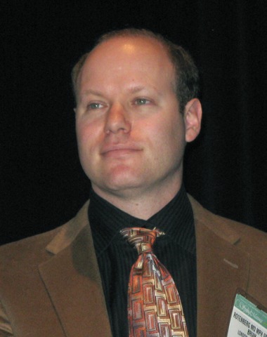
The surprising finding runs contrary to general practice and has several implications including how best to counsel patients for postoperative care, lead author Dr. Brian Rotenberg said at the Combined Otolaryngology Spring Meetings.
"If nasal steroids as done in this population don’t confer any additional benefit postoperatively, should we still prescribe them?" he asked. "Should we be prescribing something different or perhaps nothing at all? Is there a potential plus side here in terms of health-care cost savings?"
During a discussion of the study, an attendee expressed concern that insurers would interpret the results too broadly and deny coverage of postoperative nasal steroids for all patients with rhinosinusitis and polyposis, and not just those with Samter’s triad, a condition consisting of asthma, aspirin sensitivity, and nasal polyposis.
Another attendee agreed that nasal steroids are not potent enough in this population and said a pulse course of oral steroids 60 mg for 4 days can knock down symptoms in 80% of those with recurrence and be maintained with topical steroids. In the absence of a federally approved product for nasal use, he also suggested that dexamethasone eye drops can be effective.
Dr. Rotenberg replied that all patients received 3 weeks of postoperative oral prednisone, but that pulse-dosing of steroids was limited to one patient with early recurrence.
The 60 patients in the analysis had failed medical management for chronic rhinosinusitis with nasal polyposis and had a minimum preoperative Lund-Kennedy score of 8 out of 12. Nineteen patients were randomized to saline, 21 to saline plus budesonide, and 20 to saline/budesonide combination. Their mean Lund-Mackay scores were 20.6, 19.9, and 20.5, respectively.
Exclusion criteria included revision functional endoscopic sinus surgery, use of corticosteroids for other medical conditions, smoking, and concurrent disease with steroid contraindication.
At 6 months postoperatively, there were no significant differences between the saline, saline plus budesonide, and saline/budesonide combination groups with regard to Lund-Kennedy scores (1.5, 0.9, and 1.2, respectively), adrenocorticotropic hormone (ACTH) ranges (all normal), and intraocular pressure (12.4, 12.9, and 13.9 mm Hg, respectively), said Dr. Rotenberg of the University of Western Ontario in London, Ontario. Quality of life as assessed using the 21-item Sino-Nasal Outcome Test (SNOT-21) was also similar at a mean of 29.7, 27.4 and 28.2, respectively.
At 1 year of follow-up, once again there were no significant differences between the saline, saline plus budesonide, and saline/budesonide groups in Lund-Kennedy scores (3.7, 4.4, and 4.1, respectively), Lund-Mackay scores (11.8, 12.7, and 13.4, respectively), SNOT-21 scores (42.5, 47.9, and 42.2, respectively), intraocular pressure (13.1, 13.4,and 12.9 mm Hg, respectively). ACTH ranges were all normal.
A within-group analysis showed a significant improvement in all outcomes from baseline to 6 months, and a general worsening of outcomes at 1 year compared with the first 6 months, although they were still improved over baseline, Dr. Rotenberg said.
He pointed out that the literature is lacking in evidence guiding the postoperative management of patients with chronic rhinosinusitis with nasal polyps undergoing surgery. One study reported that steroid nasal spray did not influence polyp recurrence rate after surgery (Clin. Exp. Allergy. 2004;34:1395-400), while another showed that normal and buffered hypertonic saline nasal sprays had no beneficial effect on postoperative symptoms compared with no treatment (Am. J. Rhinol. 2006;20:191-6).
Dr. Rotenberg reported no relevant financial disclosures.
Session moderator Dr. Brent A. Senior said in an interview that the study was well designed and sufficiently powered, but would have been strengthened by the inclusion of a nontreatment group and more information on oral medications, as they can have a significant impact on disease. Follow-up to 18 months also would be useful, as the endoscopic appearance of the nasal cavity at 18 months has been shown to be predictive of how patients will do years after surgery.
"This is a good study, but not a game changer," he said, adding that additional work is needed to confirm the findings.
Dr. Senior is chief of rhinology, allergy and sinus surgery at the University of North Carolina at Chapel Hill.
rhinosinusitis and polyposis, dexamethasone eye drops,
Session moderator Dr. Brent A. Senior said in an interview that the study was well designed and sufficiently powered, but would have been strengthened by the inclusion of a nontreatment group and more information on oral medications, as they can have a significant impact on disease. Follow-up to 18 months also would be useful, as the endoscopic appearance of the nasal cavity at 18 months has been shown to be predictive of how patients will do years after surgery.
"This is a good study, but not a game changer," he said, adding that additional work is needed to confirm the findings.
Dr. Senior is chief of rhinology, allergy and sinus surgery at the University of North Carolina at Chapel Hill.
Session moderator Dr. Brent A. Senior said in an interview that the study was well designed and sufficiently powered, but would have been strengthened by the inclusion of a nontreatment group and more information on oral medications, as they can have a significant impact on disease. Follow-up to 18 months also would be useful, as the endoscopic appearance of the nasal cavity at 18 months has been shown to be predictive of how patients will do years after surgery.
"This is a good study, but not a game changer," he said, adding that additional work is needed to confirm the findings.
Dr. Senior is chief of rhinology, allergy and sinus surgery at the University of North Carolina at Chapel Hill.
CHICAGO – Intranasal steroids were as good as saline alone as postoperative care in a randomized, double-blinded study of patients with chronic rhinosinusitis and Samter’s triad.
There was no difference in disease recurrence rate, complications, or quality of life at 6 months and 1-year postoperative using intranasal saline, saline plus budesonide, or saline and budesonide combined.
The surprising finding runs contrary to general practice and has several implications including how best to counsel patients for postoperative care, lead author Dr. Brian Rotenberg said at the Combined Otolaryngology Spring Meetings.
"If nasal steroids as done in this population don’t confer any additional benefit postoperatively, should we still prescribe them?" he asked. "Should we be prescribing something different or perhaps nothing at all? Is there a potential plus side here in terms of health-care cost savings?"
During a discussion of the study, an attendee expressed concern that insurers would interpret the results too broadly and deny coverage of postoperative nasal steroids for all patients with rhinosinusitis and polyposis, and not just those with Samter’s triad, a condition consisting of asthma, aspirin sensitivity, and nasal polyposis.
Another attendee agreed that nasal steroids are not potent enough in this population and said a pulse course of oral steroids 60 mg for 4 days can knock down symptoms in 80% of those with recurrence and be maintained with topical steroids. In the absence of a federally approved product for nasal use, he also suggested that dexamethasone eye drops can be effective.
Dr. Rotenberg replied that all patients received 3 weeks of postoperative oral prednisone, but that pulse-dosing of steroids was limited to one patient with early recurrence.
The 60 patients in the analysis had failed medical management for chronic rhinosinusitis with nasal polyposis and had a minimum preoperative Lund-Kennedy score of 8 out of 12. Nineteen patients were randomized to saline, 21 to saline plus budesonide, and 20 to saline/budesonide combination. Their mean Lund-Mackay scores were 20.6, 19.9, and 20.5, respectively.
Exclusion criteria included revision functional endoscopic sinus surgery, use of corticosteroids for other medical conditions, smoking, and concurrent disease with steroid contraindication.
At 6 months postoperatively, there were no significant differences between the saline, saline plus budesonide, and saline/budesonide combination groups with regard to Lund-Kennedy scores (1.5, 0.9, and 1.2, respectively), adrenocorticotropic hormone (ACTH) ranges (all normal), and intraocular pressure (12.4, 12.9, and 13.9 mm Hg, respectively), said Dr. Rotenberg of the University of Western Ontario in London, Ontario. Quality of life as assessed using the 21-item Sino-Nasal Outcome Test (SNOT-21) was also similar at a mean of 29.7, 27.4 and 28.2, respectively.
At 1 year of follow-up, once again there were no significant differences between the saline, saline plus budesonide, and saline/budesonide groups in Lund-Kennedy scores (3.7, 4.4, and 4.1, respectively), Lund-Mackay scores (11.8, 12.7, and 13.4, respectively), SNOT-21 scores (42.5, 47.9, and 42.2, respectively), intraocular pressure (13.1, 13.4,and 12.9 mm Hg, respectively). ACTH ranges were all normal.
A within-group analysis showed a significant improvement in all outcomes from baseline to 6 months, and a general worsening of outcomes at 1 year compared with the first 6 months, although they were still improved over baseline, Dr. Rotenberg said.
He pointed out that the literature is lacking in evidence guiding the postoperative management of patients with chronic rhinosinusitis with nasal polyps undergoing surgery. One study reported that steroid nasal spray did not influence polyp recurrence rate after surgery (Clin. Exp. Allergy. 2004;34:1395-400), while another showed that normal and buffered hypertonic saline nasal sprays had no beneficial effect on postoperative symptoms compared with no treatment (Am. J. Rhinol. 2006;20:191-6).
Dr. Rotenberg reported no relevant financial disclosures.
CHICAGO – Intranasal steroids were as good as saline alone as postoperative care in a randomized, double-blinded study of patients with chronic rhinosinusitis and Samter’s triad.
There was no difference in disease recurrence rate, complications, or quality of life at 6 months and 1-year postoperative using intranasal saline, saline plus budesonide, or saline and budesonide combined.
The surprising finding runs contrary to general practice and has several implications including how best to counsel patients for postoperative care, lead author Dr. Brian Rotenberg said at the Combined Otolaryngology Spring Meetings.
"If nasal steroids as done in this population don’t confer any additional benefit postoperatively, should we still prescribe them?" he asked. "Should we be prescribing something different or perhaps nothing at all? Is there a potential plus side here in terms of health-care cost savings?"
During a discussion of the study, an attendee expressed concern that insurers would interpret the results too broadly and deny coverage of postoperative nasal steroids for all patients with rhinosinusitis and polyposis, and not just those with Samter’s triad, a condition consisting of asthma, aspirin sensitivity, and nasal polyposis.
Another attendee agreed that nasal steroids are not potent enough in this population and said a pulse course of oral steroids 60 mg for 4 days can knock down symptoms in 80% of those with recurrence and be maintained with topical steroids. In the absence of a federally approved product for nasal use, he also suggested that dexamethasone eye drops can be effective.
Dr. Rotenberg replied that all patients received 3 weeks of postoperative oral prednisone, but that pulse-dosing of steroids was limited to one patient with early recurrence.
The 60 patients in the analysis had failed medical management for chronic rhinosinusitis with nasal polyposis and had a minimum preoperative Lund-Kennedy score of 8 out of 12. Nineteen patients were randomized to saline, 21 to saline plus budesonide, and 20 to saline/budesonide combination. Their mean Lund-Mackay scores were 20.6, 19.9, and 20.5, respectively.
Exclusion criteria included revision functional endoscopic sinus surgery, use of corticosteroids for other medical conditions, smoking, and concurrent disease with steroid contraindication.
At 6 months postoperatively, there were no significant differences between the saline, saline plus budesonide, and saline/budesonide combination groups with regard to Lund-Kennedy scores (1.5, 0.9, and 1.2, respectively), adrenocorticotropic hormone (ACTH) ranges (all normal), and intraocular pressure (12.4, 12.9, and 13.9 mm Hg, respectively), said Dr. Rotenberg of the University of Western Ontario in London, Ontario. Quality of life as assessed using the 21-item Sino-Nasal Outcome Test (SNOT-21) was also similar at a mean of 29.7, 27.4 and 28.2, respectively.
At 1 year of follow-up, once again there were no significant differences between the saline, saline plus budesonide, and saline/budesonide groups in Lund-Kennedy scores (3.7, 4.4, and 4.1, respectively), Lund-Mackay scores (11.8, 12.7, and 13.4, respectively), SNOT-21 scores (42.5, 47.9, and 42.2, respectively), intraocular pressure (13.1, 13.4,and 12.9 mm Hg, respectively). ACTH ranges were all normal.
A within-group analysis showed a significant improvement in all outcomes from baseline to 6 months, and a general worsening of outcomes at 1 year compared with the first 6 months, although they were still improved over baseline, Dr. Rotenberg said.
He pointed out that the literature is lacking in evidence guiding the postoperative management of patients with chronic rhinosinusitis with nasal polyps undergoing surgery. One study reported that steroid nasal spray did not influence polyp recurrence rate after surgery (Clin. Exp. Allergy. 2004;34:1395-400), while another showed that normal and buffered hypertonic saline nasal sprays had no beneficial effect on postoperative symptoms compared with no treatment (Am. J. Rhinol. 2006;20:191-6).
Dr. Rotenberg reported no relevant financial disclosures.
rhinosinusitis and polyposis, dexamethasone eye drops,
rhinosinusitis and polyposis, dexamethasone eye drops,
FROM THE COMBINED OTOLARYNGOLOGY SPRING MEETINGS
Major Finding: At 6 months postoperatively, there were no significant differences between the saline, saline plus budesonide, and saline/budesonide combination groups with regard to Lund-Kennedy scores (1.5, 0.9, and 1.2, respectively), adrenocorticotropic hormone (ACTH) ranges (all normal), and intraocular pressure (12.4, 12.9, and 13.9 mm Hg, respectively).
Data Source: Prospective, double-blind study in 60 patients with chronic rhinosinusitis and Samter’s triad who underwent functional endoscopic sinus surgery.
Disclosures: Dr. Rotenberg reported no relevant financial disclosures. Dr. Senior reported receiving honorarium from and serving as a consultant for BrainLAB, ENTrigue, and Gyrus ACMI, a subsidiary of Olympus.
Intranasal Postop Steroids Same as Saline in Select Patients
CHICAGO – Intranasal steroids were as good as saline alone as postoperative care in a randomized, double-blinded study of patients with chronic rhinosinusitis and Samter’s triad.
There was no difference in disease recurrence rate, complications, or quality of life at 6 months and 1-year postoperative using intranasal saline, saline plus budesonide, or saline and budesonide combined.
The surprising finding runs contrary to general practice and has several implications including how best to counsel patients for postoperative care, lead author Dr. Brian Rotenberg said at the Combined Otolaryngology Spring Meetings.
"If nasal steroids as done in this population don’t confer any additional benefit postoperatively, should we still prescribe them?" he asked. "Should we be prescribing something different or perhaps nothing at all? Is there a potential plus side here in terms of health-care cost savings?"
During a discussion of the study, an attendee expressed concern that insurers would interpret the results too broadly and deny coverage of postoperative nasal steroids for all patients with rhinosinusitis and polyposis, and not just those with Samter’s triad, a condition consisting of asthma, aspirin sensitivity, and nasal polyposis.
Another attendee agreed that nasal steroids are not potent enough in this population and said a pulse course of oral steroids 60 mg for 4 days can knock down symptoms in 80% of those with recurrence and be maintained with topical steroids. In the absence of a federally approved product for nasal use, he also suggested that dexamethasone eye drops can be effective.
Dr. Rotenberg replied that all patients received 3 weeks of postoperative oral prednisone, but that pulse-dosing of steroids was limited to one patient with early recurrence.
The 60 patients in the analysis had failed medical management for chronic rhinosinusitis with nasal polyposis and had a minimum preoperative Lund-Kennedy score of 8 out of 12. Nineteen patients were randomized to saline, 21 to saline plus budesonide, and 20 to saline/budesonide combination. Their mean Lund-Mackay scores were 20.6, 19.9, and 20.5, respectively.
Exclusion criteria included revision functional endoscopic sinus surgery, use of corticosteroids for other medical conditions, smoking, and concurrent disease with steroid contraindication.
At 6 months postoperatively, there were no significant differences between the saline, saline plus budesonide, and saline/budesonide combination groups with regard to Lund-Kennedy scores (1.5, 0.9, and 1.2, respectively), adrenocorticotropic hormone (ACTH) ranges (all normal), and intraocular pressure (12.4, 12.9, and 13.9 mm Hg, respectively), said Dr. Rotenberg of the University of Western Ontario in London, Ontario. Quality of life as assessed using the 21-item Sino-Nasal Outcome Test (SNOT-21) was also similar at a mean of 29.7, 27.4 and 28.2, respectively.
At 1 year of follow-up, once again there were no significant differences between the saline, saline plus budesonide, and saline/budesonide groups in Lund-Kennedy scores (3.7, 4.4, and 4.1, respectively), Lund-Mackay scores (11.8, 12.7, and 13.4, respectively), SNOT-21 scores (42.5, 47.9, and 42.2, respectively), intraocular pressure (13.1, 13.4,and 12.9 mm Hg, respectively). ACTH ranges were all normal.
A within-group analysis showed a significant improvement in all outcomes from baseline to 6 months, and a general worsening of outcomes at 1 year compared with the first 6 months, although they were still improved over baseline, Dr. Rotenberg said.
He pointed out that the literature is lacking in evidence guiding the postoperative management of patients with chronic rhinosinusitis with nasal polyps undergoing surgery. One study reported that steroid nasal spray did not influence polyp recurrence rate after surgery (Clin. Exp. Allergy. 2004;34:1395-400), while another showed that normal and buffered hypertonic saline nasal sprays had no beneficial effect on postoperative symptoms compared with no treatment (Am. J. Rhinol. 2006;20:191-6).
Dr. Rotenberg reported no relevant financial disclosures.
Session moderator Dr. Brent A. Senior said in an interview that the study was well designed and sufficiently powered, but would have been strengthened by the inclusion of a nontreatment group and more information on oral medications, as they can have a significant impact on disease. Follow-up to 18 months also would be useful, as the endoscopic appearance of the nasal cavity at 18 months has been shown to be predictive of how patients will do years after surgery.
"This is a good study, but not a game changer," he said, adding that additional work is needed to confirm the findings.
Dr. Senior is chief of rhinology, allergy and sinus surgery at the University of North Carolina at Chapel Hill.
rhinosinusitis and polyposis, dexamethasone eye drops,
Session moderator Dr. Brent A. Senior said in an interview that the study was well designed and sufficiently powered, but would have been strengthened by the inclusion of a nontreatment group and more information on oral medications, as they can have a significant impact on disease. Follow-up to 18 months also would be useful, as the endoscopic appearance of the nasal cavity at 18 months has been shown to be predictive of how patients will do years after surgery.
"This is a good study, but not a game changer," he said, adding that additional work is needed to confirm the findings.
Dr. Senior is chief of rhinology, allergy and sinus surgery at the University of North Carolina at Chapel Hill.
Session moderator Dr. Brent A. Senior said in an interview that the study was well designed and sufficiently powered, but would have been strengthened by the inclusion of a nontreatment group and more information on oral medications, as they can have a significant impact on disease. Follow-up to 18 months also would be useful, as the endoscopic appearance of the nasal cavity at 18 months has been shown to be predictive of how patients will do years after surgery.
"This is a good study, but not a game changer," he said, adding that additional work is needed to confirm the findings.
Dr. Senior is chief of rhinology, allergy and sinus surgery at the University of North Carolina at Chapel Hill.
CHICAGO – Intranasal steroids were as good as saline alone as postoperative care in a randomized, double-blinded study of patients with chronic rhinosinusitis and Samter’s triad.
There was no difference in disease recurrence rate, complications, or quality of life at 6 months and 1-year postoperative using intranasal saline, saline plus budesonide, or saline and budesonide combined.
The surprising finding runs contrary to general practice and has several implications including how best to counsel patients for postoperative care, lead author Dr. Brian Rotenberg said at the Combined Otolaryngology Spring Meetings.
"If nasal steroids as done in this population don’t confer any additional benefit postoperatively, should we still prescribe them?" he asked. "Should we be prescribing something different or perhaps nothing at all? Is there a potential plus side here in terms of health-care cost savings?"
During a discussion of the study, an attendee expressed concern that insurers would interpret the results too broadly and deny coverage of postoperative nasal steroids for all patients with rhinosinusitis and polyposis, and not just those with Samter’s triad, a condition consisting of asthma, aspirin sensitivity, and nasal polyposis.
Another attendee agreed that nasal steroids are not potent enough in this population and said a pulse course of oral steroids 60 mg for 4 days can knock down symptoms in 80% of those with recurrence and be maintained with topical steroids. In the absence of a federally approved product for nasal use, he also suggested that dexamethasone eye drops can be effective.
Dr. Rotenberg replied that all patients received 3 weeks of postoperative oral prednisone, but that pulse-dosing of steroids was limited to one patient with early recurrence.
The 60 patients in the analysis had failed medical management for chronic rhinosinusitis with nasal polyposis and had a minimum preoperative Lund-Kennedy score of 8 out of 12. Nineteen patients were randomized to saline, 21 to saline plus budesonide, and 20 to saline/budesonide combination. Their mean Lund-Mackay scores were 20.6, 19.9, and 20.5, respectively.
Exclusion criteria included revision functional endoscopic sinus surgery, use of corticosteroids for other medical conditions, smoking, and concurrent disease with steroid contraindication.
At 6 months postoperatively, there were no significant differences between the saline, saline plus budesonide, and saline/budesonide combination groups with regard to Lund-Kennedy scores (1.5, 0.9, and 1.2, respectively), adrenocorticotropic hormone (ACTH) ranges (all normal), and intraocular pressure (12.4, 12.9, and 13.9 mm Hg, respectively), said Dr. Rotenberg of the University of Western Ontario in London, Ontario. Quality of life as assessed using the 21-item Sino-Nasal Outcome Test (SNOT-21) was also similar at a mean of 29.7, 27.4 and 28.2, respectively.
At 1 year of follow-up, once again there were no significant differences between the saline, saline plus budesonide, and saline/budesonide groups in Lund-Kennedy scores (3.7, 4.4, and 4.1, respectively), Lund-Mackay scores (11.8, 12.7, and 13.4, respectively), SNOT-21 scores (42.5, 47.9, and 42.2, respectively), intraocular pressure (13.1, 13.4,and 12.9 mm Hg, respectively). ACTH ranges were all normal.
A within-group analysis showed a significant improvement in all outcomes from baseline to 6 months, and a general worsening of outcomes at 1 year compared with the first 6 months, although they were still improved over baseline, Dr. Rotenberg said.
He pointed out that the literature is lacking in evidence guiding the postoperative management of patients with chronic rhinosinusitis with nasal polyps undergoing surgery. One study reported that steroid nasal spray did not influence polyp recurrence rate after surgery (Clin. Exp. Allergy. 2004;34:1395-400), while another showed that normal and buffered hypertonic saline nasal sprays had no beneficial effect on postoperative symptoms compared with no treatment (Am. J. Rhinol. 2006;20:191-6).
Dr. Rotenberg reported no relevant financial disclosures.
CHICAGO – Intranasal steroids were as good as saline alone as postoperative care in a randomized, double-blinded study of patients with chronic rhinosinusitis and Samter’s triad.
There was no difference in disease recurrence rate, complications, or quality of life at 6 months and 1-year postoperative using intranasal saline, saline plus budesonide, or saline and budesonide combined.
The surprising finding runs contrary to general practice and has several implications including how best to counsel patients for postoperative care, lead author Dr. Brian Rotenberg said at the Combined Otolaryngology Spring Meetings.
"If nasal steroids as done in this population don’t confer any additional benefit postoperatively, should we still prescribe them?" he asked. "Should we be prescribing something different or perhaps nothing at all? Is there a potential plus side here in terms of health-care cost savings?"
During a discussion of the study, an attendee expressed concern that insurers would interpret the results too broadly and deny coverage of postoperative nasal steroids for all patients with rhinosinusitis and polyposis, and not just those with Samter’s triad, a condition consisting of asthma, aspirin sensitivity, and nasal polyposis.
Another attendee agreed that nasal steroids are not potent enough in this population and said a pulse course of oral steroids 60 mg for 4 days can knock down symptoms in 80% of those with recurrence and be maintained with topical steroids. In the absence of a federally approved product for nasal use, he also suggested that dexamethasone eye drops can be effective.
Dr. Rotenberg replied that all patients received 3 weeks of postoperative oral prednisone, but that pulse-dosing of steroids was limited to one patient with early recurrence.
The 60 patients in the analysis had failed medical management for chronic rhinosinusitis with nasal polyposis and had a minimum preoperative Lund-Kennedy score of 8 out of 12. Nineteen patients were randomized to saline, 21 to saline plus budesonide, and 20 to saline/budesonide combination. Their mean Lund-Mackay scores were 20.6, 19.9, and 20.5, respectively.
Exclusion criteria included revision functional endoscopic sinus surgery, use of corticosteroids for other medical conditions, smoking, and concurrent disease with steroid contraindication.
At 6 months postoperatively, there were no significant differences between the saline, saline plus budesonide, and saline/budesonide combination groups with regard to Lund-Kennedy scores (1.5, 0.9, and 1.2, respectively), adrenocorticotropic hormone (ACTH) ranges (all normal), and intraocular pressure (12.4, 12.9, and 13.9 mm Hg, respectively), said Dr. Rotenberg of the University of Western Ontario in London, Ontario. Quality of life as assessed using the 21-item Sino-Nasal Outcome Test (SNOT-21) was also similar at a mean of 29.7, 27.4 and 28.2, respectively.
At 1 year of follow-up, once again there were no significant differences between the saline, saline plus budesonide, and saline/budesonide groups in Lund-Kennedy scores (3.7, 4.4, and 4.1, respectively), Lund-Mackay scores (11.8, 12.7, and 13.4, respectively), SNOT-21 scores (42.5, 47.9, and 42.2, respectively), intraocular pressure (13.1, 13.4,and 12.9 mm Hg, respectively). ACTH ranges were all normal.
A within-group analysis showed a significant improvement in all outcomes from baseline to 6 months, and a general worsening of outcomes at 1 year compared with the first 6 months, although they were still improved over baseline, Dr. Rotenberg said.
He pointed out that the literature is lacking in evidence guiding the postoperative management of patients with chronic rhinosinusitis with nasal polyps undergoing surgery. One study reported that steroid nasal spray did not influence polyp recurrence rate after surgery (Clin. Exp. Allergy. 2004;34:1395-400), while another showed that normal and buffered hypertonic saline nasal sprays had no beneficial effect on postoperative symptoms compared with no treatment (Am. J. Rhinol. 2006;20:191-6).
Dr. Rotenberg reported no relevant financial disclosures.
rhinosinusitis and polyposis, dexamethasone eye drops,
rhinosinusitis and polyposis, dexamethasone eye drops,
FROM THE COMBINED OTOLARYNGOLOGY SPRING MEETINGS
Major Finding: At 6 months postoperatively, there were no significant differences between the saline, saline plus budesonide, and saline/budesonide combination groups with regard to Lund-Kennedy scores (1.5, 0.9, and 1.2, respectively), adrenocorticotropic hormone (ACTH) ranges (all normal), and intraocular pressure (12.4, 12.9, and 13.9 mm Hg, respectively).
Data Source: Prospective, double-blind study in 60 patients with chronic rhinosinusitis and Samter’s triad who underwent functional endoscopic sinus surgery.
Disclosures: Dr. Rotenberg reported no relevant financial disclosures. Dr. Senior reported receiving honorarium from and serving as a consultant for BrainLAB, ENTrigue, and Gyrus ACMI, a subsidiary of Olympus.
Pirfenidone May Preserve Lung Function in Idiopathic Pulmonary Fibrosis
The immunosuppressant drug pirfenidone was shown to modestly reduce lung function decline in patients with idiopathic pulmonary fibrosis, according to results from two phase III randomized, placebo-controlled trials.
The trials, enrolling 779 patients of both sexes aged 40-80 years at 110 sites worldwide, are the largest to date of pirfenidone in this patient group. Idiopathic pulmonary fibrosis (IPF), a progressive lung disease that is generally fatal within 5 years, is classified as an orphan disease for which there are few treatment options.
The results, published online May 14 in the Lancet (doi:10.1016/S0140- 6736[11]60405-4), show a "clinically meaningful benefit" for people with IPF, wrote the investigators, led by Dr. Paul W. Noble, professor of medicine and chief of the pulmonary, allergy, and critical care medicine division at Duke University, Durham, N.C.
The trials, both sponsored by InterMune and similar in design, evaluated pirfenidone at different doses over a period of 72 weeks, with the primary end point being change from baseline in percentage predicted forced vital capacity (FVC). Secondary end points were progression-free survival and 6-minute walk test results.
In the first trial, 435 patients were randomized to receive pirfenidone 2,403 mg/day, pirfenidone 1,197 mg/day, or placebo. In the second trial, 344 patients were randomized to pirfenidone 2,403 mg/day or placebo.
The first study showed an 8.4% decline in mean FVC in the pirfenidone group, compared with 12.4% in the placebo group after 72 weeks. In the second study, the difference between groups in FVC change at week 72 was not significant (9.0% for pirfenidone vs. 9.6% for placebo). However, the investigators noted, a "consistent pirfenidone effect was apparent until week 48 (P = .005) and in an analysis of all study time points (P = .007)" in the second study.
In a pooled analysis of both studies, the pirfenidone group saw fewer overall deaths (19, or 6%) than the placebo group (29, or 8%) and fewer deaths related to idiopathic pulmonary fibrosis (12, or 3%, with treatment, compared with 25, or 7%, with placebo). However, the trials were not powered to assess mortality. In both trials, the treatment groups saw significantly more adverse effects, including nausea, dyspepsia, vomiting, anorexia, photosensitivity, rash, and dizziness.
In a pooled analysis looking at FVC change, "pirfenidone reduced the proportion of patients with a 10% or more decrement by 30% compared with placebo," Dr. Noble and his colleagues wrote, noting that a 10% or greater decline is considered to be predictive of death in clinical practice. "Moreover, pirfenidone was associated with a 26% reduction in the risk of death or disease progression in analyses of progression-free survival, a 31% reduced mean decline in [6-minute walk test] at week 72, and a consistently favorable direction of effect on mortality, despite the trials not being powered to assess mortality," they wrote.
Dr. Noble and his colleagues acknowledged as limitations their enrollment of patients with mild to moderate disease and few comorbidities, meaning the results "cannot necessarily be generalized to the broader population of patients." Also, they noted, "the lack of adjustment for multiple statistical testing has the potential for overinterpretation of the results." Safety and tolerance beyond 72 weeks were not measured.
In an editorial accompanying the report, Dr. Demosthenes Bouros of Democritus University of Thrace in Alexandropolis, Greece, wrote that pirfenidone "could be appropriate in patients who are willing to accept possible adverse consequences even if expected benefits are small." Successful treatment of IPF "is likely to require a combination of therapies targeting several pathways involved in fibroproliferation," Dr. Bouros said (Lancet 2011 May 14 [doi:10.1016/S0140- 6736(11)60546-1]).
Pirfenidone has been licensed in Japan since 2008 for the treatment of IPF, and in February, the European Medicines Agency recommended marketing authorization throughout the European Union for pirfenidone, based in part on preliminary findings from the same two phase III trials.
Pirfenidone has not been approved by the U.S. Food and Drug Administration for patients with IPF. Earlier this month, despite a favorable 9-3 vote by its expert panel, the agency said it would need to see evidence from additional trials before approving pirfenidone.
Dr. Noble and his colleagues’ research was funded by InterMune, pirfenidone’s manufacturer. InterMune was involved with the study design, data collection, data analysis, writing, analysis, and maintenance of the data. Dr. Noble disclosed having served as an investigator for InterMune, and all but 2 of his 11 coauthors disclosed relationships with InterMune or are employees of InterMune. Dr. Bouros declared that he had no relevant financial disclosures.
The immunosuppressant drug pirfenidone was shown to modestly reduce lung function decline in patients with idiopathic pulmonary fibrosis, according to results from two phase III randomized, placebo-controlled trials.
The trials, enrolling 779 patients of both sexes aged 40-80 years at 110 sites worldwide, are the largest to date of pirfenidone in this patient group. Idiopathic pulmonary fibrosis (IPF), a progressive lung disease that is generally fatal within 5 years, is classified as an orphan disease for which there are few treatment options.
The results, published online May 14 in the Lancet (doi:10.1016/S0140- 6736[11]60405-4), show a "clinically meaningful benefit" for people with IPF, wrote the investigators, led by Dr. Paul W. Noble, professor of medicine and chief of the pulmonary, allergy, and critical care medicine division at Duke University, Durham, N.C.
The trials, both sponsored by InterMune and similar in design, evaluated pirfenidone at different doses over a period of 72 weeks, with the primary end point being change from baseline in percentage predicted forced vital capacity (FVC). Secondary end points were progression-free survival and 6-minute walk test results.
In the first trial, 435 patients were randomized to receive pirfenidone 2,403 mg/day, pirfenidone 1,197 mg/day, or placebo. In the second trial, 344 patients were randomized to pirfenidone 2,403 mg/day or placebo.
The first study showed an 8.4% decline in mean FVC in the pirfenidone group, compared with 12.4% in the placebo group after 72 weeks. In the second study, the difference between groups in FVC change at week 72 was not significant (9.0% for pirfenidone vs. 9.6% for placebo). However, the investigators noted, a "consistent pirfenidone effect was apparent until week 48 (P = .005) and in an analysis of all study time points (P = .007)" in the second study.
In a pooled analysis of both studies, the pirfenidone group saw fewer overall deaths (19, or 6%) than the placebo group (29, or 8%) and fewer deaths related to idiopathic pulmonary fibrosis (12, or 3%, with treatment, compared with 25, or 7%, with placebo). However, the trials were not powered to assess mortality. In both trials, the treatment groups saw significantly more adverse effects, including nausea, dyspepsia, vomiting, anorexia, photosensitivity, rash, and dizziness.
In a pooled analysis looking at FVC change, "pirfenidone reduced the proportion of patients with a 10% or more decrement by 30% compared with placebo," Dr. Noble and his colleagues wrote, noting that a 10% or greater decline is considered to be predictive of death in clinical practice. "Moreover, pirfenidone was associated with a 26% reduction in the risk of death or disease progression in analyses of progression-free survival, a 31% reduced mean decline in [6-minute walk test] at week 72, and a consistently favorable direction of effect on mortality, despite the trials not being powered to assess mortality," they wrote.
Dr. Noble and his colleagues acknowledged as limitations their enrollment of patients with mild to moderate disease and few comorbidities, meaning the results "cannot necessarily be generalized to the broader population of patients." Also, they noted, "the lack of adjustment for multiple statistical testing has the potential for overinterpretation of the results." Safety and tolerance beyond 72 weeks were not measured.
In an editorial accompanying the report, Dr. Demosthenes Bouros of Democritus University of Thrace in Alexandropolis, Greece, wrote that pirfenidone "could be appropriate in patients who are willing to accept possible adverse consequences even if expected benefits are small." Successful treatment of IPF "is likely to require a combination of therapies targeting several pathways involved in fibroproliferation," Dr. Bouros said (Lancet 2011 May 14 [doi:10.1016/S0140- 6736(11)60546-1]).
Pirfenidone has been licensed in Japan since 2008 for the treatment of IPF, and in February, the European Medicines Agency recommended marketing authorization throughout the European Union for pirfenidone, based in part on preliminary findings from the same two phase III trials.
Pirfenidone has not been approved by the U.S. Food and Drug Administration for patients with IPF. Earlier this month, despite a favorable 9-3 vote by its expert panel, the agency said it would need to see evidence from additional trials before approving pirfenidone.
Dr. Noble and his colleagues’ research was funded by InterMune, pirfenidone’s manufacturer. InterMune was involved with the study design, data collection, data analysis, writing, analysis, and maintenance of the data. Dr. Noble disclosed having served as an investigator for InterMune, and all but 2 of his 11 coauthors disclosed relationships with InterMune or are employees of InterMune. Dr. Bouros declared that he had no relevant financial disclosures.
The immunosuppressant drug pirfenidone was shown to modestly reduce lung function decline in patients with idiopathic pulmonary fibrosis, according to results from two phase III randomized, placebo-controlled trials.
The trials, enrolling 779 patients of both sexes aged 40-80 years at 110 sites worldwide, are the largest to date of pirfenidone in this patient group. Idiopathic pulmonary fibrosis (IPF), a progressive lung disease that is generally fatal within 5 years, is classified as an orphan disease for which there are few treatment options.
The results, published online May 14 in the Lancet (doi:10.1016/S0140- 6736[11]60405-4), show a "clinically meaningful benefit" for people with IPF, wrote the investigators, led by Dr. Paul W. Noble, professor of medicine and chief of the pulmonary, allergy, and critical care medicine division at Duke University, Durham, N.C.
The trials, both sponsored by InterMune and similar in design, evaluated pirfenidone at different doses over a period of 72 weeks, with the primary end point being change from baseline in percentage predicted forced vital capacity (FVC). Secondary end points were progression-free survival and 6-minute walk test results.
In the first trial, 435 patients were randomized to receive pirfenidone 2,403 mg/day, pirfenidone 1,197 mg/day, or placebo. In the second trial, 344 patients were randomized to pirfenidone 2,403 mg/day or placebo.
The first study showed an 8.4% decline in mean FVC in the pirfenidone group, compared with 12.4% in the placebo group after 72 weeks. In the second study, the difference between groups in FVC change at week 72 was not significant (9.0% for pirfenidone vs. 9.6% for placebo). However, the investigators noted, a "consistent pirfenidone effect was apparent until week 48 (P = .005) and in an analysis of all study time points (P = .007)" in the second study.
In a pooled analysis of both studies, the pirfenidone group saw fewer overall deaths (19, or 6%) than the placebo group (29, or 8%) and fewer deaths related to idiopathic pulmonary fibrosis (12, or 3%, with treatment, compared with 25, or 7%, with placebo). However, the trials were not powered to assess mortality. In both trials, the treatment groups saw significantly more adverse effects, including nausea, dyspepsia, vomiting, anorexia, photosensitivity, rash, and dizziness.
In a pooled analysis looking at FVC change, "pirfenidone reduced the proportion of patients with a 10% or more decrement by 30% compared with placebo," Dr. Noble and his colleagues wrote, noting that a 10% or greater decline is considered to be predictive of death in clinical practice. "Moreover, pirfenidone was associated with a 26% reduction in the risk of death or disease progression in analyses of progression-free survival, a 31% reduced mean decline in [6-minute walk test] at week 72, and a consistently favorable direction of effect on mortality, despite the trials not being powered to assess mortality," they wrote.
Dr. Noble and his colleagues acknowledged as limitations their enrollment of patients with mild to moderate disease and few comorbidities, meaning the results "cannot necessarily be generalized to the broader population of patients." Also, they noted, "the lack of adjustment for multiple statistical testing has the potential for overinterpretation of the results." Safety and tolerance beyond 72 weeks were not measured.
In an editorial accompanying the report, Dr. Demosthenes Bouros of Democritus University of Thrace in Alexandropolis, Greece, wrote that pirfenidone "could be appropriate in patients who are willing to accept possible adverse consequences even if expected benefits are small." Successful treatment of IPF "is likely to require a combination of therapies targeting several pathways involved in fibroproliferation," Dr. Bouros said (Lancet 2011 May 14 [doi:10.1016/S0140- 6736(11)60546-1]).
Pirfenidone has been licensed in Japan since 2008 for the treatment of IPF, and in February, the European Medicines Agency recommended marketing authorization throughout the European Union for pirfenidone, based in part on preliminary findings from the same two phase III trials.
Pirfenidone has not been approved by the U.S. Food and Drug Administration for patients with IPF. Earlier this month, despite a favorable 9-3 vote by its expert panel, the agency said it would need to see evidence from additional trials before approving pirfenidone.
Dr. Noble and his colleagues’ research was funded by InterMune, pirfenidone’s manufacturer. InterMune was involved with the study design, data collection, data analysis, writing, analysis, and maintenance of the data. Dr. Noble disclosed having served as an investigator for InterMune, and all but 2 of his 11 coauthors disclosed relationships with InterMune or are employees of InterMune. Dr. Bouros declared that he had no relevant financial disclosures.
FROM THE LANCET
Major Finding: In a pooled analysis, pirfenidone reduced the proportion of IPF patients with a 10% or more decrement in forced vital capacity by 30%, compared with placebo.
Data Source: Two randomized placebo-controlled trials enrolling 779 patients of both sexes aged 40-80 years at 110 treatment centers worldwide.
Disclosures: Dr. Noble and his colleagues’ research was funded by InterMune, pirfenidone’s manufacturer. InterMune was involved with the study design, data collection, data analysis, writing, analysis, and maintenance of the data. Dr. Noble disclosed having served as an investigator for InterMune, and all but 2 of his 11 coauthors disclosed relationships with InterMune or are employees of InterMune. Dr. Bouros declared that he had no relevant financial disclosures.
Interstitial and Obstructive Lung Disease Risk, Mortality Increased in RA Patients
CHICAGO – The risk of both interstitial and obstructive lung disease is increased in patients with rheumatoid arthritis, as is mortality in affected, compared with unaffected, arthritis patients.
In a cohort of rheumatoid arthritis (RA) patients from a single county in Minnesota who have been followed since 1955, the risk of developing interstitial lung disease is about 8%, compared with less than 1% in the general population, Dr. Eric Matteson reported at a symposium sponsored by the American College of Rheumatology.
"The hazard ratio for developing interstitial lung disease, compared to non-RA patients, is about a ninefold increase, so this is an enormous difference," said Dr. Matteson, professor of medicine and chair of the division of rheumatology and health sciences research at the Mayo Clinic, Rochester, Minn.
Furthermore, these patients with interstitial lung disease have much higher mortality than RA patients who do not have interstitial lung disease (hazard ratio 2.14), he said.
The findings compare well with those from a recently completed survey, which used National Center for Health Statistics data and suggested that, while mortality in RA in general has declined, the rates of interstitial lung disease are increasing.
"We’re probably seeing more [interstitial lung disease in RA patients] today than we did maybe 10, 20, and certainly 30 years ago," Dr. Matteson said.
It is possible that the declines in RA mortality overall are outpacing those from interstitial lung disease. Although biologics being used to treat RA are helping joint disease and other extra-articular manifestations of RA, there is no evidence that they influence the development of interstitial lung disease, he suggested.
Obstructive lung disease is also a concern in RA patients, and although the difference in incidence between RA patients and the general population is smaller than with interstitial lung disease, this finding may be more surprising, Dr. Matteson said, noting that this information from the Minnesota cohort was new to him.
"It appears that rheumatoid arthritis is a risk factor for obstructive lung disease," he said. After correcting for age, sex, and smoking status, there is about a 50% increase in the incidence of obstructive lung disease in RA patients, compared with the general population.
The reason for this is unclear, but "intriguing new data" suggest that the cystic fibrosis genes and some related genes are risk factors for developing bronchiolitis and other forms of obstructive disease in RA, so that may be part of the biologic answer, he said.
There is a biologic reason why these patients also may be at higher risk for obstructive lung disease as well as interstitial lung disease, he said.
And like interstitial lung disease, obstructive lung disease also adversely affects mortality in RA patients (hazard ratio of 1.87 for affected vs. nonaffected RA patients), he noted.
CHICAGO – The risk of both interstitial and obstructive lung disease is increased in patients with rheumatoid arthritis, as is mortality in affected, compared with unaffected, arthritis patients.
In a cohort of rheumatoid arthritis (RA) patients from a single county in Minnesota who have been followed since 1955, the risk of developing interstitial lung disease is about 8%, compared with less than 1% in the general population, Dr. Eric Matteson reported at a symposium sponsored by the American College of Rheumatology.
"The hazard ratio for developing interstitial lung disease, compared to non-RA patients, is about a ninefold increase, so this is an enormous difference," said Dr. Matteson, professor of medicine and chair of the division of rheumatology and health sciences research at the Mayo Clinic, Rochester, Minn.
Furthermore, these patients with interstitial lung disease have much higher mortality than RA patients who do not have interstitial lung disease (hazard ratio 2.14), he said.
The findings compare well with those from a recently completed survey, which used National Center for Health Statistics data and suggested that, while mortality in RA in general has declined, the rates of interstitial lung disease are increasing.
"We’re probably seeing more [interstitial lung disease in RA patients] today than we did maybe 10, 20, and certainly 30 years ago," Dr. Matteson said.
It is possible that the declines in RA mortality overall are outpacing those from interstitial lung disease. Although biologics being used to treat RA are helping joint disease and other extra-articular manifestations of RA, there is no evidence that they influence the development of interstitial lung disease, he suggested.
Obstructive lung disease is also a concern in RA patients, and although the difference in incidence between RA patients and the general population is smaller than with interstitial lung disease, this finding may be more surprising, Dr. Matteson said, noting that this information from the Minnesota cohort was new to him.
"It appears that rheumatoid arthritis is a risk factor for obstructive lung disease," he said. After correcting for age, sex, and smoking status, there is about a 50% increase in the incidence of obstructive lung disease in RA patients, compared with the general population.
The reason for this is unclear, but "intriguing new data" suggest that the cystic fibrosis genes and some related genes are risk factors for developing bronchiolitis and other forms of obstructive disease in RA, so that may be part of the biologic answer, he said.
There is a biologic reason why these patients also may be at higher risk for obstructive lung disease as well as interstitial lung disease, he said.
And like interstitial lung disease, obstructive lung disease also adversely affects mortality in RA patients (hazard ratio of 1.87 for affected vs. nonaffected RA patients), he noted.
CHICAGO – The risk of both interstitial and obstructive lung disease is increased in patients with rheumatoid arthritis, as is mortality in affected, compared with unaffected, arthritis patients.
In a cohort of rheumatoid arthritis (RA) patients from a single county in Minnesota who have been followed since 1955, the risk of developing interstitial lung disease is about 8%, compared with less than 1% in the general population, Dr. Eric Matteson reported at a symposium sponsored by the American College of Rheumatology.
"The hazard ratio for developing interstitial lung disease, compared to non-RA patients, is about a ninefold increase, so this is an enormous difference," said Dr. Matteson, professor of medicine and chair of the division of rheumatology and health sciences research at the Mayo Clinic, Rochester, Minn.
Furthermore, these patients with interstitial lung disease have much higher mortality than RA patients who do not have interstitial lung disease (hazard ratio 2.14), he said.
The findings compare well with those from a recently completed survey, which used National Center for Health Statistics data and suggested that, while mortality in RA in general has declined, the rates of interstitial lung disease are increasing.
"We’re probably seeing more [interstitial lung disease in RA patients] today than we did maybe 10, 20, and certainly 30 years ago," Dr. Matteson said.
It is possible that the declines in RA mortality overall are outpacing those from interstitial lung disease. Although biologics being used to treat RA are helping joint disease and other extra-articular manifestations of RA, there is no evidence that they influence the development of interstitial lung disease, he suggested.
Obstructive lung disease is also a concern in RA patients, and although the difference in incidence between RA patients and the general population is smaller than with interstitial lung disease, this finding may be more surprising, Dr. Matteson said, noting that this information from the Minnesota cohort was new to him.
"It appears that rheumatoid arthritis is a risk factor for obstructive lung disease," he said. After correcting for age, sex, and smoking status, there is about a 50% increase in the incidence of obstructive lung disease in RA patients, compared with the general population.
The reason for this is unclear, but "intriguing new data" suggest that the cystic fibrosis genes and some related genes are risk factors for developing bronchiolitis and other forms of obstructive disease in RA, so that may be part of the biologic answer, he said.
There is a biologic reason why these patients also may be at higher risk for obstructive lung disease as well as interstitial lung disease, he said.
And like interstitial lung disease, obstructive lung disease also adversely affects mortality in RA patients (hazard ratio of 1.87 for affected vs. nonaffected RA patients), he noted.
FROM A SYMPOSIUM SPONSORED BY THE AMERICAN COLLEGE OF RHEUMATOLOGY
Interstitial and Obstructive Lung Disease Risk, Mortality Increased in RA Patients
CHICAGO – The risk of both interstitial and obstructive lung disease is increased in patients with rheumatoid arthritis, as is mortality in affected, compared with unaffected, arthritis patients.
In a cohort of rheumatoid arthritis (RA) patients from a single county in Minnesota who have been followed since 1955, the risk of developing interstitial lung disease is about 8%, compared with less than 1% in the general population, Dr. Eric Matteson reported at a symposium sponsored by the American College of Rheumatology.
"The hazard ratio for developing interstitial lung disease, compared to non-RA patients, is about a ninefold increase, so this is an enormous difference," said Dr. Matteson, professor of medicine and chair of the division of rheumatology and health sciences research at the Mayo Clinic, Rochester, Minn.
Furthermore, these patients with interstitial lung disease have much higher mortality than RA patients who do not have interstitial lung disease (hazard ratio 2.14), he said.
The findings compare well with those from a recently completed survey, which used National Center for Health Statistics data and suggested that, while mortality in RA in general has declined, the rates of interstitial lung disease are increasing.
"We’re probably seeing more [interstitial lung disease in RA patients] today than we did maybe 10, 20, and certainly 30 years ago," Dr. Matteson said.
It is possible that the declines in RA mortality overall are outpacing those from interstitial lung disease. Although biologics being used to treat RA are helping joint disease and other extra-articular manifestations of RA, there is no evidence that they influence the development of interstitial lung disease, he suggested.
Obstructive lung disease is also a concern in RA patients, and although the difference in incidence between RA patients and the general population is smaller than with interstitial lung disease, this finding may be more surprising, Dr. Matteson said, noting that this information from the Minnesota cohort was new to him.
"It appears that rheumatoid arthritis is a risk factor for obstructive lung disease," he said. After correcting for age, sex, and smoking status, there is about a 50% increase in the incidence of obstructive lung disease in RA patients, compared with the general population.
The reason for this is unclear, but "intriguing new data" suggest that the cystic fibrosis genes and some related genes are risk factors for developing bronchiolitis and other forms of obstructive disease in RA, so that may be part of the biologic answer, he said.
There is a biologic reason why these patients also may be at higher risk for obstructive lung disease as well as interstitial lung disease, he said.
And like interstitial lung disease, obstructive lung disease also adversely affects mortality in RA patients (hazard ratio of 1.87 for affected vs. nonaffected RA patients), he noted.
CHICAGO – The risk of both interstitial and obstructive lung disease is increased in patients with rheumatoid arthritis, as is mortality in affected, compared with unaffected, arthritis patients.
In a cohort of rheumatoid arthritis (RA) patients from a single county in Minnesota who have been followed since 1955, the risk of developing interstitial lung disease is about 8%, compared with less than 1% in the general population, Dr. Eric Matteson reported at a symposium sponsored by the American College of Rheumatology.
"The hazard ratio for developing interstitial lung disease, compared to non-RA patients, is about a ninefold increase, so this is an enormous difference," said Dr. Matteson, professor of medicine and chair of the division of rheumatology and health sciences research at the Mayo Clinic, Rochester, Minn.
Furthermore, these patients with interstitial lung disease have much higher mortality than RA patients who do not have interstitial lung disease (hazard ratio 2.14), he said.
The findings compare well with those from a recently completed survey, which used National Center for Health Statistics data and suggested that, while mortality in RA in general has declined, the rates of interstitial lung disease are increasing.
"We’re probably seeing more [interstitial lung disease in RA patients] today than we did maybe 10, 20, and certainly 30 years ago," Dr. Matteson said.
It is possible that the declines in RA mortality overall are outpacing those from interstitial lung disease. Although biologics being used to treat RA are helping joint disease and other extra-articular manifestations of RA, there is no evidence that they influence the development of interstitial lung disease, he suggested.
Obstructive lung disease is also a concern in RA patients, and although the difference in incidence between RA patients and the general population is smaller than with interstitial lung disease, this finding may be more surprising, Dr. Matteson said, noting that this information from the Minnesota cohort was new to him.
"It appears that rheumatoid arthritis is a risk factor for obstructive lung disease," he said. After correcting for age, sex, and smoking status, there is about a 50% increase in the incidence of obstructive lung disease in RA patients, compared with the general population.
The reason for this is unclear, but "intriguing new data" suggest that the cystic fibrosis genes and some related genes are risk factors for developing bronchiolitis and other forms of obstructive disease in RA, so that may be part of the biologic answer, he said.
There is a biologic reason why these patients also may be at higher risk for obstructive lung disease as well as interstitial lung disease, he said.
And like interstitial lung disease, obstructive lung disease also adversely affects mortality in RA patients (hazard ratio of 1.87 for affected vs. nonaffected RA patients), he noted.
CHICAGO – The risk of both interstitial and obstructive lung disease is increased in patients with rheumatoid arthritis, as is mortality in affected, compared with unaffected, arthritis patients.
In a cohort of rheumatoid arthritis (RA) patients from a single county in Minnesota who have been followed since 1955, the risk of developing interstitial lung disease is about 8%, compared with less than 1% in the general population, Dr. Eric Matteson reported at a symposium sponsored by the American College of Rheumatology.
"The hazard ratio for developing interstitial lung disease, compared to non-RA patients, is about a ninefold increase, so this is an enormous difference," said Dr. Matteson, professor of medicine and chair of the division of rheumatology and health sciences research at the Mayo Clinic, Rochester, Minn.
Furthermore, these patients with interstitial lung disease have much higher mortality than RA patients who do not have interstitial lung disease (hazard ratio 2.14), he said.
The findings compare well with those from a recently completed survey, which used National Center for Health Statistics data and suggested that, while mortality in RA in general has declined, the rates of interstitial lung disease are increasing.
"We’re probably seeing more [interstitial lung disease in RA patients] today than we did maybe 10, 20, and certainly 30 years ago," Dr. Matteson said.
It is possible that the declines in RA mortality overall are outpacing those from interstitial lung disease. Although biologics being used to treat RA are helping joint disease and other extra-articular manifestations of RA, there is no evidence that they influence the development of interstitial lung disease, he suggested.
Obstructive lung disease is also a concern in RA patients, and although the difference in incidence between RA patients and the general population is smaller than with interstitial lung disease, this finding may be more surprising, Dr. Matteson said, noting that this information from the Minnesota cohort was new to him.
"It appears that rheumatoid arthritis is a risk factor for obstructive lung disease," he said. After correcting for age, sex, and smoking status, there is about a 50% increase in the incidence of obstructive lung disease in RA patients, compared with the general population.
The reason for this is unclear, but "intriguing new data" suggest that the cystic fibrosis genes and some related genes are risk factors for developing bronchiolitis and other forms of obstructive disease in RA, so that may be part of the biologic answer, he said.
There is a biologic reason why these patients also may be at higher risk for obstructive lung disease as well as interstitial lung disease, he said.
And like interstitial lung disease, obstructive lung disease also adversely affects mortality in RA patients (hazard ratio of 1.87 for affected vs. nonaffected RA patients), he noted.
FROM A SYMPOSIUM SPONSORED BY THE AMERICAN COLLEGE OF RHEUMATOLOGY
Human Lung Stem Cells Discovered
Researchers believe they have isolated human lung stem cells for the first time, according to a report in the May 12 issue of the New England Journal of Medicine.
Cells from normal human lung tissue obtained from 12 unused donor lungs were identified as stem cells using the stem-cell antigen c-kit, which has been used before to identify cardiac and hematopoietic stem cells.
These lung cells were then expanded and, in a series of in vitro and in vivo studies, the researchers demonstrated the three properties fundamental to stem cells: self-renewal, clonogenicity, and multipotentiality, said Jan Kajstura, Ph.D., of the departments of anesthesia and medicine at Brigham and Women’s Hospital and Harvard Medical School, Boston, and associates.
The undifferentiated cells were nested in niches within the distal airways in the lung samples. Other types of stem cells also typically are located in such anatomical niches, the researchers noted.
In addition to these cells from adult donor lungs, identical cells also were found in lung tissue specimens from nine cases of fetal death.
When the stem cells were transplanted into deliberately injured mouse lungs, they became structurally and functionally integrated into the damaged organs and created human bronchioles, alveoli, and pulmonary vessels within 14 days, Dr. Kajstura and colleagues said (N. Engl. J. Med. 2011 May 12;364:1795-806).
These findings indicate that the lung stem cells might play a crucial role in lung tissue regeneration after injury and in lung tissue homeostasis, they said.
This study was supported by the National Institutes of Health and Cardiocentro Ticino. A patent has been filed for this class of human lung stem cells on behalf of Partners HealthCare (which includes Brigham and Women’s Hospital). Dr. Kajstura reported no conflicts of interest; a coauthor reported ties to Autologous.
If the findings by Dr. Kajstura and colleagues are borne out, this discovery "promises to overcome one of the major hurdles in human lung regeneration: the identification and isolation of a native lung cell that could be used to replenish functioning lung tissue in a patient with lung disease, averting the hazards of allogeneic transplantation or reprogramming," Dr. Harold A. Chapman said.
It is tempting to theorize that such cells also would make appealing "parents" for bioengineered lung tissue, but it’s important to keep in mind that there is no evidence thus far showing that the regenerated bronchioles, alveoli, and pulmonary vessels integrate sufficiently with host vasculature or airways to support perfusion or ventilation, he noted.
While it remains to be seen whether these stem cells "efficiently assemble into a permanent, fully functional unit," Dr. Kajstura’s findings "should energize the field," he added.
Harold A. Chapman, M.D., is in pulmonary and critical care medicine at the University of California, San Francisco. He reported ties to the National Institutes of Health, Fate Therapeutics, and Genzyme. These remarks were taken from his editorial comment accompanying Dr. Kajstura’s report (N. Engl. J. Med. 2011;364:1867-8).
If the findings by Dr. Kajstura and colleagues are borne out, this discovery "promises to overcome one of the major hurdles in human lung regeneration: the identification and isolation of a native lung cell that could be used to replenish functioning lung tissue in a patient with lung disease, averting the hazards of allogeneic transplantation or reprogramming," Dr. Harold A. Chapman said.
It is tempting to theorize that such cells also would make appealing "parents" for bioengineered lung tissue, but it’s important to keep in mind that there is no evidence thus far showing that the regenerated bronchioles, alveoli, and pulmonary vessels integrate sufficiently with host vasculature or airways to support perfusion or ventilation, he noted.
While it remains to be seen whether these stem cells "efficiently assemble into a permanent, fully functional unit," Dr. Kajstura’s findings "should energize the field," he added.
Harold A. Chapman, M.D., is in pulmonary and critical care medicine at the University of California, San Francisco. He reported ties to the National Institutes of Health, Fate Therapeutics, and Genzyme. These remarks were taken from his editorial comment accompanying Dr. Kajstura’s report (N. Engl. J. Med. 2011;364:1867-8).
If the findings by Dr. Kajstura and colleagues are borne out, this discovery "promises to overcome one of the major hurdles in human lung regeneration: the identification and isolation of a native lung cell that could be used to replenish functioning lung tissue in a patient with lung disease, averting the hazards of allogeneic transplantation or reprogramming," Dr. Harold A. Chapman said.
It is tempting to theorize that such cells also would make appealing "parents" for bioengineered lung tissue, but it’s important to keep in mind that there is no evidence thus far showing that the regenerated bronchioles, alveoli, and pulmonary vessels integrate sufficiently with host vasculature or airways to support perfusion or ventilation, he noted.
While it remains to be seen whether these stem cells "efficiently assemble into a permanent, fully functional unit," Dr. Kajstura’s findings "should energize the field," he added.
Harold A. Chapman, M.D., is in pulmonary and critical care medicine at the University of California, San Francisco. He reported ties to the National Institutes of Health, Fate Therapeutics, and Genzyme. These remarks were taken from his editorial comment accompanying Dr. Kajstura’s report (N. Engl. J. Med. 2011;364:1867-8).
Researchers believe they have isolated human lung stem cells for the first time, according to a report in the May 12 issue of the New England Journal of Medicine.
Cells from normal human lung tissue obtained from 12 unused donor lungs were identified as stem cells using the stem-cell antigen c-kit, which has been used before to identify cardiac and hematopoietic stem cells.
These lung cells were then expanded and, in a series of in vitro and in vivo studies, the researchers demonstrated the three properties fundamental to stem cells: self-renewal, clonogenicity, and multipotentiality, said Jan Kajstura, Ph.D., of the departments of anesthesia and medicine at Brigham and Women’s Hospital and Harvard Medical School, Boston, and associates.
The undifferentiated cells were nested in niches within the distal airways in the lung samples. Other types of stem cells also typically are located in such anatomical niches, the researchers noted.
In addition to these cells from adult donor lungs, identical cells also were found in lung tissue specimens from nine cases of fetal death.
When the stem cells were transplanted into deliberately injured mouse lungs, they became structurally and functionally integrated into the damaged organs and created human bronchioles, alveoli, and pulmonary vessels within 14 days, Dr. Kajstura and colleagues said (N. Engl. J. Med. 2011 May 12;364:1795-806).
These findings indicate that the lung stem cells might play a crucial role in lung tissue regeneration after injury and in lung tissue homeostasis, they said.
This study was supported by the National Institutes of Health and Cardiocentro Ticino. A patent has been filed for this class of human lung stem cells on behalf of Partners HealthCare (which includes Brigham and Women’s Hospital). Dr. Kajstura reported no conflicts of interest; a coauthor reported ties to Autologous.
Researchers believe they have isolated human lung stem cells for the first time, according to a report in the May 12 issue of the New England Journal of Medicine.
Cells from normal human lung tissue obtained from 12 unused donor lungs were identified as stem cells using the stem-cell antigen c-kit, which has been used before to identify cardiac and hematopoietic stem cells.
These lung cells were then expanded and, in a series of in vitro and in vivo studies, the researchers demonstrated the three properties fundamental to stem cells: self-renewal, clonogenicity, and multipotentiality, said Jan Kajstura, Ph.D., of the departments of anesthesia and medicine at Brigham and Women’s Hospital and Harvard Medical School, Boston, and associates.
The undifferentiated cells were nested in niches within the distal airways in the lung samples. Other types of stem cells also typically are located in such anatomical niches, the researchers noted.
In addition to these cells from adult donor lungs, identical cells also were found in lung tissue specimens from nine cases of fetal death.
When the stem cells were transplanted into deliberately injured mouse lungs, they became structurally and functionally integrated into the damaged organs and created human bronchioles, alveoli, and pulmonary vessels within 14 days, Dr. Kajstura and colleagues said (N. Engl. J. Med. 2011 May 12;364:1795-806).
These findings indicate that the lung stem cells might play a crucial role in lung tissue regeneration after injury and in lung tissue homeostasis, they said.
This study was supported by the National Institutes of Health and Cardiocentro Ticino. A patent has been filed for this class of human lung stem cells on behalf of Partners HealthCare (which includes Brigham and Women’s Hospital). Dr. Kajstura reported no conflicts of interest; a coauthor reported ties to Autologous.
FROM THE NEW ENGLAND JOURNAL OF MEDICINE
Major Finding: Cells obtained from 12 human donor lungs and 9 cadaveric fetal lungs demonstrated three fundamental properties – self-renewal, clonogenicity, and multipotentiality – indicating that they are lung stem cells with the potential to promote tissue regeneration in patients with lung disease.
Data Source: In vitro and in vivo mouse studies.
Disclosures: This study was supported by the National Institutes of Health and Cardiocentro Ticino. A patent has been filed for this class of human lung stem cells on behalf of Partners HealthCare (which includes Brigham and Women’s Hospital). Dr. Kajstura reported no conflicts of interest; a coauthor reported ties to Autologous.
Human Lung Stem Cells Discovered
Researchers believe they have isolated human lung stem cells for the first time, according to a report in the May 12 issue of the New England Journal of Medicine.
Cells from normal human lung tissue obtained from 12 unused donor lungs were identified as stem cells using the stem-cell antigen c-kit, which has been used before to identify cardiac and hematopoietic stem cells.
These lung cells were then expanded and, in a series of in vitro and in vivo studies, the researchers demonstrated the three properties fundamental to stem cells: self-renewal, clonogenicity, and multipotentiality, said Jan Kajstura, Ph.D., of the departments of anesthesia and medicine at Brigham and Women’s Hospital and Harvard Medical School, Boston, and associates.
The undifferentiated cells were nested in niches within the distal airways in the lung samples. Other types of stem cells also typically are located in such anatomical niches, the researchers noted.
In addition to these cells from adult donor lungs, identical cells also were found in lung tissue specimens from nine cases of fetal death.
When the stem cells were transplanted into deliberately injured mouse lungs, they became structurally and functionally integrated into the damaged organs and created human bronchioles, alveoli, and pulmonary vessels within 14 days, Dr. Kajstura and colleagues said (N. Engl. J. Med. 2011 May 12;364:1795-806).
These findings indicate that the lung stem cells might play a crucial role in lung tissue regeneration after injury and in lung tissue homeostasis, they said.
This study was supported by the National Institutes of Health and Cardiocentro Ticino. A patent has been filed for this class of human lung stem cells on behalf of Partners HealthCare (which includes Brigham and Women’s Hospital). Dr. Kajstura reported no conflicts of interest; a coauthor reported ties to Autologous.
If the findings by Dr. Kajstura and colleagues are borne out, this discovery "promises to overcome one of the major hurdles in human lung regeneration: the identification and isolation of a native lung cell that could be used to replenish functioning lung tissue in a patient with lung disease, averting the hazards of allogeneic transplantation or reprogramming," Dr. Harold A. Chapman said.
It is tempting to theorize that such cells also would make appealing "parents" for bioengineered lung tissue, but it’s important to keep in mind that there is no evidence thus far showing that the regenerated bronchioles, alveoli, and pulmonary vessels integrate sufficiently with host vasculature or airways to support perfusion or ventilation, he noted.
While it remains to be seen whether these stem cells "efficiently assemble into a permanent, fully functional unit," Dr. Kajstura’s findings "should energize the field," he added.
Harold A. Chapman, M.D., is in pulmonary and critical care medicine at the University of California, San Francisco. He reported ties to the National Institutes of Health, Fate Therapeutics, and Genzyme. These remarks were taken from his editorial comment accompanying Dr. Kajstura’s report (N. Engl. J. Med. 2011;364:1867-8).
If the findings by Dr. Kajstura and colleagues are borne out, this discovery "promises to overcome one of the major hurdles in human lung regeneration: the identification and isolation of a native lung cell that could be used to replenish functioning lung tissue in a patient with lung disease, averting the hazards of allogeneic transplantation or reprogramming," Dr. Harold A. Chapman said.
It is tempting to theorize that such cells also would make appealing "parents" for bioengineered lung tissue, but it’s important to keep in mind that there is no evidence thus far showing that the regenerated bronchioles, alveoli, and pulmonary vessels integrate sufficiently with host vasculature or airways to support perfusion or ventilation, he noted.
While it remains to be seen whether these stem cells "efficiently assemble into a permanent, fully functional unit," Dr. Kajstura’s findings "should energize the field," he added.
Harold A. Chapman, M.D., is in pulmonary and critical care medicine at the University of California, San Francisco. He reported ties to the National Institutes of Health, Fate Therapeutics, and Genzyme. These remarks were taken from his editorial comment accompanying Dr. Kajstura’s report (N. Engl. J. Med. 2011;364:1867-8).
If the findings by Dr. Kajstura and colleagues are borne out, this discovery "promises to overcome one of the major hurdles in human lung regeneration: the identification and isolation of a native lung cell that could be used to replenish functioning lung tissue in a patient with lung disease, averting the hazards of allogeneic transplantation or reprogramming," Dr. Harold A. Chapman said.
It is tempting to theorize that such cells also would make appealing "parents" for bioengineered lung tissue, but it’s important to keep in mind that there is no evidence thus far showing that the regenerated bronchioles, alveoli, and pulmonary vessels integrate sufficiently with host vasculature or airways to support perfusion or ventilation, he noted.
While it remains to be seen whether these stem cells "efficiently assemble into a permanent, fully functional unit," Dr. Kajstura’s findings "should energize the field," he added.
Harold A. Chapman, M.D., is in pulmonary and critical care medicine at the University of California, San Francisco. He reported ties to the National Institutes of Health, Fate Therapeutics, and Genzyme. These remarks were taken from his editorial comment accompanying Dr. Kajstura’s report (N. Engl. J. Med. 2011;364:1867-8).
Researchers believe they have isolated human lung stem cells for the first time, according to a report in the May 12 issue of the New England Journal of Medicine.
Cells from normal human lung tissue obtained from 12 unused donor lungs were identified as stem cells using the stem-cell antigen c-kit, which has been used before to identify cardiac and hematopoietic stem cells.
These lung cells were then expanded and, in a series of in vitro and in vivo studies, the researchers demonstrated the three properties fundamental to stem cells: self-renewal, clonogenicity, and multipotentiality, said Jan Kajstura, Ph.D., of the departments of anesthesia and medicine at Brigham and Women’s Hospital and Harvard Medical School, Boston, and associates.
The undifferentiated cells were nested in niches within the distal airways in the lung samples. Other types of stem cells also typically are located in such anatomical niches, the researchers noted.
In addition to these cells from adult donor lungs, identical cells also were found in lung tissue specimens from nine cases of fetal death.
When the stem cells were transplanted into deliberately injured mouse lungs, they became structurally and functionally integrated into the damaged organs and created human bronchioles, alveoli, and pulmonary vessels within 14 days, Dr. Kajstura and colleagues said (N. Engl. J. Med. 2011 May 12;364:1795-806).
These findings indicate that the lung stem cells might play a crucial role in lung tissue regeneration after injury and in lung tissue homeostasis, they said.
This study was supported by the National Institutes of Health and Cardiocentro Ticino. A patent has been filed for this class of human lung stem cells on behalf of Partners HealthCare (which includes Brigham and Women’s Hospital). Dr. Kajstura reported no conflicts of interest; a coauthor reported ties to Autologous.
Researchers believe they have isolated human lung stem cells for the first time, according to a report in the May 12 issue of the New England Journal of Medicine.
Cells from normal human lung tissue obtained from 12 unused donor lungs were identified as stem cells using the stem-cell antigen c-kit, which has been used before to identify cardiac and hematopoietic stem cells.
These lung cells were then expanded and, in a series of in vitro and in vivo studies, the researchers demonstrated the three properties fundamental to stem cells: self-renewal, clonogenicity, and multipotentiality, said Jan Kajstura, Ph.D., of the departments of anesthesia and medicine at Brigham and Women’s Hospital and Harvard Medical School, Boston, and associates.
The undifferentiated cells were nested in niches within the distal airways in the lung samples. Other types of stem cells also typically are located in such anatomical niches, the researchers noted.
In addition to these cells from adult donor lungs, identical cells also were found in lung tissue specimens from nine cases of fetal death.
When the stem cells were transplanted into deliberately injured mouse lungs, they became structurally and functionally integrated into the damaged organs and created human bronchioles, alveoli, and pulmonary vessels within 14 days, Dr. Kajstura and colleagues said (N. Engl. J. Med. 2011 May 12;364:1795-806).
These findings indicate that the lung stem cells might play a crucial role in lung tissue regeneration after injury and in lung tissue homeostasis, they said.
This study was supported by the National Institutes of Health and Cardiocentro Ticino. A patent has been filed for this class of human lung stem cells on behalf of Partners HealthCare (which includes Brigham and Women’s Hospital). Dr. Kajstura reported no conflicts of interest; a coauthor reported ties to Autologous.
FROM THE NEW ENGLAND JOURNAL OF MEDICINE
Major Finding: Cells obtained from 12 human donor lungs and 9 cadaveric fetal lungs demonstrated three fundamental properties – self-renewal, clonogenicity, and multipotentiality – indicating that they are lung stem cells with the potential to promote tissue regeneration in patients with lung disease.
Data Source: In vitro and in vivo mouse studies.
Disclosures: This study was supported by the National Institutes of Health and Cardiocentro Ticino. A patent has been filed for this class of human lung stem cells on behalf of Partners HealthCare (which includes Brigham and Women’s Hospital). Dr. Kajstura reported no conflicts of interest; a coauthor reported ties to Autologous.
Steroid-Eluting Sinus Stent May Minimize Postop Events
CHICAGO – A bioabsorbable steroid-eluting stent preserved sinus patency following endoscopic sinus surgery in 50 patients with chronic rhinosinusitis in a prospective, multicenter study.
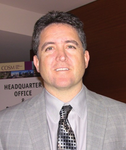
The stent was associated with low rates of polyp formation, inflammation, and adhesions, despite a challenging patient population and withholding of postoperative oral and topical steroids for 30 days, lead author Dr. Keith D. Forwith said at the Combined Otolaryngology Spring Meetings.
The results were consistent across different patient populations and consistent with the pilot study in 43 patients (Int. Forum Allergy Rhinol. 2011;1:23-32).
"I think with any kind of new technique or device, the ultimate thing is how the surgeon feels about it and his or her results," he said. "What I noticed most of all when we stopped doing the study is that I missed using this device. Patients I had used it in during the study had very clean results, very easy debridements. ... We’re very pleased with the results and think it’s a promising technology."

Study sponsor Intersect ENT Inc. has submitted the investigational stent for federal review and hopes to market the device within 6-9 months.
The springlike stent maintains patency by propping open the sinus cavity and releasing mometasone furoate into the sinus lining over 30 days before being resorbed. If approved, it could provide patients with an alternative to space-filling packing materials and silicone stents, and reduce the use of systemic steroids.
The current study involved 50 adults from seven centers with chronic rhinosinusitis for at least 8 consecutive weeks that was confirmed by CT scan (mean CT stage, 11.2). Antibiotics could be prescribed per physician standard of care, and saline irrigation was permitted postoperatively as needed. Polyps were present in 66% of patients, and 28% had undergone a prior sinus procedure. Their mean age was 44 years, and 52% were male.

Ethmoidectomy and maxillary antrostomy were performed in all 50 patients, with 28 also undergoing frontal sinusotomy and 31 sphenoidotomy. Stents were placed unilaterally in 10 patients and bilaterally in 40 patients. Device placement was successful in 100% of sinuses treated, and the implants were resorbed as predicted, Dr. Forwith said. At postoperative day 30, 15% of the material remained and 0.2% remained at day 60.
Endoscopic follow-up at 1 month revealed polypoid edema in 10% of patients, significant adhesion formation in 1%, and middle turbinate lateralization in only 4.4%, said Dr. Forwith, who is in private practice in Louisville, Ky.
When patients were asked about the procedure, their mean score on the 22-question Sino-Nasal Outcome Test improved significantly from baseline through 6 months. The same was true using the Rhinosinusitis Disability Index. "We were pretty pleased that our patients liked the results of the intervention," he said.
No clinically significant changes from baseline occurred in lens opacities or intraocular pressure, which was a theoretical concern given the close proximity of the device. The patients’ mean intraocular pressure was 15 mm Hg at baseline and 14.3 mm Hg at day 30.
One patient experienced headache with sinus pressure/irritation at day 21 that was determined to be related primarily to the surgery and was exacerbated in intensity by the presence of crust on the device. The device was removed and the event resolved without sequelae by day 28.
When asked during a discussion of the study whether the drug-eluting stent resulted in any systemic complications or adrenal corticol suppression, Dr. Forwith replied that they did not specifically look at adrenal suppression, but added that the 370-mcg dose of mometasone furoate is lower than the dose patients receive with most b.i.d. nasal spray administration.
Stent maker Intersect ENT provided funding, administrative support, and materials for the study. Dr. Forwith reported no conflicts of interest.
CHICAGO – A bioabsorbable steroid-eluting stent preserved sinus patency following endoscopic sinus surgery in 50 patients with chronic rhinosinusitis in a prospective, multicenter study.
The stent was associated with low rates of polyp formation, inflammation, and adhesions, despite a challenging patient population and withholding of postoperative oral and topical steroids for 30 days, lead author Dr. Keith D. Forwith said at the Combined Otolaryngology Spring Meetings.
The results were consistent across different patient populations and consistent with the pilot study in 43 patients (Int. Forum Allergy Rhinol. 2011;1:23-32).
"I think with any kind of new technique or device, the ultimate thing is how the surgeon feels about it and his or her results," he said. "What I noticed most of all when we stopped doing the study is that I missed using this device. Patients I had used it in during the study had very clean results, very easy debridements. ... We’re very pleased with the results and think it’s a promising technology."
Study sponsor Intersect ENT Inc. has submitted the investigational stent for federal review and hopes to market the device within 6-9 months.
The springlike stent maintains patency by propping open the sinus cavity and releasing mometasone furoate into the sinus lining over 30 days before being resorbed. If approved, it could provide patients with an alternative to space-filling packing materials and silicone stents, and reduce the use of systemic steroids.
The current study involved 50 adults from seven centers with chronic rhinosinusitis for at least 8 consecutive weeks that was confirmed by CT scan (mean CT stage, 11.2). Antibiotics could be prescribed per physician standard of care, and saline irrigation was permitted postoperatively as needed. Polyps were present in 66% of patients, and 28% had undergone a prior sinus procedure. Their mean age was 44 years, and 52% were male.
Ethmoidectomy and maxillary antrostomy were performed in all 50 patients, with 28 also undergoing frontal sinusotomy and 31 sphenoidotomy. Stents were placed unilaterally in 10 patients and bilaterally in 40 patients. Device placement was successful in 100% of sinuses treated, and the implants were resorbed as predicted, Dr. Forwith said. At postoperative day 30, 15% of the material remained and 0.2% remained at day 60.
Endoscopic follow-up at 1 month revealed polypoid edema in 10% of patients, significant adhesion formation in 1%, and middle turbinate lateralization in only 4.4%, said Dr. Forwith, who is in private practice in Louisville, Ky.
When patients were asked about the procedure, their mean score on the 22-question Sino-Nasal Outcome Test improved significantly from baseline through 6 months. The same was true using the Rhinosinusitis Disability Index. "We were pretty pleased that our patients liked the results of the intervention," he said.
No clinically significant changes from baseline occurred in lens opacities or intraocular pressure, which was a theoretical concern given the close proximity of the device. The patients’ mean intraocular pressure was 15 mm Hg at baseline and 14.3 mm Hg at day 30.
One patient experienced headache with sinus pressure/irritation at day 21 that was determined to be related primarily to the surgery and was exacerbated in intensity by the presence of crust on the device. The device was removed and the event resolved without sequelae by day 28.
When asked during a discussion of the study whether the drug-eluting stent resulted in any systemic complications or adrenal corticol suppression, Dr. Forwith replied that they did not specifically look at adrenal suppression, but added that the 370-mcg dose of mometasone furoate is lower than the dose patients receive with most b.i.d. nasal spray administration.
Stent maker Intersect ENT provided funding, administrative support, and materials for the study. Dr. Forwith reported no conflicts of interest.
CHICAGO – A bioabsorbable steroid-eluting stent preserved sinus patency following endoscopic sinus surgery in 50 patients with chronic rhinosinusitis in a prospective, multicenter study.
The stent was associated with low rates of polyp formation, inflammation, and adhesions, despite a challenging patient population and withholding of postoperative oral and topical steroids for 30 days, lead author Dr. Keith D. Forwith said at the Combined Otolaryngology Spring Meetings.
The results were consistent across different patient populations and consistent with the pilot study in 43 patients (Int. Forum Allergy Rhinol. 2011;1:23-32).
"I think with any kind of new technique or device, the ultimate thing is how the surgeon feels about it and his or her results," he said. "What I noticed most of all when we stopped doing the study is that I missed using this device. Patients I had used it in during the study had very clean results, very easy debridements. ... We’re very pleased with the results and think it’s a promising technology."
Study sponsor Intersect ENT Inc. has submitted the investigational stent for federal review and hopes to market the device within 6-9 months.
The springlike stent maintains patency by propping open the sinus cavity and releasing mometasone furoate into the sinus lining over 30 days before being resorbed. If approved, it could provide patients with an alternative to space-filling packing materials and silicone stents, and reduce the use of systemic steroids.
The current study involved 50 adults from seven centers with chronic rhinosinusitis for at least 8 consecutive weeks that was confirmed by CT scan (mean CT stage, 11.2). Antibiotics could be prescribed per physician standard of care, and saline irrigation was permitted postoperatively as needed. Polyps were present in 66% of patients, and 28% had undergone a prior sinus procedure. Their mean age was 44 years, and 52% were male.
Ethmoidectomy and maxillary antrostomy were performed in all 50 patients, with 28 also undergoing frontal sinusotomy and 31 sphenoidotomy. Stents were placed unilaterally in 10 patients and bilaterally in 40 patients. Device placement was successful in 100% of sinuses treated, and the implants were resorbed as predicted, Dr. Forwith said. At postoperative day 30, 15% of the material remained and 0.2% remained at day 60.
Endoscopic follow-up at 1 month revealed polypoid edema in 10% of patients, significant adhesion formation in 1%, and middle turbinate lateralization in only 4.4%, said Dr. Forwith, who is in private practice in Louisville, Ky.
When patients were asked about the procedure, their mean score on the 22-question Sino-Nasal Outcome Test improved significantly from baseline through 6 months. The same was true using the Rhinosinusitis Disability Index. "We were pretty pleased that our patients liked the results of the intervention," he said.
No clinically significant changes from baseline occurred in lens opacities or intraocular pressure, which was a theoretical concern given the close proximity of the device. The patients’ mean intraocular pressure was 15 mm Hg at baseline and 14.3 mm Hg at day 30.
One patient experienced headache with sinus pressure/irritation at day 21 that was determined to be related primarily to the surgery and was exacerbated in intensity by the presence of crust on the device. The device was removed and the event resolved without sequelae by day 28.
When asked during a discussion of the study whether the drug-eluting stent resulted in any systemic complications or adrenal corticol suppression, Dr. Forwith replied that they did not specifically look at adrenal suppression, but added that the 370-mcg dose of mometasone furoate is lower than the dose patients receive with most b.i.d. nasal spray administration.
Stent maker Intersect ENT provided funding, administrative support, and materials for the study. Dr. Forwith reported no conflicts of interest.
FROM THE COMBINED OTOLARYNGOLOGY SPRING MEETINGS
Major Finding: At 1 month follow-up, the occurrence of polypoid edema was 10%, significant adhesion formation 1%, and middle turbinate lateralization 4.4%.
Data Source: Prospective, multicenter study in 50 patients with chronic rhinosinusitis undergoing endoscopic sinus surgery.
Disclosures: Stent maker Intersect ENT provided funding, administrative support, and materials for the study. Dr. Forwith reported no conflicts of interest.
Steroid-Eluting Sinus Stent May Minimize Postop Events
CHICAGO – A bioabsorbable steroid-eluting stent preserved sinus patency following endoscopic sinus surgery in 50 patients with chronic rhinosinusitis in a prospective, multicenter study.
The stent was associated with low rates of polyp formation, inflammation, and adhesions, despite a challenging patient population and withholding of postoperative oral and topical steroids for 30 days, lead author Dr. Keith D. Forwith said at the Combined Otolaryngology Spring Meetings.
The results were consistent across different patient populations and consistent with the pilot study in 43 patients (Int. Forum Allergy Rhinol. 2011;1:23-32).
"I think with any kind of new technique or device, the ultimate thing is how the surgeon feels about it and his or her results," he said. "What I noticed most of all when we stopped doing the study is that I missed using this device. Patients I had used it in during the study had very clean results, very easy debridements. ... We’re very pleased with the results and think it’s a promising technology."

Study sponsor Intersect ENT Inc. has submitted the investigational stent for federal review and hopes to market the device within 6-9 months.
The springlike stent maintains patency by propping open the sinus cavity and releasing mometasone furoate into the sinus lining over 30 days before being resorbed. If approved, it could provide patients with an alternative to space-filling packing materials and silicone stents, and reduce the use of systemic steroids.
The current study involved 50 adults from seven centers with chronic rhinosinusitis for at least 8 consecutive weeks that was confirmed by CT scan (mean CT stage, 11.2). Antibiotics could be prescribed per physician standard of care, and saline irrigation was permitted postoperatively as needed. Polyps were present in 66% of patients, and 28% had undergone a prior sinus procedure. Their mean age was 44 years, and 52% were male.

Ethmoidectomy and maxillary antrostomy were performed in all 50 patients, with 28 also undergoing frontal sinusotomy and 31 sphenoidotomy. Stents were placed unilaterally in 10 patients and bilaterally in 40 patients. Device placement was successful in 100% of sinuses treated, and the implants were resorbed as predicted, Dr. Forwith said. At postoperative day 30, 15% of the material remained and 0.2% remained at day 60.
Endoscopic follow-up at 1 month revealed polypoid edema in 10% of patients, significant adhesion formation in 1%, and middle turbinate lateralization in only 4.4%, said Dr. Forwith, who is in private practice in Louisville, Ky.
When patients were asked about the procedure, their mean score on the 22-question Sino-Nasal Outcome Test improved significantly from baseline through 6 months. The same was true using the Rhinosinusitis Disability Index. "We were pretty pleased that our patients liked the results of the intervention," he said.
No clinically significant changes from baseline occurred in lens opacities or intraocular pressure, which was a theoretical concern given the close proximity of the device. The patients’ mean intraocular pressure was 15 mm Hg at baseline and 14.3 mm Hg at day 30.
One patient experienced headache with sinus pressure/irritation at day 21 that was determined to be related primarily to the surgery and was exacerbated in intensity by the presence of crust on the device. The device was removed and the event resolved without sequelae by day 28.
When asked during a discussion of the study whether the drug-eluting stent resulted in any systemic complications or adrenal corticol suppression, Dr. Forwith replied that they did not specifically look at adrenal suppression, but added that the 370-mcg dose of mometasone furoate is lower than the dose patients receive with most b.i.d. nasal spray administration.
Stent maker Intersect ENT provided funding, administrative support, and materials for the study. Dr. Forwith reported no conflicts of interest.
CHICAGO – A bioabsorbable steroid-eluting stent preserved sinus patency following endoscopic sinus surgery in 50 patients with chronic rhinosinusitis in a prospective, multicenter study.
The stent was associated with low rates of polyp formation, inflammation, and adhesions, despite a challenging patient population and withholding of postoperative oral and topical steroids for 30 days, lead author Dr. Keith D. Forwith said at the Combined Otolaryngology Spring Meetings.
The results were consistent across different patient populations and consistent with the pilot study in 43 patients (Int. Forum Allergy Rhinol. 2011;1:23-32).
"I think with any kind of new technique or device, the ultimate thing is how the surgeon feels about it and his or her results," he said. "What I noticed most of all when we stopped doing the study is that I missed using this device. Patients I had used it in during the study had very clean results, very easy debridements. ... We’re very pleased with the results and think it’s a promising technology."
Study sponsor Intersect ENT Inc. has submitted the investigational stent for federal review and hopes to market the device within 6-9 months.
The springlike stent maintains patency by propping open the sinus cavity and releasing mometasone furoate into the sinus lining over 30 days before being resorbed. If approved, it could provide patients with an alternative to space-filling packing materials and silicone stents, and reduce the use of systemic steroids.
The current study involved 50 adults from seven centers with chronic rhinosinusitis for at least 8 consecutive weeks that was confirmed by CT scan (mean CT stage, 11.2). Antibiotics could be prescribed per physician standard of care, and saline irrigation was permitted postoperatively as needed. Polyps were present in 66% of patients, and 28% had undergone a prior sinus procedure. Their mean age was 44 years, and 52% were male.
Ethmoidectomy and maxillary antrostomy were performed in all 50 patients, with 28 also undergoing frontal sinusotomy and 31 sphenoidotomy. Stents were placed unilaterally in 10 patients and bilaterally in 40 patients. Device placement was successful in 100% of sinuses treated, and the implants were resorbed as predicted, Dr. Forwith said. At postoperative day 30, 15% of the material remained and 0.2% remained at day 60.
Endoscopic follow-up at 1 month revealed polypoid edema in 10% of patients, significant adhesion formation in 1%, and middle turbinate lateralization in only 4.4%, said Dr. Forwith, who is in private practice in Louisville, Ky.
When patients were asked about the procedure, their mean score on the 22-question Sino-Nasal Outcome Test improved significantly from baseline through 6 months. The same was true using the Rhinosinusitis Disability Index. "We were pretty pleased that our patients liked the results of the intervention," he said.
No clinically significant changes from baseline occurred in lens opacities or intraocular pressure, which was a theoretical concern given the close proximity of the device. The patients’ mean intraocular pressure was 15 mm Hg at baseline and 14.3 mm Hg at day 30.
One patient experienced headache with sinus pressure/irritation at day 21 that was determined to be related primarily to the surgery and was exacerbated in intensity by the presence of crust on the device. The device was removed and the event resolved without sequelae by day 28.
When asked during a discussion of the study whether the drug-eluting stent resulted in any systemic complications or adrenal corticol suppression, Dr. Forwith replied that they did not specifically look at adrenal suppression, but added that the 370-mcg dose of mometasone furoate is lower than the dose patients receive with most b.i.d. nasal spray administration.
Stent maker Intersect ENT provided funding, administrative support, and materials for the study. Dr. Forwith reported no conflicts of interest.
CHICAGO – A bioabsorbable steroid-eluting stent preserved sinus patency following endoscopic sinus surgery in 50 patients with chronic rhinosinusitis in a prospective, multicenter study.
The stent was associated with low rates of polyp formation, inflammation, and adhesions, despite a challenging patient population and withholding of postoperative oral and topical steroids for 30 days, lead author Dr. Keith D. Forwith said at the Combined Otolaryngology Spring Meetings.
The results were consistent across different patient populations and consistent with the pilot study in 43 patients (Int. Forum Allergy Rhinol. 2011;1:23-32).
"I think with any kind of new technique or device, the ultimate thing is how the surgeon feels about it and his or her results," he said. "What I noticed most of all when we stopped doing the study is that I missed using this device. Patients I had used it in during the study had very clean results, very easy debridements. ... We’re very pleased with the results and think it’s a promising technology."
Study sponsor Intersect ENT Inc. has submitted the investigational stent for federal review and hopes to market the device within 6-9 months.
The springlike stent maintains patency by propping open the sinus cavity and releasing mometasone furoate into the sinus lining over 30 days before being resorbed. If approved, it could provide patients with an alternative to space-filling packing materials and silicone stents, and reduce the use of systemic steroids.
The current study involved 50 adults from seven centers with chronic rhinosinusitis for at least 8 consecutive weeks that was confirmed by CT scan (mean CT stage, 11.2). Antibiotics could be prescribed per physician standard of care, and saline irrigation was permitted postoperatively as needed. Polyps were present in 66% of patients, and 28% had undergone a prior sinus procedure. Their mean age was 44 years, and 52% were male.
Ethmoidectomy and maxillary antrostomy were performed in all 50 patients, with 28 also undergoing frontal sinusotomy and 31 sphenoidotomy. Stents were placed unilaterally in 10 patients and bilaterally in 40 patients. Device placement was successful in 100% of sinuses treated, and the implants were resorbed as predicted, Dr. Forwith said. At postoperative day 30, 15% of the material remained and 0.2% remained at day 60.
Endoscopic follow-up at 1 month revealed polypoid edema in 10% of patients, significant adhesion formation in 1%, and middle turbinate lateralization in only 4.4%, said Dr. Forwith, who is in private practice in Louisville, Ky.
When patients were asked about the procedure, their mean score on the 22-question Sino-Nasal Outcome Test improved significantly from baseline through 6 months. The same was true using the Rhinosinusitis Disability Index. "We were pretty pleased that our patients liked the results of the intervention," he said.
No clinically significant changes from baseline occurred in lens opacities or intraocular pressure, which was a theoretical concern given the close proximity of the device. The patients’ mean intraocular pressure was 15 mm Hg at baseline and 14.3 mm Hg at day 30.
One patient experienced headache with sinus pressure/irritation at day 21 that was determined to be related primarily to the surgery and was exacerbated in intensity by the presence of crust on the device. The device was removed and the event resolved without sequelae by day 28.
When asked during a discussion of the study whether the drug-eluting stent resulted in any systemic complications or adrenal corticol suppression, Dr. Forwith replied that they did not specifically look at adrenal suppression, but added that the 370-mcg dose of mometasone furoate is lower than the dose patients receive with most b.i.d. nasal spray administration.
Stent maker Intersect ENT provided funding, administrative support, and materials for the study. Dr. Forwith reported no conflicts of interest.
FROM THE COMBINED OTOLARYNGOLOGY SPRING MEETINGS
Major Finding: At 1 month follow-up, the occurrence of polypoid edema was 10%, significant adhesion formation 1%, and middle turbinate lateralization 4.4%.
Data Source: Prospective, multicenter study in 50 patients with chronic rhinosinusitis undergoing endoscopic sinus surgery.
Disclosures: Stent maker Intersect ENT provided funding, administrative support, and materials for the study. Dr. Forwith reported no conflicts of interest.
Lung Transplantation and Waiting List Survival Improved for PAH Patients
PHILADELPHIA – Although mortality on the waiting list is still a problem, long-term survival after lung transplantation of patients with pulmonary arterial hypertension has improved significantly over time, a study has shown.
In the study, pulmonary arterial hypertension (PAH) was classified as idiopathic (iPAH) or associated with congenital heart diseases or connective tissue diseases. Patients were divided into 1997-2004 and 2005-2010 cohorts.
Out of 2,918 patients referred to the program between January 1997 and September 2010, 316 (11%) presented with PAH (World Health Organization Group 1). In these patients, PAH was classified as iPAH (123 patients), congenital (77 patients), connective (102 patients), and other (14). The number of referrals was similar between 1997-2004 and 2005-2010. Follow-up was completed until September 2010 for all patients.
Among the 100 PAH patients listed for lung transplantation (LT), 57 underwent bilateral LT and 22 had heart LT. Eighteen patients on the waiting list died, and three are still waiting. The waiting list mortality was higher for patients with connective tissue diseases, Dr. Marc de Perrot said at the annual meeting of the American Association for Thoracic Surgery.
No patient with iPAH has died on the waiting list since 2005; 25% died before that time, he and his associates at Toronto General Hospital found.
After LT, the 30-day mortality decreased from 24% in the first cohort to 6% in the second, a significant difference. The 10-year survival was 56% after bilateral LT and 49% after heart LT, a nonsignificant difference.
However, the 10-year survival was significantly worse for iPAH patients at 42% vs. 70% for the remaining patients (P = .01). The 10-year survival was best for connective tissue disease (69%) and congenital (70%) patients.
Lung transplantation is a viable option for about a third of the patients presenting with PAH, according to Dr. de Perrot. He added that extracorporeal life support may help reduce the waiting list mortality, particularly for iPAH patients. Overall, the 30-day mortality for patients after lung transplantation has improved significantly over time.
"Patients with connective tissue diseases have a high mortality on the waiting list, but enjoy excellent long-term survival after transplant," Dr. de Perrot concluded.
Dr. de Perrot reported receiving speaker and teaching honoraria from Actelion.
PHILADELPHIA – Although mortality on the waiting list is still a problem, long-term survival after lung transplantation of patients with pulmonary arterial hypertension has improved significantly over time, a study has shown.
In the study, pulmonary arterial hypertension (PAH) was classified as idiopathic (iPAH) or associated with congenital heart diseases or connective tissue diseases. Patients were divided into 1997-2004 and 2005-2010 cohorts.
Out of 2,918 patients referred to the program between January 1997 and September 2010, 316 (11%) presented with PAH (World Health Organization Group 1). In these patients, PAH was classified as iPAH (123 patients), congenital (77 patients), connective (102 patients), and other (14). The number of referrals was similar between 1997-2004 and 2005-2010. Follow-up was completed until September 2010 for all patients.
Among the 100 PAH patients listed for lung transplantation (LT), 57 underwent bilateral LT and 22 had heart LT. Eighteen patients on the waiting list died, and three are still waiting. The waiting list mortality was higher for patients with connective tissue diseases, Dr. Marc de Perrot said at the annual meeting of the American Association for Thoracic Surgery.
No patient with iPAH has died on the waiting list since 2005; 25% died before that time, he and his associates at Toronto General Hospital found.
After LT, the 30-day mortality decreased from 24% in the first cohort to 6% in the second, a significant difference. The 10-year survival was 56% after bilateral LT and 49% after heart LT, a nonsignificant difference.
However, the 10-year survival was significantly worse for iPAH patients at 42% vs. 70% for the remaining patients (P = .01). The 10-year survival was best for connective tissue disease (69%) and congenital (70%) patients.
Lung transplantation is a viable option for about a third of the patients presenting with PAH, according to Dr. de Perrot. He added that extracorporeal life support may help reduce the waiting list mortality, particularly for iPAH patients. Overall, the 30-day mortality for patients after lung transplantation has improved significantly over time.
"Patients with connective tissue diseases have a high mortality on the waiting list, but enjoy excellent long-term survival after transplant," Dr. de Perrot concluded.
Dr. de Perrot reported receiving speaker and teaching honoraria from Actelion.
PHILADELPHIA – Although mortality on the waiting list is still a problem, long-term survival after lung transplantation of patients with pulmonary arterial hypertension has improved significantly over time, a study has shown.
In the study, pulmonary arterial hypertension (PAH) was classified as idiopathic (iPAH) or associated with congenital heart diseases or connective tissue diseases. Patients were divided into 1997-2004 and 2005-2010 cohorts.
Out of 2,918 patients referred to the program between January 1997 and September 2010, 316 (11%) presented with PAH (World Health Organization Group 1). In these patients, PAH was classified as iPAH (123 patients), congenital (77 patients), connective (102 patients), and other (14). The number of referrals was similar between 1997-2004 and 2005-2010. Follow-up was completed until September 2010 for all patients.
Among the 100 PAH patients listed for lung transplantation (LT), 57 underwent bilateral LT and 22 had heart LT. Eighteen patients on the waiting list died, and three are still waiting. The waiting list mortality was higher for patients with connective tissue diseases, Dr. Marc de Perrot said at the annual meeting of the American Association for Thoracic Surgery.
No patient with iPAH has died on the waiting list since 2005; 25% died before that time, he and his associates at Toronto General Hospital found.
After LT, the 30-day mortality decreased from 24% in the first cohort to 6% in the second, a significant difference. The 10-year survival was 56% after bilateral LT and 49% after heart LT, a nonsignificant difference.
However, the 10-year survival was significantly worse for iPAH patients at 42% vs. 70% for the remaining patients (P = .01). The 10-year survival was best for connective tissue disease (69%) and congenital (70%) patients.
Lung transplantation is a viable option for about a third of the patients presenting with PAH, according to Dr. de Perrot. He added that extracorporeal life support may help reduce the waiting list mortality, particularly for iPAH patients. Overall, the 30-day mortality for patients after lung transplantation has improved significantly over time.
"Patients with connective tissue diseases have a high mortality on the waiting list, but enjoy excellent long-term survival after transplant," Dr. de Perrot concluded.
Dr. de Perrot reported receiving speaker and teaching honoraria from Actelion.
FROM THE ANNUAL MEETING OF THE AMERICAN ASSOCIATION FOR THORACIC SURGERY
Major Finding: In the period 1997-2005, 25% of patients with idiopathic pulmonary arterial hypertension died on the waiting list. None died on the list between 2005 and 2010. After lung transplantation, the 30-day mortality decreased from 24% in the first earlier period to 6% in the later period.
Data Source: A review of 2,918 patients at a single institution who were referred for lung transplantation.
Disclosures: Dr. de Perrot reported receiving speaker and teaching honoraria from Actelion.