User login
Bringing you the latest news, research and reviews, exclusive interviews, podcasts, quizzes, and more.
Powered by CHEST Physician, Clinician Reviews, MDedge Family Medicine, Internal Medicine News, and The Journal of Clinical Outcomes Management.
Do antibiotics shorten symptoms in patients with purulent nasal discharge?
NO. For most patients with purulent nasal discharge, antibiotics don’t decrease symptom duration; they do increase adverse events (strength of recommendation [SOR]: A, 3 meta-analyses and 2 randomized controlled trials [RCTs]).
Researchers in the field don’t recommend using antibiotics as routine treatment for purulent rhinorrhea associated with symptoms of upper respiratory infection ([SOR]: C, expert opinion).
Evidence summary
A Cochrane review of antibiotics for the common cold that included 5 RCTs with a total of 772 participants with purulent nasal discharge found no benefit from antibiotics.1 The relative risk (RR) for persistent acute purulent rhinitis with antibiotics compared with placebo was 0.63 (95% confidence interval [CI], 0.38-1.07; P=.087). The antibiotic groups showed an increase in adverse effects, with an RR of 1.46 (95% CI, 1.01-1.94; P=.047).
Benefits of antibiotics tempered by adverse effects
A meta-analysis of 6 RCTs with more than 1400 subjects showed persistent nasal discharge at 5 to 8 days, on average, in 23% of patients who received antibiotics compared with 46% of patients who received placebo (RR of benefits=1.18; 95% CI, 1.05-1.33; P=.05).2 Most subjects were between 12 and 50 years of age; 2 of the trials included children between 2 months and 16 years of age. All subjects had symptoms for fewer than 10 days.
The adverse effects of antibiotic treatment, primarily rash and diarrhea, were also addressed (RR of adverse effects=1.46; 95% CI, 1.10-1.94; P=.028). Given the overlap of the number needed to treat (7-15) and number needed to harm (12-78), the authors concluded that most patients get better without antibiotics, supporting “no antibiotic as first line” treatment advice.
Other studies show minimal benefit for antibiotics
A meta-analysis of 9 placebo-controlled RCTs (2640 adult subjects with rhinosinusitis-like complaints) found that antibiotics provided minimal benefit. For patients with visible purulent drainage in the pharynx, the NNT overlapped with the NNH; patients without visible purulent discharge showed even less benefit from antibiotics.3
Clinical improvement is insufficient to recommend antibiotic treatment
Three double-blinded RCTs studied patients older than 12 years who presented to a family practice clinic complaining of purulent rhinitis.4-6 All 3 studies compared amoxicillin treatment with placebo; outcomes were based primarily on patient diaries that recorded symptoms, including nasal discharge.
The first study randomized 135 patients to either amoxicillin (n=67) or placebo (n=68) for 10 days.4 At the end of 2 weeks, both groups had similar rates of symptom improvement—although in a subgroup of 57 patients who had complete symptom resolution at 2 weeks, the median number of days until resolution of purulent nasal discharge was 8 in the amoxicillin group compared with 12 days for the placebo group (P=.039). The authors could not identify clinical characteristics favoring antibiotic treatment.
In the second study, 207 patients received amoxicillin and 209 placebo.5 After 10 days of therapy, symptom resolution rates were not significantly different (35% for amoxicillin vs 29% for placebo). However, patients in the amoxicillin group had quicker resolution of purulent nasal discharge (9 vs 14 days for 75% of patients to be free of that symptom; P=.007).5
The third study (240 adults) didn’t find a significant decrease in duration of purulent nasal discharge in the antibiotic group compared with the placebo group.6
Despite the findings of decreased duration of purulent nasal discharge in the first 2 studies, the authors of all 3 studies concluded that the clinical difference in improvement between antibiotic and placebo groups was not enough to recommend treatment with antibiotics. Although the trials didn’t measure adverse outcomes, the authors advised clinicians to consider the potential for adverse reactions before recommending antibiotic treatment.
Recommendations
Both the American Academy of Otolaryngology and the American Academy of Allergy, Asthma, and Immunology recommend watchful waiting without antibiotics for acute sinusitis with mild pain or temperature lower than 101°F and consideration of antibiotics only if symptoms worsen or fail to improve by 7 days after diagnosis. Neither group offers specific recommendations regarding patients with purulent discharge.7,8
The Centers for Disease Control and Prevention recommend reserving antibiotic treatment of acute bacterial rhinosinusitis for patients with symptoms lasting longer than 7 days and patients who have unilateral symptoms with purulent nasal discharge.9
1. Arroll B, Kenealy T. Antibiotics for the common cold and acute purulent rhinitis. Cochrane Database Syst Rev. 2005;(3):CD000247.-
2. Arroll B, Kenealy T. Are antibiotics effective for acute purulent rhinitis? Systematic review and meta-analysis of placebo controlled randomised trials. BMJ. 2006;333:279.-
3. Young J, De Sutter A, Merenstein D, et al. Antibiotics for adults with clinically diagnosed acute rhinosinusitis: a meta-analysis of individual patient data. Lancet. 2008;371:908-914.
4. Merenstein D, Whittaker C, Chadwell T, et al. Are antibiotics beneficial for patients with sinusitis complaints? A randomized double-blind clinical trial. J Fam Pract. 2005;54:144-151.
5. De Sutter AI, De Meyere MJ, Christiaens TC, et al. Does amoxicillin improve outcomes in patients with purulent rhinorrhea? A pragmatic randomized double-blind controlled trial in family practice. J Fam Pract. 2002;51:317-323.
6. Williamson IG, Rumsby K, Benge S, et al. Antibiotics and topical nasal steroid for treatment of acute maxillary sinusitis: a randomized controlled trial. JAMA. 2007;298:2487-2496.
7. Rosenfeld RM, Andes D, Bhattacharyya N, et al. Clinical practice guideline: adult sinusitis. Otolaryngol Head Neck Surg. 2007;137(3 suppl):S1-S31.
8. Slavin RG, Spector SL, Bernstein IL, et al. The diagnosis and management of sinusitis: a practice parameter update. J Allergy Clin Immunol. 2005;116(6 suppl):S13-S47.
9. Hickner JM, Bartlett JG, Besser RE, et al. Principles of appropriate antibiotic use for acute rhinosinusitis in adults: background. Ann Emerg Med. 2001;37:703-710.
NO. For most patients with purulent nasal discharge, antibiotics don’t decrease symptom duration; they do increase adverse events (strength of recommendation [SOR]: A, 3 meta-analyses and 2 randomized controlled trials [RCTs]).
Researchers in the field don’t recommend using antibiotics as routine treatment for purulent rhinorrhea associated with symptoms of upper respiratory infection ([SOR]: C, expert opinion).
Evidence summary
A Cochrane review of antibiotics for the common cold that included 5 RCTs with a total of 772 participants with purulent nasal discharge found no benefit from antibiotics.1 The relative risk (RR) for persistent acute purulent rhinitis with antibiotics compared with placebo was 0.63 (95% confidence interval [CI], 0.38-1.07; P=.087). The antibiotic groups showed an increase in adverse effects, with an RR of 1.46 (95% CI, 1.01-1.94; P=.047).
Benefits of antibiotics tempered by adverse effects
A meta-analysis of 6 RCTs with more than 1400 subjects showed persistent nasal discharge at 5 to 8 days, on average, in 23% of patients who received antibiotics compared with 46% of patients who received placebo (RR of benefits=1.18; 95% CI, 1.05-1.33; P=.05).2 Most subjects were between 12 and 50 years of age; 2 of the trials included children between 2 months and 16 years of age. All subjects had symptoms for fewer than 10 days.
The adverse effects of antibiotic treatment, primarily rash and diarrhea, were also addressed (RR of adverse effects=1.46; 95% CI, 1.10-1.94; P=.028). Given the overlap of the number needed to treat (7-15) and number needed to harm (12-78), the authors concluded that most patients get better without antibiotics, supporting “no antibiotic as first line” treatment advice.
Other studies show minimal benefit for antibiotics
A meta-analysis of 9 placebo-controlled RCTs (2640 adult subjects with rhinosinusitis-like complaints) found that antibiotics provided minimal benefit. For patients with visible purulent drainage in the pharynx, the NNT overlapped with the NNH; patients without visible purulent discharge showed even less benefit from antibiotics.3
Clinical improvement is insufficient to recommend antibiotic treatment
Three double-blinded RCTs studied patients older than 12 years who presented to a family practice clinic complaining of purulent rhinitis.4-6 All 3 studies compared amoxicillin treatment with placebo; outcomes were based primarily on patient diaries that recorded symptoms, including nasal discharge.
The first study randomized 135 patients to either amoxicillin (n=67) or placebo (n=68) for 10 days.4 At the end of 2 weeks, both groups had similar rates of symptom improvement—although in a subgroup of 57 patients who had complete symptom resolution at 2 weeks, the median number of days until resolution of purulent nasal discharge was 8 in the amoxicillin group compared with 12 days for the placebo group (P=.039). The authors could not identify clinical characteristics favoring antibiotic treatment.
In the second study, 207 patients received amoxicillin and 209 placebo.5 After 10 days of therapy, symptom resolution rates were not significantly different (35% for amoxicillin vs 29% for placebo). However, patients in the amoxicillin group had quicker resolution of purulent nasal discharge (9 vs 14 days for 75% of patients to be free of that symptom; P=.007).5
The third study (240 adults) didn’t find a significant decrease in duration of purulent nasal discharge in the antibiotic group compared with the placebo group.6
Despite the findings of decreased duration of purulent nasal discharge in the first 2 studies, the authors of all 3 studies concluded that the clinical difference in improvement between antibiotic and placebo groups was not enough to recommend treatment with antibiotics. Although the trials didn’t measure adverse outcomes, the authors advised clinicians to consider the potential for adverse reactions before recommending antibiotic treatment.
Recommendations
Both the American Academy of Otolaryngology and the American Academy of Allergy, Asthma, and Immunology recommend watchful waiting without antibiotics for acute sinusitis with mild pain or temperature lower than 101°F and consideration of antibiotics only if symptoms worsen or fail to improve by 7 days after diagnosis. Neither group offers specific recommendations regarding patients with purulent discharge.7,8
The Centers for Disease Control and Prevention recommend reserving antibiotic treatment of acute bacterial rhinosinusitis for patients with symptoms lasting longer than 7 days and patients who have unilateral symptoms with purulent nasal discharge.9
NO. For most patients with purulent nasal discharge, antibiotics don’t decrease symptom duration; they do increase adverse events (strength of recommendation [SOR]: A, 3 meta-analyses and 2 randomized controlled trials [RCTs]).
Researchers in the field don’t recommend using antibiotics as routine treatment for purulent rhinorrhea associated with symptoms of upper respiratory infection ([SOR]: C, expert opinion).
Evidence summary
A Cochrane review of antibiotics for the common cold that included 5 RCTs with a total of 772 participants with purulent nasal discharge found no benefit from antibiotics.1 The relative risk (RR) for persistent acute purulent rhinitis with antibiotics compared with placebo was 0.63 (95% confidence interval [CI], 0.38-1.07; P=.087). The antibiotic groups showed an increase in adverse effects, with an RR of 1.46 (95% CI, 1.01-1.94; P=.047).
Benefits of antibiotics tempered by adverse effects
A meta-analysis of 6 RCTs with more than 1400 subjects showed persistent nasal discharge at 5 to 8 days, on average, in 23% of patients who received antibiotics compared with 46% of patients who received placebo (RR of benefits=1.18; 95% CI, 1.05-1.33; P=.05).2 Most subjects were between 12 and 50 years of age; 2 of the trials included children between 2 months and 16 years of age. All subjects had symptoms for fewer than 10 days.
The adverse effects of antibiotic treatment, primarily rash and diarrhea, were also addressed (RR of adverse effects=1.46; 95% CI, 1.10-1.94; P=.028). Given the overlap of the number needed to treat (7-15) and number needed to harm (12-78), the authors concluded that most patients get better without antibiotics, supporting “no antibiotic as first line” treatment advice.
Other studies show minimal benefit for antibiotics
A meta-analysis of 9 placebo-controlled RCTs (2640 adult subjects with rhinosinusitis-like complaints) found that antibiotics provided minimal benefit. For patients with visible purulent drainage in the pharynx, the NNT overlapped with the NNH; patients without visible purulent discharge showed even less benefit from antibiotics.3
Clinical improvement is insufficient to recommend antibiotic treatment
Three double-blinded RCTs studied patients older than 12 years who presented to a family practice clinic complaining of purulent rhinitis.4-6 All 3 studies compared amoxicillin treatment with placebo; outcomes were based primarily on patient diaries that recorded symptoms, including nasal discharge.
The first study randomized 135 patients to either amoxicillin (n=67) or placebo (n=68) for 10 days.4 At the end of 2 weeks, both groups had similar rates of symptom improvement—although in a subgroup of 57 patients who had complete symptom resolution at 2 weeks, the median number of days until resolution of purulent nasal discharge was 8 in the amoxicillin group compared with 12 days for the placebo group (P=.039). The authors could not identify clinical characteristics favoring antibiotic treatment.
In the second study, 207 patients received amoxicillin and 209 placebo.5 After 10 days of therapy, symptom resolution rates were not significantly different (35% for amoxicillin vs 29% for placebo). However, patients in the amoxicillin group had quicker resolution of purulent nasal discharge (9 vs 14 days for 75% of patients to be free of that symptom; P=.007).5
The third study (240 adults) didn’t find a significant decrease in duration of purulent nasal discharge in the antibiotic group compared with the placebo group.6
Despite the findings of decreased duration of purulent nasal discharge in the first 2 studies, the authors of all 3 studies concluded that the clinical difference in improvement between antibiotic and placebo groups was not enough to recommend treatment with antibiotics. Although the trials didn’t measure adverse outcomes, the authors advised clinicians to consider the potential for adverse reactions before recommending antibiotic treatment.
Recommendations
Both the American Academy of Otolaryngology and the American Academy of Allergy, Asthma, and Immunology recommend watchful waiting without antibiotics for acute sinusitis with mild pain or temperature lower than 101°F and consideration of antibiotics only if symptoms worsen or fail to improve by 7 days after diagnosis. Neither group offers specific recommendations regarding patients with purulent discharge.7,8
The Centers for Disease Control and Prevention recommend reserving antibiotic treatment of acute bacterial rhinosinusitis for patients with symptoms lasting longer than 7 days and patients who have unilateral symptoms with purulent nasal discharge.9
1. Arroll B, Kenealy T. Antibiotics for the common cold and acute purulent rhinitis. Cochrane Database Syst Rev. 2005;(3):CD000247.-
2. Arroll B, Kenealy T. Are antibiotics effective for acute purulent rhinitis? Systematic review and meta-analysis of placebo controlled randomised trials. BMJ. 2006;333:279.-
3. Young J, De Sutter A, Merenstein D, et al. Antibiotics for adults with clinically diagnosed acute rhinosinusitis: a meta-analysis of individual patient data. Lancet. 2008;371:908-914.
4. Merenstein D, Whittaker C, Chadwell T, et al. Are antibiotics beneficial for patients with sinusitis complaints? A randomized double-blind clinical trial. J Fam Pract. 2005;54:144-151.
5. De Sutter AI, De Meyere MJ, Christiaens TC, et al. Does amoxicillin improve outcomes in patients with purulent rhinorrhea? A pragmatic randomized double-blind controlled trial in family practice. J Fam Pract. 2002;51:317-323.
6. Williamson IG, Rumsby K, Benge S, et al. Antibiotics and topical nasal steroid for treatment of acute maxillary sinusitis: a randomized controlled trial. JAMA. 2007;298:2487-2496.
7. Rosenfeld RM, Andes D, Bhattacharyya N, et al. Clinical practice guideline: adult sinusitis. Otolaryngol Head Neck Surg. 2007;137(3 suppl):S1-S31.
8. Slavin RG, Spector SL, Bernstein IL, et al. The diagnosis and management of sinusitis: a practice parameter update. J Allergy Clin Immunol. 2005;116(6 suppl):S13-S47.
9. Hickner JM, Bartlett JG, Besser RE, et al. Principles of appropriate antibiotic use for acute rhinosinusitis in adults: background. Ann Emerg Med. 2001;37:703-710.
1. Arroll B, Kenealy T. Antibiotics for the common cold and acute purulent rhinitis. Cochrane Database Syst Rev. 2005;(3):CD000247.-
2. Arroll B, Kenealy T. Are antibiotics effective for acute purulent rhinitis? Systematic review and meta-analysis of placebo controlled randomised trials. BMJ. 2006;333:279.-
3. Young J, De Sutter A, Merenstein D, et al. Antibiotics for adults with clinically diagnosed acute rhinosinusitis: a meta-analysis of individual patient data. Lancet. 2008;371:908-914.
4. Merenstein D, Whittaker C, Chadwell T, et al. Are antibiotics beneficial for patients with sinusitis complaints? A randomized double-blind clinical trial. J Fam Pract. 2005;54:144-151.
5. De Sutter AI, De Meyere MJ, Christiaens TC, et al. Does amoxicillin improve outcomes in patients with purulent rhinorrhea? A pragmatic randomized double-blind controlled trial in family practice. J Fam Pract. 2002;51:317-323.
6. Williamson IG, Rumsby K, Benge S, et al. Antibiotics and topical nasal steroid for treatment of acute maxillary sinusitis: a randomized controlled trial. JAMA. 2007;298:2487-2496.
7. Rosenfeld RM, Andes D, Bhattacharyya N, et al. Clinical practice guideline: adult sinusitis. Otolaryngol Head Neck Surg. 2007;137(3 suppl):S1-S31.
8. Slavin RG, Spector SL, Bernstein IL, et al. The diagnosis and management of sinusitis: a practice parameter update. J Allergy Clin Immunol. 2005;116(6 suppl):S13-S47.
9. Hickner JM, Bartlett JG, Besser RE, et al. Principles of appropriate antibiotic use for acute rhinosinusitis in adults: background. Ann Emerg Med. 2001;37:703-710.
Evidence-based answers from the Family Physicians Inquiries Network
Thorough Work-Up Crucial in Sarcoidosis Cases
SAN DIEGO – While it’s well known that sarcoidosis commonly affects pulmonary function, it’s perhaps less known that the disorder can be detrimental to cardiac function in approximately 5% of cases.
"A common way that patients present with cardiac sarcoidosis is with sudden cardiac death," Dr. Misha Rosenbach said at the annual meeting of the American Academy of Dermatology. "This is a terrible way to present to your doctor with a problem."

A multisystem disorder of unknown cause, sarcoidosis commonly affects young and middle-aged adults and frequently presents with bilateral hilar lymphadenopathy, pulmonary infiltration, and ocular and skin lesions. Other organs may be involved. The diagnosis is established when clinicoradiologic findings are supported by histologic evidence of noncaseating epithelioid cell granulomas.
"Sarcoidosis is primarily a pulmonary disease, but patients can also present with profound systemic symptoms," said Dr. Rosenbach of the departments of dermatology and internal medicine at the University of Pennsylvania, Philadelphia. "When you’re evaluating a patient with cutaneous sarcoidosis, and making a diagnosis of granulomatous disease of the skin, and looking for extracutaneous involvement, it’s important to know what else can be affected."
Although pulmonary function is affected in more than 90% of cases, other commonly affected sites include the eyes (25%-50% of cases), lymph nodes (about 33% of cases), musculoskeletal system (25%-40% of cases), endocrine system (10%-25% of cases), and liver (20%-50% of cases). The initial evaluation should consist of history and physical exam; chest x-ray; pulmonary function tests (including carbon monoxide diffusing capacity); ophthalmologic examination; complete blood count and serum chemistries (including calcium); urinalysis; EKG (plus additional testing if there is a history of palpitations); tuberculin skin test (TST) or interferon (IFN)–gamma release assay; and thyroid and vitamin D testing.
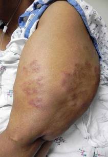
"Patients with sarcoidosis often have low levels of 25-hydroxyvitamin D, but elevated levels of 1,25-dihydroxyvitamin D3," Dr. Rosenbach said. "Inappropriate supplementation can lead to hypercalcemia."
For latent tuberculosis testing, he pointed out that the IFN-gamma release assay (IGRA) is thought to be more accurate than the TST. "IGRA significantly reduces false-positive results" in bacille Calmette-Guérin–vaccinated patients, said Dr. Rosenbach, who is also director of the cutaneous sarcoidosis clinic at the University of Pennsylvania. "Cost-benefit analyses suggest that IGRA [is] cost equivalent to TST, and the Centers for Disease Control and Prevention recommends that IGRA may be used in all circumstances in which the TST is currently used. However, both TST and IGRA have decreased responsiveness and lower sensitivity in patients with impaired immune systems."
In terms of the impact of sarcoidosis on the thyroid gland, a recent analysis of a large database in the United Kingdom found that hyper- and hypothyroidism were twice as common in patients with sarcoidosis, compared with a control population (Postgrad. Med. J. 2009;85:233-7).
A more recent study of 50 patients with cutaneous sarcoidosis conducted by Dr. Rosenbach and his colleagues found that 25% of patients had abnormal thyroid laboratory test results (J. Am. Acad. Dermatol. 2012;66:167-8).
The precise association between sarcoidosis and malignancy remains unclear, he said, but the best available studies suggest that the incidence of lymphoproliferative disorder may be increased in patients with sarcoidosis. Other granulomatous dermatitides may be associated with hematologic abnormalities. Authors of one review found that granulomatous dermatitides may be the first sign of underlying myelodysplastic syndrome (MDS), and recommended that clinicians consider looking for underlying MDS in patients with unexplained or atypical granulomatous skin eruptions (Arch. Dermatol. 2011;147:331-5).

A common stepwise approach for treating patients, Dr. Rosenbach said, begins with skin-directed therapies in the form of steroids or injections. The second step involves the use of antimalarials and tetracycline-class antibiotics; the third step involves methotrexate and/or prednisone, and the fourth step involves consideration for treatment with infliximab or adalimumab. "At this point, etanercept should probably not be used," Dr. Rosenbach said. "It appears to be less effective, and in a few reports has been associated with worsening of disease."
The data are strongest for infliximab, he said, at a recommended dosage of 5 mg/kg at 0, 2, and 6 weeks, and then with maintenance therapy every 6-8 weeks. Adalimumab appeared to work best at 40 mg every week, he said, "but the addition of low-dose methotrexate is sometimes necessary to either regimen."
Dr. Rosenbach disclosed that he was an investigator for a clinical trial sponsored by Centocor and Johnson & Johnson to investigate biologics for chronic/refractory sarcoidosis.
SAN DIEGO – While it’s well known that sarcoidosis commonly affects pulmonary function, it’s perhaps less known that the disorder can be detrimental to cardiac function in approximately 5% of cases.
"A common way that patients present with cardiac sarcoidosis is with sudden cardiac death," Dr. Misha Rosenbach said at the annual meeting of the American Academy of Dermatology. "This is a terrible way to present to your doctor with a problem."
A multisystem disorder of unknown cause, sarcoidosis commonly affects young and middle-aged adults and frequently presents with bilateral hilar lymphadenopathy, pulmonary infiltration, and ocular and skin lesions. Other organs may be involved. The diagnosis is established when clinicoradiologic findings are supported by histologic evidence of noncaseating epithelioid cell granulomas.
"Sarcoidosis is primarily a pulmonary disease, but patients can also present with profound systemic symptoms," said Dr. Rosenbach of the departments of dermatology and internal medicine at the University of Pennsylvania, Philadelphia. "When you’re evaluating a patient with cutaneous sarcoidosis, and making a diagnosis of granulomatous disease of the skin, and looking for extracutaneous involvement, it’s important to know what else can be affected."
Although pulmonary function is affected in more than 90% of cases, other commonly affected sites include the eyes (25%-50% of cases), lymph nodes (about 33% of cases), musculoskeletal system (25%-40% of cases), endocrine system (10%-25% of cases), and liver (20%-50% of cases). The initial evaluation should consist of history and physical exam; chest x-ray; pulmonary function tests (including carbon monoxide diffusing capacity); ophthalmologic examination; complete blood count and serum chemistries (including calcium); urinalysis; EKG (plus additional testing if there is a history of palpitations); tuberculin skin test (TST) or interferon (IFN)–gamma release assay; and thyroid and vitamin D testing.
"Patients with sarcoidosis often have low levels of 25-hydroxyvitamin D, but elevated levels of 1,25-dihydroxyvitamin D3," Dr. Rosenbach said. "Inappropriate supplementation can lead to hypercalcemia."
For latent tuberculosis testing, he pointed out that the IFN-gamma release assay (IGRA) is thought to be more accurate than the TST. "IGRA significantly reduces false-positive results" in bacille Calmette-Guérin–vaccinated patients, said Dr. Rosenbach, who is also director of the cutaneous sarcoidosis clinic at the University of Pennsylvania. "Cost-benefit analyses suggest that IGRA [is] cost equivalent to TST, and the Centers for Disease Control and Prevention recommends that IGRA may be used in all circumstances in which the TST is currently used. However, both TST and IGRA have decreased responsiveness and lower sensitivity in patients with impaired immune systems."
In terms of the impact of sarcoidosis on the thyroid gland, a recent analysis of a large database in the United Kingdom found that hyper- and hypothyroidism were twice as common in patients with sarcoidosis, compared with a control population (Postgrad. Med. J. 2009;85:233-7).
A more recent study of 50 patients with cutaneous sarcoidosis conducted by Dr. Rosenbach and his colleagues found that 25% of patients had abnormal thyroid laboratory test results (J. Am. Acad. Dermatol. 2012;66:167-8).
The precise association between sarcoidosis and malignancy remains unclear, he said, but the best available studies suggest that the incidence of lymphoproliferative disorder may be increased in patients with sarcoidosis. Other granulomatous dermatitides may be associated with hematologic abnormalities. Authors of one review found that granulomatous dermatitides may be the first sign of underlying myelodysplastic syndrome (MDS), and recommended that clinicians consider looking for underlying MDS in patients with unexplained or atypical granulomatous skin eruptions (Arch. Dermatol. 2011;147:331-5).
A common stepwise approach for treating patients, Dr. Rosenbach said, begins with skin-directed therapies in the form of steroids or injections. The second step involves the use of antimalarials and tetracycline-class antibiotics; the third step involves methotrexate and/or prednisone, and the fourth step involves consideration for treatment with infliximab or adalimumab. "At this point, etanercept should probably not be used," Dr. Rosenbach said. "It appears to be less effective, and in a few reports has been associated with worsening of disease."
The data are strongest for infliximab, he said, at a recommended dosage of 5 mg/kg at 0, 2, and 6 weeks, and then with maintenance therapy every 6-8 weeks. Adalimumab appeared to work best at 40 mg every week, he said, "but the addition of low-dose methotrexate is sometimes necessary to either regimen."
Dr. Rosenbach disclosed that he was an investigator for a clinical trial sponsored by Centocor and Johnson & Johnson to investigate biologics for chronic/refractory sarcoidosis.
SAN DIEGO – While it’s well known that sarcoidosis commonly affects pulmonary function, it’s perhaps less known that the disorder can be detrimental to cardiac function in approximately 5% of cases.
"A common way that patients present with cardiac sarcoidosis is with sudden cardiac death," Dr. Misha Rosenbach said at the annual meeting of the American Academy of Dermatology. "This is a terrible way to present to your doctor with a problem."
A multisystem disorder of unknown cause, sarcoidosis commonly affects young and middle-aged adults and frequently presents with bilateral hilar lymphadenopathy, pulmonary infiltration, and ocular and skin lesions. Other organs may be involved. The diagnosis is established when clinicoradiologic findings are supported by histologic evidence of noncaseating epithelioid cell granulomas.
"Sarcoidosis is primarily a pulmonary disease, but patients can also present with profound systemic symptoms," said Dr. Rosenbach of the departments of dermatology and internal medicine at the University of Pennsylvania, Philadelphia. "When you’re evaluating a patient with cutaneous sarcoidosis, and making a diagnosis of granulomatous disease of the skin, and looking for extracutaneous involvement, it’s important to know what else can be affected."
Although pulmonary function is affected in more than 90% of cases, other commonly affected sites include the eyes (25%-50% of cases), lymph nodes (about 33% of cases), musculoskeletal system (25%-40% of cases), endocrine system (10%-25% of cases), and liver (20%-50% of cases). The initial evaluation should consist of history and physical exam; chest x-ray; pulmonary function tests (including carbon monoxide diffusing capacity); ophthalmologic examination; complete blood count and serum chemistries (including calcium); urinalysis; EKG (plus additional testing if there is a history of palpitations); tuberculin skin test (TST) or interferon (IFN)–gamma release assay; and thyroid and vitamin D testing.
"Patients with sarcoidosis often have low levels of 25-hydroxyvitamin D, but elevated levels of 1,25-dihydroxyvitamin D3," Dr. Rosenbach said. "Inappropriate supplementation can lead to hypercalcemia."
For latent tuberculosis testing, he pointed out that the IFN-gamma release assay (IGRA) is thought to be more accurate than the TST. "IGRA significantly reduces false-positive results" in bacille Calmette-Guérin–vaccinated patients, said Dr. Rosenbach, who is also director of the cutaneous sarcoidosis clinic at the University of Pennsylvania. "Cost-benefit analyses suggest that IGRA [is] cost equivalent to TST, and the Centers for Disease Control and Prevention recommends that IGRA may be used in all circumstances in which the TST is currently used. However, both TST and IGRA have decreased responsiveness and lower sensitivity in patients with impaired immune systems."
In terms of the impact of sarcoidosis on the thyroid gland, a recent analysis of a large database in the United Kingdom found that hyper- and hypothyroidism were twice as common in patients with sarcoidosis, compared with a control population (Postgrad. Med. J. 2009;85:233-7).
A more recent study of 50 patients with cutaneous sarcoidosis conducted by Dr. Rosenbach and his colleagues found that 25% of patients had abnormal thyroid laboratory test results (J. Am. Acad. Dermatol. 2012;66:167-8).
The precise association between sarcoidosis and malignancy remains unclear, he said, but the best available studies suggest that the incidence of lymphoproliferative disorder may be increased in patients with sarcoidosis. Other granulomatous dermatitides may be associated with hematologic abnormalities. Authors of one review found that granulomatous dermatitides may be the first sign of underlying myelodysplastic syndrome (MDS), and recommended that clinicians consider looking for underlying MDS in patients with unexplained or atypical granulomatous skin eruptions (Arch. Dermatol. 2011;147:331-5).
A common stepwise approach for treating patients, Dr. Rosenbach said, begins with skin-directed therapies in the form of steroids or injections. The second step involves the use of antimalarials and tetracycline-class antibiotics; the third step involves methotrexate and/or prednisone, and the fourth step involves consideration for treatment with infliximab or adalimumab. "At this point, etanercept should probably not be used," Dr. Rosenbach said. "It appears to be less effective, and in a few reports has been associated with worsening of disease."
The data are strongest for infliximab, he said, at a recommended dosage of 5 mg/kg at 0, 2, and 6 weeks, and then with maintenance therapy every 6-8 weeks. Adalimumab appeared to work best at 40 mg every week, he said, "but the addition of low-dose methotrexate is sometimes necessary to either regimen."
Dr. Rosenbach disclosed that he was an investigator for a clinical trial sponsored by Centocor and Johnson & Johnson to investigate biologics for chronic/refractory sarcoidosis.
EXPERT ANALYSIS FROM THE ANNUAL MEETING OF THE AMERICAN ACADEMY OF DERMATOLOGY
Outpatient Aspirin Desensitization Restores Drug's Benefit in AERD
ORLANDO – Aspirin sensitivity can be a real headache for patients and clinicians, but a safe and effective aspirin desensitization protocol can bring the analgesic and anti-inflammatory benefits of aspirin therapy to patients with aspirin-exacerbated respiratory disease, reported a clinician at the annual meeting of the American Academy of Allergy, Asthma, and Immunology.
Patients with AERD who undergo aspirin desensitization have fewer bouts of sinusitis, show improvement in both asthma symptoms and sense of smell, and use less corticosteroid, compared with patients who don’t undergo desensitization, said Dr. Katharine M. Woessner, program director in the division of allergy, asthma, and immunology at the Scripps Clinic in San Diego.

Aspirin desensitization also blunts the response to other NSAIDS like ibuprofen and naproxen, which – like aspirin – inhibit the cyclooxygenase-1 (COX-1) enzyme, Dr. Woessner said. Aspirin and other NSAIDs induce rhinitis and asthma attacks in patients with AERD, and the disease is progressive even when patients are careful to avoid all NSAIDs.
Only 3 of 1,400 consecutive patients with AERD that was treated with aspirin desensitization at Scripps experienced systemic reactions, and all of those responded to a single dose of intramuscular epinephrine, she said.
"I don’t know why aspirin desensitization works, but we clearly have a therapy that’s easy to use and is quite effective in managing these patients," Dr. Woessner said.
Aspirin Challenge
AERD usually begins in patients in their 30s or 40s who have a prior history of tolerance to aspirin and other NSAIDs. It is more than twice as likely to occur in women, and tends to be more severe in women than in men. Patients develop chronic congestion, rhinitis, anosmia, and nasal polyps, often followed by asthma 1-5 years after the onset of rhinitis.
The disease is characterized by chronic eosinophilic rhinosinusitis and nasal polyposis that are initially intermittent but evolve into chronic, hyperplasic eosinophilic sinusitis that often requires surgical intervention, Dr. Woessner said.
The asthma that develops in patients with AERD is persistent, usually moderate to severe, and related to the severity of sinus disease. However, "asthma is not necessarily a prerequisite to make a diagnosis of aspirin-exacerbated respiratory disease," she noted.
The clinical standard for AERD diagnosis is an oral aspirin challenge. Patients typically can experience a 20% or greater decline in FEV1 (forced expiratory volume in 1 second) and a naso-ocular reaction, but purely upper airway or lower airway reactions can also occur. Patients may also have a partial asthma reaction, with a change in FEV1 from baseline of –15% to –20% and a naso-ocular reaction or laryngospasm. Patients are considered to be negative for AERD if they have no reactions following a 325-mg oral aspirin challenge.
In those who test positive, treatment consists of avoiding all COX-1–inhibiting NSAIDS. Highly selective COX-2 inhibitors such as celecoxib (Celebrex) are generally well tolerated in patients with AERD, Dr. Woesnner said, noting that more than 200 AERD-proven patients at Scripps who were challenged with a highly selective COX-2 inhibitor had no reaction. However, cross reactivity is possible with higher doses of less-selective COX-2 inhibitors such as nimesulide (Sulide) or meloxicam (Mobic). Acetaminophen at doses up to 1,000 mg are also typically well tolerated in these patients.
"There are several drugs that don’t cross-react with aspirin in patients with AERD. The problem is that they’re not very good analgesics, so if we need something that’s analgesic or anti-inflammatory, patients are not going to get much benefit from these medications," Dr. Woessner said.
Outpatient Precautions
Aspirin challenge and desensitization in the outpatient setting can be safely performed with a few caveats. The patient should have stable asthma within 10% of the best prior value and an FEV1 of at least 60%-80% of predicted, or an output of at least 1.5 L. The patient should be on inhaled steroids and a long-acting beta-agonist, and 2-4 weeks before desensitization should be started on montelukast or another leukotriene modifier.
The advent of leukotriene inhibitors has made aspirin desensitization a routine outpatient procedure, session moderator Dr. Mariana C. Castells noted in an interview.
"We used to do aspirin desensitization in the intensive care unit, just because the reactions were scary and we didn’t know how to control them. In the last 10 years, since we started to use montelukast [Singulair] and Zyflo [zileuton], less than 1% have been done in an intensive care unit," said Dr. Castells of the division of rheumatology, immunology, and allergy at Brigham and Women’s Hospital in Boston.
Patients are asked to not use antihistamines for 72 hours before the procedure so that their naso-ocular responses can be observed. Patients with nasal polyps may require debulking surgery prior to desensitization.
"We had recommended in the past that intravenous access be available, but we have data now that it may not be necessary," Dr. Woessner said.
On day 1 of sensitization, patients take 20-40 mg of aspirin at 8 a.m., 40-60 mg at 11 a.m., and 60-100 mg at 2 p.m. On day 2, the respective doses at the same times of day are 100-160 mg, 160-325 mg, and 325 mg.
During desensitization, FEV1 should be measured every hour, and should be at least 1.5 L and greater than 60% of predicted. When a patient has a reaction and that reaction is resolved, the provoking dose should be repeated, and if there is no reaction, the escalating doses can be continued every 3 hours as scheduled. Patients are considered to be densensitized or aspirin tolerant if they can take a 325-mg dose with no reaction. The patient should be instructed to start on 650-mg aspirin that night as the first daily dose, and to continue with up to 650 mg twice daily. About half of all patients can decrease to 325 mg b.i.d. at 1-6 months after the densensitization protocol is completed.
Combining a nasal ketorolac challenge with the oral aspirin challenge can make the desensitization even safer, Dr. Woessner said. The patient is given ketorolac spray at increasing doses every half-hour over 2 hours, followed 1 hour after the last spray with 60-mg aspirin in two doses 1.5 hours apart. The patient is discharged and returns the next day to receive 150-mg aspirin, followed 3 hours later by 325 mg.
No funding source was reported. Dr. Woessner disclosed that she is on the speakers bureaus of Merck and Teva, and has been a speaker for GlaxoSmithKline.
ORLANDO – Aspirin sensitivity can be a real headache for patients and clinicians, but a safe and effective aspirin desensitization protocol can bring the analgesic and anti-inflammatory benefits of aspirin therapy to patients with aspirin-exacerbated respiratory disease, reported a clinician at the annual meeting of the American Academy of Allergy, Asthma, and Immunology.
Patients with AERD who undergo aspirin desensitization have fewer bouts of sinusitis, show improvement in both asthma symptoms and sense of smell, and use less corticosteroid, compared with patients who don’t undergo desensitization, said Dr. Katharine M. Woessner, program director in the division of allergy, asthma, and immunology at the Scripps Clinic in San Diego.
Aspirin desensitization also blunts the response to other NSAIDS like ibuprofen and naproxen, which – like aspirin – inhibit the cyclooxygenase-1 (COX-1) enzyme, Dr. Woessner said. Aspirin and other NSAIDs induce rhinitis and asthma attacks in patients with AERD, and the disease is progressive even when patients are careful to avoid all NSAIDs.
Only 3 of 1,400 consecutive patients with AERD that was treated with aspirin desensitization at Scripps experienced systemic reactions, and all of those responded to a single dose of intramuscular epinephrine, she said.
"I don’t know why aspirin desensitization works, but we clearly have a therapy that’s easy to use and is quite effective in managing these patients," Dr. Woessner said.
Aspirin Challenge
AERD usually begins in patients in their 30s or 40s who have a prior history of tolerance to aspirin and other NSAIDs. It is more than twice as likely to occur in women, and tends to be more severe in women than in men. Patients develop chronic congestion, rhinitis, anosmia, and nasal polyps, often followed by asthma 1-5 years after the onset of rhinitis.
The disease is characterized by chronic eosinophilic rhinosinusitis and nasal polyposis that are initially intermittent but evolve into chronic, hyperplasic eosinophilic sinusitis that often requires surgical intervention, Dr. Woessner said.
The asthma that develops in patients with AERD is persistent, usually moderate to severe, and related to the severity of sinus disease. However, "asthma is not necessarily a prerequisite to make a diagnosis of aspirin-exacerbated respiratory disease," she noted.
The clinical standard for AERD diagnosis is an oral aspirin challenge. Patients typically can experience a 20% or greater decline in FEV1 (forced expiratory volume in 1 second) and a naso-ocular reaction, but purely upper airway or lower airway reactions can also occur. Patients may also have a partial asthma reaction, with a change in FEV1 from baseline of –15% to –20% and a naso-ocular reaction or laryngospasm. Patients are considered to be negative for AERD if they have no reactions following a 325-mg oral aspirin challenge.
In those who test positive, treatment consists of avoiding all COX-1–inhibiting NSAIDS. Highly selective COX-2 inhibitors such as celecoxib (Celebrex) are generally well tolerated in patients with AERD, Dr. Woesnner said, noting that more than 200 AERD-proven patients at Scripps who were challenged with a highly selective COX-2 inhibitor had no reaction. However, cross reactivity is possible with higher doses of less-selective COX-2 inhibitors such as nimesulide (Sulide) or meloxicam (Mobic). Acetaminophen at doses up to 1,000 mg are also typically well tolerated in these patients.
"There are several drugs that don’t cross-react with aspirin in patients with AERD. The problem is that they’re not very good analgesics, so if we need something that’s analgesic or anti-inflammatory, patients are not going to get much benefit from these medications," Dr. Woessner said.
Outpatient Precautions
Aspirin challenge and desensitization in the outpatient setting can be safely performed with a few caveats. The patient should have stable asthma within 10% of the best prior value and an FEV1 of at least 60%-80% of predicted, or an output of at least 1.5 L. The patient should be on inhaled steroids and a long-acting beta-agonist, and 2-4 weeks before desensitization should be started on montelukast or another leukotriene modifier.
The advent of leukotriene inhibitors has made aspirin desensitization a routine outpatient procedure, session moderator Dr. Mariana C. Castells noted in an interview.
"We used to do aspirin desensitization in the intensive care unit, just because the reactions were scary and we didn’t know how to control them. In the last 10 years, since we started to use montelukast [Singulair] and Zyflo [zileuton], less than 1% have been done in an intensive care unit," said Dr. Castells of the division of rheumatology, immunology, and allergy at Brigham and Women’s Hospital in Boston.
Patients are asked to not use antihistamines for 72 hours before the procedure so that their naso-ocular responses can be observed. Patients with nasal polyps may require debulking surgery prior to desensitization.
"We had recommended in the past that intravenous access be available, but we have data now that it may not be necessary," Dr. Woessner said.
On day 1 of sensitization, patients take 20-40 mg of aspirin at 8 a.m., 40-60 mg at 11 a.m., and 60-100 mg at 2 p.m. On day 2, the respective doses at the same times of day are 100-160 mg, 160-325 mg, and 325 mg.
During desensitization, FEV1 should be measured every hour, and should be at least 1.5 L and greater than 60% of predicted. When a patient has a reaction and that reaction is resolved, the provoking dose should be repeated, and if there is no reaction, the escalating doses can be continued every 3 hours as scheduled. Patients are considered to be densensitized or aspirin tolerant if they can take a 325-mg dose with no reaction. The patient should be instructed to start on 650-mg aspirin that night as the first daily dose, and to continue with up to 650 mg twice daily. About half of all patients can decrease to 325 mg b.i.d. at 1-6 months after the densensitization protocol is completed.
Combining a nasal ketorolac challenge with the oral aspirin challenge can make the desensitization even safer, Dr. Woessner said. The patient is given ketorolac spray at increasing doses every half-hour over 2 hours, followed 1 hour after the last spray with 60-mg aspirin in two doses 1.5 hours apart. The patient is discharged and returns the next day to receive 150-mg aspirin, followed 3 hours later by 325 mg.
No funding source was reported. Dr. Woessner disclosed that she is on the speakers bureaus of Merck and Teva, and has been a speaker for GlaxoSmithKline.
ORLANDO – Aspirin sensitivity can be a real headache for patients and clinicians, but a safe and effective aspirin desensitization protocol can bring the analgesic and anti-inflammatory benefits of aspirin therapy to patients with aspirin-exacerbated respiratory disease, reported a clinician at the annual meeting of the American Academy of Allergy, Asthma, and Immunology.
Patients with AERD who undergo aspirin desensitization have fewer bouts of sinusitis, show improvement in both asthma symptoms and sense of smell, and use less corticosteroid, compared with patients who don’t undergo desensitization, said Dr. Katharine M. Woessner, program director in the division of allergy, asthma, and immunology at the Scripps Clinic in San Diego.
Aspirin desensitization also blunts the response to other NSAIDS like ibuprofen and naproxen, which – like aspirin – inhibit the cyclooxygenase-1 (COX-1) enzyme, Dr. Woessner said. Aspirin and other NSAIDs induce rhinitis and asthma attacks in patients with AERD, and the disease is progressive even when patients are careful to avoid all NSAIDs.
Only 3 of 1,400 consecutive patients with AERD that was treated with aspirin desensitization at Scripps experienced systemic reactions, and all of those responded to a single dose of intramuscular epinephrine, she said.
"I don’t know why aspirin desensitization works, but we clearly have a therapy that’s easy to use and is quite effective in managing these patients," Dr. Woessner said.
Aspirin Challenge
AERD usually begins in patients in their 30s or 40s who have a prior history of tolerance to aspirin and other NSAIDs. It is more than twice as likely to occur in women, and tends to be more severe in women than in men. Patients develop chronic congestion, rhinitis, anosmia, and nasal polyps, often followed by asthma 1-5 years after the onset of rhinitis.
The disease is characterized by chronic eosinophilic rhinosinusitis and nasal polyposis that are initially intermittent but evolve into chronic, hyperplasic eosinophilic sinusitis that often requires surgical intervention, Dr. Woessner said.
The asthma that develops in patients with AERD is persistent, usually moderate to severe, and related to the severity of sinus disease. However, "asthma is not necessarily a prerequisite to make a diagnosis of aspirin-exacerbated respiratory disease," she noted.
The clinical standard for AERD diagnosis is an oral aspirin challenge. Patients typically can experience a 20% or greater decline in FEV1 (forced expiratory volume in 1 second) and a naso-ocular reaction, but purely upper airway or lower airway reactions can also occur. Patients may also have a partial asthma reaction, with a change in FEV1 from baseline of –15% to –20% and a naso-ocular reaction or laryngospasm. Patients are considered to be negative for AERD if they have no reactions following a 325-mg oral aspirin challenge.
In those who test positive, treatment consists of avoiding all COX-1–inhibiting NSAIDS. Highly selective COX-2 inhibitors such as celecoxib (Celebrex) are generally well tolerated in patients with AERD, Dr. Woesnner said, noting that more than 200 AERD-proven patients at Scripps who were challenged with a highly selective COX-2 inhibitor had no reaction. However, cross reactivity is possible with higher doses of less-selective COX-2 inhibitors such as nimesulide (Sulide) or meloxicam (Mobic). Acetaminophen at doses up to 1,000 mg are also typically well tolerated in these patients.
"There are several drugs that don’t cross-react with aspirin in patients with AERD. The problem is that they’re not very good analgesics, so if we need something that’s analgesic or anti-inflammatory, patients are not going to get much benefit from these medications," Dr. Woessner said.
Outpatient Precautions
Aspirin challenge and desensitization in the outpatient setting can be safely performed with a few caveats. The patient should have stable asthma within 10% of the best prior value and an FEV1 of at least 60%-80% of predicted, or an output of at least 1.5 L. The patient should be on inhaled steroids and a long-acting beta-agonist, and 2-4 weeks before desensitization should be started on montelukast or another leukotriene modifier.
The advent of leukotriene inhibitors has made aspirin desensitization a routine outpatient procedure, session moderator Dr. Mariana C. Castells noted in an interview.
"We used to do aspirin desensitization in the intensive care unit, just because the reactions were scary and we didn’t know how to control them. In the last 10 years, since we started to use montelukast [Singulair] and Zyflo [zileuton], less than 1% have been done in an intensive care unit," said Dr. Castells of the division of rheumatology, immunology, and allergy at Brigham and Women’s Hospital in Boston.
Patients are asked to not use antihistamines for 72 hours before the procedure so that their naso-ocular responses can be observed. Patients with nasal polyps may require debulking surgery prior to desensitization.
"We had recommended in the past that intravenous access be available, but we have data now that it may not be necessary," Dr. Woessner said.
On day 1 of sensitization, patients take 20-40 mg of aspirin at 8 a.m., 40-60 mg at 11 a.m., and 60-100 mg at 2 p.m. On day 2, the respective doses at the same times of day are 100-160 mg, 160-325 mg, and 325 mg.
During desensitization, FEV1 should be measured every hour, and should be at least 1.5 L and greater than 60% of predicted. When a patient has a reaction and that reaction is resolved, the provoking dose should be repeated, and if there is no reaction, the escalating doses can be continued every 3 hours as scheduled. Patients are considered to be densensitized or aspirin tolerant if they can take a 325-mg dose with no reaction. The patient should be instructed to start on 650-mg aspirin that night as the first daily dose, and to continue with up to 650 mg twice daily. About half of all patients can decrease to 325 mg b.i.d. at 1-6 months after the densensitization protocol is completed.
Combining a nasal ketorolac challenge with the oral aspirin challenge can make the desensitization even safer, Dr. Woessner said. The patient is given ketorolac spray at increasing doses every half-hour over 2 hours, followed 1 hour after the last spray with 60-mg aspirin in two doses 1.5 hours apart. The patient is discharged and returns the next day to receive 150-mg aspirin, followed 3 hours later by 325 mg.
No funding source was reported. Dr. Woessner disclosed that she is on the speakers bureaus of Merck and Teva, and has been a speaker for GlaxoSmithKline.
EXPERT OPINION FROM THE ANNUAL MEETING OF THE AMERICAN ACADEMY OF ALLERGY, ASTHMA, AND IMMUNOLOGY
Nonrespiratory Symptoms Precede Loss of Asthma Control
ORLANDO – Parents were able to identify patterns of nonrespiratory symptoms in their children that preceded loss of asthma control, a small study showed.
The findings, if replicated, suggest that parents could recognize these early patterns and potentially intervene to prevent loss of control, as well as asthma exacerbations. Currently, guidelines from the National Asthma Education Program recommend starting treatment once an asthma exacerbation has begun, and current asthma action plans focus mainly on respiratory tract symptoms. The guidelines do not provide specific information about what signs and symptoms might present before lower respiratory tract symptoms appear, said Dr. Lisanne Newton of the Cleveland Clinic.
Caregivers of children aged 2-11 years with persistent asthma completed daily diaries for 16 weeks, in which they categorized each of 41 signs and symptoms as usual, less than usual, or more than usual. The signs and symptoms were derived from a prior study in which caregivers had identified them as potential early warnings (J. Pediatr. 2009;154:877-81).
Of 33 caregivers (32 parents and 1 grandparent) enrolled, 27 completed at least some of the questionnaire, and 19 completed all 16 weeks. Nearly all of the parents were female, and two-thirds of the children were male. The parents had a mean age of 38 years, and the children had a mean age of 7 years. Two-thirds of the parents had a history of atopic disease themselves, Dr. Newton noted.
The caregivers completed diary entries for 82% of days overall. At least one episode of loss of control, defined as 2 or more consecutive days of increased lower respiratory symptoms, occurred in 78% of the children. A total of 41% had at least one course of oral corticosteroids, 22% had at least one emergency department visit, and 41% had at least one unexpected medical provider visit.
Overall there was a significant change from baseline in all symptom types during an episode, compared with a controlled period, which was defined as the days excluding the episodes and the 5 days prior and 2 days after an episode. The odds of a child having unusual nonrespiratory signs or symptoms were significant at 3 days, 2 days, and 1 day prior to an episode, compared with controlled periods, with odds ratios of 1.6, 2.1, and 2.3, respectively. The change from baseline in upper and lower respiratory symptoms in the 3 days prior to an episode was not significant, she said.
Specific nonrespiratory signs and symptoms included irritability, low activity levels, tiredness, and sunken eyes. Parents reported an unusual amount of irritability in their children starting 5 days before an episode, with odds ratios of 2.2 at 5 days ahead and 4.1 at 1 day ahead. Unusually low activity levels were significant starting at 3 days before an episode, with odds ratios of 2.7 at 3 days and 4.4 at 1 day prior to the episode. Tiredness was also significantly more common, with an odds ratio of 2.8 at 3 days and 7.6 at 1 day. Sunken eyes were reported starting at 2 days prior, with an odds ratio of 2.5. All of these signs and symptoms were even more common during an episode.
Surprisingly, the overall odds of a child having an unusual amount of an upper respiratory sign or symptom, compared with controlled periods, was not significant in the 5 days before an episode, though they were significant during an episode. An itchy throat was the only upper respiratory symptom that was significant, and only at 3 days (OR, 2.9) and 2 days (4.3) prior to and during an episode but not the day prior, she noted.
Though limited by a small sample size, some consistent symptom patterns were seen in 5 of the 12 children who had at least three episodes during the 16-week period. One child had an unusual amount of crying and irritability the day prior to two of his three episodes. Another child had an unusual amount of talking, activity, and energy in the 2 days before half of her episodes. A third child had sleeping problems in the 2 days prior to three-quarters of his episodes and watery eyes in the 2 days prior to half of his episodes.
There were no differences between episodes occurring during weeks when oral corticosteroids were prescribed versus episodes occurring during weeks when no oral corticosteroids were prescribed, Dr. Newton said.
Further investigation is needed to determine whether parents would treat their children when these symptoms occurred, and if treatment modification could prevent loss of asthma control and exacerbations, she said.
Dr. Newton stated that she had no relevant financial disclosures.
early patterns, asthma exacerbations, the National Asthma Education Program, asthma action plans, respiratory tract symptoms, Dr. Lisanne Newton,
ORLANDO – Parents were able to identify patterns of nonrespiratory symptoms in their children that preceded loss of asthma control, a small study showed.
The findings, if replicated, suggest that parents could recognize these early patterns and potentially intervene to prevent loss of control, as well as asthma exacerbations. Currently, guidelines from the National Asthma Education Program recommend starting treatment once an asthma exacerbation has begun, and current asthma action plans focus mainly on respiratory tract symptoms. The guidelines do not provide specific information about what signs and symptoms might present before lower respiratory tract symptoms appear, said Dr. Lisanne Newton of the Cleveland Clinic.
Caregivers of children aged 2-11 years with persistent asthma completed daily diaries for 16 weeks, in which they categorized each of 41 signs and symptoms as usual, less than usual, or more than usual. The signs and symptoms were derived from a prior study in which caregivers had identified them as potential early warnings (J. Pediatr. 2009;154:877-81).
Of 33 caregivers (32 parents and 1 grandparent) enrolled, 27 completed at least some of the questionnaire, and 19 completed all 16 weeks. Nearly all of the parents were female, and two-thirds of the children were male. The parents had a mean age of 38 years, and the children had a mean age of 7 years. Two-thirds of the parents had a history of atopic disease themselves, Dr. Newton noted.
The caregivers completed diary entries for 82% of days overall. At least one episode of loss of control, defined as 2 or more consecutive days of increased lower respiratory symptoms, occurred in 78% of the children. A total of 41% had at least one course of oral corticosteroids, 22% had at least one emergency department visit, and 41% had at least one unexpected medical provider visit.
Overall there was a significant change from baseline in all symptom types during an episode, compared with a controlled period, which was defined as the days excluding the episodes and the 5 days prior and 2 days after an episode. The odds of a child having unusual nonrespiratory signs or symptoms were significant at 3 days, 2 days, and 1 day prior to an episode, compared with controlled periods, with odds ratios of 1.6, 2.1, and 2.3, respectively. The change from baseline in upper and lower respiratory symptoms in the 3 days prior to an episode was not significant, she said.
Specific nonrespiratory signs and symptoms included irritability, low activity levels, tiredness, and sunken eyes. Parents reported an unusual amount of irritability in their children starting 5 days before an episode, with odds ratios of 2.2 at 5 days ahead and 4.1 at 1 day ahead. Unusually low activity levels were significant starting at 3 days before an episode, with odds ratios of 2.7 at 3 days and 4.4 at 1 day prior to the episode. Tiredness was also significantly more common, with an odds ratio of 2.8 at 3 days and 7.6 at 1 day. Sunken eyes were reported starting at 2 days prior, with an odds ratio of 2.5. All of these signs and symptoms were even more common during an episode.
Surprisingly, the overall odds of a child having an unusual amount of an upper respiratory sign or symptom, compared with controlled periods, was not significant in the 5 days before an episode, though they were significant during an episode. An itchy throat was the only upper respiratory symptom that was significant, and only at 3 days (OR, 2.9) and 2 days (4.3) prior to and during an episode but not the day prior, she noted.
Though limited by a small sample size, some consistent symptom patterns were seen in 5 of the 12 children who had at least three episodes during the 16-week period. One child had an unusual amount of crying and irritability the day prior to two of his three episodes. Another child had an unusual amount of talking, activity, and energy in the 2 days before half of her episodes. A third child had sleeping problems in the 2 days prior to three-quarters of his episodes and watery eyes in the 2 days prior to half of his episodes.
There were no differences between episodes occurring during weeks when oral corticosteroids were prescribed versus episodes occurring during weeks when no oral corticosteroids were prescribed, Dr. Newton said.
Further investigation is needed to determine whether parents would treat their children when these symptoms occurred, and if treatment modification could prevent loss of asthma control and exacerbations, she said.
Dr. Newton stated that she had no relevant financial disclosures.
ORLANDO – Parents were able to identify patterns of nonrespiratory symptoms in their children that preceded loss of asthma control, a small study showed.
The findings, if replicated, suggest that parents could recognize these early patterns and potentially intervene to prevent loss of control, as well as asthma exacerbations. Currently, guidelines from the National Asthma Education Program recommend starting treatment once an asthma exacerbation has begun, and current asthma action plans focus mainly on respiratory tract symptoms. The guidelines do not provide specific information about what signs and symptoms might present before lower respiratory tract symptoms appear, said Dr. Lisanne Newton of the Cleveland Clinic.
Caregivers of children aged 2-11 years with persistent asthma completed daily diaries for 16 weeks, in which they categorized each of 41 signs and symptoms as usual, less than usual, or more than usual. The signs and symptoms were derived from a prior study in which caregivers had identified them as potential early warnings (J. Pediatr. 2009;154:877-81).
Of 33 caregivers (32 parents and 1 grandparent) enrolled, 27 completed at least some of the questionnaire, and 19 completed all 16 weeks. Nearly all of the parents were female, and two-thirds of the children were male. The parents had a mean age of 38 years, and the children had a mean age of 7 years. Two-thirds of the parents had a history of atopic disease themselves, Dr. Newton noted.
The caregivers completed diary entries for 82% of days overall. At least one episode of loss of control, defined as 2 or more consecutive days of increased lower respiratory symptoms, occurred in 78% of the children. A total of 41% had at least one course of oral corticosteroids, 22% had at least one emergency department visit, and 41% had at least one unexpected medical provider visit.
Overall there was a significant change from baseline in all symptom types during an episode, compared with a controlled period, which was defined as the days excluding the episodes and the 5 days prior and 2 days after an episode. The odds of a child having unusual nonrespiratory signs or symptoms were significant at 3 days, 2 days, and 1 day prior to an episode, compared with controlled periods, with odds ratios of 1.6, 2.1, and 2.3, respectively. The change from baseline in upper and lower respiratory symptoms in the 3 days prior to an episode was not significant, she said.
Specific nonrespiratory signs and symptoms included irritability, low activity levels, tiredness, and sunken eyes. Parents reported an unusual amount of irritability in their children starting 5 days before an episode, with odds ratios of 2.2 at 5 days ahead and 4.1 at 1 day ahead. Unusually low activity levels were significant starting at 3 days before an episode, with odds ratios of 2.7 at 3 days and 4.4 at 1 day prior to the episode. Tiredness was also significantly more common, with an odds ratio of 2.8 at 3 days and 7.6 at 1 day. Sunken eyes were reported starting at 2 days prior, with an odds ratio of 2.5. All of these signs and symptoms were even more common during an episode.
Surprisingly, the overall odds of a child having an unusual amount of an upper respiratory sign or symptom, compared with controlled periods, was not significant in the 5 days before an episode, though they were significant during an episode. An itchy throat was the only upper respiratory symptom that was significant, and only at 3 days (OR, 2.9) and 2 days (4.3) prior to and during an episode but not the day prior, she noted.
Though limited by a small sample size, some consistent symptom patterns were seen in 5 of the 12 children who had at least three episodes during the 16-week period. One child had an unusual amount of crying and irritability the day prior to two of his three episodes. Another child had an unusual amount of talking, activity, and energy in the 2 days before half of her episodes. A third child had sleeping problems in the 2 days prior to three-quarters of his episodes and watery eyes in the 2 days prior to half of his episodes.
There were no differences between episodes occurring during weeks when oral corticosteroids were prescribed versus episodes occurring during weeks when no oral corticosteroids were prescribed, Dr. Newton said.
Further investigation is needed to determine whether parents would treat their children when these symptoms occurred, and if treatment modification could prevent loss of asthma control and exacerbations, she said.
Dr. Newton stated that she had no relevant financial disclosures.
early patterns, asthma exacerbations, the National Asthma Education Program, asthma action plans, respiratory tract symptoms, Dr. Lisanne Newton,
early patterns, asthma exacerbations, the National Asthma Education Program, asthma action plans, respiratory tract symptoms, Dr. Lisanne Newton,
FROM THE ANNUAL MEETING OF THE AMERICAN ASSOCIATION OF ALLERGY, ASTHMA, AND IMMUNOLOGY
Major Finding: The odds of a child having unusual nonrespiratory signs or symptoms were significant at 3 days, 2 days, and 1 day prior to an asthma exacerbation, compared with controlled periods, with odds ratios of 1.6, 2.1, and 2.3, respectively.
Data Source: The data come from 16-week diaries kept by 27 caregivers of children with persistent asthma.
Disclosures: Dr. Newton reported having no relevant financial disclosures.
Combo Treatment Beneficial in Moderate to Severe COPD
ORLANDO – A combination of fixed-dose mometasone furoate and formoterol fumarate improved lung function in patients with moderate to very severe chronic obstructive pulmonary disease.
The finding came from a 26-week, phase III, multicenter, double-blind, placebo-controlled study of 1,055 adults who were current or former smokers and had a prebronchodilator forced expiratory volume in 1 second (FEV1)/ forced vital capacity ratio of 0.70 or less. Currently, three combinations of inhaled corticosteroid plus long-acting beta agonists are available for the treatment of COPD, but not this particular combination of mometasone furoate with formoterol fumarate administered with a metered-dose inhaler, which is licensed for asthma treatment in the United States under the name Dulera, Dr. Edward Kerwin said at the annual meeting of the American Academy of Allergy, Asthma, and Immunology.
The patients were randomized to one of five twice-daily metered-dose inhaler treatment arms: mometasone furoate 400 mcg/formoterol 10 mcg (MF400/F10), mometasone furoate 200 mcg/formoterol 10 mcg (MF200/F10), mometasone furoate 400 mcg alone (MF400), formoterol 10 mcg alone (F10), or placebo. A total of 840 of the 1,055 randomized patients completed the treatment period. About 80% of the patients were male, with a mean age of 59 years, and about three-quarters were white. Nearly half were current smokers, and all had smoked at one point in time, with an average of 40 pack-years.
The findings of this study also were published online in the International Journal of Chronic Obstructive Pulmonary Disease (Int. J. Chron. Obstruct. Pulmon. Dis. 2012;7:43-55).
One of the two coprimary endpoints, the contribution of F10 to the MF400/F10 combination at week 13, was reached with a statistically significant 109 mL difference in FEV1 area under the curve, compared with MF 400 alone. The overall effect size was a 163 mL difference for MF400/F10 over placebo at 13 weeks. A comparison of MF 400/F10 with MF 400 monotherapy also demonstrated a statistically significant effect of the F10, with an improvement of 69 mL, reported Dr. Kerwin, of the Clinical Research Institute of Southern Oregon, Medford.
The other coprimary endpoint, the mean change from baseline in morning predose FEV1 at the week 13 endpoint, showed the contribution of the MF component. It was statistically significant for MF400/F10 over F10 alone, at 111 mL, and for MF200/F10 over F10 alone, at 58 mL. An overall effect size of 128 mL was seen for MF400/F10 over placebo.
Among the secondary efficacy variables, the MF 400/F10 group exceeded the 4-point minimum clinically important difference on the St. George’s Respiratory Questionnaire, compared with placebo, with a significant effect size of 4.56 points at week 26. Statistically significant improvements in questionnaire total score for MF400/F10 over placebo were demonstrated at weeks 4, 13, and 26. However, the MF200/F10 dosage did not achieve the minimum clinically important difference, with only a 2.82 reduction, compared with placebo.
The proportion of COPD symptom–free nights improved by 0.15 with MF400/F10, compared with 0.06 for placebo, a significant difference over the 26-week period. However, there was no treatment difference between MF400/F10 and placebo in the proportion of patients with partly stable COPD at 26 weeks, with percentages ranging from 38% to 46% across treatment groups.
Time to first COPD exacerbation significantly improved with MF400/F10 over F10 alone, and an analysis excluding mild exacerbations showed that moderate to severe exacerbations were significantly more frequent with placebo than with MF400/F10, at 16.5% vs. 8.8%, Dr. Kerwin said.
Treatment with MF400/F10 was well tolerated, and the proportion of patients reporting treatment-emergent adverse events during the 26-week treatment period was similar between treatment groups, ranging from 26% in the MF400/F10 group to 33% in the F10 alone group. The most common of these were headache, upper respiratory tract infection, cough, COPD, and hypertension. Pneumonia did not occur in more than 1% in any treatment group. The incidence of oral candidiasis was low, occurring in no more than 2.4% of any group (five of the MF 400 alone patients), he reported.
This study was sponsored by Merck Sharp & Dohme. Dr. Kerwin disclosed that he has received consulting fees and/or speaking fees from Dey Laboratories, GlaxoSmithKline, MAP Pharma (AstraZeneca), Merck, Teva, and Sepracor.
ORLANDO – A combination of fixed-dose mometasone furoate and formoterol fumarate improved lung function in patients with moderate to very severe chronic obstructive pulmonary disease.
The finding came from a 26-week, phase III, multicenter, double-blind, placebo-controlled study of 1,055 adults who were current or former smokers and had a prebronchodilator forced expiratory volume in 1 second (FEV1)/ forced vital capacity ratio of 0.70 or less. Currently, three combinations of inhaled corticosteroid plus long-acting beta agonists are available for the treatment of COPD, but not this particular combination of mometasone furoate with formoterol fumarate administered with a metered-dose inhaler, which is licensed for asthma treatment in the United States under the name Dulera, Dr. Edward Kerwin said at the annual meeting of the American Academy of Allergy, Asthma, and Immunology.
The patients were randomized to one of five twice-daily metered-dose inhaler treatment arms: mometasone furoate 400 mcg/formoterol 10 mcg (MF400/F10), mometasone furoate 200 mcg/formoterol 10 mcg (MF200/F10), mometasone furoate 400 mcg alone (MF400), formoterol 10 mcg alone (F10), or placebo. A total of 840 of the 1,055 randomized patients completed the treatment period. About 80% of the patients were male, with a mean age of 59 years, and about three-quarters were white. Nearly half were current smokers, and all had smoked at one point in time, with an average of 40 pack-years.
The findings of this study also were published online in the International Journal of Chronic Obstructive Pulmonary Disease (Int. J. Chron. Obstruct. Pulmon. Dis. 2012;7:43-55).
One of the two coprimary endpoints, the contribution of F10 to the MF400/F10 combination at week 13, was reached with a statistically significant 109 mL difference in FEV1 area under the curve, compared with MF 400 alone. The overall effect size was a 163 mL difference for MF400/F10 over placebo at 13 weeks. A comparison of MF 400/F10 with MF 400 monotherapy also demonstrated a statistically significant effect of the F10, with an improvement of 69 mL, reported Dr. Kerwin, of the Clinical Research Institute of Southern Oregon, Medford.
The other coprimary endpoint, the mean change from baseline in morning predose FEV1 at the week 13 endpoint, showed the contribution of the MF component. It was statistically significant for MF400/F10 over F10 alone, at 111 mL, and for MF200/F10 over F10 alone, at 58 mL. An overall effect size of 128 mL was seen for MF400/F10 over placebo.
Among the secondary efficacy variables, the MF 400/F10 group exceeded the 4-point minimum clinically important difference on the St. George’s Respiratory Questionnaire, compared with placebo, with a significant effect size of 4.56 points at week 26. Statistically significant improvements in questionnaire total score for MF400/F10 over placebo were demonstrated at weeks 4, 13, and 26. However, the MF200/F10 dosage did not achieve the minimum clinically important difference, with only a 2.82 reduction, compared with placebo.
The proportion of COPD symptom–free nights improved by 0.15 with MF400/F10, compared with 0.06 for placebo, a significant difference over the 26-week period. However, there was no treatment difference between MF400/F10 and placebo in the proportion of patients with partly stable COPD at 26 weeks, with percentages ranging from 38% to 46% across treatment groups.
Time to first COPD exacerbation significantly improved with MF400/F10 over F10 alone, and an analysis excluding mild exacerbations showed that moderate to severe exacerbations were significantly more frequent with placebo than with MF400/F10, at 16.5% vs. 8.8%, Dr. Kerwin said.
Treatment with MF400/F10 was well tolerated, and the proportion of patients reporting treatment-emergent adverse events during the 26-week treatment period was similar between treatment groups, ranging from 26% in the MF400/F10 group to 33% in the F10 alone group. The most common of these were headache, upper respiratory tract infection, cough, COPD, and hypertension. Pneumonia did not occur in more than 1% in any treatment group. The incidence of oral candidiasis was low, occurring in no more than 2.4% of any group (five of the MF 400 alone patients), he reported.
This study was sponsored by Merck Sharp & Dohme. Dr. Kerwin disclosed that he has received consulting fees and/or speaking fees from Dey Laboratories, GlaxoSmithKline, MAP Pharma (AstraZeneca), Merck, Teva, and Sepracor.
ORLANDO – A combination of fixed-dose mometasone furoate and formoterol fumarate improved lung function in patients with moderate to very severe chronic obstructive pulmonary disease.
The finding came from a 26-week, phase III, multicenter, double-blind, placebo-controlled study of 1,055 adults who were current or former smokers and had a prebronchodilator forced expiratory volume in 1 second (FEV1)/ forced vital capacity ratio of 0.70 or less. Currently, three combinations of inhaled corticosteroid plus long-acting beta agonists are available for the treatment of COPD, but not this particular combination of mometasone furoate with formoterol fumarate administered with a metered-dose inhaler, which is licensed for asthma treatment in the United States under the name Dulera, Dr. Edward Kerwin said at the annual meeting of the American Academy of Allergy, Asthma, and Immunology.
The patients were randomized to one of five twice-daily metered-dose inhaler treatment arms: mometasone furoate 400 mcg/formoterol 10 mcg (MF400/F10), mometasone furoate 200 mcg/formoterol 10 mcg (MF200/F10), mometasone furoate 400 mcg alone (MF400), formoterol 10 mcg alone (F10), or placebo. A total of 840 of the 1,055 randomized patients completed the treatment period. About 80% of the patients were male, with a mean age of 59 years, and about three-quarters were white. Nearly half were current smokers, and all had smoked at one point in time, with an average of 40 pack-years.
The findings of this study also were published online in the International Journal of Chronic Obstructive Pulmonary Disease (Int. J. Chron. Obstruct. Pulmon. Dis. 2012;7:43-55).
One of the two coprimary endpoints, the contribution of F10 to the MF400/F10 combination at week 13, was reached with a statistically significant 109 mL difference in FEV1 area under the curve, compared with MF 400 alone. The overall effect size was a 163 mL difference for MF400/F10 over placebo at 13 weeks. A comparison of MF 400/F10 with MF 400 monotherapy also demonstrated a statistically significant effect of the F10, with an improvement of 69 mL, reported Dr. Kerwin, of the Clinical Research Institute of Southern Oregon, Medford.
The other coprimary endpoint, the mean change from baseline in morning predose FEV1 at the week 13 endpoint, showed the contribution of the MF component. It was statistically significant for MF400/F10 over F10 alone, at 111 mL, and for MF200/F10 over F10 alone, at 58 mL. An overall effect size of 128 mL was seen for MF400/F10 over placebo.
Among the secondary efficacy variables, the MF 400/F10 group exceeded the 4-point minimum clinically important difference on the St. George’s Respiratory Questionnaire, compared with placebo, with a significant effect size of 4.56 points at week 26. Statistically significant improvements in questionnaire total score for MF400/F10 over placebo were demonstrated at weeks 4, 13, and 26. However, the MF200/F10 dosage did not achieve the minimum clinically important difference, with only a 2.82 reduction, compared with placebo.
The proportion of COPD symptom–free nights improved by 0.15 with MF400/F10, compared with 0.06 for placebo, a significant difference over the 26-week period. However, there was no treatment difference between MF400/F10 and placebo in the proportion of patients with partly stable COPD at 26 weeks, with percentages ranging from 38% to 46% across treatment groups.
Time to first COPD exacerbation significantly improved with MF400/F10 over F10 alone, and an analysis excluding mild exacerbations showed that moderate to severe exacerbations were significantly more frequent with placebo than with MF400/F10, at 16.5% vs. 8.8%, Dr. Kerwin said.
Treatment with MF400/F10 was well tolerated, and the proportion of patients reporting treatment-emergent adverse events during the 26-week treatment period was similar between treatment groups, ranging from 26% in the MF400/F10 group to 33% in the F10 alone group. The most common of these were headache, upper respiratory tract infection, cough, COPD, and hypertension. Pneumonia did not occur in more than 1% in any treatment group. The incidence of oral candidiasis was low, occurring in no more than 2.4% of any group (five of the MF 400 alone patients), he reported.
This study was sponsored by Merck Sharp & Dohme. Dr. Kerwin disclosed that he has received consulting fees and/or speaking fees from Dey Laboratories, GlaxoSmithKline, MAP Pharma (AstraZeneca), Merck, Teva, and Sepracor.
FROM THE ANNUAL MEETING OF THE AMERICAN ACADEMY OF ALLERGY, ASTHMA, AND IMMUNOLOGY
Major Finding: The contribution of F10 to the MF400/F10 combination at week 13 was shown by a statistically significant 109 mL difference in FEV1 area under the curve, compared with MF400 alone. The mean change from baseline in morning predose FEV1 at the week 13 endpoint showed the contribution of the MF400 component, with a statistically significant difference of 111 mL between MF400/F10 and F10 alone.
Data Source: The data come from a 26-week, phase III, multicenter, double-blind, placebo-controlled study of 1,055 adults with moderate to very severe COPD.
Disclosures: The study was sponsored by Merck Sharp & Dohme. Dr. Kerwin disclosed that he has received consulting fees and/or speaking fees from Dey Laboratories, GlaxoSmithKline, MAP Pharma (AstraZeneca), Merck, Teva, and Sepracor.
Steroid Exposure Risk High in 'Allergic Triad'
ORLANDO – Children who have at least two diagnoses of the "allergic triad" – asthma, allergic rhinitis, and atopic dermatitis – often receive prescriptions from multiple physicians and may be at risk for substantial exposure to exogenous corticosteroids.
This finding, from a chart review of 197 pediatric patients seen between 2000 and 2010 at a single U.S. allergy/immunology clinic, "reinforces the need for improved communication and coordination of care," said Dr. Min Jung Lee of Cohen Children’s Medical Center of New York.
Of the 197 patients who had been diagnosed with at least two of the three ICD-9 codes for asthma, allergic rhinitis, and/or atopic dermatitis, 48% had all three conditions. Of the patients diagnosed with two of the three conditions, 67% had both asthma and allergic rhinitis, 16.5% had asthma and atopic dermatitis, and 16.5% had allergic rhinitis and atopic dermatitis, Dr. Lee said at the annual meeting of the American Academy of Allergy, Asthma, and Immunology.
Of the patients with asthma, 74% were treated with inhaled steroids. Of those, 36% received steroid prescriptions from multiple physicians, 29% from allergists alone, 21% from primary care physicians alone, and 9% from pulmonologists alone. Of the patients with allergic rhinitis, 62% were treated with intranasal steroids, of which 20% were given prescriptions by multiple physicians, 67% by allergists alone, 5% by primary care physicians alone, and 3% by otolaryngologists alone. Of those with atopic dermatitis, 75% were treated with topical steroids; of those, 41% received steroid prescriptions from multiple physicians, 23% from dermatologists alone, 21% from allergists alone, and 7% from primary care physicians alone. (There were small numbers from each group for which the specialty of the prescriber was unknown.)
Among the children with both asthma and allergic rhinitis, 55% were treated with corticosteroids for both conditions and 38% for one of the two conditions, while just 7% were not treated with corticosteroids. For those with asthma and atopic dermatitis, 59% were prescribed corticosteroids for both conditions and 23% for just one of the two, while 18% received no corticosteroids. For the allergic rhinitis plus atopic dermatitis group, 6% were treated with corticosteroids for both conditions and 82% for just one, while 12% received none.
For the 95 patients who had all three conditions, 41% were treated with corticosteroids for all three, 36% for two of the three, and 19% for one of the three. Just 4% of that group was not treated with corticosteroids, Dr. Lee reported.
In response to an audience member’s question about whether the prescriptions were coordinated, she replied, "No, usually we didn’t see any communication between physicians."
In an interview, the study’s principal investigator, Dr. James C. Fagin, noted, "Our concern is the cumulative effect, because there are so many physicians involved and the communication is so poor that the primary care physician may not know what the [specialist] is prescribing, or if the [specialist] changes that prescription to a more potent drug. Without the electronic medical record to cement all of this, it becomes difficult to manage. We are certainly guilty in our speciality because we’re not always notifying the primary care physician every time we change a prescription."
However, "in terms of asthma care, we want the primary care physician to be actively involved, but they aren’t always good at communicating with specialists about changes they make. ... Communication has to go both ways," added Dr. Fagin, director of pediatric allergy and clinical immunology and director of the Center for Childhood Asthma at the Cohen children’s center.
In response to another audience member’s question regarding the role of electronic medical records, Dr. Lee responded that "access to electronic pharmacy records would also be very beneficial for each of us to know what our patients are getting."
Both Dr. Lee and Dr. Fagin stated that they had no disclosures.
ORLANDO – Children who have at least two diagnoses of the "allergic triad" – asthma, allergic rhinitis, and atopic dermatitis – often receive prescriptions from multiple physicians and may be at risk for substantial exposure to exogenous corticosteroids.
This finding, from a chart review of 197 pediatric patients seen between 2000 and 2010 at a single U.S. allergy/immunology clinic, "reinforces the need for improved communication and coordination of care," said Dr. Min Jung Lee of Cohen Children’s Medical Center of New York.
Of the 197 patients who had been diagnosed with at least two of the three ICD-9 codes for asthma, allergic rhinitis, and/or atopic dermatitis, 48% had all three conditions. Of the patients diagnosed with two of the three conditions, 67% had both asthma and allergic rhinitis, 16.5% had asthma and atopic dermatitis, and 16.5% had allergic rhinitis and atopic dermatitis, Dr. Lee said at the annual meeting of the American Academy of Allergy, Asthma, and Immunology.
Of the patients with asthma, 74% were treated with inhaled steroids. Of those, 36% received steroid prescriptions from multiple physicians, 29% from allergists alone, 21% from primary care physicians alone, and 9% from pulmonologists alone. Of the patients with allergic rhinitis, 62% were treated with intranasal steroids, of which 20% were given prescriptions by multiple physicians, 67% by allergists alone, 5% by primary care physicians alone, and 3% by otolaryngologists alone. Of those with atopic dermatitis, 75% were treated with topical steroids; of those, 41% received steroid prescriptions from multiple physicians, 23% from dermatologists alone, 21% from allergists alone, and 7% from primary care physicians alone. (There were small numbers from each group for which the specialty of the prescriber was unknown.)
Among the children with both asthma and allergic rhinitis, 55% were treated with corticosteroids for both conditions and 38% for one of the two conditions, while just 7% were not treated with corticosteroids. For those with asthma and atopic dermatitis, 59% were prescribed corticosteroids for both conditions and 23% for just one of the two, while 18% received no corticosteroids. For the allergic rhinitis plus atopic dermatitis group, 6% were treated with corticosteroids for both conditions and 82% for just one, while 12% received none.
For the 95 patients who had all three conditions, 41% were treated with corticosteroids for all three, 36% for two of the three, and 19% for one of the three. Just 4% of that group was not treated with corticosteroids, Dr. Lee reported.
In response to an audience member’s question about whether the prescriptions were coordinated, she replied, "No, usually we didn’t see any communication between physicians."
In an interview, the study’s principal investigator, Dr. James C. Fagin, noted, "Our concern is the cumulative effect, because there are so many physicians involved and the communication is so poor that the primary care physician may not know what the [specialist] is prescribing, or if the [specialist] changes that prescription to a more potent drug. Without the electronic medical record to cement all of this, it becomes difficult to manage. We are certainly guilty in our speciality because we’re not always notifying the primary care physician every time we change a prescription."
However, "in terms of asthma care, we want the primary care physician to be actively involved, but they aren’t always good at communicating with specialists about changes they make. ... Communication has to go both ways," added Dr. Fagin, director of pediatric allergy and clinical immunology and director of the Center for Childhood Asthma at the Cohen children’s center.
In response to another audience member’s question regarding the role of electronic medical records, Dr. Lee responded that "access to electronic pharmacy records would also be very beneficial for each of us to know what our patients are getting."
Both Dr. Lee and Dr. Fagin stated that they had no disclosures.
ORLANDO – Children who have at least two diagnoses of the "allergic triad" – asthma, allergic rhinitis, and atopic dermatitis – often receive prescriptions from multiple physicians and may be at risk for substantial exposure to exogenous corticosteroids.
This finding, from a chart review of 197 pediatric patients seen between 2000 and 2010 at a single U.S. allergy/immunology clinic, "reinforces the need for improved communication and coordination of care," said Dr. Min Jung Lee of Cohen Children’s Medical Center of New York.
Of the 197 patients who had been diagnosed with at least two of the three ICD-9 codes for asthma, allergic rhinitis, and/or atopic dermatitis, 48% had all three conditions. Of the patients diagnosed with two of the three conditions, 67% had both asthma and allergic rhinitis, 16.5% had asthma and atopic dermatitis, and 16.5% had allergic rhinitis and atopic dermatitis, Dr. Lee said at the annual meeting of the American Academy of Allergy, Asthma, and Immunology.
Of the patients with asthma, 74% were treated with inhaled steroids. Of those, 36% received steroid prescriptions from multiple physicians, 29% from allergists alone, 21% from primary care physicians alone, and 9% from pulmonologists alone. Of the patients with allergic rhinitis, 62% were treated with intranasal steroids, of which 20% were given prescriptions by multiple physicians, 67% by allergists alone, 5% by primary care physicians alone, and 3% by otolaryngologists alone. Of those with atopic dermatitis, 75% were treated with topical steroids; of those, 41% received steroid prescriptions from multiple physicians, 23% from dermatologists alone, 21% from allergists alone, and 7% from primary care physicians alone. (There were small numbers from each group for which the specialty of the prescriber was unknown.)
Among the children with both asthma and allergic rhinitis, 55% were treated with corticosteroids for both conditions and 38% for one of the two conditions, while just 7% were not treated with corticosteroids. For those with asthma and atopic dermatitis, 59% were prescribed corticosteroids for both conditions and 23% for just one of the two, while 18% received no corticosteroids. For the allergic rhinitis plus atopic dermatitis group, 6% were treated with corticosteroids for both conditions and 82% for just one, while 12% received none.
For the 95 patients who had all three conditions, 41% were treated with corticosteroids for all three, 36% for two of the three, and 19% for one of the three. Just 4% of that group was not treated with corticosteroids, Dr. Lee reported.
In response to an audience member’s question about whether the prescriptions were coordinated, she replied, "No, usually we didn’t see any communication between physicians."
In an interview, the study’s principal investigator, Dr. James C. Fagin, noted, "Our concern is the cumulative effect, because there are so many physicians involved and the communication is so poor that the primary care physician may not know what the [specialist] is prescribing, or if the [specialist] changes that prescription to a more potent drug. Without the electronic medical record to cement all of this, it becomes difficult to manage. We are certainly guilty in our speciality because we’re not always notifying the primary care physician every time we change a prescription."
However, "in terms of asthma care, we want the primary care physician to be actively involved, but they aren’t always good at communicating with specialists about changes they make. ... Communication has to go both ways," added Dr. Fagin, director of pediatric allergy and clinical immunology and director of the Center for Childhood Asthma at the Cohen children’s center.
In response to another audience member’s question regarding the role of electronic medical records, Dr. Lee responded that "access to electronic pharmacy records would also be very beneficial for each of us to know what our patients are getting."
Both Dr. Lee and Dr. Fagin stated that they had no disclosures.
FROM THE ANNUAL MEETING OF THE AMERICAN ACADEMY OF ASTHMA, ALLERGY, AND IMMUNOLOGY
Major Finding: Treatment with corticosteroids for both conditions was found in 55% of children with asthma and allergic rhinitis, 59% of those with asthma and atopic dermatitis, and 6% of those with allergic rhinitis plus atopic dermatitis. In patients who had all three conditions, 41% were treated with corticosteroids for all three and 36% for two of the three.
Data Source: The findings come from a chart review of 197 pediatric patients seen at a single allergy clinic over a 10-year period.
Disclosures: Both Dr. Lee and Dr. Fagin stated that they had no disclosures.
Cost Sharing Linked to Rise in Asthma Hospitalizations
Private health plans’ use of cost sharing for prescription drugs – the practice of shifting more of the expense of medications onto the patient – apparently has led parents of children with asthma to reduce their use of control therapy, according to a report in the March 28 issue of JAMA.
Higher out-of-pocket medication costs correlated with a significant reduction in the percentage of days covered by an asthma medication among privately insured children aged 5-18 years, and a simultaneous rise in the number of asthma-related hospitalizations, said Pinar Karaca-Mandic, Ph.D., of the division of health policy and management at the University of Minnesota School of Public Health, Minneapolis, and her associates.
Numerous studies have examined the effect of medication cost sharing on adults, consistently linking it to reduced medication use and increases in hospitalizations and emergency department visits. However, the effects on children "have been overlooked."
Dr. Karaca-Mandic and her colleagues used data on pharmacy and medical claims from a benefits consulting firm to assess how out-of-pocket drug costs affected these factors in children newly diagnosed with persistent asthma requiring long-acting control therapy.
They analyzed data on 8,834 de-identified patients covered by private health insurance provided by 37 geographically diverse U.S. employers from 1997 through 2008. The children were prescribed inhaled corticosteroids, long-acting beta-agonists, leukotriene receptor antagonists, methylxanthines, cromolyn sodium, and immunomodulators to be taken every day year-round.
The investigators followed the patients for 1 year, determining the percentage of days in which these asthma control drugs were supplied. They also calculated parents’ mean out-of-pocket costs for the drugs.
Since asthma is managed somewhat differently as children age, the researchers divided their study subjects into younger (2,921 subjects younger than 5 years) or older (5,913 subjects aged 5-18 years) groups. Approximately 60% of both age groups were prescribed more than one asthma medication.
The mean use of asthma control medications was low in both age groups, at 40%-46% of the year, Dr. Karaca-Mandic and her associates said (JAMA 2012;307:1284-91).
In the older age group, higher out-of-pocket drug costs were associated with a significant reduction in percentage of days covered by an asthma control medication, from approximately 42% to 40%. This association was not seen in younger children.
Similarly, in the older age group, higher out-of-pocket drug costs were associated with a significant increase in asthma-related hospitalizations but not other hospitalizations, from 1.7 to 2.4 per year. This association also was not seen in younger children.
Out-of-pocket drug costs did not significantly affect rates of ED visits in either age group.
The lack of an association between drug costs and drug usage in younger children "suggests that parents may be less sensitive to medication costs for younger children, who traditionally have more severe disease. In addition, parents may play a more active role in disease management for younger children with asthma compared with that for adolescents," the researchers said.
Overall, the study findings indicate that "greater prescription medication cost sharing among children with asthma may lead to small reductions in use of important medications, with unintended consequences of more frequent asthma-related hospitalizations," they said.
This study was limited in that it did not include information on family income or on asthma severity, the researchers added.
This study was funded by the National Institute of Child Health and Human Development, the National Institute on Aging, and the Roybal Center for Health Policy Simulation. Dr. Karaca-Mandic reported no financial conflicts. Her associates reported ties to Precision Health Economics, Pfizer, Novartis, and Bristol-Myers Squibb.
Since this study included only data on long-acting controller medications, it could not examine the possibility that parents were substituting the less expensive short-acting beta-agonist bronchodilators for these more costly agents, said Wendy J. Ungar, Ph.D.
There are many generic bronchodilators, but the longer-acting inhaled corticosteroids and dual agents are usually brand-name products and are the most expensive asthma treatments, "often costing upward of $100 per month," she noted.
"When faced with a fixed household spending budget, a parent may choose the wrong drug to refill," Dr. Ungar said.
Dr. Ungar is in the Child Health Evaluative Sciences program at the Hospital for Sick Children, Toronto, and in the Institute of Health Policy, Management, and Evaluation at the University of Toronto. She reported no relevant financial conflicts. These remarks were taken from her editorial accompanying Dr. Karaca-Mandic’s report (JAMA 2012;307:1316-18).
Since this study included only data on long-acting controller medications, it could not examine the possibility that parents were substituting the less expensive short-acting beta-agonist bronchodilators for these more costly agents, said Wendy J. Ungar, Ph.D.
There are many generic bronchodilators, but the longer-acting inhaled corticosteroids and dual agents are usually brand-name products and are the most expensive asthma treatments, "often costing upward of $100 per month," she noted.
"When faced with a fixed household spending budget, a parent may choose the wrong drug to refill," Dr. Ungar said.
Dr. Ungar is in the Child Health Evaluative Sciences program at the Hospital for Sick Children, Toronto, and in the Institute of Health Policy, Management, and Evaluation at the University of Toronto. She reported no relevant financial conflicts. These remarks were taken from her editorial accompanying Dr. Karaca-Mandic’s report (JAMA 2012;307:1316-18).
Since this study included only data on long-acting controller medications, it could not examine the possibility that parents were substituting the less expensive short-acting beta-agonist bronchodilators for these more costly agents, said Wendy J. Ungar, Ph.D.
There are many generic bronchodilators, but the longer-acting inhaled corticosteroids and dual agents are usually brand-name products and are the most expensive asthma treatments, "often costing upward of $100 per month," she noted.
"When faced with a fixed household spending budget, a parent may choose the wrong drug to refill," Dr. Ungar said.
Dr. Ungar is in the Child Health Evaluative Sciences program at the Hospital for Sick Children, Toronto, and in the Institute of Health Policy, Management, and Evaluation at the University of Toronto. She reported no relevant financial conflicts. These remarks were taken from her editorial accompanying Dr. Karaca-Mandic’s report (JAMA 2012;307:1316-18).
Private health plans’ use of cost sharing for prescription drugs – the practice of shifting more of the expense of medications onto the patient – apparently has led parents of children with asthma to reduce their use of control therapy, according to a report in the March 28 issue of JAMA.
Higher out-of-pocket medication costs correlated with a significant reduction in the percentage of days covered by an asthma medication among privately insured children aged 5-18 years, and a simultaneous rise in the number of asthma-related hospitalizations, said Pinar Karaca-Mandic, Ph.D., of the division of health policy and management at the University of Minnesota School of Public Health, Minneapolis, and her associates.
Numerous studies have examined the effect of medication cost sharing on adults, consistently linking it to reduced medication use and increases in hospitalizations and emergency department visits. However, the effects on children "have been overlooked."
Dr. Karaca-Mandic and her colleagues used data on pharmacy and medical claims from a benefits consulting firm to assess how out-of-pocket drug costs affected these factors in children newly diagnosed with persistent asthma requiring long-acting control therapy.
They analyzed data on 8,834 de-identified patients covered by private health insurance provided by 37 geographically diverse U.S. employers from 1997 through 2008. The children were prescribed inhaled corticosteroids, long-acting beta-agonists, leukotriene receptor antagonists, methylxanthines, cromolyn sodium, and immunomodulators to be taken every day year-round.
The investigators followed the patients for 1 year, determining the percentage of days in which these asthma control drugs were supplied. They also calculated parents’ mean out-of-pocket costs for the drugs.
Since asthma is managed somewhat differently as children age, the researchers divided their study subjects into younger (2,921 subjects younger than 5 years) or older (5,913 subjects aged 5-18 years) groups. Approximately 60% of both age groups were prescribed more than one asthma medication.
The mean use of asthma control medications was low in both age groups, at 40%-46% of the year, Dr. Karaca-Mandic and her associates said (JAMA 2012;307:1284-91).
In the older age group, higher out-of-pocket drug costs were associated with a significant reduction in percentage of days covered by an asthma control medication, from approximately 42% to 40%. This association was not seen in younger children.
Similarly, in the older age group, higher out-of-pocket drug costs were associated with a significant increase in asthma-related hospitalizations but not other hospitalizations, from 1.7 to 2.4 per year. This association also was not seen in younger children.
Out-of-pocket drug costs did not significantly affect rates of ED visits in either age group.
The lack of an association between drug costs and drug usage in younger children "suggests that parents may be less sensitive to medication costs for younger children, who traditionally have more severe disease. In addition, parents may play a more active role in disease management for younger children with asthma compared with that for adolescents," the researchers said.
Overall, the study findings indicate that "greater prescription medication cost sharing among children with asthma may lead to small reductions in use of important medications, with unintended consequences of more frequent asthma-related hospitalizations," they said.
This study was limited in that it did not include information on family income or on asthma severity, the researchers added.
This study was funded by the National Institute of Child Health and Human Development, the National Institute on Aging, and the Roybal Center for Health Policy Simulation. Dr. Karaca-Mandic reported no financial conflicts. Her associates reported ties to Precision Health Economics, Pfizer, Novartis, and Bristol-Myers Squibb.
Private health plans’ use of cost sharing for prescription drugs – the practice of shifting more of the expense of medications onto the patient – apparently has led parents of children with asthma to reduce their use of control therapy, according to a report in the March 28 issue of JAMA.
Higher out-of-pocket medication costs correlated with a significant reduction in the percentage of days covered by an asthma medication among privately insured children aged 5-18 years, and a simultaneous rise in the number of asthma-related hospitalizations, said Pinar Karaca-Mandic, Ph.D., of the division of health policy and management at the University of Minnesota School of Public Health, Minneapolis, and her associates.
Numerous studies have examined the effect of medication cost sharing on adults, consistently linking it to reduced medication use and increases in hospitalizations and emergency department visits. However, the effects on children "have been overlooked."
Dr. Karaca-Mandic and her colleagues used data on pharmacy and medical claims from a benefits consulting firm to assess how out-of-pocket drug costs affected these factors in children newly diagnosed with persistent asthma requiring long-acting control therapy.
They analyzed data on 8,834 de-identified patients covered by private health insurance provided by 37 geographically diverse U.S. employers from 1997 through 2008. The children were prescribed inhaled corticosteroids, long-acting beta-agonists, leukotriene receptor antagonists, methylxanthines, cromolyn sodium, and immunomodulators to be taken every day year-round.
The investigators followed the patients for 1 year, determining the percentage of days in which these asthma control drugs were supplied. They also calculated parents’ mean out-of-pocket costs for the drugs.
Since asthma is managed somewhat differently as children age, the researchers divided their study subjects into younger (2,921 subjects younger than 5 years) or older (5,913 subjects aged 5-18 years) groups. Approximately 60% of both age groups were prescribed more than one asthma medication.
The mean use of asthma control medications was low in both age groups, at 40%-46% of the year, Dr. Karaca-Mandic and her associates said (JAMA 2012;307:1284-91).
In the older age group, higher out-of-pocket drug costs were associated with a significant reduction in percentage of days covered by an asthma control medication, from approximately 42% to 40%. This association was not seen in younger children.
Similarly, in the older age group, higher out-of-pocket drug costs were associated with a significant increase in asthma-related hospitalizations but not other hospitalizations, from 1.7 to 2.4 per year. This association also was not seen in younger children.
Out-of-pocket drug costs did not significantly affect rates of ED visits in either age group.
The lack of an association between drug costs and drug usage in younger children "suggests that parents may be less sensitive to medication costs for younger children, who traditionally have more severe disease. In addition, parents may play a more active role in disease management for younger children with asthma compared with that for adolescents," the researchers said.
Overall, the study findings indicate that "greater prescription medication cost sharing among children with asthma may lead to small reductions in use of important medications, with unintended consequences of more frequent asthma-related hospitalizations," they said.
This study was limited in that it did not include information on family income or on asthma severity, the researchers added.
This study was funded by the National Institute of Child Health and Human Development, the National Institute on Aging, and the Roybal Center for Health Policy Simulation. Dr. Karaca-Mandic reported no financial conflicts. Her associates reported ties to Precision Health Economics, Pfizer, Novartis, and Bristol-Myers Squibb.
FROM JAMA
Major Finding: Higher out-of-pocket costs for prescription drugs correlated with a significant 2% reduction in the use of asthma control medications and a significant increase in the number of asthma-related hospitalizations per year.
Data Source: Data were taken from an analysis of pharmacy and medical claims data for 8,834 privately insured U.S. children newly diagnosed as having persistent asthma requiring long-acting control therapy during 1997-2008.
Disclosures: This study was funded by the National Institute of Child Health and Human Development, the National Institute on Aging, and the Roybal Center for Health Policy Simulation. Dr. Karaca-Mandic reported no financial conflicts. Her associates reported ties to Precision Health Economics, Pfizer, Novartis, and Bristol-Myers Squibb.
Vitamin D May Boost Fluticasone's Allergic Rhinitis Effect
ORLANDO – Daily oral treatment with a vitamin D supplement significantly improved the ability of fluticasone nasal spray to relieve the total, daytime symptoms of seasonal rhinitis in a pilot, placebo-controlled study of 35 patients.
Two weeks of daily treatment with 4,000 IU of a standard, over-the-counter vitamin D pill also produced a strong trend toward improving the impact of a standard fluticasone nasal spray on nasal symptoms, and improved rhinoconjunctivitis quality of life, James Lane reported at the annual meeting of the American Academy of Allergy, Asthma, and Immunology.
But because the study population was so small, the next step has to be an attempt to repeat the result in a larger number of patients, said Mr. Lane, a researcher in the department of surgery at the University of Chicago.

"We were not expecting vitamin D to look this good. The spread [between vitamin D treatment and placebo] was convincing and surprising; the differences we saw at days 2 and 3 were huge. We need to do it again," said Dr. Fuad M. Baroody, a professor of surgery and pediatrics at the University of Chicago and the senior investigator of the study.
"This was a shot in the dark, to see if there was any chance of it working for rhinitis. I hope we’ll get funding [from the National Institutes of Health] because industry won’t fund this," Dr. Baroody said in an interview. He and his associates tested vitamin D because of prior evidence that it can improve immune function.
He said that he has submitted a proposal to the NIH for a four-arm follow-up study: vitamin D alone, fluticasone alone, both agents together, and a double-placebo group. Dr. Baroody added that he has consulted a statistician who calculated that for a four-group study like this that involves multiple between-group comparisons, he would need to enroll about 150 patients into each arm – 600 patients in total – to produce adequate statistical power.
"There is no way we could do that by ourselves. If the government doesn’t fund it, it dies here," he said.
The study enrolled patients who were 18-45 years old and had at least a 2-year history of seasonal allergic rhinitis to tree, grass, or ragweed pollen during the pollen seasons of 2010 and 2011. The patients also needed to have a positive skin-test reaction to one of the pollen types, and had to show at least a 35% improvement in their peak nasal flow following treatment with oxymetazoline, a decongestant.
The researchers had all patients self-administer 100-mcg fluticasone propionate nasal spray into each nostril once daily, and randomized patients to daily oral treatment with 4,000 IU of vitamin D or placebo for 14 days.
The enrolled patients had an average age of about 28 years, with roughly equal numbers of men and women. At baseline, their average level of serum vitamin D was about 30 ng/mL. By the end of the study, the average vitamin D level of the patients randomized to take a daily vitamin D pill had risen to 37 ng/ml, while the average level in the placebo patients had not changed from baseline.
After 2 weeks in the study, the daytime total-symptom score throughout the study period fell by an average of 3.7 points in the placebo group and by 6.9 points in the patients taking vitamin D, a significant statistical difference. The 24-hour, nasal-symptom score throughout the study fell by an average of 7.6 points in the placebo patients and by 11.3 points in the vitamin D group, a difference that approached significance. Both the total symptom scores and nasal symptom scores fell dramatically lower in the vitamin D patients during the first week of the study and then remained low during the second week. In contrast, scores fell more gradually in the placebo patients, but by the final, 14th day of the study average scores in the placebo patients approached those of the patients on vitamin D. This pattern suggested that vitamin D helped hasten patient responses to fluticasone, Dr. Baroody said.
The researchers also measured the patients’ rhinoconjunctivitis quality of life at baseline and after 2 weeks on treatment. This measure fell by an average of 2.0 units in the placebo patients and by an average of 2.5 units in the patients who took vitamin D. With this metric, a reduction of at least 0.5 units is considered clinically meaningful. The results therefore showed that while the patients taking fluticasone alone had a meaningful quality of life improvement, the incrementally better improvement in patients also taking vitamin D was even more clinically meaningful, although the between-group difference of 0.5 units was not significant.
Mr. Lane and Dr. Baroody said that they had no relevant disclosures.
ORLANDO – Daily oral treatment with a vitamin D supplement significantly improved the ability of fluticasone nasal spray to relieve the total, daytime symptoms of seasonal rhinitis in a pilot, placebo-controlled study of 35 patients.
Two weeks of daily treatment with 4,000 IU of a standard, over-the-counter vitamin D pill also produced a strong trend toward improving the impact of a standard fluticasone nasal spray on nasal symptoms, and improved rhinoconjunctivitis quality of life, James Lane reported at the annual meeting of the American Academy of Allergy, Asthma, and Immunology.
But because the study population was so small, the next step has to be an attempt to repeat the result in a larger number of patients, said Mr. Lane, a researcher in the department of surgery at the University of Chicago.
"We were not expecting vitamin D to look this good. The spread [between vitamin D treatment and placebo] was convincing and surprising; the differences we saw at days 2 and 3 were huge. We need to do it again," said Dr. Fuad M. Baroody, a professor of surgery and pediatrics at the University of Chicago and the senior investigator of the study.
"This was a shot in the dark, to see if there was any chance of it working for rhinitis. I hope we’ll get funding [from the National Institutes of Health] because industry won’t fund this," Dr. Baroody said in an interview. He and his associates tested vitamin D because of prior evidence that it can improve immune function.
He said that he has submitted a proposal to the NIH for a four-arm follow-up study: vitamin D alone, fluticasone alone, both agents together, and a double-placebo group. Dr. Baroody added that he has consulted a statistician who calculated that for a four-group study like this that involves multiple between-group comparisons, he would need to enroll about 150 patients into each arm – 600 patients in total – to produce adequate statistical power.
"There is no way we could do that by ourselves. If the government doesn’t fund it, it dies here," he said.
The study enrolled patients who were 18-45 years old and had at least a 2-year history of seasonal allergic rhinitis to tree, grass, or ragweed pollen during the pollen seasons of 2010 and 2011. The patients also needed to have a positive skin-test reaction to one of the pollen types, and had to show at least a 35% improvement in their peak nasal flow following treatment with oxymetazoline, a decongestant.
The researchers had all patients self-administer 100-mcg fluticasone propionate nasal spray into each nostril once daily, and randomized patients to daily oral treatment with 4,000 IU of vitamin D or placebo for 14 days.
The enrolled patients had an average age of about 28 years, with roughly equal numbers of men and women. At baseline, their average level of serum vitamin D was about 30 ng/mL. By the end of the study, the average vitamin D level of the patients randomized to take a daily vitamin D pill had risen to 37 ng/ml, while the average level in the placebo patients had not changed from baseline.
After 2 weeks in the study, the daytime total-symptom score throughout the study period fell by an average of 3.7 points in the placebo group and by 6.9 points in the patients taking vitamin D, a significant statistical difference. The 24-hour, nasal-symptom score throughout the study fell by an average of 7.6 points in the placebo patients and by 11.3 points in the vitamin D group, a difference that approached significance. Both the total symptom scores and nasal symptom scores fell dramatically lower in the vitamin D patients during the first week of the study and then remained low during the second week. In contrast, scores fell more gradually in the placebo patients, but by the final, 14th day of the study average scores in the placebo patients approached those of the patients on vitamin D. This pattern suggested that vitamin D helped hasten patient responses to fluticasone, Dr. Baroody said.
The researchers also measured the patients’ rhinoconjunctivitis quality of life at baseline and after 2 weeks on treatment. This measure fell by an average of 2.0 units in the placebo patients and by an average of 2.5 units in the patients who took vitamin D. With this metric, a reduction of at least 0.5 units is considered clinically meaningful. The results therefore showed that while the patients taking fluticasone alone had a meaningful quality of life improvement, the incrementally better improvement in patients also taking vitamin D was even more clinically meaningful, although the between-group difference of 0.5 units was not significant.
Mr. Lane and Dr. Baroody said that they had no relevant disclosures.
ORLANDO – Daily oral treatment with a vitamin D supplement significantly improved the ability of fluticasone nasal spray to relieve the total, daytime symptoms of seasonal rhinitis in a pilot, placebo-controlled study of 35 patients.
Two weeks of daily treatment with 4,000 IU of a standard, over-the-counter vitamin D pill also produced a strong trend toward improving the impact of a standard fluticasone nasal spray on nasal symptoms, and improved rhinoconjunctivitis quality of life, James Lane reported at the annual meeting of the American Academy of Allergy, Asthma, and Immunology.
But because the study population was so small, the next step has to be an attempt to repeat the result in a larger number of patients, said Mr. Lane, a researcher in the department of surgery at the University of Chicago.
"We were not expecting vitamin D to look this good. The spread [between vitamin D treatment and placebo] was convincing and surprising; the differences we saw at days 2 and 3 were huge. We need to do it again," said Dr. Fuad M. Baroody, a professor of surgery and pediatrics at the University of Chicago and the senior investigator of the study.
"This was a shot in the dark, to see if there was any chance of it working for rhinitis. I hope we’ll get funding [from the National Institutes of Health] because industry won’t fund this," Dr. Baroody said in an interview. He and his associates tested vitamin D because of prior evidence that it can improve immune function.
He said that he has submitted a proposal to the NIH for a four-arm follow-up study: vitamin D alone, fluticasone alone, both agents together, and a double-placebo group. Dr. Baroody added that he has consulted a statistician who calculated that for a four-group study like this that involves multiple between-group comparisons, he would need to enroll about 150 patients into each arm – 600 patients in total – to produce adequate statistical power.
"There is no way we could do that by ourselves. If the government doesn’t fund it, it dies here," he said.
The study enrolled patients who were 18-45 years old and had at least a 2-year history of seasonal allergic rhinitis to tree, grass, or ragweed pollen during the pollen seasons of 2010 and 2011. The patients also needed to have a positive skin-test reaction to one of the pollen types, and had to show at least a 35% improvement in their peak nasal flow following treatment with oxymetazoline, a decongestant.
The researchers had all patients self-administer 100-mcg fluticasone propionate nasal spray into each nostril once daily, and randomized patients to daily oral treatment with 4,000 IU of vitamin D or placebo for 14 days.
The enrolled patients had an average age of about 28 years, with roughly equal numbers of men and women. At baseline, their average level of serum vitamin D was about 30 ng/mL. By the end of the study, the average vitamin D level of the patients randomized to take a daily vitamin D pill had risen to 37 ng/ml, while the average level in the placebo patients had not changed from baseline.
After 2 weeks in the study, the daytime total-symptom score throughout the study period fell by an average of 3.7 points in the placebo group and by 6.9 points in the patients taking vitamin D, a significant statistical difference. The 24-hour, nasal-symptom score throughout the study fell by an average of 7.6 points in the placebo patients and by 11.3 points in the vitamin D group, a difference that approached significance. Both the total symptom scores and nasal symptom scores fell dramatically lower in the vitamin D patients during the first week of the study and then remained low during the second week. In contrast, scores fell more gradually in the placebo patients, but by the final, 14th day of the study average scores in the placebo patients approached those of the patients on vitamin D. This pattern suggested that vitamin D helped hasten patient responses to fluticasone, Dr. Baroody said.
The researchers also measured the patients’ rhinoconjunctivitis quality of life at baseline and after 2 weeks on treatment. This measure fell by an average of 2.0 units in the placebo patients and by an average of 2.5 units in the patients who took vitamin D. With this metric, a reduction of at least 0.5 units is considered clinically meaningful. The results therefore showed that while the patients taking fluticasone alone had a meaningful quality of life improvement, the incrementally better improvement in patients also taking vitamin D was even more clinically meaningful, although the between-group difference of 0.5 units was not significant.
Mr. Lane and Dr. Baroody said that they had no relevant disclosures.
FROM THE ANNUAL MEETING OF THE AMERICAN ACADEMY OF ALLERGY, ASTHMA, AND IMMUNOLOGY
Traffic Proximity Linked to Adult Asthma
ORLANDO – High levels of traffic-exhaust exposure at home were linked with an increased risk for asthma or worsening asthma in adults, according to results from two separate, U.S. studies reported at the annual meeting of the American Academy of Allergy, Asthma, and Immunology.
In one study, the association occurred exclusively among adults who were also obese, said Dr. Tolly Epstein, an allergy and immunology physician at the University of Cincinnati; the second study did not include a body mass index assessment.

Dr. Epstein’s study, which included 104 people aged 65 years or older with asthma, and the second study, which included 20 adults aged 18 or older with asthma who lived in Brooklyn, N.Y., are among the first reported results to link adult asthma with regular exposure to traffic emissions at home.
Results from several prior studies had documented this link in children, but far fewer data exist that show an association in adults, said both Dr. Epstein and Dr. Maria-Anna Vastardi, who reported the Brooklyn results (J. Allergy Clin. Immunol. 2012;129[suppl.]:AB205).

Both researchers also agreed that, for the time being, there is little that asthma patients or susceptible people who live close to highways or other high-traffic roads can do with this knowledge, short of moving further away from heavy traffic.
"New buses don’t produce as much diesel exhaust," noted Dr. Vastardi, an allergy and immunology physician at Downstate Medical Center in Brooklyn. In addition, Environmental Protection Agency regulations phased in during 2007-2010 began to reduce the fine-particle emissions that seem to have the greatest asthma impact. But full compliance with those regulations won’t happen until buses and trucks that were built before 2007 and are still on U.S. highways reach the end of their commercial lives.
Until then, "the best advice is to move away from highways," Dr. Vastardi said in an interview.
The findings from Dr. Epstein’s study suggest that weight loss might also help.

"We don’t know the relationship between obesity, air pollution, and asthma, but results from another study showed the same thing in older adults exposed to ozone," she said. "We know that obesity is a risk factor for poor asthma control in all populations, so losing weight should help."
Another key finding from her study was that the minimum air level of ECAT (elemental carbon attributable to traffic, which is a surrogate marker for diesel-generated particulate exposure) that linked with significantly worse asthma control was 0.39 mcg/m3, strikingly similar to the minimum ECAT exposure level of 0.41 mcg/m3 that prior findings by her Cincinnati associates showed linked with wheezing in 3-year old children (Am. J. Respir. Crit. Care Med. 2009;180:1068-75).
"What’s important is that we now have a level of ECAT exposure that seems to associate with adverse effects" on asthma, Dr. Epstein said in an interview. "It’s something to look at for public health, this level compared with currently accepted ECAT levels."
Her study, "Cincinnati Asthma Severity in Older Adults," included 104 patients from pulmonary and asthma clinics in the Cincinnati area during March 2010-April 2011 who were at least 65 years old. The study excluded patients with chronic obstructive pulmonary disease or heart failure.
Most enrollees had moderate to severe asthma, based on their level of medication
use. Nearly three-quarters were on regular treatment with a long-acting beta agonist as well as an inhaled corticosteroid, and a quarter were on treatment with an inhaled corticosteroid alone. None of the enrolled patients was on a regimen limited to albuterol only. They averaged an FEV1 (forced expiratory volume in 1 second) of 71% prior to treatment with a bronchodilator.
Data on their ECAT exposure came from a series of air samplings made at 27 sites throughout the Cincinnati area during 2001-2006 as part of the Cincinnati Childhood Allergy and Air Pollution Study. This analysis identified 75% of the older asthma patients with a low ECAT exposure at home of 0.38 mcg/m3 or less, and 25% with a high exposure of 0.39 mcg/m3 or greater. During the sampling period, 95% of the patients reported spending an average of 18 hours a day at home.
When several demographic and clinical variables (including age, sex, race, atopy, and chronic rhinitis) were controlled, the analysis found that high ECAT exposure significantly correlated with poor asthma control, as measured by a questionnaire, and a high number of exacerbations. No such link existed for patients who had lower ECAT exposures.
In addition, high ECAT exposure and poor asthma control were linked only in patients with a body mass index of at least 30 kg/m2, Dr. Epstein said. Obese patients had a fivefold increased rate of poor asthma control, compared with leaner patients.
The results reported by Dr. Vastardi focused on 17 patients with asthma, 25 patients with seasonal rhinoconjunctivitis, and 3 patients with both conditions who were patients at Lutheran Medical Center in Brooklyn. All patients were at least 18 years old, and all lived within 2 miles of a busy, elevated interstate highway in Brooklyn. The study also included 17 healthy adults without asthma or rhinoconjunctivitis who lived in the same area. The average age of all participants was about 40 years, and about 60% were women.
The 45 patients lived an average of 0.28 miles from the highway, compared with an average of 0.57 miles among the controls, a statistically significant difference, Dr. Vastardi reported. A more focused analysis showed that the 17 patients who had only asthma lived an average of about 0.1 miles from the highway, whereas the 25 patients who had only seasonal rhinoconjunctivitis lived an average of about 0.4 miles from the highway, a distance that was not significantly different from the controls.
"Our results indicate that proximity to a heavily trafficked highway correlated with the presence of asthma in adults, but not with seasonal allergy," she said. The results suggest that vehicle emissions "increase the risk for developing inflammatory lung disease" in adults.
Dr. Epstein and Dr. Vastardi said that they had no disclosures.
ORLANDO – High levels of traffic-exhaust exposure at home were linked with an increased risk for asthma or worsening asthma in adults, according to results from two separate, U.S. studies reported at the annual meeting of the American Academy of Allergy, Asthma, and Immunology.
In one study, the association occurred exclusively among adults who were also obese, said Dr. Tolly Epstein, an allergy and immunology physician at the University of Cincinnati; the second study did not include a body mass index assessment.
Dr. Epstein’s study, which included 104 people aged 65 years or older with asthma, and the second study, which included 20 adults aged 18 or older with asthma who lived in Brooklyn, N.Y., are among the first reported results to link adult asthma with regular exposure to traffic emissions at home.
Results from several prior studies had documented this link in children, but far fewer data exist that show an association in adults, said both Dr. Epstein and Dr. Maria-Anna Vastardi, who reported the Brooklyn results (J. Allergy Clin. Immunol. 2012;129[suppl.]:AB205).
Both researchers also agreed that, for the time being, there is little that asthma patients or susceptible people who live close to highways or other high-traffic roads can do with this knowledge, short of moving further away from heavy traffic.
"New buses don’t produce as much diesel exhaust," noted Dr. Vastardi, an allergy and immunology physician at Downstate Medical Center in Brooklyn. In addition, Environmental Protection Agency regulations phased in during 2007-2010 began to reduce the fine-particle emissions that seem to have the greatest asthma impact. But full compliance with those regulations won’t happen until buses and trucks that were built before 2007 and are still on U.S. highways reach the end of their commercial lives.
Until then, "the best advice is to move away from highways," Dr. Vastardi said in an interview.
The findings from Dr. Epstein’s study suggest that weight loss might also help.
"We don’t know the relationship between obesity, air pollution, and asthma, but results from another study showed the same thing in older adults exposed to ozone," she said. "We know that obesity is a risk factor for poor asthma control in all populations, so losing weight should help."
Another key finding from her study was that the minimum air level of ECAT (elemental carbon attributable to traffic, which is a surrogate marker for diesel-generated particulate exposure) that linked with significantly worse asthma control was 0.39 mcg/m3, strikingly similar to the minimum ECAT exposure level of 0.41 mcg/m3 that prior findings by her Cincinnati associates showed linked with wheezing in 3-year old children (Am. J. Respir. Crit. Care Med. 2009;180:1068-75).
"What’s important is that we now have a level of ECAT exposure that seems to associate with adverse effects" on asthma, Dr. Epstein said in an interview. "It’s something to look at for public health, this level compared with currently accepted ECAT levels."
Her study, "Cincinnati Asthma Severity in Older Adults," included 104 patients from pulmonary and asthma clinics in the Cincinnati area during March 2010-April 2011 who were at least 65 years old. The study excluded patients with chronic obstructive pulmonary disease or heart failure.
Most enrollees had moderate to severe asthma, based on their level of medication
use. Nearly three-quarters were on regular treatment with a long-acting beta agonist as well as an inhaled corticosteroid, and a quarter were on treatment with an inhaled corticosteroid alone. None of the enrolled patients was on a regimen limited to albuterol only. They averaged an FEV1 (forced expiratory volume in 1 second) of 71% prior to treatment with a bronchodilator.
Data on their ECAT exposure came from a series of air samplings made at 27 sites throughout the Cincinnati area during 2001-2006 as part of the Cincinnati Childhood Allergy and Air Pollution Study. This analysis identified 75% of the older asthma patients with a low ECAT exposure at home of 0.38 mcg/m3 or less, and 25% with a high exposure of 0.39 mcg/m3 or greater. During the sampling period, 95% of the patients reported spending an average of 18 hours a day at home.
When several demographic and clinical variables (including age, sex, race, atopy, and chronic rhinitis) were controlled, the analysis found that high ECAT exposure significantly correlated with poor asthma control, as measured by a questionnaire, and a high number of exacerbations. No such link existed for patients who had lower ECAT exposures.
In addition, high ECAT exposure and poor asthma control were linked only in patients with a body mass index of at least 30 kg/m2, Dr. Epstein said. Obese patients had a fivefold increased rate of poor asthma control, compared with leaner patients.
The results reported by Dr. Vastardi focused on 17 patients with asthma, 25 patients with seasonal rhinoconjunctivitis, and 3 patients with both conditions who were patients at Lutheran Medical Center in Brooklyn. All patients were at least 18 years old, and all lived within 2 miles of a busy, elevated interstate highway in Brooklyn. The study also included 17 healthy adults without asthma or rhinoconjunctivitis who lived in the same area. The average age of all participants was about 40 years, and about 60% were women.
The 45 patients lived an average of 0.28 miles from the highway, compared with an average of 0.57 miles among the controls, a statistically significant difference, Dr. Vastardi reported. A more focused analysis showed that the 17 patients who had only asthma lived an average of about 0.1 miles from the highway, whereas the 25 patients who had only seasonal rhinoconjunctivitis lived an average of about 0.4 miles from the highway, a distance that was not significantly different from the controls.
"Our results indicate that proximity to a heavily trafficked highway correlated with the presence of asthma in adults, but not with seasonal allergy," she said. The results suggest that vehicle emissions "increase the risk for developing inflammatory lung disease" in adults.
Dr. Epstein and Dr. Vastardi said that they had no disclosures.
ORLANDO – High levels of traffic-exhaust exposure at home were linked with an increased risk for asthma or worsening asthma in adults, according to results from two separate, U.S. studies reported at the annual meeting of the American Academy of Allergy, Asthma, and Immunology.
In one study, the association occurred exclusively among adults who were also obese, said Dr. Tolly Epstein, an allergy and immunology physician at the University of Cincinnati; the second study did not include a body mass index assessment.
Dr. Epstein’s study, which included 104 people aged 65 years or older with asthma, and the second study, which included 20 adults aged 18 or older with asthma who lived in Brooklyn, N.Y., are among the first reported results to link adult asthma with regular exposure to traffic emissions at home.
Results from several prior studies had documented this link in children, but far fewer data exist that show an association in adults, said both Dr. Epstein and Dr. Maria-Anna Vastardi, who reported the Brooklyn results (J. Allergy Clin. Immunol. 2012;129[suppl.]:AB205).
Both researchers also agreed that, for the time being, there is little that asthma patients or susceptible people who live close to highways or other high-traffic roads can do with this knowledge, short of moving further away from heavy traffic.
"New buses don’t produce as much diesel exhaust," noted Dr. Vastardi, an allergy and immunology physician at Downstate Medical Center in Brooklyn. In addition, Environmental Protection Agency regulations phased in during 2007-2010 began to reduce the fine-particle emissions that seem to have the greatest asthma impact. But full compliance with those regulations won’t happen until buses and trucks that were built before 2007 and are still on U.S. highways reach the end of their commercial lives.
Until then, "the best advice is to move away from highways," Dr. Vastardi said in an interview.
The findings from Dr. Epstein’s study suggest that weight loss might also help.
"We don’t know the relationship between obesity, air pollution, and asthma, but results from another study showed the same thing in older adults exposed to ozone," she said. "We know that obesity is a risk factor for poor asthma control in all populations, so losing weight should help."
Another key finding from her study was that the minimum air level of ECAT (elemental carbon attributable to traffic, which is a surrogate marker for diesel-generated particulate exposure) that linked with significantly worse asthma control was 0.39 mcg/m3, strikingly similar to the minimum ECAT exposure level of 0.41 mcg/m3 that prior findings by her Cincinnati associates showed linked with wheezing in 3-year old children (Am. J. Respir. Crit. Care Med. 2009;180:1068-75).
"What’s important is that we now have a level of ECAT exposure that seems to associate with adverse effects" on asthma, Dr. Epstein said in an interview. "It’s something to look at for public health, this level compared with currently accepted ECAT levels."
Her study, "Cincinnati Asthma Severity in Older Adults," included 104 patients from pulmonary and asthma clinics in the Cincinnati area during March 2010-April 2011 who were at least 65 years old. The study excluded patients with chronic obstructive pulmonary disease or heart failure.
Most enrollees had moderate to severe asthma, based on their level of medication
use. Nearly three-quarters were on regular treatment with a long-acting beta agonist as well as an inhaled corticosteroid, and a quarter were on treatment with an inhaled corticosteroid alone. None of the enrolled patients was on a regimen limited to albuterol only. They averaged an FEV1 (forced expiratory volume in 1 second) of 71% prior to treatment with a bronchodilator.
Data on their ECAT exposure came from a series of air samplings made at 27 sites throughout the Cincinnati area during 2001-2006 as part of the Cincinnati Childhood Allergy and Air Pollution Study. This analysis identified 75% of the older asthma patients with a low ECAT exposure at home of 0.38 mcg/m3 or less, and 25% with a high exposure of 0.39 mcg/m3 or greater. During the sampling period, 95% of the patients reported spending an average of 18 hours a day at home.
When several demographic and clinical variables (including age, sex, race, atopy, and chronic rhinitis) were controlled, the analysis found that high ECAT exposure significantly correlated with poor asthma control, as measured by a questionnaire, and a high number of exacerbations. No such link existed for patients who had lower ECAT exposures.
In addition, high ECAT exposure and poor asthma control were linked only in patients with a body mass index of at least 30 kg/m2, Dr. Epstein said. Obese patients had a fivefold increased rate of poor asthma control, compared with leaner patients.
The results reported by Dr. Vastardi focused on 17 patients with asthma, 25 patients with seasonal rhinoconjunctivitis, and 3 patients with both conditions who were patients at Lutheran Medical Center in Brooklyn. All patients were at least 18 years old, and all lived within 2 miles of a busy, elevated interstate highway in Brooklyn. The study also included 17 healthy adults without asthma or rhinoconjunctivitis who lived in the same area. The average age of all participants was about 40 years, and about 60% were women.
The 45 patients lived an average of 0.28 miles from the highway, compared with an average of 0.57 miles among the controls, a statistically significant difference, Dr. Vastardi reported. A more focused analysis showed that the 17 patients who had only asthma lived an average of about 0.1 miles from the highway, whereas the 25 patients who had only seasonal rhinoconjunctivitis lived an average of about 0.4 miles from the highway, a distance that was not significantly different from the controls.
"Our results indicate that proximity to a heavily trafficked highway correlated with the presence of asthma in adults, but not with seasonal allergy," she said. The results suggest that vehicle emissions "increase the risk for developing inflammatory lung disease" in adults.
Dr. Epstein and Dr. Vastardi said that they had no disclosures.
FROM THE ANNUAL MEETING OF THE AMERICAN ACADEMY OF ALLERGY, ASTHMA, AND IMMUNOLOGY
Major Finding: Moderate to severe asthma or poorly controlled asthma in adult patients was significantly linked with high diesel-traffic exhaust exposure.
Data Source: Data came from two independent studies, one with 104 asthma patients from the Cincinnati area, and a second comparing 20 patients with asthma with 17 control individuals from Brooklyn, N.Y.
Disclosures: Dr. Epstein and Dr. Vastardi said that they had no disclosures.
Rivaroxaban Found Noninferior to Standard Therapy for Pulmonary Embolism
Rivaroxaban was noninferior to standard treatment at preventing a recurrence of pulmonary embolism in an international open-label trial reported online in the New England Journal of Medicine and presented simultaneously at the annual meeting of the American College of Cardiology.
Rates of adverse bleeding events with rivaroxaban were similar to those with the more complex standard therapy of enoxaparin plus a vitamin K antagonist, a regimen that requires INR (international normalized ratio) monitoring, said Dr. Harry R. Buller of the department of vascular medicine at the University of Amsterdam and his associated in the EINSTEIN-PE clinical trial.
The study, sponsored by Bayer HealthCare and Janssen Pharmaceuticals, involved 4,832 adults with acute symptomatic pulmonary embolism (PE), with or without deep vein thrombosis (DVT), who were treated at 263 sites in 38 countries in 2007-2011. In all, 2,419 study subjects were randomly assigned to receive oral rivaroxaban (15 mg twice daily for 3 weeks, followed by 20 mg once daily) and 2,413 to receive enoxaparin (1 mg/kg twice daily, which was discontinued when the INR reached 2.0 or greater for 2 consecutive days after at least 5 days of therapy) plus either warfarin or acenocoumarol at a dosage adjusted to maintain an INR of 2.0-3.0.
All study subjects were treated for durations of 3, 6, or 12 months, according to the wishes of their treating physicians and in keeping with current practice. The mean duration of treatment was approximately 9 months.
In the standard treatment group, the INR was in the therapeutic range 62.7% of the time. The INR was not measured in the rivaroxaban group, but adherence to therapy was high in 94.2% of patients.
The primary efficacy outcome was symptomatic recurrent venous thromboembolism, a composite of fatal and nonfatal PE or DVT. This occurred in 50 patients (2.1%) receiving rivaroxaban and 44 (1.8%) receiving standard therapy, which met the criterion for noninferiority, the investigators said (N. Engl. J. Med. 2012 March 26 [doi:10.1056/NEJMoa1113572]).
During the initial 3-week period of intensive rivaroxaban therapy, the primary efficacy outcome occurred in 18 patients (0.7%) of the rivaroxaban group and in 21 patients (0.9%) in the standard therapy group.
The efficacy results were similar in a per-protocol analysis and in an intention-to-treat analysis.
The primary safety outcome was clinically relevant bleeding, and it occurred in 249 subjects (10.3%) in the rivaroxaban group and in 274 (11.4%) in the standard treatment group. In particular, major bleeding events occurred in 26 patients (1.1%) taking rivaroxaban and 52 (2.2%) taking standard treatment.
During the initial 3-week period of intensive rivaroxaban therapy, bleeding rates were similar between the two study groups. Over the full course of treatment, "there were fewer episodes of intracranial bleeding or bleeding in critical areas in the rivaroxaban group than in the standard therapy group," Dr. Buller and his colleagues said.
With regard to other safety outcomes, the rates of acute coronary events were similar between the two study groups, at 0.6% with rivaroxaban and 0.9% with standard therapy. And abnormal findings on liver function tests were seen in 0.2% of both groups.
"The suggestion that rivaroxaban can be administered at the same dose in all patients without laboratory monitoring has raised concern," so the researchers performed multiple subgroup analyses to examine whether any particular patient group was at increased risk while taking the drug. They found that the rates of recurrent venous thromboembolism and of adverse bleeding events were similar between patients taking rivaroxaban and those on standard therapy regardless of age, sex, obesity status, level of renal function, and the extent of initial PE.
"We believe that our population is representative of the spectrum of patients who present with symptomatic PE," the investigators said, noting that nearly 25% of the study population had extensive PE and 13% had limited PE. Moreover, almost 25% also had concomitant DVT.
This study was supported by Bayer HealthCare and Janssen Pharmaceuticals. Dr. Buller reported ties to ICTOM/Bayer HealthCare, Sanofi-Aventis, Boehringer Ingelheim, Pfizer, GlaxoSmithKline, and Daiichi Sankyo, and his associates reported ties to numerous industry sources.
As soon as the FDA gives its approval, "rivaroxaban is going to be used by hospitalists and other physicians to manage patients with pulmonary embolism and deep vein thrombosis," said Dr. Amir K. Jaffer, the division chief of hospital medicine at the University of Miami. He is also a member of the Hospitalist News advisory board.
Rivaroxaban appears to be just as safe as low-molecular-weight heparin followed by coumadin, which is considered conventional therapy for pulmonary embolism (PE), which involves heparin for 5 days followed by coumadin that requires monitoring.
Compared to conventional therapy, it appears that this drug may be safer and just as effective. Now, instead of injections, you have an oral drug. In this study, the researchers used 15 mg rivaroxaban given orally twice daily for 3 weeks, followed by 20 mg once daily. I think this may allow us to send patients home early from the hospital.
"With standard therapy, patients have to be taught how to perform self injections. You have to make sure that they're doing it correctly before you send them home. …Potentially, this new drug will cut the length of hospital stay," given that patients don't need to be taught about self-injections.
"Of course, it will still be necessary to teach patients signs to look out for that could mean PE or DVT [deep vein thrombosis]," he said. Patients still will need continued care from their primary care physician or specialist.
Rivaroxaban may be safer as well. There was a 1.1% risk reduction with the use of rivaroxaban compared with standard therapy in the current study.
Of note, about a quarter of the patients in this study also had DVT. In similar trial, that came out in 2010, researchers looked at the efficacy and safety of rivaroxaban for DVT (N. Engl. J. Med. 2010;363:2499-510). Rivaroxaban was found to be safe and efficacious for DVT as well, said Dr. Jaffer.
"I haven't seen any cost-effectiveness analysis just yet. The drug may be more expensive to purchase but it may be that cost-effective analyses suggest that it will be more cost effective overall to use this newer drug."
Dr. Jaffer reported that he is a consultant for Janssen Pharmaceuticals.
As soon as the FDA gives its approval, "rivaroxaban is going to be used by hospitalists and other physicians to manage patients with pulmonary embolism and deep vein thrombosis," said Dr. Amir K. Jaffer, the division chief of hospital medicine at the University of Miami. He is also a member of the Hospitalist News advisory board.
Rivaroxaban appears to be just as safe as low-molecular-weight heparin followed by coumadin, which is considered conventional therapy for pulmonary embolism (PE), which involves heparin for 5 days followed by coumadin that requires monitoring.
Compared to conventional therapy, it appears that this drug may be safer and just as effective. Now, instead of injections, you have an oral drug. In this study, the researchers used 15 mg rivaroxaban given orally twice daily for 3 weeks, followed by 20 mg once daily. I think this may allow us to send patients home early from the hospital.
"With standard therapy, patients have to be taught how to perform self injections. You have to make sure that they're doing it correctly before you send them home. …Potentially, this new drug will cut the length of hospital stay," given that patients don't need to be taught about self-injections.
"Of course, it will still be necessary to teach patients signs to look out for that could mean PE or DVT [deep vein thrombosis]," he said. Patients still will need continued care from their primary care physician or specialist.
Rivaroxaban may be safer as well. There was a 1.1% risk reduction with the use of rivaroxaban compared with standard therapy in the current study.
Of note, about a quarter of the patients in this study also had DVT. In similar trial, that came out in 2010, researchers looked at the efficacy and safety of rivaroxaban for DVT (N. Engl. J. Med. 2010;363:2499-510). Rivaroxaban was found to be safe and efficacious for DVT as well, said Dr. Jaffer.
"I haven't seen any cost-effectiveness analysis just yet. The drug may be more expensive to purchase but it may be that cost-effective analyses suggest that it will be more cost effective overall to use this newer drug."
Dr. Jaffer reported that he is a consultant for Janssen Pharmaceuticals.
As soon as the FDA gives its approval, "rivaroxaban is going to be used by hospitalists and other physicians to manage patients with pulmonary embolism and deep vein thrombosis," said Dr. Amir K. Jaffer, the division chief of hospital medicine at the University of Miami. He is also a member of the Hospitalist News advisory board.
Rivaroxaban appears to be just as safe as low-molecular-weight heparin followed by coumadin, which is considered conventional therapy for pulmonary embolism (PE), which involves heparin for 5 days followed by coumadin that requires monitoring.
Compared to conventional therapy, it appears that this drug may be safer and just as effective. Now, instead of injections, you have an oral drug. In this study, the researchers used 15 mg rivaroxaban given orally twice daily for 3 weeks, followed by 20 mg once daily. I think this may allow us to send patients home early from the hospital.
"With standard therapy, patients have to be taught how to perform self injections. You have to make sure that they're doing it correctly before you send them home. …Potentially, this new drug will cut the length of hospital stay," given that patients don't need to be taught about self-injections.
"Of course, it will still be necessary to teach patients signs to look out for that could mean PE or DVT [deep vein thrombosis]," he said. Patients still will need continued care from their primary care physician or specialist.
Rivaroxaban may be safer as well. There was a 1.1% risk reduction with the use of rivaroxaban compared with standard therapy in the current study.
Of note, about a quarter of the patients in this study also had DVT. In similar trial, that came out in 2010, researchers looked at the efficacy and safety of rivaroxaban for DVT (N. Engl. J. Med. 2010;363:2499-510). Rivaroxaban was found to be safe and efficacious for DVT as well, said Dr. Jaffer.
"I haven't seen any cost-effectiveness analysis just yet. The drug may be more expensive to purchase but it may be that cost-effective analyses suggest that it will be more cost effective overall to use this newer drug."
Dr. Jaffer reported that he is a consultant for Janssen Pharmaceuticals.
Rivaroxaban was noninferior to standard treatment at preventing a recurrence of pulmonary embolism in an international open-label trial reported online in the New England Journal of Medicine and presented simultaneously at the annual meeting of the American College of Cardiology.
Rates of adverse bleeding events with rivaroxaban were similar to those with the more complex standard therapy of enoxaparin plus a vitamin K antagonist, a regimen that requires INR (international normalized ratio) monitoring, said Dr. Harry R. Buller of the department of vascular medicine at the University of Amsterdam and his associated in the EINSTEIN-PE clinical trial.
The study, sponsored by Bayer HealthCare and Janssen Pharmaceuticals, involved 4,832 adults with acute symptomatic pulmonary embolism (PE), with or without deep vein thrombosis (DVT), who were treated at 263 sites in 38 countries in 2007-2011. In all, 2,419 study subjects were randomly assigned to receive oral rivaroxaban (15 mg twice daily for 3 weeks, followed by 20 mg once daily) and 2,413 to receive enoxaparin (1 mg/kg twice daily, which was discontinued when the INR reached 2.0 or greater for 2 consecutive days after at least 5 days of therapy) plus either warfarin or acenocoumarol at a dosage adjusted to maintain an INR of 2.0-3.0.
All study subjects were treated for durations of 3, 6, or 12 months, according to the wishes of their treating physicians and in keeping with current practice. The mean duration of treatment was approximately 9 months.
In the standard treatment group, the INR was in the therapeutic range 62.7% of the time. The INR was not measured in the rivaroxaban group, but adherence to therapy was high in 94.2% of patients.
The primary efficacy outcome was symptomatic recurrent venous thromboembolism, a composite of fatal and nonfatal PE or DVT. This occurred in 50 patients (2.1%) receiving rivaroxaban and 44 (1.8%) receiving standard therapy, which met the criterion for noninferiority, the investigators said (N. Engl. J. Med. 2012 March 26 [doi:10.1056/NEJMoa1113572]).
During the initial 3-week period of intensive rivaroxaban therapy, the primary efficacy outcome occurred in 18 patients (0.7%) of the rivaroxaban group and in 21 patients (0.9%) in the standard therapy group.
The efficacy results were similar in a per-protocol analysis and in an intention-to-treat analysis.
The primary safety outcome was clinically relevant bleeding, and it occurred in 249 subjects (10.3%) in the rivaroxaban group and in 274 (11.4%) in the standard treatment group. In particular, major bleeding events occurred in 26 patients (1.1%) taking rivaroxaban and 52 (2.2%) taking standard treatment.
During the initial 3-week period of intensive rivaroxaban therapy, bleeding rates were similar between the two study groups. Over the full course of treatment, "there were fewer episodes of intracranial bleeding or bleeding in critical areas in the rivaroxaban group than in the standard therapy group," Dr. Buller and his colleagues said.
With regard to other safety outcomes, the rates of acute coronary events were similar between the two study groups, at 0.6% with rivaroxaban and 0.9% with standard therapy. And abnormal findings on liver function tests were seen in 0.2% of both groups.
"The suggestion that rivaroxaban can be administered at the same dose in all patients without laboratory monitoring has raised concern," so the researchers performed multiple subgroup analyses to examine whether any particular patient group was at increased risk while taking the drug. They found that the rates of recurrent venous thromboembolism and of adverse bleeding events were similar between patients taking rivaroxaban and those on standard therapy regardless of age, sex, obesity status, level of renal function, and the extent of initial PE.
"We believe that our population is representative of the spectrum of patients who present with symptomatic PE," the investigators said, noting that nearly 25% of the study population had extensive PE and 13% had limited PE. Moreover, almost 25% also had concomitant DVT.
This study was supported by Bayer HealthCare and Janssen Pharmaceuticals. Dr. Buller reported ties to ICTOM/Bayer HealthCare, Sanofi-Aventis, Boehringer Ingelheim, Pfizer, GlaxoSmithKline, and Daiichi Sankyo, and his associates reported ties to numerous industry sources.
Rivaroxaban was noninferior to standard treatment at preventing a recurrence of pulmonary embolism in an international open-label trial reported online in the New England Journal of Medicine and presented simultaneously at the annual meeting of the American College of Cardiology.
Rates of adverse bleeding events with rivaroxaban were similar to those with the more complex standard therapy of enoxaparin plus a vitamin K antagonist, a regimen that requires INR (international normalized ratio) monitoring, said Dr. Harry R. Buller of the department of vascular medicine at the University of Amsterdam and his associated in the EINSTEIN-PE clinical trial.
The study, sponsored by Bayer HealthCare and Janssen Pharmaceuticals, involved 4,832 adults with acute symptomatic pulmonary embolism (PE), with or without deep vein thrombosis (DVT), who were treated at 263 sites in 38 countries in 2007-2011. In all, 2,419 study subjects were randomly assigned to receive oral rivaroxaban (15 mg twice daily for 3 weeks, followed by 20 mg once daily) and 2,413 to receive enoxaparin (1 mg/kg twice daily, which was discontinued when the INR reached 2.0 or greater for 2 consecutive days after at least 5 days of therapy) plus either warfarin or acenocoumarol at a dosage adjusted to maintain an INR of 2.0-3.0.
All study subjects were treated for durations of 3, 6, or 12 months, according to the wishes of their treating physicians and in keeping with current practice. The mean duration of treatment was approximately 9 months.
In the standard treatment group, the INR was in the therapeutic range 62.7% of the time. The INR was not measured in the rivaroxaban group, but adherence to therapy was high in 94.2% of patients.
The primary efficacy outcome was symptomatic recurrent venous thromboembolism, a composite of fatal and nonfatal PE or DVT. This occurred in 50 patients (2.1%) receiving rivaroxaban and 44 (1.8%) receiving standard therapy, which met the criterion for noninferiority, the investigators said (N. Engl. J. Med. 2012 March 26 [doi:10.1056/NEJMoa1113572]).
During the initial 3-week period of intensive rivaroxaban therapy, the primary efficacy outcome occurred in 18 patients (0.7%) of the rivaroxaban group and in 21 patients (0.9%) in the standard therapy group.
The efficacy results were similar in a per-protocol analysis and in an intention-to-treat analysis.
The primary safety outcome was clinically relevant bleeding, and it occurred in 249 subjects (10.3%) in the rivaroxaban group and in 274 (11.4%) in the standard treatment group. In particular, major bleeding events occurred in 26 patients (1.1%) taking rivaroxaban and 52 (2.2%) taking standard treatment.
During the initial 3-week period of intensive rivaroxaban therapy, bleeding rates were similar between the two study groups. Over the full course of treatment, "there were fewer episodes of intracranial bleeding or bleeding in critical areas in the rivaroxaban group than in the standard therapy group," Dr. Buller and his colleagues said.
With regard to other safety outcomes, the rates of acute coronary events were similar between the two study groups, at 0.6% with rivaroxaban and 0.9% with standard therapy. And abnormal findings on liver function tests were seen in 0.2% of both groups.
"The suggestion that rivaroxaban can be administered at the same dose in all patients without laboratory monitoring has raised concern," so the researchers performed multiple subgroup analyses to examine whether any particular patient group was at increased risk while taking the drug. They found that the rates of recurrent venous thromboembolism and of adverse bleeding events were similar between patients taking rivaroxaban and those on standard therapy regardless of age, sex, obesity status, level of renal function, and the extent of initial PE.
"We believe that our population is representative of the spectrum of patients who present with symptomatic PE," the investigators said, noting that nearly 25% of the study population had extensive PE and 13% had limited PE. Moreover, almost 25% also had concomitant DVT.
This study was supported by Bayer HealthCare and Janssen Pharmaceuticals. Dr. Buller reported ties to ICTOM/Bayer HealthCare, Sanofi-Aventis, Boehringer Ingelheim, Pfizer, GlaxoSmithKline, and Daiichi Sankyo, and his associates reported ties to numerous industry sources.
FROM THE NEW ENGLAND JOURNAL OF MEDICINE
Major Finding: PE, with or without DVT, recurred in 2.1% of patients receiving rivaroxaban and in 1.8% of those taking standard preventive therapy, meeting the prespecified criterion for noninferiority.
Data Source: This was a 4-year, randomized, open-label, noninferiority trial comparing rivaroxaban (2,419 subjects) and standard treatment (2,413 subjects) in 38 countries.
Disclosures: This study was supported by Bayer HealthCare and Janssen Pharmaceuticals. Dr. Buller reported ties to ICTOM/Bayer HealthCare, Sanofi-Aventis, Boehringer Ingelheim, Pfizer, GlaxoSmithKline, and Daiichi Sankyo, and his associates reported ties to numerous industry sources.