User login
Bringing you the latest news, research and reviews, exclusive interviews, podcasts, quizzes, and more.
Powered by CHEST Physician, Clinician Reviews, MDedge Family Medicine, Internal Medicine News, and The Journal of Clinical Outcomes Management.
ESC13 Commentary: More studies link smoking and heart disease
Several studies presented at the European Society of Cardiology's 2013 Congress re-itereated what research has shown so far: quitting smoking reduces the risk of cardiovascular disease.
Prof. Freek Verheugt and Prof. Eva Prescott summarize the findings of some of the abstracts.
Several studies presented at the European Society of Cardiology's 2013 Congress re-itereated what research has shown so far: quitting smoking reduces the risk of cardiovascular disease.
Prof. Freek Verheugt and Prof. Eva Prescott summarize the findings of some of the abstracts.
Several studies presented at the European Society of Cardiology's 2013 Congress re-itereated what research has shown so far: quitting smoking reduces the risk of cardiovascular disease.
Prof. Freek Verheugt and Prof. Eva Prescott summarize the findings of some of the abstracts.

Almost 2 million try to quit smoking in wake of CDC campaign
Almost 2 million Americans tried to quit smoking in the wake of a 2012 government educational campaign, and at least 100,000 of them have quit permanently.
That’s according to an analysis of the Centers for Disease Control and Prevention’s (CDC’s) Tips From Former Smokers campaign that was published online in the Lancet on Sept. 9 (doi: 10.1016/S0140-6736(13)61686-4). The analysis by CDC officials estimates that 1.6 million Americans tried to quit after the campaign’s launch in March 2012. By June 2012, when it ended, at least 100,000 of them could be defined as having permanently quit.
"These are really minimal estimates," said Dr. Thomas Frieden, director of the CDC, in a briefing with reporters. "We think the actual impact may have been even larger than this."

The Tips From Former Smokers campaign was made possible by a $54 million grant from the Affordable Care Act’s Public Health and Prevention Fund. Print ads featured graphic photos of former smokers with stomas, or scars from open heart surgery. Former smokers also described tobacco’s toll on their health in broadcast and radio ads and videos posted to the CDC website. The TV ads directed viewers to the 1-800-QUIT-NOW quit line or to the National Cancer Institute’s quit assistance website, www.smokefree.gov.
A testimonial from former smoker Terrie Hall has been the most-visited page on the entire CDC site, receiving 2.5 million hits so far, Dr. Frieden said. In it, Ms. Hall tells smokers: "Record your voice for loved ones while you still can." Ms. Hall was diagnosed with throat cancer, had her larynx removed, and now speaks with the aid of an artificial voice box.
Overall, the tips campaign was seen by four out of five smokers, the Lancet report estimated.
To assess how well the campaign worked, the CDC used a nationally representative online survey. Current smokers – those who had smoked at least 100 cigarettes in their lifetime and now smoked every day or some days – and nonsmokers (all others) were compared. There was a baseline survey before the campaign started and another immediately after the campaign ended.
Of the invited smokers, 70% (4,108) responded, and 58% (3,000) of the invited nonsmokers responded to the baseline survey. After the campaign ended, 74% (3,058) of the smokers and 74% (2,220) of the nonsmokers responded. About 75% of the smokers and nonsmokers said they recalled seeing at least one tips ad.
The prevalence of smokers who tried to quit in the past 3 months increased from 31% before the tips campaign to 35% after the campaign. At the end of the 12-week campaign, 13% of smokers who tried to quit said they had not smoked again.
After stratifying the results of the overall response to the campaign, the CDC researchers found that there were more quit attempts among younger smokers, lighter smokers, African American smokers, and smokers with less education.
Calls to the 1-800-QUIT-NOW line increased 132% during the 12-week campaign, 200,000 more calls than during the same period the previous year. There were also 500,000 unique visitors to the www.smokefree.gov website.
The analysis showed that the campaign spurred a large number of nonsmokers to talk to their friends or family about the dangers of smoking and quitting. Applying the findings to the U.S. population, the researchers reported that almost 5 million nonsmokers recommended a smoking cessation service to a friend or family member, and 6 million discussed the dangers of smoking.
Lisha Hancock was one of those smokers who heard from a family member about quitting, but she also said that she was influenced greatly by Terrie Hall’s story. Ms. Hancock told reporters that she smoked for 17 years, starting at age 21. Family and peer pressure did not motivate her to quit. But her 5-year-old son’s questions and response to Ms. Hall’s ads, along with her own impressions from Ms. Hall’s testimonial, moved her. "You can see the regret and sadness in her eyes," said Ms. Hancock, in a conference call.
After seeing the ads and online testimonials, Ms. Hancock decided to make a plan, made some diet and exercise changes, and used nicotine lozenges to help her quit. She has gone about 6 months without smoking, she said.
The CDC report found that thanks to more people quitting, the campaign may have added 500,000 quality-adjusted life-years to the U.S. population, which suggests a cost per life-year saved of less than $200. That ranks the campaign "among the most cost-effective preventive interventions," said the CDC authors.

Meanwhile, the $54 million spent on the campaign is less than what the tobacco industry spends for 3 days of marketing, according to the report. The industry’s $8 billion in annual spending far outweighs the federal government’s capabilities, but Dr. Frieden said he was optimistic. "We’re going to win that David and Goliath battle," he told reporters.
Dr. Frieden said that the study results validated a large, national educational antismoking campaign. The CDC will continue to find ways to alert the public to the dangers of smoking, he said. The agency ran additional Tips From Former Smokers ads between March and June this year. That campaign included exhortations for smokers to talk to their physicians about quitting.
The authors reported having no financial disclosures.
On Twitter @aliciaault
Almost 2 million Americans tried to quit smoking in the wake of a 2012 government educational campaign, and at least 100,000 of them have quit permanently.
That’s according to an analysis of the Centers for Disease Control and Prevention’s (CDC’s) Tips From Former Smokers campaign that was published online in the Lancet on Sept. 9 (doi: 10.1016/S0140-6736(13)61686-4). The analysis by CDC officials estimates that 1.6 million Americans tried to quit after the campaign’s launch in March 2012. By June 2012, when it ended, at least 100,000 of them could be defined as having permanently quit.
"These are really minimal estimates," said Dr. Thomas Frieden, director of the CDC, in a briefing with reporters. "We think the actual impact may have been even larger than this."
The Tips From Former Smokers campaign was made possible by a $54 million grant from the Affordable Care Act’s Public Health and Prevention Fund. Print ads featured graphic photos of former smokers with stomas, or scars from open heart surgery. Former smokers also described tobacco’s toll on their health in broadcast and radio ads and videos posted to the CDC website. The TV ads directed viewers to the 1-800-QUIT-NOW quit line or to the National Cancer Institute’s quit assistance website, www.smokefree.gov.
A testimonial from former smoker Terrie Hall has been the most-visited page on the entire CDC site, receiving 2.5 million hits so far, Dr. Frieden said. In it, Ms. Hall tells smokers: "Record your voice for loved ones while you still can." Ms. Hall was diagnosed with throat cancer, had her larynx removed, and now speaks with the aid of an artificial voice box.
Overall, the tips campaign was seen by four out of five smokers, the Lancet report estimated.
To assess how well the campaign worked, the CDC used a nationally representative online survey. Current smokers – those who had smoked at least 100 cigarettes in their lifetime and now smoked every day or some days – and nonsmokers (all others) were compared. There was a baseline survey before the campaign started and another immediately after the campaign ended.
Of the invited smokers, 70% (4,108) responded, and 58% (3,000) of the invited nonsmokers responded to the baseline survey. After the campaign ended, 74% (3,058) of the smokers and 74% (2,220) of the nonsmokers responded. About 75% of the smokers and nonsmokers said they recalled seeing at least one tips ad.
The prevalence of smokers who tried to quit in the past 3 months increased from 31% before the tips campaign to 35% after the campaign. At the end of the 12-week campaign, 13% of smokers who tried to quit said they had not smoked again.
After stratifying the results of the overall response to the campaign, the CDC researchers found that there were more quit attempts among younger smokers, lighter smokers, African American smokers, and smokers with less education.
Calls to the 1-800-QUIT-NOW line increased 132% during the 12-week campaign, 200,000 more calls than during the same period the previous year. There were also 500,000 unique visitors to the www.smokefree.gov website.
The analysis showed that the campaign spurred a large number of nonsmokers to talk to their friends or family about the dangers of smoking and quitting. Applying the findings to the U.S. population, the researchers reported that almost 5 million nonsmokers recommended a smoking cessation service to a friend or family member, and 6 million discussed the dangers of smoking.
Lisha Hancock was one of those smokers who heard from a family member about quitting, but she also said that she was influenced greatly by Terrie Hall’s story. Ms. Hancock told reporters that she smoked for 17 years, starting at age 21. Family and peer pressure did not motivate her to quit. But her 5-year-old son’s questions and response to Ms. Hall’s ads, along with her own impressions from Ms. Hall’s testimonial, moved her. "You can see the regret and sadness in her eyes," said Ms. Hancock, in a conference call.
After seeing the ads and online testimonials, Ms. Hancock decided to make a plan, made some diet and exercise changes, and used nicotine lozenges to help her quit. She has gone about 6 months without smoking, she said.
The CDC report found that thanks to more people quitting, the campaign may have added 500,000 quality-adjusted life-years to the U.S. population, which suggests a cost per life-year saved of less than $200. That ranks the campaign "among the most cost-effective preventive interventions," said the CDC authors.
Meanwhile, the $54 million spent on the campaign is less than what the tobacco industry spends for 3 days of marketing, according to the report. The industry’s $8 billion in annual spending far outweighs the federal government’s capabilities, but Dr. Frieden said he was optimistic. "We’re going to win that David and Goliath battle," he told reporters.
Dr. Frieden said that the study results validated a large, national educational antismoking campaign. The CDC will continue to find ways to alert the public to the dangers of smoking, he said. The agency ran additional Tips From Former Smokers ads between March and June this year. That campaign included exhortations for smokers to talk to their physicians about quitting.
The authors reported having no financial disclosures.
On Twitter @aliciaault
Almost 2 million Americans tried to quit smoking in the wake of a 2012 government educational campaign, and at least 100,000 of them have quit permanently.
That’s according to an analysis of the Centers for Disease Control and Prevention’s (CDC’s) Tips From Former Smokers campaign that was published online in the Lancet on Sept. 9 (doi: 10.1016/S0140-6736(13)61686-4). The analysis by CDC officials estimates that 1.6 million Americans tried to quit after the campaign’s launch in March 2012. By June 2012, when it ended, at least 100,000 of them could be defined as having permanently quit.
"These are really minimal estimates," said Dr. Thomas Frieden, director of the CDC, in a briefing with reporters. "We think the actual impact may have been even larger than this."
The Tips From Former Smokers campaign was made possible by a $54 million grant from the Affordable Care Act’s Public Health and Prevention Fund. Print ads featured graphic photos of former smokers with stomas, or scars from open heart surgery. Former smokers also described tobacco’s toll on their health in broadcast and radio ads and videos posted to the CDC website. The TV ads directed viewers to the 1-800-QUIT-NOW quit line or to the National Cancer Institute’s quit assistance website, www.smokefree.gov.
A testimonial from former smoker Terrie Hall has been the most-visited page on the entire CDC site, receiving 2.5 million hits so far, Dr. Frieden said. In it, Ms. Hall tells smokers: "Record your voice for loved ones while you still can." Ms. Hall was diagnosed with throat cancer, had her larynx removed, and now speaks with the aid of an artificial voice box.
Overall, the tips campaign was seen by four out of five smokers, the Lancet report estimated.
To assess how well the campaign worked, the CDC used a nationally representative online survey. Current smokers – those who had smoked at least 100 cigarettes in their lifetime and now smoked every day or some days – and nonsmokers (all others) were compared. There was a baseline survey before the campaign started and another immediately after the campaign ended.
Of the invited smokers, 70% (4,108) responded, and 58% (3,000) of the invited nonsmokers responded to the baseline survey. After the campaign ended, 74% (3,058) of the smokers and 74% (2,220) of the nonsmokers responded. About 75% of the smokers and nonsmokers said they recalled seeing at least one tips ad.
The prevalence of smokers who tried to quit in the past 3 months increased from 31% before the tips campaign to 35% after the campaign. At the end of the 12-week campaign, 13% of smokers who tried to quit said they had not smoked again.
After stratifying the results of the overall response to the campaign, the CDC researchers found that there were more quit attempts among younger smokers, lighter smokers, African American smokers, and smokers with less education.
Calls to the 1-800-QUIT-NOW line increased 132% during the 12-week campaign, 200,000 more calls than during the same period the previous year. There were also 500,000 unique visitors to the www.smokefree.gov website.
The analysis showed that the campaign spurred a large number of nonsmokers to talk to their friends or family about the dangers of smoking and quitting. Applying the findings to the U.S. population, the researchers reported that almost 5 million nonsmokers recommended a smoking cessation service to a friend or family member, and 6 million discussed the dangers of smoking.
Lisha Hancock was one of those smokers who heard from a family member about quitting, but she also said that she was influenced greatly by Terrie Hall’s story. Ms. Hancock told reporters that she smoked for 17 years, starting at age 21. Family and peer pressure did not motivate her to quit. But her 5-year-old son’s questions and response to Ms. Hall’s ads, along with her own impressions from Ms. Hall’s testimonial, moved her. "You can see the regret and sadness in her eyes," said Ms. Hancock, in a conference call.
After seeing the ads and online testimonials, Ms. Hancock decided to make a plan, made some diet and exercise changes, and used nicotine lozenges to help her quit. She has gone about 6 months without smoking, she said.
The CDC report found that thanks to more people quitting, the campaign may have added 500,000 quality-adjusted life-years to the U.S. population, which suggests a cost per life-year saved of less than $200. That ranks the campaign "among the most cost-effective preventive interventions," said the CDC authors.
Meanwhile, the $54 million spent on the campaign is less than what the tobacco industry spends for 3 days of marketing, according to the report. The industry’s $8 billion in annual spending far outweighs the federal government’s capabilities, but Dr. Frieden said he was optimistic. "We’re going to win that David and Goliath battle," he told reporters.
Dr. Frieden said that the study results validated a large, national educational antismoking campaign. The CDC will continue to find ways to alert the public to the dangers of smoking, he said. The agency ran additional Tips From Former Smokers ads between March and June this year. That campaign included exhortations for smokers to talk to their physicians about quitting.
The authors reported having no financial disclosures.
On Twitter @aliciaault
FROM THE LANCET
Major finding: About 2 million Americans tried to quit in March 2012, and by June 2012 at least 100,000 remained abstinent following a public education campaign.
Data source: Data compiled from a baseline and follow-up survey of nationally representative samples of adult smokers and nonsmokers.
Disclosures: The authors reported having no financial disclosures.

Rotigotine patch improves restless legs symptoms
The dopamine receptor agonist rotigotine significantly reduced symptoms of restless legs syndrome in a multicenter placebo-controlled trial.
Administered via transdermal patch, rotigotine improved scores on both the International Restless Legs Syndrome Study Group rating scale (IRLS) total score and the Pittsburgh Sleep Quality Index (PSQI), Dr. Yuichi Inoue and his colleagues wrote in Sleep Medicine (2013 Aug. 21 [doi: 10.1016/j.sleep.2013.07.007]).
An interaction analysis suggested that baseline symptoms significantly correlated with treatment results, according to Dr. Inoue of Tokyo Medical University and his coauthors.
Rotigotine is a non-ergotamine dopaminergic agonist that was approved in the United States in 2007 for Parkinson’s disease. A year later, it was pulled from the market because the drug tended to crystallize within the patch, leading to underdosing. It was reapproved last year for moderate to severe restless legs syndrome.
The study was conducted at 44 institutions in Japan from February to December 2010. It randomized 284 adults with restless legs syndrome either to the rotigotine patch in a dosage of 2 mg or 3 mg or to placebo patch. The patients’ mean age was 51 years. About 60% of each group had severe restless legs syndrome as characterized by their IRLS score. The mean PSQI score was 7.6, indicating poor sleep quality.
A 5-week titration period was followed by 8 weeks of steady-state treatment. Down-titration was permitted if adverse events occurred; 5 patients were down-titrated. Treatment was discontinued in 31 patients; the most common reason was an adverse event, usually a skin reaction.
Patients in the active groups showed improvements in both outcome measures as early as 1 week after treatment began, the investigators reported. By the end of the study, the mean IRLS score decreased by 14.3 points in the 2-mg group, 14.6 points in the 3-mg group, and 11.6 points in the placebo group – a significant difference. Sixty percent of those taking 2 mg were considered responders, as were 66% of those taking 3 mg and 47% of those taking placebo.
The mean changes in the total PSQI were not significantly different between the active and placebo groups. However, the authors said, at the end of the study, the proportion of patients with a score of less than 5.5 (a total score of 5.5 or higher was defined as pathologic sleep disturbance) was significantly greater in the 2- and 3-mg groups than in the placebo group (77% and 74% vs. 56%, respectively).
The investigators also found some significant associations when they performed an interaction analysis. "The improvements ... among patients with more severe insomnia were greater in both rotigotine groups than in the placebo group when the patients were stratified by the PSQI total score. Patients with more severe insomnia also showed a greater improvement in insomnia symptoms following treatment with rotigotine."
Most patients taking the study drug experienced some form of adverse event (80% at 2 mg, 86% at 3 mg) – significantly more than those taking placebo (52%). Most of the problems were application-site reactions. Other adverse events that were more common in the active groups included nausea and somnolence.
The study was supported by Otsuka Pharmaceutical.
The dopamine receptor agonist rotigotine significantly reduced symptoms of restless legs syndrome in a multicenter placebo-controlled trial.
Administered via transdermal patch, rotigotine improved scores on both the International Restless Legs Syndrome Study Group rating scale (IRLS) total score and the Pittsburgh Sleep Quality Index (PSQI), Dr. Yuichi Inoue and his colleagues wrote in Sleep Medicine (2013 Aug. 21 [doi: 10.1016/j.sleep.2013.07.007]).
An interaction analysis suggested that baseline symptoms significantly correlated with treatment results, according to Dr. Inoue of Tokyo Medical University and his coauthors.
Rotigotine is a non-ergotamine dopaminergic agonist that was approved in the United States in 2007 for Parkinson’s disease. A year later, it was pulled from the market because the drug tended to crystallize within the patch, leading to underdosing. It was reapproved last year for moderate to severe restless legs syndrome.
The study was conducted at 44 institutions in Japan from February to December 2010. It randomized 284 adults with restless legs syndrome either to the rotigotine patch in a dosage of 2 mg or 3 mg or to placebo patch. The patients’ mean age was 51 years. About 60% of each group had severe restless legs syndrome as characterized by their IRLS score. The mean PSQI score was 7.6, indicating poor sleep quality.
A 5-week titration period was followed by 8 weeks of steady-state treatment. Down-titration was permitted if adverse events occurred; 5 patients were down-titrated. Treatment was discontinued in 31 patients; the most common reason was an adverse event, usually a skin reaction.
Patients in the active groups showed improvements in both outcome measures as early as 1 week after treatment began, the investigators reported. By the end of the study, the mean IRLS score decreased by 14.3 points in the 2-mg group, 14.6 points in the 3-mg group, and 11.6 points in the placebo group – a significant difference. Sixty percent of those taking 2 mg were considered responders, as were 66% of those taking 3 mg and 47% of those taking placebo.
The mean changes in the total PSQI were not significantly different between the active and placebo groups. However, the authors said, at the end of the study, the proportion of patients with a score of less than 5.5 (a total score of 5.5 or higher was defined as pathologic sleep disturbance) was significantly greater in the 2- and 3-mg groups than in the placebo group (77% and 74% vs. 56%, respectively).
The investigators also found some significant associations when they performed an interaction analysis. "The improvements ... among patients with more severe insomnia were greater in both rotigotine groups than in the placebo group when the patients were stratified by the PSQI total score. Patients with more severe insomnia also showed a greater improvement in insomnia symptoms following treatment with rotigotine."
Most patients taking the study drug experienced some form of adverse event (80% at 2 mg, 86% at 3 mg) – significantly more than those taking placebo (52%). Most of the problems were application-site reactions. Other adverse events that were more common in the active groups included nausea and somnolence.
The study was supported by Otsuka Pharmaceutical.
The dopamine receptor agonist rotigotine significantly reduced symptoms of restless legs syndrome in a multicenter placebo-controlled trial.
Administered via transdermal patch, rotigotine improved scores on both the International Restless Legs Syndrome Study Group rating scale (IRLS) total score and the Pittsburgh Sleep Quality Index (PSQI), Dr. Yuichi Inoue and his colleagues wrote in Sleep Medicine (2013 Aug. 21 [doi: 10.1016/j.sleep.2013.07.007]).
An interaction analysis suggested that baseline symptoms significantly correlated with treatment results, according to Dr. Inoue of Tokyo Medical University and his coauthors.
Rotigotine is a non-ergotamine dopaminergic agonist that was approved in the United States in 2007 for Parkinson’s disease. A year later, it was pulled from the market because the drug tended to crystallize within the patch, leading to underdosing. It was reapproved last year for moderate to severe restless legs syndrome.
The study was conducted at 44 institutions in Japan from February to December 2010. It randomized 284 adults with restless legs syndrome either to the rotigotine patch in a dosage of 2 mg or 3 mg or to placebo patch. The patients’ mean age was 51 years. About 60% of each group had severe restless legs syndrome as characterized by their IRLS score. The mean PSQI score was 7.6, indicating poor sleep quality.
A 5-week titration period was followed by 8 weeks of steady-state treatment. Down-titration was permitted if adverse events occurred; 5 patients were down-titrated. Treatment was discontinued in 31 patients; the most common reason was an adverse event, usually a skin reaction.
Patients in the active groups showed improvements in both outcome measures as early as 1 week after treatment began, the investigators reported. By the end of the study, the mean IRLS score decreased by 14.3 points in the 2-mg group, 14.6 points in the 3-mg group, and 11.6 points in the placebo group – a significant difference. Sixty percent of those taking 2 mg were considered responders, as were 66% of those taking 3 mg and 47% of those taking placebo.
The mean changes in the total PSQI were not significantly different between the active and placebo groups. However, the authors said, at the end of the study, the proportion of patients with a score of less than 5.5 (a total score of 5.5 or higher was defined as pathologic sleep disturbance) was significantly greater in the 2- and 3-mg groups than in the placebo group (77% and 74% vs. 56%, respectively).
The investigators also found some significant associations when they performed an interaction analysis. "The improvements ... among patients with more severe insomnia were greater in both rotigotine groups than in the placebo group when the patients were stratified by the PSQI total score. Patients with more severe insomnia also showed a greater improvement in insomnia symptoms following treatment with rotigotine."
Most patients taking the study drug experienced some form of adverse event (80% at 2 mg, 86% at 3 mg) – significantly more than those taking placebo (52%). Most of the problems were application-site reactions. Other adverse events that were more common in the active groups included nausea and somnolence.
The study was supported by Otsuka Pharmaceutical.
FROM SLEEP MEDICINE
Major finding: Patients with restless legs syndrome who used a rotigotine patch experienced a decrease of about 15 points on the International Restless Legs Syndrome Study Group rating scale, compared with a 12-point decrease among those taking placebo, a significant difference.
Data source: A randomized placebo-controlled study comprising 284 patients.
Disclosures: The study was supported by Otsuka Pharmaceutical.
Tiotropium via Respimat didn’t raise COPD mortality risk
Delivering the bronchodilator tiotropium with the Respimat inhaler did not increase the risk of death in chronic obstructive pulmonary disease, compared with the HandiHaler delivery system, according to a large study.
A randomized trial of more than 17,000 patients with COPD found that tiotropium Respimat 2.5 mcg and 5 mcg were both noninferior to tiotropium HandiHaler 18 mcg with respect to mortality, Dr. Robert A. Wise and his colleagues reported in the Sept. 8 issue of the New England Journal of Medicine and presented simultaneously at the European Respiratory Society Annual Congress (N. Eng. J. Med. 2013 [doi:10.1056/NEJMoa1303342]).
The 5-mcg Respimat dose also was as effective as the HandiHaler at preventing a first exacerbation of COPD, wrote Dr. Wise of Johns Hopkins Medical Center, Baltimore, and his colleagues.
Boehringer Ingelheim, which makes the Respimat inhaler, sponsored the study.
The 5-mcg Respimat dose and the 18-mcg HandiHaler dose have been shown to be pharmacokinetically equivalent. However, "concern about the safety of tiotropium Respimat was expressed when a post hoc pooled analysis of three 1-year trials and one 6-month placebo-controlled trial showed that [the 5-mcg dose] was associated with excess mortality in the planned treatment period (relative risk, 1.33), particularly among patients with known cardiac-rhythm disorders," Dr. Wise and the coauthors said. Subsequent meta-analyses appeared to confirm the finding.
The researchers designed the TIOSPIR (Tiotropium Safety and Performance in Respimat) trial to investigate the possible increased mortality risk in a large number of patients – many with preexisting cardiac disease. TIOSPIR included 1,825 patients with cardiac arrhythmias and 3,152 with ischemic heart disease, coronary artery disease, or heart failure.
The study comprised 17,135 patients with COPD, who were randomized to once-daily tiotropium at 2.5 mcg or 5 mcg delivered by Respimat, or 18 mcg delivered by HandiHaler. The study was designed to continue until at least 1,266 deaths had occurred. The mean follow-up was 2.3 years.
Average age of the patients was 65 years; 71% were male. About a third (38%) were current smokers. The mean forced expiratory volume in 1 second was 48% of predicted value.
Cardiac disease was not uncommon: 11% had a history of arrhythmia; 6% a prior heart attack; 2% a prior stroke; and 15% ischemic heart disease or coronary artery disease. Most (62%) were taking a long-acting beta2-agonist, and 68% used inhaled glucocorticoids.
The primary endpoint was the risk of death from any cause. Death occurred in 7.7% of the 2.5-mcg Respimat group, 7.4% of the 5-mcg Respimat group, and 7.7% of the HandiHaler group. There was no significant difference in the risk of death between the HandiHaler and either the 2.5-mcg or 5-mcg Respimat dose (hazard ratios, 1.00 and 0.96, respectively).
Cause of death was judged to be cardiovascular in 2% of cases from each group, and respiratory in about 2.5% of cases in each group.
"In particular, there was no increased risk of death among the 1,221 patients with a history of cardiac arrhythmia in the Respimat 5-mcg group as compared with the HandiHaler group (10.6% and 12.9%, respectively)," the authors noted, resulting in a nonsignificant hazard ratio of 0.81.
The risk of first COPD exacerbation was the secondary endpoint. In that measure, all of the treatments performed similarly. The hazard ratio for Respimat 5 mcg vs. HandiHaler was 0.98; exacerbations occurred in 48% of the Respimat 5-mcg group and 49% of the HandiHaler group, with median time to exacerbation of 756 and 719 days, respectively.
The investigators also performed a spirometry substudy. That showed that Respimat 5 mcg was noninferior to the HandiHaler treatment; the 2.5-mcg dose, however, was not as effective as the HandiHaler.
About a third of patients in each group experienced a serious adverse event. Most were respiratory in nature (about 17% in each group), followed by cardiovascular events (4% in each group).
Dr. Wise reported that he receives consulting fees from the company. Of the 11 coauthors, 6 are Boehringer Ingelheim employees and 4 reported consulting fees, research support, or other financial relationships with the company.

|
| Dr. Darcy Marciniuk |
Dr. Darcy D. Marciniuk, FCCP, comments: In this study of more than 17,100 participants, tiotropium delivered via the Respimat device was as effective and safe, as when delivered via the HandiHaler. Post-hoc analysis and meta-analyses of prior smaller studies suggested a possible increased risk of death, but this large and carefully performed study, which included a significant number of participants with pre-existing cardiac disease, did not replicate those concerns.
These results demonstrate, once again, the value of large, well-performed clinical research trials designed to directly address important clinical issues.
COPD, tiotropium Respimat, tiotropium HandiHaler, Dr. Robert A. Wise, New England Journal of Medicine, European Respiratory Society Annual Congress,
|
|
| Dr. Darcy Marciniuk |
Dr. Darcy D. Marciniuk, FCCP, comments: In this study of more than 17,100 participants, tiotropium delivered via the Respimat device was as effective and safe, as when delivered via the HandiHaler. Post-hoc analysis and meta-analyses of prior smaller studies suggested a possible increased risk of death, but this large and carefully performed study, which included a significant number of participants with pre-existing cardiac disease, did not replicate those concerns.
These results demonstrate, once again, the value of large, well-performed clinical research trials designed to directly address important clinical issues.
|
|
| Dr. Darcy Marciniuk |
Dr. Darcy D. Marciniuk, FCCP, comments: In this study of more than 17,100 participants, tiotropium delivered via the Respimat device was as effective and safe, as when delivered via the HandiHaler. Post-hoc analysis and meta-analyses of prior smaller studies suggested a possible increased risk of death, but this large and carefully performed study, which included a significant number of participants with pre-existing cardiac disease, did not replicate those concerns.
These results demonstrate, once again, the value of large, well-performed clinical research trials designed to directly address important clinical issues.
Delivering the bronchodilator tiotropium with the Respimat inhaler did not increase the risk of death in chronic obstructive pulmonary disease, compared with the HandiHaler delivery system, according to a large study.
A randomized trial of more than 17,000 patients with COPD found that tiotropium Respimat 2.5 mcg and 5 mcg were both noninferior to tiotropium HandiHaler 18 mcg with respect to mortality, Dr. Robert A. Wise and his colleagues reported in the Sept. 8 issue of the New England Journal of Medicine and presented simultaneously at the European Respiratory Society Annual Congress (N. Eng. J. Med. 2013 [doi:10.1056/NEJMoa1303342]).
The 5-mcg Respimat dose also was as effective as the HandiHaler at preventing a first exacerbation of COPD, wrote Dr. Wise of Johns Hopkins Medical Center, Baltimore, and his colleagues.
Boehringer Ingelheim, which makes the Respimat inhaler, sponsored the study.
The 5-mcg Respimat dose and the 18-mcg HandiHaler dose have been shown to be pharmacokinetically equivalent. However, "concern about the safety of tiotropium Respimat was expressed when a post hoc pooled analysis of three 1-year trials and one 6-month placebo-controlled trial showed that [the 5-mcg dose] was associated with excess mortality in the planned treatment period (relative risk, 1.33), particularly among patients with known cardiac-rhythm disorders," Dr. Wise and the coauthors said. Subsequent meta-analyses appeared to confirm the finding.
The researchers designed the TIOSPIR (Tiotropium Safety and Performance in Respimat) trial to investigate the possible increased mortality risk in a large number of patients – many with preexisting cardiac disease. TIOSPIR included 1,825 patients with cardiac arrhythmias and 3,152 with ischemic heart disease, coronary artery disease, or heart failure.
The study comprised 17,135 patients with COPD, who were randomized to once-daily tiotropium at 2.5 mcg or 5 mcg delivered by Respimat, or 18 mcg delivered by HandiHaler. The study was designed to continue until at least 1,266 deaths had occurred. The mean follow-up was 2.3 years.
Average age of the patients was 65 years; 71% were male. About a third (38%) were current smokers. The mean forced expiratory volume in 1 second was 48% of predicted value.
Cardiac disease was not uncommon: 11% had a history of arrhythmia; 6% a prior heart attack; 2% a prior stroke; and 15% ischemic heart disease or coronary artery disease. Most (62%) were taking a long-acting beta2-agonist, and 68% used inhaled glucocorticoids.
The primary endpoint was the risk of death from any cause. Death occurred in 7.7% of the 2.5-mcg Respimat group, 7.4% of the 5-mcg Respimat group, and 7.7% of the HandiHaler group. There was no significant difference in the risk of death between the HandiHaler and either the 2.5-mcg or 5-mcg Respimat dose (hazard ratios, 1.00 and 0.96, respectively).
Cause of death was judged to be cardiovascular in 2% of cases from each group, and respiratory in about 2.5% of cases in each group.
"In particular, there was no increased risk of death among the 1,221 patients with a history of cardiac arrhythmia in the Respimat 5-mcg group as compared with the HandiHaler group (10.6% and 12.9%, respectively)," the authors noted, resulting in a nonsignificant hazard ratio of 0.81.
The risk of first COPD exacerbation was the secondary endpoint. In that measure, all of the treatments performed similarly. The hazard ratio for Respimat 5 mcg vs. HandiHaler was 0.98; exacerbations occurred in 48% of the Respimat 5-mcg group and 49% of the HandiHaler group, with median time to exacerbation of 756 and 719 days, respectively.
The investigators also performed a spirometry substudy. That showed that Respimat 5 mcg was noninferior to the HandiHaler treatment; the 2.5-mcg dose, however, was not as effective as the HandiHaler.
About a third of patients in each group experienced a serious adverse event. Most were respiratory in nature (about 17% in each group), followed by cardiovascular events (4% in each group).
Dr. Wise reported that he receives consulting fees from the company. Of the 11 coauthors, 6 are Boehringer Ingelheim employees and 4 reported consulting fees, research support, or other financial relationships with the company.
Delivering the bronchodilator tiotropium with the Respimat inhaler did not increase the risk of death in chronic obstructive pulmonary disease, compared with the HandiHaler delivery system, according to a large study.
A randomized trial of more than 17,000 patients with COPD found that tiotropium Respimat 2.5 mcg and 5 mcg were both noninferior to tiotropium HandiHaler 18 mcg with respect to mortality, Dr. Robert A. Wise and his colleagues reported in the Sept. 8 issue of the New England Journal of Medicine and presented simultaneously at the European Respiratory Society Annual Congress (N. Eng. J. Med. 2013 [doi:10.1056/NEJMoa1303342]).
The 5-mcg Respimat dose also was as effective as the HandiHaler at preventing a first exacerbation of COPD, wrote Dr. Wise of Johns Hopkins Medical Center, Baltimore, and his colleagues.
Boehringer Ingelheim, which makes the Respimat inhaler, sponsored the study.
The 5-mcg Respimat dose and the 18-mcg HandiHaler dose have been shown to be pharmacokinetically equivalent. However, "concern about the safety of tiotropium Respimat was expressed when a post hoc pooled analysis of three 1-year trials and one 6-month placebo-controlled trial showed that [the 5-mcg dose] was associated with excess mortality in the planned treatment period (relative risk, 1.33), particularly among patients with known cardiac-rhythm disorders," Dr. Wise and the coauthors said. Subsequent meta-analyses appeared to confirm the finding.
The researchers designed the TIOSPIR (Tiotropium Safety and Performance in Respimat) trial to investigate the possible increased mortality risk in a large number of patients – many with preexisting cardiac disease. TIOSPIR included 1,825 patients with cardiac arrhythmias and 3,152 with ischemic heart disease, coronary artery disease, or heart failure.
The study comprised 17,135 patients with COPD, who were randomized to once-daily tiotropium at 2.5 mcg or 5 mcg delivered by Respimat, or 18 mcg delivered by HandiHaler. The study was designed to continue until at least 1,266 deaths had occurred. The mean follow-up was 2.3 years.
Average age of the patients was 65 years; 71% were male. About a third (38%) were current smokers. The mean forced expiratory volume in 1 second was 48% of predicted value.
Cardiac disease was not uncommon: 11% had a history of arrhythmia; 6% a prior heart attack; 2% a prior stroke; and 15% ischemic heart disease or coronary artery disease. Most (62%) were taking a long-acting beta2-agonist, and 68% used inhaled glucocorticoids.
The primary endpoint was the risk of death from any cause. Death occurred in 7.7% of the 2.5-mcg Respimat group, 7.4% of the 5-mcg Respimat group, and 7.7% of the HandiHaler group. There was no significant difference in the risk of death between the HandiHaler and either the 2.5-mcg or 5-mcg Respimat dose (hazard ratios, 1.00 and 0.96, respectively).
Cause of death was judged to be cardiovascular in 2% of cases from each group, and respiratory in about 2.5% of cases in each group.
"In particular, there was no increased risk of death among the 1,221 patients with a history of cardiac arrhythmia in the Respimat 5-mcg group as compared with the HandiHaler group (10.6% and 12.9%, respectively)," the authors noted, resulting in a nonsignificant hazard ratio of 0.81.
The risk of first COPD exacerbation was the secondary endpoint. In that measure, all of the treatments performed similarly. The hazard ratio for Respimat 5 mcg vs. HandiHaler was 0.98; exacerbations occurred in 48% of the Respimat 5-mcg group and 49% of the HandiHaler group, with median time to exacerbation of 756 and 719 days, respectively.
The investigators also performed a spirometry substudy. That showed that Respimat 5 mcg was noninferior to the HandiHaler treatment; the 2.5-mcg dose, however, was not as effective as the HandiHaler.
About a third of patients in each group experienced a serious adverse event. Most were respiratory in nature (about 17% in each group), followed by cardiovascular events (4% in each group).
Dr. Wise reported that he receives consulting fees from the company. Of the 11 coauthors, 6 are Boehringer Ingelheim employees and 4 reported consulting fees, research support, or other financial relationships with the company.
COPD, tiotropium Respimat, tiotropium HandiHaler, Dr. Robert A. Wise, New England Journal of Medicine, European Respiratory Society Annual Congress,
COPD, tiotropium Respimat, tiotropium HandiHaler, Dr. Robert A. Wise, New England Journal of Medicine, European Respiratory Society Annual Congress,
FROM THE ERS ANNUAL CONGRESS
Major finding: Patients with chronic obstructive pulmonary were at no increased risk of death if they used 2.5 or 5 mcg tiotropium delivered by the Respimat inhaler, compared with 18 mcg tiotropium delivered by HandiHaler (HR, 1.00 and 0.98, respectively).
Data source: The randomized, double-blind study comprised 17,135 patients with COPD.
Disclosures: Boehringer Ingelheim sponsored the study. Dr. Wise reported that he receives consulting fees from the company. Of the 11 coauthors, 6 are Boehringer Ingelheim employees and 4 reported consulting fees, research support, or other financial relationships with the company.
Meta-analysis confirms pramipexole calms restless legs
The dopamine agonist pramipexole allays the symptoms of primary restless leg syndrome and improves patients’ sleep quality, at least in the short-term, according to a meta-analysis of randomized, double-blind, placebo-controlled clinical trials.
The mean change in restless leg syndrome (RLS) symptoms assessed using the International RLS Study Group Rating Scale (IRLS) was –5.96, compared with placebo (P less than .00001). The mean change in sleep quality assessed by a variety of patient assessment scales was -0.48 (P less than .00001).
RLS can be categorized as primary or secondary, with the latter due to a variety of causes such as pregnancy, uremia, iron deficiency, anemia, or Parkinson’s disease. Managing those conditions effectively may subsequently improve or even cure RLS.
Primary RLS, however, is defined as that due to no other known cause, and it affects between 1.9% and 4.6% of the European and North American adult populations (Sleep Med. Rev. 2012;16:283-95).
Treatments for the condition include lifestyle changes and medications such as pramipexole and other dopamine agonists, levodopa, and painkillers. Results with dopamine agonists to date have been modest.
"Pramipexole has been proven to be a first-line drug for the treatment of movement disorders such as Parkinson’s disease and RLS," observe Dr. Wei Zhang and associates, who undertook the meta-analysis.
The researchers, who are based at the Shengjing Hospital of China Medical University, Shenyang, People’s Republic of China, added that European guidelines state that pramipexole is effective at providing short-term relief but are less certain on the long-term benefits.
"We conducted a meta-analysis in order to summarize the efficacy and tolerability of pramipexole for the treatment of primary RLS," they explained (Neuropsychiatr. Dis. Treat. 2013;9:1035-43).
Six trials published between 2006 and 2012 were identified that met the Preferred Reporting Items for Systematic reviews and Meta-Analyses (PRISMA) statement criteria and had used the IRLS as a primary outcome measure.
Altogether, this included 1,506 patients, 961 of who had received the active treatment. Treatment with pramipexole or placebo lasted for between 3 and 12 weeks, with four trials using flexible and two fixed dosing schedules (0.125, 0.25, 0.5, or 0.75 mg/day).
Pramipexole-treated patients were more likely to be identified as IRLS responders (greater than or equal to a 50% reduction in total scores from baseline) than were the patients given placebo (odds ratio = 2.51). Clinicians and patients were also more likely to report higher response rates ("much" or "very much" improved) with the active therapy, with respective odds ratios of 3.13 (P less than .00001) and 2.80 (P = .05).
Pramipexole was well tolerated in patients with primary RLS, the authors say. As expected, nausea and fatigue were the most common adverse events experienced, with respective relative risks of 2.68 (P less than .001) and 1.82 (P = .013) comparing active with placebo treatment.
There were no differences in the incidence of other common adverse effects between the groups, including headache, dizziness, somnolence, or nasopharyngitis.
The researchers note that the trials included in the meta-analysis were all "high quality" and that "might minimize selection and measurement bias." However, the fact that they all used an intention-to-treat approach to avoid overestimating the efficacy effects could mean that the side effects are underestimated, they observed.
The IRLS and other efficacy measures used in the trials were subjective, and future trials could benefit from objective measurement of RLS. At the present time, the only means of doing this is by polysomnography.
Both long-term studies as well as more evidence of head-to-head comparisons of pramipexole with other dopamine agonists, anticonvulsants, and levodopa are needed, the researchers suggested.
The authors reported that they had no relevant conflicts of interest.
The dopamine agonist pramipexole allays the symptoms of primary restless leg syndrome and improves patients’ sleep quality, at least in the short-term, according to a meta-analysis of randomized, double-blind, placebo-controlled clinical trials.
The mean change in restless leg syndrome (RLS) symptoms assessed using the International RLS Study Group Rating Scale (IRLS) was –5.96, compared with placebo (P less than .00001). The mean change in sleep quality assessed by a variety of patient assessment scales was -0.48 (P less than .00001).
RLS can be categorized as primary or secondary, with the latter due to a variety of causes such as pregnancy, uremia, iron deficiency, anemia, or Parkinson’s disease. Managing those conditions effectively may subsequently improve or even cure RLS.
Primary RLS, however, is defined as that due to no other known cause, and it affects between 1.9% and 4.6% of the European and North American adult populations (Sleep Med. Rev. 2012;16:283-95).
Treatments for the condition include lifestyle changes and medications such as pramipexole and other dopamine agonists, levodopa, and painkillers. Results with dopamine agonists to date have been modest.
"Pramipexole has been proven to be a first-line drug for the treatment of movement disorders such as Parkinson’s disease and RLS," observe Dr. Wei Zhang and associates, who undertook the meta-analysis.
The researchers, who are based at the Shengjing Hospital of China Medical University, Shenyang, People’s Republic of China, added that European guidelines state that pramipexole is effective at providing short-term relief but are less certain on the long-term benefits.
"We conducted a meta-analysis in order to summarize the efficacy and tolerability of pramipexole for the treatment of primary RLS," they explained (Neuropsychiatr. Dis. Treat. 2013;9:1035-43).
Six trials published between 2006 and 2012 were identified that met the Preferred Reporting Items for Systematic reviews and Meta-Analyses (PRISMA) statement criteria and had used the IRLS as a primary outcome measure.
Altogether, this included 1,506 patients, 961 of who had received the active treatment. Treatment with pramipexole or placebo lasted for between 3 and 12 weeks, with four trials using flexible and two fixed dosing schedules (0.125, 0.25, 0.5, or 0.75 mg/day).
Pramipexole-treated patients were more likely to be identified as IRLS responders (greater than or equal to a 50% reduction in total scores from baseline) than were the patients given placebo (odds ratio = 2.51). Clinicians and patients were also more likely to report higher response rates ("much" or "very much" improved) with the active therapy, with respective odds ratios of 3.13 (P less than .00001) and 2.80 (P = .05).
Pramipexole was well tolerated in patients with primary RLS, the authors say. As expected, nausea and fatigue were the most common adverse events experienced, with respective relative risks of 2.68 (P less than .001) and 1.82 (P = .013) comparing active with placebo treatment.
There were no differences in the incidence of other common adverse effects between the groups, including headache, dizziness, somnolence, or nasopharyngitis.
The researchers note that the trials included in the meta-analysis were all "high quality" and that "might minimize selection and measurement bias." However, the fact that they all used an intention-to-treat approach to avoid overestimating the efficacy effects could mean that the side effects are underestimated, they observed.
The IRLS and other efficacy measures used in the trials were subjective, and future trials could benefit from objective measurement of RLS. At the present time, the only means of doing this is by polysomnography.
Both long-term studies as well as more evidence of head-to-head comparisons of pramipexole with other dopamine agonists, anticonvulsants, and levodopa are needed, the researchers suggested.
The authors reported that they had no relevant conflicts of interest.
The dopamine agonist pramipexole allays the symptoms of primary restless leg syndrome and improves patients’ sleep quality, at least in the short-term, according to a meta-analysis of randomized, double-blind, placebo-controlled clinical trials.
The mean change in restless leg syndrome (RLS) symptoms assessed using the International RLS Study Group Rating Scale (IRLS) was –5.96, compared with placebo (P less than .00001). The mean change in sleep quality assessed by a variety of patient assessment scales was -0.48 (P less than .00001).
RLS can be categorized as primary or secondary, with the latter due to a variety of causes such as pregnancy, uremia, iron deficiency, anemia, or Parkinson’s disease. Managing those conditions effectively may subsequently improve or even cure RLS.
Primary RLS, however, is defined as that due to no other known cause, and it affects between 1.9% and 4.6% of the European and North American adult populations (Sleep Med. Rev. 2012;16:283-95).
Treatments for the condition include lifestyle changes and medications such as pramipexole and other dopamine agonists, levodopa, and painkillers. Results with dopamine agonists to date have been modest.
"Pramipexole has been proven to be a first-line drug for the treatment of movement disorders such as Parkinson’s disease and RLS," observe Dr. Wei Zhang and associates, who undertook the meta-analysis.
The researchers, who are based at the Shengjing Hospital of China Medical University, Shenyang, People’s Republic of China, added that European guidelines state that pramipexole is effective at providing short-term relief but are less certain on the long-term benefits.
"We conducted a meta-analysis in order to summarize the efficacy and tolerability of pramipexole for the treatment of primary RLS," they explained (Neuropsychiatr. Dis. Treat. 2013;9:1035-43).
Six trials published between 2006 and 2012 were identified that met the Preferred Reporting Items for Systematic reviews and Meta-Analyses (PRISMA) statement criteria and had used the IRLS as a primary outcome measure.
Altogether, this included 1,506 patients, 961 of who had received the active treatment. Treatment with pramipexole or placebo lasted for between 3 and 12 weeks, with four trials using flexible and two fixed dosing schedules (0.125, 0.25, 0.5, or 0.75 mg/day).
Pramipexole-treated patients were more likely to be identified as IRLS responders (greater than or equal to a 50% reduction in total scores from baseline) than were the patients given placebo (odds ratio = 2.51). Clinicians and patients were also more likely to report higher response rates ("much" or "very much" improved) with the active therapy, with respective odds ratios of 3.13 (P less than .00001) and 2.80 (P = .05).
Pramipexole was well tolerated in patients with primary RLS, the authors say. As expected, nausea and fatigue were the most common adverse events experienced, with respective relative risks of 2.68 (P less than .001) and 1.82 (P = .013) comparing active with placebo treatment.
There were no differences in the incidence of other common adverse effects between the groups, including headache, dizziness, somnolence, or nasopharyngitis.
The researchers note that the trials included in the meta-analysis were all "high quality" and that "might minimize selection and measurement bias." However, the fact that they all used an intention-to-treat approach to avoid overestimating the efficacy effects could mean that the side effects are underestimated, they observed.
The IRLS and other efficacy measures used in the trials were subjective, and future trials could benefit from objective measurement of RLS. At the present time, the only means of doing this is by polysomnography.
Both long-term studies as well as more evidence of head-to-head comparisons of pramipexole with other dopamine agonists, anticonvulsants, and levodopa are needed, the researchers suggested.
The authors reported that they had no relevant conflicts of interest.
FROM NEUROPSYCHIATRIC DISEASE AND TREATMENT
Major finding: Pramipexole improved symptoms and sleep quality to a greater extent than did placebo (both P less than .00001).
Data source: Meta-analysis of six randomized, placebo-controlled trials involving 1,506 patients with primary restless leg syndrome treated with pramipexole (n = 961) or placebo (n = 545).
Disclosures: The authors had no relevant conflicts of interest.
Predictive models revealed lung nodules’ cancer risks
New statistical models can be used to predict with "excellent" accuracy which lung nodules found on screening low-dose computed tomography scans of high-risk patients are likely to be cancerous, according to a report published online Sept. 4 in the New England Journal of Medicine.
"Our models are coupled with risk calculators, which make possible the rapid and easy calculation of lung-cancer risk given the characteristics of the person and the nodules," said Dr. Annette McWilliams of Vancouver General Hospital and the British Columbia Cancer Agency, and her associates (N. Engl. J. Med. 2013;369:910-9).
Several previous studies have reported that more than 20% of people who participate in low-dose computed tomography (LDCT) screening programs are found to have at least one lung nodule that requires further investigation. Yet only a small percentage of these nodules turn out to be malignant.
In one large screening trial, 25% of the surgical procedures that were prompted by an initial LDCT scan were performed on nodules that proved to be benign. That means that many patients were needlessly exposed to invasive diagnostic procedures, Dr. McWilliams and her colleagues said.
They developed models that would accurately predict the likelihood that such nodules are malignant, so as to guide clinical decision making and thereby reduce the risks and the costs of morbidity and mortality in LDCT screening programs.
The first step was to collect a large amount of detailed data regarding nodules identified on LDCT that were either cancerous when detected or later developed into cancers. To do that, the researchers analyzed data from the multicenter Pan-Canadian Early Detection of Lung Cancer Study, a population-based sample of current and former smokers aged 50-75 years who had no history of lung cancer but had a 3-year risk of at least 2% of developing the disease.
A total of 1,871 of the 2,537 participants in that study (74%) were found to have 7,008 lung nodules on initial scanning. They were followed for a median of 3.1 years using repeat LDCT at 3- to 12-month intervals, depending on the size of the largest nodule.
Lung cancer was diagnosed on the basis of histopathologic assessment of resection specimens or cytopathologic examination of needle-aspiration biopsy samples. A total of 102 nodules (5.5%) were found to be malignant.
Tumor characteristics that strongly correlated with malignancy were larger nodule size, partially solid type (as opposed to nonsolid, fully solid, or perifissural types), location of the nodule in the upper lung lobes, a small number of nodules (rather than numerous nodules), and the presence of spiculation.
Patient characteristics that strongly correlated with malignancy were female sex, older age, family history of lung cancer, and emphysema.
Dr. McWilliams and her associates used these traits to develop prediction models. They then tested the models in a validation cohort: 1,090 current and former smokers aged 50-74 years who underwent LDCT as part of several chemoprevention trials conducted by the British Columbia Cancer Agency over a 10-year period.
Those study subjects were found to have 5,021 lung nodules. Forty of those patients were found to have 42 lung cancers during a median follow-up of 8.6 years.
"Our models showed excellent predictive accuracy," clearly differentiating study subjects who had or developed lung cancer from those who did not. "The models performed well even when applied to nodules 10 mm or smaller, which are the most clinically challenging and most numerous nodules," the investigators noted.
None of the 571 perifissural nodules found on LDCT in those two cohorts proved to be malignant. "When the data from the two studies were pooled, the probability of lung cancer in perifissural nodules was 0," Dr. McWilliams and her associates said.
Such nodules "probably do not require longitudinal follow-up with CT," they added.
Their models are not intended for use in low-risk people for whom LDCT is not currently recommended. The models also "do not apply to persons with hilar or mediastinal lymphadenopathy, for whom further investigations are warranted irrespective of the nodule size," the researchers added.
The Terry Fox Research Institute, the Canadian Partnership Against Cancer, the U.S. Public Health Service, and the National Cancer Institute supported the study. Dr. McWilliams reported no financial conflicts of interest; her associates reported ties to CareFusion, Eli Lilly, GlaxoSmithKline, Med Biogene, Olympus, Pfizer, Roche, Siemens, and Toshiba.
New statistical models can be used to predict with "excellent" accuracy which lung nodules found on screening low-dose computed tomography scans of high-risk patients are likely to be cancerous, according to a report published online Sept. 4 in the New England Journal of Medicine.
"Our models are coupled with risk calculators, which make possible the rapid and easy calculation of lung-cancer risk given the characteristics of the person and the nodules," said Dr. Annette McWilliams of Vancouver General Hospital and the British Columbia Cancer Agency, and her associates (N. Engl. J. Med. 2013;369:910-9).
Several previous studies have reported that more than 20% of people who participate in low-dose computed tomography (LDCT) screening programs are found to have at least one lung nodule that requires further investigation. Yet only a small percentage of these nodules turn out to be malignant.
In one large screening trial, 25% of the surgical procedures that were prompted by an initial LDCT scan were performed on nodules that proved to be benign. That means that many patients were needlessly exposed to invasive diagnostic procedures, Dr. McWilliams and her colleagues said.
They developed models that would accurately predict the likelihood that such nodules are malignant, so as to guide clinical decision making and thereby reduce the risks and the costs of morbidity and mortality in LDCT screening programs.
The first step was to collect a large amount of detailed data regarding nodules identified on LDCT that were either cancerous when detected or later developed into cancers. To do that, the researchers analyzed data from the multicenter Pan-Canadian Early Detection of Lung Cancer Study, a population-based sample of current and former smokers aged 50-75 years who had no history of lung cancer but had a 3-year risk of at least 2% of developing the disease.
A total of 1,871 of the 2,537 participants in that study (74%) were found to have 7,008 lung nodules on initial scanning. They were followed for a median of 3.1 years using repeat LDCT at 3- to 12-month intervals, depending on the size of the largest nodule.
Lung cancer was diagnosed on the basis of histopathologic assessment of resection specimens or cytopathologic examination of needle-aspiration biopsy samples. A total of 102 nodules (5.5%) were found to be malignant.
Tumor characteristics that strongly correlated with malignancy were larger nodule size, partially solid type (as opposed to nonsolid, fully solid, or perifissural types), location of the nodule in the upper lung lobes, a small number of nodules (rather than numerous nodules), and the presence of spiculation.
Patient characteristics that strongly correlated with malignancy were female sex, older age, family history of lung cancer, and emphysema.
Dr. McWilliams and her associates used these traits to develop prediction models. They then tested the models in a validation cohort: 1,090 current and former smokers aged 50-74 years who underwent LDCT as part of several chemoprevention trials conducted by the British Columbia Cancer Agency over a 10-year period.
Those study subjects were found to have 5,021 lung nodules. Forty of those patients were found to have 42 lung cancers during a median follow-up of 8.6 years.
"Our models showed excellent predictive accuracy," clearly differentiating study subjects who had or developed lung cancer from those who did not. "The models performed well even when applied to nodules 10 mm or smaller, which are the most clinically challenging and most numerous nodules," the investigators noted.
None of the 571 perifissural nodules found on LDCT in those two cohorts proved to be malignant. "When the data from the two studies were pooled, the probability of lung cancer in perifissural nodules was 0," Dr. McWilliams and her associates said.
Such nodules "probably do not require longitudinal follow-up with CT," they added.
Their models are not intended for use in low-risk people for whom LDCT is not currently recommended. The models also "do not apply to persons with hilar or mediastinal lymphadenopathy, for whom further investigations are warranted irrespective of the nodule size," the researchers added.
The Terry Fox Research Institute, the Canadian Partnership Against Cancer, the U.S. Public Health Service, and the National Cancer Institute supported the study. Dr. McWilliams reported no financial conflicts of interest; her associates reported ties to CareFusion, Eli Lilly, GlaxoSmithKline, Med Biogene, Olympus, Pfizer, Roche, Siemens, and Toshiba.
New statistical models can be used to predict with "excellent" accuracy which lung nodules found on screening low-dose computed tomography scans of high-risk patients are likely to be cancerous, according to a report published online Sept. 4 in the New England Journal of Medicine.
"Our models are coupled with risk calculators, which make possible the rapid and easy calculation of lung-cancer risk given the characteristics of the person and the nodules," said Dr. Annette McWilliams of Vancouver General Hospital and the British Columbia Cancer Agency, and her associates (N. Engl. J. Med. 2013;369:910-9).
Several previous studies have reported that more than 20% of people who participate in low-dose computed tomography (LDCT) screening programs are found to have at least one lung nodule that requires further investigation. Yet only a small percentage of these nodules turn out to be malignant.
In one large screening trial, 25% of the surgical procedures that were prompted by an initial LDCT scan were performed on nodules that proved to be benign. That means that many patients were needlessly exposed to invasive diagnostic procedures, Dr. McWilliams and her colleagues said.
They developed models that would accurately predict the likelihood that such nodules are malignant, so as to guide clinical decision making and thereby reduce the risks and the costs of morbidity and mortality in LDCT screening programs.
The first step was to collect a large amount of detailed data regarding nodules identified on LDCT that were either cancerous when detected or later developed into cancers. To do that, the researchers analyzed data from the multicenter Pan-Canadian Early Detection of Lung Cancer Study, a population-based sample of current and former smokers aged 50-75 years who had no history of lung cancer but had a 3-year risk of at least 2% of developing the disease.
A total of 1,871 of the 2,537 participants in that study (74%) were found to have 7,008 lung nodules on initial scanning. They were followed for a median of 3.1 years using repeat LDCT at 3- to 12-month intervals, depending on the size of the largest nodule.
Lung cancer was diagnosed on the basis of histopathologic assessment of resection specimens or cytopathologic examination of needle-aspiration biopsy samples. A total of 102 nodules (5.5%) were found to be malignant.
Tumor characteristics that strongly correlated with malignancy were larger nodule size, partially solid type (as opposed to nonsolid, fully solid, or perifissural types), location of the nodule in the upper lung lobes, a small number of nodules (rather than numerous nodules), and the presence of spiculation.
Patient characteristics that strongly correlated with malignancy were female sex, older age, family history of lung cancer, and emphysema.
Dr. McWilliams and her associates used these traits to develop prediction models. They then tested the models in a validation cohort: 1,090 current and former smokers aged 50-74 years who underwent LDCT as part of several chemoprevention trials conducted by the British Columbia Cancer Agency over a 10-year period.
Those study subjects were found to have 5,021 lung nodules. Forty of those patients were found to have 42 lung cancers during a median follow-up of 8.6 years.
"Our models showed excellent predictive accuracy," clearly differentiating study subjects who had or developed lung cancer from those who did not. "The models performed well even when applied to nodules 10 mm or smaller, which are the most clinically challenging and most numerous nodules," the investigators noted.
None of the 571 perifissural nodules found on LDCT in those two cohorts proved to be malignant. "When the data from the two studies were pooled, the probability of lung cancer in perifissural nodules was 0," Dr. McWilliams and her associates said.
Such nodules "probably do not require longitudinal follow-up with CT," they added.
Their models are not intended for use in low-risk people for whom LDCT is not currently recommended. The models also "do not apply to persons with hilar or mediastinal lymphadenopathy, for whom further investigations are warranted irrespective of the nodule size," the researchers added.
The Terry Fox Research Institute, the Canadian Partnership Against Cancer, the U.S. Public Health Service, and the National Cancer Institute supported the study. Dr. McWilliams reported no financial conflicts of interest; her associates reported ties to CareFusion, Eli Lilly, GlaxoSmithKline, Med Biogene, Olympus, Pfizer, Roche, Siemens, and Toshiba.
FROM THE NEW ENGLAND JOURNAL OF MEDICINE
Major finding: New predictive models clearly differentiated study subjects who had or who soon developed lung cancer following the detection of nodules on low-dose CT screening from those who did not.
Data source: The development of statistical models based on an analysis of data from two large population-based cohorts of adults at high risk of developing lung cancer who underwent LDCT scans and were followed for a median of 3 years.
Disclosures: The Terry Fox Research Institute, the Canadian Partnership Against Cancer, the U.S. Public Health Service, and the National Cancer Institute supported the study. Dr. McWilliams reported no financial conflicts of interest; her associates reported ties to CareFusion, Eli Lilly, GlaxoSmithKline, Med Biogene, Olympus, Pfizer, Roche, Siemens, and Toshiba.
LDCT detects threefold more early lung cancers
Low-dose CT screening of adults at high risk for lung cancer was three times better than radiography at detecting early, more treatable malignancies in the National Lung Screening Trial, according to a report published online Sept. 4 in the New England Journal of Medicine.
The initial findings from the NLST showed that low-dose CT (LDCT) lung screening reduced lung-cancer mortality by 20%, relative to radiographic screening. The investigators now report more detailed findings from the first two rounds of screening, which show that this decrease in lung-cancer mortality "was coupled with a shift to detection of earlier-stage non-small-cell lung cancers," which are potentially curable, said Dr. Denise R. Aberle of the department of radiological sciences, University of California, Los Angeles, and her associates.
In the NLST, 53,454 adults at high risk for lung cancer were randomly assigned to undergo three annual screenings using either LDCT or radiography at 33 medical centers across the country. The screening took place between August 2002 and September 2007.
At the first round of screening, the sensitivity of LDCT was 94.4%, the specificity was 72.6%, the positive predictive value was 2.4%, and the negative predictive value was 99.9%. In comparison, the sensitivity of radiography was 59.6%, the specificity was 94.1%, the positive predictive value was 4.4%, and the negative predictive value was 99.8%.
At the second round of screening, LDCT’s sensitivity was 93%, specificity was 83.9%, positive predictive value was 5.2%, and negative predictive value was 99.9%. In comparison, radiography’s sensitivity was 63.9%, specificity was 95.3%, positive predictive value was 6.7%, and negative predictive value was 99.8%.
During the first round of screening, nearly half (47.5%) of the staged cancers detected on LDCT were stage IA, compared with only 23.5% of those detected on radiography. In contrast, only 31.1% of the staged cancers detected on LDCT were advanced stage III or IV cancers, compared with 59.1% of those detected on radiography.
This discrepancy in the distribution of early- vs. late-stage cancers persisted during the second round of screening, Dr. Aberle and her associates reported (N. Engl. J. Med. 2013 Sept. 4 [doi: 10.1056/NEJMoa1208962]).
In the future, "the performance characteristics of LDCT may be enhanced by determining the most appropriate risk cohort, refining both algorithms for interpreting the results of screening and definitions of positive findings, and determining the appropriate duration and timing of screening," they added.
The NLST was funded by the National Cancer Institute. Dr. Aberle reported no potential financial conflicts of interest; one of her associates reported ties to Endocyte, Frontier Science, and other companies.
Low-dose CT screening of adults at high risk for lung cancer was three times better than radiography at detecting early, more treatable malignancies in the National Lung Screening Trial, according to a report published online Sept. 4 in the New England Journal of Medicine.
The initial findings from the NLST showed that low-dose CT (LDCT) lung screening reduced lung-cancer mortality by 20%, relative to radiographic screening. The investigators now report more detailed findings from the first two rounds of screening, which show that this decrease in lung-cancer mortality "was coupled with a shift to detection of earlier-stage non-small-cell lung cancers," which are potentially curable, said Dr. Denise R. Aberle of the department of radiological sciences, University of California, Los Angeles, and her associates.
In the NLST, 53,454 adults at high risk for lung cancer were randomly assigned to undergo three annual screenings using either LDCT or radiography at 33 medical centers across the country. The screening took place between August 2002 and September 2007.
At the first round of screening, the sensitivity of LDCT was 94.4%, the specificity was 72.6%, the positive predictive value was 2.4%, and the negative predictive value was 99.9%. In comparison, the sensitivity of radiography was 59.6%, the specificity was 94.1%, the positive predictive value was 4.4%, and the negative predictive value was 99.8%.
At the second round of screening, LDCT’s sensitivity was 93%, specificity was 83.9%, positive predictive value was 5.2%, and negative predictive value was 99.9%. In comparison, radiography’s sensitivity was 63.9%, specificity was 95.3%, positive predictive value was 6.7%, and negative predictive value was 99.8%.
During the first round of screening, nearly half (47.5%) of the staged cancers detected on LDCT were stage IA, compared with only 23.5% of those detected on radiography. In contrast, only 31.1% of the staged cancers detected on LDCT were advanced stage III or IV cancers, compared with 59.1% of those detected on radiography.
This discrepancy in the distribution of early- vs. late-stage cancers persisted during the second round of screening, Dr. Aberle and her associates reported (N. Engl. J. Med. 2013 Sept. 4 [doi: 10.1056/NEJMoa1208962]).
In the future, "the performance characteristics of LDCT may be enhanced by determining the most appropriate risk cohort, refining both algorithms for interpreting the results of screening and definitions of positive findings, and determining the appropriate duration and timing of screening," they added.
The NLST was funded by the National Cancer Institute. Dr. Aberle reported no potential financial conflicts of interest; one of her associates reported ties to Endocyte, Frontier Science, and other companies.
Low-dose CT screening of adults at high risk for lung cancer was three times better than radiography at detecting early, more treatable malignancies in the National Lung Screening Trial, according to a report published online Sept. 4 in the New England Journal of Medicine.
The initial findings from the NLST showed that low-dose CT (LDCT) lung screening reduced lung-cancer mortality by 20%, relative to radiographic screening. The investigators now report more detailed findings from the first two rounds of screening, which show that this decrease in lung-cancer mortality "was coupled with a shift to detection of earlier-stage non-small-cell lung cancers," which are potentially curable, said Dr. Denise R. Aberle of the department of radiological sciences, University of California, Los Angeles, and her associates.
In the NLST, 53,454 adults at high risk for lung cancer were randomly assigned to undergo three annual screenings using either LDCT or radiography at 33 medical centers across the country. The screening took place between August 2002 and September 2007.
At the first round of screening, the sensitivity of LDCT was 94.4%, the specificity was 72.6%, the positive predictive value was 2.4%, and the negative predictive value was 99.9%. In comparison, the sensitivity of radiography was 59.6%, the specificity was 94.1%, the positive predictive value was 4.4%, and the negative predictive value was 99.8%.
At the second round of screening, LDCT’s sensitivity was 93%, specificity was 83.9%, positive predictive value was 5.2%, and negative predictive value was 99.9%. In comparison, radiography’s sensitivity was 63.9%, specificity was 95.3%, positive predictive value was 6.7%, and negative predictive value was 99.8%.
During the first round of screening, nearly half (47.5%) of the staged cancers detected on LDCT were stage IA, compared with only 23.5% of those detected on radiography. In contrast, only 31.1% of the staged cancers detected on LDCT were advanced stage III or IV cancers, compared with 59.1% of those detected on radiography.
This discrepancy in the distribution of early- vs. late-stage cancers persisted during the second round of screening, Dr. Aberle and her associates reported (N. Engl. J. Med. 2013 Sept. 4 [doi: 10.1056/NEJMoa1208962]).
In the future, "the performance characteristics of LDCT may be enhanced by determining the most appropriate risk cohort, refining both algorithms for interpreting the results of screening and definitions of positive findings, and determining the appropriate duration and timing of screening," they added.
The NLST was funded by the National Cancer Institute. Dr. Aberle reported no potential financial conflicts of interest; one of her associates reported ties to Endocyte, Frontier Science, and other companies.
FROM THE NEW ENGLAND JOURNAL OF MEDICINE
Major finding: In the first round of screening, 47.5% of the staged cancers detected on LDCT were stage IA, compared with only 23.5% of those detected on radiography, and this discrepancy persisted during the second round of screening.
Data source: An extended, detailed analysis of data collected in the first two screening rounds of the National Lung Screening Trial, a randomized study in which 53,454 adults at high risk for lung cancer underwent serial screening with either LDCT or radiography over a 5-year period.
Disclosures: The NLST was funded by the National Cancer Institute. Dr. Aberle reported no potential financial conflicts of interest; one of her associates reported ties to Endocyte, Frontier Science, and other companies.
Edoxaban pivotal trial shows VTE efficacy, safety
AMSTERDAM – The growing role for the new factor Xa inhibitors in treating acute venous thromboembolism received another boost with the performance of a new drug from the class, edoxaban, in a pivotal, randomized, international trial with more than 8,000 patients.
Edoxaban showed noninferior efficacy and superior safety compared with the standard treatment, warfarin, during 12 months of follow-up, performance that closely matched prior results for the first two direct factor Xa inhibitors that were tested for treating venous thromboembolism (VTE), rivaroxaban (Xarelto) and apixaban (Eliquis). The edoxaban trial also showed an important new feature for drugs in this class, a statistically significant benefit compared with warfarin in the roughly one-third of enrolled patients with a severe pulmonary embolism causing right ventricular dysfunction.
"The most interesting observation is that the third of patients [with pulmonary embolism and] with right ventricular dysfunction were better off with edoxaban," Dr. Harry R. Büller said at the European Society of Cardiology Congress 2013.
"A criticism raised with [the pivotal rivaroxaban and apixaban trials treating acute VTE] was that the investigators were reluctant not to use low molecular weight heparin in the patients with large clots. In that group we’ve made a novel observation" in this prespecified subgroup analysis, said Dr. Büller, professor and chairman of vascular medicine at the Academic Medical Center in Amsterdam.
Investigators in the edoxaban trial may have been more willing to enroll their severely ill VTE patients because the treatment regimens compared began all patients with at least 5 days of treatment with either enoxaparin or unfractionated heparin. Intravenous treatment stopped after about a week and then continued with either oral warfarin or edoxaban. This treatment strategy contrasted with the prior rivaroxaban (EINSTEIN, N. Engl. J. Med. 2012;366:1287-97 and apixaban (AMPLIFY, N. Engl. J. Med. 2013;369:799-808) pivotal studies that began acute VTE patients on the oral drugs from the start of treatment. In the edoxaban study, participating physicians enrolled "the full spectrum of VTE patients," Dr. Büller said.
Physicians "feel comfortable intervening with heparin in these patients because you have quick, intravenous treatment with an anticoagulant," commented Dr. William A. Zoghbi, professor of medicine and director of the Cardiovascular Imaging Institute at the Debakey Heart and Vascular Center at Methodist Hospital in Houston. The results showing superior efficacy of edoxaban over warfarin in the most severely affected pulmonary embolism patients "makes you more confident that in the higher-risk patients edoxaban was not only not inferior but may even have an advantage," Dr. Zoghbi said in an interview.
The Hokusai-VTE trial randomized 4,921 patients with deep-vein thrombosis and 3,319 patients with pulmonary embolism at 439 centers in 37 countries during January 2010–October 2012. Patients averaged 56 years of age, a bit more than half were men, and they received heparin for a median of 7 days.
After 1 year, the primary efficacy endpoint of recurrent VTE in all patients occurred in 3.2% of the 4,118 patients randomized to received edoxaban and in 3.5% of the 4,122 randomized to receive warfarin, results that meet the study’s criterion for noninferiority.
Among the prespecified subgroup of pulmonary embolism patients with a larger clot and inferred right-ventricular dysfunction – based on both the anatomic size of their clot and on their blood level of N-terminal pro-brain natriuretic peptide – the rate of recurrent VTE during follow-up was 3.3% in the edoxaban group and 6.2% in the warfarin group, a statistically significant difference.
The study’s primary safety outcome was the rate of clinically relevant major or nonmajor bleeds, which occurred in 8.5% of the edoxaban patients and in 10.3% of the warfarin patients, for a relative hazard reduction of 19% by edoxaban that was statistically significant for superiority, reported Dr. Büller. Concurrent with his talk at the meeting a report of the trial results was published online (N. Engl. J. Med. 2013;doi:10.1056NEJMoa1306638).
The safety data also showed that the edoxaban-treated patients had a total of 5 intracranial or retroperitoneal bleeds, compared with 22 in the patients treated with warfarin, a difference that Dr. Büller said he believed was real and due to the effect of warfarin on clotting factor VII, and is something that has also been seen in the trials testing the other new factor Xa inhibitors.
The Hokusai-VTE study is unique among the trials of oral factor Xa inhibitors by being the only study that allowed for a flexible duration of anticoagulation treatment, followed all patients for 12 months, and used imaging and biomarkers to risk-stratify patients to prove efficacy in patients with severe pulmonary embolism, commented Dr. Stavros V. Konstantinides, professor and deputy scientific director of the Center for Thrombosis and Haemostasis of Johannes Gutenberg University in Mainz, Germany.
The data collected on all three oral factor X inhibitors so far suggest that they all are "noninferior to standard treatment and are also not inferior and most likely superior for safety, with apixaban showing the best safety profile so far," said Dr. Konstantinides, who was the designated discussant for Dr. Büller’s report. But Dr. Konstantinides also warned that these new drugs will need to "justify their high cost" by showing improvements in patient satisfaction with their treatment and quality of life, and by lowering overall health-care costs by lowering the number of rehospitalizations in VTE patients.
Hokusai-VTE was sponsored by Daiichi-Sankyo, the company developing endoxaban. Dr. Büller said that he has received honoraria from Daiichi Sankyo, as well as from Bayer, Boehringer Ingelheim, Pfizer/Bristol-Myers Squibb, Isis, and Thrombogenics. Dr. Büller also served as a principal investigator for the VTE pivotal trials of rivaroxaban and apixaban. Dr. Zoghbi said that he had no relevant disclosures. Dr. Konstantinides has been a speaker for and an advisor to Boehringer Ingelheim, Bayer, and Pfizer/Bristol-Myers Squibb.
Twitter:@mitchelzoler
It appears that the Hokusai-VTE results proved the hypothesis that edoxaban is noninferior to warfarin, and also important was that in higher-risk patients with significant pulmonary embolisms, edoxaban was better.
The emergence of the oral factor X inhibitors is giving physicians a new option for treatment. These drugs are much more convenient to use than warfarin; diet does not interfere with treatment, and blood draws aren’t necessary. It looks like the benefits from the new factor X inhibitors may be class effects; the results have been consistent from one drug to the next. But there is the limitation right now of their relatively high cost. I hope that eventually the cost differential won’t be as prohibitive as it probably is today for some patients.
Physicians and patients in the United States are slowly starting to use these drugs routinely in patients with VTE, and in other settings, such as to prevent clot formation and strokes in patients with atrial fibrillation. In mid-2013, about 10%-12% of patients with atrial fibrillation are being treated with one of the new oral anticoagulants, a group of drugs that also includes dabigatran (Pradaxa). I think there will gradually be increasing use of these drugs in place of warfarin as people grow more familiar and comfortable with them.
Dr. William A. Zoghbi is professor of medicine and director of the Cardiovascular Imaging Institute at the DeBakey Heart and Vascular Center of Methodist Hospital in Houston. He said that he had no relevant disclosures. Dr. Zoghbi made these comments in an interview.
It appears that the Hokusai-VTE results proved the hypothesis that edoxaban is noninferior to warfarin, and also important was that in higher-risk patients with significant pulmonary embolisms, edoxaban was better.
The emergence of the oral factor X inhibitors is giving physicians a new option for treatment. These drugs are much more convenient to use than warfarin; diet does not interfere with treatment, and blood draws aren’t necessary. It looks like the benefits from the new factor X inhibitors may be class effects; the results have been consistent from one drug to the next. But there is the limitation right now of their relatively high cost. I hope that eventually the cost differential won’t be as prohibitive as it probably is today for some patients.
Physicians and patients in the United States are slowly starting to use these drugs routinely in patients with VTE, and in other settings, such as to prevent clot formation and strokes in patients with atrial fibrillation. In mid-2013, about 10%-12% of patients with atrial fibrillation are being treated with one of the new oral anticoagulants, a group of drugs that also includes dabigatran (Pradaxa). I think there will gradually be increasing use of these drugs in place of warfarin as people grow more familiar and comfortable with them.
Dr. William A. Zoghbi is professor of medicine and director of the Cardiovascular Imaging Institute at the DeBakey Heart and Vascular Center of Methodist Hospital in Houston. He said that he had no relevant disclosures. Dr. Zoghbi made these comments in an interview.
It appears that the Hokusai-VTE results proved the hypothesis that edoxaban is noninferior to warfarin, and also important was that in higher-risk patients with significant pulmonary embolisms, edoxaban was better.
The emergence of the oral factor X inhibitors is giving physicians a new option for treatment. These drugs are much more convenient to use than warfarin; diet does not interfere with treatment, and blood draws aren’t necessary. It looks like the benefits from the new factor X inhibitors may be class effects; the results have been consistent from one drug to the next. But there is the limitation right now of their relatively high cost. I hope that eventually the cost differential won’t be as prohibitive as it probably is today for some patients.
Physicians and patients in the United States are slowly starting to use these drugs routinely in patients with VTE, and in other settings, such as to prevent clot formation and strokes in patients with atrial fibrillation. In mid-2013, about 10%-12% of patients with atrial fibrillation are being treated with one of the new oral anticoagulants, a group of drugs that also includes dabigatran (Pradaxa). I think there will gradually be increasing use of these drugs in place of warfarin as people grow more familiar and comfortable with them.
Dr. William A. Zoghbi is professor of medicine and director of the Cardiovascular Imaging Institute at the DeBakey Heart and Vascular Center of Methodist Hospital in Houston. He said that he had no relevant disclosures. Dr. Zoghbi made these comments in an interview.
AMSTERDAM – The growing role for the new factor Xa inhibitors in treating acute venous thromboembolism received another boost with the performance of a new drug from the class, edoxaban, in a pivotal, randomized, international trial with more than 8,000 patients.
Edoxaban showed noninferior efficacy and superior safety compared with the standard treatment, warfarin, during 12 months of follow-up, performance that closely matched prior results for the first two direct factor Xa inhibitors that were tested for treating venous thromboembolism (VTE), rivaroxaban (Xarelto) and apixaban (Eliquis). The edoxaban trial also showed an important new feature for drugs in this class, a statistically significant benefit compared with warfarin in the roughly one-third of enrolled patients with a severe pulmonary embolism causing right ventricular dysfunction.
"The most interesting observation is that the third of patients [with pulmonary embolism and] with right ventricular dysfunction were better off with edoxaban," Dr. Harry R. Büller said at the European Society of Cardiology Congress 2013.
"A criticism raised with [the pivotal rivaroxaban and apixaban trials treating acute VTE] was that the investigators were reluctant not to use low molecular weight heparin in the patients with large clots. In that group we’ve made a novel observation" in this prespecified subgroup analysis, said Dr. Büller, professor and chairman of vascular medicine at the Academic Medical Center in Amsterdam.
Investigators in the edoxaban trial may have been more willing to enroll their severely ill VTE patients because the treatment regimens compared began all patients with at least 5 days of treatment with either enoxaparin or unfractionated heparin. Intravenous treatment stopped after about a week and then continued with either oral warfarin or edoxaban. This treatment strategy contrasted with the prior rivaroxaban (EINSTEIN, N. Engl. J. Med. 2012;366:1287-97 and apixaban (AMPLIFY, N. Engl. J. Med. 2013;369:799-808) pivotal studies that began acute VTE patients on the oral drugs from the start of treatment. In the edoxaban study, participating physicians enrolled "the full spectrum of VTE patients," Dr. Büller said.
Physicians "feel comfortable intervening with heparin in these patients because you have quick, intravenous treatment with an anticoagulant," commented Dr. William A. Zoghbi, professor of medicine and director of the Cardiovascular Imaging Institute at the Debakey Heart and Vascular Center at Methodist Hospital in Houston. The results showing superior efficacy of edoxaban over warfarin in the most severely affected pulmonary embolism patients "makes you more confident that in the higher-risk patients edoxaban was not only not inferior but may even have an advantage," Dr. Zoghbi said in an interview.
The Hokusai-VTE trial randomized 4,921 patients with deep-vein thrombosis and 3,319 patients with pulmonary embolism at 439 centers in 37 countries during January 2010–October 2012. Patients averaged 56 years of age, a bit more than half were men, and they received heparin for a median of 7 days.
After 1 year, the primary efficacy endpoint of recurrent VTE in all patients occurred in 3.2% of the 4,118 patients randomized to received edoxaban and in 3.5% of the 4,122 randomized to receive warfarin, results that meet the study’s criterion for noninferiority.
Among the prespecified subgroup of pulmonary embolism patients with a larger clot and inferred right-ventricular dysfunction – based on both the anatomic size of their clot and on their blood level of N-terminal pro-brain natriuretic peptide – the rate of recurrent VTE during follow-up was 3.3% in the edoxaban group and 6.2% in the warfarin group, a statistically significant difference.
The study’s primary safety outcome was the rate of clinically relevant major or nonmajor bleeds, which occurred in 8.5% of the edoxaban patients and in 10.3% of the warfarin patients, for a relative hazard reduction of 19% by edoxaban that was statistically significant for superiority, reported Dr. Büller. Concurrent with his talk at the meeting a report of the trial results was published online (N. Engl. J. Med. 2013;doi:10.1056NEJMoa1306638).
The safety data also showed that the edoxaban-treated patients had a total of 5 intracranial or retroperitoneal bleeds, compared with 22 in the patients treated with warfarin, a difference that Dr. Büller said he believed was real and due to the effect of warfarin on clotting factor VII, and is something that has also been seen in the trials testing the other new factor Xa inhibitors.
The Hokusai-VTE study is unique among the trials of oral factor Xa inhibitors by being the only study that allowed for a flexible duration of anticoagulation treatment, followed all patients for 12 months, and used imaging and biomarkers to risk-stratify patients to prove efficacy in patients with severe pulmonary embolism, commented Dr. Stavros V. Konstantinides, professor and deputy scientific director of the Center for Thrombosis and Haemostasis of Johannes Gutenberg University in Mainz, Germany.
The data collected on all three oral factor X inhibitors so far suggest that they all are "noninferior to standard treatment and are also not inferior and most likely superior for safety, with apixaban showing the best safety profile so far," said Dr. Konstantinides, who was the designated discussant for Dr. Büller’s report. But Dr. Konstantinides also warned that these new drugs will need to "justify their high cost" by showing improvements in patient satisfaction with their treatment and quality of life, and by lowering overall health-care costs by lowering the number of rehospitalizations in VTE patients.
Hokusai-VTE was sponsored by Daiichi-Sankyo, the company developing endoxaban. Dr. Büller said that he has received honoraria from Daiichi Sankyo, as well as from Bayer, Boehringer Ingelheim, Pfizer/Bristol-Myers Squibb, Isis, and Thrombogenics. Dr. Büller also served as a principal investigator for the VTE pivotal trials of rivaroxaban and apixaban. Dr. Zoghbi said that he had no relevant disclosures. Dr. Konstantinides has been a speaker for and an advisor to Boehringer Ingelheim, Bayer, and Pfizer/Bristol-Myers Squibb.
Twitter:@mitchelzoler
AMSTERDAM – The growing role for the new factor Xa inhibitors in treating acute venous thromboembolism received another boost with the performance of a new drug from the class, edoxaban, in a pivotal, randomized, international trial with more than 8,000 patients.
Edoxaban showed noninferior efficacy and superior safety compared with the standard treatment, warfarin, during 12 months of follow-up, performance that closely matched prior results for the first two direct factor Xa inhibitors that were tested for treating venous thromboembolism (VTE), rivaroxaban (Xarelto) and apixaban (Eliquis). The edoxaban trial also showed an important new feature for drugs in this class, a statistically significant benefit compared with warfarin in the roughly one-third of enrolled patients with a severe pulmonary embolism causing right ventricular dysfunction.
"The most interesting observation is that the third of patients [with pulmonary embolism and] with right ventricular dysfunction were better off with edoxaban," Dr. Harry R. Büller said at the European Society of Cardiology Congress 2013.
"A criticism raised with [the pivotal rivaroxaban and apixaban trials treating acute VTE] was that the investigators were reluctant not to use low molecular weight heparin in the patients with large clots. In that group we’ve made a novel observation" in this prespecified subgroup analysis, said Dr. Büller, professor and chairman of vascular medicine at the Academic Medical Center in Amsterdam.
Investigators in the edoxaban trial may have been more willing to enroll their severely ill VTE patients because the treatment regimens compared began all patients with at least 5 days of treatment with either enoxaparin or unfractionated heparin. Intravenous treatment stopped after about a week and then continued with either oral warfarin or edoxaban. This treatment strategy contrasted with the prior rivaroxaban (EINSTEIN, N. Engl. J. Med. 2012;366:1287-97 and apixaban (AMPLIFY, N. Engl. J. Med. 2013;369:799-808) pivotal studies that began acute VTE patients on the oral drugs from the start of treatment. In the edoxaban study, participating physicians enrolled "the full spectrum of VTE patients," Dr. Büller said.
Physicians "feel comfortable intervening with heparin in these patients because you have quick, intravenous treatment with an anticoagulant," commented Dr. William A. Zoghbi, professor of medicine and director of the Cardiovascular Imaging Institute at the Debakey Heart and Vascular Center at Methodist Hospital in Houston. The results showing superior efficacy of edoxaban over warfarin in the most severely affected pulmonary embolism patients "makes you more confident that in the higher-risk patients edoxaban was not only not inferior but may even have an advantage," Dr. Zoghbi said in an interview.
The Hokusai-VTE trial randomized 4,921 patients with deep-vein thrombosis and 3,319 patients with pulmonary embolism at 439 centers in 37 countries during January 2010–October 2012. Patients averaged 56 years of age, a bit more than half were men, and they received heparin for a median of 7 days.
After 1 year, the primary efficacy endpoint of recurrent VTE in all patients occurred in 3.2% of the 4,118 patients randomized to received edoxaban and in 3.5% of the 4,122 randomized to receive warfarin, results that meet the study’s criterion for noninferiority.
Among the prespecified subgroup of pulmonary embolism patients with a larger clot and inferred right-ventricular dysfunction – based on both the anatomic size of their clot and on their blood level of N-terminal pro-brain natriuretic peptide – the rate of recurrent VTE during follow-up was 3.3% in the edoxaban group and 6.2% in the warfarin group, a statistically significant difference.
The study’s primary safety outcome was the rate of clinically relevant major or nonmajor bleeds, which occurred in 8.5% of the edoxaban patients and in 10.3% of the warfarin patients, for a relative hazard reduction of 19% by edoxaban that was statistically significant for superiority, reported Dr. Büller. Concurrent with his talk at the meeting a report of the trial results was published online (N. Engl. J. Med. 2013;doi:10.1056NEJMoa1306638).
The safety data also showed that the edoxaban-treated patients had a total of 5 intracranial or retroperitoneal bleeds, compared with 22 in the patients treated with warfarin, a difference that Dr. Büller said he believed was real and due to the effect of warfarin on clotting factor VII, and is something that has also been seen in the trials testing the other new factor Xa inhibitors.
The Hokusai-VTE study is unique among the trials of oral factor Xa inhibitors by being the only study that allowed for a flexible duration of anticoagulation treatment, followed all patients for 12 months, and used imaging and biomarkers to risk-stratify patients to prove efficacy in patients with severe pulmonary embolism, commented Dr. Stavros V. Konstantinides, professor and deputy scientific director of the Center for Thrombosis and Haemostasis of Johannes Gutenberg University in Mainz, Germany.
The data collected on all three oral factor X inhibitors so far suggest that they all are "noninferior to standard treatment and are also not inferior and most likely superior for safety, with apixaban showing the best safety profile so far," said Dr. Konstantinides, who was the designated discussant for Dr. Büller’s report. But Dr. Konstantinides also warned that these new drugs will need to "justify their high cost" by showing improvements in patient satisfaction with their treatment and quality of life, and by lowering overall health-care costs by lowering the number of rehospitalizations in VTE patients.
Hokusai-VTE was sponsored by Daiichi-Sankyo, the company developing endoxaban. Dr. Büller said that he has received honoraria from Daiichi Sankyo, as well as from Bayer, Boehringer Ingelheim, Pfizer/Bristol-Myers Squibb, Isis, and Thrombogenics. Dr. Büller also served as a principal investigator for the VTE pivotal trials of rivaroxaban and apixaban. Dr. Zoghbi said that he had no relevant disclosures. Dr. Konstantinides has been a speaker for and an advisor to Boehringer Ingelheim, Bayer, and Pfizer/Bristol-Myers Squibb.
Twitter:@mitchelzoler
AT ESC 2013
Major finding: Treatment of acute venous thromboembolism with edoxaban reduced clinically relevant bleeds by a relative 19%, compared with warfarin.
Data source: The data came from Hokusai-VTE, a multicenter, randomized, pivotal trial that enrolled 8,240 patients with acute venous thromboembolism.
Disclosures: Hokusai-VTE was sponsored by Daiichi-Sankyo, the company developing edoxaban. Dr. Büller said that he has received honoraria from Daiichi Sankyo, as well as from Bayer, Boehringer Ingelheim, Pfizer/Bristol-Myers Squibb, Isis, and Thrombogenics. Dr. Büller also served as a principal investigator for the VTE pivotal trials of rivaroxaban and apixaban. Dr. Zoghbi said that he had no relevant disclosures. Dr. Konstantinides has been a speaker for and an advisor to Boehringer Ingelheim, Bayer, and Pfizer/Bristol-Myers Squibb.
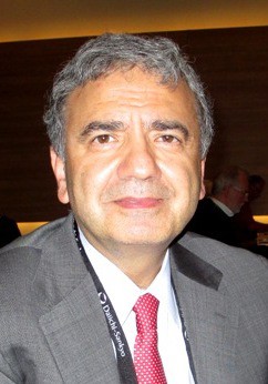
Lung cancer screening: The USPSTF's latest proposal

Pearls in clinical diagnosis of pertussis
VAIL, COLO. – One of the most useful signs that a young infant with an afebrile coughing illness has pertussis is the combination of an elevated white blood cell count of 20,000 cells/mcL or more plus a lymphocyte count of at least 10,000 cells/mcL.
"This is a pediatric pearl. It’s really a poor man’s way of diagnosing pertussis. It’s an effect of the pertussis toxin spreading throughout the neonate’s body, causing a very high white cell count with absolute lymphocytosis," Dr. Ann-Christine Nyquist said at a conference on pediatric infectious diseases sponsored by the Children’s Hospital Colorado.
This clinical pearl is part of a highly useful algorithm put forth by the Global Pertussis Initiative in an effort to update and standardize the case definitions of pertussis. Existing case definitions were developed more than 40 years ago and have numerous shortcomings.
The group developed a three-pronged, age-based algorithm, reflecting an understanding that the key manifestations of pertussis are different in infants aged 0-3 months, children aged 4 months to 9 years, and adolescents or adults.
According to the algorithm, the presence of an elevated WBC count with absolute lymphocytosis in an infant up to 3 months old with an afebrile illness and a cough of less than 3 weeks’ duration that’s increasing in frequency and severity is "virtually diagnostic" of pertussis (Clin. Infect. Dis. 2012;54:1756-64).
Another key feature of pertussis – and this one applies across the age spectrum, from infants to adults – is that the coryza remains watery and doesn’t become purulent, unlike in most viral respiratory infections.
"That green snotty nose doesn’t usually happen when kids have pertussis," explained Dr. Nyquist, professor of pediatrics at the University of Colorado, Denver.
Similarly, in patients of all ages the pertussis cough, even as it worsens, does not become truly productive.
To help nail down the diagnosis of pertussis in infants, the key question to ask parents is, "Is there an adult or adolescent in your family who’s had the most severe cough in their life?" Most infants with pertussis will have had close exposure to an older family member with a prolonged afebrile coughing illness, Dr. Nyquist noted.
In the 4-month to 9-year-old age group, the cough becomes more paroxysmal. The key indicators of pertussis in this age group, according to the Global Pertussis Initiative algorithm, are worsening paroxysmal nonproductive cough of at least 7 days’ duration in an afebrile child with nonpurulent coryza.
This same triad – worsening paroxysmal nonproductive cough, afebrile illness, and nonpurulent coryza – also has high sensitivity and good specificity for the clinical diagnosis of pertussis in adolescents and adults. In addition, the algorithm highlights another useful clue to the diagnosis in patients in this age range: the occurrence of sweating episodes between coughing paroxysms.
In terms of laboratory diagnostics, real-time PCR and culture of nasopharyngeal mucus are most useful in the first 3 weeks after illness onset. Serology is a challenge because the results are influenced by the effects of vaccination; it shouldn’t be used to diagnose pertussis within 1 year following inoculation with any pertussis vaccine because it’s impossible to tell if a positive result represents a response to the vaccine or to infection.

Also, serology can’t distinguish between Bordetella pertussis and B. parapertussis infection. Most PCR tests can. It’s an important distinction because B. parapertussis turns out to be the pathogen in roughly 15% of cases of coughing illnesses similar to pertussis. B. parapertussis infection isn’t vaccine preventable, and its treatment hasn’t been well studied.
The expert consensus is that direct fluorescent antibody testing should be discouraged as a tool to diagnose pertussis because of its unreliable sensitivity and specificity. IgG anti–pertussis toxin ELISA testing is superior to IgA anti–pertussis toxin ELISA, which has a high false-negative rate, Dr. Nyquist observed.
She reported having no financial relationships with any commercial interests.
VAIL, COLO. – One of the most useful signs that a young infant with an afebrile coughing illness has pertussis is the combination of an elevated white blood cell count of 20,000 cells/mcL or more plus a lymphocyte count of at least 10,000 cells/mcL.
"This is a pediatric pearl. It’s really a poor man’s way of diagnosing pertussis. It’s an effect of the pertussis toxin spreading throughout the neonate’s body, causing a very high white cell count with absolute lymphocytosis," Dr. Ann-Christine Nyquist said at a conference on pediatric infectious diseases sponsored by the Children’s Hospital Colorado.
This clinical pearl is part of a highly useful algorithm put forth by the Global Pertussis Initiative in an effort to update and standardize the case definitions of pertussis. Existing case definitions were developed more than 40 years ago and have numerous shortcomings.
The group developed a three-pronged, age-based algorithm, reflecting an understanding that the key manifestations of pertussis are different in infants aged 0-3 months, children aged 4 months to 9 years, and adolescents or adults.
According to the algorithm, the presence of an elevated WBC count with absolute lymphocytosis in an infant up to 3 months old with an afebrile illness and a cough of less than 3 weeks’ duration that’s increasing in frequency and severity is "virtually diagnostic" of pertussis (Clin. Infect. Dis. 2012;54:1756-64).
Another key feature of pertussis – and this one applies across the age spectrum, from infants to adults – is that the coryza remains watery and doesn’t become purulent, unlike in most viral respiratory infections.
"That green snotty nose doesn’t usually happen when kids have pertussis," explained Dr. Nyquist, professor of pediatrics at the University of Colorado, Denver.
Similarly, in patients of all ages the pertussis cough, even as it worsens, does not become truly productive.
To help nail down the diagnosis of pertussis in infants, the key question to ask parents is, "Is there an adult or adolescent in your family who’s had the most severe cough in their life?" Most infants with pertussis will have had close exposure to an older family member with a prolonged afebrile coughing illness, Dr. Nyquist noted.
In the 4-month to 9-year-old age group, the cough becomes more paroxysmal. The key indicators of pertussis in this age group, according to the Global Pertussis Initiative algorithm, are worsening paroxysmal nonproductive cough of at least 7 days’ duration in an afebrile child with nonpurulent coryza.
This same triad – worsening paroxysmal nonproductive cough, afebrile illness, and nonpurulent coryza – also has high sensitivity and good specificity for the clinical diagnosis of pertussis in adolescents and adults. In addition, the algorithm highlights another useful clue to the diagnosis in patients in this age range: the occurrence of sweating episodes between coughing paroxysms.
In terms of laboratory diagnostics, real-time PCR and culture of nasopharyngeal mucus are most useful in the first 3 weeks after illness onset. Serology is a challenge because the results are influenced by the effects of vaccination; it shouldn’t be used to diagnose pertussis within 1 year following inoculation with any pertussis vaccine because it’s impossible to tell if a positive result represents a response to the vaccine or to infection.
Also, serology can’t distinguish between Bordetella pertussis and B. parapertussis infection. Most PCR tests can. It’s an important distinction because B. parapertussis turns out to be the pathogen in roughly 15% of cases of coughing illnesses similar to pertussis. B. parapertussis infection isn’t vaccine preventable, and its treatment hasn’t been well studied.
The expert consensus is that direct fluorescent antibody testing should be discouraged as a tool to diagnose pertussis because of its unreliable sensitivity and specificity. IgG anti–pertussis toxin ELISA testing is superior to IgA anti–pertussis toxin ELISA, which has a high false-negative rate, Dr. Nyquist observed.
She reported having no financial relationships with any commercial interests.
VAIL, COLO. – One of the most useful signs that a young infant with an afebrile coughing illness has pertussis is the combination of an elevated white blood cell count of 20,000 cells/mcL or more plus a lymphocyte count of at least 10,000 cells/mcL.
"This is a pediatric pearl. It’s really a poor man’s way of diagnosing pertussis. It’s an effect of the pertussis toxin spreading throughout the neonate’s body, causing a very high white cell count with absolute lymphocytosis," Dr. Ann-Christine Nyquist said at a conference on pediatric infectious diseases sponsored by the Children’s Hospital Colorado.
This clinical pearl is part of a highly useful algorithm put forth by the Global Pertussis Initiative in an effort to update and standardize the case definitions of pertussis. Existing case definitions were developed more than 40 years ago and have numerous shortcomings.
The group developed a three-pronged, age-based algorithm, reflecting an understanding that the key manifestations of pertussis are different in infants aged 0-3 months, children aged 4 months to 9 years, and adolescents or adults.
According to the algorithm, the presence of an elevated WBC count with absolute lymphocytosis in an infant up to 3 months old with an afebrile illness and a cough of less than 3 weeks’ duration that’s increasing in frequency and severity is "virtually diagnostic" of pertussis (Clin. Infect. Dis. 2012;54:1756-64).
Another key feature of pertussis – and this one applies across the age spectrum, from infants to adults – is that the coryza remains watery and doesn’t become purulent, unlike in most viral respiratory infections.
"That green snotty nose doesn’t usually happen when kids have pertussis," explained Dr. Nyquist, professor of pediatrics at the University of Colorado, Denver.
Similarly, in patients of all ages the pertussis cough, even as it worsens, does not become truly productive.
To help nail down the diagnosis of pertussis in infants, the key question to ask parents is, "Is there an adult or adolescent in your family who’s had the most severe cough in their life?" Most infants with pertussis will have had close exposure to an older family member with a prolonged afebrile coughing illness, Dr. Nyquist noted.
In the 4-month to 9-year-old age group, the cough becomes more paroxysmal. The key indicators of pertussis in this age group, according to the Global Pertussis Initiative algorithm, are worsening paroxysmal nonproductive cough of at least 7 days’ duration in an afebrile child with nonpurulent coryza.
This same triad – worsening paroxysmal nonproductive cough, afebrile illness, and nonpurulent coryza – also has high sensitivity and good specificity for the clinical diagnosis of pertussis in adolescents and adults. In addition, the algorithm highlights another useful clue to the diagnosis in patients in this age range: the occurrence of sweating episodes between coughing paroxysms.
In terms of laboratory diagnostics, real-time PCR and culture of nasopharyngeal mucus are most useful in the first 3 weeks after illness onset. Serology is a challenge because the results are influenced by the effects of vaccination; it shouldn’t be used to diagnose pertussis within 1 year following inoculation with any pertussis vaccine because it’s impossible to tell if a positive result represents a response to the vaccine or to infection.
Also, serology can’t distinguish between Bordetella pertussis and B. parapertussis infection. Most PCR tests can. It’s an important distinction because B. parapertussis turns out to be the pathogen in roughly 15% of cases of coughing illnesses similar to pertussis. B. parapertussis infection isn’t vaccine preventable, and its treatment hasn’t been well studied.
The expert consensus is that direct fluorescent antibody testing should be discouraged as a tool to diagnose pertussis because of its unreliable sensitivity and specificity. IgG anti–pertussis toxin ELISA testing is superior to IgA anti–pertussis toxin ELISA, which has a high false-negative rate, Dr. Nyquist observed.
She reported having no financial relationships with any commercial interests.
EXPERT ANALYSIS FROM THE ANNUAL PEDIATRIC INFECTIOUS DISEASES CONFERENCE