User login
Bringing you the latest news, research and reviews, exclusive interviews, podcasts, quizzes, and more.
Powered by CHEST Physician, Clinician Reviews, MDedge Family Medicine, Internal Medicine News, and The Journal of Clinical Outcomes Management.
Failure to spot CHF leads to heart transplant
Failure to spot CHF leads to heart transplant
A 49-YEAR-OLD MAN SOUGHT TREATMENT AT AN URGENT CARE FACILITY after having shortness of breath every morning for 2 weeks. His heart rate was 119 beats/min, his blood pressure was 170/101 mm Hg, and he did not have chest pain. An electrocardiogram (EKG) was abnormal and chest x-ray showed fluid in the lung. The patient was diagnosed with pneumonia, prescribed antibiotics, and told to follow up with his physician. A follow-up chest x-ray 2 weeks later showed an enlarged heart and more fluid in the lung. A computed tomography scan indicated congestive heart failure and an EKG showed signs of a heart attack. The patient underwent a heart transplant and requires immunosuppressants.
PLAINTIFF'S CLAIM If the physician at the urgent care facility had noticed the patient’s enlarged heart, there would have been less heart damage, and the patient might have required a bypass, rather than a transplant.
THE DEFENSE No information about the defense is available.
VERDICT $1 million New Jersey verdict.
COMMENT When evaluating shortness of breath, always think lungs and heart until you have a definite diagnosis. Remember that neurological disease can present with shortness of breath, too. Consider amyotrophic lateral sclerosis, Guillain-Barré syndrome, and myasthenia gravis.
Infant suffers brain injury after delayed lab results
PARENTS BROUGHT THEIR 2-WEEK-OLD DAUGHTER TO THE EMERGENCY DEPARTMENT (ED) after she had missed several feedings and was short of breath. The ED physician ordered blood tests, but discharged the patient before receiving the results and told the parents to follow up with the infant’s pediatrician. Blood work subsequently revealed that the child had a Group B streptococcus infection, but by the time these results were communicated to the parents and treatment had begun, the infant had developed meningitis. She suffered brain injury, and was diagnosed with cerebral palsy.
PLAINTIFF'S CLAIM There was a delay in the diagnosis and treatment of the infant. Blood test results showing a bacterial infection were available the morning after discharge, but instead of notifying the parents, an additional blood culture was ordered to determine the type of bacteria present. The parents were then contacted 6 hours after the bacteria was identified as Group B streptococcus.
THE DEFENSE The defendants denied any negligence, although a nurse who cared for the infant claimed she had expressed concerns about the decision to discharge the patient.
VERDICT $7.15 million Maryland verdict.
COMMENT In newborns, the differential diagnosis for shortness of breath widens to include infection. In this case, I suspect the problem was a lack of tight follow-up, which can lead to bad outcomes—especially in newborns.
Suctioning neonates at birth: Time to change our approach
Stop suctioning neonates at birth. There is no benefit to this practice, and it can cause bradycardia and apnea. Instead, wipe the baby’s mouth and nose with a towel to clear excess secretions and stimulate respiration.1
Strength of recommendation
B: Based on a single randomized equivalency trial.
Kelleher J, Bhat, R, Salas AA, et al. Oronasopharyngeal suction versus wiping of the mouth and nose at birth: a randomised equivalency trial. Lancet. 2013;382:326-330.
Illustrative case
A healthy neonate is born through clear amniotic fluid with no meconium. She is vigorous and has no major congenital anomalies. Does she need oronasopharyngeal suctioning?
No, she does not need suctioning. Although it is still standard practice to perform oronasopharyngeal suctioning with a bulb syringe immediately after delivery, multiple studies have found no benefit to routine suctioning.2-7 Guidelines from the Neonatal Resuscitation Program (NRP) and other organizations recommend against the practice, even for neonates born through meconium-stained amniotic fluid.8,9 Suctioning is done because some clinicians believe it reduces the risk of aspiration, especially if there is meconium, and to stimulate breathing, but the evidence suggests that suctioning can stimulate the vagus nerve, which can lead to bradycardia.2 Studies that compared babies who did and didn’t receive suctioning found that those who received it had lower Apgar scores and oxygen saturation levels.2-4
Wiping the neonate’s mouth and nose with a towel is an alternative to suctioning, but until now no trials have compared the outcomes of these 2 methods. Kelleher et al1 conducted an equivalency trial to determine if wiping the mouth and nose is as effective as oronasopharyngeal suctioning.
STUDY SUMMARY: No difference in breathing after wiping or suctioning
Of 506 neonates randomized, 15 were excluded because they were not vigorous and had meconium-stained fluid, and 3 were excluded when their parents withdrew consent. Baseline characteristics for the 2 groups—including maternal age, presence of chronic medical conditions, and body mass index; vaginal vs cesarean delivery; umbilical artery pH; and neonatal sex, ethnic origin, and birth weight—were similar.
In the first 24 hours after birth, the average respiratory rate in the wiping group was 51 breaths/min (standard deviation [SD] ± 8) vs 50 breaths/min (SD ± 6) in the suctioning group. There was no difference in respiratory rates between the 2 groups at 1, 8, or 16 hours after birth. There was also no difference between the 2 groups in Apgar scores or need for advanced resuscitation. More neonates in the wiping group than in the suctioning group were admitted to the neonatal intensive care unit (45 of 246 [18%] vs 30 of 242 [12%]; P=.07), but the study was not powered to assess this outcome.
WHAT'S NEW: Wiping is as effective as suctioning, but there are no adverse effects
This study gives us evidence that wiping the face, mouth, and nose is equivalent to suctioning newborns at delivery, and it supports the NRP recommendation against routine suctioning in vigorous neonates born at term. Wiping avoids the potential adverse effects on the respiratory mucosa, bradycardia, and lower Apgar scores associated with suctioning via bulb syringes.
CAVEATS: Wiping is not best if a neonate’s airway is obstructed
This study looked only at neonates born after 35 weeks’ gestation who did not have meconium-stained amniotic fluid or congenital abnormalities. Also, NRP guidelines do recommend clearing the airways with a bulb syringe or suction catheter if airway obstruction is evident or positive-pressure ventilation is required.8
Another caveat ... In this study,1 there were 98 treatment crossovers: 64 of the 246 neonates in the wiping group received suctioning, and 34 of the 242 neonates in the suctioning group received wiping. However, this was not likely to change the study’s overall conclusion because a per-treatment analysis also found that wiping and suctioning were equivalent.
CHALLENGES TO IMPLEMENTATION: “We’ve always done it this way”
Practice patterns in a delivery room can be difficult to change. As we work on improving our delivery room environment and changing ingrained habits, the evidence from this study should help support the use of wiping in place of suctioning. The transition from suctioning to wiping also would be facilitated by having easily accessible towels designated for wiping.
Acknowledgement
The PURLs Surveillance System was supported in part by Grant Number UL1RR024999 from the National Center For Research Resources, a Clinical Translational Science Award to the University of Chicago. The content is solely the responsibility of the authors and does not necessarily represent the official views of the National Center For Research Resources or the National Institutes of Health.
1. Kelleher J, Bhat R, Salas AA, et al. Oronasopharyngeal suction versus wiping of the mouth and nose at birth: a randomised equivalency trial. Lancet. 2013;382:326-330.
2. Gungor S, Kurt E, Teksoz E, et al. Oronasopharyngeal suction versus no suction in normal and term infants delivered by elective cesarean section: a prospective randomized controlled trial. Gynecol Obstet Invest. 2006;61:9-14.
3. Gungor S, Teksoz E, Ceyhan T, et al. Oronasopharyngeal suction versus no suction in normal, term and vaginally born infants: a prospective randomized controlled trial. Aust N Z J Obstet Gynaecol. 2005;45:453-456.
4. Carrasco M, Martell M, Estol PC. Oronasopharyngeal suction at birth: effects on arterial oxygen saturation. J Pediatr. 1997;130:832-834.
5. Estol PC, Piriz H, Basalo S, et al. Oro-naso-pharyngeal suction at birth: effects on respiratory adaptation of normal term vaginally born infants. J Perinat Med. 1992;20:297-305.
6. Wiswell TE, Gannon CM, Jacob J, et al. Delivery room management of the apparently vigorous meconium-stained neonate: results of the multicenter, international collaborative trial. Pediatrics. 2000;105(1 pt 1):1-7.
7. Vain NE, Szyld EG, Prudent LM, et al. Oropharyngeal and nasopharyngeal suctioning of meconium-stained neonates before delivery of their shoulders: multicentre, randomized controlled trial. Lancet. 2004;364:597-602.
8. Kattwinkel J, Perlman JM, Aziz K, et al. Part 15: neonatal resuscitation: 2010 American Heart Association guidelines for cardiopulmonary resuscitation and emergency cardiovascular care. Circulation. 2010;122(18 suppl 3):S909-S919.
9. Perlman JM, Wyllie J, Kattwinkel J, et al; Neonatal Resuscitation Chapter Collaborators. Neonatal resuscitation: 2010 International Consensus on Cardiopulmonary Resuscitation and Emergency Cardiovascular Care Science with Treatment Recommendations. Pediatrics. 2010;126:e1319-1344.
Stop suctioning neonates at birth. There is no benefit to this practice, and it can cause bradycardia and apnea. Instead, wipe the baby’s mouth and nose with a towel to clear excess secretions and stimulate respiration.1
Strength of recommendation
B: Based on a single randomized equivalency trial.
Kelleher J, Bhat, R, Salas AA, et al. Oronasopharyngeal suction versus wiping of the mouth and nose at birth: a randomised equivalency trial. Lancet. 2013;382:326-330.
Illustrative case
A healthy neonate is born through clear amniotic fluid with no meconium. She is vigorous and has no major congenital anomalies. Does she need oronasopharyngeal suctioning?
No, she does not need suctioning. Although it is still standard practice to perform oronasopharyngeal suctioning with a bulb syringe immediately after delivery, multiple studies have found no benefit to routine suctioning.2-7 Guidelines from the Neonatal Resuscitation Program (NRP) and other organizations recommend against the practice, even for neonates born through meconium-stained amniotic fluid.8,9 Suctioning is done because some clinicians believe it reduces the risk of aspiration, especially if there is meconium, and to stimulate breathing, but the evidence suggests that suctioning can stimulate the vagus nerve, which can lead to bradycardia.2 Studies that compared babies who did and didn’t receive suctioning found that those who received it had lower Apgar scores and oxygen saturation levels.2-4
Wiping the neonate’s mouth and nose with a towel is an alternative to suctioning, but until now no trials have compared the outcomes of these 2 methods. Kelleher et al1 conducted an equivalency trial to determine if wiping the mouth and nose is as effective as oronasopharyngeal suctioning.
STUDY SUMMARY: No difference in breathing after wiping or suctioning
Of 506 neonates randomized, 15 were excluded because they were not vigorous and had meconium-stained fluid, and 3 were excluded when their parents withdrew consent. Baseline characteristics for the 2 groups—including maternal age, presence of chronic medical conditions, and body mass index; vaginal vs cesarean delivery; umbilical artery pH; and neonatal sex, ethnic origin, and birth weight—were similar.
In the first 24 hours after birth, the average respiratory rate in the wiping group was 51 breaths/min (standard deviation [SD] ± 8) vs 50 breaths/min (SD ± 6) in the suctioning group. There was no difference in respiratory rates between the 2 groups at 1, 8, or 16 hours after birth. There was also no difference between the 2 groups in Apgar scores or need for advanced resuscitation. More neonates in the wiping group than in the suctioning group were admitted to the neonatal intensive care unit (45 of 246 [18%] vs 30 of 242 [12%]; P=.07), but the study was not powered to assess this outcome.
WHAT'S NEW: Wiping is as effective as suctioning, but there are no adverse effects
This study gives us evidence that wiping the face, mouth, and nose is equivalent to suctioning newborns at delivery, and it supports the NRP recommendation against routine suctioning in vigorous neonates born at term. Wiping avoids the potential adverse effects on the respiratory mucosa, bradycardia, and lower Apgar scores associated with suctioning via bulb syringes.
CAVEATS: Wiping is not best if a neonate’s airway is obstructed
This study looked only at neonates born after 35 weeks’ gestation who did not have meconium-stained amniotic fluid or congenital abnormalities. Also, NRP guidelines do recommend clearing the airways with a bulb syringe or suction catheter if airway obstruction is evident or positive-pressure ventilation is required.8
Another caveat ... In this study,1 there were 98 treatment crossovers: 64 of the 246 neonates in the wiping group received suctioning, and 34 of the 242 neonates in the suctioning group received wiping. However, this was not likely to change the study’s overall conclusion because a per-treatment analysis also found that wiping and suctioning were equivalent.
CHALLENGES TO IMPLEMENTATION: “We’ve always done it this way”
Practice patterns in a delivery room can be difficult to change. As we work on improving our delivery room environment and changing ingrained habits, the evidence from this study should help support the use of wiping in place of suctioning. The transition from suctioning to wiping also would be facilitated by having easily accessible towels designated for wiping.
Acknowledgement
The PURLs Surveillance System was supported in part by Grant Number UL1RR024999 from the National Center For Research Resources, a Clinical Translational Science Award to the University of Chicago. The content is solely the responsibility of the authors and does not necessarily represent the official views of the National Center For Research Resources or the National Institutes of Health.
1. Kelleher J, Bhat R, Salas AA, et al. Oronasopharyngeal suction versus wiping of the mouth and nose at birth: a randomised equivalency trial. Lancet. 2013;382:326-330.
2. Gungor S, Kurt E, Teksoz E, et al. Oronasopharyngeal suction versus no suction in normal and term infants delivered by elective cesarean section: a prospective randomized controlled trial. Gynecol Obstet Invest. 2006;61:9-14.
3. Gungor S, Teksoz E, Ceyhan T, et al. Oronasopharyngeal suction versus no suction in normal, term and vaginally born infants: a prospective randomized controlled trial. Aust N Z J Obstet Gynaecol. 2005;45:453-456.
4. Carrasco M, Martell M, Estol PC. Oronasopharyngeal suction at birth: effects on arterial oxygen saturation. J Pediatr. 1997;130:832-834.
5. Estol PC, Piriz H, Basalo S, et al. Oro-naso-pharyngeal suction at birth: effects on respiratory adaptation of normal term vaginally born infants. J Perinat Med. 1992;20:297-305.
6. Wiswell TE, Gannon CM, Jacob J, et al. Delivery room management of the apparently vigorous meconium-stained neonate: results of the multicenter, international collaborative trial. Pediatrics. 2000;105(1 pt 1):1-7.
7. Vain NE, Szyld EG, Prudent LM, et al. Oropharyngeal and nasopharyngeal suctioning of meconium-stained neonates before delivery of their shoulders: multicentre, randomized controlled trial. Lancet. 2004;364:597-602.
8. Kattwinkel J, Perlman JM, Aziz K, et al. Part 15: neonatal resuscitation: 2010 American Heart Association guidelines for cardiopulmonary resuscitation and emergency cardiovascular care. Circulation. 2010;122(18 suppl 3):S909-S919.
9. Perlman JM, Wyllie J, Kattwinkel J, et al; Neonatal Resuscitation Chapter Collaborators. Neonatal resuscitation: 2010 International Consensus on Cardiopulmonary Resuscitation and Emergency Cardiovascular Care Science with Treatment Recommendations. Pediatrics. 2010;126:e1319-1344.
Copyright © 2014 Family Physicians Inquiries Network. All rights reserved.
Olodaterol LABA approved for some COPD patients
The Food and Drug Administration approved olodaterol inhalation spray to treat airflow obstruction in patients with chronic obstructive pulmonary disease.
The drug (Striverdi Respimat, by Boehringer Ingelheim Pharmaceuticals) can be used once daily over a long period of time but should not be used as a rescue therapy to treat sudden breathing problems or in patients with acutely deteriorating chronic obstructive pulmonary disease (COPD), according to an FDA statement.
"This new long-term maintenance medication provides an additional treatment option for the millions of Americans who suffer with COPD," Dr. Curtis Rosebraugh of the FDA said in the statement.
Olodaterol is a long-acting beta-adrenergic agonist (LABA) that relaxes muscles around the lungs’ airways and helps them stay relaxed to prevent symptoms. In controlled, 48-week clinical trials in 3,104 patients with diagnosed COPD, investigators found improved lung function in those on olodaterol, compared with placebo.
The FDA’s approval included a boxed warning that LABA medications increase the risk of asthma-related death. Olodaterol is not approved to treat asthma, and the safety and effectiveness of the drug in people with asthma have not been established.
Serious side effects from olodaterol can include narrowing and obstruction of the respiratory airway (paradoxical bronchospasm) and cardiovascular effects. The most common side effects reported in clinical trials are runny nose, upper respiratory tract infection, bronchitis, cough, urinary tract infection, dizziness, rash, diarrhea, back pain, and arthralgia.
On Twitter @sherryboschert
CDC targets West Africa Ebola outbreak
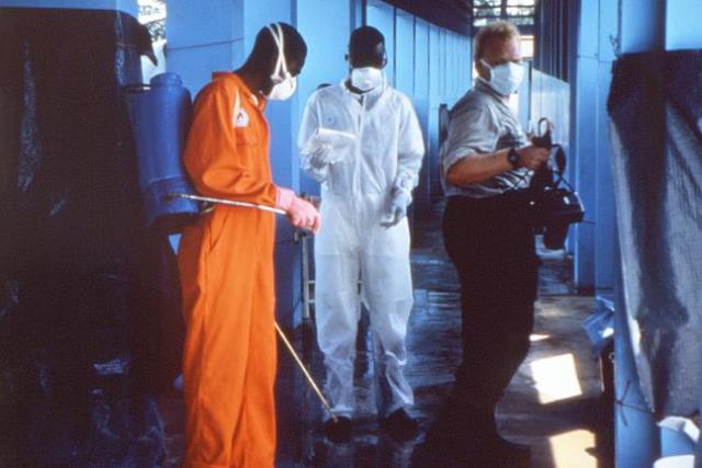
ATLANTA – The U.S. Centers for Disease Control and Prevention warned against nonessential travel to three West African countries facing the largest outbreak of Ebola virus in history and announced that it will send 50 more CDC personnel to assist those countries in the next 30 days.
The Ebola outbreak along the borders shared by Guinea, Liberia, and Sierra Leone is worsening, prompting the CDC to upgrade its response 4 days after it advised the public to take sensible, enhanced precautions when considering travel to that region.

The current outbreak is "the largest, most complex outbreak [of Ebola virus] that we know of in history," said Dr. Tom Frieden, director of the CDC. There is no known effective treatment for Ebola or vaccine against it.
The U.S. population is not at risk of an Ebola outbreak, however, because it has quarantine stations at all major ports of entry and isolation facilities in every hospital that has an intensive care unit, he said. The United States has a health care infrastructure better equipped to identify people who might have Ebola, rapidly test for the virus, monitor patients for signs of illness, and provide supportive care with infection control, Dr. Frieden added.
If health care workers see a patient who has a fever or other serious illness and has traveled to any of the three African countries recently, they should contact the CDC to arrange rapid testing and appropriate follow-up. An estimated 10,000 people travel between the United States and the three countries during a normal 3- or 4-month period, he said.
It’s important to note that Ebola is not spread by airborne routes, food, or water. People infected with Ebola are not contagious unless they are sick and someone comes in close contact with their infected bodily fluids. Anyone who may have been exposed to Ebolavirus (and all of their contacts) should be watched daily for 21 days to see if fever develops, Dr. Frieden said.
Many of the people who have died in the West African outbreak have been health care workers or people attending traditional funerals that include contact with the deceased person’s body.
The travel warning grew out of concern that people traveling to the West African countries might need medical attention for reasons unrelated to Ebola (such as appendicitis, a broken bone, or injuries sustained in a car accident), further stressing the local medical facilities and potentially exposing the travelers to Ebola when seeking care.
The surge in CDC infection control specialists headed to West Africa will help those countries develop the capacity to rapidly identify and isolate infected people and to screen travelers who are thinking of flying out of the country.
The CDC has worked with other African countries in the past to quell Ebola outbreaks through boots-on-the-ground infection control procedures. In Uganda, for example, working with traditional healers to recognize signs of Ebola and to make burial practices safer changed the environment so that "where there used to be large outbreaks, there are now single cases," Dr. Frieden said.
Quelling the West African outbreak would take at least 3-6 months under ideal conditions, but surely will take longer in West Africa, facing its first Ebola outbreak, he noted. The medical and public health infrastructures are limited and not used to dealing with Ebola, and public misconceptions about the virus have led to violence against some infection control workers.
"We already had one team that was confronted by an angry group of people and had to retreat across border to a safe country," Dr. Frieden said.
On Twitter @sherryboschert
ATLANTA – The U.S. Centers for Disease Control and Prevention warned against nonessential travel to three West African countries facing the largest outbreak of Ebola virus in history and announced that it will send 50 more CDC personnel to assist those countries in the next 30 days.
The Ebola outbreak along the borders shared by Guinea, Liberia, and Sierra Leone is worsening, prompting the CDC to upgrade its response 4 days after it advised the public to take sensible, enhanced precautions when considering travel to that region.
The current outbreak is "the largest, most complex outbreak [of Ebola virus] that we know of in history," said Dr. Tom Frieden, director of the CDC. There is no known effective treatment for Ebola or vaccine against it.
The U.S. population is not at risk of an Ebola outbreak, however, because it has quarantine stations at all major ports of entry and isolation facilities in every hospital that has an intensive care unit, he said. The United States has a health care infrastructure better equipped to identify people who might have Ebola, rapidly test for the virus, monitor patients for signs of illness, and provide supportive care with infection control, Dr. Frieden added.
If health care workers see a patient who has a fever or other serious illness and has traveled to any of the three African countries recently, they should contact the CDC to arrange rapid testing and appropriate follow-up. An estimated 10,000 people travel between the United States and the three countries during a normal 3- or 4-month period, he said.
It’s important to note that Ebola is not spread by airborne routes, food, or water. People infected with Ebola are not contagious unless they are sick and someone comes in close contact with their infected bodily fluids. Anyone who may have been exposed to Ebolavirus (and all of their contacts) should be watched daily for 21 days to see if fever develops, Dr. Frieden said.
Many of the people who have died in the West African outbreak have been health care workers or people attending traditional funerals that include contact with the deceased person’s body.
The travel warning grew out of concern that people traveling to the West African countries might need medical attention for reasons unrelated to Ebola (such as appendicitis, a broken bone, or injuries sustained in a car accident), further stressing the local medical facilities and potentially exposing the travelers to Ebola when seeking care.
The surge in CDC infection control specialists headed to West Africa will help those countries develop the capacity to rapidly identify and isolate infected people and to screen travelers who are thinking of flying out of the country.
The CDC has worked with other African countries in the past to quell Ebola outbreaks through boots-on-the-ground infection control procedures. In Uganda, for example, working with traditional healers to recognize signs of Ebola and to make burial practices safer changed the environment so that "where there used to be large outbreaks, there are now single cases," Dr. Frieden said.
Quelling the West African outbreak would take at least 3-6 months under ideal conditions, but surely will take longer in West Africa, facing its first Ebola outbreak, he noted. The medical and public health infrastructures are limited and not used to dealing with Ebola, and public misconceptions about the virus have led to violence against some infection control workers.
"We already had one team that was confronted by an angry group of people and had to retreat across border to a safe country," Dr. Frieden said.
On Twitter @sherryboschert
FROM A CDC PRESS CONFERENCE
Home sleep tests have lower initial cost, but less value, too
A sleep physician, when presented with probable obstructive sleep apnea, needs to study the patient to confirm diagnosis, then treat the patient. The hard part is the treatment. The physician needs to convince people to accept continuous positive airway pressure (CPAP) therapy.
Dr. Charles W. Atwood, FCCP, associate professor of medicine at the University of Pittsburgh, recently reported that in a controlled Veterans Affairs environment, over 2 years the cost of a home sleep test was $564 less than that of an in-lab study (CHEST Physician, July 2014 p. 27). It is no surprise that the test cost less; however, he also stated that there was no difference in clinical outcomes after 2 years of follow up.
I have practiced sleep medicine in a group of five physicians, all of whom are board certified in pulmonary disease and sleep medicine. Our studies are usually performed in an American Academy of Sleep Medicine–accredited eight-bed lab or a neighboring, accredited two-bed lab.
Whenever possible, we perform split-night studies on OSA patients. We utilize home sleep tests (HSTs) when appropriate, in accord with AASM guidelines. Our HST patients are taught how to wear the testing device and if the study is positive, patients are given a 3-day trial of autotitrating CPAP, after a mask fitting, individual instructions, and the opportunity to call for help, 24/7.
On the other hand, we sometimes see HST devices that have been mailed to patients; and if the study is positive, a CPAP machine is mailed. We see patients who have had an HST ordered by a non-sleep physician, and the treatment is left to the durable medical equipment company. The physician seems to assume that the DME supplier will appropriately fit patient with mask and follow patient’s compliance.
Unfortunately, this does not always happen. Patients may never be followed. Patients have appeared in our office, after 5 years of using CPAP, because they heard that there is a way to check compliance, or that they can get a new machine. Some patients’ masks are held together with masking tape and superglue; and some patients have significant ulcerations on the bridge of their nose because of improper mask fit.
In our five-physician practice of sleep medicine, we use the equivalent of a full-time staff person to interact with the DME companies on the patient’s behalf, and to correct problems with CPAP machines and masks.
Clearly, an HST without follow-up does not yield equivalent compliance compared with a lab split-night study. In the lab, the patient is introduced to CPAP by a compassionate sleep technologist, who is present during the titration to allay anxiety, to change masks as needed, and to reassure apprehensive patients. Without follow-up care, there is a much higher noncompliance rate.
Not only does the patient suffer when noncompliant with CPAP because of lack of adequate follow-up after an HST, but the bed partner’s sleep is disturbed, families are disturbed by the patient’s symptoms, job performance may suffer, and a sleepy driver is a potential danger to self and community. Payers may save dollars by paying for the HST per se; but without adequate follow-up of a patient after an HST, future health issues or accidents obviate any savings to the payer.
Conclusion: In a controlled setting, such as a Veterans Affairs hospital, where a sleep physician has control of testing, CPAP delivery, and follow-up, an HST may offer compliance similar to that of an in-lab study. However, in a community practice, even in a suburb where patients are well informed regarding their health issues, unless an HST is performed in a sleep center or by a physician knowledgeable in sleep medicine, who follows the patient and checks compliance, there is a major difference in the clinical success rate.
Without clinical follow-up of patient compliance, an HST per se may initially cost less, but the HST provides less value.
Dr. Cohen is a founding board member of the California Sleep Society and is in private practice in the San Francisco Bay area.

Evidence base is still missing in pediatric bronchiolitis care
A recent study titled "Racial/Ethnic differences in the presentation and management of severe bronchiolitis" investigates the multiple factors related to the presentation and management of bronchiolitis in the United States and hypothesizes that there are disparities among ethnic groups.
The study also expands on the concerns that the 2006 Academy of Pediatrics Bronchiolitis guidelines are not being followed (Pediatrics 2006;118:1774-93). The study brings up important topics for practitioners, epidemiologists, and insurance companies. Jonathan Santiago, MPH, and his colleagues at Yale University, New Haven, Conn., conclude that non-Hispanic black children were more likely to receive albuterol before admission and less likely to receive chest radiographs during hospitalization while Hispanic children are most likely to be discharged on inhaled corticosteroids.

However, it is important to review the study to understand the potential implications:
The study by Mr. Santiago, a medical student at Yale, and his colleagues has significant flaws with the inclusion and exclusion criteria. These flaws could have significantly affected the authors’ conclusions. Still, the research highlights important issues: "Why are clinicians not following the guidelines for all our U.S. infants and toddlers?" And, "Why do clinicians not use evidence-based medicine?" If clinicians are treating young children from ethnic groups differently, why is that happening?
The AAP guidelines were developed with the support of the American Academy of Family Physicians, the American College of Chest Physicians (CHEST), the American Thoracic Society, and the European Respiratory Society. The guidelines outline that clinicians should diagnose bronchiolitis and assess severity based on a standard history and physical. Chest radiographs should not be routinely ordered. The guidelines also recommend that a carefully monitored trial of alpha-adrenergic or beta-adrenergic medication is an option. However, inhaled bronchodilators should be continued only if there is a documented positive response. Among many other recommendations, corticosteroids are definitively not recommended for routine bronchiolitis treatment.
The Journal of Pediatrics recently published an article by Dr. Todd A. Florin of the Cincinnati Children’s Hospital and his colleagues on variation in the management of hospitalized infants with bronchiolitis (2014;pii: S0022-3476[14]00507-1 [doi:10.1016/j.jpeds.2014.05.057]). More than 60,000 hospitalizations were analyzed for infants aged 12 months and younger. After adjustment for patient characteristics, obtaining a chest radiograph was the one factor that had a great variation between hospitals. There was an 8.6% decrease in obtaining chest x-rays during the study period of 2007-2012. There also was wide variation among hospitals in regard to bronchodilator use, and there was no decrease in its use observed over the study period, despite the guidelines. Finally, a decrease of only 3.3% in corticosteroid use occurred during 2007-2012 – after the guidelines came out!
There is a theme: Family physicians, pediatricians, and other health care providers are not assessing and managing bronchiolitis using evidence-based medicine.
Mr. Santiago’s multicenter trial looked at 2,130 subjects and 24% were non-Hispanic blacks and 38% were Hispanic. Their median age was 4 months, while the mean age of the children in Dr. Florin’s study was 3.7 months. Many points of these studies can be teased out. For example, in Mr. Santiago’s study, non-Hispanic black children were more likely to receive albuterol before admission with an odds ratio of 1.58, and in the larger study by Dr. Florin, use of albuterol, in general, increased the patients’ length of stay. If Mr. Santiago’s study were expanded with stricter entry criteria and more hospitals, would a similar increased length of stay be found among non-Hispanic black children?
The guidelines are now 8 years old, and new guidelines are coming. But this important information, thoroughly analyzed by respected thought leaders, should be well disseminated among our peers. Our common goal should be to make sure that children at risk are not subjected to unnecessary x-rays, breathing treatments, and medications for bronchiolitis. The Hippocratic Oath, loosely translated, states: "I will prescribe regimens for the good of my patients according to my ability and my judgment and never do harm to anyone."
Dr. Millard is a pediatric pulmonologist at Helen DeVos Children’s Hospital (HDVCH) in Grand Rapids, Michigan, and an adviser for CHEST Physician. She is an associate professor of pediatrics and human development at Michigan State University and is the director of the pediatric pulmonary diagnostics laboratory at HDVCH and is in charge of clinical research for the division.
A recent study titled "Racial/Ethnic differences in the presentation and management of severe bronchiolitis" investigates the multiple factors related to the presentation and management of bronchiolitis in the United States and hypothesizes that there are disparities among ethnic groups.
The study also expands on the concerns that the 2006 Academy of Pediatrics Bronchiolitis guidelines are not being followed (Pediatrics 2006;118:1774-93). The study brings up important topics for practitioners, epidemiologists, and insurance companies. Jonathan Santiago, MPH, and his colleagues at Yale University, New Haven, Conn., conclude that non-Hispanic black children were more likely to receive albuterol before admission and less likely to receive chest radiographs during hospitalization while Hispanic children are most likely to be discharged on inhaled corticosteroids.
However, it is important to review the study to understand the potential implications:
The study by Mr. Santiago, a medical student at Yale, and his colleagues has significant flaws with the inclusion and exclusion criteria. These flaws could have significantly affected the authors’ conclusions. Still, the research highlights important issues: "Why are clinicians not following the guidelines for all our U.S. infants and toddlers?" And, "Why do clinicians not use evidence-based medicine?" If clinicians are treating young children from ethnic groups differently, why is that happening?
The AAP guidelines were developed with the support of the American Academy of Family Physicians, the American College of Chest Physicians (CHEST), the American Thoracic Society, and the European Respiratory Society. The guidelines outline that clinicians should diagnose bronchiolitis and assess severity based on a standard history and physical. Chest radiographs should not be routinely ordered. The guidelines also recommend that a carefully monitored trial of alpha-adrenergic or beta-adrenergic medication is an option. However, inhaled bronchodilators should be continued only if there is a documented positive response. Among many other recommendations, corticosteroids are definitively not recommended for routine bronchiolitis treatment.
The Journal of Pediatrics recently published an article by Dr. Todd A. Florin of the Cincinnati Children’s Hospital and his colleagues on variation in the management of hospitalized infants with bronchiolitis (2014;pii: S0022-3476[14]00507-1 [doi:10.1016/j.jpeds.2014.05.057]). More than 60,000 hospitalizations were analyzed for infants aged 12 months and younger. After adjustment for patient characteristics, obtaining a chest radiograph was the one factor that had a great variation between hospitals. There was an 8.6% decrease in obtaining chest x-rays during the study period of 2007-2012. There also was wide variation among hospitals in regard to bronchodilator use, and there was no decrease in its use observed over the study period, despite the guidelines. Finally, a decrease of only 3.3% in corticosteroid use occurred during 2007-2012 – after the guidelines came out!
There is a theme: Family physicians, pediatricians, and other health care providers are not assessing and managing bronchiolitis using evidence-based medicine.
Mr. Santiago’s multicenter trial looked at 2,130 subjects and 24% were non-Hispanic blacks and 38% were Hispanic. Their median age was 4 months, while the mean age of the children in Dr. Florin’s study was 3.7 months. Many points of these studies can be teased out. For example, in Mr. Santiago’s study, non-Hispanic black children were more likely to receive albuterol before admission with an odds ratio of 1.58, and in the larger study by Dr. Florin, use of albuterol, in general, increased the patients’ length of stay. If Mr. Santiago’s study were expanded with stricter entry criteria and more hospitals, would a similar increased length of stay be found among non-Hispanic black children?
The guidelines are now 8 years old, and new guidelines are coming. But this important information, thoroughly analyzed by respected thought leaders, should be well disseminated among our peers. Our common goal should be to make sure that children at risk are not subjected to unnecessary x-rays, breathing treatments, and medications for bronchiolitis. The Hippocratic Oath, loosely translated, states: "I will prescribe regimens for the good of my patients according to my ability and my judgment and never do harm to anyone."
Dr. Millard is a pediatric pulmonologist at Helen DeVos Children’s Hospital (HDVCH) in Grand Rapids, Michigan, and an adviser for CHEST Physician. She is an associate professor of pediatrics and human development at Michigan State University and is the director of the pediatric pulmonary diagnostics laboratory at HDVCH and is in charge of clinical research for the division.
New recommendations issued for palivizumab in RSV prophylaxis
New guidelines regarding the use of palivizumab to reduce the risk of respiratory syncytial virus now further restrict which infants should receive the prophylaxis. The new guidelines, issued by the American Academy of Pediatrics, replace the previous ones from 2012.
Approximately 2.1 million children under age 5 develop an RSV infection requiring medical care each year, including about 58,000 who are hospitalized during their first few years of life. The highest-risk age for RSV hospitalization is 2 months.
However, palivizumab (Synagis), an immunoglobulin monoclonal antibody, can reduce the risk of hospitalization for RSV. The standard prophylactic dose is 15 mg/kg every 30 days during RSV season, for up to five doses. Palivizumab does not interfere with immunizations and can be administered at the same time as vaccines.
The main changes in the updated guidelines reduce the number of infants who will qualify for the prophylaxis, according to the policy statement published online by the AAP Committee on Infectious Diseases, chaired by Red Book Associate Editor Michael T. Brady, and the AAP Bronchiolitis Guidelines Committee (Pediatrics 2014 July 28 [doi: 10.1542/peds.2014-1665]).
Whereas all otherwise healthy preterm infants born at less than 32 weeks were previously recommended to receive palivizumab, now only those born at less than 29 weeks are recommended to receive it.
However, recommendations for palivizumab remain in place for preterm infants younger than 32 weeks’ gestational age who have chronic lung disease of prematurity and needed more than 21% oxygen for at least the first 28 days after birth. In addition, as in the previous guidelines, these children should receive palivizumab only during their first year of life unless, in their second year, they still need medical support in the 6 months before RSV season starts. Medical support includes supplemental oxygen, chronic corticosteroid therapy, or diuretic therapy.
Infants less than 12 months old with hemodynamically significant congenital heart disease should still receive palivizumab, particularly if they have moderate to severe pulmonary hypertension or if they have acyanotic heart disease and either take medication to control congestive heart failure or else will need heart surgery. However, children older than 12 months are no longer recommended to receive the prophylaxis (previous guidelines went up to 24 months), nor are those who have hemodynamically insignificant heart disease, who have surgically corrected lesions, who do not need drugs for congestive heart failure, or who have mild cardiomyopathy but do not need therapy.
Unless they have another qualifying condition, children with Down syndrome or cystic fibrosis also are not recommended to receive palivizumab, even though some evidence shows that these children may be at higher risk for RSV. The 2012 guidelines did not have a recommendation for or against palivizumab for children with cystic fibrosis, but data from studies since then bolster the case to exclude these children from receiving the prophylaxis because of limited evidence of clinical benefit.
Further, a group no longer recommended to receive palivizumab are those born between 32 and 35 weeks who were born within 3 months of RSV season and either attended childcare or had a sibling under age 5.
As in 2012, palivizumab "may be considered" for two groups of children: those less than 24 months old undergoing chemotherapy or otherwise severely immunocompromised, and those with pulmonary abnormalities or neuromuscular disorders that prevent them from coughing sufficiently to clear upper-airway secretions.
Another change in the updated recommendations is to cease prophylactic doses of palivizumab if a child is hospitalized with a breakthrough RSV infection. Previous guidelines advised that doses continue through the maximum recommended amount, but this has been removed in the new statement because fewer than 0.5% of infants are rehospitalized with RSV in the same season. Palivizumab is still not recommended for prevention of health care–associated RSV or to treat RSV.
The updated guidelines are based on new data, including revised (lower) estimates of RSV mortality in hospitalized children, information on RSV seasonality, declining bronchiolitis hospitalizations, data on palivizumab benefits for children with cystic fibrosis or Down syndrome, and "reports describing palivizumab-resistant RSV isolates from hospitalized patients who receive prophylaxis." These data are summarized in the technical report by the AAP Committee on Infectious Diseases (Pediatrics 2014 July 28 [doi: 10.1542/peds.2014-1666]).
Palivizumab was licensed by the Food and Drug Administration in June 1998, largely based on the results of a randomized controlled trial in 1996-1997 involving 1,501 preterm infants and young children (some with chronic lung disease of prematurity). A second randomized controlled trial was performed in 1998-2002 involving 1,287 children with hemodynamically significant congenital heart disease.
No external funding was used to develop the guidelines. No disclosures were reported for the members of the AAP Committee on Infectious Diseases.
To many of us in the northern hemisphere, January through March is "flu season." To pediatricians, it’s bronchiolitis season. We wish it weren’t; about one-third of all hospitalizations in children under 5 years are due to bronchiolitis, specifically to respiratory syncytial virus.
Life would be so much easier for all of us, let alone infants, if we could prevent RSV disease. And we can: Palivizumab (Synagis) has been passively preventing RSV since 1998. Its use has been restricted by its cost: If a season’s course of palivizumab cost $5 or even $50 instead of a yearly cost of upward of $5,000 (including the cost of administration), discussion of its use would be much different.

|
|
Some argue that other physicians don’t watch costs, so why should we? If we can prevent a nasty disease, why shouldn’t we, at any price? Where bronchiolitis is especially common, severe, and costly, such as Alaska’s Yukon-Kuskokwim Delta, it has been proposed that palivizumab be given to all newborns. But it won’t be paid for, even there.
Insurance companies and Medicaid payers have long used AAP standards to determine which infants can be given this expensive preventive measure. It is very likely that they will seize upon these new standards as well, and we will have to follow suit. I don’t have many parents of patients in my practice who will choose to pay out of pocket for palivizumab.
The new guidelines further restrict the use of palivizumab, continuing a process of restriction as more information has become available over the past 16 years. Hospitalization for bronchiolitis has become less common, even in premature infants. The technical report clearly lays out the committee’s reasoning and the new data underlying it. Many practitioners will be unhappy with the changes. Some of our patients will be hospitalized with RSV bronchiolitis when previous guidelines would have had them receiving palivizumab. Some of our patients will also die from having driven coupes instead of Panzer tanks. I’ll trust the careful analysis of the Committee on Infectious Diseases.
Lance Chilton, M.D., is a professor of pediatrics at the University of New Mexico in Albuquerque and a pediatrician at Young Children’s Health Center. He is on the speakers bureau for Merck.
To many of us in the northern hemisphere, January through March is "flu season." To pediatricians, it’s bronchiolitis season. We wish it weren’t; about one-third of all hospitalizations in children under 5 years are due to bronchiolitis, specifically to respiratory syncytial virus.
Life would be so much easier for all of us, let alone infants, if we could prevent RSV disease. And we can: Palivizumab (Synagis) has been passively preventing RSV since 1998. Its use has been restricted by its cost: If a season’s course of palivizumab cost $5 or even $50 instead of a yearly cost of upward of $5,000 (including the cost of administration), discussion of its use would be much different.
|
|
|
Some argue that other physicians don’t watch costs, so why should we? If we can prevent a nasty disease, why shouldn’t we, at any price? Where bronchiolitis is especially common, severe, and costly, such as Alaska’s Yukon-Kuskokwim Delta, it has been proposed that palivizumab be given to all newborns. But it won’t be paid for, even there.
Insurance companies and Medicaid payers have long used AAP standards to determine which infants can be given this expensive preventive measure. It is very likely that they will seize upon these new standards as well, and we will have to follow suit. I don’t have many parents of patients in my practice who will choose to pay out of pocket for palivizumab.
The new guidelines further restrict the use of palivizumab, continuing a process of restriction as more information has become available over the past 16 years. Hospitalization for bronchiolitis has become less common, even in premature infants. The technical report clearly lays out the committee’s reasoning and the new data underlying it. Many practitioners will be unhappy with the changes. Some of our patients will be hospitalized with RSV bronchiolitis when previous guidelines would have had them receiving palivizumab. Some of our patients will also die from having driven coupes instead of Panzer tanks. I’ll trust the careful analysis of the Committee on Infectious Diseases.
Lance Chilton, M.D., is a professor of pediatrics at the University of New Mexico in Albuquerque and a pediatrician at Young Children’s Health Center. He is on the speakers bureau for Merck.
New guidelines regarding the use of palivizumab to reduce the risk of respiratory syncytial virus now further restrict which infants should receive the prophylaxis. The new guidelines, issued by the American Academy of Pediatrics, replace the previous ones from 2012.
Approximately 2.1 million children under age 5 develop an RSV infection requiring medical care each year, including about 58,000 who are hospitalized during their first few years of life. The highest-risk age for RSV hospitalization is 2 months.
However, palivizumab (Synagis), an immunoglobulin monoclonal antibody, can reduce the risk of hospitalization for RSV. The standard prophylactic dose is 15 mg/kg every 30 days during RSV season, for up to five doses. Palivizumab does not interfere with immunizations and can be administered at the same time as vaccines.
The main changes in the updated guidelines reduce the number of infants who will qualify for the prophylaxis, according to the policy statement published online by the AAP Committee on Infectious Diseases, chaired by Red Book Associate Editor Michael T. Brady, and the AAP Bronchiolitis Guidelines Committee (Pediatrics 2014 July 28 [doi: 10.1542/peds.2014-1665]).
Whereas all otherwise healthy preterm infants born at less than 32 weeks were previously recommended to receive palivizumab, now only those born at less than 29 weeks are recommended to receive it.
However, recommendations for palivizumab remain in place for preterm infants younger than 32 weeks’ gestational age who have chronic lung disease of prematurity and needed more than 21% oxygen for at least the first 28 days after birth. In addition, as in the previous guidelines, these children should receive palivizumab only during their first year of life unless, in their second year, they still need medical support in the 6 months before RSV season starts. Medical support includes supplemental oxygen, chronic corticosteroid therapy, or diuretic therapy.
Infants less than 12 months old with hemodynamically significant congenital heart disease should still receive palivizumab, particularly if they have moderate to severe pulmonary hypertension or if they have acyanotic heart disease and either take medication to control congestive heart failure or else will need heart surgery. However, children older than 12 months are no longer recommended to receive the prophylaxis (previous guidelines went up to 24 months), nor are those who have hemodynamically insignificant heart disease, who have surgically corrected lesions, who do not need drugs for congestive heart failure, or who have mild cardiomyopathy but do not need therapy.
Unless they have another qualifying condition, children with Down syndrome or cystic fibrosis also are not recommended to receive palivizumab, even though some evidence shows that these children may be at higher risk for RSV. The 2012 guidelines did not have a recommendation for or against palivizumab for children with cystic fibrosis, but data from studies since then bolster the case to exclude these children from receiving the prophylaxis because of limited evidence of clinical benefit.
Further, a group no longer recommended to receive palivizumab are those born between 32 and 35 weeks who were born within 3 months of RSV season and either attended childcare or had a sibling under age 5.
As in 2012, palivizumab "may be considered" for two groups of children: those less than 24 months old undergoing chemotherapy or otherwise severely immunocompromised, and those with pulmonary abnormalities or neuromuscular disorders that prevent them from coughing sufficiently to clear upper-airway secretions.
Another change in the updated recommendations is to cease prophylactic doses of palivizumab if a child is hospitalized with a breakthrough RSV infection. Previous guidelines advised that doses continue through the maximum recommended amount, but this has been removed in the new statement because fewer than 0.5% of infants are rehospitalized with RSV in the same season. Palivizumab is still not recommended for prevention of health care–associated RSV or to treat RSV.
The updated guidelines are based on new data, including revised (lower) estimates of RSV mortality in hospitalized children, information on RSV seasonality, declining bronchiolitis hospitalizations, data on palivizumab benefits for children with cystic fibrosis or Down syndrome, and "reports describing palivizumab-resistant RSV isolates from hospitalized patients who receive prophylaxis." These data are summarized in the technical report by the AAP Committee on Infectious Diseases (Pediatrics 2014 July 28 [doi: 10.1542/peds.2014-1666]).
Palivizumab was licensed by the Food and Drug Administration in June 1998, largely based on the results of a randomized controlled trial in 1996-1997 involving 1,501 preterm infants and young children (some with chronic lung disease of prematurity). A second randomized controlled trial was performed in 1998-2002 involving 1,287 children with hemodynamically significant congenital heart disease.
No external funding was used to develop the guidelines. No disclosures were reported for the members of the AAP Committee on Infectious Diseases.
FROM PEDIATRICS
Adenotonsillectomy for OSAS led to weight gain, even in overweight children
Adenotonsillectomy for obstructive sleep apnea syndrome led to clinically significant weight gain, even in children who were overweight at baseline, according to a first-in-kind randomized clinical trial published in the August issue of Pediatrics.
More than 52% of overweight children with obstructive sleep apnea syndrome (OSAS) who underwent adenotonsillectomy developed obesity within 7 months, compared with 21% of those randomized to watchful waiting and supportive care (P less than .05), reported Dr. Eliot S. Katz of Boston Children’s Hospital and his associates.
Based on those results, children who undergo adenotonsillectomy for OSAS should be monitored for weight changes, receive nutritional counseling, and be encouraged to exercise, the researchers said (Pediatrics 2014;134:282-9).
For the multicenter, single-blind clinical trial, the investigators randomized 464 children aged 5-9.9 years with OSAS to undergo either early adenotonsillectomy – defined as surgery within 4 weeks of randomization – or watchful waiting and supportive care.
Children classified as failing to thrive at baseline were more likely to attain a normal weight after adenotonsillectomy, compared with watchful waiting, although the difference was not statistically significant, Dr. Katz and his associates said.
The National Institutes of Health funded the study. One coauthor reported having served as a consultant for Zansors and receiving grants from Respironics/Phillips and Fisher-Paykel. The other authors reported no conflicts of interest.
FROM PEDIATRICS
Key clinical point: Adenotonsillectomy for obstructive sleep apnea syndrome increases the risk of obesity in overweight children.
Major finding: Almost 52% of overweight children randomized to adenotonsillectomy developed obesity in the subsequent 7 months, compared with 21% of overweight children in the control group (P less than .05).
Data source: Multicenter, single-blind, clinical trial of 464 children with OSAS randomized either to early adenotonsillectomy or watchful waiting and supportive care.
Disclosures: The National Institutes of Health funded the study. One coauthor reported having served as a consultant for Zansors and receiving grants from Respironics/Phillips and Fisher-Paykel. The other authors reported no conflicts of interest.
Prevention program led to steep drop in postop pneumonia cases
A quality improvement program cut postoperative pneumonia rates by 43.6%, investigators reported July 23 in JAMA Surgery.
The small, single-center retrospective cohort study is the first to present long-term findings for such a program, said Dr. Hadiza Kazaure at Stanford (Calif.) University and her associates.

The program, which investigators started at a Stanford-affiliated Veterans Affairs hospital, included coughing and deep-breathing exercises with incentive spirometry, a twice-daily oral chlorhexidine hygiene, ambulation with pain control, elevating the head of the bed by 30 degrees and sitting for meals, educating surgical nursing staff about their roles in preventing pneumonia, and automating physician orders for pneumonia prevention, the researchers said (JAMA Surg. 2014 July 23 [doi:10.1001/jamasurg.2014.1216]).
The investigators calculated postimplementation rates of ward-acquired pneumonia in all noncardiac, unventilated surgical patients in the 5 years after the program started, they said.
Among 4,099 hospitalized patients who were at risk, 18 (0.44%) developed postoperative pneumonia, a 43.6% drop from the preprogram rate of 0.78%, the investigators reported.
By comparison, the postintervention rate for the American College of Surgeons National Surgical Quality Improvement Program (ACS-NSQIP) was 582% higher than that of the VA hospital for 2008-2012. By achieving the VA hospital program’s results, the hospitals in the ACS-NSQIP could save about $280 million, Dr. Kazaure and her associates said.
The database abstraction methods used to choose patients were validated, but nonetheless could have caused selection errors. Also, few patients developed pneumonia, which kept the investigators from tracking trends over time on the ward, they said. "Based on published data, we used a national average of $46,400 cost of care per postoperative pneumonia case to estimate potential cost savings in our analyses," they added. "The validity of cost estimates in this study to individual hospitals must be considered because geographic variation in cost of care is well established."
The authors reported no funding sources or conflicts of interest.
The "remarkably low" baseline rate of postoperative pneumonia "calls into question the adequacy of detection and reporting of pneumonia" on the surgical ward, making findings "of somewhat less consequence," Dr. Catherine Lewis said.
"Moreover, the authors’ program was implemented and evaluated over a period in which there was an ongoing shift from open to laparoscopic or robotic surgical techniques, which has been shown to significantly decrease the rates of postoperative pneumonia, particularly in colorectal and prostate operations," she said.
The authors did not examine whether changes in patient care or surgical technique affected postoperative pneumonia rates, Dr. Lewis noted. She pointed to other research showing that using a neuraxial blockade instead of general anesthesia led to a significant drop in postoperative pneumonia, as did using selective nasogastric decompression (Ann. Intern. Med. 2006;144:596-608).
Previously, the authors of the current study reported that postoperative non–ventilator-associated pneumonia rates did not change after they started their program, Dr. Lewis said. The number of ward cases decreased, but the number of non–ventilator-associated pneumonia intensive care unit cases increased, so the decrease could reflect redistribution in the location of patients, she said.
But the authors also deserved praise for developing and starting a relatively cost- and time-efficient quality improvement measure, Dr. Lewis added. She said that such programs could increase awareness and education among health care providers who care for patients after surgery.
Dr. Lewis specializes in general, critical care, and trauma surgery at the University of California, Los Angeles. The remarks were excerpted from her invited editorial accompanying Dr. Kazaure’s report (JAMA Surg. 2014 July 23 [doi:10.1001/jamasurg.2014.1249]).
A quality improvement program cut postoperative pneumonia rates by 43.6%, investigators reported July 23 in JAMA Surgery.
The small, single-center retrospective cohort study is the first to present long-term findings for such a program, said Dr. Hadiza Kazaure at Stanford (Calif.) University and her associates.
The program, which investigators started at a Stanford-affiliated Veterans Affairs hospital, included coughing and deep-breathing exercises with incentive spirometry, a twice-daily oral chlorhexidine hygiene, ambulation with pain control, elevating the head of the bed by 30 degrees and sitting for meals, educating surgical nursing staff about their roles in preventing pneumonia, and automating physician orders for pneumonia prevention, the researchers said (JAMA Surg. 2014 July 23 [doi:10.1001/jamasurg.2014.1216]).
The investigators calculated postimplementation rates of ward-acquired pneumonia in all noncardiac, unventilated surgical patients in the 5 years after the program started, they said.
Among 4,099 hospitalized patients who were at risk, 18 (0.44%) developed postoperative pneumonia, a 43.6% drop from the preprogram rate of 0.78%, the investigators reported.
By comparison, the postintervention rate for the American College of Surgeons National Surgical Quality Improvement Program (ACS-NSQIP) was 582% higher than that of the VA hospital for 2008-2012. By achieving the VA hospital program’s results, the hospitals in the ACS-NSQIP could save about $280 million, Dr. Kazaure and her associates said.
The database abstraction methods used to choose patients were validated, but nonetheless could have caused selection errors. Also, few patients developed pneumonia, which kept the investigators from tracking trends over time on the ward, they said. "Based on published data, we used a national average of $46,400 cost of care per postoperative pneumonia case to estimate potential cost savings in our analyses," they added. "The validity of cost estimates in this study to individual hospitals must be considered because geographic variation in cost of care is well established."
The authors reported no funding sources or conflicts of interest.
FROM JAMA SURGERY
Key clinical point: Cost-effective quality improvement programs can potentially prevent postoperative pneumonia in unventilated patients.
Major finding: Among 4,099 hospitalized patients at risk between 2008 and 2012, 18 (0.44%) developed postoperative pneumonia, a 43.6% decrease from the preintervention rate of 0.78%.
Data source: Retrospective cohort study of all noncardiac surgical patients who acquired postoperative pneumonia on a Veterans Affairs hospital ward from 2008 to 2012.
Disclosures: The authors reported no funding sources or conflicts of interest.

Sleep Society: Screen for Apnea at First Medicare Visit
MINNEAPOLIS – The American Academy of Sleep Medicine is pushing to have a simple sleep apnea questionnaire included in the initial Welcome to Medicare preventive care visit.
Including such a screening tool would help identify obstructive sleep apnea (OSA) when patients first join the Medicare program and thus improve the odds of diagnosing and treating the condition, said Dr. Timothy Morgenthaler, president of the AASM. Getting a handle on OSA could also reduce the potential that the beneficiary will develop related chronic conditions, and that will help Medicare curb expenditures, he said.

An estimated 20% of current Medicare beneficiaries have OSA. That number is expected to grow with the rising obesity rates, he said. Untreated OSA can increase the risk of hypertension, heart disease, type 2 diabetes, and stroke, said Dr. Morgenthaler, who is professor of medicine at the Mayo Clinic in Rochester, Minn.
The AASM has been lobbying Congress to include a validated OSA screen in the initial Medicare visit and found sponsors in Rep. Michael Burgess (R-Tex.) and Rep. Bobby Rush (D-Ill.). The two congressmen introduced a bill (H.R. 4695) that would do just that on May 21.
"This important legislation addresses the barriers that prevent new Medicare beneficiaries from receiving what we know to be required sleep apnea services," Dr. Morgenthaler said at the annual meeting of the Associated Professional Sleep Societies.
Rep. Erik Paulsen (R-Minn.), who recently signed on to the bill as a cosponsor, told AASM attendees that adding an OSA screen to the initial Medicare visit would help increase detection of disease, raise patient awareness, and "improve health care quality and reduce costs to the Medicare program," over the long term.
The AASM is asking its members to back the legislation and educate local lawmakers and patients through the group’s Seniors Sleep Campaign.
The association also wants to make it easier for board-certified sleep medicine specialists to care for Medicare patients from start to finish. Currently, antikickback laws prevent sleep specialists and sleep centers from directly providing therapeutic durable medical equipment to Medicare patients, said Dr. Morgenthaler.
The AASM has developed model language for an exception to that statute, which it hopes legislators or regulators will approve, he said. It would allow board-certified specialists to provide the continuum of care from start to finish, including durable medical equipment such as continuous positive airway pressure devices.
Eliminating the current fragmented system of care would eliminate waste, simplify the work flow, and improve the quality of care and reduce costs, said Dr. Morgenthaler.
On Twitter @aliciaault
MINNEAPOLIS – The American Academy of Sleep Medicine is pushing to have a simple sleep apnea questionnaire included in the initial Welcome to Medicare preventive care visit.
Including such a screening tool would help identify obstructive sleep apnea (OSA) when patients first join the Medicare program and thus improve the odds of diagnosing and treating the condition, said Dr. Timothy Morgenthaler, president of the AASM. Getting a handle on OSA could also reduce the potential that the beneficiary will develop related chronic conditions, and that will help Medicare curb expenditures, he said.
An estimated 20% of current Medicare beneficiaries have OSA. That number is expected to grow with the rising obesity rates, he said. Untreated OSA can increase the risk of hypertension, heart disease, type 2 diabetes, and stroke, said Dr. Morgenthaler, who is professor of medicine at the Mayo Clinic in Rochester, Minn.
The AASM has been lobbying Congress to include a validated OSA screen in the initial Medicare visit and found sponsors in Rep. Michael Burgess (R-Tex.) and Rep. Bobby Rush (D-Ill.). The two congressmen introduced a bill (H.R. 4695) that would do just that on May 21.
"This important legislation addresses the barriers that prevent new Medicare beneficiaries from receiving what we know to be required sleep apnea services," Dr. Morgenthaler said at the annual meeting of the Associated Professional Sleep Societies.
Rep. Erik Paulsen (R-Minn.), who recently signed on to the bill as a cosponsor, told AASM attendees that adding an OSA screen to the initial Medicare visit would help increase detection of disease, raise patient awareness, and "improve health care quality and reduce costs to the Medicare program," over the long term.
The AASM is asking its members to back the legislation and educate local lawmakers and patients through the group’s Seniors Sleep Campaign.
The association also wants to make it easier for board-certified sleep medicine specialists to care for Medicare patients from start to finish. Currently, antikickback laws prevent sleep specialists and sleep centers from directly providing therapeutic durable medical equipment to Medicare patients, said Dr. Morgenthaler.
The AASM has developed model language for an exception to that statute, which it hopes legislators or regulators will approve, he said. It would allow board-certified specialists to provide the continuum of care from start to finish, including durable medical equipment such as continuous positive airway pressure devices.
Eliminating the current fragmented system of care would eliminate waste, simplify the work flow, and improve the quality of care and reduce costs, said Dr. Morgenthaler.
On Twitter @aliciaault
AT SLEEP 2014