User login
ENZAMET trial: Early enzalutamide delays progression, improves survival in mHSPC
CHICAGO – Adding the oral androgen receptor inhibitor enzalutamide to standard first-line testosterone suppression delays progression and improves survival in men with metastatic hormone-sensitive prostate cancer (mHSPC), according to “practice-informing” interim results from the randomized phase 3 ENZAMET trial.
The survival rate at 3 years in 563 men with mHSPC who were enrolled in the international trial and who received early testosterone suppression and enzalutamide was 80%, compared with 72% among 562 men who received testosterone suppression and standard nonsteroidal antiandrogen therapy with or without docetaxel, study cochair Christopher Sweeney, MBBS, reported at the annual meeting of the American Society of Clinical Oncology (Abstract LBA2).
The findings of the Australian and New Zealand Urogenital and Prostate (ANZUP) Cancer Trials Group study (ANZUP 1304/ENZAMET) were published simultaneously in the New England Journal of Medicine.
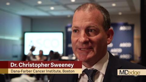
“So ... we’re moving forward by going backwards in the disease setting where the disease is more sensitive and responds better to therapy,” Dr. Sweeney, of Dana-Farber Cancer Institute’s Lank Center for Genitourinary Oncology and professor of medicine at Harvard Medical School, Boston, explained in this video interview.
He also described the future directions for the research – in particular the need for longer follow-up to clarify the effects of docetaxel in this setting – and how the current findings will be reflected in his own management of patients with prostate cancer.
The findings have immediate implications for practice, ASCO expert Neeraj Agarwal, MD, professor of medicine and investigator at the Huntsman Cancer Institute, University of Utah, Salt Lake City, said during a press briefing at the meeting.
“In my view, using enzalutamide early on will allow our patients to avoid chemotherapy and steroids for many years, and thus, hopefully, improve their quality of life,” he said, noting that the findings are particularly exciting when considered in the context of the “equally impressive margin of benefit” seen with the similar drug apalutamide in the TITAN trial, which was presented separately during the ASCO meeting.
“One study is encouraging, but two large studies ... demonstrating similar findings, is even better,” he said. “This increases my confidence that targeting [the androgen receptor] is the optimal approach for newly diagnosed patients with advanced prostate cancer.”
Dr. Sweeney reported relationships (stock and other ownership interests, consulting or advisory roles, research funding to his institution, and/or patents/royalties/other intellectual property) with Leuchemix, Amgen, Astellas Pharma, AstraZeneca, Bayer, Genentech/Roche, Janssen Biotech, Pfizer, Sanofi, Dendreon, Sotio, and Exelixis. Dr. Agarwal reported consultancy or research for Pfizer, Novartis, Exelixis, Eisai, Genentech, Medivation, Clovis, Merck, Bayer, GlaxoSmithKline, AstraZeneca, EMD Serono, and Bristol-Myers Squibb.
CHICAGO – Adding the oral androgen receptor inhibitor enzalutamide to standard first-line testosterone suppression delays progression and improves survival in men with metastatic hormone-sensitive prostate cancer (mHSPC), according to “practice-informing” interim results from the randomized phase 3 ENZAMET trial.
The survival rate at 3 years in 563 men with mHSPC who were enrolled in the international trial and who received early testosterone suppression and enzalutamide was 80%, compared with 72% among 562 men who received testosterone suppression and standard nonsteroidal antiandrogen therapy with or without docetaxel, study cochair Christopher Sweeney, MBBS, reported at the annual meeting of the American Society of Clinical Oncology (Abstract LBA2).
The findings of the Australian and New Zealand Urogenital and Prostate (ANZUP) Cancer Trials Group study (ANZUP 1304/ENZAMET) were published simultaneously in the New England Journal of Medicine.
“So ... we’re moving forward by going backwards in the disease setting where the disease is more sensitive and responds better to therapy,” Dr. Sweeney, of Dana-Farber Cancer Institute’s Lank Center for Genitourinary Oncology and professor of medicine at Harvard Medical School, Boston, explained in this video interview.
He also described the future directions for the research – in particular the need for longer follow-up to clarify the effects of docetaxel in this setting – and how the current findings will be reflected in his own management of patients with prostate cancer.
The findings have immediate implications for practice, ASCO expert Neeraj Agarwal, MD, professor of medicine and investigator at the Huntsman Cancer Institute, University of Utah, Salt Lake City, said during a press briefing at the meeting.
“In my view, using enzalutamide early on will allow our patients to avoid chemotherapy and steroids for many years, and thus, hopefully, improve their quality of life,” he said, noting that the findings are particularly exciting when considered in the context of the “equally impressive margin of benefit” seen with the similar drug apalutamide in the TITAN trial, which was presented separately during the ASCO meeting.
“One study is encouraging, but two large studies ... demonstrating similar findings, is even better,” he said. “This increases my confidence that targeting [the androgen receptor] is the optimal approach for newly diagnosed patients with advanced prostate cancer.”
Dr. Sweeney reported relationships (stock and other ownership interests, consulting or advisory roles, research funding to his institution, and/or patents/royalties/other intellectual property) with Leuchemix, Amgen, Astellas Pharma, AstraZeneca, Bayer, Genentech/Roche, Janssen Biotech, Pfizer, Sanofi, Dendreon, Sotio, and Exelixis. Dr. Agarwal reported consultancy or research for Pfizer, Novartis, Exelixis, Eisai, Genentech, Medivation, Clovis, Merck, Bayer, GlaxoSmithKline, AstraZeneca, EMD Serono, and Bristol-Myers Squibb.
CHICAGO – Adding the oral androgen receptor inhibitor enzalutamide to standard first-line testosterone suppression delays progression and improves survival in men with metastatic hormone-sensitive prostate cancer (mHSPC), according to “practice-informing” interim results from the randomized phase 3 ENZAMET trial.
The survival rate at 3 years in 563 men with mHSPC who were enrolled in the international trial and who received early testosterone suppression and enzalutamide was 80%, compared with 72% among 562 men who received testosterone suppression and standard nonsteroidal antiandrogen therapy with or without docetaxel, study cochair Christopher Sweeney, MBBS, reported at the annual meeting of the American Society of Clinical Oncology (Abstract LBA2).
The findings of the Australian and New Zealand Urogenital and Prostate (ANZUP) Cancer Trials Group study (ANZUP 1304/ENZAMET) were published simultaneously in the New England Journal of Medicine.
“So ... we’re moving forward by going backwards in the disease setting where the disease is more sensitive and responds better to therapy,” Dr. Sweeney, of Dana-Farber Cancer Institute’s Lank Center for Genitourinary Oncology and professor of medicine at Harvard Medical School, Boston, explained in this video interview.
He also described the future directions for the research – in particular the need for longer follow-up to clarify the effects of docetaxel in this setting – and how the current findings will be reflected in his own management of patients with prostate cancer.
The findings have immediate implications for practice, ASCO expert Neeraj Agarwal, MD, professor of medicine and investigator at the Huntsman Cancer Institute, University of Utah, Salt Lake City, said during a press briefing at the meeting.
“In my view, using enzalutamide early on will allow our patients to avoid chemotherapy and steroids for many years, and thus, hopefully, improve their quality of life,” he said, noting that the findings are particularly exciting when considered in the context of the “equally impressive margin of benefit” seen with the similar drug apalutamide in the TITAN trial, which was presented separately during the ASCO meeting.
“One study is encouraging, but two large studies ... demonstrating similar findings, is even better,” he said. “This increases my confidence that targeting [the androgen receptor] is the optimal approach for newly diagnosed patients with advanced prostate cancer.”
Dr. Sweeney reported relationships (stock and other ownership interests, consulting or advisory roles, research funding to his institution, and/or patents/royalties/other intellectual property) with Leuchemix, Amgen, Astellas Pharma, AstraZeneca, Bayer, Genentech/Roche, Janssen Biotech, Pfizer, Sanofi, Dendreon, Sotio, and Exelixis. Dr. Agarwal reported consultancy or research for Pfizer, Novartis, Exelixis, Eisai, Genentech, Medivation, Clovis, Merck, Bayer, GlaxoSmithKline, AstraZeneca, EMD Serono, and Bristol-Myers Squibb.
REPORTING FROM ASCO 2019
Increasingly violent storms may strain mental health
SAN FRANCISCO – Longer and more powerful storms caused by climate change will put increasing pressure on mental health care. The unusually powerful 2017 hurricane season, highlighted by damage done to Puerto Rico by Hurricane Maria and to Houston by Hurricane Harvey, may serve as a harbinger of more intense storm seasons to come, according to James M. Shultz, PhD, director of the Center for Disaster and Extreme Event Preparedness at the University of Miami.

Overall, 2017 was something of a “perfect storm” season. “We’ve had predictions about what climate change would do to extreme storms. It was quite exceptional in bringing together all of the elements we have seen predicted by climate scientists,” Dr. Shultz said during a press conference at the annual meeting of the American Psychiatric Association. Study coauthor Zelde Espinel, MD, MPH, also of the center at the university, presented the poster at the meeting.
Aside from greater intensity, climate change is causing a slowing of storms once they make landfall, which increases rainfall and the risks of floods. Nowhere was that more apparent than in Houston in the aftermath of Hurricane Harvey, where tens of thousands of spontaneous rescue efforts arose to rescue people trapped in their homes.
These storms put tremendous pressure on health care systems, as in Puerto Rico when Hurricane Maria knocked out electrical grids, some of which stayed down for 6 months or more. This kind of upheaval interrupts health care infrastructure, including psychiatric services, leaving vulnerable individuals at even greater risk.
Then there are the direct and indirect effects of storms on mental health. When air conditioning and fans are inoperative because of power outages, people get exposed to extreme and relentless heat. They may experience food and water shortages. In worst cases, they may be forced out of their homes on a temporary or even permanent basis. Dr. Shultz recounted research looking at victims of Hurricane Maria.
Researchers used standardized measures to assess both survivors who remained in Puerto Rico, and others who were forced to relocate, mostly to Florida. Sixty-six percent of those interviewed had clinically significant elevated symptoms of PTSD, major depression, or generalized anxiety. A study looking at people displaced from Puerto Rico and those who stayed also found high rates of posttraumatic stress disorder and depression in both samples, and rates were actually higher in those who were displaced to Florida, Dr. Shultz said (Disaster Med Public Health Prep. 2019 Feb;13[13]:24-7).
These effects will only worsen as climate change brings more and more powerful storms, and psychiatrists must be ready to help. The year 2017 “is just a snapshot. It may in fact be just a garden variety year when we look back later in this century.
Many countries most affected by climate change are poor in resources and may have few psychiatrists available in the first place. After a storm, infrastructure and the number of trained mental health professionals may further decline. That calls for outside assistance: “We’ve been talking about the possibility of bringing interpersonal psychotherapy (to affected areas) and to have lay personnel supervised by psychiatrists be able to deliver these sorts of interventions,” he said.
Dr. Shultz has no relevant financial disclosures.
SAN FRANCISCO – Longer and more powerful storms caused by climate change will put increasing pressure on mental health care. The unusually powerful 2017 hurricane season, highlighted by damage done to Puerto Rico by Hurricane Maria and to Houston by Hurricane Harvey, may serve as a harbinger of more intense storm seasons to come, according to James M. Shultz, PhD, director of the Center for Disaster and Extreme Event Preparedness at the University of Miami.
Overall, 2017 was something of a “perfect storm” season. “We’ve had predictions about what climate change would do to extreme storms. It was quite exceptional in bringing together all of the elements we have seen predicted by climate scientists,” Dr. Shultz said during a press conference at the annual meeting of the American Psychiatric Association. Study coauthor Zelde Espinel, MD, MPH, also of the center at the university, presented the poster at the meeting.
Aside from greater intensity, climate change is causing a slowing of storms once they make landfall, which increases rainfall and the risks of floods. Nowhere was that more apparent than in Houston in the aftermath of Hurricane Harvey, where tens of thousands of spontaneous rescue efforts arose to rescue people trapped in their homes.
These storms put tremendous pressure on health care systems, as in Puerto Rico when Hurricane Maria knocked out electrical grids, some of which stayed down for 6 months or more. This kind of upheaval interrupts health care infrastructure, including psychiatric services, leaving vulnerable individuals at even greater risk.
Then there are the direct and indirect effects of storms on mental health. When air conditioning and fans are inoperative because of power outages, people get exposed to extreme and relentless heat. They may experience food and water shortages. In worst cases, they may be forced out of their homes on a temporary or even permanent basis. Dr. Shultz recounted research looking at victims of Hurricane Maria.
Researchers used standardized measures to assess both survivors who remained in Puerto Rico, and others who were forced to relocate, mostly to Florida. Sixty-six percent of those interviewed had clinically significant elevated symptoms of PTSD, major depression, or generalized anxiety. A study looking at people displaced from Puerto Rico and those who stayed also found high rates of posttraumatic stress disorder and depression in both samples, and rates were actually higher in those who were displaced to Florida, Dr. Shultz said (Disaster Med Public Health Prep. 2019 Feb;13[13]:24-7).
These effects will only worsen as climate change brings more and more powerful storms, and psychiatrists must be ready to help. The year 2017 “is just a snapshot. It may in fact be just a garden variety year when we look back later in this century.
Many countries most affected by climate change are poor in resources and may have few psychiatrists available in the first place. After a storm, infrastructure and the number of trained mental health professionals may further decline. That calls for outside assistance: “We’ve been talking about the possibility of bringing interpersonal psychotherapy (to affected areas) and to have lay personnel supervised by psychiatrists be able to deliver these sorts of interventions,” he said.
Dr. Shultz has no relevant financial disclosures.
SAN FRANCISCO – Longer and more powerful storms caused by climate change will put increasing pressure on mental health care. The unusually powerful 2017 hurricane season, highlighted by damage done to Puerto Rico by Hurricane Maria and to Houston by Hurricane Harvey, may serve as a harbinger of more intense storm seasons to come, according to James M. Shultz, PhD, director of the Center for Disaster and Extreme Event Preparedness at the University of Miami.
Overall, 2017 was something of a “perfect storm” season. “We’ve had predictions about what climate change would do to extreme storms. It was quite exceptional in bringing together all of the elements we have seen predicted by climate scientists,” Dr. Shultz said during a press conference at the annual meeting of the American Psychiatric Association. Study coauthor Zelde Espinel, MD, MPH, also of the center at the university, presented the poster at the meeting.
Aside from greater intensity, climate change is causing a slowing of storms once they make landfall, which increases rainfall and the risks of floods. Nowhere was that more apparent than in Houston in the aftermath of Hurricane Harvey, where tens of thousands of spontaneous rescue efforts arose to rescue people trapped in their homes.
These storms put tremendous pressure on health care systems, as in Puerto Rico when Hurricane Maria knocked out electrical grids, some of which stayed down for 6 months or more. This kind of upheaval interrupts health care infrastructure, including psychiatric services, leaving vulnerable individuals at even greater risk.
Then there are the direct and indirect effects of storms on mental health. When air conditioning and fans are inoperative because of power outages, people get exposed to extreme and relentless heat. They may experience food and water shortages. In worst cases, they may be forced out of their homes on a temporary or even permanent basis. Dr. Shultz recounted research looking at victims of Hurricane Maria.
Researchers used standardized measures to assess both survivors who remained in Puerto Rico, and others who were forced to relocate, mostly to Florida. Sixty-six percent of those interviewed had clinically significant elevated symptoms of PTSD, major depression, or generalized anxiety. A study looking at people displaced from Puerto Rico and those who stayed also found high rates of posttraumatic stress disorder and depression in both samples, and rates were actually higher in those who were displaced to Florida, Dr. Shultz said (Disaster Med Public Health Prep. 2019 Feb;13[13]:24-7).
These effects will only worsen as climate change brings more and more powerful storms, and psychiatrists must be ready to help. The year 2017 “is just a snapshot. It may in fact be just a garden variety year when we look back later in this century.
Many countries most affected by climate change are poor in resources and may have few psychiatrists available in the first place. After a storm, infrastructure and the number of trained mental health professionals may further decline. That calls for outside assistance: “We’ve been talking about the possibility of bringing interpersonal psychotherapy (to affected areas) and to have lay personnel supervised by psychiatrists be able to deliver these sorts of interventions,” he said.
Dr. Shultz has no relevant financial disclosures.
REPORTING FROM APA 2019

Get a Free Head Shot at SVS Booth
Are you in need of a new headshot for your website or institution’s site? Good news for you! Attendees can get a professional head shot taken from 10am to 2pm on Thursday and Friday at the Vascular Annual Meeting. Stop by the SVS Member Booth, #331, in the Exhibit Hall to take advantage of the opportunity! SVS reserves the right to use the photos, but you may use them however you’d like. Still need to register for the meeting? Do so today.
Are you in need of a new headshot for your website or institution’s site? Good news for you! Attendees can get a professional head shot taken from 10am to 2pm on Thursday and Friday at the Vascular Annual Meeting. Stop by the SVS Member Booth, #331, in the Exhibit Hall to take advantage of the opportunity! SVS reserves the right to use the photos, but you may use them however you’d like. Still need to register for the meeting? Do so today.
Are you in need of a new headshot for your website or institution’s site? Good news for you! Attendees can get a professional head shot taken from 10am to 2pm on Thursday and Friday at the Vascular Annual Meeting. Stop by the SVS Member Booth, #331, in the Exhibit Hall to take advantage of the opportunity! SVS reserves the right to use the photos, but you may use them however you’d like. Still need to register for the meeting? Do so today.
Bidding for Silent Auction Now Open
Many fabulous prizes are now available for bidding. The Society for Vascular Surgery has compiled nearly 70 packages for items graciously donated for the silent auction portion of the ‘Vascular Spectacular’ gala. The event takes place at the Vascular Annual Meeting on Friday, June 14, in National Harbor, MD. Everyone, including non-attendees, may participate in the silent auction until it closes during the gala itself. Sign up to participate in the auction here.
Many fabulous prizes are now available for bidding. The Society for Vascular Surgery has compiled nearly 70 packages for items graciously donated for the silent auction portion of the ‘Vascular Spectacular’ gala. The event takes place at the Vascular Annual Meeting on Friday, June 14, in National Harbor, MD. Everyone, including non-attendees, may participate in the silent auction until it closes during the gala itself. Sign up to participate in the auction here.
Many fabulous prizes are now available for bidding. The Society for Vascular Surgery has compiled nearly 70 packages for items graciously donated for the silent auction portion of the ‘Vascular Spectacular’ gala. The event takes place at the Vascular Annual Meeting on Friday, June 14, in National Harbor, MD. Everyone, including non-attendees, may participate in the silent auction until it closes during the gala itself. Sign up to participate in the auction here.
Methotrexate significantly reduced knee OA pain
TORONTO – Philip G. Conaghan, MD, PhD, reported at the OARSI 2019 World Congress.

There is, however, an asterisk attached to these findings. “Despite a moderate standard effect size, the treatment effect was smaller than some of the thresholds for what is considered clinically meaningful,” he noted at the meeting sponsored by the Osteoarthritis Research Society International.
That being said, the rheumatologist is convinced further investigation of methotrexate in osteoarthritis is warranted.
“I have to say that, unlike our earlier hydroxychloroquine trial, which was robustly negative with nothing more to say, I think there is a signal in this study. I need to understand the results of this trial better to understand if there is a subgroup we could treat with methotrexate. It’s a cheap drug, it’s readily available, and we’ve got a lot of experience with it,” noted Dr. Conaghan, professor of musculoskeletal medicine at the University of Leeds (England) and director of the Leeds Institute of Rheumatic and Musculoskeletal Medicine.
The rationale for the 15-center PROMOTE trial is that synovitis is common in OA. Synovitis is associated with pain, methotrexate is the gold-standard treatment for synovitis in inflammatory forms of arthritis, and current treatments for OA are, to say the least, severely limited. Also, an earlier 30-patient, open-label pilot study of methotrexate in patients with painful knee OA conducted by Dr. Conaghan and coworkers suggested the drug was promising (Rheumatology [Oxford]. 2013 May;52[5]:888-92).
PROMOTE included 134 patients with symptomatic and radiographic knee OA who were randomized in double-blind fashion to 6 months of oral methotrexate at 10 mg titrated to a target dose of 25 mg/week or to placebo. All patients also received usual care with oral NSAIDs and/or acetaminophen. Their mean baseline knee pain on a 0-10 numeric rating scale was 6.6.
The primary endpoint, assessed at 6 months, was the difference between the two study arms in average knee pain during the previous week on a 0-10 scale. The score was 5.1 in the methotrexate group and 6.2 in the placebo arm, for a baseline-adjusted treatment difference of 0.83 points, which works out to a standard effect size of 0.36. When the data were reanalyzed after excluding the 15 patients who missed more than four doses of medication within any 3-month period, the between-group difference in pain scores increased to 0.95 points in favor of the methotrexate group.
A significant difference in favor of the methotrexate group was documented in the OARSI-OMERACT response rate at 6 months: 45% in the methotrexate group and 26% in the controls. Some secondary endpoints were positive as well, with statistically significant differences seen at 6 months in Western Ontario and McMaster Universities Osteoarthritis Index (WOMAC) stiffness, WOMAC physical function, and several other endpoints. But there were no significant differences in WOMAC pain, SF-12 physical component or SF-12 mental component scores, or in an OA quality of life measure.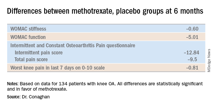
The mean dose of methotrexate used in the study was about 17 mg/week. Dr. Conaghan said that if he could do the trial over again, he would have used subcutaneous methotrexate.
“It’s a more reliable way of getting a dose into people and probably of getting a slightly higher dose into people. In the rheumatoid arthritis world, we use a lot more subcutaneous methotrexate now than we did 10 years ago because it gets around a lot of the minor side effects and helps compliance,” he said.
One audience member suggested that one potentially useful way to zero in on a subgroup of knee OA patients likely to derive the most benefit from methotrexate would be to have screened potential study participants for comorbid fibromyalgia and exclude those with the disorder. Dr. Conaghan replied that the PROMOTE investigators did gather data on participants’ pain at sites other than the knee. That data can be used to identify those at increased likelihood of fibromyalgia, and he agreed that’s worth looking into.
Dr. Conaghan reported having no financial conflicts regarding PROMOTE, which was funded by the U.K. National Institute for Health Research and Versus Arthritis.
SOURCE: Conaghan PG et al. OARSI 2019, Abstract 86.
TORONTO – Philip G. Conaghan, MD, PhD, reported at the OARSI 2019 World Congress.
There is, however, an asterisk attached to these findings. “Despite a moderate standard effect size, the treatment effect was smaller than some of the thresholds for what is considered clinically meaningful,” he noted at the meeting sponsored by the Osteoarthritis Research Society International.
That being said, the rheumatologist is convinced further investigation of methotrexate in osteoarthritis is warranted.
“I have to say that, unlike our earlier hydroxychloroquine trial, which was robustly negative with nothing more to say, I think there is a signal in this study. I need to understand the results of this trial better to understand if there is a subgroup we could treat with methotrexate. It’s a cheap drug, it’s readily available, and we’ve got a lot of experience with it,” noted Dr. Conaghan, professor of musculoskeletal medicine at the University of Leeds (England) and director of the Leeds Institute of Rheumatic and Musculoskeletal Medicine.
The rationale for the 15-center PROMOTE trial is that synovitis is common in OA. Synovitis is associated with pain, methotrexate is the gold-standard treatment for synovitis in inflammatory forms of arthritis, and current treatments for OA are, to say the least, severely limited. Also, an earlier 30-patient, open-label pilot study of methotrexate in patients with painful knee OA conducted by Dr. Conaghan and coworkers suggested the drug was promising (Rheumatology [Oxford]. 2013 May;52[5]:888-92).
PROMOTE included 134 patients with symptomatic and radiographic knee OA who were randomized in double-blind fashion to 6 months of oral methotrexate at 10 mg titrated to a target dose of 25 mg/week or to placebo. All patients also received usual care with oral NSAIDs and/or acetaminophen. Their mean baseline knee pain on a 0-10 numeric rating scale was 6.6.
The primary endpoint, assessed at 6 months, was the difference between the two study arms in average knee pain during the previous week on a 0-10 scale. The score was 5.1 in the methotrexate group and 6.2 in the placebo arm, for a baseline-adjusted treatment difference of 0.83 points, which works out to a standard effect size of 0.36. When the data were reanalyzed after excluding the 15 patients who missed more than four doses of medication within any 3-month period, the between-group difference in pain scores increased to 0.95 points in favor of the methotrexate group.
A significant difference in favor of the methotrexate group was documented in the OARSI-OMERACT response rate at 6 months: 45% in the methotrexate group and 26% in the controls. Some secondary endpoints were positive as well, with statistically significant differences seen at 6 months in Western Ontario and McMaster Universities Osteoarthritis Index (WOMAC) stiffness, WOMAC physical function, and several other endpoints. But there were no significant differences in WOMAC pain, SF-12 physical component or SF-12 mental component scores, or in an OA quality of life measure.
The mean dose of methotrexate used in the study was about 17 mg/week. Dr. Conaghan said that if he could do the trial over again, he would have used subcutaneous methotrexate.
“It’s a more reliable way of getting a dose into people and probably of getting a slightly higher dose into people. In the rheumatoid arthritis world, we use a lot more subcutaneous methotrexate now than we did 10 years ago because it gets around a lot of the minor side effects and helps compliance,” he said.
One audience member suggested that one potentially useful way to zero in on a subgroup of knee OA patients likely to derive the most benefit from methotrexate would be to have screened potential study participants for comorbid fibromyalgia and exclude those with the disorder. Dr. Conaghan replied that the PROMOTE investigators did gather data on participants’ pain at sites other than the knee. That data can be used to identify those at increased likelihood of fibromyalgia, and he agreed that’s worth looking into.
Dr. Conaghan reported having no financial conflicts regarding PROMOTE, which was funded by the U.K. National Institute for Health Research and Versus Arthritis.
SOURCE: Conaghan PG et al. OARSI 2019, Abstract 86.
TORONTO – Philip G. Conaghan, MD, PhD, reported at the OARSI 2019 World Congress.
There is, however, an asterisk attached to these findings. “Despite a moderate standard effect size, the treatment effect was smaller than some of the thresholds for what is considered clinically meaningful,” he noted at the meeting sponsored by the Osteoarthritis Research Society International.
That being said, the rheumatologist is convinced further investigation of methotrexate in osteoarthritis is warranted.
“I have to say that, unlike our earlier hydroxychloroquine trial, which was robustly negative with nothing more to say, I think there is a signal in this study. I need to understand the results of this trial better to understand if there is a subgroup we could treat with methotrexate. It’s a cheap drug, it’s readily available, and we’ve got a lot of experience with it,” noted Dr. Conaghan, professor of musculoskeletal medicine at the University of Leeds (England) and director of the Leeds Institute of Rheumatic and Musculoskeletal Medicine.
The rationale for the 15-center PROMOTE trial is that synovitis is common in OA. Synovitis is associated with pain, methotrexate is the gold-standard treatment for synovitis in inflammatory forms of arthritis, and current treatments for OA are, to say the least, severely limited. Also, an earlier 30-patient, open-label pilot study of methotrexate in patients with painful knee OA conducted by Dr. Conaghan and coworkers suggested the drug was promising (Rheumatology [Oxford]. 2013 May;52[5]:888-92).
PROMOTE included 134 patients with symptomatic and radiographic knee OA who were randomized in double-blind fashion to 6 months of oral methotrexate at 10 mg titrated to a target dose of 25 mg/week or to placebo. All patients also received usual care with oral NSAIDs and/or acetaminophen. Their mean baseline knee pain on a 0-10 numeric rating scale was 6.6.
The primary endpoint, assessed at 6 months, was the difference between the two study arms in average knee pain during the previous week on a 0-10 scale. The score was 5.1 in the methotrexate group and 6.2 in the placebo arm, for a baseline-adjusted treatment difference of 0.83 points, which works out to a standard effect size of 0.36. When the data were reanalyzed after excluding the 15 patients who missed more than four doses of medication within any 3-month period, the between-group difference in pain scores increased to 0.95 points in favor of the methotrexate group.
A significant difference in favor of the methotrexate group was documented in the OARSI-OMERACT response rate at 6 months: 45% in the methotrexate group and 26% in the controls. Some secondary endpoints were positive as well, with statistically significant differences seen at 6 months in Western Ontario and McMaster Universities Osteoarthritis Index (WOMAC) stiffness, WOMAC physical function, and several other endpoints. But there were no significant differences in WOMAC pain, SF-12 physical component or SF-12 mental component scores, or in an OA quality of life measure.
The mean dose of methotrexate used in the study was about 17 mg/week. Dr. Conaghan said that if he could do the trial over again, he would have used subcutaneous methotrexate.
“It’s a more reliable way of getting a dose into people and probably of getting a slightly higher dose into people. In the rheumatoid arthritis world, we use a lot more subcutaneous methotrexate now than we did 10 years ago because it gets around a lot of the minor side effects and helps compliance,” he said.
One audience member suggested that one potentially useful way to zero in on a subgroup of knee OA patients likely to derive the most benefit from methotrexate would be to have screened potential study participants for comorbid fibromyalgia and exclude those with the disorder. Dr. Conaghan replied that the PROMOTE investigators did gather data on participants’ pain at sites other than the knee. That data can be used to identify those at increased likelihood of fibromyalgia, and he agreed that’s worth looking into.
Dr. Conaghan reported having no financial conflicts regarding PROMOTE, which was funded by the U.K. National Institute for Health Research and Versus Arthritis.
SOURCE: Conaghan PG et al. OARSI 2019, Abstract 86.
REPORTING FROM OARSI 2019

Switching from interferon beta-1a to alemtuzumab improves MS outcomes
SEATTLE – , according to an analysis presented at the annual meeting of the Consortium of Multiple Sclerosis Centers. These outcomes may be maintained for 6 years without continuous treatment, said the investigators.
The CARE-MS II study demonstrated alemtuzumab’s superior efficacy, compared with subcutaneous interferon beta-1a, over 2 years in patients with active relapsing-remitting MS who had had an inadequate response to previous therapy. The trial was followed by a 4-year extension, during which patients who had received interferon beta-1a were given the option of discontinuing that therapy and initiating alemtuzumab. The alemtuzumab regimen for these patients was 12 mg/day for 5 consecutive days at baseline, and the same dose for 3 consecutive days at 1 year. Additional annual alemtuzumab as needed for disease activity was allowed. At investigators’ discretion, patients could receive other disease-modifying therapy (DMT) at any time. After the 4-year extension, patients could continue in the 5-year TOPAZ extension.
Carolina Ionete, MD, a neurologist at University of Massachusetts Memorial Medical Center in Worcester, and colleagues examined data from the TOPAZ extension study to evaluate the efficacy and safety of alemtuzumab over 6 years in patients with relapsing-remitting MS from CARE-MS II who discontinued interferon beta-1a. In TOPAZ, patients can receive additional alemtuzumab (12 mg/day on 3 consecutive days at 12 or more months after the most recent course) or other DMTs at any time at investigators’ discretion.
In all, 143 patients started alemtuzumab in the extension study. Of this group 117 patients (82%) completed year 2 of TOPAZ (i.e., year 6 after initiating alemtuzumab). The annualized relapse rate at year 6 was 0.19, and the annual rate of freedom from relapse ranged from 83% to 90% during years 1 through 6. At year 6, Expanded Disability Status Scale (EDSS) scores were stable (51%) or improved (17%) in 68% of patients, compared with the baseline of the main study. At year 6, the mean EDSS score change was 0.43. Over 6 years, 69% of patients were free from 6-month confirmed disability worsening, and 23% achieved 6-month confirmed disability improvement.
In addition, 69% of patients were free of MRI disease activity in year 6. The median percent cumulative brain volume loss from alemtuzumab initiation through year 6 was 0.53%, compared with 0.81% over 2 years with interferon beta-1a. Brain volume loss was 0.32% or less annually during years 2 through 6 after initiating alemtuzumab (0.04% at year 2, 0.15% at year 3, 0.14% at year 4, 0.07% at year 5, and 0.32% at year 6). These efficacy outcomes were observed as 57% of patients received neither additional alemtuzumab nor another DMT through year 6. Safety results were consistent with those for the alemtuzumab-treated patients in the core and extension studies.
Sanofi and Bayer HealthCare Pharmaceuticals supported this study. Dr. Ionete received research support from Biogen, Roche, and Sanofi. She reported receiving compensation for advisory board participation from Sanofi.
SEATTLE – , according to an analysis presented at the annual meeting of the Consortium of Multiple Sclerosis Centers. These outcomes may be maintained for 6 years without continuous treatment, said the investigators.
The CARE-MS II study demonstrated alemtuzumab’s superior efficacy, compared with subcutaneous interferon beta-1a, over 2 years in patients with active relapsing-remitting MS who had had an inadequate response to previous therapy. The trial was followed by a 4-year extension, during which patients who had received interferon beta-1a were given the option of discontinuing that therapy and initiating alemtuzumab. The alemtuzumab regimen for these patients was 12 mg/day for 5 consecutive days at baseline, and the same dose for 3 consecutive days at 1 year. Additional annual alemtuzumab as needed for disease activity was allowed. At investigators’ discretion, patients could receive other disease-modifying therapy (DMT) at any time. After the 4-year extension, patients could continue in the 5-year TOPAZ extension.
Carolina Ionete, MD, a neurologist at University of Massachusetts Memorial Medical Center in Worcester, and colleagues examined data from the TOPAZ extension study to evaluate the efficacy and safety of alemtuzumab over 6 years in patients with relapsing-remitting MS from CARE-MS II who discontinued interferon beta-1a. In TOPAZ, patients can receive additional alemtuzumab (12 mg/day on 3 consecutive days at 12 or more months after the most recent course) or other DMTs at any time at investigators’ discretion.
In all, 143 patients started alemtuzumab in the extension study. Of this group 117 patients (82%) completed year 2 of TOPAZ (i.e., year 6 after initiating alemtuzumab). The annualized relapse rate at year 6 was 0.19, and the annual rate of freedom from relapse ranged from 83% to 90% during years 1 through 6. At year 6, Expanded Disability Status Scale (EDSS) scores were stable (51%) or improved (17%) in 68% of patients, compared with the baseline of the main study. At year 6, the mean EDSS score change was 0.43. Over 6 years, 69% of patients were free from 6-month confirmed disability worsening, and 23% achieved 6-month confirmed disability improvement.
In addition, 69% of patients were free of MRI disease activity in year 6. The median percent cumulative brain volume loss from alemtuzumab initiation through year 6 was 0.53%, compared with 0.81% over 2 years with interferon beta-1a. Brain volume loss was 0.32% or less annually during years 2 through 6 after initiating alemtuzumab (0.04% at year 2, 0.15% at year 3, 0.14% at year 4, 0.07% at year 5, and 0.32% at year 6). These efficacy outcomes were observed as 57% of patients received neither additional alemtuzumab nor another DMT through year 6. Safety results were consistent with those for the alemtuzumab-treated patients in the core and extension studies.
Sanofi and Bayer HealthCare Pharmaceuticals supported this study. Dr. Ionete received research support from Biogen, Roche, and Sanofi. She reported receiving compensation for advisory board participation from Sanofi.
SEATTLE – , according to an analysis presented at the annual meeting of the Consortium of Multiple Sclerosis Centers. These outcomes may be maintained for 6 years without continuous treatment, said the investigators.
The CARE-MS II study demonstrated alemtuzumab’s superior efficacy, compared with subcutaneous interferon beta-1a, over 2 years in patients with active relapsing-remitting MS who had had an inadequate response to previous therapy. The trial was followed by a 4-year extension, during which patients who had received interferon beta-1a were given the option of discontinuing that therapy and initiating alemtuzumab. The alemtuzumab regimen for these patients was 12 mg/day for 5 consecutive days at baseline, and the same dose for 3 consecutive days at 1 year. Additional annual alemtuzumab as needed for disease activity was allowed. At investigators’ discretion, patients could receive other disease-modifying therapy (DMT) at any time. After the 4-year extension, patients could continue in the 5-year TOPAZ extension.
Carolina Ionete, MD, a neurologist at University of Massachusetts Memorial Medical Center in Worcester, and colleagues examined data from the TOPAZ extension study to evaluate the efficacy and safety of alemtuzumab over 6 years in patients with relapsing-remitting MS from CARE-MS II who discontinued interferon beta-1a. In TOPAZ, patients can receive additional alemtuzumab (12 mg/day on 3 consecutive days at 12 or more months after the most recent course) or other DMTs at any time at investigators’ discretion.
In all, 143 patients started alemtuzumab in the extension study. Of this group 117 patients (82%) completed year 2 of TOPAZ (i.e., year 6 after initiating alemtuzumab). The annualized relapse rate at year 6 was 0.19, and the annual rate of freedom from relapse ranged from 83% to 90% during years 1 through 6. At year 6, Expanded Disability Status Scale (EDSS) scores were stable (51%) or improved (17%) in 68% of patients, compared with the baseline of the main study. At year 6, the mean EDSS score change was 0.43. Over 6 years, 69% of patients were free from 6-month confirmed disability worsening, and 23% achieved 6-month confirmed disability improvement.
In addition, 69% of patients were free of MRI disease activity in year 6. The median percent cumulative brain volume loss from alemtuzumab initiation through year 6 was 0.53%, compared with 0.81% over 2 years with interferon beta-1a. Brain volume loss was 0.32% or less annually during years 2 through 6 after initiating alemtuzumab (0.04% at year 2, 0.15% at year 3, 0.14% at year 4, 0.07% at year 5, and 0.32% at year 6). These efficacy outcomes were observed as 57% of patients received neither additional alemtuzumab nor another DMT through year 6. Safety results were consistent with those for the alemtuzumab-treated patients in the core and extension studies.
Sanofi and Bayer HealthCare Pharmaceuticals supported this study. Dr. Ionete received research support from Biogen, Roche, and Sanofi. She reported receiving compensation for advisory board participation from Sanofi.
REPORTING FROM CMSC 2019
Racial disparities in time to cancer care erased with Medicaid expansion
CHICAGO – For decades investigators have documented racial disparities in access to cancer care and in clinical outcomes, with socioeconomic factors suspected – but not conclusively proven – to play a role
Now a new study based on electronic health record (EHR) data shows that after Medicaid expansion under the Affordable Care Act (ACA), racial differences in timely cancer treatment effectively disappeared. Before Medicaid expansion, African Americans were 4.8% less likely than whites to receive timely cancer treatment, defined as treatment starting within 30 days of diagnosis of an advanced or metastatic solid tumor. After Medicaid expansion, however, the difference between the racial groups had dwindled to just 0.8% and was no longer statistically significant.
The findings suggest that the expanded availability of health insurance has had a salutary effect on cancer care.
In this video interview, co-authors Amy J. Davidoff, PhD, MS, from the Yale Cancer Center in New Haven Connecticut, and Blythe J.S. Adamson, PhD, from Flatiron Health in New York, New York, discuss the study findings and the possible implications for health care policy in the United States.
The study was funded by Flatiron Health. Dr. Adamson is an employee of the company. Dr. Davidoff disclosed consulting or advisory roles with and honoraria from several pharmaceutical companies.
CHICAGO – For decades investigators have documented racial disparities in access to cancer care and in clinical outcomes, with socioeconomic factors suspected – but not conclusively proven – to play a role
Now a new study based on electronic health record (EHR) data shows that after Medicaid expansion under the Affordable Care Act (ACA), racial differences in timely cancer treatment effectively disappeared. Before Medicaid expansion, African Americans were 4.8% less likely than whites to receive timely cancer treatment, defined as treatment starting within 30 days of diagnosis of an advanced or metastatic solid tumor. After Medicaid expansion, however, the difference between the racial groups had dwindled to just 0.8% and was no longer statistically significant.
The findings suggest that the expanded availability of health insurance has had a salutary effect on cancer care.
In this video interview, co-authors Amy J. Davidoff, PhD, MS, from the Yale Cancer Center in New Haven Connecticut, and Blythe J.S. Adamson, PhD, from Flatiron Health in New York, New York, discuss the study findings and the possible implications for health care policy in the United States.
The study was funded by Flatiron Health. Dr. Adamson is an employee of the company. Dr. Davidoff disclosed consulting or advisory roles with and honoraria from several pharmaceutical companies.
CHICAGO – For decades investigators have documented racial disparities in access to cancer care and in clinical outcomes, with socioeconomic factors suspected – but not conclusively proven – to play a role
Now a new study based on electronic health record (EHR) data shows that after Medicaid expansion under the Affordable Care Act (ACA), racial differences in timely cancer treatment effectively disappeared. Before Medicaid expansion, African Americans were 4.8% less likely than whites to receive timely cancer treatment, defined as treatment starting within 30 days of diagnosis of an advanced or metastatic solid tumor. After Medicaid expansion, however, the difference between the racial groups had dwindled to just 0.8% and was no longer statistically significant.
The findings suggest that the expanded availability of health insurance has had a salutary effect on cancer care.
In this video interview, co-authors Amy J. Davidoff, PhD, MS, from the Yale Cancer Center in New Haven Connecticut, and Blythe J.S. Adamson, PhD, from Flatiron Health in New York, New York, discuss the study findings and the possible implications for health care policy in the United States.
The study was funded by Flatiron Health. Dr. Adamson is an employee of the company. Dr. Davidoff disclosed consulting or advisory roles with and honoraria from several pharmaceutical companies.
REPORTING FROM ASCO 2019

Cleveland Clinic targets time to treat in cancer
CHICAGO – In 2014, the average time from diagnosis to treatment initiation for new cancer patients at the Cleveland Clinic was 29-41 days, depending on whether the patient was diagnosed internally or externally. That figure was not acceptable, said Brian J. Bolwell, MD, chairman of the Cleveland Clinic’s Taussig Cancer Institute.
Since then, the time-to-treat metric has improved dramatically, dropping 33%. Today, time to treat for new cancer patients is 25-31 days, depending on the site of diagnosis.
To get there, leaders at the cancer center examined the causes of delay within each of their disease programs. The analysis revealed that less than 20% of the time it was patient preferences that slowed down the initiation of treatment, but that more than 80% of the time the delay was on the part of their institution.
Dr. Bolwell said this led them to start tracking every newly diagnosed patient who came through the cancer center to ensure they didn’t fall through the cracks, and that they were treated as rapidly as possible.
But figuring out how to get patients to treatment quicker depended on the type of cancer they had, since each type of cancer had different challenges and different points of entry to the health care system.
“So for breast cancer, it turns out a lot of the challenges might be coordination of surgery because sometimes a general surgeon has to work with a reconstructive-plastic surgeon and coordinating the surgical schedules might drastically lengthen time to treat,” he said during an interview at the annual meeting of the American Society of Clinical Oncology.
They helped address that problem by scheduling breast cancer patients for surgery by the next available operating room slot, rather than doing the scheduling by surgeon.
There are additional barriers to achieving a rapid time to treat standard, including prior authorization, Dr. Bolwell said. But they are continuing to chip away at the metric, working within each cancer type to lower the obstacles to treatment. “I don’t think we’ll ever be satisfied with where we are,” Dr. Bolwell said.
Dr. Bolwell reported having no relevant financial disclosures.
CHICAGO – In 2014, the average time from diagnosis to treatment initiation for new cancer patients at the Cleveland Clinic was 29-41 days, depending on whether the patient was diagnosed internally or externally. That figure was not acceptable, said Brian J. Bolwell, MD, chairman of the Cleveland Clinic’s Taussig Cancer Institute.
Since then, the time-to-treat metric has improved dramatically, dropping 33%. Today, time to treat for new cancer patients is 25-31 days, depending on the site of diagnosis.
To get there, leaders at the cancer center examined the causes of delay within each of their disease programs. The analysis revealed that less than 20% of the time it was patient preferences that slowed down the initiation of treatment, but that more than 80% of the time the delay was on the part of their institution.
Dr. Bolwell said this led them to start tracking every newly diagnosed patient who came through the cancer center to ensure they didn’t fall through the cracks, and that they were treated as rapidly as possible.
But figuring out how to get patients to treatment quicker depended on the type of cancer they had, since each type of cancer had different challenges and different points of entry to the health care system.
“So for breast cancer, it turns out a lot of the challenges might be coordination of surgery because sometimes a general surgeon has to work with a reconstructive-plastic surgeon and coordinating the surgical schedules might drastically lengthen time to treat,” he said during an interview at the annual meeting of the American Society of Clinical Oncology.
They helped address that problem by scheduling breast cancer patients for surgery by the next available operating room slot, rather than doing the scheduling by surgeon.
There are additional barriers to achieving a rapid time to treat standard, including prior authorization, Dr. Bolwell said. But they are continuing to chip away at the metric, working within each cancer type to lower the obstacles to treatment. “I don’t think we’ll ever be satisfied with where we are,” Dr. Bolwell said.
Dr. Bolwell reported having no relevant financial disclosures.
CHICAGO – In 2014, the average time from diagnosis to treatment initiation for new cancer patients at the Cleveland Clinic was 29-41 days, depending on whether the patient was diagnosed internally or externally. That figure was not acceptable, said Brian J. Bolwell, MD, chairman of the Cleveland Clinic’s Taussig Cancer Institute.
Since then, the time-to-treat metric has improved dramatically, dropping 33%. Today, time to treat for new cancer patients is 25-31 days, depending on the site of diagnosis.
To get there, leaders at the cancer center examined the causes of delay within each of their disease programs. The analysis revealed that less than 20% of the time it was patient preferences that slowed down the initiation of treatment, but that more than 80% of the time the delay was on the part of their institution.
Dr. Bolwell said this led them to start tracking every newly diagnosed patient who came through the cancer center to ensure they didn’t fall through the cracks, and that they were treated as rapidly as possible.
But figuring out how to get patients to treatment quicker depended on the type of cancer they had, since each type of cancer had different challenges and different points of entry to the health care system.
“So for breast cancer, it turns out a lot of the challenges might be coordination of surgery because sometimes a general surgeon has to work with a reconstructive-plastic surgeon and coordinating the surgical schedules might drastically lengthen time to treat,” he said during an interview at the annual meeting of the American Society of Clinical Oncology.
They helped address that problem by scheduling breast cancer patients for surgery by the next available operating room slot, rather than doing the scheduling by surgeon.
There are additional barriers to achieving a rapid time to treat standard, including prior authorization, Dr. Bolwell said. But they are continuing to chip away at the metric, working within each cancer type to lower the obstacles to treatment. “I don’t think we’ll ever be satisfied with where we are,” Dr. Bolwell said.
Dr. Bolwell reported having no relevant financial disclosures.
FROM ASCO 2019

Meta-analysis finds no link between PPI use and risk of dementia

The finding runs counter to recent studies, including a large pharmacoepidemiological claims data analysis from Germany, that propose an association between proton pump inhibitor (PPI) use and the development of dementia (JAMA Neurol. 2016;73[4]:410-6). “The issue with these studies is that they’re based on retrospective claims data and pharmacoepidemiological studies and insurance databases that don’t really give you a good causality basis,” lead study author Saad Alrajhi, MD, said in an interview at the annual Digestive Disease Week.
In an effort to better characterize the association between PPI exposure and dementia, Dr. Alrajhi, a gastroenterology fellow at McGill University, Montreal, and colleagues conducted a meta-analysis of all fully published randomized clinical trials or observational studies comparing use of PPIs and occurrence of dementia. The researchers queried Embase, MEDLINE, and ISI Web of Knowledge for relevant studies that were published from 1995 through September 2018. Next, they assessed the quality of the studies by using the Cochrane risk assessment tool for RCTs or the Newcastle-Ottawa Scale for observational studies.
As the primary outcome, the researchers compared dementia incidence after PPI exposure (experimental group) versus no PPI exposure (control group). Development of Alzheimer’s dementia was a secondary outcome. Sensitivity analyses consisted of excluding one study at a time, and assessing results among studies of highest qualities. Subgroup analyses included stratifying patients by age. To report odds ratios, Dr. Alrajhi and colleagues used fixed or random effects models based on the absence or presence of heterogeneity.
Of 549 studies assessed, 5 met the criteria for inclusion in the final analysis: 3 case-control studies and 2 cohort studies, with a total of 472,933 patients. All of the studies scored 8 or 9 on the Newcastle-Ottawa scale, indicating high quality. Significant heterogeneity was noted for all analyses. The researchers found that the incidence of dementia was not significantly increased among patients in the PPI-exposed group (odd ratio, 1.08 (95% confidence interval, 0.97-1.20; P = .18). Sensitivity analyses confirmed the robustness of the results. Subgroup analysis showed no between-group differences among studies that included a minimum age above 65 years (three studies) or less than age 65 (two studies). PPI exposure was not associated with the development of Alzheimer’s dementia (two studies) (OR, 1.32 (95% CI, 0.80-2.17; P = .27).
“In the absence of randomized trial evidence, a PPI prescribing approach based on appropriate utilization of guideline-based prescription should be done without the extra fear of the association of dementia,” Dr. Alrajhi said.
The researchers reported having no financial disclosures.
The finding runs counter to recent studies, including a large pharmacoepidemiological claims data analysis from Germany, that propose an association between proton pump inhibitor (PPI) use and the development of dementia (JAMA Neurol. 2016;73[4]:410-6). “The issue with these studies is that they’re based on retrospective claims data and pharmacoepidemiological studies and insurance databases that don’t really give you a good causality basis,” lead study author Saad Alrajhi, MD, said in an interview at the annual Digestive Disease Week.
In an effort to better characterize the association between PPI exposure and dementia, Dr. Alrajhi, a gastroenterology fellow at McGill University, Montreal, and colleagues conducted a meta-analysis of all fully published randomized clinical trials or observational studies comparing use of PPIs and occurrence of dementia. The researchers queried Embase, MEDLINE, and ISI Web of Knowledge for relevant studies that were published from 1995 through September 2018. Next, they assessed the quality of the studies by using the Cochrane risk assessment tool for RCTs or the Newcastle-Ottawa Scale for observational studies.
As the primary outcome, the researchers compared dementia incidence after PPI exposure (experimental group) versus no PPI exposure (control group). Development of Alzheimer’s dementia was a secondary outcome. Sensitivity analyses consisted of excluding one study at a time, and assessing results among studies of highest qualities. Subgroup analyses included stratifying patients by age. To report odds ratios, Dr. Alrajhi and colleagues used fixed or random effects models based on the absence or presence of heterogeneity.
Of 549 studies assessed, 5 met the criteria for inclusion in the final analysis: 3 case-control studies and 2 cohort studies, with a total of 472,933 patients. All of the studies scored 8 or 9 on the Newcastle-Ottawa scale, indicating high quality. Significant heterogeneity was noted for all analyses. The researchers found that the incidence of dementia was not significantly increased among patients in the PPI-exposed group (odd ratio, 1.08 (95% confidence interval, 0.97-1.20; P = .18). Sensitivity analyses confirmed the robustness of the results. Subgroup analysis showed no between-group differences among studies that included a minimum age above 65 years (three studies) or less than age 65 (two studies). PPI exposure was not associated with the development of Alzheimer’s dementia (two studies) (OR, 1.32 (95% CI, 0.80-2.17; P = .27).
“In the absence of randomized trial evidence, a PPI prescribing approach based on appropriate utilization of guideline-based prescription should be done without the extra fear of the association of dementia,” Dr. Alrajhi said.
The researchers reported having no financial disclosures.
The finding runs counter to recent studies, including a large pharmacoepidemiological claims data analysis from Germany, that propose an association between proton pump inhibitor (PPI) use and the development of dementia (JAMA Neurol. 2016;73[4]:410-6). “The issue with these studies is that they’re based on retrospective claims data and pharmacoepidemiological studies and insurance databases that don’t really give you a good causality basis,” lead study author Saad Alrajhi, MD, said in an interview at the annual Digestive Disease Week.
In an effort to better characterize the association between PPI exposure and dementia, Dr. Alrajhi, a gastroenterology fellow at McGill University, Montreal, and colleagues conducted a meta-analysis of all fully published randomized clinical trials or observational studies comparing use of PPIs and occurrence of dementia. The researchers queried Embase, MEDLINE, and ISI Web of Knowledge for relevant studies that were published from 1995 through September 2018. Next, they assessed the quality of the studies by using the Cochrane risk assessment tool for RCTs or the Newcastle-Ottawa Scale for observational studies.
As the primary outcome, the researchers compared dementia incidence after PPI exposure (experimental group) versus no PPI exposure (control group). Development of Alzheimer’s dementia was a secondary outcome. Sensitivity analyses consisted of excluding one study at a time, and assessing results among studies of highest qualities. Subgroup analyses included stratifying patients by age. To report odds ratios, Dr. Alrajhi and colleagues used fixed or random effects models based on the absence or presence of heterogeneity.
Of 549 studies assessed, 5 met the criteria for inclusion in the final analysis: 3 case-control studies and 2 cohort studies, with a total of 472,933 patients. All of the studies scored 8 or 9 on the Newcastle-Ottawa scale, indicating high quality. Significant heterogeneity was noted for all analyses. The researchers found that the incidence of dementia was not significantly increased among patients in the PPI-exposed group (odd ratio, 1.08 (95% confidence interval, 0.97-1.20; P = .18). Sensitivity analyses confirmed the robustness of the results. Subgroup analysis showed no between-group differences among studies that included a minimum age above 65 years (three studies) or less than age 65 (two studies). PPI exposure was not associated with the development of Alzheimer’s dementia (two studies) (OR, 1.32 (95% CI, 0.80-2.17; P = .27).
“In the absence of randomized trial evidence, a PPI prescribing approach based on appropriate utilization of guideline-based prescription should be done without the extra fear of the association of dementia,” Dr. Alrajhi said.
The researchers reported having no financial disclosures.
REPORTING FROM DDW 2019
Even if successful, IVF may boost relapses in MS
SEATTLE – Failed in-vitro fertilization (IVF) treatment appears to boost the risk of relapse in women with multiple sclerosis (MS). Does successful IVF have the same effect? The preliminary results of a new study suggests it does, a finding that may influence how physicians track patients during pregnancy.
“We found that IVF can still cause a relapse even if it is successful,” study lead author Maria Claudia Manieri, a graduate student at Harvard Medical School’s Partners MS Center, said in an interview at the annual meeting of the Consortium of Multiple Sclerosis Centers, where she presented the findings.
Multiple studies have linked infertility treatment in women with MS to relapses. In a 2013 review, researchers analyzed several papers, and “all of them reported an increase in annualized relapse rate after ART [assisted reproductive treatment]. Furthermore, in a recent study, clinical worsening was associated with an increase in MRI activity” (Clin Immunol. 2013 Nov;149(2):219-24).
For the new report, based on statistics from the New England Multiple Sclerosis Pregnancy Prospective Cohort Study, Ms. Manieri and colleagues collected data on 91 women (mean age = 33). Eleven were unsuccessful in conceiving, and 80 successfully conceived.
Three of the 91 women (3%) used intrauterine insemination as a fertility treatment. Another 9 (10%) relied on ART; all used IVF except for 1 who underwent intracytoplasmic sperm injection.
The new report is a preliminary analysis of early data, Ms. Manieri said. The study has recruited about one-sixth of its participants, she said, and will track women beyond pregnancy to explore long-term outcomes in their children.
Eleven women relapsed during pregnancy, including 9 who were using fertility treatment (P = .003). Of those 9, 7 women (78%) used ART.
No other factor other than fertility treatment predicted intrapartum relapses. The relapses during pregnancy started at 21 weeks (± 12 weeks) of gestational age and lasted for 4 weeks (± 2 weeks).
Of those who successfully conceived, 4 of 5 (80%) who used fertility treatment relapsed, compared with 7 of 64 (11%) who didn’t use fertility treatment. Of women who did not successfully conceive, 2 of 3 (67%) relapsed among those who used fertility treatment vs. 1 of 7 (14%) of those who didn’t.
It’s not clear how infertility treatment may be boosting MS relapse in women, but the 2013 review offered these possibilities: “temporary interruption of disease modified therapies, stressful events associated with infertility, and immunological changes induced by hormones such as increase in pro-inflammatory cytokines and anti-MOG antibodies, as well as an increase in immune cell migration across the blood-brain-barrier.”
MS tends to improve during pregnancy, and it’s common for neurologists to not see patients for extended periods, Ms. Manieri said. In light of the findings, she said, it may be wise for neurologists to continue follow-up appointments during pregnancy. “Avoid delaying care and keep monitoring the patient,” she advised.
The study was funded by Sanofi Genzyme and a gift from Michelle and Christopher Rondeau. The study authors report no relevant disclosures.
SEATTLE – Failed in-vitro fertilization (IVF) treatment appears to boost the risk of relapse in women with multiple sclerosis (MS). Does successful IVF have the same effect? The preliminary results of a new study suggests it does, a finding that may influence how physicians track patients during pregnancy.
“We found that IVF can still cause a relapse even if it is successful,” study lead author Maria Claudia Manieri, a graduate student at Harvard Medical School’s Partners MS Center, said in an interview at the annual meeting of the Consortium of Multiple Sclerosis Centers, where she presented the findings.
Multiple studies have linked infertility treatment in women with MS to relapses. In a 2013 review, researchers analyzed several papers, and “all of them reported an increase in annualized relapse rate after ART [assisted reproductive treatment]. Furthermore, in a recent study, clinical worsening was associated with an increase in MRI activity” (Clin Immunol. 2013 Nov;149(2):219-24).
For the new report, based on statistics from the New England Multiple Sclerosis Pregnancy Prospective Cohort Study, Ms. Manieri and colleagues collected data on 91 women (mean age = 33). Eleven were unsuccessful in conceiving, and 80 successfully conceived.
Three of the 91 women (3%) used intrauterine insemination as a fertility treatment. Another 9 (10%) relied on ART; all used IVF except for 1 who underwent intracytoplasmic sperm injection.
The new report is a preliminary analysis of early data, Ms. Manieri said. The study has recruited about one-sixth of its participants, she said, and will track women beyond pregnancy to explore long-term outcomes in their children.
Eleven women relapsed during pregnancy, including 9 who were using fertility treatment (P = .003). Of those 9, 7 women (78%) used ART.
No other factor other than fertility treatment predicted intrapartum relapses. The relapses during pregnancy started at 21 weeks (± 12 weeks) of gestational age and lasted for 4 weeks (± 2 weeks).
Of those who successfully conceived, 4 of 5 (80%) who used fertility treatment relapsed, compared with 7 of 64 (11%) who didn’t use fertility treatment. Of women who did not successfully conceive, 2 of 3 (67%) relapsed among those who used fertility treatment vs. 1 of 7 (14%) of those who didn’t.
It’s not clear how infertility treatment may be boosting MS relapse in women, but the 2013 review offered these possibilities: “temporary interruption of disease modified therapies, stressful events associated with infertility, and immunological changes induced by hormones such as increase in pro-inflammatory cytokines and anti-MOG antibodies, as well as an increase in immune cell migration across the blood-brain-barrier.”
MS tends to improve during pregnancy, and it’s common for neurologists to not see patients for extended periods, Ms. Manieri said. In light of the findings, she said, it may be wise for neurologists to continue follow-up appointments during pregnancy. “Avoid delaying care and keep monitoring the patient,” she advised.
The study was funded by Sanofi Genzyme and a gift from Michelle and Christopher Rondeau. The study authors report no relevant disclosures.
SEATTLE – Failed in-vitro fertilization (IVF) treatment appears to boost the risk of relapse in women with multiple sclerosis (MS). Does successful IVF have the same effect? The preliminary results of a new study suggests it does, a finding that may influence how physicians track patients during pregnancy.
“We found that IVF can still cause a relapse even if it is successful,” study lead author Maria Claudia Manieri, a graduate student at Harvard Medical School’s Partners MS Center, said in an interview at the annual meeting of the Consortium of Multiple Sclerosis Centers, where she presented the findings.
Multiple studies have linked infertility treatment in women with MS to relapses. In a 2013 review, researchers analyzed several papers, and “all of them reported an increase in annualized relapse rate after ART [assisted reproductive treatment]. Furthermore, in a recent study, clinical worsening was associated with an increase in MRI activity” (Clin Immunol. 2013 Nov;149(2):219-24).
For the new report, based on statistics from the New England Multiple Sclerosis Pregnancy Prospective Cohort Study, Ms. Manieri and colleagues collected data on 91 women (mean age = 33). Eleven were unsuccessful in conceiving, and 80 successfully conceived.
Three of the 91 women (3%) used intrauterine insemination as a fertility treatment. Another 9 (10%) relied on ART; all used IVF except for 1 who underwent intracytoplasmic sperm injection.
The new report is a preliminary analysis of early data, Ms. Manieri said. The study has recruited about one-sixth of its participants, she said, and will track women beyond pregnancy to explore long-term outcomes in their children.
Eleven women relapsed during pregnancy, including 9 who were using fertility treatment (P = .003). Of those 9, 7 women (78%) used ART.
No other factor other than fertility treatment predicted intrapartum relapses. The relapses during pregnancy started at 21 weeks (± 12 weeks) of gestational age and lasted for 4 weeks (± 2 weeks).
Of those who successfully conceived, 4 of 5 (80%) who used fertility treatment relapsed, compared with 7 of 64 (11%) who didn’t use fertility treatment. Of women who did not successfully conceive, 2 of 3 (67%) relapsed among those who used fertility treatment vs. 1 of 7 (14%) of those who didn’t.
It’s not clear how infertility treatment may be boosting MS relapse in women, but the 2013 review offered these possibilities: “temporary interruption of disease modified therapies, stressful events associated with infertility, and immunological changes induced by hormones such as increase in pro-inflammatory cytokines and anti-MOG antibodies, as well as an increase in immune cell migration across the blood-brain-barrier.”
MS tends to improve during pregnancy, and it’s common for neurologists to not see patients for extended periods, Ms. Manieri said. In light of the findings, she said, it may be wise for neurologists to continue follow-up appointments during pregnancy. “Avoid delaying care and keep monitoring the patient,” she advised.
The study was funded by Sanofi Genzyme and a gift from Michelle and Christopher Rondeau. The study authors report no relevant disclosures.
REPORTING FROM CMSC 2019