User login
Psoriasis Journal Scan: October 2019
Psoriasis Associated with Tumor Necrosis Factor-Alpha Inhibitors in Children with Inflammatory Diseases.
Buckley, L. H., Xiao, R. , Perman, M. et al. Arthritis Care Res. 2019 Oct 23.
The study aimed to estimate the incidence rate (IR) of psoriasis in children with inflammatory bowel disease (IBD), juvenile idiopathic arthritis (JIA), and chronic noninfectious osteomyelitis (CNO) with tumor necrosis factor‐alpha inhibitor (TNFi) exposure as compared to those without TNFi exposure and to the general pediatric population. Researchers found that children with IBD, JIA, and CNO had an increased rate of psoriasis compared to the general pediatric population, with the highest rate in those with TNFi exposure.
Skin Patterning in Psoriasis by Spatial Interactions between Pathogenic Cytokines.
Ringham, L, Prusinkiewicz, P, Gniadeck R. iScience. 2019 Oct 25;20:546-553.
This study shows that all known patterns of psoriasis, a common inflammatory skin disease, can be explained in terms of reaction-diffusion. Researchers constructed a computational model based on the known interactions between the main pathogenic cytokines: interleukins IL-17 and IL-23, and tumor necrosis factor TNF-α. Simulations revealed that the parameter space of the model contained all classes of psoriatic lesion patterns. They also faithfully reproduced the growth and evolution of the plaques and the response to treatment by cytokine targeting. Thus the pathogenesis of inflammatory diseases, such as psoriasis, may be readily understood in the framework of the stimulatory and inhibitory interactions between a few diffusing mediators.
SEfficacy of Secukinumab for Plaque Psoriasis in a Patient on Hemodialysis.
Ikuma D, Oguro M, Hoshino J, et al. CEN Case Rep. 2019 Oct 25.
The case report discusses the safety and efficiency of secukinumab on a 60-year-old patient on hemodialysis. The psoriasis area and severity index (PASI) score decreased from 49.8 to 14.8 after 2 weeks and to 0 after 6 weeks, with remission being maintained after 28 months. No adverse reactions were seen. This case indicates that secukinumab may be effective for severe psoriasis in patients on hemodialysis for end-stage renal disease.
Comparison of pharmacokinetics, safety and tolerability of secukinumab administered subcutaneously using different delivery systems in healthy volunteers and in psoriasis patients.
Bruin, G, Hockey, H‐UP, La Stella, P, et al. Br J Clin Pharmacol. 2019.
The aim of the study was to compare the pharmacokinetics, safety and tolerability of secukinumab with different devices for subcutaneous (s.c.) administration of 2 mL. Collective evidence from both studies demonstrated that 2 mL injections of secukinumab into the abdomen or thigh using different devices resulted in comparable PK characteristics and were all well tolerated without noticable local reactions.
Dual biologic therapy for recalcitrant psoriasis and psoriatic arthritis
Thibodeaux, Quinn et al. JAAD Case Reports, Volume 5, Issue 10, 928 – 930.
This study presents a patient with severe psoriatic skin and joint disease who has been treated with multiple combinations of dual biologic therapy, including ustekinumab plus etanercept for 12 months, secukinumab plus etanercept for 6 months, and guselkumab plus etanercept for 15 months. Throughout the patient's treatment, adverse events only occurred with the ustekinumab plus etanercept combination and consisted of an increased incidence of urinary tract and upper respiratory infections, including a hospitalization for H2N1 flu.
Psoriasis Associated with Tumor Necrosis Factor-Alpha Inhibitors in Children with Inflammatory Diseases.
Buckley, L. H., Xiao, R. , Perman, M. et al. Arthritis Care Res. 2019 Oct 23.
The study aimed to estimate the incidence rate (IR) of psoriasis in children with inflammatory bowel disease (IBD), juvenile idiopathic arthritis (JIA), and chronic noninfectious osteomyelitis (CNO) with tumor necrosis factor‐alpha inhibitor (TNFi) exposure as compared to those without TNFi exposure and to the general pediatric population. Researchers found that children with IBD, JIA, and CNO had an increased rate of psoriasis compared to the general pediatric population, with the highest rate in those with TNFi exposure.
Skin Patterning in Psoriasis by Spatial Interactions between Pathogenic Cytokines.
Ringham, L, Prusinkiewicz, P, Gniadeck R. iScience. 2019 Oct 25;20:546-553.
This study shows that all known patterns of psoriasis, a common inflammatory skin disease, can be explained in terms of reaction-diffusion. Researchers constructed a computational model based on the known interactions between the main pathogenic cytokines: interleukins IL-17 and IL-23, and tumor necrosis factor TNF-α. Simulations revealed that the parameter space of the model contained all classes of psoriatic lesion patterns. They also faithfully reproduced the growth and evolution of the plaques and the response to treatment by cytokine targeting. Thus the pathogenesis of inflammatory diseases, such as psoriasis, may be readily understood in the framework of the stimulatory and inhibitory interactions between a few diffusing mediators.
SEfficacy of Secukinumab for Plaque Psoriasis in a Patient on Hemodialysis.
Ikuma D, Oguro M, Hoshino J, et al. CEN Case Rep. 2019 Oct 25.
The case report discusses the safety and efficiency of secukinumab on a 60-year-old patient on hemodialysis. The psoriasis area and severity index (PASI) score decreased from 49.8 to 14.8 after 2 weeks and to 0 after 6 weeks, with remission being maintained after 28 months. No adverse reactions were seen. This case indicates that secukinumab may be effective for severe psoriasis in patients on hemodialysis for end-stage renal disease.
Comparison of pharmacokinetics, safety and tolerability of secukinumab administered subcutaneously using different delivery systems in healthy volunteers and in psoriasis patients.
Bruin, G, Hockey, H‐UP, La Stella, P, et al. Br J Clin Pharmacol. 2019.
The aim of the study was to compare the pharmacokinetics, safety and tolerability of secukinumab with different devices for subcutaneous (s.c.) administration of 2 mL. Collective evidence from both studies demonstrated that 2 mL injections of secukinumab into the abdomen or thigh using different devices resulted in comparable PK characteristics and were all well tolerated without noticable local reactions.
Dual biologic therapy for recalcitrant psoriasis and psoriatic arthritis
Thibodeaux, Quinn et al. JAAD Case Reports, Volume 5, Issue 10, 928 – 930.
This study presents a patient with severe psoriatic skin and joint disease who has been treated with multiple combinations of dual biologic therapy, including ustekinumab plus etanercept for 12 months, secukinumab plus etanercept for 6 months, and guselkumab plus etanercept for 15 months. Throughout the patient's treatment, adverse events only occurred with the ustekinumab plus etanercept combination and consisted of an increased incidence of urinary tract and upper respiratory infections, including a hospitalization for H2N1 flu.
Psoriasis Associated with Tumor Necrosis Factor-Alpha Inhibitors in Children with Inflammatory Diseases.
Buckley, L. H., Xiao, R. , Perman, M. et al. Arthritis Care Res. 2019 Oct 23.
The study aimed to estimate the incidence rate (IR) of psoriasis in children with inflammatory bowel disease (IBD), juvenile idiopathic arthritis (JIA), and chronic noninfectious osteomyelitis (CNO) with tumor necrosis factor‐alpha inhibitor (TNFi) exposure as compared to those without TNFi exposure and to the general pediatric population. Researchers found that children with IBD, JIA, and CNO had an increased rate of psoriasis compared to the general pediatric population, with the highest rate in those with TNFi exposure.
Skin Patterning in Psoriasis by Spatial Interactions between Pathogenic Cytokines.
Ringham, L, Prusinkiewicz, P, Gniadeck R. iScience. 2019 Oct 25;20:546-553.
This study shows that all known patterns of psoriasis, a common inflammatory skin disease, can be explained in terms of reaction-diffusion. Researchers constructed a computational model based on the known interactions between the main pathogenic cytokines: interleukins IL-17 and IL-23, and tumor necrosis factor TNF-α. Simulations revealed that the parameter space of the model contained all classes of psoriatic lesion patterns. They also faithfully reproduced the growth and evolution of the plaques and the response to treatment by cytokine targeting. Thus the pathogenesis of inflammatory diseases, such as psoriasis, may be readily understood in the framework of the stimulatory and inhibitory interactions between a few diffusing mediators.
SEfficacy of Secukinumab for Plaque Psoriasis in a Patient on Hemodialysis.
Ikuma D, Oguro M, Hoshino J, et al. CEN Case Rep. 2019 Oct 25.
The case report discusses the safety and efficiency of secukinumab on a 60-year-old patient on hemodialysis. The psoriasis area and severity index (PASI) score decreased from 49.8 to 14.8 after 2 weeks and to 0 after 6 weeks, with remission being maintained after 28 months. No adverse reactions were seen. This case indicates that secukinumab may be effective for severe psoriasis in patients on hemodialysis for end-stage renal disease.
Comparison of pharmacokinetics, safety and tolerability of secukinumab administered subcutaneously using different delivery systems in healthy volunteers and in psoriasis patients.
Bruin, G, Hockey, H‐UP, La Stella, P, et al. Br J Clin Pharmacol. 2019.
The aim of the study was to compare the pharmacokinetics, safety and tolerability of secukinumab with different devices for subcutaneous (s.c.) administration of 2 mL. Collective evidence from both studies demonstrated that 2 mL injections of secukinumab into the abdomen or thigh using different devices resulted in comparable PK characteristics and were all well tolerated without noticable local reactions.
Dual biologic therapy for recalcitrant psoriasis and psoriatic arthritis
Thibodeaux, Quinn et al. JAAD Case Reports, Volume 5, Issue 10, 928 – 930.
This study presents a patient with severe psoriatic skin and joint disease who has been treated with multiple combinations of dual biologic therapy, including ustekinumab plus etanercept for 12 months, secukinumab plus etanercept for 6 months, and guselkumab plus etanercept for 15 months. Throughout the patient's treatment, adverse events only occurred with the ustekinumab plus etanercept combination and consisted of an increased incidence of urinary tract and upper respiratory infections, including a hospitalization for H2N1 flu.
Signs and Symptoms of Psoriatic Arthritis
Psoriasis risk rises with TNF inhibitor use in children with inflammatory disorders
, a retrospective cohort study has determined.
“The incidence rate and risk factors of psoriasis in children with IBD [inflammatory bowel disease], JIA [juvenile idiopathic arthritis], or CNO [chronic nonbacterial osteomyelitis] who are exposed to TNFi [tumor necrosis factor inhibitors] are unknown. Additionally, there is a well-established association between these inflammatory conditions and psoriasis development. Yet, as TNFi can both treat and trigger psoriasis, it is not clear how TNFi exposure affects this relationship,” wrote Lisa H. Buckley, MD, of Children’s Hospital at Vanderbilt, Nashville, Tenn., and colleagues. Their report is in Arthritis Care & Research.
The team examined the relationship in children who were treated for an inflammatory disorder at Children’s Hospital of Philadelphia during 2008-2018. IBD was most common at 74%, followed by JIA at 24% and CNO at 2%.
Among 4,111 children with those inflammatory disorders, the psoriasis incidence was 12.3 per 1,000 person-years in exposed children and 3.8 per 1,000 person-years in unexposed. This significant difference equated to a hazard ratio of 3.84 for developing psoriasis after TNFi exposure.
“These data reflect the established association between inflammatory conditions and psoriasis development and suggest that TNFi exposure further increases the risk of psoriasis,” Dr. Buckley and coauthors wrote.
The median duration of follow-up in this study was about 2.5 years for patients exposed to TNFi and 2 years for those unexposed. Among the entire cohort, 39% had been exposed to a TNFi, with 4,705 person-years of follow-up. Among the unexposed children (61%), there were 6,604 person-years of follow-up.
In all, 83 cases of psoriasis developed: 58 in the exposed group and 25 in the unexposed group. Psoriasis incidence varied by disorder. Exposed children with IBD had a higher incidence than did unexposed children (10.9 vs. 2.6 per 1,000 person-years; HR = 4.52). Exposed children with JIA also had a higher incidence than did unexposed children (14.7 vs. 5.5 per 1,000 person-years; HR = 2.90). Among those with CNO, incidences were similar for exposed and unexposed children (33.5 and 38.9 per 1,000 person-years).
A family history of psoriasis significantly increased the risk of psoriasis with a hazard ratio of 3.11, the authors noted. But none of the other covariates (age, sex, race, obesity, methotrexate exposure, and underlying diagnosis) exerted a significant additional risk.
The study had no outside funding source. The authors had no financial disclosures. Dr. Buckley conducted the research when she was a pediatric rheumatology fellow at Children’s Hospital of Philadelphia.
SOURCE: Buckley LH et al. Arthritis Care Res. 2019 Oct 23. doi: 10.1002/ACR.24100
, a retrospective cohort study has determined.
“The incidence rate and risk factors of psoriasis in children with IBD [inflammatory bowel disease], JIA [juvenile idiopathic arthritis], or CNO [chronic nonbacterial osteomyelitis] who are exposed to TNFi [tumor necrosis factor inhibitors] are unknown. Additionally, there is a well-established association between these inflammatory conditions and psoriasis development. Yet, as TNFi can both treat and trigger psoriasis, it is not clear how TNFi exposure affects this relationship,” wrote Lisa H. Buckley, MD, of Children’s Hospital at Vanderbilt, Nashville, Tenn., and colleagues. Their report is in Arthritis Care & Research.
The team examined the relationship in children who were treated for an inflammatory disorder at Children’s Hospital of Philadelphia during 2008-2018. IBD was most common at 74%, followed by JIA at 24% and CNO at 2%.
Among 4,111 children with those inflammatory disorders, the psoriasis incidence was 12.3 per 1,000 person-years in exposed children and 3.8 per 1,000 person-years in unexposed. This significant difference equated to a hazard ratio of 3.84 for developing psoriasis after TNFi exposure.
“These data reflect the established association between inflammatory conditions and psoriasis development and suggest that TNFi exposure further increases the risk of psoriasis,” Dr. Buckley and coauthors wrote.
The median duration of follow-up in this study was about 2.5 years for patients exposed to TNFi and 2 years for those unexposed. Among the entire cohort, 39% had been exposed to a TNFi, with 4,705 person-years of follow-up. Among the unexposed children (61%), there were 6,604 person-years of follow-up.
In all, 83 cases of psoriasis developed: 58 in the exposed group and 25 in the unexposed group. Psoriasis incidence varied by disorder. Exposed children with IBD had a higher incidence than did unexposed children (10.9 vs. 2.6 per 1,000 person-years; HR = 4.52). Exposed children with JIA also had a higher incidence than did unexposed children (14.7 vs. 5.5 per 1,000 person-years; HR = 2.90). Among those with CNO, incidences were similar for exposed and unexposed children (33.5 and 38.9 per 1,000 person-years).
A family history of psoriasis significantly increased the risk of psoriasis with a hazard ratio of 3.11, the authors noted. But none of the other covariates (age, sex, race, obesity, methotrexate exposure, and underlying diagnosis) exerted a significant additional risk.
The study had no outside funding source. The authors had no financial disclosures. Dr. Buckley conducted the research when she was a pediatric rheumatology fellow at Children’s Hospital of Philadelphia.
SOURCE: Buckley LH et al. Arthritis Care Res. 2019 Oct 23. doi: 10.1002/ACR.24100
, a retrospective cohort study has determined.
“The incidence rate and risk factors of psoriasis in children with IBD [inflammatory bowel disease], JIA [juvenile idiopathic arthritis], or CNO [chronic nonbacterial osteomyelitis] who are exposed to TNFi [tumor necrosis factor inhibitors] are unknown. Additionally, there is a well-established association between these inflammatory conditions and psoriasis development. Yet, as TNFi can both treat and trigger psoriasis, it is not clear how TNFi exposure affects this relationship,” wrote Lisa H. Buckley, MD, of Children’s Hospital at Vanderbilt, Nashville, Tenn., and colleagues. Their report is in Arthritis Care & Research.
The team examined the relationship in children who were treated for an inflammatory disorder at Children’s Hospital of Philadelphia during 2008-2018. IBD was most common at 74%, followed by JIA at 24% and CNO at 2%.
Among 4,111 children with those inflammatory disorders, the psoriasis incidence was 12.3 per 1,000 person-years in exposed children and 3.8 per 1,000 person-years in unexposed. This significant difference equated to a hazard ratio of 3.84 for developing psoriasis after TNFi exposure.
“These data reflect the established association between inflammatory conditions and psoriasis development and suggest that TNFi exposure further increases the risk of psoriasis,” Dr. Buckley and coauthors wrote.
The median duration of follow-up in this study was about 2.5 years for patients exposed to TNFi and 2 years for those unexposed. Among the entire cohort, 39% had been exposed to a TNFi, with 4,705 person-years of follow-up. Among the unexposed children (61%), there were 6,604 person-years of follow-up.
In all, 83 cases of psoriasis developed: 58 in the exposed group and 25 in the unexposed group. Psoriasis incidence varied by disorder. Exposed children with IBD had a higher incidence than did unexposed children (10.9 vs. 2.6 per 1,000 person-years; HR = 4.52). Exposed children with JIA also had a higher incidence than did unexposed children (14.7 vs. 5.5 per 1,000 person-years; HR = 2.90). Among those with CNO, incidences were similar for exposed and unexposed children (33.5 and 38.9 per 1,000 person-years).
A family history of psoriasis significantly increased the risk of psoriasis with a hazard ratio of 3.11, the authors noted. But none of the other covariates (age, sex, race, obesity, methotrexate exposure, and underlying diagnosis) exerted a significant additional risk.
The study had no outside funding source. The authors had no financial disclosures. Dr. Buckley conducted the research when she was a pediatric rheumatology fellow at Children’s Hospital of Philadelphia.
SOURCE: Buckley LH et al. Arthritis Care Res. 2019 Oct 23. doi: 10.1002/ACR.24100
FROM ARTHRITIS RESEARCH & CARE
Management program improves adherence in specialty pharmacy patients
NATIONAL HARBOR, MD. – , according to research presented at the annual meeting of the Academy of Managed Care Pharmacy.
Patient management programs can help improve quality, satisfaction, and health outcomes for pharmacy patients, according to Brian MacDonald, PharmD, of Magellan Rx Management, and colleagues.
“The goal of a successful patient management program is to improve medication use and overall wellness, which can be achieved through patient engagement and empowerment,” they wrote in a poster presented at the meeting, adding that engaging patients can be a challenge.
Dr. MacDonald and colleagues analyzed claims data from January 2016 through April 2019 for more than 14,000 specialty pharmacy patients aged 18 years and older. Eligible patients – defined as those with at least one paid claim for a self-administered specialty drug in 10 eligible categories – were offered monthly coaching services via a patient management program. Baseline data were collected in 2016.
Over the course of the intervention, several changes were made to the patient management program in an effort to improve patient satisfaction.
Staffing was increased and priority was given to new patients. In addition, digital support tools were expanded, and staff engaged in continuous attempts to engage patients.
Patient engagement in the management program increased significantly from 21.6% in the baseline period to 33.4% during the intervention, and increased across all disease categories.
Patients eligible for the management program showed improved medication adherence, measured by the proportion of days covered, at 89.4% vs. 88.1% for ineligible patients. Further, a significantly greater percentage of eligible patients reached a target adherence of more than 85% over that same time period.
The investigators noted that a longer follow-up period may provide improved information on the impact of patient management programs on improved adherence and medical outcomes.
Magellan Rx Management funded the study. Dr. MacDonald and his colleagues are employees of the company.
SOURCE: MacDonald B et al. AMCP Nexus 2019, poster U11.
NATIONAL HARBOR, MD. – , according to research presented at the annual meeting of the Academy of Managed Care Pharmacy.
Patient management programs can help improve quality, satisfaction, and health outcomes for pharmacy patients, according to Brian MacDonald, PharmD, of Magellan Rx Management, and colleagues.
“The goal of a successful patient management program is to improve medication use and overall wellness, which can be achieved through patient engagement and empowerment,” they wrote in a poster presented at the meeting, adding that engaging patients can be a challenge.
Dr. MacDonald and colleagues analyzed claims data from January 2016 through April 2019 for more than 14,000 specialty pharmacy patients aged 18 years and older. Eligible patients – defined as those with at least one paid claim for a self-administered specialty drug in 10 eligible categories – were offered monthly coaching services via a patient management program. Baseline data were collected in 2016.
Over the course of the intervention, several changes were made to the patient management program in an effort to improve patient satisfaction.
Staffing was increased and priority was given to new patients. In addition, digital support tools were expanded, and staff engaged in continuous attempts to engage patients.
Patient engagement in the management program increased significantly from 21.6% in the baseline period to 33.4% during the intervention, and increased across all disease categories.
Patients eligible for the management program showed improved medication adherence, measured by the proportion of days covered, at 89.4% vs. 88.1% for ineligible patients. Further, a significantly greater percentage of eligible patients reached a target adherence of more than 85% over that same time period.
The investigators noted that a longer follow-up period may provide improved information on the impact of patient management programs on improved adherence and medical outcomes.
Magellan Rx Management funded the study. Dr. MacDonald and his colleagues are employees of the company.
SOURCE: MacDonald B et al. AMCP Nexus 2019, poster U11.
NATIONAL HARBOR, MD. – , according to research presented at the annual meeting of the Academy of Managed Care Pharmacy.
Patient management programs can help improve quality, satisfaction, and health outcomes for pharmacy patients, according to Brian MacDonald, PharmD, of Magellan Rx Management, and colleagues.
“The goal of a successful patient management program is to improve medication use and overall wellness, which can be achieved through patient engagement and empowerment,” they wrote in a poster presented at the meeting, adding that engaging patients can be a challenge.
Dr. MacDonald and colleagues analyzed claims data from January 2016 through April 2019 for more than 14,000 specialty pharmacy patients aged 18 years and older. Eligible patients – defined as those with at least one paid claim for a self-administered specialty drug in 10 eligible categories – were offered monthly coaching services via a patient management program. Baseline data were collected in 2016.
Over the course of the intervention, several changes were made to the patient management program in an effort to improve patient satisfaction.
Staffing was increased and priority was given to new patients. In addition, digital support tools were expanded, and staff engaged in continuous attempts to engage patients.
Patient engagement in the management program increased significantly from 21.6% in the baseline period to 33.4% during the intervention, and increased across all disease categories.
Patients eligible for the management program showed improved medication adherence, measured by the proportion of days covered, at 89.4% vs. 88.1% for ineligible patients. Further, a significantly greater percentage of eligible patients reached a target adherence of more than 85% over that same time period.
The investigators noted that a longer follow-up period may provide improved information on the impact of patient management programs on improved adherence and medical outcomes.
Magellan Rx Management funded the study. Dr. MacDonald and his colleagues are employees of the company.
SOURCE: MacDonald B et al. AMCP Nexus 2019, poster U11.
REPORTING FROM AMCP NEXUS 2019
Bedside near infrared spectroscopy could reveal brain injury during ECMO
SAN FRANCISCO – In patients on extracorporeal membrane oxygenation (ECMO) support, a 25% drop in regional oxygen saturation index (rSO2) value on a bedside near-infrared spectroscopy (NIRS) system is a potential indicator of acute brain injury (ABI), according to a new pilot study. Patients on ECMO are in dire straits, with about a 50% survival rate, but the odds become much worse in the presence of brain injury – the fatality rate rises to about 90%. In addition, ABIs and other neurological complications are common in ECMO patients, occurring in about half.
Currently, clinicians must rely on clinical signs to pinpoint and address emerging injuries, but a bedside NIRS system could flag problems for earlier intervention. “Changes in brain oxygenation could prompt clinicians to do earlier investigations of what’s going on, and maybe they can intervene and reduce the extent of the injury,” Toan Huynh, MD, director of research at Carolinas Medical Center, Charlotte, N.C., said in an interview. Dr. Huynh comoderated the session at the annual clinical congress of the American College of Surgeons, where the research was presented. The study was “very novel,” Dr. Huynh added.
NIRS monitors oxygenation noninvasively. Its rSO2 value balances cerebral oxygen delivery and oxygen consumption, reflecting aerobic metabolism, and therefore has the potential to reveal a patient’s status. A previous study (Artif Organs. 2012;36:659-67) showed that a drop in rSO2 of more than 25%, or absolute values below 40, could predict ABI in ECMO patients, but the work was done retrospectively and is subject to the usual limitations.
The current work, which was presented by medical student Megan Hunt of Johns Hopkins School of Medicine, Baltimore, sought to determine the potential value of NIRS in a prospective study, as well as to determine the interpretation of NIRS that had the best predictive value.
The researchers enrolled 47 consecutive ECMO patients (39 venoarterial, 8 venovenous). Twenty-seven of 39 (69%) of venoarterial ECMO patients, and 6 of 8 (75%) of venovenous ECMO patients had a desaturation event; 53.2% of patients overall experienced an ABI – a number that was in line with previous studies. The researchers found that ABI risk factors such as atrial fibrillation, a lower pre-ECMO Glasgow Coma Score, and more blood transfusions were linked to greater odds of ABI. “Generally, more critically ill patients [had] acute brain injuries,” Ms. Hunt said during her talk.
A total of 65.6% of patients who had an ABI registered a 25% or more drop in rSO2 from baseline, while 26.7% had no change (P = .03). No other measure showed a statistically significant association. A multivariate regression model of ABI variables found that a 25% decline in rSO2 from baseline predicted ABI (adjusted odds ratio, 7.6; P = .03), which outperformed APACHE II (aOR, 1.2; P = .002). A receiver operating characteristic (ROC) curve showed that a 25% drop from baseline had a sensitivity of 85% and a specificity of 55% for ABI.
The study is limited by its small sample size, the fact that it was done at a single institution, and its lack of a control group for validation, but Ms. Hunt was confident in its value. “Ultimately, I think this data is significant in that it shows that NIRS can serve as a real-time bedside monitoring tool for detecting acute brain injury. A drop of greater than 25% should be worrisome for clinicians using this monitoring tool,” she said, and she called for further research to establish a consistent protocol.
The source of funding was not disclosed. Ms. Hunt and Dr. Huynh had no relevant disclosures.
SOURCE: Hunt MF et al. Clinical Congress 2019. Abstract, doi: 10.1016/j.jamcollsurg.2019.08.666.
SAN FRANCISCO – In patients on extracorporeal membrane oxygenation (ECMO) support, a 25% drop in regional oxygen saturation index (rSO2) value on a bedside near-infrared spectroscopy (NIRS) system is a potential indicator of acute brain injury (ABI), according to a new pilot study. Patients on ECMO are in dire straits, with about a 50% survival rate, but the odds become much worse in the presence of brain injury – the fatality rate rises to about 90%. In addition, ABIs and other neurological complications are common in ECMO patients, occurring in about half.
Currently, clinicians must rely on clinical signs to pinpoint and address emerging injuries, but a bedside NIRS system could flag problems for earlier intervention. “Changes in brain oxygenation could prompt clinicians to do earlier investigations of what’s going on, and maybe they can intervene and reduce the extent of the injury,” Toan Huynh, MD, director of research at Carolinas Medical Center, Charlotte, N.C., said in an interview. Dr. Huynh comoderated the session at the annual clinical congress of the American College of Surgeons, where the research was presented. The study was “very novel,” Dr. Huynh added.
NIRS monitors oxygenation noninvasively. Its rSO2 value balances cerebral oxygen delivery and oxygen consumption, reflecting aerobic metabolism, and therefore has the potential to reveal a patient’s status. A previous study (Artif Organs. 2012;36:659-67) showed that a drop in rSO2 of more than 25%, or absolute values below 40, could predict ABI in ECMO patients, but the work was done retrospectively and is subject to the usual limitations.
The current work, which was presented by medical student Megan Hunt of Johns Hopkins School of Medicine, Baltimore, sought to determine the potential value of NIRS in a prospective study, as well as to determine the interpretation of NIRS that had the best predictive value.
The researchers enrolled 47 consecutive ECMO patients (39 venoarterial, 8 venovenous). Twenty-seven of 39 (69%) of venoarterial ECMO patients, and 6 of 8 (75%) of venovenous ECMO patients had a desaturation event; 53.2% of patients overall experienced an ABI – a number that was in line with previous studies. The researchers found that ABI risk factors such as atrial fibrillation, a lower pre-ECMO Glasgow Coma Score, and more blood transfusions were linked to greater odds of ABI. “Generally, more critically ill patients [had] acute brain injuries,” Ms. Hunt said during her talk.
A total of 65.6% of patients who had an ABI registered a 25% or more drop in rSO2 from baseline, while 26.7% had no change (P = .03). No other measure showed a statistically significant association. A multivariate regression model of ABI variables found that a 25% decline in rSO2 from baseline predicted ABI (adjusted odds ratio, 7.6; P = .03), which outperformed APACHE II (aOR, 1.2; P = .002). A receiver operating characteristic (ROC) curve showed that a 25% drop from baseline had a sensitivity of 85% and a specificity of 55% for ABI.
The study is limited by its small sample size, the fact that it was done at a single institution, and its lack of a control group for validation, but Ms. Hunt was confident in its value. “Ultimately, I think this data is significant in that it shows that NIRS can serve as a real-time bedside monitoring tool for detecting acute brain injury. A drop of greater than 25% should be worrisome for clinicians using this monitoring tool,” she said, and she called for further research to establish a consistent protocol.
The source of funding was not disclosed. Ms. Hunt and Dr. Huynh had no relevant disclosures.
SOURCE: Hunt MF et al. Clinical Congress 2019. Abstract, doi: 10.1016/j.jamcollsurg.2019.08.666.
SAN FRANCISCO – In patients on extracorporeal membrane oxygenation (ECMO) support, a 25% drop in regional oxygen saturation index (rSO2) value on a bedside near-infrared spectroscopy (NIRS) system is a potential indicator of acute brain injury (ABI), according to a new pilot study. Patients on ECMO are in dire straits, with about a 50% survival rate, but the odds become much worse in the presence of brain injury – the fatality rate rises to about 90%. In addition, ABIs and other neurological complications are common in ECMO patients, occurring in about half.
Currently, clinicians must rely on clinical signs to pinpoint and address emerging injuries, but a bedside NIRS system could flag problems for earlier intervention. “Changes in brain oxygenation could prompt clinicians to do earlier investigations of what’s going on, and maybe they can intervene and reduce the extent of the injury,” Toan Huynh, MD, director of research at Carolinas Medical Center, Charlotte, N.C., said in an interview. Dr. Huynh comoderated the session at the annual clinical congress of the American College of Surgeons, where the research was presented. The study was “very novel,” Dr. Huynh added.
NIRS monitors oxygenation noninvasively. Its rSO2 value balances cerebral oxygen delivery and oxygen consumption, reflecting aerobic metabolism, and therefore has the potential to reveal a patient’s status. A previous study (Artif Organs. 2012;36:659-67) showed that a drop in rSO2 of more than 25%, or absolute values below 40, could predict ABI in ECMO patients, but the work was done retrospectively and is subject to the usual limitations.
The current work, which was presented by medical student Megan Hunt of Johns Hopkins School of Medicine, Baltimore, sought to determine the potential value of NIRS in a prospective study, as well as to determine the interpretation of NIRS that had the best predictive value.
The researchers enrolled 47 consecutive ECMO patients (39 venoarterial, 8 venovenous). Twenty-seven of 39 (69%) of venoarterial ECMO patients, and 6 of 8 (75%) of venovenous ECMO patients had a desaturation event; 53.2% of patients overall experienced an ABI – a number that was in line with previous studies. The researchers found that ABI risk factors such as atrial fibrillation, a lower pre-ECMO Glasgow Coma Score, and more blood transfusions were linked to greater odds of ABI. “Generally, more critically ill patients [had] acute brain injuries,” Ms. Hunt said during her talk.
A total of 65.6% of patients who had an ABI registered a 25% or more drop in rSO2 from baseline, while 26.7% had no change (P = .03). No other measure showed a statistically significant association. A multivariate regression model of ABI variables found that a 25% decline in rSO2 from baseline predicted ABI (adjusted odds ratio, 7.6; P = .03), which outperformed APACHE II (aOR, 1.2; P = .002). A receiver operating characteristic (ROC) curve showed that a 25% drop from baseline had a sensitivity of 85% and a specificity of 55% for ABI.
The study is limited by its small sample size, the fact that it was done at a single institution, and its lack of a control group for validation, but Ms. Hunt was confident in its value. “Ultimately, I think this data is significant in that it shows that NIRS can serve as a real-time bedside monitoring tool for detecting acute brain injury. A drop of greater than 25% should be worrisome for clinicians using this monitoring tool,” she said, and she called for further research to establish a consistent protocol.
The source of funding was not disclosed. Ms. Hunt and Dr. Huynh had no relevant disclosures.
SOURCE: Hunt MF et al. Clinical Congress 2019. Abstract, doi: 10.1016/j.jamcollsurg.2019.08.666.
REPORTING FROM CLINICAL CONGRESS 2019
KRAS-mutation colon, rectal cancers have distinct survival profiles
SAN FRANCISCO – When it comes to KRAS mutational status, liver metastases originating from left-sided colon tumors have different clinical and survival characteristics from those originating from primary rectal tumors. There was no significant difference in survival between bearers of mutated versus wild-type (WT) KRAS in rectal tumor cases, while there was a significant difference in survival from left-sided colon cases, according to a first-time analysis of the effect of KRAS status in this specific population.
The work was presented at the annual clinical congress of the American College of Surgeons by Neda Amini, MD. “The liver metastasis originating from a rectal tumor might have a different biology than from a primary colon tumor, and we should have stratification according to the primary tumor location in clinical trials testing chemotherapy and targeted agents,” said Dr. Amini, who is a surgical resident at Sinai Hospital of Baltimore, during her presentation of the research.
“I thought that was interesting because most of the studies that have been done look at KRAS mutations in colorectal cancers, and [colon and rectal cancers] are two completely different entities,” said session comoderator Valentine Nfonsam, MD, associate professor of surgery at the University of Arizona, Tucson, in an interview. The findings could also impact clinical practice. “If a patient has rectal cancer, if they have a KRAS mutation, whether you treat them with cetuximab or not, the overall survival doesn’t really change. Whereas for colon cancer patients you really want to make that distinction. You want to truly personalize their therapy, because of the difference in survival in a patient with the KRAS mutation in colon cancer” Dr. Nfonsam said.
“It gets to the heart that there might be different biology between colon cancers and rectal cancers. It’s important to understand the differences in the basic biology, which affects the treatment and the surgery,” said the other comoderator, Jonathan Mitchem, MD, in an interview. Dr. Mitchem is an assistant professor at the University of Missouri–Columbia.
KRAS is common in colorectal cancer, occurring in 30% of cases, and multiple trials have shown it is associated with nonresponse to the epidermal growth factor receptor inhibitors cetuximab or panitumumab. All colorectal cancer patients with liver metastases should be screened for KRAS mutations, according to National Comprehensive Cancer Network guidelines.
The researchers conducted a retrospective analysis of 1,304 patients who underwent curative-intent surgery for colorectal liver metastases at nine institutions between 2000 and 2016. The KRAS mutation rate was similar in the primary colon and rectal tumors (34.2% vs. 30.9%; P = .24). The frequency was highest in right-sided colon tumors (39.4%). There was a statistically significant difference in the frequency of KRAS mutation between primary rectal tumors (30.9%), and left-sided colon tumors (21.1%; P = .001).
There were several differences in clinical characteristics between left-sided colon cancers and rectal cancers. Rectal cancer patients were more likely to be male (73.4% vs. 62.4%; P = .001); more likely to be stage T1-T2 (16.6% vs. 10.6%; P = .012); less likely to have serum carcinoembryonic antigen greater than 100 ng/mL (8.4% vs. 14.1%; P = .018); and less likely to have a liver metastasis under 3 cm (36.1% vs. 49.3%).
There were significant differences between KRAS mutant and KRAS wild-type patients with a colon primary tumor, including greater likelihood of lymph node metastasis in WT (65.2% vs. 55.37%; P = .004), greater likelihood of liver metastasis greater than 3 cm in WT (48.8% versus 39.3%; P = .01), greater likelihood of extrahepatic disease in mutant KRAS (16.3% vs. 10.4%; P = .01), greater likelihood of prehepatic resection chemotherapy in WT (65.5% vs. 56.0%; P = .005), greater likelihood of posthepatic resection chemotherapy in mutant KRAS (64.5% vs. 55.5%; P = .01), and greater likelihood of receiving anti–epidermal growth factor therapy in WT (5.7% vs. 0.3%; P less than .001). The only difference seen in patients with rectal primary tumors was the odds of receiving post-hepatic surgery chemotherapy, which was higher among patients with mutated KRAS (70.8% vs. 59.0%; P = .03).
After a median follow-up of 26.4 months, the 1-, 3-, and 5-year overall survival rates were 88.9%, 62.5%, and 44.5%. Among patients with primary colon cancer, there was a statistically significant lower survival curve in patients with a KRAS mutation overall and in those with left-sided colon tumors (log rank P less than .001 for both), but there was no significant survival difference between mutation bearers and wild-type patients with a primary rectal tumor (log rank P = .53). A multivariate analysis showed an 82% risk of death from KRAS mutation in primary colon cancer (hazard ratio, 1.82; P less than .001), but a univariate analysis showed no significant mortality association in rectal primary tumors (hazard ratio, 1.13; P = .46).
The funding source was not disclosed. The authors had no relevant financial disclosures.
SOURCE: Amini N et al. J Am Coll Surg. 2019 Oct;229(4):Suppl 1, S69-70.
SAN FRANCISCO – When it comes to KRAS mutational status, liver metastases originating from left-sided colon tumors have different clinical and survival characteristics from those originating from primary rectal tumors. There was no significant difference in survival between bearers of mutated versus wild-type (WT) KRAS in rectal tumor cases, while there was a significant difference in survival from left-sided colon cases, according to a first-time analysis of the effect of KRAS status in this specific population.
The work was presented at the annual clinical congress of the American College of Surgeons by Neda Amini, MD. “The liver metastasis originating from a rectal tumor might have a different biology than from a primary colon tumor, and we should have stratification according to the primary tumor location in clinical trials testing chemotherapy and targeted agents,” said Dr. Amini, who is a surgical resident at Sinai Hospital of Baltimore, during her presentation of the research.
“I thought that was interesting because most of the studies that have been done look at KRAS mutations in colorectal cancers, and [colon and rectal cancers] are two completely different entities,” said session comoderator Valentine Nfonsam, MD, associate professor of surgery at the University of Arizona, Tucson, in an interview. The findings could also impact clinical practice. “If a patient has rectal cancer, if they have a KRAS mutation, whether you treat them with cetuximab or not, the overall survival doesn’t really change. Whereas for colon cancer patients you really want to make that distinction. You want to truly personalize their therapy, because of the difference in survival in a patient with the KRAS mutation in colon cancer” Dr. Nfonsam said.
“It gets to the heart that there might be different biology between colon cancers and rectal cancers. It’s important to understand the differences in the basic biology, which affects the treatment and the surgery,” said the other comoderator, Jonathan Mitchem, MD, in an interview. Dr. Mitchem is an assistant professor at the University of Missouri–Columbia.
KRAS is common in colorectal cancer, occurring in 30% of cases, and multiple trials have shown it is associated with nonresponse to the epidermal growth factor receptor inhibitors cetuximab or panitumumab. All colorectal cancer patients with liver metastases should be screened for KRAS mutations, according to National Comprehensive Cancer Network guidelines.
The researchers conducted a retrospective analysis of 1,304 patients who underwent curative-intent surgery for colorectal liver metastases at nine institutions between 2000 and 2016. The KRAS mutation rate was similar in the primary colon and rectal tumors (34.2% vs. 30.9%; P = .24). The frequency was highest in right-sided colon tumors (39.4%). There was a statistically significant difference in the frequency of KRAS mutation between primary rectal tumors (30.9%), and left-sided colon tumors (21.1%; P = .001).
There were several differences in clinical characteristics between left-sided colon cancers and rectal cancers. Rectal cancer patients were more likely to be male (73.4% vs. 62.4%; P = .001); more likely to be stage T1-T2 (16.6% vs. 10.6%; P = .012); less likely to have serum carcinoembryonic antigen greater than 100 ng/mL (8.4% vs. 14.1%; P = .018); and less likely to have a liver metastasis under 3 cm (36.1% vs. 49.3%).
There were significant differences between KRAS mutant and KRAS wild-type patients with a colon primary tumor, including greater likelihood of lymph node metastasis in WT (65.2% vs. 55.37%; P = .004), greater likelihood of liver metastasis greater than 3 cm in WT (48.8% versus 39.3%; P = .01), greater likelihood of extrahepatic disease in mutant KRAS (16.3% vs. 10.4%; P = .01), greater likelihood of prehepatic resection chemotherapy in WT (65.5% vs. 56.0%; P = .005), greater likelihood of posthepatic resection chemotherapy in mutant KRAS (64.5% vs. 55.5%; P = .01), and greater likelihood of receiving anti–epidermal growth factor therapy in WT (5.7% vs. 0.3%; P less than .001). The only difference seen in patients with rectal primary tumors was the odds of receiving post-hepatic surgery chemotherapy, which was higher among patients with mutated KRAS (70.8% vs. 59.0%; P = .03).
After a median follow-up of 26.4 months, the 1-, 3-, and 5-year overall survival rates were 88.9%, 62.5%, and 44.5%. Among patients with primary colon cancer, there was a statistically significant lower survival curve in patients with a KRAS mutation overall and in those with left-sided colon tumors (log rank P less than .001 for both), but there was no significant survival difference between mutation bearers and wild-type patients with a primary rectal tumor (log rank P = .53). A multivariate analysis showed an 82% risk of death from KRAS mutation in primary colon cancer (hazard ratio, 1.82; P less than .001), but a univariate analysis showed no significant mortality association in rectal primary tumors (hazard ratio, 1.13; P = .46).
The funding source was not disclosed. The authors had no relevant financial disclosures.
SOURCE: Amini N et al. J Am Coll Surg. 2019 Oct;229(4):Suppl 1, S69-70.
SAN FRANCISCO – When it comes to KRAS mutational status, liver metastases originating from left-sided colon tumors have different clinical and survival characteristics from those originating from primary rectal tumors. There was no significant difference in survival between bearers of mutated versus wild-type (WT) KRAS in rectal tumor cases, while there was a significant difference in survival from left-sided colon cases, according to a first-time analysis of the effect of KRAS status in this specific population.
The work was presented at the annual clinical congress of the American College of Surgeons by Neda Amini, MD. “The liver metastasis originating from a rectal tumor might have a different biology than from a primary colon tumor, and we should have stratification according to the primary tumor location in clinical trials testing chemotherapy and targeted agents,” said Dr. Amini, who is a surgical resident at Sinai Hospital of Baltimore, during her presentation of the research.
“I thought that was interesting because most of the studies that have been done look at KRAS mutations in colorectal cancers, and [colon and rectal cancers] are two completely different entities,” said session comoderator Valentine Nfonsam, MD, associate professor of surgery at the University of Arizona, Tucson, in an interview. The findings could also impact clinical practice. “If a patient has rectal cancer, if they have a KRAS mutation, whether you treat them with cetuximab or not, the overall survival doesn’t really change. Whereas for colon cancer patients you really want to make that distinction. You want to truly personalize their therapy, because of the difference in survival in a patient with the KRAS mutation in colon cancer” Dr. Nfonsam said.
“It gets to the heart that there might be different biology between colon cancers and rectal cancers. It’s important to understand the differences in the basic biology, which affects the treatment and the surgery,” said the other comoderator, Jonathan Mitchem, MD, in an interview. Dr. Mitchem is an assistant professor at the University of Missouri–Columbia.
KRAS is common in colorectal cancer, occurring in 30% of cases, and multiple trials have shown it is associated with nonresponse to the epidermal growth factor receptor inhibitors cetuximab or panitumumab. All colorectal cancer patients with liver metastases should be screened for KRAS mutations, according to National Comprehensive Cancer Network guidelines.
The researchers conducted a retrospective analysis of 1,304 patients who underwent curative-intent surgery for colorectal liver metastases at nine institutions between 2000 and 2016. The KRAS mutation rate was similar in the primary colon and rectal tumors (34.2% vs. 30.9%; P = .24). The frequency was highest in right-sided colon tumors (39.4%). There was a statistically significant difference in the frequency of KRAS mutation between primary rectal tumors (30.9%), and left-sided colon tumors (21.1%; P = .001).
There were several differences in clinical characteristics between left-sided colon cancers and rectal cancers. Rectal cancer patients were more likely to be male (73.4% vs. 62.4%; P = .001); more likely to be stage T1-T2 (16.6% vs. 10.6%; P = .012); less likely to have serum carcinoembryonic antigen greater than 100 ng/mL (8.4% vs. 14.1%; P = .018); and less likely to have a liver metastasis under 3 cm (36.1% vs. 49.3%).
There were significant differences between KRAS mutant and KRAS wild-type patients with a colon primary tumor, including greater likelihood of lymph node metastasis in WT (65.2% vs. 55.37%; P = .004), greater likelihood of liver metastasis greater than 3 cm in WT (48.8% versus 39.3%; P = .01), greater likelihood of extrahepatic disease in mutant KRAS (16.3% vs. 10.4%; P = .01), greater likelihood of prehepatic resection chemotherapy in WT (65.5% vs. 56.0%; P = .005), greater likelihood of posthepatic resection chemotherapy in mutant KRAS (64.5% vs. 55.5%; P = .01), and greater likelihood of receiving anti–epidermal growth factor therapy in WT (5.7% vs. 0.3%; P less than .001). The only difference seen in patients with rectal primary tumors was the odds of receiving post-hepatic surgery chemotherapy, which was higher among patients with mutated KRAS (70.8% vs. 59.0%; P = .03).
After a median follow-up of 26.4 months, the 1-, 3-, and 5-year overall survival rates were 88.9%, 62.5%, and 44.5%. Among patients with primary colon cancer, there was a statistically significant lower survival curve in patients with a KRAS mutation overall and in those with left-sided colon tumors (log rank P less than .001 for both), but there was no significant survival difference between mutation bearers and wild-type patients with a primary rectal tumor (log rank P = .53). A multivariate analysis showed an 82% risk of death from KRAS mutation in primary colon cancer (hazard ratio, 1.82; P less than .001), but a univariate analysis showed no significant mortality association in rectal primary tumors (hazard ratio, 1.13; P = .46).
The funding source was not disclosed. The authors had no relevant financial disclosures.
SOURCE: Amini N et al. J Am Coll Surg. 2019 Oct;229(4):Suppl 1, S69-70.
REPORTING FROM CLINICAL CONGRESS 2019
Macitentan produces similar results in PAH-SSc and IPAH/HPAH
NEW ORLEANS – Real-world data support the use of macitentan to treat pulmonary arterial hypertension (PAH) associated with connective tissue disease, according to a speaker at the annual meeting of the American College of Chest Physicians.
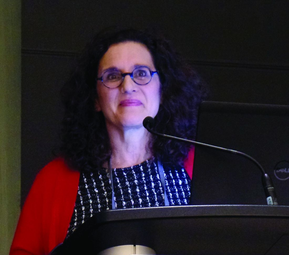
Outcomes of macitentan (Opsumit) treatment were similar in patients who had PAH associated with systemic sclerosis (PAH-SSc) and patients who had idiopathic PAH (IPAH) or heritable PAH (HPAH), Vallerie McLaughlin, MD, of the University of Michigan, Ann Arbor, said at the meeting.
“Within the limits of a real-world registry, these data add to the growing body of evidence supporting the use of macitentan for treatment in patients with CTD [connective tissue disease],” Dr. McLaughlin said.
She and her colleagues evaluated data from the prospective OPUS registry (NCT02126943) and the retrospective OrPHeUS study (NCT03197688), both of which included patients who were newly started on macitentan.
Dr. McLaughlin presented data on 2,311 patients with IPAH/HPAH and 668 patients with PAH-SSc. She also presented data on patients with PAH-systemic lupus erythematosus and PAH-mixed CTD, but numbers in these groups were small, and outcomes were similar to those in the PAH-SSc group.
Demographic and disease characteristics at the start of macitentan were similar between the IPAH/HPAH and PAH-SSc groups. The median age was 64 years in both groups. The median time from PAH diagnosis was 7.6 months in the IPAH/HPAH group and 8.5 months in the PAH-SSc group.
The median duration of macitentan exposure was 13.4 months in the IPAH/HPAH group and 14.4 months in the PAH-SSc group. The proportion of patients receiving macitentan in combination with other therapies (double or triple combinations) increased from baseline to 6 months in both groups.
Hepatic adverse events occurred in 7.4% of IPAH/HPAH patients and 7.9% of PAH-SSc patients. The most common adverse events among the IPAH/HPAH and PAH-SSc groups in the OPUS registry alone were dyspnea (19% and 26.1%, respectively), peripheral edema (9.8% and 12.4%), fatigue (6.8% and 11.7%), anemia (6.7% and 11.7%), headache (10.2% and 11%), and dizziness (6.7% and 10.7%).
About 39% of patients in both groups discontinued macitentan. Similar proportions in each group discontinued because of adverse events (17% in the IPAH/HPAH group and 18.3% in the PAH-SSc group) and hepatic adverse events (0.2% and 0.7%, respectively).
The proportion of patients with at least one hospitalization was 36.2% in the IPAH/HPAH group and 40.1% in the PAH-SSc group.
The 12-month Kaplan-Meier survival estimate was 92.9% in the IPAH/HPAH group and 91.3% in the PAH-SSc group. The 24-month estimated survival rate was 85.6% and 82.1%, respectively.
The OPUS registry and OrPHeUS study are sponsored by Actelion. Dr. McLaughlin disclosed relationships with Actelion, Acceleron, Bayer, Caremark, CiVi Biopharma, Reata, Sonovie, and United Therapeutics.
SOURCE: McLaughlin V et al. CHEST 2019. Abstract, doi: 10.1016/j.chest.2019.08.827.
NEW ORLEANS – Real-world data support the use of macitentan to treat pulmonary arterial hypertension (PAH) associated with connective tissue disease, according to a speaker at the annual meeting of the American College of Chest Physicians.
Outcomes of macitentan (Opsumit) treatment were similar in patients who had PAH associated with systemic sclerosis (PAH-SSc) and patients who had idiopathic PAH (IPAH) or heritable PAH (HPAH), Vallerie McLaughlin, MD, of the University of Michigan, Ann Arbor, said at the meeting.
“Within the limits of a real-world registry, these data add to the growing body of evidence supporting the use of macitentan for treatment in patients with CTD [connective tissue disease],” Dr. McLaughlin said.
She and her colleagues evaluated data from the prospective OPUS registry (NCT02126943) and the retrospective OrPHeUS study (NCT03197688), both of which included patients who were newly started on macitentan.
Dr. McLaughlin presented data on 2,311 patients with IPAH/HPAH and 668 patients with PAH-SSc. She also presented data on patients with PAH-systemic lupus erythematosus and PAH-mixed CTD, but numbers in these groups were small, and outcomes were similar to those in the PAH-SSc group.
Demographic and disease characteristics at the start of macitentan were similar between the IPAH/HPAH and PAH-SSc groups. The median age was 64 years in both groups. The median time from PAH diagnosis was 7.6 months in the IPAH/HPAH group and 8.5 months in the PAH-SSc group.
The median duration of macitentan exposure was 13.4 months in the IPAH/HPAH group and 14.4 months in the PAH-SSc group. The proportion of patients receiving macitentan in combination with other therapies (double or triple combinations) increased from baseline to 6 months in both groups.
Hepatic adverse events occurred in 7.4% of IPAH/HPAH patients and 7.9% of PAH-SSc patients. The most common adverse events among the IPAH/HPAH and PAH-SSc groups in the OPUS registry alone were dyspnea (19% and 26.1%, respectively), peripheral edema (9.8% and 12.4%), fatigue (6.8% and 11.7%), anemia (6.7% and 11.7%), headache (10.2% and 11%), and dizziness (6.7% and 10.7%).
About 39% of patients in both groups discontinued macitentan. Similar proportions in each group discontinued because of adverse events (17% in the IPAH/HPAH group and 18.3% in the PAH-SSc group) and hepatic adverse events (0.2% and 0.7%, respectively).
The proportion of patients with at least one hospitalization was 36.2% in the IPAH/HPAH group and 40.1% in the PAH-SSc group.
The 12-month Kaplan-Meier survival estimate was 92.9% in the IPAH/HPAH group and 91.3% in the PAH-SSc group. The 24-month estimated survival rate was 85.6% and 82.1%, respectively.
The OPUS registry and OrPHeUS study are sponsored by Actelion. Dr. McLaughlin disclosed relationships with Actelion, Acceleron, Bayer, Caremark, CiVi Biopharma, Reata, Sonovie, and United Therapeutics.
SOURCE: McLaughlin V et al. CHEST 2019. Abstract, doi: 10.1016/j.chest.2019.08.827.
NEW ORLEANS – Real-world data support the use of macitentan to treat pulmonary arterial hypertension (PAH) associated with connective tissue disease, according to a speaker at the annual meeting of the American College of Chest Physicians.
Outcomes of macitentan (Opsumit) treatment were similar in patients who had PAH associated with systemic sclerosis (PAH-SSc) and patients who had idiopathic PAH (IPAH) or heritable PAH (HPAH), Vallerie McLaughlin, MD, of the University of Michigan, Ann Arbor, said at the meeting.
“Within the limits of a real-world registry, these data add to the growing body of evidence supporting the use of macitentan for treatment in patients with CTD [connective tissue disease],” Dr. McLaughlin said.
She and her colleagues evaluated data from the prospective OPUS registry (NCT02126943) and the retrospective OrPHeUS study (NCT03197688), both of which included patients who were newly started on macitentan.
Dr. McLaughlin presented data on 2,311 patients with IPAH/HPAH and 668 patients with PAH-SSc. She also presented data on patients with PAH-systemic lupus erythematosus and PAH-mixed CTD, but numbers in these groups were small, and outcomes were similar to those in the PAH-SSc group.
Demographic and disease characteristics at the start of macitentan were similar between the IPAH/HPAH and PAH-SSc groups. The median age was 64 years in both groups. The median time from PAH diagnosis was 7.6 months in the IPAH/HPAH group and 8.5 months in the PAH-SSc group.
The median duration of macitentan exposure was 13.4 months in the IPAH/HPAH group and 14.4 months in the PAH-SSc group. The proportion of patients receiving macitentan in combination with other therapies (double or triple combinations) increased from baseline to 6 months in both groups.
Hepatic adverse events occurred in 7.4% of IPAH/HPAH patients and 7.9% of PAH-SSc patients. The most common adverse events among the IPAH/HPAH and PAH-SSc groups in the OPUS registry alone were dyspnea (19% and 26.1%, respectively), peripheral edema (9.8% and 12.4%), fatigue (6.8% and 11.7%), anemia (6.7% and 11.7%), headache (10.2% and 11%), and dizziness (6.7% and 10.7%).
About 39% of patients in both groups discontinued macitentan. Similar proportions in each group discontinued because of adverse events (17% in the IPAH/HPAH group and 18.3% in the PAH-SSc group) and hepatic adverse events (0.2% and 0.7%, respectively).
The proportion of patients with at least one hospitalization was 36.2% in the IPAH/HPAH group and 40.1% in the PAH-SSc group.
The 12-month Kaplan-Meier survival estimate was 92.9% in the IPAH/HPAH group and 91.3% in the PAH-SSc group. The 24-month estimated survival rate was 85.6% and 82.1%, respectively.
The OPUS registry and OrPHeUS study are sponsored by Actelion. Dr. McLaughlin disclosed relationships with Actelion, Acceleron, Bayer, Caremark, CiVi Biopharma, Reata, Sonovie, and United Therapeutics.
SOURCE: McLaughlin V et al. CHEST 2019. Abstract, doi: 10.1016/j.chest.2019.08.827.
REPORTING FROM CHEST 2019
Net prices of drugs rising four-times faster than inflation
NATIONAL HARBOR, MD. – The net prices of drugs are increasing four times faster than the rate of inflation, despite being offset 43% from list prices.

List prices increased by 232% from 2007 to 2018 (12% per year) and net prices increased 133% during that same time period. For Medicaid, the gross-to-net discount increased from 40% in 2007 to 68% in 2018. For all other payers, the increase was 22%-50% during that same period, Inmaculada Hernandez, PharmD, and colleagues reported at annual meeting of the Academy of Managed Care Pharmacy.
The investigators also found a wide variation on discounts across therapeutic classes. For example, list price for drugs in the multiple sclerosis category increased 407% over the study period while net price increased 221%. Insulins came in second in terms of gross price increases (337%) but saw only net prices increases by 83% due to increasing discounts, according to Dr. Hernandez, assistant professor of pharmacy and therapeutics at the University of Pittsburgh.
List prices for noninsulin diabetes treatments tripled during the observation period, but net prices went up only 24%. List price increases were lowest in the antineoplastic class, averaging 135%, though there were only 34% in rebates to offset the list price, resulting in an average net price increase of 89%.
Research was based on pricing data supplied by investment firm SSR Health for branded products and U.S. sales reported by publicly traded companies. The National Heart, Lung, and Blood Institute sponsored the study.
SOURCE: Hernandez I et a. AMCP Nexus, poster U2.
NATIONAL HARBOR, MD. – The net prices of drugs are increasing four times faster than the rate of inflation, despite being offset 43% from list prices.
List prices increased by 232% from 2007 to 2018 (12% per year) and net prices increased 133% during that same time period. For Medicaid, the gross-to-net discount increased from 40% in 2007 to 68% in 2018. For all other payers, the increase was 22%-50% during that same period, Inmaculada Hernandez, PharmD, and colleagues reported at annual meeting of the Academy of Managed Care Pharmacy.
The investigators also found a wide variation on discounts across therapeutic classes. For example, list price for drugs in the multiple sclerosis category increased 407% over the study period while net price increased 221%. Insulins came in second in terms of gross price increases (337%) but saw only net prices increases by 83% due to increasing discounts, according to Dr. Hernandez, assistant professor of pharmacy and therapeutics at the University of Pittsburgh.
List prices for noninsulin diabetes treatments tripled during the observation period, but net prices went up only 24%. List price increases were lowest in the antineoplastic class, averaging 135%, though there were only 34% in rebates to offset the list price, resulting in an average net price increase of 89%.
Research was based on pricing data supplied by investment firm SSR Health for branded products and U.S. sales reported by publicly traded companies. The National Heart, Lung, and Blood Institute sponsored the study.
SOURCE: Hernandez I et a. AMCP Nexus, poster U2.
NATIONAL HARBOR, MD. – The net prices of drugs are increasing four times faster than the rate of inflation, despite being offset 43% from list prices.
List prices increased by 232% from 2007 to 2018 (12% per year) and net prices increased 133% during that same time period. For Medicaid, the gross-to-net discount increased from 40% in 2007 to 68% in 2018. For all other payers, the increase was 22%-50% during that same period, Inmaculada Hernandez, PharmD, and colleagues reported at annual meeting of the Academy of Managed Care Pharmacy.
The investigators also found a wide variation on discounts across therapeutic classes. For example, list price for drugs in the multiple sclerosis category increased 407% over the study period while net price increased 221%. Insulins came in second in terms of gross price increases (337%) but saw only net prices increases by 83% due to increasing discounts, according to Dr. Hernandez, assistant professor of pharmacy and therapeutics at the University of Pittsburgh.
List prices for noninsulin diabetes treatments tripled during the observation period, but net prices went up only 24%. List price increases were lowest in the antineoplastic class, averaging 135%, though there were only 34% in rebates to offset the list price, resulting in an average net price increase of 89%.
Research was based on pricing data supplied by investment firm SSR Health for branded products and U.S. sales reported by publicly traded companies. The National Heart, Lung, and Blood Institute sponsored the study.
SOURCE: Hernandez I et a. AMCP Nexus, poster U2.
REPORTING FROM AMCP NEXUS 2019
Quick Byte: DeepMind emerges
The science of prediction
Hundreds of scientists around the world enter a competition every 2 years called the Critical Assessment of Structure Prediction.

“Tackling a biological puzzle they call ‘the protein folding problem,’ they try to predict the three-dimensional shape of proteins in the human body. No one knows how to solve the problem. Even the winners only chip away at it. But a solution could streamline the way scientists create new medicines and fight disease,” according to a report in the New York Times.
In 2019, those scientists did not win the contest. “It was won by DeepMind, the artificial intelligence lab owned by Google’s parent company. DeepMind specializes in ‘deep learning,’ a type of artificial intelligence that is rapidly changing drug discovery science.”
Reference
1. Metz C. “Making New Drugs With a Dose of Artificial Intelligence,” New York Times. Feb. 5, 2019. https://www.nytimes.com/2019/02/05/technology/artificial-intelligence-drug-research-deepmind.html. Accessed Feb 7, 2019.
The science of prediction
The science of prediction
Hundreds of scientists around the world enter a competition every 2 years called the Critical Assessment of Structure Prediction.
“Tackling a biological puzzle they call ‘the protein folding problem,’ they try to predict the three-dimensional shape of proteins in the human body. No one knows how to solve the problem. Even the winners only chip away at it. But a solution could streamline the way scientists create new medicines and fight disease,” according to a report in the New York Times.
In 2019, those scientists did not win the contest. “It was won by DeepMind, the artificial intelligence lab owned by Google’s parent company. DeepMind specializes in ‘deep learning,’ a type of artificial intelligence that is rapidly changing drug discovery science.”
Reference
1. Metz C. “Making New Drugs With a Dose of Artificial Intelligence,” New York Times. Feb. 5, 2019. https://www.nytimes.com/2019/02/05/technology/artificial-intelligence-drug-research-deepmind.html. Accessed Feb 7, 2019.
Hundreds of scientists around the world enter a competition every 2 years called the Critical Assessment of Structure Prediction.
“Tackling a biological puzzle they call ‘the protein folding problem,’ they try to predict the three-dimensional shape of proteins in the human body. No one knows how to solve the problem. Even the winners only chip away at it. But a solution could streamline the way scientists create new medicines and fight disease,” according to a report in the New York Times.
In 2019, those scientists did not win the contest. “It was won by DeepMind, the artificial intelligence lab owned by Google’s parent company. DeepMind specializes in ‘deep learning,’ a type of artificial intelligence that is rapidly changing drug discovery science.”
Reference
1. Metz C. “Making New Drugs With a Dose of Artificial Intelligence,” New York Times. Feb. 5, 2019. https://www.nytimes.com/2019/02/05/technology/artificial-intelligence-drug-research-deepmind.html. Accessed Feb 7, 2019.
Thousands of Emergency-Care Claims Were Denied; Court Rules VA Must Reimburse
When veterans receive emergency care at non-VA facilities, they can file for reimbursement of non-VA emergency care costs. If the claims are denied or rejected, non-VA facilities and providers can bill the veterans for some or all of the costs.
A stunning VA Office of Inspector General (OIG) report issued in August revealed that the VA had wrongfully rejected thousands of those emergency-care claims between April 1, 2017 and September 30, 2017. The OIG report said VA supervisors had pressured staff to quickly decide claims and even deny claims to maximize productivity. That culture of “speed over accuracy” led to 31% of claims being “inappropriately processed.”
Under regulations, when a claim is denied it is because there is no basis for payment, but a claim may also be rejected, meaning it cannot be decided until the claimant provides additional or corrected information. When the Claims Adjudication and Reimbursement Directorate (CAR) in the VA Office of Community Care (OCC) denies a claim, the claimant may have to pay out of pocket for emergency care.
The billed amount of inappropriately processed claims in those 5 months that were denied or rejected was “large in the aggregate,” the report said: an estimated $716 million, presenting potential undue financial risk to an estimated 60,800 veterans. The OIG estimated that about 17,400 veterans, with bills of at least $53.3 million, were negatively affected during the audit period. If corrective actions are not taken, the OIG estimates that the errors could result in $533 million in improper underpayments over 5 years.
The OIG audit was initiated after then Representative Tim Walz (D-MN) raised the concern in September 2017 about claims processors denying non-VA claims not only to meet production goals, but also to receive incentives such as high-performance ratings, bonuses, and telework privileges. The OIG audit also found that examiners who did not consistently meet the production numbers were not considered for overtime and had their telework privileges removed.
Other processing errors were also significant, the report says. For example, claimants did not receive complete and accurate information regarding why their claims were denied or rejected. Those errors created a risk that claimants could not effectively respond with information to get their claims approved.
The pressure to approve or deny claims quickly stemmed from a backlog of claims > 30 days old, which OCC and CAR leaders tried to reduce, the report says.
The OIG notes that since FY 2017 the OCC “experienced several leadership changes,” including 3 CAR directors. While the audit’s findings cannot be tied directly to those changes, the instability “provided the context in which problems were allowed to continue without remediation.”
In May, VHA officials reported to the audit team that they had implemented process improvements and initiatives that affect claims processing nationwide, and “the environment in which claims are processed.” Richard Stone, executive in charge of the VHA, submitted plans to comply with the IG’s 11 recommendations, which included tying incentives to all performance standards rather than just production quantity.
The US Court of Appeals for Veterans Claims recently ruled that the VA must reimburse veterans for emergency medical care at non-VA facilities—potentially between $1.8 billion and $6.5 billion in reimbursements for veterans who have filed or will file claims between 2016 and 2025. The court also struck down an internal VA regulation that blocked such payments. In 2015, the court had struck down a previous version of the internal regulation that refused coverage for an emergency claim when another form of insurance covered even a small part of the bill. The court said that regulation violated a 2010 federal law. In last month’s ruling, the court found the department had violated the same federal law with its revision of the reimbursement regulation, issued in 2018. The new rule, the panel said, created a new obstacle for veterans.
When veterans receive emergency care at non-VA facilities, they can file for reimbursement of non-VA emergency care costs. If the claims are denied or rejected, non-VA facilities and providers can bill the veterans for some or all of the costs.
A stunning VA Office of Inspector General (OIG) report issued in August revealed that the VA had wrongfully rejected thousands of those emergency-care claims between April 1, 2017 and September 30, 2017. The OIG report said VA supervisors had pressured staff to quickly decide claims and even deny claims to maximize productivity. That culture of “speed over accuracy” led to 31% of claims being “inappropriately processed.”
Under regulations, when a claim is denied it is because there is no basis for payment, but a claim may also be rejected, meaning it cannot be decided until the claimant provides additional or corrected information. When the Claims Adjudication and Reimbursement Directorate (CAR) in the VA Office of Community Care (OCC) denies a claim, the claimant may have to pay out of pocket for emergency care.
The billed amount of inappropriately processed claims in those 5 months that were denied or rejected was “large in the aggregate,” the report said: an estimated $716 million, presenting potential undue financial risk to an estimated 60,800 veterans. The OIG estimated that about 17,400 veterans, with bills of at least $53.3 million, were negatively affected during the audit period. If corrective actions are not taken, the OIG estimates that the errors could result in $533 million in improper underpayments over 5 years.
The OIG audit was initiated after then Representative Tim Walz (D-MN) raised the concern in September 2017 about claims processors denying non-VA claims not only to meet production goals, but also to receive incentives such as high-performance ratings, bonuses, and telework privileges. The OIG audit also found that examiners who did not consistently meet the production numbers were not considered for overtime and had their telework privileges removed.
Other processing errors were also significant, the report says. For example, claimants did not receive complete and accurate information regarding why their claims were denied or rejected. Those errors created a risk that claimants could not effectively respond with information to get their claims approved.
The pressure to approve or deny claims quickly stemmed from a backlog of claims > 30 days old, which OCC and CAR leaders tried to reduce, the report says.
The OIG notes that since FY 2017 the OCC “experienced several leadership changes,” including 3 CAR directors. While the audit’s findings cannot be tied directly to those changes, the instability “provided the context in which problems were allowed to continue without remediation.”
In May, VHA officials reported to the audit team that they had implemented process improvements and initiatives that affect claims processing nationwide, and “the environment in which claims are processed.” Richard Stone, executive in charge of the VHA, submitted plans to comply with the IG’s 11 recommendations, which included tying incentives to all performance standards rather than just production quantity.
The US Court of Appeals for Veterans Claims recently ruled that the VA must reimburse veterans for emergency medical care at non-VA facilities—potentially between $1.8 billion and $6.5 billion in reimbursements for veterans who have filed or will file claims between 2016 and 2025. The court also struck down an internal VA regulation that blocked such payments. In 2015, the court had struck down a previous version of the internal regulation that refused coverage for an emergency claim when another form of insurance covered even a small part of the bill. The court said that regulation violated a 2010 federal law. In last month’s ruling, the court found the department had violated the same federal law with its revision of the reimbursement regulation, issued in 2018. The new rule, the panel said, created a new obstacle for veterans.
When veterans receive emergency care at non-VA facilities, they can file for reimbursement of non-VA emergency care costs. If the claims are denied or rejected, non-VA facilities and providers can bill the veterans for some or all of the costs.
A stunning VA Office of Inspector General (OIG) report issued in August revealed that the VA had wrongfully rejected thousands of those emergency-care claims between April 1, 2017 and September 30, 2017. The OIG report said VA supervisors had pressured staff to quickly decide claims and even deny claims to maximize productivity. That culture of “speed over accuracy” led to 31% of claims being “inappropriately processed.”
Under regulations, when a claim is denied it is because there is no basis for payment, but a claim may also be rejected, meaning it cannot be decided until the claimant provides additional or corrected information. When the Claims Adjudication and Reimbursement Directorate (CAR) in the VA Office of Community Care (OCC) denies a claim, the claimant may have to pay out of pocket for emergency care.
The billed amount of inappropriately processed claims in those 5 months that were denied or rejected was “large in the aggregate,” the report said: an estimated $716 million, presenting potential undue financial risk to an estimated 60,800 veterans. The OIG estimated that about 17,400 veterans, with bills of at least $53.3 million, were negatively affected during the audit period. If corrective actions are not taken, the OIG estimates that the errors could result in $533 million in improper underpayments over 5 years.
The OIG audit was initiated after then Representative Tim Walz (D-MN) raised the concern in September 2017 about claims processors denying non-VA claims not only to meet production goals, but also to receive incentives such as high-performance ratings, bonuses, and telework privileges. The OIG audit also found that examiners who did not consistently meet the production numbers were not considered for overtime and had their telework privileges removed.
Other processing errors were also significant, the report says. For example, claimants did not receive complete and accurate information regarding why their claims were denied or rejected. Those errors created a risk that claimants could not effectively respond with information to get their claims approved.
The pressure to approve or deny claims quickly stemmed from a backlog of claims > 30 days old, which OCC and CAR leaders tried to reduce, the report says.
The OIG notes that since FY 2017 the OCC “experienced several leadership changes,” including 3 CAR directors. While the audit’s findings cannot be tied directly to those changes, the instability “provided the context in which problems were allowed to continue without remediation.”
In May, VHA officials reported to the audit team that they had implemented process improvements and initiatives that affect claims processing nationwide, and “the environment in which claims are processed.” Richard Stone, executive in charge of the VHA, submitted plans to comply with the IG’s 11 recommendations, which included tying incentives to all performance standards rather than just production quantity.
The US Court of Appeals for Veterans Claims recently ruled that the VA must reimburse veterans for emergency medical care at non-VA facilities—potentially between $1.8 billion and $6.5 billion in reimbursements for veterans who have filed or will file claims between 2016 and 2025. The court also struck down an internal VA regulation that blocked such payments. In 2015, the court had struck down a previous version of the internal regulation that refused coverage for an emergency claim when another form of insurance covered even a small part of the bill. The court said that regulation violated a 2010 federal law. In last month’s ruling, the court found the department had violated the same federal law with its revision of the reimbursement regulation, issued in 2018. The new rule, the panel said, created a new obstacle for veterans.