User login
Private practice to private equity-backed MSO - perspectives from the United Digestive team - Part 1
Author's note: This is the first of a two-part series. In December 2018, Atlanta Gastroenterology Associates partnered with Frazier Healthcare Partners to form the practice management company United Digestive (UD). Since that time, colleagues across the country have evaluated their own private equity prospects and partnerships, as well as monitored the progress of our transition.
Our guiding principle is to provide a best-in-class operational infrastructure, so independent gastroenterologists can focus on delivering the highest quality patient care. Thus, in the first year, significant efforts and capital have been invested into United Digestive’s scalable platform to promote organic growth, as well as facilitate a smooth transition for other groups and physicians joining the team. So how are things going? Enjoy this two-part article where we reached out to several team members from all levels within the organization and asked them to share their personal experiences – both highlights and challenges – during UD’s first year. Here’s what they had to say.
During the COVID-19 crisis, how has UD management responded? How has the UD management services organization model affected partner level physician (PLP) compensation?
Dr. Marc Rosenberg, UD Physician Executive Committee member:
- “Immediately, the entire leadership team recognized the threat of COVID to our community, patients, staff, and business. A multidisciplinary task force including clinical and business leaders utilized our PE partner’s vast resources, local hospital expertise, national societal recommendations and colleagues’ experiences from around the country to focus on protocols and procedures to protect our patients and staff. A few of the team’s timely decisions included: closing the majority of our patient fronting services, transitioning to telehealth, hiring an infection control consultant, allowing physical distancing of our staff with off sight work, instituting symptomatic pathways, donating personal protective gear to local hospitals, covering all benefits as well as provided resources to obtain government benefits to furloughed team members, developing a provider wellness program, and encouraging hospital coverage considerations for high risk providers. I also know the team is focusing on how we re-open at the appropriate time with necessary safety considerations, like investigating testing options and ensuring appropriate PPE is in place. The collaboration between clinical and business leadership has been tremendous through this evolving and challenging period. As for UD physician partner compensation, our model is uniquely organized such that the MSO covers all overhead expenses with partners contributing a fixed percentage of a partner’s collections, while others typically share overhead expenses. During uncertain times, like the COVID-19 crisis, it is reassuring to partners to know that they are not responsible for the cost of infrastructure (i.e. leases, capital equipment, EHR system, consultants, etc.) and staffing, including current and new associates.”
With formation of United Digestive as an MSO, has your day-to-day work life changed or your clinical decision-making been impacted?
Dr. Aja McCutchen, UD Physician Executive Committee Member:
- “With the formation of UD, my daily work life has changed very little; however, with their focus on improving “back-office” functions, my schedule is now fully optimized by reducing gaps from cancellations with same-day/next-day scheduling. In addition, the patient experience has been enhanced with decreased wait times, easier appointment scheduling, and quicker access to support staff. The procurement of business intelligence tools, and, more importantly, the implementation of dashboards, has provided much needed visibility across the organization allowing managerial decisions to be driven by accurate data.
From a clinical decision-making standpoint, Atlanta Gastroenterology was already armed with strong clinical teams and committees. We have been able to build upon our pre-existing committees and optimize their ability to steward best practices and develop clinical pathways. This, in turn, translates to consistency across the organization in the delivery of evidence-based, comprehensive GI care.”
Kimberly Orleck, PA-C, Advanced Practice Provider (APP) Supervisor:
- “The formation of UD has not affected my clinical decision-making abilities. In fact, this new platform is dedicated to empowering and establishing APPs as independent clinicians with appropriate physician oversight. As a result, I have welcomed more administrative responsibilities and have become more involved in business meetings and decision making. We have worked together to better utilize APPs using data to match supply with demand.”
Physician compensation improvement is typically a key concern for physicians who work with private equity MSOs. How has United Digestive performed for its partner-level physicians in year one?
Dr. Marc Rosenberg:
- “The MSO has helped to improve physician income – slowly at first and now on a steeper trajectory. We have been ahead of expected income improvement based on models we reviewed when evaluating the formations of an MSO in potential partnership with Frazier Healthcare Partners. United Digestive’s EBIDTA, of which each partner-level physician owns a significant percentage through shares from rollover proceeds, has grown impressively in one year. This has been achieved mostly through significant organic growth and to a lesser degree through mergers and acquisitions. UD has helped to enhance the bottom line through increased reimbursements from payor negotiated contracts, new revenue-generating service lines, and operational efficiencies.”
Dr. Patel and Dr. Sonnenshine are with Atlanta Gastroenterology Associates in Atlanta, which is part of United Digestive. They have no conflicts.
*This story was updated on 6/1/2020.
Author's note: This is the first of a two-part series. In December 2018, Atlanta Gastroenterology Associates partnered with Frazier Healthcare Partners to form the practice management company United Digestive (UD). Since that time, colleagues across the country have evaluated their own private equity prospects and partnerships, as well as monitored the progress of our transition.
Our guiding principle is to provide a best-in-class operational infrastructure, so independent gastroenterologists can focus on delivering the highest quality patient care. Thus, in the first year, significant efforts and capital have been invested into United Digestive’s scalable platform to promote organic growth, as well as facilitate a smooth transition for other groups and physicians joining the team. So how are things going? Enjoy this two-part article where we reached out to several team members from all levels within the organization and asked them to share their personal experiences – both highlights and challenges – during UD’s first year. Here’s what they had to say.
During the COVID-19 crisis, how has UD management responded? How has the UD management services organization model affected partner level physician (PLP) compensation?
Dr. Marc Rosenberg, UD Physician Executive Committee member:
- “Immediately, the entire leadership team recognized the threat of COVID to our community, patients, staff, and business. A multidisciplinary task force including clinical and business leaders utilized our PE partner’s vast resources, local hospital expertise, national societal recommendations and colleagues’ experiences from around the country to focus on protocols and procedures to protect our patients and staff. A few of the team’s timely decisions included: closing the majority of our patient fronting services, transitioning to telehealth, hiring an infection control consultant, allowing physical distancing of our staff with off sight work, instituting symptomatic pathways, donating personal protective gear to local hospitals, covering all benefits as well as provided resources to obtain government benefits to furloughed team members, developing a provider wellness program, and encouraging hospital coverage considerations for high risk providers. I also know the team is focusing on how we re-open at the appropriate time with necessary safety considerations, like investigating testing options and ensuring appropriate PPE is in place. The collaboration between clinical and business leadership has been tremendous through this evolving and challenging period. As for UD physician partner compensation, our model is uniquely organized such that the MSO covers all overhead expenses with partners contributing a fixed percentage of a partner’s collections, while others typically share overhead expenses. During uncertain times, like the COVID-19 crisis, it is reassuring to partners to know that they are not responsible for the cost of infrastructure (i.e. leases, capital equipment, EHR system, consultants, etc.) and staffing, including current and new associates.”
With formation of United Digestive as an MSO, has your day-to-day work life changed or your clinical decision-making been impacted?
Dr. Aja McCutchen, UD Physician Executive Committee Member:
- “With the formation of UD, my daily work life has changed very little; however, with their focus on improving “back-office” functions, my schedule is now fully optimized by reducing gaps from cancellations with same-day/next-day scheduling. In addition, the patient experience has been enhanced with decreased wait times, easier appointment scheduling, and quicker access to support staff. The procurement of business intelligence tools, and, more importantly, the implementation of dashboards, has provided much needed visibility across the organization allowing managerial decisions to be driven by accurate data.
From a clinical decision-making standpoint, Atlanta Gastroenterology was already armed with strong clinical teams and committees. We have been able to build upon our pre-existing committees and optimize their ability to steward best practices and develop clinical pathways. This, in turn, translates to consistency across the organization in the delivery of evidence-based, comprehensive GI care.”
Kimberly Orleck, PA-C, Advanced Practice Provider (APP) Supervisor:
- “The formation of UD has not affected my clinical decision-making abilities. In fact, this new platform is dedicated to empowering and establishing APPs as independent clinicians with appropriate physician oversight. As a result, I have welcomed more administrative responsibilities and have become more involved in business meetings and decision making. We have worked together to better utilize APPs using data to match supply with demand.”
Physician compensation improvement is typically a key concern for physicians who work with private equity MSOs. How has United Digestive performed for its partner-level physicians in year one?
Dr. Marc Rosenberg:
- “The MSO has helped to improve physician income – slowly at first and now on a steeper trajectory. We have been ahead of expected income improvement based on models we reviewed when evaluating the formations of an MSO in potential partnership with Frazier Healthcare Partners. United Digestive’s EBIDTA, of which each partner-level physician owns a significant percentage through shares from rollover proceeds, has grown impressively in one year. This has been achieved mostly through significant organic growth and to a lesser degree through mergers and acquisitions. UD has helped to enhance the bottom line through increased reimbursements from payor negotiated contracts, new revenue-generating service lines, and operational efficiencies.”
Dr. Patel and Dr. Sonnenshine are with Atlanta Gastroenterology Associates in Atlanta, which is part of United Digestive. They have no conflicts.
*This story was updated on 6/1/2020.
Author's note: This is the first of a two-part series. In December 2018, Atlanta Gastroenterology Associates partnered with Frazier Healthcare Partners to form the practice management company United Digestive (UD). Since that time, colleagues across the country have evaluated their own private equity prospects and partnerships, as well as monitored the progress of our transition.
Our guiding principle is to provide a best-in-class operational infrastructure, so independent gastroenterologists can focus on delivering the highest quality patient care. Thus, in the first year, significant efforts and capital have been invested into United Digestive’s scalable platform to promote organic growth, as well as facilitate a smooth transition for other groups and physicians joining the team. So how are things going? Enjoy this two-part article where we reached out to several team members from all levels within the organization and asked them to share their personal experiences – both highlights and challenges – during UD’s first year. Here’s what they had to say.
During the COVID-19 crisis, how has UD management responded? How has the UD management services organization model affected partner level physician (PLP) compensation?
Dr. Marc Rosenberg, UD Physician Executive Committee member:
- “Immediately, the entire leadership team recognized the threat of COVID to our community, patients, staff, and business. A multidisciplinary task force including clinical and business leaders utilized our PE partner’s vast resources, local hospital expertise, national societal recommendations and colleagues’ experiences from around the country to focus on protocols and procedures to protect our patients and staff. A few of the team’s timely decisions included: closing the majority of our patient fronting services, transitioning to telehealth, hiring an infection control consultant, allowing physical distancing of our staff with off sight work, instituting symptomatic pathways, donating personal protective gear to local hospitals, covering all benefits as well as provided resources to obtain government benefits to furloughed team members, developing a provider wellness program, and encouraging hospital coverage considerations for high risk providers. I also know the team is focusing on how we re-open at the appropriate time with necessary safety considerations, like investigating testing options and ensuring appropriate PPE is in place. The collaboration between clinical and business leadership has been tremendous through this evolving and challenging period. As for UD physician partner compensation, our model is uniquely organized such that the MSO covers all overhead expenses with partners contributing a fixed percentage of a partner’s collections, while others typically share overhead expenses. During uncertain times, like the COVID-19 crisis, it is reassuring to partners to know that they are not responsible for the cost of infrastructure (i.e. leases, capital equipment, EHR system, consultants, etc.) and staffing, including current and new associates.”
With formation of United Digestive as an MSO, has your day-to-day work life changed or your clinical decision-making been impacted?
Dr. Aja McCutchen, UD Physician Executive Committee Member:
- “With the formation of UD, my daily work life has changed very little; however, with their focus on improving “back-office” functions, my schedule is now fully optimized by reducing gaps from cancellations with same-day/next-day scheduling. In addition, the patient experience has been enhanced with decreased wait times, easier appointment scheduling, and quicker access to support staff. The procurement of business intelligence tools, and, more importantly, the implementation of dashboards, has provided much needed visibility across the organization allowing managerial decisions to be driven by accurate data.
From a clinical decision-making standpoint, Atlanta Gastroenterology was already armed with strong clinical teams and committees. We have been able to build upon our pre-existing committees and optimize their ability to steward best practices and develop clinical pathways. This, in turn, translates to consistency across the organization in the delivery of evidence-based, comprehensive GI care.”
Kimberly Orleck, PA-C, Advanced Practice Provider (APP) Supervisor:
- “The formation of UD has not affected my clinical decision-making abilities. In fact, this new platform is dedicated to empowering and establishing APPs as independent clinicians with appropriate physician oversight. As a result, I have welcomed more administrative responsibilities and have become more involved in business meetings and decision making. We have worked together to better utilize APPs using data to match supply with demand.”
Physician compensation improvement is typically a key concern for physicians who work with private equity MSOs. How has United Digestive performed for its partner-level physicians in year one?
Dr. Marc Rosenberg:
- “The MSO has helped to improve physician income – slowly at first and now on a steeper trajectory. We have been ahead of expected income improvement based on models we reviewed when evaluating the formations of an MSO in potential partnership with Frazier Healthcare Partners. United Digestive’s EBIDTA, of which each partner-level physician owns a significant percentage through shares from rollover proceeds, has grown impressively in one year. This has been achieved mostly through significant organic growth and to a lesser degree through mergers and acquisitions. UD has helped to enhance the bottom line through increased reimbursements from payor negotiated contracts, new revenue-generating service lines, and operational efficiencies.”
Dr. Patel and Dr. Sonnenshine are with Atlanta Gastroenterology Associates in Atlanta, which is part of United Digestive. They have no conflicts.
*This story was updated on 6/1/2020.
Standardized protocol guides therapies to reduce VTE after arthroplasty
A simple tool to guide choice of antithrombotic therapy following total joint arthroplasty led to a reduction in pulmonary embolism (PE) after being introduced systemwide, according to a prospectively tracked evaluation of a large patient cohort. The results of the study were reported in an abstract scheduled for release at the annual meeting of the American Academy of Orthopaedic Surgeons. The meeting was canceled because of COVID-19.
“We developed a simplified scoring system for evaluating risk of thromboembolism and guiding prophylaxis that led to a significant reduction in events across a large integrated health care system,” reported James Wylie, MD, associate medical director for hip and knee preservation and orthopedic research at Intermountain Healthcare, Salt Lake City, Utah.
The goal of the methodology was to create a uniform and evidence-based approach to risk assessment in order to guide selection of appropriate venous thromboembolism (VTE) prophylaxis. The tool takes into account the need to individualize antithrombotic drugs for risk of both VTE and for bleeding.
“VTE is a major threat following total joint replacement, but not all patients require anticoagulants. Recent evidence supports a shift to aspirin for low-risk patients,” explained Dr. Wylie in an interview.
The risk tool assigns points for such factors as history of VTE, older age, history of coronary artery disease, history of cancer, and increased body mass index. There are two possible ratings to guide strategies. Those with standard risk are candidates for 81 mg of aspirin twice daily. Those with high risk are candidates for 2.5 mg of apixaban, also administered twice daily. Custom dosing of warfarin is an alternative for the latter group. Regardless of strategy, prophylaxis is administered for 30 days following arthroplasty
“The risk score is calculated automatically, because you have to click a box in the electronic medical record for all of those factors as part of admission orders,” Dr. Wylie said.
The protocol was introduced in July 2017 and adoption was tracked prospectively over 18 months. In an evaluable cohort of 20,284 patients, PE rates in the 71% of patients adherent to the protocol were compared with the 29% who were not.
Over the observation period, the rates of PE were 0.34% and 0.62% (P = .004) for those adherent and nonadherent, respectively. The rate of unplanned readmissions and death, which were secondary outcomes, were both numerically lower in the group treated by adherent surgeons, but the differences did not reach statistical significance.
Adoption of the protocol by surgeons did increase over the course of the observation period, and this correlated with a decrease in unplanned readmissions. Bleeding-related readmission was a rare event in this analysis and did not significantly increase over time, according to Dr. Wylie.
The risk assessment tool, developed by a multispecialty team at Intermountain Healthcare, was based on a review of hundreds of published papers and guidelines, according to Nathan Momberger, MD, who is the associate medical director of total joint replacement at Intermountain and was a coauthor on this study. A member of the team that developed the risk assessment tool, Dr. Momberger noted that new risk score was developed at a time when clinicians have been moving quickly away from warfarin to direct oral anticoagulants.
“None of our surgeons were using the same VTE prophylaxis when we started this project,” Dr. Momberger said. This was a motivation for developing a systemwide approach. In the 22 participating hospitals, there were 50 surgeons performing total knee arthroplasty and 40 surgeons were performing total hip surgery at the time the new protocol was introduced.
Further analyses will provide a more detailed analysis of the effect of the protocol on other thrombotic events, including deep vein thrombosis, and on cost. Since these data were analyzed, protocol adoption has increased and now exceeds 80%, according to Dr. Wylie.
Although a standardized approach to VTE prophylaxis following total joint arthroplasty is attractive, the ideal strategy remains controversial, according to Sunny Parikh, MD, an orthopedic surgeon affiliated with Colchester (England) General Hospital.
As a coauthor of a recent study that quantified symptomatic VTE rates at his and a neighboring hospital over a 3-year period (BMC Musculoskelet Disord. 2020;21:95), Dr. Parikh reported that VTE rates did not reach zero even with a prolonged course of the low-molecular-weight heparin enoxaparin.
At 90 days, the symptomatic VTE rate was only 0.3% for total knee arthroplasty but reached 1.2% for total hip arthroplasty.
“At the time of this study we were using enoxaparin for 28 days following total hip replacements and for 14 days following total knee replacements,” Dr. Parikh reported. Since this study, his institution has switched to a regimen recommended by the U.K.’s National Institute for Health and Clinical Excellence (NICE).
Under the NICE guidelines, VTE prophylaxis for total hip arthroplasty is 40 mg enoxaparin once daily for 14 days followed by 75 mg aspirin for another 14 days, according to Dr. Parikh. For total knee arthroplasty, the standard regimen is 75 mg aspirin for 14 days.
For those who might not be best managed with the standard approach, “there is no clear guideline.” Rather, in patients with renal or liver impairment, “we discuss the case with the hematology team to adjust the doses,” Dr. Parikh reported.
The advantage of a standardized approach applied to all or most patients is that is eliminates disparities, but Dr. Parikh agreed that risk-adjusted prophylaxis might be warranted for optimal outcomes.
Dr. Wylie reported a financial relationship with Arthrex.
A simple tool to guide choice of antithrombotic therapy following total joint arthroplasty led to a reduction in pulmonary embolism (PE) after being introduced systemwide, according to a prospectively tracked evaluation of a large patient cohort. The results of the study were reported in an abstract scheduled for release at the annual meeting of the American Academy of Orthopaedic Surgeons. The meeting was canceled because of COVID-19.
“We developed a simplified scoring system for evaluating risk of thromboembolism and guiding prophylaxis that led to a significant reduction in events across a large integrated health care system,” reported James Wylie, MD, associate medical director for hip and knee preservation and orthopedic research at Intermountain Healthcare, Salt Lake City, Utah.
The goal of the methodology was to create a uniform and evidence-based approach to risk assessment in order to guide selection of appropriate venous thromboembolism (VTE) prophylaxis. The tool takes into account the need to individualize antithrombotic drugs for risk of both VTE and for bleeding.
“VTE is a major threat following total joint replacement, but not all patients require anticoagulants. Recent evidence supports a shift to aspirin for low-risk patients,” explained Dr. Wylie in an interview.
The risk tool assigns points for such factors as history of VTE, older age, history of coronary artery disease, history of cancer, and increased body mass index. There are two possible ratings to guide strategies. Those with standard risk are candidates for 81 mg of aspirin twice daily. Those with high risk are candidates for 2.5 mg of apixaban, also administered twice daily. Custom dosing of warfarin is an alternative for the latter group. Regardless of strategy, prophylaxis is administered for 30 days following arthroplasty
“The risk score is calculated automatically, because you have to click a box in the electronic medical record for all of those factors as part of admission orders,” Dr. Wylie said.
The protocol was introduced in July 2017 and adoption was tracked prospectively over 18 months. In an evaluable cohort of 20,284 patients, PE rates in the 71% of patients adherent to the protocol were compared with the 29% who were not.
Over the observation period, the rates of PE were 0.34% and 0.62% (P = .004) for those adherent and nonadherent, respectively. The rate of unplanned readmissions and death, which were secondary outcomes, were both numerically lower in the group treated by adherent surgeons, but the differences did not reach statistical significance.
Adoption of the protocol by surgeons did increase over the course of the observation period, and this correlated with a decrease in unplanned readmissions. Bleeding-related readmission was a rare event in this analysis and did not significantly increase over time, according to Dr. Wylie.
The risk assessment tool, developed by a multispecialty team at Intermountain Healthcare, was based on a review of hundreds of published papers and guidelines, according to Nathan Momberger, MD, who is the associate medical director of total joint replacement at Intermountain and was a coauthor on this study. A member of the team that developed the risk assessment tool, Dr. Momberger noted that new risk score was developed at a time when clinicians have been moving quickly away from warfarin to direct oral anticoagulants.
“None of our surgeons were using the same VTE prophylaxis when we started this project,” Dr. Momberger said. This was a motivation for developing a systemwide approach. In the 22 participating hospitals, there were 50 surgeons performing total knee arthroplasty and 40 surgeons were performing total hip surgery at the time the new protocol was introduced.
Further analyses will provide a more detailed analysis of the effect of the protocol on other thrombotic events, including deep vein thrombosis, and on cost. Since these data were analyzed, protocol adoption has increased and now exceeds 80%, according to Dr. Wylie.
Although a standardized approach to VTE prophylaxis following total joint arthroplasty is attractive, the ideal strategy remains controversial, according to Sunny Parikh, MD, an orthopedic surgeon affiliated with Colchester (England) General Hospital.
As a coauthor of a recent study that quantified symptomatic VTE rates at his and a neighboring hospital over a 3-year period (BMC Musculoskelet Disord. 2020;21:95), Dr. Parikh reported that VTE rates did not reach zero even with a prolonged course of the low-molecular-weight heparin enoxaparin.
At 90 days, the symptomatic VTE rate was only 0.3% for total knee arthroplasty but reached 1.2% for total hip arthroplasty.
“At the time of this study we were using enoxaparin for 28 days following total hip replacements and for 14 days following total knee replacements,” Dr. Parikh reported. Since this study, his institution has switched to a regimen recommended by the U.K.’s National Institute for Health and Clinical Excellence (NICE).
Under the NICE guidelines, VTE prophylaxis for total hip arthroplasty is 40 mg enoxaparin once daily for 14 days followed by 75 mg aspirin for another 14 days, according to Dr. Parikh. For total knee arthroplasty, the standard regimen is 75 mg aspirin for 14 days.
For those who might not be best managed with the standard approach, “there is no clear guideline.” Rather, in patients with renal or liver impairment, “we discuss the case with the hematology team to adjust the doses,” Dr. Parikh reported.
The advantage of a standardized approach applied to all or most patients is that is eliminates disparities, but Dr. Parikh agreed that risk-adjusted prophylaxis might be warranted for optimal outcomes.
Dr. Wylie reported a financial relationship with Arthrex.
A simple tool to guide choice of antithrombotic therapy following total joint arthroplasty led to a reduction in pulmonary embolism (PE) after being introduced systemwide, according to a prospectively tracked evaluation of a large patient cohort. The results of the study were reported in an abstract scheduled for release at the annual meeting of the American Academy of Orthopaedic Surgeons. The meeting was canceled because of COVID-19.
“We developed a simplified scoring system for evaluating risk of thromboembolism and guiding prophylaxis that led to a significant reduction in events across a large integrated health care system,” reported James Wylie, MD, associate medical director for hip and knee preservation and orthopedic research at Intermountain Healthcare, Salt Lake City, Utah.
The goal of the methodology was to create a uniform and evidence-based approach to risk assessment in order to guide selection of appropriate venous thromboembolism (VTE) prophylaxis. The tool takes into account the need to individualize antithrombotic drugs for risk of both VTE and for bleeding.
“VTE is a major threat following total joint replacement, but not all patients require anticoagulants. Recent evidence supports a shift to aspirin for low-risk patients,” explained Dr. Wylie in an interview.
The risk tool assigns points for such factors as history of VTE, older age, history of coronary artery disease, history of cancer, and increased body mass index. There are two possible ratings to guide strategies. Those with standard risk are candidates for 81 mg of aspirin twice daily. Those with high risk are candidates for 2.5 mg of apixaban, also administered twice daily. Custom dosing of warfarin is an alternative for the latter group. Regardless of strategy, prophylaxis is administered for 30 days following arthroplasty
“The risk score is calculated automatically, because you have to click a box in the electronic medical record for all of those factors as part of admission orders,” Dr. Wylie said.
The protocol was introduced in July 2017 and adoption was tracked prospectively over 18 months. In an evaluable cohort of 20,284 patients, PE rates in the 71% of patients adherent to the protocol were compared with the 29% who were not.
Over the observation period, the rates of PE were 0.34% and 0.62% (P = .004) for those adherent and nonadherent, respectively. The rate of unplanned readmissions and death, which were secondary outcomes, were both numerically lower in the group treated by adherent surgeons, but the differences did not reach statistical significance.
Adoption of the protocol by surgeons did increase over the course of the observation period, and this correlated with a decrease in unplanned readmissions. Bleeding-related readmission was a rare event in this analysis and did not significantly increase over time, according to Dr. Wylie.
The risk assessment tool, developed by a multispecialty team at Intermountain Healthcare, was based on a review of hundreds of published papers and guidelines, according to Nathan Momberger, MD, who is the associate medical director of total joint replacement at Intermountain and was a coauthor on this study. A member of the team that developed the risk assessment tool, Dr. Momberger noted that new risk score was developed at a time when clinicians have been moving quickly away from warfarin to direct oral anticoagulants.
“None of our surgeons were using the same VTE prophylaxis when we started this project,” Dr. Momberger said. This was a motivation for developing a systemwide approach. In the 22 participating hospitals, there were 50 surgeons performing total knee arthroplasty and 40 surgeons were performing total hip surgery at the time the new protocol was introduced.
Further analyses will provide a more detailed analysis of the effect of the protocol on other thrombotic events, including deep vein thrombosis, and on cost. Since these data were analyzed, protocol adoption has increased and now exceeds 80%, according to Dr. Wylie.
Although a standardized approach to VTE prophylaxis following total joint arthroplasty is attractive, the ideal strategy remains controversial, according to Sunny Parikh, MD, an orthopedic surgeon affiliated with Colchester (England) General Hospital.
As a coauthor of a recent study that quantified symptomatic VTE rates at his and a neighboring hospital over a 3-year period (BMC Musculoskelet Disord. 2020;21:95), Dr. Parikh reported that VTE rates did not reach zero even with a prolonged course of the low-molecular-weight heparin enoxaparin.
At 90 days, the symptomatic VTE rate was only 0.3% for total knee arthroplasty but reached 1.2% for total hip arthroplasty.
“At the time of this study we were using enoxaparin for 28 days following total hip replacements and for 14 days following total knee replacements,” Dr. Parikh reported. Since this study, his institution has switched to a regimen recommended by the U.K.’s National Institute for Health and Clinical Excellence (NICE).
Under the NICE guidelines, VTE prophylaxis for total hip arthroplasty is 40 mg enoxaparin once daily for 14 days followed by 75 mg aspirin for another 14 days, according to Dr. Parikh. For total knee arthroplasty, the standard regimen is 75 mg aspirin for 14 days.
For those who might not be best managed with the standard approach, “there is no clear guideline.” Rather, in patients with renal or liver impairment, “we discuss the case with the hematology team to adjust the doses,” Dr. Parikh reported.
The advantage of a standardized approach applied to all or most patients is that is eliminates disparities, but Dr. Parikh agreed that risk-adjusted prophylaxis might be warranted for optimal outcomes.
Dr. Wylie reported a financial relationship with Arthrex.
REPORTING FROM AAOS 2020
LOTUS: Ipatasertib plus paclitaxel may prolong OS in TNBC
according to final results of the phase 2 LOTUS trial.
The median OS was 25.8 months in patients who received ipatasertib plus paclitaxel and 16.9 months in patients who received paclitaxel plus placebo. Although this difference was not statistically significant, it was “clinically meaningful,” according to Rebecca Dent, MD, of the National Cancer Center Singapore. Dr. Dent presented OS data from the LOTUS trial at the European Society of Medical Oncology: Breast Cancer virtual meeting.
Previously reported data showed a progression-free survival benefit in the ipatasertib arm, compared with the placebo arm – 6.2 months and 4.9 months, respectively (Lancet Oncol. 2017 Oct;18[10]:1360-72). An enhanced effect was noted in patients with PIK3CA/AKT1/PTEN–altered tumors, but the OS data were not mature at the time of that primary analysis.
Patients, treatment, and safety
LOTUS participants had measurable locally advanced/metastatic TNBC that was previously untreated with systemic therapy, and they were stratified by prior adjuvant therapy, chemotherapy-free interval, and tumor immunohistochemistry PTEN status.
Patients were randomized 1:1 to receive paclitaxel at a dose of 80 mg/m2 on days 1, 8, and 15 of each 28-day cycle plus either placebo (n = 62) or ipatasertib at a dose of 400 mg on days 1-21 (n = 62).
The safety results didn’t differ between the primary and updated results, Dr. Dent noted.
“What we can say is that the combination is extremely well tolerated, especially when we compare it to other targeted agents targeting this pathway,” she said. “What we do see is noticeable diarrhea ... [and] an increase in sensory neuropathy, which is not entirely unexpected.”
Subsequent systemic anticancer therapy was required in 77% of patients in the ipatasertib arm and 90% of patients in the placebo arm.
OS results
The final OS data show a numerical advantage for patients in the ipatasertib arm compared with the placebo arm. The 1-year OS was 83% in the ipatasertib arm and 68% in the placebo arm. The median OS was 25.8 months and 16.9 months, respectively (stratified hazard ratio, 0.80).
“This is a clinically meaningful improvement of 9 months in overall survival, which is indeed not too dissimilar to what we have seen in patients with PD-L1-positive TNBC who are receiving immune checkpoint inhibition in combination with chemotherapy,” Dr. Dent said.
However, the 95% confidence interval for the stratified OS hazard ratio crossed 1 (0.50-1.28). Therefore, the findings require confirmatory phase 3 trial results, Dr. Dent said.
The OS improvements with ipatasertib were seen “in all biomarker-defined subgroups – PTEN normal or low, PIK3CA/AKT1/PTEN altered or non-altered,” she said. OS benefits were more pronounced in patients with altered PIK3C/AKT1/PTEN status, but the numbers are too small to make definitive conclusions, according to Dr. Dent.
‘Promising’ results, confirmation needed
Invited discussant Suzette Delaloge, MD, head of the breast cancer department at Gustave Roussy, Paris, said the OS findings from LOTUS are “quite promising,” and the safety data are “reassuring.”
The findings are comparable to those of the recently published PAKT trial (J Clin Oncol. 2020 Feb 10;38[5]:423-33), she said, noting that “AKT inhibition in combination with paclitaxel deserves phase 3 development, and this is ongoing in major phase 3 trials.”
Dr. Delaloge also noted that the efficacy may not be limited to PIK3CA/AKT–altered tumors, and given the heterogeneity of TNBC – which may explain the differences seen among various studies – “it is very important that such phase 3 trials ideally involve extensive genomical definitions of tumors so that we understand what it is we are talking about and what is the real effect of the drug.”
“The adequate positioning of AKT inhibition in competition/complementation with other ongoing strategies remains to be defined,” she said. “I think these drugs are ready for translation to early phases.”
Dr. Dent said the results of LOTUS warrant confirmation in the ongoing, randomized phase 3 IPATunity130 trial , in which researchers are evaluating first-line ipatasertib plus paclitaxel for metastatic TNBC. She noted that the small sample sizes and heterogeneity of TNBC among LOTUS participants limited the interpretation of the findings.
In another trial, IPATunity170, researchers are evaluating the first-line role of ipatasertib plus paclitaxel and atezolizumab in locally advanced or metastatic TNBC.
“This is an exciting triplet combination that’s been evaluated already in the phase 1 setting, and now we have a phase 3 study evaluating the triplet,” Dr. Dent said.
The LOTUS trial was funded by Roche/Genentech. Dr. Dent reported receiving honoraria from Roche, Novartis, Lilly, Pfizer, Eisai, Merck, and AstraZeneca. Dr. Delaloge reported personal financial interests in AstraZeneca until 2018 and travel and accommodation support from AstraZeneca, Pfizer, Roche, and Pierre Fabre.
SOURCE: Dent R et al. ESMO Breast Cancer 2020, Abstract 139O
according to final results of the phase 2 LOTUS trial.
The median OS was 25.8 months in patients who received ipatasertib plus paclitaxel and 16.9 months in patients who received paclitaxel plus placebo. Although this difference was not statistically significant, it was “clinically meaningful,” according to Rebecca Dent, MD, of the National Cancer Center Singapore. Dr. Dent presented OS data from the LOTUS trial at the European Society of Medical Oncology: Breast Cancer virtual meeting.
Previously reported data showed a progression-free survival benefit in the ipatasertib arm, compared with the placebo arm – 6.2 months and 4.9 months, respectively (Lancet Oncol. 2017 Oct;18[10]:1360-72). An enhanced effect was noted in patients with PIK3CA/AKT1/PTEN–altered tumors, but the OS data were not mature at the time of that primary analysis.
Patients, treatment, and safety
LOTUS participants had measurable locally advanced/metastatic TNBC that was previously untreated with systemic therapy, and they were stratified by prior adjuvant therapy, chemotherapy-free interval, and tumor immunohistochemistry PTEN status.
Patients were randomized 1:1 to receive paclitaxel at a dose of 80 mg/m2 on days 1, 8, and 15 of each 28-day cycle plus either placebo (n = 62) or ipatasertib at a dose of 400 mg on days 1-21 (n = 62).
The safety results didn’t differ between the primary and updated results, Dr. Dent noted.
“What we can say is that the combination is extremely well tolerated, especially when we compare it to other targeted agents targeting this pathway,” she said. “What we do see is noticeable diarrhea ... [and] an increase in sensory neuropathy, which is not entirely unexpected.”
Subsequent systemic anticancer therapy was required in 77% of patients in the ipatasertib arm and 90% of patients in the placebo arm.
OS results
The final OS data show a numerical advantage for patients in the ipatasertib arm compared with the placebo arm. The 1-year OS was 83% in the ipatasertib arm and 68% in the placebo arm. The median OS was 25.8 months and 16.9 months, respectively (stratified hazard ratio, 0.80).
“This is a clinically meaningful improvement of 9 months in overall survival, which is indeed not too dissimilar to what we have seen in patients with PD-L1-positive TNBC who are receiving immune checkpoint inhibition in combination with chemotherapy,” Dr. Dent said.
However, the 95% confidence interval for the stratified OS hazard ratio crossed 1 (0.50-1.28). Therefore, the findings require confirmatory phase 3 trial results, Dr. Dent said.
The OS improvements with ipatasertib were seen “in all biomarker-defined subgroups – PTEN normal or low, PIK3CA/AKT1/PTEN altered or non-altered,” she said. OS benefits were more pronounced in patients with altered PIK3C/AKT1/PTEN status, but the numbers are too small to make definitive conclusions, according to Dr. Dent.
‘Promising’ results, confirmation needed
Invited discussant Suzette Delaloge, MD, head of the breast cancer department at Gustave Roussy, Paris, said the OS findings from LOTUS are “quite promising,” and the safety data are “reassuring.”
The findings are comparable to those of the recently published PAKT trial (J Clin Oncol. 2020 Feb 10;38[5]:423-33), she said, noting that “AKT inhibition in combination with paclitaxel deserves phase 3 development, and this is ongoing in major phase 3 trials.”
Dr. Delaloge also noted that the efficacy may not be limited to PIK3CA/AKT–altered tumors, and given the heterogeneity of TNBC – which may explain the differences seen among various studies – “it is very important that such phase 3 trials ideally involve extensive genomical definitions of tumors so that we understand what it is we are talking about and what is the real effect of the drug.”
“The adequate positioning of AKT inhibition in competition/complementation with other ongoing strategies remains to be defined,” she said. “I think these drugs are ready for translation to early phases.”
Dr. Dent said the results of LOTUS warrant confirmation in the ongoing, randomized phase 3 IPATunity130 trial , in which researchers are evaluating first-line ipatasertib plus paclitaxel for metastatic TNBC. She noted that the small sample sizes and heterogeneity of TNBC among LOTUS participants limited the interpretation of the findings.
In another trial, IPATunity170, researchers are evaluating the first-line role of ipatasertib plus paclitaxel and atezolizumab in locally advanced or metastatic TNBC.
“This is an exciting triplet combination that’s been evaluated already in the phase 1 setting, and now we have a phase 3 study evaluating the triplet,” Dr. Dent said.
The LOTUS trial was funded by Roche/Genentech. Dr. Dent reported receiving honoraria from Roche, Novartis, Lilly, Pfizer, Eisai, Merck, and AstraZeneca. Dr. Delaloge reported personal financial interests in AstraZeneca until 2018 and travel and accommodation support from AstraZeneca, Pfizer, Roche, and Pierre Fabre.
SOURCE: Dent R et al. ESMO Breast Cancer 2020, Abstract 139O
according to final results of the phase 2 LOTUS trial.
The median OS was 25.8 months in patients who received ipatasertib plus paclitaxel and 16.9 months in patients who received paclitaxel plus placebo. Although this difference was not statistically significant, it was “clinically meaningful,” according to Rebecca Dent, MD, of the National Cancer Center Singapore. Dr. Dent presented OS data from the LOTUS trial at the European Society of Medical Oncology: Breast Cancer virtual meeting.
Previously reported data showed a progression-free survival benefit in the ipatasertib arm, compared with the placebo arm – 6.2 months and 4.9 months, respectively (Lancet Oncol. 2017 Oct;18[10]:1360-72). An enhanced effect was noted in patients with PIK3CA/AKT1/PTEN–altered tumors, but the OS data were not mature at the time of that primary analysis.
Patients, treatment, and safety
LOTUS participants had measurable locally advanced/metastatic TNBC that was previously untreated with systemic therapy, and they were stratified by prior adjuvant therapy, chemotherapy-free interval, and tumor immunohistochemistry PTEN status.
Patients were randomized 1:1 to receive paclitaxel at a dose of 80 mg/m2 on days 1, 8, and 15 of each 28-day cycle plus either placebo (n = 62) or ipatasertib at a dose of 400 mg on days 1-21 (n = 62).
The safety results didn’t differ between the primary and updated results, Dr. Dent noted.
“What we can say is that the combination is extremely well tolerated, especially when we compare it to other targeted agents targeting this pathway,” she said. “What we do see is noticeable diarrhea ... [and] an increase in sensory neuropathy, which is not entirely unexpected.”
Subsequent systemic anticancer therapy was required in 77% of patients in the ipatasertib arm and 90% of patients in the placebo arm.
OS results
The final OS data show a numerical advantage for patients in the ipatasertib arm compared with the placebo arm. The 1-year OS was 83% in the ipatasertib arm and 68% in the placebo arm. The median OS was 25.8 months and 16.9 months, respectively (stratified hazard ratio, 0.80).
“This is a clinically meaningful improvement of 9 months in overall survival, which is indeed not too dissimilar to what we have seen in patients with PD-L1-positive TNBC who are receiving immune checkpoint inhibition in combination with chemotherapy,” Dr. Dent said.
However, the 95% confidence interval for the stratified OS hazard ratio crossed 1 (0.50-1.28). Therefore, the findings require confirmatory phase 3 trial results, Dr. Dent said.
The OS improvements with ipatasertib were seen “in all biomarker-defined subgroups – PTEN normal or low, PIK3CA/AKT1/PTEN altered or non-altered,” she said. OS benefits were more pronounced in patients with altered PIK3C/AKT1/PTEN status, but the numbers are too small to make definitive conclusions, according to Dr. Dent.
‘Promising’ results, confirmation needed
Invited discussant Suzette Delaloge, MD, head of the breast cancer department at Gustave Roussy, Paris, said the OS findings from LOTUS are “quite promising,” and the safety data are “reassuring.”
The findings are comparable to those of the recently published PAKT trial (J Clin Oncol. 2020 Feb 10;38[5]:423-33), she said, noting that “AKT inhibition in combination with paclitaxel deserves phase 3 development, and this is ongoing in major phase 3 trials.”
Dr. Delaloge also noted that the efficacy may not be limited to PIK3CA/AKT–altered tumors, and given the heterogeneity of TNBC – which may explain the differences seen among various studies – “it is very important that such phase 3 trials ideally involve extensive genomical definitions of tumors so that we understand what it is we are talking about and what is the real effect of the drug.”
“The adequate positioning of AKT inhibition in competition/complementation with other ongoing strategies remains to be defined,” she said. “I think these drugs are ready for translation to early phases.”
Dr. Dent said the results of LOTUS warrant confirmation in the ongoing, randomized phase 3 IPATunity130 trial , in which researchers are evaluating first-line ipatasertib plus paclitaxel for metastatic TNBC. She noted that the small sample sizes and heterogeneity of TNBC among LOTUS participants limited the interpretation of the findings.
In another trial, IPATunity170, researchers are evaluating the first-line role of ipatasertib plus paclitaxel and atezolizumab in locally advanced or metastatic TNBC.
“This is an exciting triplet combination that’s been evaluated already in the phase 1 setting, and now we have a phase 3 study evaluating the triplet,” Dr. Dent said.
The LOTUS trial was funded by Roche/Genentech. Dr. Dent reported receiving honoraria from Roche, Novartis, Lilly, Pfizer, Eisai, Merck, and AstraZeneca. Dr. Delaloge reported personal financial interests in AstraZeneca until 2018 and travel and accommodation support from AstraZeneca, Pfizer, Roche, and Pierre Fabre.
SOURCE: Dent R et al. ESMO Breast Cancer 2020, Abstract 139O
FROM ESMO BREAST CANCER 2020
Incidence of CNS tumors appears lower in Chinese children
, results of a population-based study suggest.
Adjusted incidence rates of CNS tumors were significantly higher among children from the U.S. Surveillance, Epidemiology, and End Results (SEER) database than among children from a cancer registry in Hong Kong.
Anthony P. Y. Liu, MBBS, MMedSc, of St. Jude Children’s Research Hospital in Memphis, Tenn., and colleagues reported these findings in JCO Global Oncology.
The Hong Kong cohort originally included 526 patients, but only 508 of them were included in the comparison with the SEER patients. The SEER cohorts included 447 patients of API descent and 5,047 patients of all ethnicities.
In all cohorts, patients were younger than 18 years of age and had been diagnosed with a primary CNS tumor during 1999-2016.
Results
Age-, sex-, and study period–adjusted incidence rates of CNS tumors were significantly higher in the SEER cohorts than in the Hong Kong cohort (P < .001). The adjusted incidence rates were:
- 2.51 per 100,000 children in the Hong Kong cohort.
- 3.26 per 100,000 children among APIs in the SEER cohort.
- 4.10 per 100,000 children among all ethnicities in the SEER cohort.
Incidence rates of most tumor types were significantly lower in the Hong Kong cohort than in the entire SEER cohort. This includes choroid plexus tumors (0.08 vs. 0.16; P = .045), ependymomas (0.18 vs. 0.31; P = .005), and glial and neuronal tumors (0.94 vs. 2.61; P < .001). However, incidence rates of germ cell tumors were significantly higher in the Hong Kong cohort (0.57 vs. 0.24; P < .001).
For the most part, there were no significant differences in incidence by histology between the Hong Kong cohort and the API SEER cohort. The exception was glial and neuronal tumors, for which the incidence rate was lower in the Hong Kong cohort than in the API SEER cohort (0.94 vs. 1.74; P < .001).
The 5-year overall survival rate was significantly lower in the Hong Kong cohort than in the API and entire SEER cohorts – 66.8% , 75.3%, and 78.7%, respectively (P < .001). This appears to be driven by inferior survival among Hong Kong patients with glial and neuronal tumors. For other tumor types, there were no significant differences in survival across the cohorts.
Interpretation
“Dr. Liu and colleagues have conducted a very nice epidemiological study, and their results suggest that the incidence of pediatric brain tumors is much lower in Hong Kong compared to the incidence in the United States,” noted Eric Bouffet, MD, of the University of Toronto.
“These results are intriguing, and it is clear that large epidemiological studies are needed to better understand the impact of ethnic, genetic, and socio-environmental factors linked to the incidence of childhood cancer, and in particular childhood brain tumors,” Dr. Bouffet added.
“My suspicion is that the lower incidence of brain tumors in Hong Kong may relate to the omission of patients who did not have a biopsy from the dataset,” said David Ziegler, MD, PhD, of the University of New South Wales in Kensington, Australia.
Dr. Liu and colleagues acknowledged that this study had limitations. The Hong Kong data do not represent the entire Chinese population, the SEER registry represents only 34.6% of the U.S. population, and the SEER registry has substandard ancestry designation for APIs. In addition, neither dataset included information on disease progression/recurrence or treatment details.
The study was supported by the American Lebanese Syrian Associated Charities and the Children’s Cancer Foundation, Hong Kong. Study authors disclosed relationships with MSD Oncology, Genentech, and Kazia Pharmaceutical.
Dr. Ziegler reported having no conflicts of interest. Dr. Bouffet disclosed relationships with Bristol-Myers Squibb and Roche.
SOURCE: Liu APY et al. JCO Glob Oncol. 2020 May 11. doi: 10.1200/JGO.19.00378.
, results of a population-based study suggest.
Adjusted incidence rates of CNS tumors were significantly higher among children from the U.S. Surveillance, Epidemiology, and End Results (SEER) database than among children from a cancer registry in Hong Kong.
Anthony P. Y. Liu, MBBS, MMedSc, of St. Jude Children’s Research Hospital in Memphis, Tenn., and colleagues reported these findings in JCO Global Oncology.
The Hong Kong cohort originally included 526 patients, but only 508 of them were included in the comparison with the SEER patients. The SEER cohorts included 447 patients of API descent and 5,047 patients of all ethnicities.
In all cohorts, patients were younger than 18 years of age and had been diagnosed with a primary CNS tumor during 1999-2016.
Results
Age-, sex-, and study period–adjusted incidence rates of CNS tumors were significantly higher in the SEER cohorts than in the Hong Kong cohort (P < .001). The adjusted incidence rates were:
- 2.51 per 100,000 children in the Hong Kong cohort.
- 3.26 per 100,000 children among APIs in the SEER cohort.
- 4.10 per 100,000 children among all ethnicities in the SEER cohort.
Incidence rates of most tumor types were significantly lower in the Hong Kong cohort than in the entire SEER cohort. This includes choroid plexus tumors (0.08 vs. 0.16; P = .045), ependymomas (0.18 vs. 0.31; P = .005), and glial and neuronal tumors (0.94 vs. 2.61; P < .001). However, incidence rates of germ cell tumors were significantly higher in the Hong Kong cohort (0.57 vs. 0.24; P < .001).
For the most part, there were no significant differences in incidence by histology between the Hong Kong cohort and the API SEER cohort. The exception was glial and neuronal tumors, for which the incidence rate was lower in the Hong Kong cohort than in the API SEER cohort (0.94 vs. 1.74; P < .001).
The 5-year overall survival rate was significantly lower in the Hong Kong cohort than in the API and entire SEER cohorts – 66.8% , 75.3%, and 78.7%, respectively (P < .001). This appears to be driven by inferior survival among Hong Kong patients with glial and neuronal tumors. For other tumor types, there were no significant differences in survival across the cohorts.
Interpretation
“Dr. Liu and colleagues have conducted a very nice epidemiological study, and their results suggest that the incidence of pediatric brain tumors is much lower in Hong Kong compared to the incidence in the United States,” noted Eric Bouffet, MD, of the University of Toronto.
“These results are intriguing, and it is clear that large epidemiological studies are needed to better understand the impact of ethnic, genetic, and socio-environmental factors linked to the incidence of childhood cancer, and in particular childhood brain tumors,” Dr. Bouffet added.
“My suspicion is that the lower incidence of brain tumors in Hong Kong may relate to the omission of patients who did not have a biopsy from the dataset,” said David Ziegler, MD, PhD, of the University of New South Wales in Kensington, Australia.
Dr. Liu and colleagues acknowledged that this study had limitations. The Hong Kong data do not represent the entire Chinese population, the SEER registry represents only 34.6% of the U.S. population, and the SEER registry has substandard ancestry designation for APIs. In addition, neither dataset included information on disease progression/recurrence or treatment details.
The study was supported by the American Lebanese Syrian Associated Charities and the Children’s Cancer Foundation, Hong Kong. Study authors disclosed relationships with MSD Oncology, Genentech, and Kazia Pharmaceutical.
Dr. Ziegler reported having no conflicts of interest. Dr. Bouffet disclosed relationships with Bristol-Myers Squibb and Roche.
SOURCE: Liu APY et al. JCO Glob Oncol. 2020 May 11. doi: 10.1200/JGO.19.00378.
, results of a population-based study suggest.
Adjusted incidence rates of CNS tumors were significantly higher among children from the U.S. Surveillance, Epidemiology, and End Results (SEER) database than among children from a cancer registry in Hong Kong.
Anthony P. Y. Liu, MBBS, MMedSc, of St. Jude Children’s Research Hospital in Memphis, Tenn., and colleagues reported these findings in JCO Global Oncology.
The Hong Kong cohort originally included 526 patients, but only 508 of them were included in the comparison with the SEER patients. The SEER cohorts included 447 patients of API descent and 5,047 patients of all ethnicities.
In all cohorts, patients were younger than 18 years of age and had been diagnosed with a primary CNS tumor during 1999-2016.
Results
Age-, sex-, and study period–adjusted incidence rates of CNS tumors were significantly higher in the SEER cohorts than in the Hong Kong cohort (P < .001). The adjusted incidence rates were:
- 2.51 per 100,000 children in the Hong Kong cohort.
- 3.26 per 100,000 children among APIs in the SEER cohort.
- 4.10 per 100,000 children among all ethnicities in the SEER cohort.
Incidence rates of most tumor types were significantly lower in the Hong Kong cohort than in the entire SEER cohort. This includes choroid plexus tumors (0.08 vs. 0.16; P = .045), ependymomas (0.18 vs. 0.31; P = .005), and glial and neuronal tumors (0.94 vs. 2.61; P < .001). However, incidence rates of germ cell tumors were significantly higher in the Hong Kong cohort (0.57 vs. 0.24; P < .001).
For the most part, there were no significant differences in incidence by histology between the Hong Kong cohort and the API SEER cohort. The exception was glial and neuronal tumors, for which the incidence rate was lower in the Hong Kong cohort than in the API SEER cohort (0.94 vs. 1.74; P < .001).
The 5-year overall survival rate was significantly lower in the Hong Kong cohort than in the API and entire SEER cohorts – 66.8% , 75.3%, and 78.7%, respectively (P < .001). This appears to be driven by inferior survival among Hong Kong patients with glial and neuronal tumors. For other tumor types, there were no significant differences in survival across the cohorts.
Interpretation
“Dr. Liu and colleagues have conducted a very nice epidemiological study, and their results suggest that the incidence of pediatric brain tumors is much lower in Hong Kong compared to the incidence in the United States,” noted Eric Bouffet, MD, of the University of Toronto.
“These results are intriguing, and it is clear that large epidemiological studies are needed to better understand the impact of ethnic, genetic, and socio-environmental factors linked to the incidence of childhood cancer, and in particular childhood brain tumors,” Dr. Bouffet added.
“My suspicion is that the lower incidence of brain tumors in Hong Kong may relate to the omission of patients who did not have a biopsy from the dataset,” said David Ziegler, MD, PhD, of the University of New South Wales in Kensington, Australia.
Dr. Liu and colleagues acknowledged that this study had limitations. The Hong Kong data do not represent the entire Chinese population, the SEER registry represents only 34.6% of the U.S. population, and the SEER registry has substandard ancestry designation for APIs. In addition, neither dataset included information on disease progression/recurrence or treatment details.
The study was supported by the American Lebanese Syrian Associated Charities and the Children’s Cancer Foundation, Hong Kong. Study authors disclosed relationships with MSD Oncology, Genentech, and Kazia Pharmaceutical.
Dr. Ziegler reported having no conflicts of interest. Dr. Bouffet disclosed relationships with Bristol-Myers Squibb and Roche.
SOURCE: Liu APY et al. JCO Glob Oncol. 2020 May 11. doi: 10.1200/JGO.19.00378.
FROM JCO GLOBAL ONCOLOGY
IBD: Steroids, but not TNF blockers, raise risk of severe COVID-19
For patients with inflammatory bowel disease (IBD) who develop coronavirus disease of 2019 (COVID-19), corticosteroid use may significantly increase risk of severe disease, according to data from more than 500 patients.
Use of sulfasalazine or 5-aminosalicylates (5-ASAs) also increased risk of severe COVID-19, albeit to a lesser degree, reported co-lead authors Erica J. Brenner, MD, of University of North Carolina Children’s Hospital, Chapel Hill, and Ryan C. Ungaro, MD, of Icahn School of Medicine at Mount Sinai, New York, and colleagues.
In contrast, tumor necrosis factor (TNF) blockers were not an independent risk factor for severe COVID-19.
“As TNF antagonists are the most commonly prescribed biologic therapy for patients with IBD, these initial findings should be reassuring to the large number of patients receiving TNF antagonist therapy and support their continued use during this current pandemic,” the investigators wrote in Gastroenterology.
These conclusions were drawn from the Surveillance Epidemiology of Coronavirus Under Research Exclusion for Inflammatory Bowel Disease (SECURE-IBD) database, a large registry actively collecting data from clinicians around the world.
In the present analysis, which involved 525 patients from 33 countries, the investigators searched for independent risk factors for severe COVID-19. Various factors were tested through multivariable regression, including age, comorbidities, usage of specific medications, and more.
The primary outcome was defined by a composite of hospitalization, ventilator use, or death, while secondary outcomes included a composite of hospitalization or death, as well as death alone.
The analysis revealed that patients receiving corticosteroids had an adjusted odds ratio of 6.87 (95% confidence interval, 2.30-20.51) for severe COVID-19, with increased risks also detected for both secondary outcomes. In contrast, TNF antagonist use was not significantly associated with the primary outcome; in fact, a possible protective effect was detected for hospitalization or death (aOR, 0.60; 95% CI, 0.38-0.96).
The investigators noted that the above findings aligned with extensive literature concerning infectious complications with corticosteroid use and “more recent commentary” surrounding TNF antagonists. Similarly, increased age and the presence of at least two comorbidities were each independently associated with increased risk of severe COVID-19, both of which are correlations that have been previously described.
But the threefold increased risk of severe COVID-19 associated with use of sulfasalazine or 5-ASAs (aOR, 3.14; 95% CI, 1.28-7.71) was a “surprising” finding, the investigators noted.
“In a direct comparison, we observed that 5-ASA/sulfasalazine–treated patients fared worse than those treated with TNF inhibitors,” the investigators wrote. “Although we cannot exclude unmeasured confounding, further exploration of biological mechanisms is warranted.”
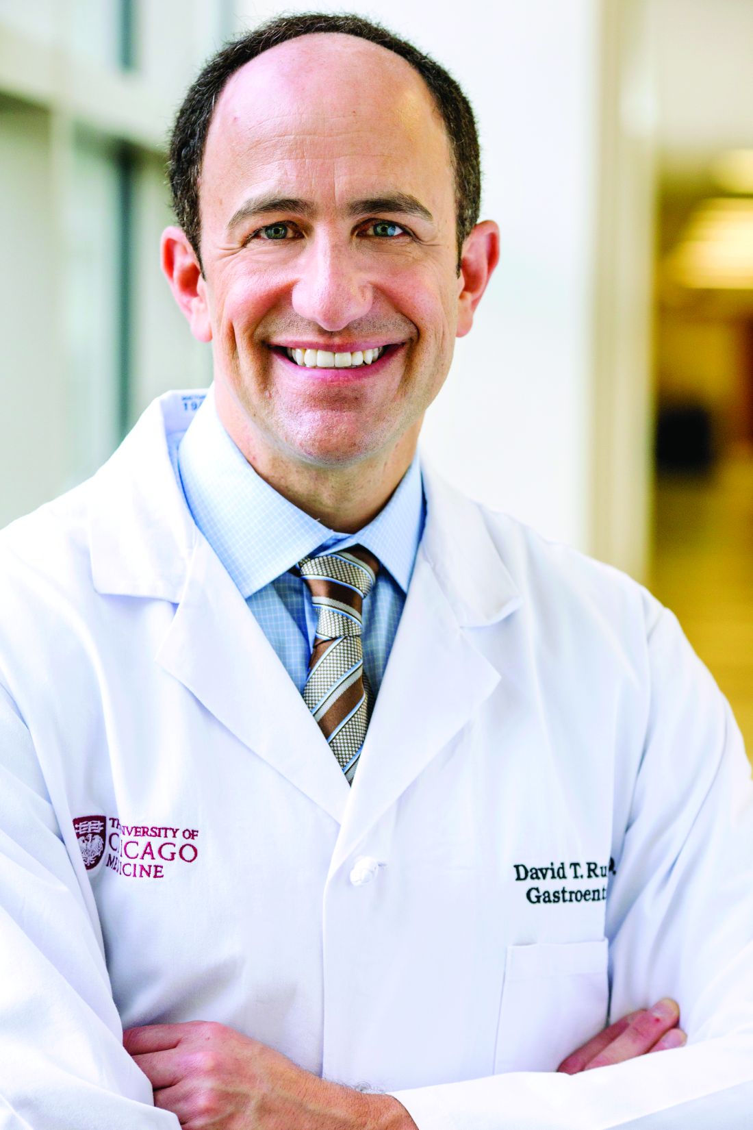
David T. Rubin, MD, AGAF, of the University of Chicago agreed that the finding deserves further investigation, particularly since sulfasalazine and 5-ASAs represent the second most commonly prescribed medication class for IBD.
“The risk with 5-ASAs is of interest but not well explained by what we know about the safety or the mechanism of these therapies,” Dr. Rubin said. “Clearly, more work is needed.”
The risks associated with corticosteroids were particularly concerning, Dr. Rubin said, because 10%-20% of patients with IBD may be taking corticosteroids at any given time.
“Steroids are still the number one prescribed therapy for Crohn’s and colitis,” he said.
Still, Dr. Rubin advised against abrupt changes to drug regimens, especially if they are effectively controlling IBD.
“Patients should stay on their existing therapies and stay in remission,” Dr. Rubin said. “If you stop your therapies … you are more likely to relapse. When you relapse, you’re more likely to need steroids as a rescue therapy … or end up in the hospital, and those are not places we want you to be.”
Despite the risks associated with steroids and sulfasalazine/5-ASAs, Dr. Rubin had an optimistic take on the study, calling the findings “very reassuring” because they support continued usage of TNF inhibitors and other biologic agents during the pandemic. He also noted that the SECURE-IBD registry, which he has contributed to, represents “an extraordinary effort” from around the world.
“[This is] an unprecedented collaboration across a scale and timeframe that has really never been seen before in our field, and I would hazard a guess that it’s probably never been seen in most other fields right now,” he said.
Clinicians seeking more information about managing patients with IBD during the COVID-19 pandemic can find guidance in the recent AGA practice update, of which Dr. Rubin was the lead author. Clinicians who would like to contribute to the SECURE-IBD registry may do so at covidibd.org. The registry now includes more than 1,000 patients.
The study was funded by Clinical and Translational Science Award grants through Dr. Ungaro. The investigators disclosed relationships with Takeda, Janssen, Pfizer, and others. Dr. Rubin disclosed relationships with Gilead, Eli Lilly, Shire, and others.
Check out the AGA COVID-19 Resource Library for new clinical guidance, education, research and physician resources, including recent guidance on how to treat patients with IBD during the pandemic, at www.gastro.org/covid.
SOURCE: Brenner EJ et al. Gastroenterology. 2020 May 18. doi: 10.1053/j.gastro.2020.05.032.
For patients with inflammatory bowel disease (IBD) who develop coronavirus disease of 2019 (COVID-19), corticosteroid use may significantly increase risk of severe disease, according to data from more than 500 patients.
Use of sulfasalazine or 5-aminosalicylates (5-ASAs) also increased risk of severe COVID-19, albeit to a lesser degree, reported co-lead authors Erica J. Brenner, MD, of University of North Carolina Children’s Hospital, Chapel Hill, and Ryan C. Ungaro, MD, of Icahn School of Medicine at Mount Sinai, New York, and colleagues.
In contrast, tumor necrosis factor (TNF) blockers were not an independent risk factor for severe COVID-19.
“As TNF antagonists are the most commonly prescribed biologic therapy for patients with IBD, these initial findings should be reassuring to the large number of patients receiving TNF antagonist therapy and support their continued use during this current pandemic,” the investigators wrote in Gastroenterology.
These conclusions were drawn from the Surveillance Epidemiology of Coronavirus Under Research Exclusion for Inflammatory Bowel Disease (SECURE-IBD) database, a large registry actively collecting data from clinicians around the world.
In the present analysis, which involved 525 patients from 33 countries, the investigators searched for independent risk factors for severe COVID-19. Various factors were tested through multivariable regression, including age, comorbidities, usage of specific medications, and more.
The primary outcome was defined by a composite of hospitalization, ventilator use, or death, while secondary outcomes included a composite of hospitalization or death, as well as death alone.
The analysis revealed that patients receiving corticosteroids had an adjusted odds ratio of 6.87 (95% confidence interval, 2.30-20.51) for severe COVID-19, with increased risks also detected for both secondary outcomes. In contrast, TNF antagonist use was not significantly associated with the primary outcome; in fact, a possible protective effect was detected for hospitalization or death (aOR, 0.60; 95% CI, 0.38-0.96).
The investigators noted that the above findings aligned with extensive literature concerning infectious complications with corticosteroid use and “more recent commentary” surrounding TNF antagonists. Similarly, increased age and the presence of at least two comorbidities were each independently associated with increased risk of severe COVID-19, both of which are correlations that have been previously described.
But the threefold increased risk of severe COVID-19 associated with use of sulfasalazine or 5-ASAs (aOR, 3.14; 95% CI, 1.28-7.71) was a “surprising” finding, the investigators noted.
“In a direct comparison, we observed that 5-ASA/sulfasalazine–treated patients fared worse than those treated with TNF inhibitors,” the investigators wrote. “Although we cannot exclude unmeasured confounding, further exploration of biological mechanisms is warranted.”
David T. Rubin, MD, AGAF, of the University of Chicago agreed that the finding deserves further investigation, particularly since sulfasalazine and 5-ASAs represent the second most commonly prescribed medication class for IBD.
“The risk with 5-ASAs is of interest but not well explained by what we know about the safety or the mechanism of these therapies,” Dr. Rubin said. “Clearly, more work is needed.”
The risks associated with corticosteroids were particularly concerning, Dr. Rubin said, because 10%-20% of patients with IBD may be taking corticosteroids at any given time.
“Steroids are still the number one prescribed therapy for Crohn’s and colitis,” he said.
Still, Dr. Rubin advised against abrupt changes to drug regimens, especially if they are effectively controlling IBD.
“Patients should stay on their existing therapies and stay in remission,” Dr. Rubin said. “If you stop your therapies … you are more likely to relapse. When you relapse, you’re more likely to need steroids as a rescue therapy … or end up in the hospital, and those are not places we want you to be.”
Despite the risks associated with steroids and sulfasalazine/5-ASAs, Dr. Rubin had an optimistic take on the study, calling the findings “very reassuring” because they support continued usage of TNF inhibitors and other biologic agents during the pandemic. He also noted that the SECURE-IBD registry, which he has contributed to, represents “an extraordinary effort” from around the world.
“[This is] an unprecedented collaboration across a scale and timeframe that has really never been seen before in our field, and I would hazard a guess that it’s probably never been seen in most other fields right now,” he said.
Clinicians seeking more information about managing patients with IBD during the COVID-19 pandemic can find guidance in the recent AGA practice update, of which Dr. Rubin was the lead author. Clinicians who would like to contribute to the SECURE-IBD registry may do so at covidibd.org. The registry now includes more than 1,000 patients.
The study was funded by Clinical and Translational Science Award grants through Dr. Ungaro. The investigators disclosed relationships with Takeda, Janssen, Pfizer, and others. Dr. Rubin disclosed relationships with Gilead, Eli Lilly, Shire, and others.
Check out the AGA COVID-19 Resource Library for new clinical guidance, education, research and physician resources, including recent guidance on how to treat patients with IBD during the pandemic, at www.gastro.org/covid.
SOURCE: Brenner EJ et al. Gastroenterology. 2020 May 18. doi: 10.1053/j.gastro.2020.05.032.
For patients with inflammatory bowel disease (IBD) who develop coronavirus disease of 2019 (COVID-19), corticosteroid use may significantly increase risk of severe disease, according to data from more than 500 patients.
Use of sulfasalazine or 5-aminosalicylates (5-ASAs) also increased risk of severe COVID-19, albeit to a lesser degree, reported co-lead authors Erica J. Brenner, MD, of University of North Carolina Children’s Hospital, Chapel Hill, and Ryan C. Ungaro, MD, of Icahn School of Medicine at Mount Sinai, New York, and colleagues.
In contrast, tumor necrosis factor (TNF) blockers were not an independent risk factor for severe COVID-19.
“As TNF antagonists are the most commonly prescribed biologic therapy for patients with IBD, these initial findings should be reassuring to the large number of patients receiving TNF antagonist therapy and support their continued use during this current pandemic,” the investigators wrote in Gastroenterology.
These conclusions were drawn from the Surveillance Epidemiology of Coronavirus Under Research Exclusion for Inflammatory Bowel Disease (SECURE-IBD) database, a large registry actively collecting data from clinicians around the world.
In the present analysis, which involved 525 patients from 33 countries, the investigators searched for independent risk factors for severe COVID-19. Various factors were tested through multivariable regression, including age, comorbidities, usage of specific medications, and more.
The primary outcome was defined by a composite of hospitalization, ventilator use, or death, while secondary outcomes included a composite of hospitalization or death, as well as death alone.
The analysis revealed that patients receiving corticosteroids had an adjusted odds ratio of 6.87 (95% confidence interval, 2.30-20.51) for severe COVID-19, with increased risks also detected for both secondary outcomes. In contrast, TNF antagonist use was not significantly associated with the primary outcome; in fact, a possible protective effect was detected for hospitalization or death (aOR, 0.60; 95% CI, 0.38-0.96).
The investigators noted that the above findings aligned with extensive literature concerning infectious complications with corticosteroid use and “more recent commentary” surrounding TNF antagonists. Similarly, increased age and the presence of at least two comorbidities were each independently associated with increased risk of severe COVID-19, both of which are correlations that have been previously described.
But the threefold increased risk of severe COVID-19 associated with use of sulfasalazine or 5-ASAs (aOR, 3.14; 95% CI, 1.28-7.71) was a “surprising” finding, the investigators noted.
“In a direct comparison, we observed that 5-ASA/sulfasalazine–treated patients fared worse than those treated with TNF inhibitors,” the investigators wrote. “Although we cannot exclude unmeasured confounding, further exploration of biological mechanisms is warranted.”
David T. Rubin, MD, AGAF, of the University of Chicago agreed that the finding deserves further investigation, particularly since sulfasalazine and 5-ASAs represent the second most commonly prescribed medication class for IBD.
“The risk with 5-ASAs is of interest but not well explained by what we know about the safety or the mechanism of these therapies,” Dr. Rubin said. “Clearly, more work is needed.”
The risks associated with corticosteroids were particularly concerning, Dr. Rubin said, because 10%-20% of patients with IBD may be taking corticosteroids at any given time.
“Steroids are still the number one prescribed therapy for Crohn’s and colitis,” he said.
Still, Dr. Rubin advised against abrupt changes to drug regimens, especially if they are effectively controlling IBD.
“Patients should stay on their existing therapies and stay in remission,” Dr. Rubin said. “If you stop your therapies … you are more likely to relapse. When you relapse, you’re more likely to need steroids as a rescue therapy … or end up in the hospital, and those are not places we want you to be.”
Despite the risks associated with steroids and sulfasalazine/5-ASAs, Dr. Rubin had an optimistic take on the study, calling the findings “very reassuring” because they support continued usage of TNF inhibitors and other biologic agents during the pandemic. He also noted that the SECURE-IBD registry, which he has contributed to, represents “an extraordinary effort” from around the world.
“[This is] an unprecedented collaboration across a scale and timeframe that has really never been seen before in our field, and I would hazard a guess that it’s probably never been seen in most other fields right now,” he said.
Clinicians seeking more information about managing patients with IBD during the COVID-19 pandemic can find guidance in the recent AGA practice update, of which Dr. Rubin was the lead author. Clinicians who would like to contribute to the SECURE-IBD registry may do so at covidibd.org. The registry now includes more than 1,000 patients.
The study was funded by Clinical and Translational Science Award grants through Dr. Ungaro. The investigators disclosed relationships with Takeda, Janssen, Pfizer, and others. Dr. Rubin disclosed relationships with Gilead, Eli Lilly, Shire, and others.
Check out the AGA COVID-19 Resource Library for new clinical guidance, education, research and physician resources, including recent guidance on how to treat patients with IBD during the pandemic, at www.gastro.org/covid.
SOURCE: Brenner EJ et al. Gastroenterology. 2020 May 18. doi: 10.1053/j.gastro.2020.05.032.
FROM GASTROENTEROLOGY
Placental injury reported in women with COVID-19
Neonates appear healthy so far
Maternal vascular malperfusion and intervillous thrombi were more common in the placentas of women infected with SARS-CoV-2, compared with historic controls, report researchers who conducted the first-of-its-kind case series in the English literature. Nevertheless, the neonates in the report appear to be healthy so far and all tested negative for the virus.
Although the series examining placentas from 16 women is small, it carries a larger implication – that increased antenatal surveillance for pregnant women infected with SARS-CoV-2 may be indicated, the researchers noted.
Furthermore, the results could align with other reports of coagulation and vascular abnormalities among people with COVID-19. “I would say that our findings fit into that larger picture of vascular injury. This is developing, and there are some significant ways that these feeder vessels to the placenta are different, but if this is the emerging paradigm, our findings can fit into it,” Jeffrey A. Goldstein, MD, PhD, assistant professor of pathology at Northwestern University, Chicago, said in an interview.
The research was published in the American Journal of Clinical Pathology.
Prior case series reported in Wuhan, China, do not currently suggest that pregnant women are more likely to experience severe COVID-19, in contrast to observations during severe acute respiratory syndrome and Middle East respiratory syndrome outbreaks. “However,” the researchers noted, “adverse perinatal outcomes have been reported, including increased risks of miscarriage, preeclampsia, preterm birth, and stillbirth.”
To learn more, Dr. Goldstein, lead author Elisheva D. Shanes, MD, and colleagues examined the histology of placentas from women with COVID-19 giving birth between March 18 and May 5, 2020. They compared these placentas with over 17,000 historic controls and 215 women who had their placentas evaluated as part of a melanoma history study.
A total of 10 women were diagnosed with COVID-19 upon presentation to labor and delivery, 4 others were diagnosed approximately 1 month before delivery and the remaining 2 within 1 week of delivery. Ten of the patients were symptomatic and two required oxygen. None of the patients received intubation or died. A total of 14 patients delivered at term, 1 delivered at 34 weeks, and the remaining case experienced a 16-week intrauterine fetal demise (IUFD). The IUFD was excluded from subsequent statistical analysis.
The neonates each had a 5-minute Apgar score of 9. Most infants were discharged on the first or second day of life, and there were no neonatal deaths.
Key findings
Of the 15 placentas, 12 featured maternal vascular malperfusion. This rate was significantly higher than historic controls (P = .046) and melanoma study controls (P = .001).
Specific features varied between groups, with decidual arteriopathy, atherosis and fibrinoid necrosis of maternal vessels, and mural hypertrophy of membrane arterioles observed more often in COVID-19 cases than in all historical controls. In addition, peripheral infarctions, decidual arteriopathy, atherosis, and fibrinoid necrosis, and mural hypertrophy being more common in COVID-19 cases than in placentas of women with a history of melanoma.
In contrast, features of fetal vascular malperfusion were observed in 12 of 15 cases, but not at rates significantly different from the control groups. Chorangiosis, villous edema, and intervillous thrombi also were more common in the COVID-19 cohort.
Dr. Goldstein was surprised they did not observe much acute or chronic inflammation. “We see chronic inflammation in the placenta in response to many viruses, such as cytomegalovirus, so you might expect similar findings, but we didn’t see any increase above the controls.”
There are a couple of case reports of histiocytic intervillositis – a particularly severe form of chronic inflammation – associated with COVID-19, “but we didn’t see that in our study,” he added.
Clinical implications
The healthy neonatal outcomes reported in the study occurred despite the placental injury, which may be caused by the redundancy built into placentas for delivering oxygen and nutrients and for removing waste.
The negative COVID-19 test results in all infants also supports existing evidence that vertical transmission of the virus is uncommon. The finding also suggests that any damage to the placenta is likely related to maternal infection.
Only one mother in the COVID-19 cohort was hypertensive, which surprised the researchers because intervillous thrombi have been associated with maternal high blood pressure. “In the context of research suggesting an increase of thrombotic and thromboembolic disorders in COVID-19,” the researchers noted, “these may represent placental formation or deposition of thrombi in response to the virus.”
One of the priorities for the researchers going forward is to monitor the longer-term outcomes of the infants, Dr. Goldstein said. “We know the people in utero during the 1918-1919 flu pandemic had higher rates of heart disease and other long-term problems, so we want to be on the lookout for something similar.”
Valuable insight
“This is a comprehensive case series of this topic, with findings worth noting and sharing in a timely fashion,” Karen Mestan, MD, associate professor of pediatrics within the division of neonatology at Northwestern University, said when asked to comment on the study.
“The information is valuable to neonatologists as the short- and long-term effects of COVID-19 exposure on newborn infants are still largely unknown,” she added. “Details of placental pathology provide emerging insight and may help us understand mother-baby vertical transmission during the current pandemic.”
Dr. Goldstein and Dr. Mestan had no relevant financial disclosures.
SOURCE: Shanes ED et al. Am J Clin Pathol. 2020 May 22. doi: 10.1093/ajcp/aqaa089.
Neonates appear healthy so far
Neonates appear healthy so far
Maternal vascular malperfusion and intervillous thrombi were more common in the placentas of women infected with SARS-CoV-2, compared with historic controls, report researchers who conducted the first-of-its-kind case series in the English literature. Nevertheless, the neonates in the report appear to be healthy so far and all tested negative for the virus.
Although the series examining placentas from 16 women is small, it carries a larger implication – that increased antenatal surveillance for pregnant women infected with SARS-CoV-2 may be indicated, the researchers noted.
Furthermore, the results could align with other reports of coagulation and vascular abnormalities among people with COVID-19. “I would say that our findings fit into that larger picture of vascular injury. This is developing, and there are some significant ways that these feeder vessels to the placenta are different, but if this is the emerging paradigm, our findings can fit into it,” Jeffrey A. Goldstein, MD, PhD, assistant professor of pathology at Northwestern University, Chicago, said in an interview.
The research was published in the American Journal of Clinical Pathology.
Prior case series reported in Wuhan, China, do not currently suggest that pregnant women are more likely to experience severe COVID-19, in contrast to observations during severe acute respiratory syndrome and Middle East respiratory syndrome outbreaks. “However,” the researchers noted, “adverse perinatal outcomes have been reported, including increased risks of miscarriage, preeclampsia, preterm birth, and stillbirth.”
To learn more, Dr. Goldstein, lead author Elisheva D. Shanes, MD, and colleagues examined the histology of placentas from women with COVID-19 giving birth between March 18 and May 5, 2020. They compared these placentas with over 17,000 historic controls and 215 women who had their placentas evaluated as part of a melanoma history study.
A total of 10 women were diagnosed with COVID-19 upon presentation to labor and delivery, 4 others were diagnosed approximately 1 month before delivery and the remaining 2 within 1 week of delivery. Ten of the patients were symptomatic and two required oxygen. None of the patients received intubation or died. A total of 14 patients delivered at term, 1 delivered at 34 weeks, and the remaining case experienced a 16-week intrauterine fetal demise (IUFD). The IUFD was excluded from subsequent statistical analysis.
The neonates each had a 5-minute Apgar score of 9. Most infants were discharged on the first or second day of life, and there were no neonatal deaths.
Key findings
Of the 15 placentas, 12 featured maternal vascular malperfusion. This rate was significantly higher than historic controls (P = .046) and melanoma study controls (P = .001).
Specific features varied between groups, with decidual arteriopathy, atherosis and fibrinoid necrosis of maternal vessels, and mural hypertrophy of membrane arterioles observed more often in COVID-19 cases than in all historical controls. In addition, peripheral infarctions, decidual arteriopathy, atherosis, and fibrinoid necrosis, and mural hypertrophy being more common in COVID-19 cases than in placentas of women with a history of melanoma.
In contrast, features of fetal vascular malperfusion were observed in 12 of 15 cases, but not at rates significantly different from the control groups. Chorangiosis, villous edema, and intervillous thrombi also were more common in the COVID-19 cohort.
Dr. Goldstein was surprised they did not observe much acute or chronic inflammation. “We see chronic inflammation in the placenta in response to many viruses, such as cytomegalovirus, so you might expect similar findings, but we didn’t see any increase above the controls.”
There are a couple of case reports of histiocytic intervillositis – a particularly severe form of chronic inflammation – associated with COVID-19, “but we didn’t see that in our study,” he added.
Clinical implications
The healthy neonatal outcomes reported in the study occurred despite the placental injury, which may be caused by the redundancy built into placentas for delivering oxygen and nutrients and for removing waste.
The negative COVID-19 test results in all infants also supports existing evidence that vertical transmission of the virus is uncommon. The finding also suggests that any damage to the placenta is likely related to maternal infection.
Only one mother in the COVID-19 cohort was hypertensive, which surprised the researchers because intervillous thrombi have been associated with maternal high blood pressure. “In the context of research suggesting an increase of thrombotic and thromboembolic disorders in COVID-19,” the researchers noted, “these may represent placental formation or deposition of thrombi in response to the virus.”
One of the priorities for the researchers going forward is to monitor the longer-term outcomes of the infants, Dr. Goldstein said. “We know the people in utero during the 1918-1919 flu pandemic had higher rates of heart disease and other long-term problems, so we want to be on the lookout for something similar.”
Valuable insight
“This is a comprehensive case series of this topic, with findings worth noting and sharing in a timely fashion,” Karen Mestan, MD, associate professor of pediatrics within the division of neonatology at Northwestern University, said when asked to comment on the study.
“The information is valuable to neonatologists as the short- and long-term effects of COVID-19 exposure on newborn infants are still largely unknown,” she added. “Details of placental pathology provide emerging insight and may help us understand mother-baby vertical transmission during the current pandemic.”
Dr. Goldstein and Dr. Mestan had no relevant financial disclosures.
SOURCE: Shanes ED et al. Am J Clin Pathol. 2020 May 22. doi: 10.1093/ajcp/aqaa089.
Maternal vascular malperfusion and intervillous thrombi were more common in the placentas of women infected with SARS-CoV-2, compared with historic controls, report researchers who conducted the first-of-its-kind case series in the English literature. Nevertheless, the neonates in the report appear to be healthy so far and all tested negative for the virus.
Although the series examining placentas from 16 women is small, it carries a larger implication – that increased antenatal surveillance for pregnant women infected with SARS-CoV-2 may be indicated, the researchers noted.
Furthermore, the results could align with other reports of coagulation and vascular abnormalities among people with COVID-19. “I would say that our findings fit into that larger picture of vascular injury. This is developing, and there are some significant ways that these feeder vessels to the placenta are different, but if this is the emerging paradigm, our findings can fit into it,” Jeffrey A. Goldstein, MD, PhD, assistant professor of pathology at Northwestern University, Chicago, said in an interview.
The research was published in the American Journal of Clinical Pathology.
Prior case series reported in Wuhan, China, do not currently suggest that pregnant women are more likely to experience severe COVID-19, in contrast to observations during severe acute respiratory syndrome and Middle East respiratory syndrome outbreaks. “However,” the researchers noted, “adverse perinatal outcomes have been reported, including increased risks of miscarriage, preeclampsia, preterm birth, and stillbirth.”
To learn more, Dr. Goldstein, lead author Elisheva D. Shanes, MD, and colleagues examined the histology of placentas from women with COVID-19 giving birth between March 18 and May 5, 2020. They compared these placentas with over 17,000 historic controls and 215 women who had their placentas evaluated as part of a melanoma history study.
A total of 10 women were diagnosed with COVID-19 upon presentation to labor and delivery, 4 others were diagnosed approximately 1 month before delivery and the remaining 2 within 1 week of delivery. Ten of the patients were symptomatic and two required oxygen. None of the patients received intubation or died. A total of 14 patients delivered at term, 1 delivered at 34 weeks, and the remaining case experienced a 16-week intrauterine fetal demise (IUFD). The IUFD was excluded from subsequent statistical analysis.
The neonates each had a 5-minute Apgar score of 9. Most infants were discharged on the first or second day of life, and there were no neonatal deaths.
Key findings
Of the 15 placentas, 12 featured maternal vascular malperfusion. This rate was significantly higher than historic controls (P = .046) and melanoma study controls (P = .001).
Specific features varied between groups, with decidual arteriopathy, atherosis and fibrinoid necrosis of maternal vessels, and mural hypertrophy of membrane arterioles observed more often in COVID-19 cases than in all historical controls. In addition, peripheral infarctions, decidual arteriopathy, atherosis, and fibrinoid necrosis, and mural hypertrophy being more common in COVID-19 cases than in placentas of women with a history of melanoma.
In contrast, features of fetal vascular malperfusion were observed in 12 of 15 cases, but not at rates significantly different from the control groups. Chorangiosis, villous edema, and intervillous thrombi also were more common in the COVID-19 cohort.
Dr. Goldstein was surprised they did not observe much acute or chronic inflammation. “We see chronic inflammation in the placenta in response to many viruses, such as cytomegalovirus, so you might expect similar findings, but we didn’t see any increase above the controls.”
There are a couple of case reports of histiocytic intervillositis – a particularly severe form of chronic inflammation – associated with COVID-19, “but we didn’t see that in our study,” he added.
Clinical implications
The healthy neonatal outcomes reported in the study occurred despite the placental injury, which may be caused by the redundancy built into placentas for delivering oxygen and nutrients and for removing waste.
The negative COVID-19 test results in all infants also supports existing evidence that vertical transmission of the virus is uncommon. The finding also suggests that any damage to the placenta is likely related to maternal infection.
Only one mother in the COVID-19 cohort was hypertensive, which surprised the researchers because intervillous thrombi have been associated with maternal high blood pressure. “In the context of research suggesting an increase of thrombotic and thromboembolic disorders in COVID-19,” the researchers noted, “these may represent placental formation or deposition of thrombi in response to the virus.”
One of the priorities for the researchers going forward is to monitor the longer-term outcomes of the infants, Dr. Goldstein said. “We know the people in utero during the 1918-1919 flu pandemic had higher rates of heart disease and other long-term problems, so we want to be on the lookout for something similar.”
Valuable insight
“This is a comprehensive case series of this topic, with findings worth noting and sharing in a timely fashion,” Karen Mestan, MD, associate professor of pediatrics within the division of neonatology at Northwestern University, said when asked to comment on the study.
“The information is valuable to neonatologists as the short- and long-term effects of COVID-19 exposure on newborn infants are still largely unknown,” she added. “Details of placental pathology provide emerging insight and may help us understand mother-baby vertical transmission during the current pandemic.”
Dr. Goldstein and Dr. Mestan had no relevant financial disclosures.
SOURCE: Shanes ED et al. Am J Clin Pathol. 2020 May 22. doi: 10.1093/ajcp/aqaa089.
FROM THE AMERICAN JOURNAL OF CLINICAL PATHOLOGY
SARS-CoV-2 infection rate 16% in asymptomatic pregnant women at delivery
Among women with a planned delivery in a New York City health system during the first half of April, the rate of asymptomatic SARS-CoV-2 infection was 16%, according to a study published in Obstetrics & Gynecology. Among the patients’ designated support persons, the asymptomatic carrier rate was 10%.
“If universal testing of pregnant patients in a high prevalence area is not performed, health care workers will be inadvertently exposed to COVID-19, unless universal precautions with personal protective equipment are taken,” wrote the researchers affiliated with the department of obstetrics, gynecology, and reproductive medicine at Icahn School of Medicine at Mount Sinai, New York.
Angela Bianco, MD, and colleagues conducted an observational study of women who were scheduled for a planned delivery within the Mount Sinai Health System between April 4 and April 15, 2020. Patients and their designated support person completed a telephone screen and underwent COVID-19 testing the day before a scheduled delivery. If support persons screened positive during the telephone interview about COVID-19 symptoms, they could not attend the birth, and patients could contact a different support person to be screened and tested. “All patients and their support persons were informed of their SARS-CoV-2 test results before admission,” the investigators wrote. “Those who tested positive were counseled regarding symptomatology that should prompt medical attention.”
In all, researchers screened 158 patients with a planned delivery, and 155 agreed to undergo COVID-19 testing. Of the 155 women tested, 24 (16%) tested positive for SARS CoV-2 infection. Among 146 support persons who had a negative interview screen and underwent SARS-CoV-2 testing, 14 (10%) tested positive for SARS-CoV-2 infection.
Test results were substantially concordant among patient and support person pairs. “Among patients who tested positive for COVID-19 infection and had a support person present, 11 of 19 (58%) support persons also tested positive for COVID-19 infection,” the authors reported. “Among patients who tested negative for COVID-19 infection and had a support person present, only 3 of 127 (2.4%) support persons tested positive for COVID-19 infection.”
Telephone screening did not identify any of the COVID-19–positive cases. Of the 24 patients with SARS-CoV-2 infection, none of their newborns tested positive at birth.
“Universal testing ... provides a mechanism for more accurate counseling of patients regarding issues such as newborn skin-to-skin contact and breastfeeding,” noted Dr. Bianco and colleagues. At their institution, parents with COVID-19 are instructed to wear a mask and practice proper hand hygiene when caring for their newborns.
Kristina Adams Waldorf, MD, said in an interview that the study by Bianco et al. underscores the high rate of asymptomatic or mildly symptomatic COVID-19 infections detected with universal screening in a hospital at the U.S. epicenter of the pandemic. “Each state and hospital will need to evaluate their own data to determine the value of universal screening for their patient population. In rural parts of America that have yet to see cases, universal screening may not make sense, but these areas are likely to be few and far between. The rest of America will need to quickly get on board with universal screening to protect their labor and delivery staff.”
Testing the partner was a strength of the study. “It is reassuring that when a pregnant woman tested negative for SARS-CoV-2, the rate was very, very low (2.4%) that her partner would test positive. However, it was disconcerting that telephone screening for common symptoms associated with COVID-19 was not very helpful in identifying cases,” said Dr. Waldorf, a professor of obstetrics and gynecology at the University of Washington, Seattle. She was not involved in the study by Bianco et al.
One study author receives payment from the American Board of Obstetrics and Gynecology for serving as a board examiner, receives payment from UpToDate, and serves as an expert witness in malpractice and products liability cases. The other authors did not report any potential conflicts of interest. Dr. Waldorf said she had no relevant financial disclosures.
SOURCE: Bianco A et al. Obstet Gynecol. 2020 May 19. doi: 10.1097/AOG.0000000000003985.
Among women with a planned delivery in a New York City health system during the first half of April, the rate of asymptomatic SARS-CoV-2 infection was 16%, according to a study published in Obstetrics & Gynecology. Among the patients’ designated support persons, the asymptomatic carrier rate was 10%.
“If universal testing of pregnant patients in a high prevalence area is not performed, health care workers will be inadvertently exposed to COVID-19, unless universal precautions with personal protective equipment are taken,” wrote the researchers affiliated with the department of obstetrics, gynecology, and reproductive medicine at Icahn School of Medicine at Mount Sinai, New York.
Angela Bianco, MD, and colleagues conducted an observational study of women who were scheduled for a planned delivery within the Mount Sinai Health System between April 4 and April 15, 2020. Patients and their designated support person completed a telephone screen and underwent COVID-19 testing the day before a scheduled delivery. If support persons screened positive during the telephone interview about COVID-19 symptoms, they could not attend the birth, and patients could contact a different support person to be screened and tested. “All patients and their support persons were informed of their SARS-CoV-2 test results before admission,” the investigators wrote. “Those who tested positive were counseled regarding symptomatology that should prompt medical attention.”
In all, researchers screened 158 patients with a planned delivery, and 155 agreed to undergo COVID-19 testing. Of the 155 women tested, 24 (16%) tested positive for SARS CoV-2 infection. Among 146 support persons who had a negative interview screen and underwent SARS-CoV-2 testing, 14 (10%) tested positive for SARS-CoV-2 infection.
Test results were substantially concordant among patient and support person pairs. “Among patients who tested positive for COVID-19 infection and had a support person present, 11 of 19 (58%) support persons also tested positive for COVID-19 infection,” the authors reported. “Among patients who tested negative for COVID-19 infection and had a support person present, only 3 of 127 (2.4%) support persons tested positive for COVID-19 infection.”
Telephone screening did not identify any of the COVID-19–positive cases. Of the 24 patients with SARS-CoV-2 infection, none of their newborns tested positive at birth.
“Universal testing ... provides a mechanism for more accurate counseling of patients regarding issues such as newborn skin-to-skin contact and breastfeeding,” noted Dr. Bianco and colleagues. At their institution, parents with COVID-19 are instructed to wear a mask and practice proper hand hygiene when caring for their newborns.
Kristina Adams Waldorf, MD, said in an interview that the study by Bianco et al. underscores the high rate of asymptomatic or mildly symptomatic COVID-19 infections detected with universal screening in a hospital at the U.S. epicenter of the pandemic. “Each state and hospital will need to evaluate their own data to determine the value of universal screening for their patient population. In rural parts of America that have yet to see cases, universal screening may not make sense, but these areas are likely to be few and far between. The rest of America will need to quickly get on board with universal screening to protect their labor and delivery staff.”
Testing the partner was a strength of the study. “It is reassuring that when a pregnant woman tested negative for SARS-CoV-2, the rate was very, very low (2.4%) that her partner would test positive. However, it was disconcerting that telephone screening for common symptoms associated with COVID-19 was not very helpful in identifying cases,” said Dr. Waldorf, a professor of obstetrics and gynecology at the University of Washington, Seattle. She was not involved in the study by Bianco et al.
One study author receives payment from the American Board of Obstetrics and Gynecology for serving as a board examiner, receives payment from UpToDate, and serves as an expert witness in malpractice and products liability cases. The other authors did not report any potential conflicts of interest. Dr. Waldorf said she had no relevant financial disclosures.
SOURCE: Bianco A et al. Obstet Gynecol. 2020 May 19. doi: 10.1097/AOG.0000000000003985.
Among women with a planned delivery in a New York City health system during the first half of April, the rate of asymptomatic SARS-CoV-2 infection was 16%, according to a study published in Obstetrics & Gynecology. Among the patients’ designated support persons, the asymptomatic carrier rate was 10%.
“If universal testing of pregnant patients in a high prevalence area is not performed, health care workers will be inadvertently exposed to COVID-19, unless universal precautions with personal protective equipment are taken,” wrote the researchers affiliated with the department of obstetrics, gynecology, and reproductive medicine at Icahn School of Medicine at Mount Sinai, New York.
Angela Bianco, MD, and colleagues conducted an observational study of women who were scheduled for a planned delivery within the Mount Sinai Health System between April 4 and April 15, 2020. Patients and their designated support person completed a telephone screen and underwent COVID-19 testing the day before a scheduled delivery. If support persons screened positive during the telephone interview about COVID-19 symptoms, they could not attend the birth, and patients could contact a different support person to be screened and tested. “All patients and their support persons were informed of their SARS-CoV-2 test results before admission,” the investigators wrote. “Those who tested positive were counseled regarding symptomatology that should prompt medical attention.”
In all, researchers screened 158 patients with a planned delivery, and 155 agreed to undergo COVID-19 testing. Of the 155 women tested, 24 (16%) tested positive for SARS CoV-2 infection. Among 146 support persons who had a negative interview screen and underwent SARS-CoV-2 testing, 14 (10%) tested positive for SARS-CoV-2 infection.
Test results were substantially concordant among patient and support person pairs. “Among patients who tested positive for COVID-19 infection and had a support person present, 11 of 19 (58%) support persons also tested positive for COVID-19 infection,” the authors reported. “Among patients who tested negative for COVID-19 infection and had a support person present, only 3 of 127 (2.4%) support persons tested positive for COVID-19 infection.”
Telephone screening did not identify any of the COVID-19–positive cases. Of the 24 patients with SARS-CoV-2 infection, none of their newborns tested positive at birth.
“Universal testing ... provides a mechanism for more accurate counseling of patients regarding issues such as newborn skin-to-skin contact and breastfeeding,” noted Dr. Bianco and colleagues. At their institution, parents with COVID-19 are instructed to wear a mask and practice proper hand hygiene when caring for their newborns.
Kristina Adams Waldorf, MD, said in an interview that the study by Bianco et al. underscores the high rate of asymptomatic or mildly symptomatic COVID-19 infections detected with universal screening in a hospital at the U.S. epicenter of the pandemic. “Each state and hospital will need to evaluate their own data to determine the value of universal screening for their patient population. In rural parts of America that have yet to see cases, universal screening may not make sense, but these areas are likely to be few and far between. The rest of America will need to quickly get on board with universal screening to protect their labor and delivery staff.”
Testing the partner was a strength of the study. “It is reassuring that when a pregnant woman tested negative for SARS-CoV-2, the rate was very, very low (2.4%) that her partner would test positive. However, it was disconcerting that telephone screening for common symptoms associated with COVID-19 was not very helpful in identifying cases,” said Dr. Waldorf, a professor of obstetrics and gynecology at the University of Washington, Seattle. She was not involved in the study by Bianco et al.
One study author receives payment from the American Board of Obstetrics and Gynecology for serving as a board examiner, receives payment from UpToDate, and serves as an expert witness in malpractice and products liability cases. The other authors did not report any potential conflicts of interest. Dr. Waldorf said she had no relevant financial disclosures.
SOURCE: Bianco A et al. Obstet Gynecol. 2020 May 19. doi: 10.1097/AOG.0000000000003985.
FROM OBSTETRICS & GYNECOLOGY
Whether to test laboring women for SARS-CoV-2 may hinge on regional prevalence
at the time of admission, research published online in Obstetrics & Gynecology suggests.
In Los Angeles, researchers stopped universal testing after none of the first 80 asymptomatic women had positive results. Researchers in Chicago, on the other hand, found a positive rate of approximately 1.6% among 614 asymptomatic patients and continue to test all patients.
“Decisions regarding universal testing need to be made in the context of regional prevalence of COVID-19 infection, with recognition that a ‘one-size-fits-all’ approach is unlikely to be justifiable,” Torri D. Metz, MD,of University of Utah Health in Salt Lake City said in an editorial accompanying research letters that described the experience in Los Angeles and Chicago. “In the setting of low population prevalence of COVID-19 infection or in locations with limited testing availability, deferring universal testing may represent the better part of valor when weighing risks, benefits, economic burden, and unintended consequences of testing for SARS-CoV-2 infection. In high-prevalence regions, universal testing may be a valuable addition to obstetric care that will prevent infections in health care workers and neonates.”
Testing all patients also may provide valuable population-level surveillance, added Dr. Metz, who is an associate professor of obstetrics and gynecology, a maternal-fetal medicine subspecialist, and vice-chair of research in obstetrics and gynecology.
One week of data
After New York hospitals reported an approximately 13% prevalence of SARS-CoV-2 infection among asymptomatic laboring women, Cedars-Sinai Medical Center in Los Angeles changed its policy from testing only women with COVID-19 symptoms to testing all women beginning April 4, 2020. “Data from New York made us very concerned about the possibility of asymptomatic infections among our own pregnant patients,” Mariam Naqvi, MD, a maternal-fetal medicine specialist at Cedars-Sinai Medical Center, said in a news release. “This would have implications for them, their babies, their households, and for the health of our staff caring for them.”
In 1 week, 82 pregnant women admitted to the obstetric unit were tested for SARS-CoV-2 infection. Of two women who reported COVID-19 symptoms, one tested positive for SARS-CoV-2. “Of the remaining 80 asymptomatic women, none tested positive for SARS-CoV-2 infection, and all remained symptom free throughout their hospitalizations,” Dr. Naqvi and colleagues reported. “One asymptomatic patient had an inadequate nasopharyngeal specimen and declined repeat testing.”
Precautions taken during universal testing meant that all members of the treatment team used valuable personal protective equipment. In some cases, mothers and newborns were separated until test results were available.
“We discontinued universal testing after a 7-day period, because we could not justify continued testing of asymptomatic women in the absence of positive test results for SARS-CoV-2 infection,” they noted. “Though universal testing did not yield enough positive results on our obstetric unit to warrant continued testing at this time, our approach may change if local rates of infection increase.”
20 days of testing
In a prospective case series of pregnant women admitted to Northwestern Memorial Hospital in Chicago from April 8 to April 27, 2020, universal testing did detect asymptomatic infections. Women with scheduled admissions were tested 12-36 hours before admission in a drive-through testing center, and women with unscheduled admissions received a test that has a 2- to 3-hour turnaround time. In addition, patients were screened for symptoms such as fever, shortness of breath, cough, sore throat, body aches, chills, new-onset vomiting, diarrhea, loss of taste or smell, and red or painful eyes.
“Asymptomatic women with pending tests were managed on the routine labor floor, but health care workers used personal protective equipment that included a respirator during the second stage of labor and delivery until the test result became available,” wrote Emily S. Miller, MD, MPH, of Northwestern University, Chicago, and colleagues.
During the first 20 days of universal testing, 635 pregnant women were admitted, and 23 (3.6%) tested positive for SARS-CoV-2 infection. Of 21 women with COVID-19 symptoms, 13 (62%) tested positive for SARS-CoV-2 infection. Of 614 women who were asymptomatic, 10 (1.6%) tested positive for SARS-CoV-2. “Our data corroborate the observation that pregnant women with SARS-CoV-2 infection on admission do not seem to be reliably identified using symptom screening alone,” the researchers wrote.
Unintended consequences
Despite a lack of effective treatments for mild to moderate COVID-19, “knowledge of the disease state allows ... health care workers to wear appropriate personal protective equipment to avoid exposure,” Dr. Metz wrote. It also allows “women to be counseled about ways to decrease transmission to neonates” and enables close monitoring of patients with infection.
At the same time, universal testing may have unintended consequences for infected patients, such as stigmatization, separation from the newborn, and delays in care related to health care providers spending more time donning personal protective equipment or changes in medical decision-making regarding cesarean delivery, she emphasized.
“Obstetricians should remain aware of disease prevalence in their communities and consider universal screening of asymptomatic women on an ongoing basis as new ‘hot spots’ for COVID-19 infection are identified,” Dr. Metz concluded.
One of Dr. Naqvi’s coauthors disclosed receiving funds from Contemporary OB/GYN, Keneka, and the American College of Obstetricians and Gynecologists and serving as a board examiner for the American Board of Obstetrics and Gynecology; her coauthors did not report any relevant financial disclosures. Dr. Metz disclosed that money was paid to her institution from Pfizer and GestVision for work related to an RSV vaccination trial and a preeclampsia test, respectively. Dr. Miller and colleagues did not report any potential conflicts of interest.
SOURCES: Naqvi M et al. Obstet Gynecol. 2020 May 19. doi: 10.1097/AOG.0000000000003987; Miller ES et al. Obstet Gynecol. 2020 May 19. doi: 10.1097/AOG.0000000000003983; Metz TD. Obstet Gynecol. 2020 May 19. doi: 10.1097/AOG.0000000000003972.
at the time of admission, research published online in Obstetrics & Gynecology suggests.
In Los Angeles, researchers stopped universal testing after none of the first 80 asymptomatic women had positive results. Researchers in Chicago, on the other hand, found a positive rate of approximately 1.6% among 614 asymptomatic patients and continue to test all patients.
“Decisions regarding universal testing need to be made in the context of regional prevalence of COVID-19 infection, with recognition that a ‘one-size-fits-all’ approach is unlikely to be justifiable,” Torri D. Metz, MD,of University of Utah Health in Salt Lake City said in an editorial accompanying research letters that described the experience in Los Angeles and Chicago. “In the setting of low population prevalence of COVID-19 infection or in locations with limited testing availability, deferring universal testing may represent the better part of valor when weighing risks, benefits, economic burden, and unintended consequences of testing for SARS-CoV-2 infection. In high-prevalence regions, universal testing may be a valuable addition to obstetric care that will prevent infections in health care workers and neonates.”
Testing all patients also may provide valuable population-level surveillance, added Dr. Metz, who is an associate professor of obstetrics and gynecology, a maternal-fetal medicine subspecialist, and vice-chair of research in obstetrics and gynecology.
One week of data
After New York hospitals reported an approximately 13% prevalence of SARS-CoV-2 infection among asymptomatic laboring women, Cedars-Sinai Medical Center in Los Angeles changed its policy from testing only women with COVID-19 symptoms to testing all women beginning April 4, 2020. “Data from New York made us very concerned about the possibility of asymptomatic infections among our own pregnant patients,” Mariam Naqvi, MD, a maternal-fetal medicine specialist at Cedars-Sinai Medical Center, said in a news release. “This would have implications for them, their babies, their households, and for the health of our staff caring for them.”
In 1 week, 82 pregnant women admitted to the obstetric unit were tested for SARS-CoV-2 infection. Of two women who reported COVID-19 symptoms, one tested positive for SARS-CoV-2. “Of the remaining 80 asymptomatic women, none tested positive for SARS-CoV-2 infection, and all remained symptom free throughout their hospitalizations,” Dr. Naqvi and colleagues reported. “One asymptomatic patient had an inadequate nasopharyngeal specimen and declined repeat testing.”
Precautions taken during universal testing meant that all members of the treatment team used valuable personal protective equipment. In some cases, mothers and newborns were separated until test results were available.
“We discontinued universal testing after a 7-day period, because we could not justify continued testing of asymptomatic women in the absence of positive test results for SARS-CoV-2 infection,” they noted. “Though universal testing did not yield enough positive results on our obstetric unit to warrant continued testing at this time, our approach may change if local rates of infection increase.”
20 days of testing
In a prospective case series of pregnant women admitted to Northwestern Memorial Hospital in Chicago from April 8 to April 27, 2020, universal testing did detect asymptomatic infections. Women with scheduled admissions were tested 12-36 hours before admission in a drive-through testing center, and women with unscheduled admissions received a test that has a 2- to 3-hour turnaround time. In addition, patients were screened for symptoms such as fever, shortness of breath, cough, sore throat, body aches, chills, new-onset vomiting, diarrhea, loss of taste or smell, and red or painful eyes.
“Asymptomatic women with pending tests were managed on the routine labor floor, but health care workers used personal protective equipment that included a respirator during the second stage of labor and delivery until the test result became available,” wrote Emily S. Miller, MD, MPH, of Northwestern University, Chicago, and colleagues.
During the first 20 days of universal testing, 635 pregnant women were admitted, and 23 (3.6%) tested positive for SARS-CoV-2 infection. Of 21 women with COVID-19 symptoms, 13 (62%) tested positive for SARS-CoV-2 infection. Of 614 women who were asymptomatic, 10 (1.6%) tested positive for SARS-CoV-2. “Our data corroborate the observation that pregnant women with SARS-CoV-2 infection on admission do not seem to be reliably identified using symptom screening alone,” the researchers wrote.
Unintended consequences
Despite a lack of effective treatments for mild to moderate COVID-19, “knowledge of the disease state allows ... health care workers to wear appropriate personal protective equipment to avoid exposure,” Dr. Metz wrote. It also allows “women to be counseled about ways to decrease transmission to neonates” and enables close monitoring of patients with infection.
At the same time, universal testing may have unintended consequences for infected patients, such as stigmatization, separation from the newborn, and delays in care related to health care providers spending more time donning personal protective equipment or changes in medical decision-making regarding cesarean delivery, she emphasized.
“Obstetricians should remain aware of disease prevalence in their communities and consider universal screening of asymptomatic women on an ongoing basis as new ‘hot spots’ for COVID-19 infection are identified,” Dr. Metz concluded.
One of Dr. Naqvi’s coauthors disclosed receiving funds from Contemporary OB/GYN, Keneka, and the American College of Obstetricians and Gynecologists and serving as a board examiner for the American Board of Obstetrics and Gynecology; her coauthors did not report any relevant financial disclosures. Dr. Metz disclosed that money was paid to her institution from Pfizer and GestVision for work related to an RSV vaccination trial and a preeclampsia test, respectively. Dr. Miller and colleagues did not report any potential conflicts of interest.
SOURCES: Naqvi M et al. Obstet Gynecol. 2020 May 19. doi: 10.1097/AOG.0000000000003987; Miller ES et al. Obstet Gynecol. 2020 May 19. doi: 10.1097/AOG.0000000000003983; Metz TD. Obstet Gynecol. 2020 May 19. doi: 10.1097/AOG.0000000000003972.
at the time of admission, research published online in Obstetrics & Gynecology suggests.
In Los Angeles, researchers stopped universal testing after none of the first 80 asymptomatic women had positive results. Researchers in Chicago, on the other hand, found a positive rate of approximately 1.6% among 614 asymptomatic patients and continue to test all patients.
“Decisions regarding universal testing need to be made in the context of regional prevalence of COVID-19 infection, with recognition that a ‘one-size-fits-all’ approach is unlikely to be justifiable,” Torri D. Metz, MD,of University of Utah Health in Salt Lake City said in an editorial accompanying research letters that described the experience in Los Angeles and Chicago. “In the setting of low population prevalence of COVID-19 infection or in locations with limited testing availability, deferring universal testing may represent the better part of valor when weighing risks, benefits, economic burden, and unintended consequences of testing for SARS-CoV-2 infection. In high-prevalence regions, universal testing may be a valuable addition to obstetric care that will prevent infections in health care workers and neonates.”
Testing all patients also may provide valuable population-level surveillance, added Dr. Metz, who is an associate professor of obstetrics and gynecology, a maternal-fetal medicine subspecialist, and vice-chair of research in obstetrics and gynecology.
One week of data
After New York hospitals reported an approximately 13% prevalence of SARS-CoV-2 infection among asymptomatic laboring women, Cedars-Sinai Medical Center in Los Angeles changed its policy from testing only women with COVID-19 symptoms to testing all women beginning April 4, 2020. “Data from New York made us very concerned about the possibility of asymptomatic infections among our own pregnant patients,” Mariam Naqvi, MD, a maternal-fetal medicine specialist at Cedars-Sinai Medical Center, said in a news release. “This would have implications for them, their babies, their households, and for the health of our staff caring for them.”
In 1 week, 82 pregnant women admitted to the obstetric unit were tested for SARS-CoV-2 infection. Of two women who reported COVID-19 symptoms, one tested positive for SARS-CoV-2. “Of the remaining 80 asymptomatic women, none tested positive for SARS-CoV-2 infection, and all remained symptom free throughout their hospitalizations,” Dr. Naqvi and colleagues reported. “One asymptomatic patient had an inadequate nasopharyngeal specimen and declined repeat testing.”
Precautions taken during universal testing meant that all members of the treatment team used valuable personal protective equipment. In some cases, mothers and newborns were separated until test results were available.
“We discontinued universal testing after a 7-day period, because we could not justify continued testing of asymptomatic women in the absence of positive test results for SARS-CoV-2 infection,” they noted. “Though universal testing did not yield enough positive results on our obstetric unit to warrant continued testing at this time, our approach may change if local rates of infection increase.”
20 days of testing
In a prospective case series of pregnant women admitted to Northwestern Memorial Hospital in Chicago from April 8 to April 27, 2020, universal testing did detect asymptomatic infections. Women with scheduled admissions were tested 12-36 hours before admission in a drive-through testing center, and women with unscheduled admissions received a test that has a 2- to 3-hour turnaround time. In addition, patients were screened for symptoms such as fever, shortness of breath, cough, sore throat, body aches, chills, new-onset vomiting, diarrhea, loss of taste or smell, and red or painful eyes.
“Asymptomatic women with pending tests were managed on the routine labor floor, but health care workers used personal protective equipment that included a respirator during the second stage of labor and delivery until the test result became available,” wrote Emily S. Miller, MD, MPH, of Northwestern University, Chicago, and colleagues.
During the first 20 days of universal testing, 635 pregnant women were admitted, and 23 (3.6%) tested positive for SARS-CoV-2 infection. Of 21 women with COVID-19 symptoms, 13 (62%) tested positive for SARS-CoV-2 infection. Of 614 women who were asymptomatic, 10 (1.6%) tested positive for SARS-CoV-2. “Our data corroborate the observation that pregnant women with SARS-CoV-2 infection on admission do not seem to be reliably identified using symptom screening alone,” the researchers wrote.
Unintended consequences
Despite a lack of effective treatments for mild to moderate COVID-19, “knowledge of the disease state allows ... health care workers to wear appropriate personal protective equipment to avoid exposure,” Dr. Metz wrote. It also allows “women to be counseled about ways to decrease transmission to neonates” and enables close monitoring of patients with infection.
At the same time, universal testing may have unintended consequences for infected patients, such as stigmatization, separation from the newborn, and delays in care related to health care providers spending more time donning personal protective equipment or changes in medical decision-making regarding cesarean delivery, she emphasized.
“Obstetricians should remain aware of disease prevalence in their communities and consider universal screening of asymptomatic women on an ongoing basis as new ‘hot spots’ for COVID-19 infection are identified,” Dr. Metz concluded.
One of Dr. Naqvi’s coauthors disclosed receiving funds from Contemporary OB/GYN, Keneka, and the American College of Obstetricians and Gynecologists and serving as a board examiner for the American Board of Obstetrics and Gynecology; her coauthors did not report any relevant financial disclosures. Dr. Metz disclosed that money was paid to her institution from Pfizer and GestVision for work related to an RSV vaccination trial and a preeclampsia test, respectively. Dr. Miller and colleagues did not report any potential conflicts of interest.
SOURCES: Naqvi M et al. Obstet Gynecol. 2020 May 19. doi: 10.1097/AOG.0000000000003987; Miller ES et al. Obstet Gynecol. 2020 May 19. doi: 10.1097/AOG.0000000000003983; Metz TD. Obstet Gynecol. 2020 May 19. doi: 10.1097/AOG.0000000000003972.
FROM OBSTETRICS & GYNECOLOGY
Unacceptable RA pain may drop with TNFi treatment but still lingers in many patients
according to findings from 21 months of follow-up in a post hoc analysis of data from the randomized, controlled Swedish Farmacotherapy (SWEFOT) trial.

Although RA patients who took biologic combination therapy had 32% lower risk for unacceptable pain (rated at >40 mm on a 0- to 100-mm visual analog scale) at 21 months, they still had no difference from patients taking triple therapy in the rate of pain described as refractory, or unacceptable despite inflammation control (C-reactive protein <10 mg/L).
While these results lend “some support to a better effect on long-term pain for the biological treatment, compared with triple therapy ... our findings are also in line with insufficient effects of current treatment strategies to prevent development of inflammation-independent pain components, warranting early alternative treatment approaches in affected patients,” Tor Olofsson, MD, PhD, of Lund (Sweden) University, and colleagues wrote in Arthritis Care & Research.
The pain outcomes analyzed in this post hoc study were all secondary outcomes of the original open-label SWEFOT trial, which during 2002-2005 enrolled 258 RA patients with less than a year of symptoms who did not reach low disease activity (28-joint Disease Activity Score ≤3.2) after 3 months of methotrexate and randomized them to an addition of either infliximab (3 mg/kg rounded up to nearest 100-mg increment) or sulfasalazine 1,000 mg twice daily plus hydroxychloroquine 400 mg once daily.
Overall, 90 of 128 patients in the infliximab group and 74 of 130 in the triple-therapy group continued the protocol until the 21-month follow-up. Patients in the infliximab group had a significantly lower area under the curve for visual analog scale for pain, most of which was accounted for during months 9-21. The percentage of patients in the infliximab group with unacceptable pain also dropped significantly from 57% at randomization to 32% at 21 months, while no difference was seen for triple therapy patients, of whom 45% had unacceptable pain at 21 months.
While patients in the infliximab group had a significantly lower risk of unacceptable pain without inflammatory control at 21 months, neither treatment arm showed a within-group difference in refractory pain from randomization to the 21-month follow-up.
Nearly one-third of patients overall still reported unacceptable pain 21 months after addition of either infliximab or sulfasalazine plus hydroxychloroquine. And at that time point, refractory pain constituted 82% of all unacceptable pain. “Notably, this pattern – with a domination of refractory pain – was evident already 3 months after starting combination therapy,” Dr. Olofsson and colleagues wrote.
The original SWEFOT study was supported in part by a grant from the Swedish Rheumatism Association, and in part by an annual unrestricted grant from Schering-Plough Sweden (now Merck Sharp & Dohme). The post hoc analysis was supported by Lund University and the Kockska Foundation, the Swedish Research Council, and the Stockholm County Council. Two authors disclosed financial relationships with multiple pharmaceutical companies.
SOURCE: Olofsson T et al. Arthritis Care Res. 2020 May 20. doi: 10.1002/acr.24264.
according to findings from 21 months of follow-up in a post hoc analysis of data from the randomized, controlled Swedish Farmacotherapy (SWEFOT) trial.
Although RA patients who took biologic combination therapy had 32% lower risk for unacceptable pain (rated at >40 mm on a 0- to 100-mm visual analog scale) at 21 months, they still had no difference from patients taking triple therapy in the rate of pain described as refractory, or unacceptable despite inflammation control (C-reactive protein <10 mg/L).
While these results lend “some support to a better effect on long-term pain for the biological treatment, compared with triple therapy ... our findings are also in line with insufficient effects of current treatment strategies to prevent development of inflammation-independent pain components, warranting early alternative treatment approaches in affected patients,” Tor Olofsson, MD, PhD, of Lund (Sweden) University, and colleagues wrote in Arthritis Care & Research.
The pain outcomes analyzed in this post hoc study were all secondary outcomes of the original open-label SWEFOT trial, which during 2002-2005 enrolled 258 RA patients with less than a year of symptoms who did not reach low disease activity (28-joint Disease Activity Score ≤3.2) after 3 months of methotrexate and randomized them to an addition of either infliximab (3 mg/kg rounded up to nearest 100-mg increment) or sulfasalazine 1,000 mg twice daily plus hydroxychloroquine 400 mg once daily.
Overall, 90 of 128 patients in the infliximab group and 74 of 130 in the triple-therapy group continued the protocol until the 21-month follow-up. Patients in the infliximab group had a significantly lower area under the curve for visual analog scale for pain, most of which was accounted for during months 9-21. The percentage of patients in the infliximab group with unacceptable pain also dropped significantly from 57% at randomization to 32% at 21 months, while no difference was seen for triple therapy patients, of whom 45% had unacceptable pain at 21 months.
While patients in the infliximab group had a significantly lower risk of unacceptable pain without inflammatory control at 21 months, neither treatment arm showed a within-group difference in refractory pain from randomization to the 21-month follow-up.
Nearly one-third of patients overall still reported unacceptable pain 21 months after addition of either infliximab or sulfasalazine plus hydroxychloroquine. And at that time point, refractory pain constituted 82% of all unacceptable pain. “Notably, this pattern – with a domination of refractory pain – was evident already 3 months after starting combination therapy,” Dr. Olofsson and colleagues wrote.
The original SWEFOT study was supported in part by a grant from the Swedish Rheumatism Association, and in part by an annual unrestricted grant from Schering-Plough Sweden (now Merck Sharp & Dohme). The post hoc analysis was supported by Lund University and the Kockska Foundation, the Swedish Research Council, and the Stockholm County Council. Two authors disclosed financial relationships with multiple pharmaceutical companies.
SOURCE: Olofsson T et al. Arthritis Care Res. 2020 May 20. doi: 10.1002/acr.24264.
according to findings from 21 months of follow-up in a post hoc analysis of data from the randomized, controlled Swedish Farmacotherapy (SWEFOT) trial.
Although RA patients who took biologic combination therapy had 32% lower risk for unacceptable pain (rated at >40 mm on a 0- to 100-mm visual analog scale) at 21 months, they still had no difference from patients taking triple therapy in the rate of pain described as refractory, or unacceptable despite inflammation control (C-reactive protein <10 mg/L).
While these results lend “some support to a better effect on long-term pain for the biological treatment, compared with triple therapy ... our findings are also in line with insufficient effects of current treatment strategies to prevent development of inflammation-independent pain components, warranting early alternative treatment approaches in affected patients,” Tor Olofsson, MD, PhD, of Lund (Sweden) University, and colleagues wrote in Arthritis Care & Research.
The pain outcomes analyzed in this post hoc study were all secondary outcomes of the original open-label SWEFOT trial, which during 2002-2005 enrolled 258 RA patients with less than a year of symptoms who did not reach low disease activity (28-joint Disease Activity Score ≤3.2) after 3 months of methotrexate and randomized them to an addition of either infliximab (3 mg/kg rounded up to nearest 100-mg increment) or sulfasalazine 1,000 mg twice daily plus hydroxychloroquine 400 mg once daily.
Overall, 90 of 128 patients in the infliximab group and 74 of 130 in the triple-therapy group continued the protocol until the 21-month follow-up. Patients in the infliximab group had a significantly lower area under the curve for visual analog scale for pain, most of which was accounted for during months 9-21. The percentage of patients in the infliximab group with unacceptable pain also dropped significantly from 57% at randomization to 32% at 21 months, while no difference was seen for triple therapy patients, of whom 45% had unacceptable pain at 21 months.
While patients in the infliximab group had a significantly lower risk of unacceptable pain without inflammatory control at 21 months, neither treatment arm showed a within-group difference in refractory pain from randomization to the 21-month follow-up.
Nearly one-third of patients overall still reported unacceptable pain 21 months after addition of either infliximab or sulfasalazine plus hydroxychloroquine. And at that time point, refractory pain constituted 82% of all unacceptable pain. “Notably, this pattern – with a domination of refractory pain – was evident already 3 months after starting combination therapy,” Dr. Olofsson and colleagues wrote.
The original SWEFOT study was supported in part by a grant from the Swedish Rheumatism Association, and in part by an annual unrestricted grant from Schering-Plough Sweden (now Merck Sharp & Dohme). The post hoc analysis was supported by Lund University and the Kockska Foundation, the Swedish Research Council, and the Stockholm County Council. Two authors disclosed financial relationships with multiple pharmaceutical companies.
SOURCE: Olofsson T et al. Arthritis Care Res. 2020 May 20. doi: 10.1002/acr.24264.
FROM ARTHRITIS CARE & RESEARCH
More from REDUCE-IT: Icosapent ethyl cuts revascularization by a third
A new analysis from the REDUCE-IT trial has shown that the high-strength eicosapentaenoic acid product icosapent ethyl (Vascepa; Amarin) reduced the number of revascularization procedures by more than one-third in statin-treated patients whose triglyceride levels were elevated and who were at increased cardiovascular risk.
The new data were presented at the Society for Cardiovascular Angiography & Interventions virtual annual scientific sessions.
REDUCE-IT, a multicenter, double-blind, placebo-controlled trial, randomly assigned statin-treated patients whose triglyceride levels were elevated (135-499 mg/dL), whose LDL cholesterol levels were controlled (41-100 mg/dL), and who had established cardiovascular disease or diabetes plus risk factors to receive either icosapent ethyl 4 g daily or placebo.
The primary composite and other cardiovascular endpoints were substantially reduced. Prespecified analyses examined all coronary revascularizations, recurrent revascularizations, and revascularization subtypes.
“Compared with placebo, icosapent ethyl 4 g/day significantly reduced first and total revascularization events by 34% and 36%, respectively,” REDUCE-IT investigator Benjamin Peterson, MD, of Brigham and Women’s Hospital, Boston, concluded during his presentation.
This reduction was consistent with respect to urgent, emergent, and elective revascularization procedures overall, as well as percutaneous coronary intervention (PCI) and coronary artery bypass grafting (CABG) individually, he reported.
“Prior therapies aimed at patients with elevated triglycerides have not demonstrated a consistent benefit in reducing coronary revascularization, and to the best of our knowledge, this is the first non–LDL cholesterol intervention in a major randomized trial in which statin-treated patients underwent fewer CABG surgeries,” Dr. Peterson stated.
“These data highlight the substantial impact of icosapent ethyl on the underlying atherothrombotic burden in the at-risk REDUCE-IT population,” he added.
Detailed results showed that the percentage of patients who underwent first revascularizations was 9.2% with icosapent ethyl versus 13.3% with placebo (hazard ratio, 0.66; P < .0001; number needed to treat, 25).
Similar reductions were observed in total (first and subsequent) revascularizations (risk ratio, 0.64; P < .0001) and across urgent, emergent, and elective revascularizations. Icosapent ethyl significantly reduced the need for PCI (HR, 0.68; P < .0001) and CABG (HR, 0.61; P = .0005).
The moderator of a SCAI press conference, Kirk Garratt, MD, of the Center for Heart and Vascular Health at Christiana Care Health System in Wilmington, Del., said that “this is an impressive impact. I couldn’t count the number of zeros in the P value.”
Timothy Henry, MD, of Christ Hospital in Cincinnati, said that REDUCE-IT was an important trial. “It showed a very impressive effect on revascularizations along with all the other benefits.” But he suggested that the uptake in usage of icosapent ethyl in the United States has been slow, and he asked what could be done to enhance this.
REDUCE-IT senior investigator Deepak Bhatt, MD, replied that the product was only approved for the REDUCE-IT indication in December 2019, and he suggested that initial uptake may have been affected by the current COVID-19 pandemic.
“This new REDUCE-IT indication ― patients with established cardiovascular disease or diabetes plus risk factors who have moderately elevated triglycerides and controlled LDL ― includes a lot of patients, between 15% and 50% of all cardiovascular patients,” he said.
“We wanted to present this revascularization data at the SCAI meeting, as it is superimportant that interventional cardiologists know about this. Interventionalists have now taken ownership of LDL and make sure patients are on statins, and we hope they will now do the same thing for triglycerides,” Dr. Bhatt commented.
Dr. Henry agreed. “It should be a simple thing to take all secondary prevention patients with eligible triglycerides and be aggressive with this new therapy.”
Dr. Bhatt noted that a cost-effectiveness analysis of the REDUCE-IT trial that was presented at last year’s American Heart Association meeting “has shown the drug to be highly cost effective and actually cost saving at the current list price.”
Asked what the mechanism of benefit is, Dr. Bhatt said that “there has been good basic science showing that EPA [eicosapentaenoic acid] stabilizes cell membranes and reduces plaque vulnerability and progression.”
REDUCE-IT was sponsored by Amarin. Brigham and Women’s Hospital receives research funding from Amarin for Dr. Bhatt’s role as chair of the trial.
A version of this article originally appeared on Medscape.com.
A new analysis from the REDUCE-IT trial has shown that the high-strength eicosapentaenoic acid product icosapent ethyl (Vascepa; Amarin) reduced the number of revascularization procedures by more than one-third in statin-treated patients whose triglyceride levels were elevated and who were at increased cardiovascular risk.
The new data were presented at the Society for Cardiovascular Angiography & Interventions virtual annual scientific sessions.
REDUCE-IT, a multicenter, double-blind, placebo-controlled trial, randomly assigned statin-treated patients whose triglyceride levels were elevated (135-499 mg/dL), whose LDL cholesterol levels were controlled (41-100 mg/dL), and who had established cardiovascular disease or diabetes plus risk factors to receive either icosapent ethyl 4 g daily or placebo.
The primary composite and other cardiovascular endpoints were substantially reduced. Prespecified analyses examined all coronary revascularizations, recurrent revascularizations, and revascularization subtypes.
“Compared with placebo, icosapent ethyl 4 g/day significantly reduced first and total revascularization events by 34% and 36%, respectively,” REDUCE-IT investigator Benjamin Peterson, MD, of Brigham and Women’s Hospital, Boston, concluded during his presentation.
This reduction was consistent with respect to urgent, emergent, and elective revascularization procedures overall, as well as percutaneous coronary intervention (PCI) and coronary artery bypass grafting (CABG) individually, he reported.
“Prior therapies aimed at patients with elevated triglycerides have not demonstrated a consistent benefit in reducing coronary revascularization, and to the best of our knowledge, this is the first non–LDL cholesterol intervention in a major randomized trial in which statin-treated patients underwent fewer CABG surgeries,” Dr. Peterson stated.
“These data highlight the substantial impact of icosapent ethyl on the underlying atherothrombotic burden in the at-risk REDUCE-IT population,” he added.
Detailed results showed that the percentage of patients who underwent first revascularizations was 9.2% with icosapent ethyl versus 13.3% with placebo (hazard ratio, 0.66; P < .0001; number needed to treat, 25).
Similar reductions were observed in total (first and subsequent) revascularizations (risk ratio, 0.64; P < .0001) and across urgent, emergent, and elective revascularizations. Icosapent ethyl significantly reduced the need for PCI (HR, 0.68; P < .0001) and CABG (HR, 0.61; P = .0005).
The moderator of a SCAI press conference, Kirk Garratt, MD, of the Center for Heart and Vascular Health at Christiana Care Health System in Wilmington, Del., said that “this is an impressive impact. I couldn’t count the number of zeros in the P value.”
Timothy Henry, MD, of Christ Hospital in Cincinnati, said that REDUCE-IT was an important trial. “It showed a very impressive effect on revascularizations along with all the other benefits.” But he suggested that the uptake in usage of icosapent ethyl in the United States has been slow, and he asked what could be done to enhance this.
REDUCE-IT senior investigator Deepak Bhatt, MD, replied that the product was only approved for the REDUCE-IT indication in December 2019, and he suggested that initial uptake may have been affected by the current COVID-19 pandemic.
“This new REDUCE-IT indication ― patients with established cardiovascular disease or diabetes plus risk factors who have moderately elevated triglycerides and controlled LDL ― includes a lot of patients, between 15% and 50% of all cardiovascular patients,” he said.
“We wanted to present this revascularization data at the SCAI meeting, as it is superimportant that interventional cardiologists know about this. Interventionalists have now taken ownership of LDL and make sure patients are on statins, and we hope they will now do the same thing for triglycerides,” Dr. Bhatt commented.
Dr. Henry agreed. “It should be a simple thing to take all secondary prevention patients with eligible triglycerides and be aggressive with this new therapy.”
Dr. Bhatt noted that a cost-effectiveness analysis of the REDUCE-IT trial that was presented at last year’s American Heart Association meeting “has shown the drug to be highly cost effective and actually cost saving at the current list price.”
Asked what the mechanism of benefit is, Dr. Bhatt said that “there has been good basic science showing that EPA [eicosapentaenoic acid] stabilizes cell membranes and reduces plaque vulnerability and progression.”
REDUCE-IT was sponsored by Amarin. Brigham and Women’s Hospital receives research funding from Amarin for Dr. Bhatt’s role as chair of the trial.
A version of this article originally appeared on Medscape.com.
A new analysis from the REDUCE-IT trial has shown that the high-strength eicosapentaenoic acid product icosapent ethyl (Vascepa; Amarin) reduced the number of revascularization procedures by more than one-third in statin-treated patients whose triglyceride levels were elevated and who were at increased cardiovascular risk.
The new data were presented at the Society for Cardiovascular Angiography & Interventions virtual annual scientific sessions.
REDUCE-IT, a multicenter, double-blind, placebo-controlled trial, randomly assigned statin-treated patients whose triglyceride levels were elevated (135-499 mg/dL), whose LDL cholesterol levels were controlled (41-100 mg/dL), and who had established cardiovascular disease or diabetes plus risk factors to receive either icosapent ethyl 4 g daily or placebo.
The primary composite and other cardiovascular endpoints were substantially reduced. Prespecified analyses examined all coronary revascularizations, recurrent revascularizations, and revascularization subtypes.
“Compared with placebo, icosapent ethyl 4 g/day significantly reduced first and total revascularization events by 34% and 36%, respectively,” REDUCE-IT investigator Benjamin Peterson, MD, of Brigham and Women’s Hospital, Boston, concluded during his presentation.
This reduction was consistent with respect to urgent, emergent, and elective revascularization procedures overall, as well as percutaneous coronary intervention (PCI) and coronary artery bypass grafting (CABG) individually, he reported.
“Prior therapies aimed at patients with elevated triglycerides have not demonstrated a consistent benefit in reducing coronary revascularization, and to the best of our knowledge, this is the first non–LDL cholesterol intervention in a major randomized trial in which statin-treated patients underwent fewer CABG surgeries,” Dr. Peterson stated.
“These data highlight the substantial impact of icosapent ethyl on the underlying atherothrombotic burden in the at-risk REDUCE-IT population,” he added.
Detailed results showed that the percentage of patients who underwent first revascularizations was 9.2% with icosapent ethyl versus 13.3% with placebo (hazard ratio, 0.66; P < .0001; number needed to treat, 25).
Similar reductions were observed in total (first and subsequent) revascularizations (risk ratio, 0.64; P < .0001) and across urgent, emergent, and elective revascularizations. Icosapent ethyl significantly reduced the need for PCI (HR, 0.68; P < .0001) and CABG (HR, 0.61; P = .0005).
The moderator of a SCAI press conference, Kirk Garratt, MD, of the Center for Heart and Vascular Health at Christiana Care Health System in Wilmington, Del., said that “this is an impressive impact. I couldn’t count the number of zeros in the P value.”
Timothy Henry, MD, of Christ Hospital in Cincinnati, said that REDUCE-IT was an important trial. “It showed a very impressive effect on revascularizations along with all the other benefits.” But he suggested that the uptake in usage of icosapent ethyl in the United States has been slow, and he asked what could be done to enhance this.
REDUCE-IT senior investigator Deepak Bhatt, MD, replied that the product was only approved for the REDUCE-IT indication in December 2019, and he suggested that initial uptake may have been affected by the current COVID-19 pandemic.
“This new REDUCE-IT indication ― patients with established cardiovascular disease or diabetes plus risk factors who have moderately elevated triglycerides and controlled LDL ― includes a lot of patients, between 15% and 50% of all cardiovascular patients,” he said.
“We wanted to present this revascularization data at the SCAI meeting, as it is superimportant that interventional cardiologists know about this. Interventionalists have now taken ownership of LDL and make sure patients are on statins, and we hope they will now do the same thing for triglycerides,” Dr. Bhatt commented.
Dr. Henry agreed. “It should be a simple thing to take all secondary prevention patients with eligible triglycerides and be aggressive with this new therapy.”
Dr. Bhatt noted that a cost-effectiveness analysis of the REDUCE-IT trial that was presented at last year’s American Heart Association meeting “has shown the drug to be highly cost effective and actually cost saving at the current list price.”
Asked what the mechanism of benefit is, Dr. Bhatt said that “there has been good basic science showing that EPA [eicosapentaenoic acid] stabilizes cell membranes and reduces plaque vulnerability and progression.”
REDUCE-IT was sponsored by Amarin. Brigham and Women’s Hospital receives research funding from Amarin for Dr. Bhatt’s role as chair of the trial.
A version of this article originally appeared on Medscape.com.