User login
U.S. Pay-for-Performance Program Works in England
A hospital pay-for-performance program developed in the United States and adopted in Northwest England reduced patient mortality there, according to a report published Nov. 7 in the New England Journal of Medicine.
This result is surprising for three reasons: The American and British health care systems are quite different; to date, there has been very little evidence that any pay-for-performance programs have any effect on any patient outcomes; and this particular program has shown, at best, only "modest and short-term effects" on hospital processes of care in the United States, said Matt Sutton, Ph.D., of the Centre for Health Economics, Institute of Population Health, University of Manchester (England).

The Hospital Quality Incentive Demonstration (HQID), adopted in the United States by the Centers for Medicare and Medicaid Services in 2003, has since shown no effect on patient mortality and only brief, weakly positive effects on other patient outcomes. But a very similar program was adopted at 24 National Health Service hospitals serving a population of 6.8 million people in Northwest England in 2008.
Dr. Sutton and his colleagues assessed the program’s effect on patient mortality by comparing patient-level data from these hospitals against data from all 132 other hospitals in England for the 18 months before and the 18 months after the program was introduced. They focused on acute MI, heart failure, and pneumonia and, as a control condition, assessed six medical disorders not included in the program (acute renal failure, alcoholic liver disease, intracranial injury, paralytic ileus and intestinal obstruction without hernia, pulmonary embolism, and duodenal ulcer).
The final study sample comprised 410,384 patients with pneumonia, 201,003 with heart failure, 1,305 with acute MI, and 241,009 with the control disorders.

Overall, patient mortality was reduced by 1.3 percentage points in the hospitals participating in the pay-for-performance program, compared with the other hospitals. There was no such reduction in patient mortality from the six medical conditions not included in the program.
"This represents a substantial relative reduction rate of 6% and, during the 18-month period that we studied, equates to a reduction of 890 deaths in the total population of 70,644 patients with these conditions in the Northwest region of England," Dr. Sutton and his associates wrote (N. Engl. J. Med. 2012 [doi:10.1056/NEJMsa1114951]).
Further analyses of the data showed a strong correlation between the pay-for-performance program and decreased patient mortality. The final result was unchanged when the data were adjusted to account for baseline patient volume and mortality rates at the hospitals, and it persisted across all types of hospitals.
The finding that a nearly identical program produced different results in the two countries indicates that the details of incentive programs and the context in which they are introduced have an important bearing on their success, the researchers said.
This study was funded by the British National Health Service. No financial conflicts of interest were reported.
The Advancing Quality program was structured so that participating hospitals received reasonable bonuses – 4% of revenue for performing in the top quartile – and no penalties. Bonuses totaling more than $7.5 million were paid during the first 18 months of the program. The CEOs of the participating hospitals agreed in advance that any bonus money would be invested to further improve clinical care, said Dr. Michael J. Pistoria.
Regarding outcomes, Dr. Sutton and his colleagues found that risk-adjusted mortality for all studied conditions decreased during the study period, both in Northwestern England and the entire country, but that reductions were greater in Northwestern England. Conversely, the U.S. pay-for-performance program on which Advancing Quality was based – the Premier Hospital Quality Incentive Demonstration (HQID) – showed no change in 30-day mortality over the course of the 6-year demonstration project.
So why did Advancing Quality work? First, the program was focused geographically and included all the hospitals in its region. This localization may have allowed for greater collaboration and productivity. By contrast, HQID involved 5% of hospitals across the United States.
Second, participating hospitals received a larger bonus in Advancing Quality – 4% compared with 2% in HQID. In addition, a greater proportion of hospitals received the highest bonus – 25% vs. 10% in HQID.
Pay-for-performance programs will continue to impact the U.S. health care system, Dr. Pistoria said. Medicare is introducing pay for performance in the form of its Value-Based Purchasing Program next year. Hospitalists are an essential part of the success of any pay-for-performance program in an individual hospital or health care system. With appropriate incentives – enough to justify the cost of additional person-hours and resources to implement quality improvement measures – this study shows that pay-for-performance programs may positively impact outcomes. We need to continue to advocate nationally for constructive pay-for-performance programs and work locally to ensure their success, he said.
Dr. Pistoria is chief of Hospital Medicine at Coordinated Health, Bethlehem, Pa.
A hospital pay-for-performance program developed in the United States and adopted in Northwest England reduced patient mortality there, according to a report published Nov. 7 in the New England Journal of Medicine.
This result is surprising for three reasons: The American and British health care systems are quite different; to date, there has been very little evidence that any pay-for-performance programs have any effect on any patient outcomes; and this particular program has shown, at best, only "modest and short-term effects" on hospital processes of care in the United States, said Matt Sutton, Ph.D., of the Centre for Health Economics, Institute of Population Health, University of Manchester (England).
The Hospital Quality Incentive Demonstration (HQID), adopted in the United States by the Centers for Medicare and Medicaid Services in 2003, has since shown no effect on patient mortality and only brief, weakly positive effects on other patient outcomes. But a very similar program was adopted at 24 National Health Service hospitals serving a population of 6.8 million people in Northwest England in 2008.
Dr. Sutton and his colleagues assessed the program’s effect on patient mortality by comparing patient-level data from these hospitals against data from all 132 other hospitals in England for the 18 months before and the 18 months after the program was introduced. They focused on acute MI, heart failure, and pneumonia and, as a control condition, assessed six medical disorders not included in the program (acute renal failure, alcoholic liver disease, intracranial injury, paralytic ileus and intestinal obstruction without hernia, pulmonary embolism, and duodenal ulcer).
The final study sample comprised 410,384 patients with pneumonia, 201,003 with heart failure, 1,305 with acute MI, and 241,009 with the control disorders.
Overall, patient mortality was reduced by 1.3 percentage points in the hospitals participating in the pay-for-performance program, compared with the other hospitals. There was no such reduction in patient mortality from the six medical conditions not included in the program.
"This represents a substantial relative reduction rate of 6% and, during the 18-month period that we studied, equates to a reduction of 890 deaths in the total population of 70,644 patients with these conditions in the Northwest region of England," Dr. Sutton and his associates wrote (N. Engl. J. Med. 2012 [doi:10.1056/NEJMsa1114951]).
Further analyses of the data showed a strong correlation between the pay-for-performance program and decreased patient mortality. The final result was unchanged when the data were adjusted to account for baseline patient volume and mortality rates at the hospitals, and it persisted across all types of hospitals.
The finding that a nearly identical program produced different results in the two countries indicates that the details of incentive programs and the context in which they are introduced have an important bearing on their success, the researchers said.
This study was funded by the British National Health Service. No financial conflicts of interest were reported.
FROM THE NEW ENGLAND JOURNAL OF MEDICINE
Major Finding: Patient mortality was 1.3 percentage points lower in U.K. hospitals that adopted an American-designed pay-for-performance program than in hospitals that did not, which translates into 890 fewer patient deaths in the participating hospitals.
Data Source: Data are from a study comparing mortality rates between U.K. hospitals that adopted the American pay-for-performance program and similar hospitals that did not.
Disclosures: This study was funded by the British National Health Service. No financial conflicts of interest were reported.

Stem Cell Treatment Post PCI Didn't Improve Outcomes
Intracoronary delivery of bone marrow mononuclear cells to the infarct zone of patients who have undergone percutaneous coronary intervention following an ST-segment elevation myocardial infarction did not improve left ventricular function at 6 months in the TIME trial.
The trial was designed to examine the difference in effect of infusing the bone marrow mononuclear cells (BMCs) into the infarct-related artery at 7 days after PCI and at 3 days, Dr. Jay H. Traverse reported at the annual scientific sessions of the American Heart Association.

Unfortunately, neither of these approaches were any better than placebo infusion at improving the recovery of left ventricular function, improving left ventricular volume, or reducing infarct size at 6 months’ follow-up in this double-blind Timing in Myocardial Infarction Evaluation (TIME) clinical trial, said Dr. Traverse, of the Minneapolis Heart Institute at Abbott Northwestern Hospital.
"However, long-term follow-up of these patients and the development of new composite end points may still reveal a role for this cell type after AMI [acute myocardial infarction]," Dr. Traverse and his associates said in an article published online simultaneously with his presentation (JAMA 2012 Nov. 6 [doi: 10.1001/jama.2012.28726]).
Recent research indicates that the timing of BMC infusion after PCI due to ST-segment elevation myocardial infarction (STEMI) might be critical. In the days following STEMI, there are significant temporal changes in the release of cytokines and growth factors "that may support stem-cell homing and angiogenesis, leading to improved cell survival and engraftment," they noted.
"Conversely, reactive oxygen species and inflammatory cytokines ... released by [damaged] myocardium and circulating inflammatory cells may adversely affect the bone marrow and stem cell function and/or survival." This could in turn impair the quality and potency of BMCs harvested from the patient.
The results of one large study indicated that stem cells harvested 5-7 days after an acute MI performed better than those harvested earlier, but "this important variable has never been evaluated in a prospective trial that randomly selects the day of cell delivery," the researchers said.
The TIME trial was designed to do so.
A total of 132 high-risk patients were enrolled during a 3-year period after they had experienced STEMI, shown a left ventricular ejection fraction (LVEF) of 45% or less, and were slated for PCI with stenting. These subjects were randomly assigned to have stem cell therapy on either day 3 or day 7 after PCI; they underwent bone marrow aspiration on the day of the procedure, and their BMCs were isolated and stored.
After some of these patients were excluded or withdrawn from the study, the remaining 120 patients underwent a second randomization to receive either the autologous BMCs (79 patients) or placebo infusions (41 patients).
The primary end points of TIME were changes in global and regional left ventricular function on MRI scanning at 6 months post PCI. There were no significant differences in these outcomes between subjects who received stem-cell therapy 3 days after PCI and those who received it 7 days after PCI, the investigators reported.
There also were no significant differences among the study groups in secondary outcomes such as reduction in infarct volume or change in ventricular volumes.
However, there also were no significant differences in any outcomes between subjects who received active BMCs and those who received placebo infusions. This may indicate that, contrary to previous findings, BMCs do not improve left ventricular function when administered in the immediate post-STEMI setting.
But it also is possible that "the phenotype and functionality of the BMC product in this population may be an issue," and that allogeneic BMCs procured from younger, healthier donors may have a better regenerative capacity, Dr. Traverse and his colleagues said.
It was encouraging that despite their high risk, these study subjects had few clinical events during follow-up. One death occurred (due to subarachnoid hemorrhage) before stem-cell therapy was administered. Eleven patients required repeat revascularization, and 6 received implantable cardioverter-defibrillators (ICDs). There were no significant differences among the study groups in the rates of these adverse events.
This study was funded by the National Heart, Lung, and Blood Institute. Dr. Traverse reported no financial conflicts of interest, but his associates reported numerous ties to industry sources.
The TIME trial, like two others in the series of studies from the Cardiovascular Cell Therapy Research Network, a consortium funded by the National Institutes of Health, is "well-intentioned, nicely designed, and impeccably executed, but difficult to interpret," said Dr. Eduardo Marban and Dr. Konstantinos Malliaras.
It may be that this treatment is ineffective in this patient population, "in which case the previous positive clinical studies were red herrings." Or it may be that, as Dr. Traverse suggests, the cell product used in TIME was somehow deficient, they said.
Dr. Marban and Dr. Malliaras are at Cedars-Sinai Heart Institute in Los Angeles. Dr. Marban reported being a founder of Capricor, a developer of cardiac stem-cell treatments; both he and Dr. Malliaras reported financial ties to Capricor. These remarks were taken from their editorial accompanying Dr. Traverse’s report (JAMA 2012 Nov. 6 [doi: 10.1001/jama.2012.64751]).
Intracoronary delivery of bone marrow mononuclear cells to the infarct zone of patients who have undergone percutaneous coronary intervention following an ST-segment elevation myocardial infarction did not improve left ventricular function at 6 months in the TIME trial.
The trial was designed to examine the difference in effect of infusing the bone marrow mononuclear cells (BMCs) into the infarct-related artery at 7 days after PCI and at 3 days, Dr. Jay H. Traverse reported at the annual scientific sessions of the American Heart Association.
Unfortunately, neither of these approaches were any better than placebo infusion at improving the recovery of left ventricular function, improving left ventricular volume, or reducing infarct size at 6 months’ follow-up in this double-blind Timing in Myocardial Infarction Evaluation (TIME) clinical trial, said Dr. Traverse, of the Minneapolis Heart Institute at Abbott Northwestern Hospital.
"However, long-term follow-up of these patients and the development of new composite end points may still reveal a role for this cell type after AMI [acute myocardial infarction]," Dr. Traverse and his associates said in an article published online simultaneously with his presentation (JAMA 2012 Nov. 6 [doi: 10.1001/jama.2012.28726]).
Recent research indicates that the timing of BMC infusion after PCI due to ST-segment elevation myocardial infarction (STEMI) might be critical. In the days following STEMI, there are significant temporal changes in the release of cytokines and growth factors "that may support stem-cell homing and angiogenesis, leading to improved cell survival and engraftment," they noted.
"Conversely, reactive oxygen species and inflammatory cytokines ... released by [damaged] myocardium and circulating inflammatory cells may adversely affect the bone marrow and stem cell function and/or survival." This could in turn impair the quality and potency of BMCs harvested from the patient.
The results of one large study indicated that stem cells harvested 5-7 days after an acute MI performed better than those harvested earlier, but "this important variable has never been evaluated in a prospective trial that randomly selects the day of cell delivery," the researchers said.
The TIME trial was designed to do so.
A total of 132 high-risk patients were enrolled during a 3-year period after they had experienced STEMI, shown a left ventricular ejection fraction (LVEF) of 45% or less, and were slated for PCI with stenting. These subjects were randomly assigned to have stem cell therapy on either day 3 or day 7 after PCI; they underwent bone marrow aspiration on the day of the procedure, and their BMCs were isolated and stored.
After some of these patients were excluded or withdrawn from the study, the remaining 120 patients underwent a second randomization to receive either the autologous BMCs (79 patients) or placebo infusions (41 patients).
The primary end points of TIME were changes in global and regional left ventricular function on MRI scanning at 6 months post PCI. There were no significant differences in these outcomes between subjects who received stem-cell therapy 3 days after PCI and those who received it 7 days after PCI, the investigators reported.
There also were no significant differences among the study groups in secondary outcomes such as reduction in infarct volume or change in ventricular volumes.
However, there also were no significant differences in any outcomes between subjects who received active BMCs and those who received placebo infusions. This may indicate that, contrary to previous findings, BMCs do not improve left ventricular function when administered in the immediate post-STEMI setting.
But it also is possible that "the phenotype and functionality of the BMC product in this population may be an issue," and that allogeneic BMCs procured from younger, healthier donors may have a better regenerative capacity, Dr. Traverse and his colleagues said.
It was encouraging that despite their high risk, these study subjects had few clinical events during follow-up. One death occurred (due to subarachnoid hemorrhage) before stem-cell therapy was administered. Eleven patients required repeat revascularization, and 6 received implantable cardioverter-defibrillators (ICDs). There were no significant differences among the study groups in the rates of these adverse events.
This study was funded by the National Heart, Lung, and Blood Institute. Dr. Traverse reported no financial conflicts of interest, but his associates reported numerous ties to industry sources.
FROM THE ANNUAL SCIENTIFIC SESSIONS OF THE AMERICAN HEART ASSOCIATION
Major Finding: The recovery of left ventricular function and volume did not differ between patients who received stem-cell (BMC) therapy in an infarcted artery 3 days after PCI and those who received it 7 days after PCI.
Data Source: This was a randomized, double-blind, placebo-controlled clinical trial involving 120 STEMI patients who underwent PCI with stent placement, received BMC or placebo infusions either 3 or 7 days later, and were followed for 6 months.
Disclosures: This study was funded by the National Heart, Lung, and Blood Institute. Dr. Traverse reported no financial conflicts of interest, but his associates reported numerous ties to industry sources.

Donor, Autologous Stem Cells Equally Safe for Cardiomyopathy
Allogeneic and autologous stem-cell infusions were found equally safe for ischemic cardiomyopathy in a pilot study comparing the two approaches, Dr. Joshua M. Hare reported at the annual scientific sessions of the American Heart Association.
Transendocardial injections of both types of bone-marrow–derived mesenchymal stem cells produced low rates of adverse events, including immunologic reactions. Both also demonstrated "potential regenerative bioactivity" in patients with chronic left ventricular dysfunction secondary to MI that had occurred years earlier, said Dr. Hare of the Interdisciplinary Stem Cell Institute, University of Miami.
The primary goal of the trial was to establish the safety of allogeneic (donor-derived) stem-cell grafts in the myocardium. Theoretically, they should have an advantage over autologous (self-derived) grafts because they can be stockpiled for immediate use, while autologous grafts must be harvested and grown for several weeks in tissue culture.
In addition, autologous stem cells may be impaired in older patients who have comorbidities, so they may not perform as well when transplanted. Allogeneic stem cells, in contrast, can be taken from younger, healthier donors for optimal performance.
On the other hand, allogeneic mesenchymal stem cells don’t appear to persist as long as autologous stem cells once they are transplanted into the myocardium, possibly because they may be cleared from the body more rapidly due to formation of alloreactive antibodies. So there is some question as to whether their effect will wear off prematurely, Dr. Hare and his associates in the POSEIDON clinical trial said in an article published online simultaneously with the AHA presentation (JAMA 2012 Nov. 6 [doi:10.1001/jama.2012.25321]).
A secondary goal of this phase I/II clinical trial was to assess different doses of stem cells.
In all 30 study subjects, the ventricles had undergone extensive structural remodelling and their function was significantly impaired. All the subjects had heart failure symptoms, impaired 6-minute walk times, and low scores on the Minnesota Living With Heart Failure Questionnaire that measures HF-related quality of life.
All the study subjects underwent bone marrow aspiration from the iliac crest to harvest mesenchymal stem cells 4-6 weeks before undergoing cardiac catheterization. Then they were randomized to receive infusions of either those autologous grafts (15 patients) or allogeneic grafts (15 patients) derived from bone marrow aspirates of healthy donors.
In each of these study groups, subjects were assigned to receive doses of 10 million, 100 million, or 200 million stem cells. These were injected into 10 sites in the infarcted territory of the myocardium during retrograde left heart catheterization.
The primary end point of the study was the incidence of any treatment-related serious adverse event within 1 month of the procedure. This was defined as a composite of death, nonfatal MI, stroke, hospitalization for worsening HF, cardiac perforation, pericardial tamponade, or sustained ventricular arrhythmias.
One subject in each group reached this end point. Both required hospitalization for worsening HF within 30 days. Therefore, there were no differences between the two study groups in the primary end point.
Other, less serious adverse events were slightly more common with autologous than with allogeneic transplants. In particular, events "suggestive of an acute immunogenic reaction, such as fever, urticaria, hemolysis, hypotension, liver dysfunction, and/or thrombocytopenia, did not occur in any patient," the investigators said.
During a further 12 months of follow-up, there was no difference between patients who received allogeneic stem cells and those who received autologous stem cells in the rates of HF or major adverse cardiovascular events. In particular, the rates of arrhythmias, rehospitalization, and worsening HF were either similar between the two study groups or favored the allogeneic-transplant group.
Similarly, the rates of less serious adverse events were slightly higher with autologous than with allogeneic transplants. No ectopic tissue formation was observed in any patient on CT scans of the chest, abdomen, and pelvis.
Regarding the effectiveness of the two transplant approaches, both groups of patients showed equivalent, significant improvements in 6-minute walk times, NYHA class, and HF-related quality of life. Both groups also showed dramatic, 33% reductions in a CT measure of infarct size, as well as decreases in LV systolic and diastolic volumes and increases in ejection fraction.
"Our findings strongly support the ongoing development of allogeneic [stem-cell] therapy," Dr. Hare and his associates said.
Their results must be substantiated in larger phase II studies, given the relatively small sample size, open-label design, and lack of a placebo group in this clinical trial, they added.
And one surprising finding – an inverse dose-response indicating that the lowest dose of stem cells was more effective than the higher doses – was notable. "Future studies are planned to further investigate this important observation," they said.
This study was funded by the National Heart, Lung, and Blood Institute and the National Institutes of Health; Biocardia supplied the cardiac infusion catheters. Dr. Hare reported holding a patent for cardiac cell-based therapy and having ties to Biocardia, Vestion, and Kardia, and his associates reported ties to numerous industry sources.
The major finding of the POSEIDON trial is that transcardial allogeneic stem-cell therapy appears to be as safe and at least as effective as autologous stem-cell therapy, which is encouraging, said Dr. Eduardo Marban and Dr. Konstantinos Malliaras.
"By inference, POSEIDON illustrates that rejection of allogeneic cells may not matter if the cells survive long enough to trigger a regenerative cascade, resulting in sustained benefit despite evanescent cell survival," they noted.
Dr. Marban and Dr. Malliaras are at Cedars-Sinai Heart Institute, Los Angeles. Dr. Marban reported being a founder of Capricor, a developer of cardiac stem-cell treatments; both he and Dr. Malliaras reported financial ties to Capricor. These remarks were taken from their editorial accompanying Dr. Hare’s report (JAMA 2012 Nov. 6 [doi:10.1001/jama.2012.64751]).
Allogeneic and autologous stem-cell infusions were found equally safe for ischemic cardiomyopathy in a pilot study comparing the two approaches, Dr. Joshua M. Hare reported at the annual scientific sessions of the American Heart Association.
Transendocardial injections of both types of bone-marrow–derived mesenchymal stem cells produced low rates of adverse events, including immunologic reactions. Both also demonstrated "potential regenerative bioactivity" in patients with chronic left ventricular dysfunction secondary to MI that had occurred years earlier, said Dr. Hare of the Interdisciplinary Stem Cell Institute, University of Miami.
The primary goal of the trial was to establish the safety of allogeneic (donor-derived) stem-cell grafts in the myocardium. Theoretically, they should have an advantage over autologous (self-derived) grafts because they can be stockpiled for immediate use, while autologous grafts must be harvested and grown for several weeks in tissue culture.
In addition, autologous stem cells may be impaired in older patients who have comorbidities, so they may not perform as well when transplanted. Allogeneic stem cells, in contrast, can be taken from younger, healthier donors for optimal performance.
On the other hand, allogeneic mesenchymal stem cells don’t appear to persist as long as autologous stem cells once they are transplanted into the myocardium, possibly because they may be cleared from the body more rapidly due to formation of alloreactive antibodies. So there is some question as to whether their effect will wear off prematurely, Dr. Hare and his associates in the POSEIDON clinical trial said in an article published online simultaneously with the AHA presentation (JAMA 2012 Nov. 6 [doi:10.1001/jama.2012.25321]).
A secondary goal of this phase I/II clinical trial was to assess different doses of stem cells.
In all 30 study subjects, the ventricles had undergone extensive structural remodelling and their function was significantly impaired. All the subjects had heart failure symptoms, impaired 6-minute walk times, and low scores on the Minnesota Living With Heart Failure Questionnaire that measures HF-related quality of life.
All the study subjects underwent bone marrow aspiration from the iliac crest to harvest mesenchymal stem cells 4-6 weeks before undergoing cardiac catheterization. Then they were randomized to receive infusions of either those autologous grafts (15 patients) or allogeneic grafts (15 patients) derived from bone marrow aspirates of healthy donors.
In each of these study groups, subjects were assigned to receive doses of 10 million, 100 million, or 200 million stem cells. These were injected into 10 sites in the infarcted territory of the myocardium during retrograde left heart catheterization.
The primary end point of the study was the incidence of any treatment-related serious adverse event within 1 month of the procedure. This was defined as a composite of death, nonfatal MI, stroke, hospitalization for worsening HF, cardiac perforation, pericardial tamponade, or sustained ventricular arrhythmias.
One subject in each group reached this end point. Both required hospitalization for worsening HF within 30 days. Therefore, there were no differences between the two study groups in the primary end point.
Other, less serious adverse events were slightly more common with autologous than with allogeneic transplants. In particular, events "suggestive of an acute immunogenic reaction, such as fever, urticaria, hemolysis, hypotension, liver dysfunction, and/or thrombocytopenia, did not occur in any patient," the investigators said.
During a further 12 months of follow-up, there was no difference between patients who received allogeneic stem cells and those who received autologous stem cells in the rates of HF or major adverse cardiovascular events. In particular, the rates of arrhythmias, rehospitalization, and worsening HF were either similar between the two study groups or favored the allogeneic-transplant group.
Similarly, the rates of less serious adverse events were slightly higher with autologous than with allogeneic transplants. No ectopic tissue formation was observed in any patient on CT scans of the chest, abdomen, and pelvis.
Regarding the effectiveness of the two transplant approaches, both groups of patients showed equivalent, significant improvements in 6-minute walk times, NYHA class, and HF-related quality of life. Both groups also showed dramatic, 33% reductions in a CT measure of infarct size, as well as decreases in LV systolic and diastolic volumes and increases in ejection fraction.
"Our findings strongly support the ongoing development of allogeneic [stem-cell] therapy," Dr. Hare and his associates said.
Their results must be substantiated in larger phase II studies, given the relatively small sample size, open-label design, and lack of a placebo group in this clinical trial, they added.
And one surprising finding – an inverse dose-response indicating that the lowest dose of stem cells was more effective than the higher doses – was notable. "Future studies are planned to further investigate this important observation," they said.
This study was funded by the National Heart, Lung, and Blood Institute and the National Institutes of Health; Biocardia supplied the cardiac infusion catheters. Dr. Hare reported holding a patent for cardiac cell-based therapy and having ties to Biocardia, Vestion, and Kardia, and his associates reported ties to numerous industry sources.
FROM THE ANNUAL SCIENTIFIC SESSIONS OF THE AMERICAN HEART ASSOCIATION
Major Finding: The composite primary end point of serious adverse events possibly related to treatment occurred in 1 patient receiving autologous stem cells and 1 receiving allogeneic stem cells; other short- and long-term adverse events were either equivalent between the two groups or favored the allogeneic group.
Data Source: POSEIDON, a phase I/II pilot study comparing the safety of allogeneic vs. autologous stem-cell transplantation in 30 patients with ischemic cardiomyopathy secondary to MI who were followed for 1 year.
Disclosures: This study was funded by the National Heart, Lung, and Blood Institute and the National Institutes of Health; Biocardia supplied the cardiac infusion catheters. Dr. Hare reported holding a patent for cardiac cell-based therapy and having ties to Biocardia, Vestion, and Kardia, and his associates reported ties to numerous industry sources.
Studies Identify Early Changes in Familial Alzheimer's Disease
Young adult carriers of the presenilin gene mutation that invariably induces Alzheimer’s disease around the age of 45 years already show distinctive changes on functional and structural brain MRI and in amyloid-beta biomarkers when they are in their 20s and are still asymptomatic. However, amyloid-beta deposition in the grey matter of the brain does not appear until age 30 years, according to findings from two cross-sectional studies of the same cohort.
More than 2 decades before the median age of onset for familial Alzheimer’s disease (AD), functional and structural MRI changes were detectable in young adult carriers of the presenilin-1 (PSEN1) E280A mutation, "along with CSF [cerebrospinal fluid] and plasma biomarker findings consistent with amyloid-beta overproduction," wrote investigators led by Dr. Eric M. Reiman of Banner Alzheimer’s Institute, Phoenix.

Dr. Reiman was lead author of the amyloid-beta biomarkers and MRI study and coauthor of a second study; both were published online Nov. 6 in Lancet Neurology. The studies involved members of the largest known kindred of familial AD, located in Medellín, Antioquia, Colombia, as a part of the Alzheimer’s Prevention Initiative.
"Our study shows some of the earliest known brain changes in autosomal dominant AD mutation carriers," Dr. Reiman and his associates noted. Because the changes appear to be present before amyloid-beta plaque deposition begins, the findings don’t fit with the current AD model in which amyloid plaque deposition precedes markers of downstream neurodegeneration.
The investigators assessed changes in biomarkers and brain imaging studies among 20 young adults (aged 18-26 years). They first performed CSF and plasma assays in 10 carriers of the PSEN1 E280A mutation and 10 noncarriers who were matched for demographic characteristics.
Carriers had significantly higher CSF concentrations of amyloid-beta than did noncarriers, but they did not differ in CSF total tau or phosphorylated tau concentrations. Carriers also had significantly higher plasma levels of amyloid-beta.
Both findings are consistent with overproduction of amyloid-beta in the earliest stages of AD. In contrast, CSF and plasma levels of amyloid-beta are reduced in later stages of the disease, likely because the excess is being deposited in the diffuse and neuritic plaques that characterize those stages, Dr. Reiman and his colleagues said.
Sixteen of the study subjects (eight carriers and eight noncarriers of the presenilin mutation) underwent brain MRI while performing memory encoding and novel viewing tasks. The mutation carriers showed significantly greater activation in the hippocampal and parahippocampal regions than did noncarriers, as well as significantly reduced volumes of grey matter in bilateral parietal regions.
These MRI abnormalities are similar to those found in the later stages of familial AD and in late-onset AD. But in this case, the MRI abnormalities were present before any evidence of plaque accumulation, the investigators said (Lancet Neurol. 2012 Nov. 6 [doi:10.1016/S1474-4422(12)70228-4]).
"We postulate that the reductions in regional grey matter are related to a very early age-related or neurodevelopmental reduction in the density of terminal neuronal fields innervating the implicated regions. ... Regardless of whether these reductions begin before or after amyloid-beta plaque deposition, increases in hippocampal activity during a memory encoding and novel viewing task could be a result of the effort to compensate for neuronal or synaptic impairments or an inefficient inhibition of synaptic functions," Dr. Reiman and his associates wrote.
The study findings highlight the need to clarify our understanding of the earliest brain changes in AD. They also suggest that clinical trials of potential treatments, such as anti-amyloid therapies, should begin at much earlier ages than previously recognized.
Because this study was cross-sectional rather than longitudinal, we cannot know how long the high concentrations of amyloid-beta have been present, when they will begin to fall, or even whether they may have begun to fall already, wrote Dr. Nick Fox of the Dementia Research Centre in the Institute of Neurology at University College London.
Similarly, we can’t tell whether the reduced volume of grey matter and the altered synaptic function on MRI are part of a neurodegenerative process or have always been present and are developmental. Either way, "we must treat the results with great caution" because of the small study sample and cross-sectional design, Dr. Fox wrote (Lancet Neurol. 2012 Nov. 6 [doi:10.1016/s1474-4422(12)70256-9]).
The second study, led by Dr. Adam S. Fleisher (also of the Banner Alzheimer’s Institute), involved florbetapir (Amyvid) PET scan assessments of members of the familial AD kindred aged 20-56 years. A total of 11 subjects were carriers of the mutation who were already symptomatic, 19 were carriers who had not yet developed symptoms, and 20 were asymptomatic noncarriers of the mutation.
In the grey matter of the brain, florbetapir binding with amyloid-beta was evident in all the mutation carriers who were aged 30 years or older, regardless of whether they were symptomatic. In contrast, there was no such binding in the noncarriers or in the carriers who were younger than 30 years.
The cerebral pattern of this deposition was similar to that seen in patients with other forms of AD, primarily affecting the posterior cingulate, precuneus, parietotemporal, frontal, and basal ganglial regions, the researchers said (Lancet Neurol. 2012 Nov. 6 [doi:10.1016/S1474-4422(12)70227-2]).
Based on their findings, Dr. Fleisher and his associates estimated that fibrillar amyloid-beta begins to accumulate in the brain about 16 years before the typical onset of mild cognitive impairment and about 21 years before the typical onset of full dementia in familial AD. It seems to peak during the following decade, then plateau just a few years before symptoms start to appear.
It is important to note that the findings of Dr. Fleisher and his colleagues concerning people with autosomal-dominant AD may not be generalizable to more common forms of the disease, Dr. William Jagust wrote in an editorial accompanying the study.
Autosomal-dominant disease is thought to be related to the overproduction of amyloid-beta. In contrast, AD related to the apolipoprotein E genotype "is more closely associated with reduced clearance of amyloid-beta." It is still unclear whether these different mechanisms will respond the same way to amyloid-lowering therapies, said Dr. Jagust of the Helen Wills Neuroscience Institute and the School of Public Health at the University of California, Berkeley (Lancet Neurol. 2012 Nov. 6 [doi:10.1016/S1474-4422(12)70255-7]).
Both studies were funded in part by the Banner Alzheimer’s Foundation, Colciencias, the National Institute on Aging, and the state of Arizona. The florbetapir-PET scan study was funded in part by Avid Pharmaceuticals. The biomarker assay and MRI study also was funded in part by Boston University and the National Institute of Neurological Disorders and Stroke.
Dr. Fleisher reported ties to Eli Lilly and Avid Pharmaceuticals, and his associates reported ties to numerous industry sources. Dr. Jagust reported having served as a consultant to Synarc, TauRx, Genentech, and Siemens. Dr. Fox reported receiving institutional research support from Alzheimer’s Research UK, the Alzheimer’s Society, Bristol-Myers Squibb, Eisai, Elan, GE Healthcare, Janssen, Lilly, Lundbeck, the National Institute for Health Research, the U.K. Medical Research Council, Pfizer/Wyeth, and the Wolfson Foundation.
Young adult carriers of the presenilin gene mutation that invariably induces Alzheimer’s disease around the age of 45 years already show distinctive changes on functional and structural brain MRI and in amyloid-beta biomarkers when they are in their 20s and are still asymptomatic. However, amyloid-beta deposition in the grey matter of the brain does not appear until age 30 years, according to findings from two cross-sectional studies of the same cohort.
More than 2 decades before the median age of onset for familial Alzheimer’s disease (AD), functional and structural MRI changes were detectable in young adult carriers of the presenilin-1 (PSEN1) E280A mutation, "along with CSF [cerebrospinal fluid] and plasma biomarker findings consistent with amyloid-beta overproduction," wrote investigators led by Dr. Eric M. Reiman of Banner Alzheimer’s Institute, Phoenix.
Dr. Reiman was lead author of the amyloid-beta biomarkers and MRI study and coauthor of a second study; both were published online Nov. 6 in Lancet Neurology. The studies involved members of the largest known kindred of familial AD, located in Medellín, Antioquia, Colombia, as a part of the Alzheimer’s Prevention Initiative.
"Our study shows some of the earliest known brain changes in autosomal dominant AD mutation carriers," Dr. Reiman and his associates noted. Because the changes appear to be present before amyloid-beta plaque deposition begins, the findings don’t fit with the current AD model in which amyloid plaque deposition precedes markers of downstream neurodegeneration.
The investigators assessed changes in biomarkers and brain imaging studies among 20 young adults (aged 18-26 years). They first performed CSF and plasma assays in 10 carriers of the PSEN1 E280A mutation and 10 noncarriers who were matched for demographic characteristics.
Carriers had significantly higher CSF concentrations of amyloid-beta than did noncarriers, but they did not differ in CSF total tau or phosphorylated tau concentrations. Carriers also had significantly higher plasma levels of amyloid-beta.
Both findings are consistent with overproduction of amyloid-beta in the earliest stages of AD. In contrast, CSF and plasma levels of amyloid-beta are reduced in later stages of the disease, likely because the excess is being deposited in the diffuse and neuritic plaques that characterize those stages, Dr. Reiman and his colleagues said.
Sixteen of the study subjects (eight carriers and eight noncarriers of the presenilin mutation) underwent brain MRI while performing memory encoding and novel viewing tasks. The mutation carriers showed significantly greater activation in the hippocampal and parahippocampal regions than did noncarriers, as well as significantly reduced volumes of grey matter in bilateral parietal regions.
These MRI abnormalities are similar to those found in the later stages of familial AD and in late-onset AD. But in this case, the MRI abnormalities were present before any evidence of plaque accumulation, the investigators said (Lancet Neurol. 2012 Nov. 6 [doi:10.1016/S1474-4422(12)70228-4]).
"We postulate that the reductions in regional grey matter are related to a very early age-related or neurodevelopmental reduction in the density of terminal neuronal fields innervating the implicated regions. ... Regardless of whether these reductions begin before or after amyloid-beta plaque deposition, increases in hippocampal activity during a memory encoding and novel viewing task could be a result of the effort to compensate for neuronal or synaptic impairments or an inefficient inhibition of synaptic functions," Dr. Reiman and his associates wrote.
The study findings highlight the need to clarify our understanding of the earliest brain changes in AD. They also suggest that clinical trials of potential treatments, such as anti-amyloid therapies, should begin at much earlier ages than previously recognized.
Because this study was cross-sectional rather than longitudinal, we cannot know how long the high concentrations of amyloid-beta have been present, when they will begin to fall, or even whether they may have begun to fall already, wrote Dr. Nick Fox of the Dementia Research Centre in the Institute of Neurology at University College London.
Similarly, we can’t tell whether the reduced volume of grey matter and the altered synaptic function on MRI are part of a neurodegenerative process or have always been present and are developmental. Either way, "we must treat the results with great caution" because of the small study sample and cross-sectional design, Dr. Fox wrote (Lancet Neurol. 2012 Nov. 6 [doi:10.1016/s1474-4422(12)70256-9]).
The second study, led by Dr. Adam S. Fleisher (also of the Banner Alzheimer’s Institute), involved florbetapir (Amyvid) PET scan assessments of members of the familial AD kindred aged 20-56 years. A total of 11 subjects were carriers of the mutation who were already symptomatic, 19 were carriers who had not yet developed symptoms, and 20 were asymptomatic noncarriers of the mutation.
In the grey matter of the brain, florbetapir binding with amyloid-beta was evident in all the mutation carriers who were aged 30 years or older, regardless of whether they were symptomatic. In contrast, there was no such binding in the noncarriers or in the carriers who were younger than 30 years.
The cerebral pattern of this deposition was similar to that seen in patients with other forms of AD, primarily affecting the posterior cingulate, precuneus, parietotemporal, frontal, and basal ganglial regions, the researchers said (Lancet Neurol. 2012 Nov. 6 [doi:10.1016/S1474-4422(12)70227-2]).
Based on their findings, Dr. Fleisher and his associates estimated that fibrillar amyloid-beta begins to accumulate in the brain about 16 years before the typical onset of mild cognitive impairment and about 21 years before the typical onset of full dementia in familial AD. It seems to peak during the following decade, then plateau just a few years before symptoms start to appear.
It is important to note that the findings of Dr. Fleisher and his colleagues concerning people with autosomal-dominant AD may not be generalizable to more common forms of the disease, Dr. William Jagust wrote in an editorial accompanying the study.
Autosomal-dominant disease is thought to be related to the overproduction of amyloid-beta. In contrast, AD related to the apolipoprotein E genotype "is more closely associated with reduced clearance of amyloid-beta." It is still unclear whether these different mechanisms will respond the same way to amyloid-lowering therapies, said Dr. Jagust of the Helen Wills Neuroscience Institute and the School of Public Health at the University of California, Berkeley (Lancet Neurol. 2012 Nov. 6 [doi:10.1016/S1474-4422(12)70255-7]).
Both studies were funded in part by the Banner Alzheimer’s Foundation, Colciencias, the National Institute on Aging, and the state of Arizona. The florbetapir-PET scan study was funded in part by Avid Pharmaceuticals. The biomarker assay and MRI study also was funded in part by Boston University and the National Institute of Neurological Disorders and Stroke.
Dr. Fleisher reported ties to Eli Lilly and Avid Pharmaceuticals, and his associates reported ties to numerous industry sources. Dr. Jagust reported having served as a consultant to Synarc, TauRx, Genentech, and Siemens. Dr. Fox reported receiving institutional research support from Alzheimer’s Research UK, the Alzheimer’s Society, Bristol-Myers Squibb, Eisai, Elan, GE Healthcare, Janssen, Lilly, Lundbeck, the National Institute for Health Research, the U.K. Medical Research Council, Pfizer/Wyeth, and the Wolfson Foundation.
FROM LANCET NEUROLOGY
Major Finding: Asymptomatic carriers of the PSEN1 E280A mutation aged 18-26 years showed higher CSF and plasma levels of amyloid-beta, greater activation of hippocampal and parahippocampal regions of the brain, and reduced grey matter volume in parietal regions of the brain, compared with asymptomatic noncarriers. Amyloid-beta deposition in the grey matter of the brain already was evident by age 30 years in asymptomatic and symptomatic mutation carriers but not in noncarriers.
Data Source: In two cross-sectional studies of members of the largest cohort of patients carrying the PSEN1 E280A mutation for early-onset Alzheimer’s disease, researchers compared florbetapir-PET brain imaging among 11 symptomatic carriers, 19 presymptomatic carriers, and 20 asymptomatic noncarriers, as well as changes in amyloid-beta levels in CSF and plasma, and functional and structural MRI scans between 10 asymptomatic carriers and 10 asymptomatic noncarriers.
Disclosures: Both studies were funded in part by the Banner Alzheimer’s Foundation, Colciencias, the National Institute on Aging, and the state of Arizona. The florbetapir-PET scan study was funded in part by Avid Pharmaceuticals. The biomarker assay and MRI study also was funded in part by Boston University and the National Institute of Neurological Disorders and Stroke. Dr. Fleisher reported ties to Eli Lilly and Avid Pharmaceuticals, and his associates reported ties to numerous industry sources. Dr. Jagust reported having served as a consultant to Synarc, TauRx, Genentech, and Siemens. Dr. Fox reported receiving institutional research support from Alzheimer’s Research UK, the Alzheimer’s Society, Bristol-Myers Squibb, Eisai, Elan, GE Healthcare, Janssen, Lilly, Lundbeck, the National Institute for Health Research, the U.K. Medical Research Council, Pfizer/Wyeth, and the Wolfson Foundation.

Despite Ban, 18% of Hospitalized Smokers Light Up During Their Stay
More than 18% of smokers who were inpatients at a large urban teaching hospital reported that they smoked during their stay, even though smoking was prohibited in the hospital buildings, according to a report published online Nov. 5 in the Archives of Internal Medicine.
Receiving nicotine replacement therapy at admission delayed but did not prevent these patients from smoking eventually, said Susan Regan, Ph.D., of the tobacco research and treatment center at Massachusetts General Hospital, Boston, and her associates.

Virtually all hospitals now prohibit smoking indoors, but many allow patients, as well as staff, to smoke outdoors on the hospital grounds. "The fact that patients may go outside to smoke, especially without supervision or in inclement weather, raises safety concerns," the investigators said.
It also may compromise quality of care and hospital efficiency if patients aren’t available for assessments or treatments because they’re outside smoking. And smoking during a hospitalization can delay recovery and impair wound healing. In addition, inpatients who duck outside to smoke "deprive themselves of an opportunity to initiate a quit attempt in a supportive, smoke-free environment," Dr. Regan and her colleagues said.
They studied inpatient smoking during a 3-year period at the hospital, where smoking is banned indoors and outdoors except for two outdoor shelters. The study subjects were 2,185 adult inpatients who were automatically referred to the facility’s tobacco treatment service at admission, which facilitated the ordering of nicotine replacement therapy and provided a bedside counselor to assist in managing nicotine withdrawal.
The counselor also gave brief (5 minutes or less) advice on quitting smoking, as well as longer (20 minutes) cessation counseling for patients who expressed interest in quitting. The counseling included motivational interviewing, plus discussion of behavioral strategies and the use of medications to maintain smoking abstinence.
Patients’ in-hospital smoking was assessed by self-report during their hospital stays and telephone follow-up in the 2 weeks after discharge. Median length of stay was 5 days, and 62% of the subjects received nicotine replacement therapy; one-third received the therapy on the first day of their stay. The mean patient age was 53 years, and 58% of the study subjects were men.
Overall, 18.4% of these patients reported that they smoked at some time during their hospital stay. Patients were more likely to report such smoking if they were younger, had longer hospitalizations, and had no plans to quit.
Patients who received nicotine replacement therapy on admission were less likely to smoke early in their hospital stay, but they resumed smoking later on in their stay at the same rate as did patients who never received the treatment. "Patients with longer stays might require increasing nicotine dose or supplementation of patch with shorter-acting forms of nicotine replacement therapy" such as nicotine gum or lozenges, the investigators said (Arch. Intern. Med. 2012 [doi:10.1001/2013.jamainternmend.300]).
The number of cigarettes that subjects typically smoked was not as predictive of smoking during hospitalization as was the intensity of their cigarette cravings.
"It may be difficult to predict the intensity of cravings during an admission from preadmission smoking level, due to individual variability in response to illness and the hospital environment. Routine ongoing assessment of cigarette craving, although more time-consuming, might be a more effective means of identifying patients who will have difficulty remaining abstinent during their hospital stay and assist in titrating nicotine dose for patients already receiving nicotine replacement therapy," Dr. Regan and her associates said.
Patients hospitalized during the winter months were less likely to smoke (14.4%) than were those hospitalized during the other seasons (19.7%). This is probably because of the severity of Boston’s winters, but it also may be related to a seasonal difference in case mix, researchers said. This study did not assess discharge diagnoses, so the latter possibility couldn’t be examined, they noted.
This study was limited in that it relied on patient self-report and subjects may have been reluctant to disclose their smoking or may not have remembered it accurately. In addition, the study was restricted to patients who received counseling from the hospital’s tobacco treatment service and who responded to telephone follow-up, which likely introduced a response bias to the data.
This study was supported by the National Heart, Lung, and Blood Institute. Dr. Regan reported no financial conflicts of interest, but one of her associates reported ties to Nabi Biopharmaceuticals, Pfizer, and Alere Wellbeing.
The study by Dr. Regan and her associates reminds us that many smokers will find a way to smoke during a hospitalization. Perhaps extending the prohibition to the entire hospital campus might further cut down on the 18% who do, said Dr. Steven A. Schroeder.
Even if a hospital cannot have a tobacco treatment service like the one at Massachusetts General, "at the very least there should be a health professional group (e.g., physicians, nurses, respiratory therapists, pharmacists, behavioral psychologists, or some combination of these) that can work with smokers to prevent nicotine withdrawal symptoms and help them quit. Referral to a toll-free telephone quit line, available in every state, can be accomplished by fax or telephone (1-800-QUITNOW)," he wrote.
Dr. Schroeder is with the department of medicine at the University of California, San Francisco. He reported no financial conflicts of interest. These remarks were taken from his invited commentary accompanying Dr. Regan’s report (Arch. Intern. Med. 2012;doi:10.1001/2013.jamainternmed.308).
More than 18% of smokers who were inpatients at a large urban teaching hospital reported that they smoked during their stay, even though smoking was prohibited in the hospital buildings, according to a report published online Nov. 5 in the Archives of Internal Medicine.
Receiving nicotine replacement therapy at admission delayed but did not prevent these patients from smoking eventually, said Susan Regan, Ph.D., of the tobacco research and treatment center at Massachusetts General Hospital, Boston, and her associates.
Virtually all hospitals now prohibit smoking indoors, but many allow patients, as well as staff, to smoke outdoors on the hospital grounds. "The fact that patients may go outside to smoke, especially without supervision or in inclement weather, raises safety concerns," the investigators said.
It also may compromise quality of care and hospital efficiency if patients aren’t available for assessments or treatments because they’re outside smoking. And smoking during a hospitalization can delay recovery and impair wound healing. In addition, inpatients who duck outside to smoke "deprive themselves of an opportunity to initiate a quit attempt in a supportive, smoke-free environment," Dr. Regan and her colleagues said.
They studied inpatient smoking during a 3-year period at the hospital, where smoking is banned indoors and outdoors except for two outdoor shelters. The study subjects were 2,185 adult inpatients who were automatically referred to the facility’s tobacco treatment service at admission, which facilitated the ordering of nicotine replacement therapy and provided a bedside counselor to assist in managing nicotine withdrawal.
The counselor also gave brief (5 minutes or less) advice on quitting smoking, as well as longer (20 minutes) cessation counseling for patients who expressed interest in quitting. The counseling included motivational interviewing, plus discussion of behavioral strategies and the use of medications to maintain smoking abstinence.
Patients’ in-hospital smoking was assessed by self-report during their hospital stays and telephone follow-up in the 2 weeks after discharge. Median length of stay was 5 days, and 62% of the subjects received nicotine replacement therapy; one-third received the therapy on the first day of their stay. The mean patient age was 53 years, and 58% of the study subjects were men.
Overall, 18.4% of these patients reported that they smoked at some time during their hospital stay. Patients were more likely to report such smoking if they were younger, had longer hospitalizations, and had no plans to quit.
Patients who received nicotine replacement therapy on admission were less likely to smoke early in their hospital stay, but they resumed smoking later on in their stay at the same rate as did patients who never received the treatment. "Patients with longer stays might require increasing nicotine dose or supplementation of patch with shorter-acting forms of nicotine replacement therapy" such as nicotine gum or lozenges, the investigators said (Arch. Intern. Med. 2012 [doi:10.1001/2013.jamainternmend.300]).
The number of cigarettes that subjects typically smoked was not as predictive of smoking during hospitalization as was the intensity of their cigarette cravings.
"It may be difficult to predict the intensity of cravings during an admission from preadmission smoking level, due to individual variability in response to illness and the hospital environment. Routine ongoing assessment of cigarette craving, although more time-consuming, might be a more effective means of identifying patients who will have difficulty remaining abstinent during their hospital stay and assist in titrating nicotine dose for patients already receiving nicotine replacement therapy," Dr. Regan and her associates said.
Patients hospitalized during the winter months were less likely to smoke (14.4%) than were those hospitalized during the other seasons (19.7%). This is probably because of the severity of Boston’s winters, but it also may be related to a seasonal difference in case mix, researchers said. This study did not assess discharge diagnoses, so the latter possibility couldn’t be examined, they noted.
This study was limited in that it relied on patient self-report and subjects may have been reluctant to disclose their smoking or may not have remembered it accurately. In addition, the study was restricted to patients who received counseling from the hospital’s tobacco treatment service and who responded to telephone follow-up, which likely introduced a response bias to the data.
This study was supported by the National Heart, Lung, and Blood Institute. Dr. Regan reported no financial conflicts of interest, but one of her associates reported ties to Nabi Biopharmaceuticals, Pfizer, and Alere Wellbeing.
FROM THE ARCHIVES OF INTERNAL MEDICINE
Major Finding: 18.4% of the study subjects reported that they smoked at some time during their hospitalization, even though smoking was prohibited in the hospital buildings.
Data Source: An observational study of 2,185 adult smokers who were hospitalized for a variety of indications at a single medical center over the course of 3 years.
Disclosures: This study was supported by the National Heart, Lung, and Blood Institute. Dr. Regan reported no financial conflicts of interest, but one of her associates reported ties to Nabi Biopharmaceuticals, Pfizer, and Alere Wellbeing.

Analysis Details the GI Disease Burden in U.S.
Clostridium difficile contributes mightily to the overall burden of gastrointestinal disease in the United States and was associated with a 237% increase in hospitalizations in the last decade.
Researchers who examined the latest data on the nationwide toll of GI and liver disease also found a 314% rise in hospitalizations related to morbid obesity and a continuing national health burden exacted by reflux symptoms, Barrett’s esophagus, and colorectal cancer.
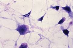
Video from the American Gastroenterological Association (http://www.youtube.com/amergastroassn)
"We compiled the most recently available statistics on GI symptoms, quality of life, outpatient diagnoses, hospitalizations, costs, mortality, and endoscopic utilization from a variety of publicly and privately held databases," Dr. Anne F. Peery of the University of North Carolina, Chapel Hill, and her colleagues reported in the November issue of Gastroenterology (doi:10.1053/j.gastro.2012.08.002).
"Payers, policy makers, clinicians, and others interested in resource utilization may use these statistics to better understand evolving disease trends, and the best way to meet the challenge of these diseases."
The findings are based on data for 2009, the most recent year for which complete information was available, from the National Ambulatory Medical Care Survey, sponsored by the U.S. Centers for Disease Control and Prevention; the United States National Health and Wellness Survey, sponsored by the private company Kantar Health; the Nationwide Inpatient Sample, sponsored by the Agency for Healthcare Research and Quality; the Surveillance, Epidemiology, and End Results database of the National Cancer Institute; the National Vital Statistics System, sponsored by the National Center for Health Statistics and the CDC; and the Thomson Reuters MarketScan’s databases of commercial, Medicare, and Medicaid records.
Among the findings:
• C. difficile hospitalizations have increased 237% since 2000 and were associated with 4% in-hospital mortality. Now the ninth leading GI cause of mortality, with an absolute increase of 230% in the number of C. difficile–related deaths since 2002, the infection also markedly impairs quality of life and the capacity for work and other activities.
• Hospitalizations related to obesity remained relatively stable since 2000, but those associated with morbid obesity rose by 314%, and many were likely caused by the marked increase in bariatric surgery.
• Gastroesophageal reflux remains the most common GI-associated diagnosis in primary care, accounting for 9 million outpatient visits in 2009, and the most common GI-associated discharge diagnosis, with 4.4 million such diagnoses in 2009. Obesity was associated with 1.7 million discharge diagnoses and constipation with 1 million.
• Barrett’s esophagus accounted for almost half a million outpatient visits in 2009, when an estimated 3.3 million Americans had this diagnosis. Given that endoscopic surveillance is recommended every 3-5 years, Barrett’s contributes substantially to resource utilization.
• Colorectal cancer, with an estimated 147,000 patients diagnosed in 2008, accounts for more than half of all GI cancer diagnoses and continues to be the primary cause of GI-associated mortality. Pancreatic and hepatobiliary cancers are the next most frequently diagnosed GI cancers.
• Of the approximately 2.5 million deaths in the United States in 2009, 10% were attributed to an underlying GI cause. Chronic liver disease and cirrhosis are the 12th leading causes of death in the country.
• The total outpatient cost for GI endoscopy in 2009 was estimated to be $32.4 billion, which is higher than previously published estimates. An estimated 6.9 million upper endoscopies, 11.5 million lower endoscopies, and 228,000 biliary endoscopies were performed in the United States in 2009.
• Chronic liver disease and viral hepatitis were associated with 6% mortality and cost an estimated $1.8 billion per year in inpatient cost.
• Hospitalizations for nonalcoholic fatty liver disease increased 97% since 2000.
This study was supported in part by the National Institutes of Health. No financial conflicts of interest were reported.
Digestive (GI and liver) diseases constitute a substantial and growing burden in the United States. As detailed in the report by Dr. Peery and colleagues, there were over 46 million outpatient encounters associated with the top 20 digestive disease diagnoses in 2009, with approximately 10% of deaths nationwide with an underlying digestive disease cause. The observed increased prevalence of hospitalizations for many diagnoses (e.g., a 14% increase with principal discharge diagnosis of chronic liver disease with viral hepatitis) and procedures (e.g., a 17% increase in lower GI endoscopies among commercially insured patients) between 2000 and 2009 is expected given population growth and aging. In addition, dramatic increases in hospitalizations associated with C. difficile and morbid obesity were also noted.
The report provides crucial information for diverse constituencies, including healthcare planners, clinicians and researchers. However, as acknowledged by the authors, there are some important caveats with respect to coverage or quality for some data sources to bear in mind when interpreting these results. This is particularly relevant for non-alcoholic fatty liver disease, which is likely underestimated because of well-known problems in diagnostic code specificity and use.

Several factors suggest that the prevalence and costs of digestive diseases will increase substantially during the next decade. These include: an aging population with the number of people aged 65 years or older projected to be greater than 54 million by 2020;the estimated tens of millions of individuals with newly available healthcare coverage as of 2014 as part of the Affordable Care Act; continued increases in obesity rates; and the recent CDC recommendation that all baby-boomers be screened for hepatitis C. This report will facilitate timely planning and also serve as benchmark to help measure the impact of these forces on the scope and burden of digestive diseases and their clinical management.
DONNA L. WHITE, PH.D., MPH, is an investigator in the Clinical Epidemiology and Outcomes Program in the Houston VA Health Services Research and Development Center of Excellence at the Michael E. DeBakey VA Medical Center, Houston. She also is an assistant professor in the department of medicine at Baylor College of Medicine, Houston.
Digestive (GI and liver) diseases constitute a substantial and growing burden in the United States. As detailed in the report by Dr. Peery and colleagues, there were over 46 million outpatient encounters associated with the top 20 digestive disease diagnoses in 2009, with approximately 10% of deaths nationwide with an underlying digestive disease cause. The observed increased prevalence of hospitalizations for many diagnoses (e.g., a 14% increase with principal discharge diagnosis of chronic liver disease with viral hepatitis) and procedures (e.g., a 17% increase in lower GI endoscopies among commercially insured patients) between 2000 and 2009 is expected given population growth and aging. In addition, dramatic increases in hospitalizations associated with C. difficile and morbid obesity were also noted.
The report provides crucial information for diverse constituencies, including healthcare planners, clinicians and researchers. However, as acknowledged by the authors, there are some important caveats with respect to coverage or quality for some data sources to bear in mind when interpreting these results. This is particularly relevant for non-alcoholic fatty liver disease, which is likely underestimated because of well-known problems in diagnostic code specificity and use.
Several factors suggest that the prevalence and costs of digestive diseases will increase substantially during the next decade. These include: an aging population with the number of people aged 65 years or older projected to be greater than 54 million by 2020;the estimated tens of millions of individuals with newly available healthcare coverage as of 2014 as part of the Affordable Care Act; continued increases in obesity rates; and the recent CDC recommendation that all baby-boomers be screened for hepatitis C. This report will facilitate timely planning and also serve as benchmark to help measure the impact of these forces on the scope and burden of digestive diseases and their clinical management.
DONNA L. WHITE, PH.D., MPH, is an investigator in the Clinical Epidemiology and Outcomes Program in the Houston VA Health Services Research and Development Center of Excellence at the Michael E. DeBakey VA Medical Center, Houston. She also is an assistant professor in the department of medicine at Baylor College of Medicine, Houston.
Clostridium difficile contributes mightily to the overall burden of gastrointestinal disease in the United States and was associated with a 237% increase in hospitalizations in the last decade.
Researchers who examined the latest data on the nationwide toll of GI and liver disease also found a 314% rise in hospitalizations related to morbid obesity and a continuing national health burden exacted by reflux symptoms, Barrett’s esophagus, and colorectal cancer.
Video from the American Gastroenterological Association (http://www.youtube.com/amergastroassn)
"We compiled the most recently available statistics on GI symptoms, quality of life, outpatient diagnoses, hospitalizations, costs, mortality, and endoscopic utilization from a variety of publicly and privately held databases," Dr. Anne F. Peery of the University of North Carolina, Chapel Hill, and her colleagues reported in the November issue of Gastroenterology (doi:10.1053/j.gastro.2012.08.002).
"Payers, policy makers, clinicians, and others interested in resource utilization may use these statistics to better understand evolving disease trends, and the best way to meet the challenge of these diseases."
The findings are based on data for 2009, the most recent year for which complete information was available, from the National Ambulatory Medical Care Survey, sponsored by the U.S. Centers for Disease Control and Prevention; the United States National Health and Wellness Survey, sponsored by the private company Kantar Health; the Nationwide Inpatient Sample, sponsored by the Agency for Healthcare Research and Quality; the Surveillance, Epidemiology, and End Results database of the National Cancer Institute; the National Vital Statistics System, sponsored by the National Center for Health Statistics and the CDC; and the Thomson Reuters MarketScan’s databases of commercial, Medicare, and Medicaid records.
Among the findings:
• C. difficile hospitalizations have increased 237% since 2000 and were associated with 4% in-hospital mortality. Now the ninth leading GI cause of mortality, with an absolute increase of 230% in the number of C. difficile–related deaths since 2002, the infection also markedly impairs quality of life and the capacity for work and other activities.
• Hospitalizations related to obesity remained relatively stable since 2000, but those associated with morbid obesity rose by 314%, and many were likely caused by the marked increase in bariatric surgery.
• Gastroesophageal reflux remains the most common GI-associated diagnosis in primary care, accounting for 9 million outpatient visits in 2009, and the most common GI-associated discharge diagnosis, with 4.4 million such diagnoses in 2009. Obesity was associated with 1.7 million discharge diagnoses and constipation with 1 million.
• Barrett’s esophagus accounted for almost half a million outpatient visits in 2009, when an estimated 3.3 million Americans had this diagnosis. Given that endoscopic surveillance is recommended every 3-5 years, Barrett’s contributes substantially to resource utilization.
• Colorectal cancer, with an estimated 147,000 patients diagnosed in 2008, accounts for more than half of all GI cancer diagnoses and continues to be the primary cause of GI-associated mortality. Pancreatic and hepatobiliary cancers are the next most frequently diagnosed GI cancers.
• Of the approximately 2.5 million deaths in the United States in 2009, 10% were attributed to an underlying GI cause. Chronic liver disease and cirrhosis are the 12th leading causes of death in the country.
• The total outpatient cost for GI endoscopy in 2009 was estimated to be $32.4 billion, which is higher than previously published estimates. An estimated 6.9 million upper endoscopies, 11.5 million lower endoscopies, and 228,000 biliary endoscopies were performed in the United States in 2009.
• Chronic liver disease and viral hepatitis were associated with 6% mortality and cost an estimated $1.8 billion per year in inpatient cost.
• Hospitalizations for nonalcoholic fatty liver disease increased 97% since 2000.
This study was supported in part by the National Institutes of Health. No financial conflicts of interest were reported.
FROM GASTROENTEROLOGY
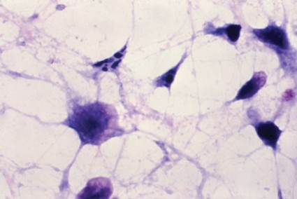
Management of Zollinger-Ellison May Depend on Presence of Tumors
Patients with Zollinger-Ellison syndrome who also have multiple endocrine neoplasia type 1 usually follow an indolent clinical course that rarely results in disease-related death, unlike those who have the sporadic form of the syndrome, Dr. Maneesh H. Singh and his colleagues reported in the November issue of Clinical Gastroenterology and Hepatology.
This finding, from a retrospective study of 49 patients treated at a single tertiary care center since 1994, argues against surgery for the estimated 25%-50% of Zollinger-Ellison syndrome (ZES) patients who have multiple endocrine neoplasia type 1 (MEN-1). "Given these results, we support a conservative approach to disease management ... focusing on symptom control with pharmacologic agents," the investigators wrote (Clin. Gastroenterol. Hepatol. 2012 Aug. 20 [doi:10.1016/j.cgh.2012.08.014]).
Early and aggressive surgery is recommended for sporadic ZES because it improves survival and can be curative. But surgery’s role in those with MEN-1 has been contentious because it doesn’t appear to improve survival in these patients. Only the most radical surgery, which carries a 40% complication rate, appears to be curative in those with MEN-1,reported Dr. Singh and his colleagues at the Hospital of the University of Pennsylvania, Philadelphia.
Because ZES is such a rare disorder, this follow-up study of 34 ZES patients who underwent surgery and 15 who did not "represents one of the largest long-term studies of surgical outcomes from a tertiary care hospital" conducted to date, they wrote.
The study subjects’ mean age at diagnosis was 47 years. The mean duration of follow-up from the time of diagnosis was 7 years, ranging from 0 to 5 years for 19 patients, from 5 to 10 years for 10, from 10 to 20 years for 15, and for more than 20 years for 5.
Of the 15 patients who did not undergo surgery, 5 declined after a discussion of the risks and benefits of the procedure, 2 because they had no lesions greater than 2 cm in diameter in imaging studies, 3 because of extensive liver involvement that was deemed unresectable, and 5 because they had unrelated comorbidities that made them poor surgical candidates.
A total of 33 subjects had sporadic ZES, while the other 16 had associated MEN-1. In the latter group, there was no significant difference between the median survival for the nine who underwent surgery (22.4 years) and for the seven who did not (25.5 years).
Standard gastrinoma resection with duodenotomy did not achieve a cure in any of the ZES patients with accompanying MEN-1.
In contrast, surgery improved both disease-related and all-cause mortality in the sporadic form of the disorder, and surgery was deemed curative in 6 (32%) of the patients with sporadic ZES.
None of the patients with MEN-1died of progressive ZES, compared with 28 (85%) of the patients with sporadic ZES. Thus, the form of the disease associated with MEN-1 appears to have a much more benign course than the sporadic form, Dr. Singh and his associates reported.
ZES associated with MEN-1 also tends to have an earlier symptom onset than does sporadic ZES, but because it is more indolent, the mean age at death was nearly identical between the two groups.
In this study, ZES patients with MEN-1had rates of liver involvement at diagnosis and rates of later liver metastases that were similar to those of patients with sporadic ZES. But again, this did not reduce their survival as it did with sporadic ZES.
This finding suggests that there may be a fundamental difference in the basic tumor biology between the two forms of the disease. It supports the theory that sporadic ZES is "a rapidly progressive, malignant form of gastrinoma that defies prediction and advocates for swift surgical intervention," the researchers wrote.
They reported no industry support for this study and no other financial conflicts.
FROM CLINICAL GASTROENTEROLOGY AND HEPATOLOGY
Liver Candidates Decline Many Organ Offers
Eighty-four percent of candidates on the wait-list for liver transplant who either died or were removed from the list before they were able to undergo transplantation declined at least one offer of a donor liver, Dr. Jennifer Cindy Lai of the University of California, San Francisco, and her colleagues reported in the November issue of Gastroenterology.
Even more surprising, most of these candidates declined "not just one or two but a median of six liver offers during their time on the wait-list."
The "declined" donor organs were then successfully transplanted into lower-priority recipients.
These findings suggest that mortality among wait-listed patients "is not simply a result of not having the opportunity for transplantation, as many of us assume. Rather, wait-list mortality appears to result from opportunities for transplantation that were declined," Dr. Lai and her associates wrote.
The reasons that so many viable donor livers were initially declined are not yet clear. General, somewhat vague reasons were listed but not fully explained in the records the researchers analyzed for this study, which they obtained from the United Network for Organ Sharing/Organ Procurement Transplantation Network database.
The investigators assessed organ offers to 33,389 liver transplant candidates aged 18 years and older who were wait-listed across the United States between 2005 and 2010.
The reasons that proffered organs were declined, as listed in the medical records, fit into six broad categories: unfavorable donor age or quality of organ; unfavorable donor organ size/weight; other unfavorable donor factors, such as ABO blood transfusion incompatibility, "social history," "positive serologic tests," or "organ anatomical damage or defect"; unreadiness of the recipient, usually because he or she was ill, unavailable, refused the organ, or required multiple organ transplants at the same time; problems with the transplant program itself, such as a "heavy workload" or unavailability of a surgeon or operating room at the recipient’s medical center, failure to respond to the offer in a timely way, or excessive distance to ship the organ.
A total of 20% of the study population (6,737 patients) died or were removed from the wait-list because they became too sick before they could undergo transplantation. A total of 5,680 (84%) of those patients had been offered one or more donor livers before they died or were taken off the list.
Offers of donor livers were declined most often (68%) because of "unfavorable donor age or quality of organ," whereas 9% were declined because of unfavorable organ size, 15% because of "other donor factors," 4% because the recipient wasn’t ready, and 4% because of transplant program or miscellaneous other factors.
However, the dominant use of the "donor quality or age" refusal code in the database almost certainly "does not accurately or fully capture the true refusal reason," Dr. Lai and her associates said.
Even livers judged to be of high quality according to standard criteria were declined because of supposed "unfavorable donor age or quality of organ." But the investigators found no difference in the risk of graft failure between such high-quality livers that were declined and other high-quality livers that were accepted on the first offer.
Other reasons must be playing an important role in this high rate refusal, but "the nuances of these refusals cannot be determined" without more individualized data, they said.
Dr. Lai and her colleagues suggested that to cut down on refusals of apparently viable organs, the transplant community should "reduce the stigma associated with non–ideal livers, and set realistic expectations for wait-listed candidates" so that they’re less likely to pass up a suitable donation while assuming that a better offer will come along.
Patients also should be educated about the unpredictability of death or of sudden worsening of liver disease while on the wait-list. They should be advised that there is a survival benefit associated with the transplantation of any graft, compared with continuing on the wait-list.
In addition, the current regulatory environment focuses on transplant centers’ outcomes, which may influence some centers to discourage the acceptance of less than optimal donor organs. "This may be especially relevant for low-volume transplant centers, for whom even a small number of poor outcomes ... may make a relatively large difference in the centers’ perceived performance," the researchers wrote.
Finally, wait-list candidates should be encouraged to complete their transplant work-ups as expeditiously as possible to avoid having to refuse a donor offer simply because they have not yet undergone the necessary cardiac testing or cancer screening.
This study was supported by the National Institute of Diabetes and Digestive and Kidney Diseases and the University of California, San Francisco. No financial conflicts of interest were reported.
FROM GASTROENTEROLOGY
Pretreatment Care Predicts HCV Outcomes
Patients with hepatitis C infections are more likely to initiate appropriate antiviral therapy and achieve a sustained virologic response if they receive high quality health care, Dr. Fasiha Kanwal of the Michael E. DeBakey Veterans Affairs Medical Center, Houston, and her colleagues reported in the November issue of Clinical Gastroenterology and Hepatology.
In their study of nearly 35,000 adults with HCV, the odds of initiating antiviral therapy were threefold higher in patients who received optimal care from the moment HCV infection was diagnosed than in those who did not. And among patients who initiated antiviral therapy, the quality of care they received before that therapy even began strongly predicted whether they would complete antiviral therapy and achieve a sustained virologic response, the investigators said.

The study showed, however, that only 11% of patients received all of the appropriate initial care.
Process-of-care measures are frequently used to assess the quality of care for HCV, but until now no study has assessed whether these measures actually correlate with better outcomes. To address this issue, Dr. Kanwal and her associates "evaluated the relationship between adherence to a broad set of process-based measures in HCV and 3 subsequent HCV-specific endpoints: receipt of antiviral treatment, completion of antiviral treatment, and the clinical outcome associated with improved survival: sustained virologic response."
The investigators used data from the VA registry on HCV clinical care, which covers patient demographics, lab tests, pharmacy information, and data on inpatient and outpatient care for approximately 300,000 patients across the country. For this study, they included data on 34,749 adults.
The mean subject age was 53 years, and 97% were men. Approximately half the study population was white and 26% was black; ethnicity was not reported for the others.
The researchers assessed seven process-of-care measures: confirmation of HCV viremia, evaluation by HCV specialists, HCV genotype testing, liver biopsy for those found to have genotype 1 HCV, and the ruling out of liver diseases related to hepatitis B, autoimmunity, or iron overload.
They also assessed seven process-of-care measures related to the prevention and management of comorbid conditions: HIV testing; hepatitis A and B serology testing, hepatitis A and B vaccination if serology results proved negative; treatment of depression; and treatment of substance abuse disorder.
Finally, they assessed six process-of-care measures related to monitoring of antiviral therapy’s effects: testing of viral load before antivirals were initiated and again at weeks 12, 24, and 48; reduction of ribavirin dose if anemia developed during treatment; and avoidance of prescribing growth-stimulating factors for leukopenia during antiviral therapy.
Overall, only 11% of the study subjects received all of the appropriate initial care, and 8% received all the appropriate care related to prevention and management of comorbid conditions. Moreover, of the study subjects who received antiviral therapy, just 37% received all the appropriate monitoring of treatment effects Dr. Kanwal and her associates said.
In patients who received optimal care before a definitive diagnosis was made, the odds of receiving antiviral therapy were 3.2 times higher than in patients who did not receive optimal care before diagnosis, they reported.
Similarly, patients who received optimal preventive and comorbid-condition care showed rates of antiviral therapy that were 36% higher than those of patients who received suboptimal preventive and comorbid-condition care.
The strong association between fulfillment of these process-of-care measures and appropriate antiviral therapy remained robust in a series of sensitivity analyses, which means it’s likely that meeting process-of-care goals directly leads to better HCV outcomes, Dr. Kanwal and her colleagues said.
The investigators could not, however, rule out the possibility that meeting these goals is simply a marker of more compliant patients, which in turn produces better outcomes.
The study findings imply that the effectiveness of the two new direct-acting antiviral agents that recently became available for HCV may hinge on the quality of care patients are receiving before they even start taking these drugs, rather than simply on the effectiveness of the drugs alone, Dr. Kanwal and her associates said.
This study was supported by the U.S. Department of Veterans Affairs Health Services Research and Development Service. No financial conflicts of interest were reported.
Financial reimbursement in medicine has long been driven by volume rather than quality. This incentive structure is changing, and in the near future practitioners will experience increased scrutiny of the quality of care we provide.

|
|
Quality can be divided into structure (having the proper equipment to clean endoscopes), process (testing for latent tuberculosis before beginning anti–tumor necrosis factor therapy), and outcomes (perforation rate during colonoscopy). The latter is, of course, the most important, but it is also the most difficult to measure because of low event rates and inadequate risk adjustment. Therefore, most quality measures are based on processes of care. For quality measurement to yield any true value to the patient, however, it is important that these processes are clearly linked to better patient outcomes.
Hepatitis C, which affects 1.3% of Americans, has recently been a target disease for measuring and improving quality of care. Dr. Kanwal and her colleagues found that patients receiving optimum process-related quality care were more likely to undergo antiviral therapy. Such patients also were more likely to complete treatment once started and to achieve sustained virologic response if treatment was completed. Since these findings persisted despite adjustment for numerous potential confounders such as comorbidities, it appears that higher quality of care (as measured by processes) may truly lead to better patient outcomes.
What does this mean for practitioners? Particularly in the era of triple therapy, hepatitis C virus (HCV) infection cannot be managed like any other disease. Protocols and tracking systems need to be developed to ensure that quality measures are met. Many practices find it helpful to funnel HCV patients to a single person for case management, such as a nurse or midlevel provider. Hopefully, these efforts, in conjunction with newer antivirals, will soon eradicate hepatitis C altogether.
Michael Volk, M.D., is assistant professor of hepatology at the University of Michigan, Ann Arbor.
Financial reimbursement in medicine has long been driven by volume rather than quality. This incentive structure is changing, and in the near future practitioners will experience increased scrutiny of the quality of care we provide.
|
|
|
Quality can be divided into structure (having the proper equipment to clean endoscopes), process (testing for latent tuberculosis before beginning anti–tumor necrosis factor therapy), and outcomes (perforation rate during colonoscopy). The latter is, of course, the most important, but it is also the most difficult to measure because of low event rates and inadequate risk adjustment. Therefore, most quality measures are based on processes of care. For quality measurement to yield any true value to the patient, however, it is important that these processes are clearly linked to better patient outcomes.
Hepatitis C, which affects 1.3% of Americans, has recently been a target disease for measuring and improving quality of care. Dr. Kanwal and her colleagues found that patients receiving optimum process-related quality care were more likely to undergo antiviral therapy. Such patients also were more likely to complete treatment once started and to achieve sustained virologic response if treatment was completed. Since these findings persisted despite adjustment for numerous potential confounders such as comorbidities, it appears that higher quality of care (as measured by processes) may truly lead to better patient outcomes.
What does this mean for practitioners? Particularly in the era of triple therapy, hepatitis C virus (HCV) infection cannot be managed like any other disease. Protocols and tracking systems need to be developed to ensure that quality measures are met. Many practices find it helpful to funnel HCV patients to a single person for case management, such as a nurse or midlevel provider. Hopefully, these efforts, in conjunction with newer antivirals, will soon eradicate hepatitis C altogether.
Michael Volk, M.D., is assistant professor of hepatology at the University of Michigan, Ann Arbor.
Patients with hepatitis C infections are more likely to initiate appropriate antiviral therapy and achieve a sustained virologic response if they receive high quality health care, Dr. Fasiha Kanwal of the Michael E. DeBakey Veterans Affairs Medical Center, Houston, and her colleagues reported in the November issue of Clinical Gastroenterology and Hepatology.
In their study of nearly 35,000 adults with HCV, the odds of initiating antiviral therapy were threefold higher in patients who received optimal care from the moment HCV infection was diagnosed than in those who did not. And among patients who initiated antiviral therapy, the quality of care they received before that therapy even began strongly predicted whether they would complete antiviral therapy and achieve a sustained virologic response, the investigators said.
The study showed, however, that only 11% of patients received all of the appropriate initial care.
Process-of-care measures are frequently used to assess the quality of care for HCV, but until now no study has assessed whether these measures actually correlate with better outcomes. To address this issue, Dr. Kanwal and her associates "evaluated the relationship between adherence to a broad set of process-based measures in HCV and 3 subsequent HCV-specific endpoints: receipt of antiviral treatment, completion of antiviral treatment, and the clinical outcome associated with improved survival: sustained virologic response."
The investigators used data from the VA registry on HCV clinical care, which covers patient demographics, lab tests, pharmacy information, and data on inpatient and outpatient care for approximately 300,000 patients across the country. For this study, they included data on 34,749 adults.
The mean subject age was 53 years, and 97% were men. Approximately half the study population was white and 26% was black; ethnicity was not reported for the others.
The researchers assessed seven process-of-care measures: confirmation of HCV viremia, evaluation by HCV specialists, HCV genotype testing, liver biopsy for those found to have genotype 1 HCV, and the ruling out of liver diseases related to hepatitis B, autoimmunity, or iron overload.
They also assessed seven process-of-care measures related to the prevention and management of comorbid conditions: HIV testing; hepatitis A and B serology testing, hepatitis A and B vaccination if serology results proved negative; treatment of depression; and treatment of substance abuse disorder.
Finally, they assessed six process-of-care measures related to monitoring of antiviral therapy’s effects: testing of viral load before antivirals were initiated and again at weeks 12, 24, and 48; reduction of ribavirin dose if anemia developed during treatment; and avoidance of prescribing growth-stimulating factors for leukopenia during antiviral therapy.
Overall, only 11% of the study subjects received all of the appropriate initial care, and 8% received all the appropriate care related to prevention and management of comorbid conditions. Moreover, of the study subjects who received antiviral therapy, just 37% received all the appropriate monitoring of treatment effects Dr. Kanwal and her associates said.
In patients who received optimal care before a definitive diagnosis was made, the odds of receiving antiviral therapy were 3.2 times higher than in patients who did not receive optimal care before diagnosis, they reported.
Similarly, patients who received optimal preventive and comorbid-condition care showed rates of antiviral therapy that were 36% higher than those of patients who received suboptimal preventive and comorbid-condition care.
The strong association between fulfillment of these process-of-care measures and appropriate antiviral therapy remained robust in a series of sensitivity analyses, which means it’s likely that meeting process-of-care goals directly leads to better HCV outcomes, Dr. Kanwal and her colleagues said.
The investigators could not, however, rule out the possibility that meeting these goals is simply a marker of more compliant patients, which in turn produces better outcomes.
The study findings imply that the effectiveness of the two new direct-acting antiviral agents that recently became available for HCV may hinge on the quality of care patients are receiving before they even start taking these drugs, rather than simply on the effectiveness of the drugs alone, Dr. Kanwal and her associates said.
This study was supported by the U.S. Department of Veterans Affairs Health Services Research and Development Service. No financial conflicts of interest were reported.
FROM CLINICAL GASTROENTEROLOGY AND HEPATOLOGY

Zoledronic acid cuts vertebral fracture risk 67% in men with osteoporosis
Annual infusions of zoledronic acid significantly decreased the risk of new morphometric vertebral fractures by 67% in men who had osteoporosis, according to an industry-sponsored clinical trial published online Oct. 31 in the New England Journal of Medicine.
In a 2-year double-blind study conducted in Europe, South America, Africa, and Australia, 553 men with osteoporosis were randomly assigned to receive once-yearly infusions of 5 mg zoledronic acid and 574 were assigned to receive placebo infusions. Only the active drug produced significant and sustained increases in bone mineral density at the lumbar spine, total hip, and femoral neck, and it did so across all levels of serum testosterone, said Dr. Steven Boonen of the Center for Metabolic Bone diseases and the division of geriatric medicine, University Hospitals Leuven (Belgium), and his associates.
Only 1.6% of the men receiving zoledronic acid developed new vertebral fractures, compared with 4.9% of those receiving placebo, a difference that corresponded with an absolute risk reduction of 3.3 percentage points and a relative risk reduction of 67%. This reduction is similar to that seen in women with osteoporosis who receive zoledronic acid, suggesting that the antifracture effect of the drug is independent of patient sex, the investigators said (N. Engl. J. Med. 2012 Oct. 31 [doi:10.1056/NEJMoa1204061]).
Now that an effective treatment has been identified for men with osteoporosis, public health efforts to detect the disease and prevent fractures in the male population, which have been inadequate to date, can be stepped up, Dr. Boonen and his colleagues added.
This study was funded by Novartis Pharma, maker of zoledronic acid. The authors reported numerous ties to industry sources.
FROM THE NEW ENGLAND JOURNAL OF MEDICINE
Major Finding: Of the men receiving zoledronic acid, 1.6% developed new vertebral fractures during 2-year follow-up, compared with 4.9% of those receiving placebo.
Data Source: These findings come from an international randomized double-blind clinical trial involving 553 men with osteoporosis who received annual infusions of zoledronic acid and 574 who received placebo infusions.
Disclosures: This study was funded by Novartis Pharma, maker of zoledronic acid. The authors reported numerous ties to industry sources.