User login
Histone discovery may have implications for blood cancers
Credit: Eric Smith
Investigators have uncovered an unanticipated mechanism underlying trimethylation of a histone that activates gene expression.
And this finding could have implications for the treatment of leukemias and lymphomas.
Ali Shilatifard, PhD, of the Stowers Institute for Medical Research in Kansas City, Missouri, and his colleagues described the discovery in Genes & Development.
Histones, which come in 4 subtypes—H2A, H2B, H3, and H4—can either coil DNA into inaccessible, silent regions or untwist it to allow gene expression. And small chemical flags, such as methyl groups, affect whether histones silence or activate genes.
Among activator histones is a form of H3 decorated at a precise location with 3 methyl groups, known as H3K4me3.
Previous research showed that the presence of H2B exhibiting a single ubiquitin molecule stimulated the methylase that modifies H3K4, thereby increasing H3K4me3 levels.
But how the methylase’s activity was directed toward the appropriate targets was unclear.
Now, Dr Shilatifard and his colleagues have discovered a mechanism underlying H3K4 trimethylation. Their research explains why H3K4me3 is deposited adjacent to a target gene promoter rather than haphazardly across the entire gene.
The team said this finding is significant because mutations in the human gene encoding the methylase responsible for H3K4me3 are associated with leukemias, lymphomas, and other malignancies.
The methylase in question, named SET1 in yeast and MLL in mammals, is part of a protein aggregate called COMPASS (COMplex of Proteins ASsociated with Set1). Dr Shilatifard was the first to define the role of COMPASS in chromatin modification.
“Over a decade ago, our lab used yeast to show that COMPASS was an H3 methylase,” he said. “Since these fundamental systems are highly conserved from yeast to Drosophila to humans, we took advantage of the awesome power of yeast genetics to identify what regulates H3K4 methylation activity.”
Part of his group’s latest paper addresses SET1/MLL regulation by different proteins within yeast COMPASS.
The investigators knew that if more than half of SET1’s front end was removed, levels of DNA-bound trimethylated H3K4 in cells harboring the remaining “stub” were equal to those in cells containing the full-length protein when analyzed in bulk.
This finding led some researchers to presume that the entire front end of SET1/MLL, as well as factors that interact with it, must not be needed to regulate H3K4me3 activity.
But Dr Shilatifard and his colleagues found evidence suggesting this presumption is incorrect.
The team first employed biochemical methods to capture every piece of DNA bound to H3K4me3 in the genome of yeast harboring either full-length SET1 or the stub missing the front end. They then sequenced all of those DNA fragments and mapped their position in the yeast genome.
Results showed that even though H3K4me3 levels in bulk were equivalent in normal and mutant cells, H3K4me3 was differentially distributed throughout the genome.
In normal cells, H3K4me3 complexes sat primarily on DNA promoter regions. By contrast, the DNA of cells harboring the stub exhibited DNA-binding H3K4me3 complexes in the middle of or between genes.
The work shows that COMPASS factors that bind to the SET1/MLL front end limit H3K4me3 deposition to the correct genomic sites (the promoter regions), while factors that bind the SET1/MLL stub increase the protein’s half-life.
The investigators also discovered how H2B ubiquitin modification machineries stimulate the entire process.
The team said understanding COMPASS regulation is essential, as genes encoding factors in the complex are mutant in numerous cancers. 
Credit: Eric Smith
Investigators have uncovered an unanticipated mechanism underlying trimethylation of a histone that activates gene expression.
And this finding could have implications for the treatment of leukemias and lymphomas.
Ali Shilatifard, PhD, of the Stowers Institute for Medical Research in Kansas City, Missouri, and his colleagues described the discovery in Genes & Development.
Histones, which come in 4 subtypes—H2A, H2B, H3, and H4—can either coil DNA into inaccessible, silent regions or untwist it to allow gene expression. And small chemical flags, such as methyl groups, affect whether histones silence or activate genes.
Among activator histones is a form of H3 decorated at a precise location with 3 methyl groups, known as H3K4me3.
Previous research showed that the presence of H2B exhibiting a single ubiquitin molecule stimulated the methylase that modifies H3K4, thereby increasing H3K4me3 levels.
But how the methylase’s activity was directed toward the appropriate targets was unclear.
Now, Dr Shilatifard and his colleagues have discovered a mechanism underlying H3K4 trimethylation. Their research explains why H3K4me3 is deposited adjacent to a target gene promoter rather than haphazardly across the entire gene.
The team said this finding is significant because mutations in the human gene encoding the methylase responsible for H3K4me3 are associated with leukemias, lymphomas, and other malignancies.
The methylase in question, named SET1 in yeast and MLL in mammals, is part of a protein aggregate called COMPASS (COMplex of Proteins ASsociated with Set1). Dr Shilatifard was the first to define the role of COMPASS in chromatin modification.
“Over a decade ago, our lab used yeast to show that COMPASS was an H3 methylase,” he said. “Since these fundamental systems are highly conserved from yeast to Drosophila to humans, we took advantage of the awesome power of yeast genetics to identify what regulates H3K4 methylation activity.”
Part of his group’s latest paper addresses SET1/MLL regulation by different proteins within yeast COMPASS.
The investigators knew that if more than half of SET1’s front end was removed, levels of DNA-bound trimethylated H3K4 in cells harboring the remaining “stub” were equal to those in cells containing the full-length protein when analyzed in bulk.
This finding led some researchers to presume that the entire front end of SET1/MLL, as well as factors that interact with it, must not be needed to regulate H3K4me3 activity.
But Dr Shilatifard and his colleagues found evidence suggesting this presumption is incorrect.
The team first employed biochemical methods to capture every piece of DNA bound to H3K4me3 in the genome of yeast harboring either full-length SET1 or the stub missing the front end. They then sequenced all of those DNA fragments and mapped their position in the yeast genome.
Results showed that even though H3K4me3 levels in bulk were equivalent in normal and mutant cells, H3K4me3 was differentially distributed throughout the genome.
In normal cells, H3K4me3 complexes sat primarily on DNA promoter regions. By contrast, the DNA of cells harboring the stub exhibited DNA-binding H3K4me3 complexes in the middle of or between genes.
The work shows that COMPASS factors that bind to the SET1/MLL front end limit H3K4me3 deposition to the correct genomic sites (the promoter regions), while factors that bind the SET1/MLL stub increase the protein’s half-life.
The investigators also discovered how H2B ubiquitin modification machineries stimulate the entire process.
The team said understanding COMPASS regulation is essential, as genes encoding factors in the complex are mutant in numerous cancers.
Credit: Eric Smith
Investigators have uncovered an unanticipated mechanism underlying trimethylation of a histone that activates gene expression.
And this finding could have implications for the treatment of leukemias and lymphomas.
Ali Shilatifard, PhD, of the Stowers Institute for Medical Research in Kansas City, Missouri, and his colleagues described the discovery in Genes & Development.
Histones, which come in 4 subtypes—H2A, H2B, H3, and H4—can either coil DNA into inaccessible, silent regions or untwist it to allow gene expression. And small chemical flags, such as methyl groups, affect whether histones silence or activate genes.
Among activator histones is a form of H3 decorated at a precise location with 3 methyl groups, known as H3K4me3.
Previous research showed that the presence of H2B exhibiting a single ubiquitin molecule stimulated the methylase that modifies H3K4, thereby increasing H3K4me3 levels.
But how the methylase’s activity was directed toward the appropriate targets was unclear.
Now, Dr Shilatifard and his colleagues have discovered a mechanism underlying H3K4 trimethylation. Their research explains why H3K4me3 is deposited adjacent to a target gene promoter rather than haphazardly across the entire gene.
The team said this finding is significant because mutations in the human gene encoding the methylase responsible for H3K4me3 are associated with leukemias, lymphomas, and other malignancies.
The methylase in question, named SET1 in yeast and MLL in mammals, is part of a protein aggregate called COMPASS (COMplex of Proteins ASsociated with Set1). Dr Shilatifard was the first to define the role of COMPASS in chromatin modification.
“Over a decade ago, our lab used yeast to show that COMPASS was an H3 methylase,” he said. “Since these fundamental systems are highly conserved from yeast to Drosophila to humans, we took advantage of the awesome power of yeast genetics to identify what regulates H3K4 methylation activity.”
Part of his group’s latest paper addresses SET1/MLL regulation by different proteins within yeast COMPASS.
The investigators knew that if more than half of SET1’s front end was removed, levels of DNA-bound trimethylated H3K4 in cells harboring the remaining “stub” were equal to those in cells containing the full-length protein when analyzed in bulk.
This finding led some researchers to presume that the entire front end of SET1/MLL, as well as factors that interact with it, must not be needed to regulate H3K4me3 activity.
But Dr Shilatifard and his colleagues found evidence suggesting this presumption is incorrect.
The team first employed biochemical methods to capture every piece of DNA bound to H3K4me3 in the genome of yeast harboring either full-length SET1 or the stub missing the front end. They then sequenced all of those DNA fragments and mapped their position in the yeast genome.
Results showed that even though H3K4me3 levels in bulk were equivalent in normal and mutant cells, H3K4me3 was differentially distributed throughout the genome.
In normal cells, H3K4me3 complexes sat primarily on DNA promoter regions. By contrast, the DNA of cells harboring the stub exhibited DNA-binding H3K4me3 complexes in the middle of or between genes.
The work shows that COMPASS factors that bind to the SET1/MLL front end limit H3K4me3 deposition to the correct genomic sites (the promoter regions), while factors that bind the SET1/MLL stub increase the protein’s half-life.
The investigators also discovered how H2B ubiquitin modification machineries stimulate the entire process.
The team said understanding COMPASS regulation is essential, as genes encoding factors in the complex are mutant in numerous cancers.
FDA committee votes in favor of vorapaxar

Credit: Andre E.X. Brown
An advisory committee is recommending that the US Food and Drug Administration (FDA) approve the antiplatelet agent vorapaxar as prophylaxis for atherothrombotic events in patients with a history of myocardial infarction.
The committee voted 10-1 in favor of vorapaxar, saying trial data suggest the drug’s potential benefits outweigh the risks for this patient population.
The FDA will take this opinion into account when deciding whether or not to approve the drug.
The committee evaluated data from the TRA 2P-TIMI 50 and TRACER trials.
In the TRACER study, researchers compared vorapaxar to placebo in 12,944 patients who had acute coronary syndromes without ST-segment elevation.
The trial was terminated early due to a significantly increased risk of bleeding in patients receiving vorapaxar. Rates of moderate and severe bleeding were 7.2% in the vorapaxar arm and 5.2% in the placebo arm (P<0.001). And the rates of intracranial hemorrhage were 1.1% and 0.2%, respectively (P<0.001).
The TRA 2P-TIMI 50 trial also showed an increased risk of bleeding with vorapaxar. In that study, investigators compared the drug to placebo in 26,449 patients with a history of myocardial infarction, ischemic stroke, or peripheral arterial disease.
Moderate or severe bleeding occurred in 4.2% of vorapaxar-treated patients and 2.5% of patients in the placebo arm (P<0.001). The rates of intracranial hemorrhage were 1.0% and 0.5%, respectively (P<0.001).
However, data from this trial also showed that vorapaxar can prevent thrombosis and decrease the likelihood of cardiac events.
And a subgroup analysis of patients with a history of myocardial infarction suggested the drug can reduce vascular events in these patients without increasing the risk of intracranial hemorrhage, although it did increase the risk of moderate or severe bleeding.
These results prompted the drug’s developer, Merck, to file a New Drug Application for vorapaxar to treat patients with a history of myocardial infarction, and not those with a history of ischemic stroke or peripheral arterial disease.
The FDA advisory committee agreed with the company’s decision to exclude patients with a history of ischemic stroke, but not those with peripheral arterial disease, as there were no significant safety issues in this population.
The committee also expressed concerns about some of the trial data, including analyses suggesting worse outcomes with vorapaxar in patients weighing less than 60 kg.
Nevertheless, the committee concluded that, overall, the benefits of vorapaxar outweigh the risks.
For more details and vorapaxar data, see the briefing information compiled for the advisory committee’s meeting.
Credit: Andre E.X. Brown
An advisory committee is recommending that the US Food and Drug Administration (FDA) approve the antiplatelet agent vorapaxar as prophylaxis for atherothrombotic events in patients with a history of myocardial infarction.
The committee voted 10-1 in favor of vorapaxar, saying trial data suggest the drug’s potential benefits outweigh the risks for this patient population.
The FDA will take this opinion into account when deciding whether or not to approve the drug.
The committee evaluated data from the TRA 2P-TIMI 50 and TRACER trials.
In the TRACER study, researchers compared vorapaxar to placebo in 12,944 patients who had acute coronary syndromes without ST-segment elevation.
The trial was terminated early due to a significantly increased risk of bleeding in patients receiving vorapaxar. Rates of moderate and severe bleeding were 7.2% in the vorapaxar arm and 5.2% in the placebo arm (P<0.001). And the rates of intracranial hemorrhage were 1.1% and 0.2%, respectively (P<0.001).
The TRA 2P-TIMI 50 trial also showed an increased risk of bleeding with vorapaxar. In that study, investigators compared the drug to placebo in 26,449 patients with a history of myocardial infarction, ischemic stroke, or peripheral arterial disease.
Moderate or severe bleeding occurred in 4.2% of vorapaxar-treated patients and 2.5% of patients in the placebo arm (P<0.001). The rates of intracranial hemorrhage were 1.0% and 0.5%, respectively (P<0.001).
However, data from this trial also showed that vorapaxar can prevent thrombosis and decrease the likelihood of cardiac events.
And a subgroup analysis of patients with a history of myocardial infarction suggested the drug can reduce vascular events in these patients without increasing the risk of intracranial hemorrhage, although it did increase the risk of moderate or severe bleeding.
These results prompted the drug’s developer, Merck, to file a New Drug Application for vorapaxar to treat patients with a history of myocardial infarction, and not those with a history of ischemic stroke or peripheral arterial disease.
The FDA advisory committee agreed with the company’s decision to exclude patients with a history of ischemic stroke, but not those with peripheral arterial disease, as there were no significant safety issues in this population.
The committee also expressed concerns about some of the trial data, including analyses suggesting worse outcomes with vorapaxar in patients weighing less than 60 kg.
Nevertheless, the committee concluded that, overall, the benefits of vorapaxar outweigh the risks.
For more details and vorapaxar data, see the briefing information compiled for the advisory committee’s meeting.
Credit: Andre E.X. Brown
An advisory committee is recommending that the US Food and Drug Administration (FDA) approve the antiplatelet agent vorapaxar as prophylaxis for atherothrombotic events in patients with a history of myocardial infarction.
The committee voted 10-1 in favor of vorapaxar, saying trial data suggest the drug’s potential benefits outweigh the risks for this patient population.
The FDA will take this opinion into account when deciding whether or not to approve the drug.
The committee evaluated data from the TRA 2P-TIMI 50 and TRACER trials.
In the TRACER study, researchers compared vorapaxar to placebo in 12,944 patients who had acute coronary syndromes without ST-segment elevation.
The trial was terminated early due to a significantly increased risk of bleeding in patients receiving vorapaxar. Rates of moderate and severe bleeding were 7.2% in the vorapaxar arm and 5.2% in the placebo arm (P<0.001). And the rates of intracranial hemorrhage were 1.1% and 0.2%, respectively (P<0.001).
The TRA 2P-TIMI 50 trial also showed an increased risk of bleeding with vorapaxar. In that study, investigators compared the drug to placebo in 26,449 patients with a history of myocardial infarction, ischemic stroke, or peripheral arterial disease.
Moderate or severe bleeding occurred in 4.2% of vorapaxar-treated patients and 2.5% of patients in the placebo arm (P<0.001). The rates of intracranial hemorrhage were 1.0% and 0.5%, respectively (P<0.001).
However, data from this trial also showed that vorapaxar can prevent thrombosis and decrease the likelihood of cardiac events.
And a subgroup analysis of patients with a history of myocardial infarction suggested the drug can reduce vascular events in these patients without increasing the risk of intracranial hemorrhage, although it did increase the risk of moderate or severe bleeding.
These results prompted the drug’s developer, Merck, to file a New Drug Application for vorapaxar to treat patients with a history of myocardial infarction, and not those with a history of ischemic stroke or peripheral arterial disease.
The FDA advisory committee agreed with the company’s decision to exclude patients with a history of ischemic stroke, but not those with peripheral arterial disease, as there were no significant safety issues in this population.
The committee also expressed concerns about some of the trial data, including analyses suggesting worse outcomes with vorapaxar in patients weighing less than 60 kg.
Nevertheless, the committee concluded that, overall, the benefits of vorapaxar outweigh the risks.
For more details and vorapaxar data, see the briefing information compiled for the advisory committee’s meeting.
Case raises questions about BRAF inhibitor’s mechanism

Credit: University of Leicester
Results of a case study suggest the BRAF inhibitor vemurafenib does not treat hairy cell leukemia (HCL) in the way researchers thought.
The BRAF V600E mutation is present in nearly all cases of HCL, so it’s not surprising that vemurafenib has elicited responses in patients with the disease.
Researchers thought the drug did this by inhibiting phosphorylation of extracellular signal-regulated kinase (ERK) and mitogen-activated protein–ERK kinase (MEK).
But new results in a patient with HCL suggest otherwise.
Salvador Macip, MD, PhD, of the University of Leicester in the UK, and his colleagues described this case in a letter to NEJM.
The patient had purine analogue-refractory disease, biallelic BRAF V600E mutations, and a high leukemic burden. Because the patient had such high numbers of circulating HCL cells, the researchers were able to study the effects of vemurafenib in vivo.
They found that vemurafenib cleared malignant cells from the patient’s blood and led to a complete clinical recovery within days of treatment initiation.
But BRAF inhibition was not associated with major changes in phosphorylation of MEK or ERK.
“[T]he drug did not work in the way we expected it to,” Dr Macip said. “Whilst it successfully blocked BRAF and killed the cancerous cells, there was no ability to block the downstream cascade of signals.”
The researchers said they could not rule out the possibility that BRAF inhibition eventually resulted in suppression of ERK activation in some anatomical compartment other than the blood. But they believe this is unlikely.
A more plausible explanation is that an alternative signaling pathway may be affected by vemurafenib, either directly or through BRAF inhibition.
“[M]ore research is required to better understand how this drug works, to ensure we are able to use it in the best possible way,” Dr Macip concluded.
Credit: University of Leicester
Results of a case study suggest the BRAF inhibitor vemurafenib does not treat hairy cell leukemia (HCL) in the way researchers thought.
The BRAF V600E mutation is present in nearly all cases of HCL, so it’s not surprising that vemurafenib has elicited responses in patients with the disease.
Researchers thought the drug did this by inhibiting phosphorylation of extracellular signal-regulated kinase (ERK) and mitogen-activated protein–ERK kinase (MEK).
But new results in a patient with HCL suggest otherwise.
Salvador Macip, MD, PhD, of the University of Leicester in the UK, and his colleagues described this case in a letter to NEJM.
The patient had purine analogue-refractory disease, biallelic BRAF V600E mutations, and a high leukemic burden. Because the patient had such high numbers of circulating HCL cells, the researchers were able to study the effects of vemurafenib in vivo.
They found that vemurafenib cleared malignant cells from the patient’s blood and led to a complete clinical recovery within days of treatment initiation.
But BRAF inhibition was not associated with major changes in phosphorylation of MEK or ERK.
“[T]he drug did not work in the way we expected it to,” Dr Macip said. “Whilst it successfully blocked BRAF and killed the cancerous cells, there was no ability to block the downstream cascade of signals.”
The researchers said they could not rule out the possibility that BRAF inhibition eventually resulted in suppression of ERK activation in some anatomical compartment other than the blood. But they believe this is unlikely.
A more plausible explanation is that an alternative signaling pathway may be affected by vemurafenib, either directly or through BRAF inhibition.
“[M]ore research is required to better understand how this drug works, to ensure we are able to use it in the best possible way,” Dr Macip concluded.
Credit: University of Leicester
Results of a case study suggest the BRAF inhibitor vemurafenib does not treat hairy cell leukemia (HCL) in the way researchers thought.
The BRAF V600E mutation is present in nearly all cases of HCL, so it’s not surprising that vemurafenib has elicited responses in patients with the disease.
Researchers thought the drug did this by inhibiting phosphorylation of extracellular signal-regulated kinase (ERK) and mitogen-activated protein–ERK kinase (MEK).
But new results in a patient with HCL suggest otherwise.
Salvador Macip, MD, PhD, of the University of Leicester in the UK, and his colleagues described this case in a letter to NEJM.
The patient had purine analogue-refractory disease, biallelic BRAF V600E mutations, and a high leukemic burden. Because the patient had such high numbers of circulating HCL cells, the researchers were able to study the effects of vemurafenib in vivo.
They found that vemurafenib cleared malignant cells from the patient’s blood and led to a complete clinical recovery within days of treatment initiation.
But BRAF inhibition was not associated with major changes in phosphorylation of MEK or ERK.
“[T]he drug did not work in the way we expected it to,” Dr Macip said. “Whilst it successfully blocked BRAF and killed the cancerous cells, there was no ability to block the downstream cascade of signals.”
The researchers said they could not rule out the possibility that BRAF inhibition eventually resulted in suppression of ERK activation in some anatomical compartment other than the blood. But they believe this is unlikely.
A more plausible explanation is that an alternative signaling pathway may be affected by vemurafenib, either directly or through BRAF inhibition.
“[M]ore research is required to better understand how this drug works, to ensure we are able to use it in the best possible way,” Dr Macip concluded.
Inhibitor attacks PEL in vitro and in vivo
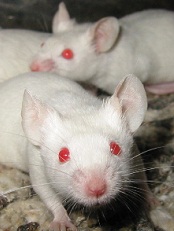
Credit: Aaron Logan
A small molecule targeting sphingosine kinase (SPHK) demonstrates considerable activity against virus-associated lymphoma, according to preclinical research published in Molecular Cancer Therapeutics.
The research focused on primary effusion lymphoma (PEL), a variant of diffuse large B-cell lymphoma etiologically linked to Kaposi’s sarcoma-associated herpesvirus (KSHV).
Investigators found that SPHK generates biologically active sphingolipids that keep PEL cells alive.
“It is still early in our understanding of how these special lipids contribute to viral cancers, but this is a major potential advance,” said senior study author Christopher Parsons, MD, of Louisiana State University Health Sciences Center in New Orleans.
“There are no therapies available to fight viral tumors by selectively blocking these pathways, all while not harming normal, uninfected cells.”
So the researchers decided to test ABC294640, a novel small molecule that selectively targets SPHK, in cells from PEL patients and mouse models of PEL.
ABC294640 induced dose-dependent apoptosis in a number of KSHV+ PEL cell lines: KSHV+/EBV- BCBL-1 cells, KSHV+/EBV+ BC-1 cells, KSHV+/EBV- BC-3 cells, and KSHV+/EBV- BCP-1 cells. But the drug showed little to no activity in KSHV-/EBV- BL-41 cells.
Further analyses revealed that ABC294640 induces apoptosis through suppression of KSHV-associated signal transduction.
The investigators observed dose-dependent suppression of ERK, Akt, and NF-kB p65 phosphorylation, as well as cleavage of caspase-3 and caspase-9, in BCBL-1 cells exposed to the drug. But the same effects did not occur in drug-resistant BL-41 cells.
Overexpression of either ERK or p65 in BCBL-1 cells partially suppressed apoptosis induced by ABC294640. And when the researchers used RNAi to target SPHK2, PEL cells exhibited reduced activation of ERK, Akt, and p65 phosphorylation, as well as a 5- to 6-fold increase in apoptosis.
Additionally, the team found that ABC294640 suppressed PEL progression and induced regression of PEL tumors in vivo.
The investigators injected BCL-1 cells into NOD/SCID mice and observed PEL expansion within 3 to 4 weeks. However, when they administered ABC294640 within 24 hours of PEL cell injections, they observed significant reductions in tumor expansion.
The researchers also evaluated ABC294640 activity after PEL tumors had been established. And treated mice showed significant tumor regression compared to untreated mice.
The investigators said these results suggest ABC294640 should be evaluated in clinical trials of KSHV-associated lymphoma.
“Our research thus far indicates that this molecule is safe, with the potential to stand alone as a single, orally administered drug,” Dr Parsons said.
Credit: Aaron Logan
A small molecule targeting sphingosine kinase (SPHK) demonstrates considerable activity against virus-associated lymphoma, according to preclinical research published in Molecular Cancer Therapeutics.
The research focused on primary effusion lymphoma (PEL), a variant of diffuse large B-cell lymphoma etiologically linked to Kaposi’s sarcoma-associated herpesvirus (KSHV).
Investigators found that SPHK generates biologically active sphingolipids that keep PEL cells alive.
“It is still early in our understanding of how these special lipids contribute to viral cancers, but this is a major potential advance,” said senior study author Christopher Parsons, MD, of Louisiana State University Health Sciences Center in New Orleans.
“There are no therapies available to fight viral tumors by selectively blocking these pathways, all while not harming normal, uninfected cells.”
So the researchers decided to test ABC294640, a novel small molecule that selectively targets SPHK, in cells from PEL patients and mouse models of PEL.
ABC294640 induced dose-dependent apoptosis in a number of KSHV+ PEL cell lines: KSHV+/EBV- BCBL-1 cells, KSHV+/EBV+ BC-1 cells, KSHV+/EBV- BC-3 cells, and KSHV+/EBV- BCP-1 cells. But the drug showed little to no activity in KSHV-/EBV- BL-41 cells.
Further analyses revealed that ABC294640 induces apoptosis through suppression of KSHV-associated signal transduction.
The investigators observed dose-dependent suppression of ERK, Akt, and NF-kB p65 phosphorylation, as well as cleavage of caspase-3 and caspase-9, in BCBL-1 cells exposed to the drug. But the same effects did not occur in drug-resistant BL-41 cells.
Overexpression of either ERK or p65 in BCBL-1 cells partially suppressed apoptosis induced by ABC294640. And when the researchers used RNAi to target SPHK2, PEL cells exhibited reduced activation of ERK, Akt, and p65 phosphorylation, as well as a 5- to 6-fold increase in apoptosis.
Additionally, the team found that ABC294640 suppressed PEL progression and induced regression of PEL tumors in vivo.
The investigators injected BCL-1 cells into NOD/SCID mice and observed PEL expansion within 3 to 4 weeks. However, when they administered ABC294640 within 24 hours of PEL cell injections, they observed significant reductions in tumor expansion.
The researchers also evaluated ABC294640 activity after PEL tumors had been established. And treated mice showed significant tumor regression compared to untreated mice.
The investigators said these results suggest ABC294640 should be evaluated in clinical trials of KSHV-associated lymphoma.
“Our research thus far indicates that this molecule is safe, with the potential to stand alone as a single, orally administered drug,” Dr Parsons said.
Credit: Aaron Logan
A small molecule targeting sphingosine kinase (SPHK) demonstrates considerable activity against virus-associated lymphoma, according to preclinical research published in Molecular Cancer Therapeutics.
The research focused on primary effusion lymphoma (PEL), a variant of diffuse large B-cell lymphoma etiologically linked to Kaposi’s sarcoma-associated herpesvirus (KSHV).
Investigators found that SPHK generates biologically active sphingolipids that keep PEL cells alive.
“It is still early in our understanding of how these special lipids contribute to viral cancers, but this is a major potential advance,” said senior study author Christopher Parsons, MD, of Louisiana State University Health Sciences Center in New Orleans.
“There are no therapies available to fight viral tumors by selectively blocking these pathways, all while not harming normal, uninfected cells.”
So the researchers decided to test ABC294640, a novel small molecule that selectively targets SPHK, in cells from PEL patients and mouse models of PEL.
ABC294640 induced dose-dependent apoptosis in a number of KSHV+ PEL cell lines: KSHV+/EBV- BCBL-1 cells, KSHV+/EBV+ BC-1 cells, KSHV+/EBV- BC-3 cells, and KSHV+/EBV- BCP-1 cells. But the drug showed little to no activity in KSHV-/EBV- BL-41 cells.
Further analyses revealed that ABC294640 induces apoptosis through suppression of KSHV-associated signal transduction.
The investigators observed dose-dependent suppression of ERK, Akt, and NF-kB p65 phosphorylation, as well as cleavage of caspase-3 and caspase-9, in BCBL-1 cells exposed to the drug. But the same effects did not occur in drug-resistant BL-41 cells.
Overexpression of either ERK or p65 in BCBL-1 cells partially suppressed apoptosis induced by ABC294640. And when the researchers used RNAi to target SPHK2, PEL cells exhibited reduced activation of ERK, Akt, and p65 phosphorylation, as well as a 5- to 6-fold increase in apoptosis.
Additionally, the team found that ABC294640 suppressed PEL progression and induced regression of PEL tumors in vivo.
The investigators injected BCL-1 cells into NOD/SCID mice and observed PEL expansion within 3 to 4 weeks. However, when they administered ABC294640 within 24 hours of PEL cell injections, they observed significant reductions in tumor expansion.
The researchers also evaluated ABC294640 activity after PEL tumors had been established. And treated mice showed significant tumor regression compared to untreated mice.
The investigators said these results suggest ABC294640 should be evaluated in clinical trials of KSHV-associated lymphoma.
“Our research thus far indicates that this molecule is safe, with the potential to stand alone as a single, orally administered drug,” Dr Parsons said.
Group discovers how drugs fight APL

Credit: The Armed Forces
Institute of Pathology
Results of a new study appear to explain how retinoic acid and arsenic trioxide work against acute promyelocytic leukemia (APL).
Researchers found that retinoic acid, either alone or in combination with arsenic trioxide, causes a cascade of molecular events that lead to cellular senescence.
And this halts APL-initiating activity in patient samples and mouse models of the disease.
The team said this mechanism could be activated to fight malignancies other than APL as well.
Hugues de Thé, MD, PhD, of Université Paris Diderot in France, and his colleagues described this research in Nature Medicine.
The group knew that APL is driven by the promyelocytic leukemia-retinoic acid receptor fusion protein (PML-RARA), which interferes with nuclear receptor signaling and PML nuclear body assembly.
Furthermore, APL can be cured by retinoic acid and arsenic trioxide (alone or in combination), both of which trigger PML-RARA degradation through non-overlapping pathways.
However, exactly how these treatments work in APL has been unclear. Dr de Thé’s research indicates that both drugs incite a cascade of events leading to senescence.
The researchers found evidence suggesting that a functional PML-transformation-related protein 53 (Trp53) axis is required to halt APL-initiating activity.
When retinoic acid induces PML-RARA degradation, normal PML elicits nuclear body reformation and prompts a Trp53 response that exhibits features of senescence. And this halts APL-initiating activity.
In addition, normal PML seems to play a role in the synergy between retinoic acid and arsenic trioxide. The researchers discovered that arsenic increases retinoic acid-induced PML-RARA degradation.
But arsenic also appears to cooperate with retinoic acid to cure APL by binding PML, accelerating nuclear body reformation, and enhancing downstream TP53 signaling.
The researchers said it seems likely that the same PML/p53 pathway can be activated in cancers other than PML as well.
Credit: The Armed Forces
Institute of Pathology
Results of a new study appear to explain how retinoic acid and arsenic trioxide work against acute promyelocytic leukemia (APL).
Researchers found that retinoic acid, either alone or in combination with arsenic trioxide, causes a cascade of molecular events that lead to cellular senescence.
And this halts APL-initiating activity in patient samples and mouse models of the disease.
The team said this mechanism could be activated to fight malignancies other than APL as well.
Hugues de Thé, MD, PhD, of Université Paris Diderot in France, and his colleagues described this research in Nature Medicine.
The group knew that APL is driven by the promyelocytic leukemia-retinoic acid receptor fusion protein (PML-RARA), which interferes with nuclear receptor signaling and PML nuclear body assembly.
Furthermore, APL can be cured by retinoic acid and arsenic trioxide (alone or in combination), both of which trigger PML-RARA degradation through non-overlapping pathways.
However, exactly how these treatments work in APL has been unclear. Dr de Thé’s research indicates that both drugs incite a cascade of events leading to senescence.
The researchers found evidence suggesting that a functional PML-transformation-related protein 53 (Trp53) axis is required to halt APL-initiating activity.
When retinoic acid induces PML-RARA degradation, normal PML elicits nuclear body reformation and prompts a Trp53 response that exhibits features of senescence. And this halts APL-initiating activity.
In addition, normal PML seems to play a role in the synergy between retinoic acid and arsenic trioxide. The researchers discovered that arsenic increases retinoic acid-induced PML-RARA degradation.
But arsenic also appears to cooperate with retinoic acid to cure APL by binding PML, accelerating nuclear body reformation, and enhancing downstream TP53 signaling.
The researchers said it seems likely that the same PML/p53 pathway can be activated in cancers other than PML as well.
Credit: The Armed Forces
Institute of Pathology
Results of a new study appear to explain how retinoic acid and arsenic trioxide work against acute promyelocytic leukemia (APL).
Researchers found that retinoic acid, either alone or in combination with arsenic trioxide, causes a cascade of molecular events that lead to cellular senescence.
And this halts APL-initiating activity in patient samples and mouse models of the disease.
The team said this mechanism could be activated to fight malignancies other than APL as well.
Hugues de Thé, MD, PhD, of Université Paris Diderot in France, and his colleagues described this research in Nature Medicine.
The group knew that APL is driven by the promyelocytic leukemia-retinoic acid receptor fusion protein (PML-RARA), which interferes with nuclear receptor signaling and PML nuclear body assembly.
Furthermore, APL can be cured by retinoic acid and arsenic trioxide (alone or in combination), both of which trigger PML-RARA degradation through non-overlapping pathways.
However, exactly how these treatments work in APL has been unclear. Dr de Thé’s research indicates that both drugs incite a cascade of events leading to senescence.
The researchers found evidence suggesting that a functional PML-transformation-related protein 53 (Trp53) axis is required to halt APL-initiating activity.
When retinoic acid induces PML-RARA degradation, normal PML elicits nuclear body reformation and prompts a Trp53 response that exhibits features of senescence. And this halts APL-initiating activity.
In addition, normal PML seems to play a role in the synergy between retinoic acid and arsenic trioxide. The researchers discovered that arsenic increases retinoic acid-induced PML-RARA degradation.
But arsenic also appears to cooperate with retinoic acid to cure APL by binding PML, accelerating nuclear body reformation, and enhancing downstream TP53 signaling.
The researchers said it seems likely that the same PML/p53 pathway can be activated in cancers other than PML as well.
Compound active against a range of cancers
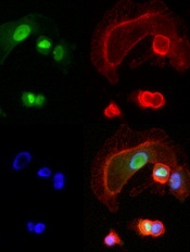
expressing NFAT3c-GFP
A little-studied chemical compound has “wide and potent” anticancer activity, investigators have reported in Cancer Cell.
The compound, BMH-21, works by inhibiting the RNA polymerase transcription pathway (Pol I), thereby preventing cancer cell communication and replication.
“Without this transcription machinery, cancer cells cannot function,” said study author Marikki Laiho, MD, PhD, of the Johns Hopkins University School of Medicine in Baltimore, Maryland.
She and her colleagues homed in on BMH-21 by screening a library of chemical compounds thought to have potential for anticancer activity.
Specifically, the team looked at the compounds’ ability to interfere with transcription in the National Cancer Institute’s collection of 60 human tumor cell lines (known as NCI-60).
BMH-21 demonstrated activity against all 9 cancer types studied—leukemia and melanoma, as well as breast, CNS, colon, lung, ovarian, prostate, and renal cancers.
The drug also repressed tumor growth in mouse models of colon cancer and melanoma.
Additional analyses showed that BMH-21 inhibited Pol I transcription and caused disintegration of the nucleolus. The drug activated loss of the Pol I catalytic subunit RPA194, which led to disassembly of the Pol I holocomplex from the ribosomal DNA.
And the loss of RPA194, which was a result of increased proteasome-mediated turnover, was associated with decreased cancer cell viability.
Dr Laiho and her colleagues are continuing studies of BMH-21 in animal models to confirm the drug’s anticancer activity, identify any toxicities associated with the compound, and determine the optimal dose.
And because Pol I activity is frequently deregulated in cancers, the investigators believe BMH-21 could have therapeutic potential for many malignancies.
Dr Laiho is currently collaborating with experts in multiple myeloma, medullary thyroid cancer, and prostate cancer to explore the drug’s activity in these malignancies.
expressing NFAT3c-GFP
A little-studied chemical compound has “wide and potent” anticancer activity, investigators have reported in Cancer Cell.
The compound, BMH-21, works by inhibiting the RNA polymerase transcription pathway (Pol I), thereby preventing cancer cell communication and replication.
“Without this transcription machinery, cancer cells cannot function,” said study author Marikki Laiho, MD, PhD, of the Johns Hopkins University School of Medicine in Baltimore, Maryland.
She and her colleagues homed in on BMH-21 by screening a library of chemical compounds thought to have potential for anticancer activity.
Specifically, the team looked at the compounds’ ability to interfere with transcription in the National Cancer Institute’s collection of 60 human tumor cell lines (known as NCI-60).
BMH-21 demonstrated activity against all 9 cancer types studied—leukemia and melanoma, as well as breast, CNS, colon, lung, ovarian, prostate, and renal cancers.
The drug also repressed tumor growth in mouse models of colon cancer and melanoma.
Additional analyses showed that BMH-21 inhibited Pol I transcription and caused disintegration of the nucleolus. The drug activated loss of the Pol I catalytic subunit RPA194, which led to disassembly of the Pol I holocomplex from the ribosomal DNA.
And the loss of RPA194, which was a result of increased proteasome-mediated turnover, was associated with decreased cancer cell viability.
Dr Laiho and her colleagues are continuing studies of BMH-21 in animal models to confirm the drug’s anticancer activity, identify any toxicities associated with the compound, and determine the optimal dose.
And because Pol I activity is frequently deregulated in cancers, the investigators believe BMH-21 could have therapeutic potential for many malignancies.
Dr Laiho is currently collaborating with experts in multiple myeloma, medullary thyroid cancer, and prostate cancer to explore the drug’s activity in these malignancies.
expressing NFAT3c-GFP
A little-studied chemical compound has “wide and potent” anticancer activity, investigators have reported in Cancer Cell.
The compound, BMH-21, works by inhibiting the RNA polymerase transcription pathway (Pol I), thereby preventing cancer cell communication and replication.
“Without this transcription machinery, cancer cells cannot function,” said study author Marikki Laiho, MD, PhD, of the Johns Hopkins University School of Medicine in Baltimore, Maryland.
She and her colleagues homed in on BMH-21 by screening a library of chemical compounds thought to have potential for anticancer activity.
Specifically, the team looked at the compounds’ ability to interfere with transcription in the National Cancer Institute’s collection of 60 human tumor cell lines (known as NCI-60).
BMH-21 demonstrated activity against all 9 cancer types studied—leukemia and melanoma, as well as breast, CNS, colon, lung, ovarian, prostate, and renal cancers.
The drug also repressed tumor growth in mouse models of colon cancer and melanoma.
Additional analyses showed that BMH-21 inhibited Pol I transcription and caused disintegration of the nucleolus. The drug activated loss of the Pol I catalytic subunit RPA194, which led to disassembly of the Pol I holocomplex from the ribosomal DNA.
And the loss of RPA194, which was a result of increased proteasome-mediated turnover, was associated with decreased cancer cell viability.
Dr Laiho and her colleagues are continuing studies of BMH-21 in animal models to confirm the drug’s anticancer activity, identify any toxicities associated with the compound, and determine the optimal dose.
And because Pol I activity is frequently deregulated in cancers, the investigators believe BMH-21 could have therapeutic potential for many malignancies.
Dr Laiho is currently collaborating with experts in multiple myeloma, medullary thyroid cancer, and prostate cancer to explore the drug’s activity in these malignancies.
E coli has applications for malaria vaccine
Credit: USDA
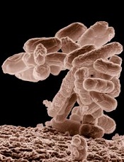
E coli bacteria may enable inexpensive production of a transmission-blocking malaria vaccine, according to a paper published in Infection and Immunity.
Scientists used E coli to create a new process to purify and refold codon harmonized recombinant Pfs25 (CHrPfs25).
Pfs25 is a sexual-stage antigen of Plasmodium falciparum expressed on the surface of zygote and ookinete forms of the parasite.
Research has shown that monoclonal antibodies directed against native Pfs25 can prevent development of P falciparum oocysts in the midgut of the mosquito.
So Pfs25 is a potential vaccine candidate, but producing it has proven challenging and costly.
“Malaria affects the poorest of the poor,” said study author Nirbhay Kumar, PhD, of Tulane University School of Public Health and Tropical Medicine in New Orleans, Louisiana.
“And if you are trying to make a vaccine for those billions of people who are at risk, you need to make it cheaper to manufacture. We think that producing this protein in bacteria will make it very cost-effective for large-scale vaccine production.”
To create the vaccine for the current study, Dr Kumar and his colleagues expressed CHrPfs25 in E coli, purified the protein after simple oxidative refolding steps, and formulated it in several adjuvants.
The team then tested the final product in mice. Antibodies present after vaccination recognized native Pfs25 on the surface of live gametes of P falciparum and demonstrated complete malaria transmission-blocking activity.
The transmission-blocking efficacy of CHrPfs25 was 100% whether the researchers were testing Anopheles gambiae mosquitoes or Anopheles stephensi mosquitoes.
Dr Kumar said the next step for this research will be to develop a version of the vaccine that can be used in clinical trials.
Transmission-blocking vaccines, though not yet widely tested in humans, have the potential to be used in conjunction with more traditional malaria vaccines and other interventions—such as malaria drugs and bed nets—to fight the disease and ultimately aid in the gradual elimination of malaria parasites.
Credit: USDA
E coli bacteria may enable inexpensive production of a transmission-blocking malaria vaccine, according to a paper published in Infection and Immunity.
Scientists used E coli to create a new process to purify and refold codon harmonized recombinant Pfs25 (CHrPfs25).
Pfs25 is a sexual-stage antigen of Plasmodium falciparum expressed on the surface of zygote and ookinete forms of the parasite.
Research has shown that monoclonal antibodies directed against native Pfs25 can prevent development of P falciparum oocysts in the midgut of the mosquito.
So Pfs25 is a potential vaccine candidate, but producing it has proven challenging and costly.
“Malaria affects the poorest of the poor,” said study author Nirbhay Kumar, PhD, of Tulane University School of Public Health and Tropical Medicine in New Orleans, Louisiana.
“And if you are trying to make a vaccine for those billions of people who are at risk, you need to make it cheaper to manufacture. We think that producing this protein in bacteria will make it very cost-effective for large-scale vaccine production.”
To create the vaccine for the current study, Dr Kumar and his colleagues expressed CHrPfs25 in E coli, purified the protein after simple oxidative refolding steps, and formulated it in several adjuvants.
The team then tested the final product in mice. Antibodies present after vaccination recognized native Pfs25 on the surface of live gametes of P falciparum and demonstrated complete malaria transmission-blocking activity.
The transmission-blocking efficacy of CHrPfs25 was 100% whether the researchers were testing Anopheles gambiae mosquitoes or Anopheles stephensi mosquitoes.
Dr Kumar said the next step for this research will be to develop a version of the vaccine that can be used in clinical trials.
Transmission-blocking vaccines, though not yet widely tested in humans, have the potential to be used in conjunction with more traditional malaria vaccines and other interventions—such as malaria drugs and bed nets—to fight the disease and ultimately aid in the gradual elimination of malaria parasites.
Credit: USDA
E coli bacteria may enable inexpensive production of a transmission-blocking malaria vaccine, according to a paper published in Infection and Immunity.
Scientists used E coli to create a new process to purify and refold codon harmonized recombinant Pfs25 (CHrPfs25).
Pfs25 is a sexual-stage antigen of Plasmodium falciparum expressed on the surface of zygote and ookinete forms of the parasite.
Research has shown that monoclonal antibodies directed against native Pfs25 can prevent development of P falciparum oocysts in the midgut of the mosquito.
So Pfs25 is a potential vaccine candidate, but producing it has proven challenging and costly.
“Malaria affects the poorest of the poor,” said study author Nirbhay Kumar, PhD, of Tulane University School of Public Health and Tropical Medicine in New Orleans, Louisiana.
“And if you are trying to make a vaccine for those billions of people who are at risk, you need to make it cheaper to manufacture. We think that producing this protein in bacteria will make it very cost-effective for large-scale vaccine production.”
To create the vaccine for the current study, Dr Kumar and his colleagues expressed CHrPfs25 in E coli, purified the protein after simple oxidative refolding steps, and formulated it in several adjuvants.
The team then tested the final product in mice. Antibodies present after vaccination recognized native Pfs25 on the surface of live gametes of P falciparum and demonstrated complete malaria transmission-blocking activity.
The transmission-blocking efficacy of CHrPfs25 was 100% whether the researchers were testing Anopheles gambiae mosquitoes or Anopheles stephensi mosquitoes.
Dr Kumar said the next step for this research will be to develop a version of the vaccine that can be used in clinical trials.
Transmission-blocking vaccines, though not yet widely tested in humans, have the potential to be used in conjunction with more traditional malaria vaccines and other interventions—such as malaria drugs and bed nets—to fight the disease and ultimately aid in the gradual elimination of malaria parasites.
Study reveals ‘widespread’ genetic heterogeneity in MM
different colored cells
Credit: Lauren Solomon
Results of a new study suggest the genetic landscape of multiple myeloma (MM) may be more complex than we thought.
The research revealed “widespread” heterogeneity in samples from more than 200 MM patients.
In some cases, a single patient had multiple mutations in the same pathway. And most of the patients harbored at least 3 detectable subclonal mutations.
The researchers said these findings, published in Cancer Cell, might explain why targeted therapies are not always effective in MM and why some patients relapse after treatment.
“What this new work shows us is that when we treat an individual patient with multiple myeloma, it’s possible that we’re not just looking at one disease, but at many,” said study author Todd Golub, MD, of the Dana-Farber Cancer Institute in Cambridge, Massachusetts.
“In the same person, there could be cancer cells with different genetic make-ups. These findings indicate a need to identify the extent of genetic diversity within a tumor as we move toward precision cancer medicine and genome-based diagnostics.”
Dr Golub and his colleagues studied samples from 203 MM patients and identified frequent mutations in genes known to play an important role in MM, including KRAS, NRAS, and BRAF.
But many of these telltale mutations were not present in all MM cells. Instead, they were often observed only in a subclonal population.
This suggests targeted therapies may have limitations in patients whose tumors are made up of these subclonal populations, the researchers said.
To explore the therapeutic implications of this research, the team performed follow-up experiments looking specifically at BRAF, a gene for which several inhibitors exist.
Previous studies indicated that roughly 4% of MM patients may have mutations in this gene. And a recent report on a single MM patient treated with drugs targeting BRAF showed promising results.
However, Dr Golub and his colleagues found evidence that treating a tumor harboring subclonal BRAF mutations with one of these agents may, at best, kill a fraction of the cells and, at worst, stimulate another cancer cell subpopulation to grow.
“There’s clearly potential for these drugs in some patients with multiple myeloma, but we show that there are also potential problems for others,” said study author Jens Lohr, MD, PhD, also of Dana-Farber.
“If a patient has a BRAF mutation in less than 100% of his cells, or if he has mutations in KRAS or NRAS at the same time, [it] may influence the response to an inhibitor.”
This suggests subclonal populations could be one of the reasons many patients suffer relapse after treatment, the researchers said.
“Matching the right drug to the right patient may not be as easy as finding a mutation and having a drug that targets it,” Dr Lohr said. “We have to keep this additional parameter of heterogeneity in mind and keep exploring what it means for therapy.”
different colored cells
Credit: Lauren Solomon
Results of a new study suggest the genetic landscape of multiple myeloma (MM) may be more complex than we thought.
The research revealed “widespread” heterogeneity in samples from more than 200 MM patients.
In some cases, a single patient had multiple mutations in the same pathway. And most of the patients harbored at least 3 detectable subclonal mutations.
The researchers said these findings, published in Cancer Cell, might explain why targeted therapies are not always effective in MM and why some patients relapse after treatment.
“What this new work shows us is that when we treat an individual patient with multiple myeloma, it’s possible that we’re not just looking at one disease, but at many,” said study author Todd Golub, MD, of the Dana-Farber Cancer Institute in Cambridge, Massachusetts.
“In the same person, there could be cancer cells with different genetic make-ups. These findings indicate a need to identify the extent of genetic diversity within a tumor as we move toward precision cancer medicine and genome-based diagnostics.”
Dr Golub and his colleagues studied samples from 203 MM patients and identified frequent mutations in genes known to play an important role in MM, including KRAS, NRAS, and BRAF.
But many of these telltale mutations were not present in all MM cells. Instead, they were often observed only in a subclonal population.
This suggests targeted therapies may have limitations in patients whose tumors are made up of these subclonal populations, the researchers said.
To explore the therapeutic implications of this research, the team performed follow-up experiments looking specifically at BRAF, a gene for which several inhibitors exist.
Previous studies indicated that roughly 4% of MM patients may have mutations in this gene. And a recent report on a single MM patient treated with drugs targeting BRAF showed promising results.
However, Dr Golub and his colleagues found evidence that treating a tumor harboring subclonal BRAF mutations with one of these agents may, at best, kill a fraction of the cells and, at worst, stimulate another cancer cell subpopulation to grow.
“There’s clearly potential for these drugs in some patients with multiple myeloma, but we show that there are also potential problems for others,” said study author Jens Lohr, MD, PhD, also of Dana-Farber.
“If a patient has a BRAF mutation in less than 100% of his cells, or if he has mutations in KRAS or NRAS at the same time, [it] may influence the response to an inhibitor.”
This suggests subclonal populations could be one of the reasons many patients suffer relapse after treatment, the researchers said.
“Matching the right drug to the right patient may not be as easy as finding a mutation and having a drug that targets it,” Dr Lohr said. “We have to keep this additional parameter of heterogeneity in mind and keep exploring what it means for therapy.”
different colored cells
Credit: Lauren Solomon
Results of a new study suggest the genetic landscape of multiple myeloma (MM) may be more complex than we thought.
The research revealed “widespread” heterogeneity in samples from more than 200 MM patients.
In some cases, a single patient had multiple mutations in the same pathway. And most of the patients harbored at least 3 detectable subclonal mutations.
The researchers said these findings, published in Cancer Cell, might explain why targeted therapies are not always effective in MM and why some patients relapse after treatment.
“What this new work shows us is that when we treat an individual patient with multiple myeloma, it’s possible that we’re not just looking at one disease, but at many,” said study author Todd Golub, MD, of the Dana-Farber Cancer Institute in Cambridge, Massachusetts.
“In the same person, there could be cancer cells with different genetic make-ups. These findings indicate a need to identify the extent of genetic diversity within a tumor as we move toward precision cancer medicine and genome-based diagnostics.”
Dr Golub and his colleagues studied samples from 203 MM patients and identified frequent mutations in genes known to play an important role in MM, including KRAS, NRAS, and BRAF.
But many of these telltale mutations were not present in all MM cells. Instead, they were often observed only in a subclonal population.
This suggests targeted therapies may have limitations in patients whose tumors are made up of these subclonal populations, the researchers said.
To explore the therapeutic implications of this research, the team performed follow-up experiments looking specifically at BRAF, a gene for which several inhibitors exist.
Previous studies indicated that roughly 4% of MM patients may have mutations in this gene. And a recent report on a single MM patient treated with drugs targeting BRAF showed promising results.
However, Dr Golub and his colleagues found evidence that treating a tumor harboring subclonal BRAF mutations with one of these agents may, at best, kill a fraction of the cells and, at worst, stimulate another cancer cell subpopulation to grow.
“There’s clearly potential for these drugs in some patients with multiple myeloma, but we show that there are also potential problems for others,” said study author Jens Lohr, MD, PhD, also of Dana-Farber.
“If a patient has a BRAF mutation in less than 100% of his cells, or if he has mutations in KRAS or NRAS at the same time, [it] may influence the response to an inhibitor.”
This suggests subclonal populations could be one of the reasons many patients suffer relapse after treatment, the researchers said.
“Matching the right drug to the right patient may not be as easy as finding a mutation and having a drug that targets it,” Dr Lohr said. “We have to keep this additional parameter of heterogeneity in mind and keep exploring what it means for therapy.”
Team reproduces HSPCs in artificial bone marrow

artificial bone marrow
Credit: C. Lee-Thedieck
Researchers say they have developed artificial bone marrow analogs that can be used to reproduce hematopoietic stem and progenitor cells (HSPCs).
The team created macroporous hydrogel scaffolds that mimic the stem cell niche of the bone marrow.
When they introduced mesenchymal stem cells (MSCs) from actual bone marrow into the analogs, the MSCs promoted HSPC proliferation.
In fact, the MSCs preserved HSPC stemness more effectively in the analogs than in standard 2-dimensional cell culture systems.
Annamarija Raic, of the Max Planck Institute for Intelligent Systems in Stuttgart, Germany, and her colleagues reported these results in Biomaterials.
The researchers noted that reproducing functional HSPCs in the lab has proven challenging. The cells cannot be cultured in vitro for a feasible period of time without differentiating.
So the team set out to create a culture system that mimics the important physical and biological parameters of the stem cell niche: the 3D architecture, the adhesive extracellular matrix, soluble factors, and the stromal cell compartment.
They used salt leaching technology to produce poly(ethylene glycol) diacrylate hydrogel scaffolds that could soak cells into their pores. To biofunctionalize the scaffolds, the investigators added an RGD peptide carrying an acrylate moiety.
They then introduced 3 different cell types into the scaffolds—the human osteosarcoma cell line CAL72, MSCs from bone marrow, and MSCs from umbilical cord blood—to see which best supported the proliferation of CD34+ HSPCs isolated from cord blood.
Each of the cell types supported HSPC proliferation, but bone marrow MSCs were the most effective. The researchers therefore decided to use bone marrow MSCs when they compared their 3D scaffolds to a 2D culture system.
The bone marrow MSCs had a beneficial effect on HSPC proliferation in the 2D cell cultures. Over 4 days, HSPCs divided 1 to 2 times more often when they were cultured with bone marrow MSCs than without the cells.
In the 3D scaffolds, HSPC proliferation was comparable or slightly lower than that observed in the 2D cultures. However, the scaffolds had a higher percentage of CD34+ HSPCs after 4 days.
The investigators therefore concluded that their hydrogel scaffolds meet the basic requirements for creating artificial stem cell niches.
artificial bone marrow
Credit: C. Lee-Thedieck
Researchers say they have developed artificial bone marrow analogs that can be used to reproduce hematopoietic stem and progenitor cells (HSPCs).
The team created macroporous hydrogel scaffolds that mimic the stem cell niche of the bone marrow.
When they introduced mesenchymal stem cells (MSCs) from actual bone marrow into the analogs, the MSCs promoted HSPC proliferation.
In fact, the MSCs preserved HSPC stemness more effectively in the analogs than in standard 2-dimensional cell culture systems.
Annamarija Raic, of the Max Planck Institute for Intelligent Systems in Stuttgart, Germany, and her colleagues reported these results in Biomaterials.
The researchers noted that reproducing functional HSPCs in the lab has proven challenging. The cells cannot be cultured in vitro for a feasible period of time without differentiating.
So the team set out to create a culture system that mimics the important physical and biological parameters of the stem cell niche: the 3D architecture, the adhesive extracellular matrix, soluble factors, and the stromal cell compartment.
They used salt leaching technology to produce poly(ethylene glycol) diacrylate hydrogel scaffolds that could soak cells into their pores. To biofunctionalize the scaffolds, the investigators added an RGD peptide carrying an acrylate moiety.
They then introduced 3 different cell types into the scaffolds—the human osteosarcoma cell line CAL72, MSCs from bone marrow, and MSCs from umbilical cord blood—to see which best supported the proliferation of CD34+ HSPCs isolated from cord blood.
Each of the cell types supported HSPC proliferation, but bone marrow MSCs were the most effective. The researchers therefore decided to use bone marrow MSCs when they compared their 3D scaffolds to a 2D culture system.
The bone marrow MSCs had a beneficial effect on HSPC proliferation in the 2D cell cultures. Over 4 days, HSPCs divided 1 to 2 times more often when they were cultured with bone marrow MSCs than without the cells.
In the 3D scaffolds, HSPC proliferation was comparable or slightly lower than that observed in the 2D cultures. However, the scaffolds had a higher percentage of CD34+ HSPCs after 4 days.
The investigators therefore concluded that their hydrogel scaffolds meet the basic requirements for creating artificial stem cell niches.
artificial bone marrow
Credit: C. Lee-Thedieck
Researchers say they have developed artificial bone marrow analogs that can be used to reproduce hematopoietic stem and progenitor cells (HSPCs).
The team created macroporous hydrogel scaffolds that mimic the stem cell niche of the bone marrow.
When they introduced mesenchymal stem cells (MSCs) from actual bone marrow into the analogs, the MSCs promoted HSPC proliferation.
In fact, the MSCs preserved HSPC stemness more effectively in the analogs than in standard 2-dimensional cell culture systems.
Annamarija Raic, of the Max Planck Institute for Intelligent Systems in Stuttgart, Germany, and her colleagues reported these results in Biomaterials.
The researchers noted that reproducing functional HSPCs in the lab has proven challenging. The cells cannot be cultured in vitro for a feasible period of time without differentiating.
So the team set out to create a culture system that mimics the important physical and biological parameters of the stem cell niche: the 3D architecture, the adhesive extracellular matrix, soluble factors, and the stromal cell compartment.
They used salt leaching technology to produce poly(ethylene glycol) diacrylate hydrogel scaffolds that could soak cells into their pores. To biofunctionalize the scaffolds, the investigators added an RGD peptide carrying an acrylate moiety.
They then introduced 3 different cell types into the scaffolds—the human osteosarcoma cell line CAL72, MSCs from bone marrow, and MSCs from umbilical cord blood—to see which best supported the proliferation of CD34+ HSPCs isolated from cord blood.
Each of the cell types supported HSPC proliferation, but bone marrow MSCs were the most effective. The researchers therefore decided to use bone marrow MSCs when they compared their 3D scaffolds to a 2D culture system.
The bone marrow MSCs had a beneficial effect on HSPC proliferation in the 2D cell cultures. Over 4 days, HSPCs divided 1 to 2 times more often when they were cultured with bone marrow MSCs than without the cells.
In the 3D scaffolds, HSPC proliferation was comparable or slightly lower than that observed in the 2D cultures. However, the scaffolds had a higher percentage of CD34+ HSPCs after 4 days.
The investigators therefore concluded that their hydrogel scaffolds meet the basic requirements for creating artificial stem cell niches.
NK cells target malaria-infected RBCs

Credit: Bjorn Onfelt/Dan Davis
The parasites that cause malaria are adapted to the hosts they infect, so studying the disease in mice doesn’t necessarily reveal information that translates to human disease.
But scientists believe they may have overcome this limitation. They’ve developed a strain of mice that mimics many features of the human immune system and can be infected with Plasmodium falciparum.
Using this strain, the researchers discovered that natural killer (NK) cells preferentially interact with and kill infected red blood cells (RBCs) in a contact-dependent manner.
The group recounted this discovery in PNAS.
“Human malaria studies have been hampered by a lack of animal models,” said study author Jianzhu Chen, PhD, of the Singapore-MIT Alliance for Research and Technology in Singapore.
“This [research] paves the way to start dissecting how the host human immune system interacts with the pathogen.”
Scientists studying malaria in mice previously generated mice with human RBCs. But these mice have compromised immune systems, so they can’t be used to study the immune response to malaria infection.
Over the past several years, Dr Chen and his colleagues have developed strains of mice that have the human cells necessary for a comprehensive immune response.
To generate these cells, the researchers deliver human hematopoietic stem cells, along with cytokines that help them mature into B and T cells, NK cells, and macrophages. These mice have already proven useful to study other diseases, such as dengue fever.
To adapt the mice for the study of malaria, the scientists injected them with human RBCs every day for a week, at which point 25% of their RBCs were human. And this was enough for the malaria parasite to cause an infection.
The researchers investigated the role of NK cells and macrophages during the first 2 days of malaria infection. And they found that eliminating macrophages had very little impact on the immune response during those early stages.
However, in mice lacking NK cells, parasite levels went up 7-fold, suggesting that NK cells are critical to controlling infection early on.
To further investigate the role of NK cells, the scientists placed human NK cells in a sample of infected and uninfected RBCs. The NK cells randomly interacted with both types of cells, but they latched onto infected cells much longer, eventually killing them.
The researchers also identified a cell adhesion protein called LFA-1 that helps NK cells bind to RBCs. They are now studying this process in more detail and trying to determine what other molecules, including those produced by the malaria parasite, might be involved.
Dr Chen and his colleagues also hope to use these mice to study experimental malaria vaccines or drugs. And in another future study, they plan to inject the mice with RBCs from patients with sickle cell anemia to investigate how the sickle-shaped cells help people survive malaria infection.
Credit: Bjorn Onfelt/Dan Davis
The parasites that cause malaria are adapted to the hosts they infect, so studying the disease in mice doesn’t necessarily reveal information that translates to human disease.
But scientists believe they may have overcome this limitation. They’ve developed a strain of mice that mimics many features of the human immune system and can be infected with Plasmodium falciparum.
Using this strain, the researchers discovered that natural killer (NK) cells preferentially interact with and kill infected red blood cells (RBCs) in a contact-dependent manner.
The group recounted this discovery in PNAS.
“Human malaria studies have been hampered by a lack of animal models,” said study author Jianzhu Chen, PhD, of the Singapore-MIT Alliance for Research and Technology in Singapore.
“This [research] paves the way to start dissecting how the host human immune system interacts with the pathogen.”
Scientists studying malaria in mice previously generated mice with human RBCs. But these mice have compromised immune systems, so they can’t be used to study the immune response to malaria infection.
Over the past several years, Dr Chen and his colleagues have developed strains of mice that have the human cells necessary for a comprehensive immune response.
To generate these cells, the researchers deliver human hematopoietic stem cells, along with cytokines that help them mature into B and T cells, NK cells, and macrophages. These mice have already proven useful to study other diseases, such as dengue fever.
To adapt the mice for the study of malaria, the scientists injected them with human RBCs every day for a week, at which point 25% of their RBCs were human. And this was enough for the malaria parasite to cause an infection.
The researchers investigated the role of NK cells and macrophages during the first 2 days of malaria infection. And they found that eliminating macrophages had very little impact on the immune response during those early stages.
However, in mice lacking NK cells, parasite levels went up 7-fold, suggesting that NK cells are critical to controlling infection early on.
To further investigate the role of NK cells, the scientists placed human NK cells in a sample of infected and uninfected RBCs. The NK cells randomly interacted with both types of cells, but they latched onto infected cells much longer, eventually killing them.
The researchers also identified a cell adhesion protein called LFA-1 that helps NK cells bind to RBCs. They are now studying this process in more detail and trying to determine what other molecules, including those produced by the malaria parasite, might be involved.
Dr Chen and his colleagues also hope to use these mice to study experimental malaria vaccines or drugs. And in another future study, they plan to inject the mice with RBCs from patients with sickle cell anemia to investigate how the sickle-shaped cells help people survive malaria infection.
Credit: Bjorn Onfelt/Dan Davis
The parasites that cause malaria are adapted to the hosts they infect, so studying the disease in mice doesn’t necessarily reveal information that translates to human disease.
But scientists believe they may have overcome this limitation. They’ve developed a strain of mice that mimics many features of the human immune system and can be infected with Plasmodium falciparum.
Using this strain, the researchers discovered that natural killer (NK) cells preferentially interact with and kill infected red blood cells (RBCs) in a contact-dependent manner.
The group recounted this discovery in PNAS.
“Human malaria studies have been hampered by a lack of animal models,” said study author Jianzhu Chen, PhD, of the Singapore-MIT Alliance for Research and Technology in Singapore.
“This [research] paves the way to start dissecting how the host human immune system interacts with the pathogen.”
Scientists studying malaria in mice previously generated mice with human RBCs. But these mice have compromised immune systems, so they can’t be used to study the immune response to malaria infection.
Over the past several years, Dr Chen and his colleagues have developed strains of mice that have the human cells necessary for a comprehensive immune response.
To generate these cells, the researchers deliver human hematopoietic stem cells, along with cytokines that help them mature into B and T cells, NK cells, and macrophages. These mice have already proven useful to study other diseases, such as dengue fever.
To adapt the mice for the study of malaria, the scientists injected them with human RBCs every day for a week, at which point 25% of their RBCs were human. And this was enough for the malaria parasite to cause an infection.
The researchers investigated the role of NK cells and macrophages during the first 2 days of malaria infection. And they found that eliminating macrophages had very little impact on the immune response during those early stages.
However, in mice lacking NK cells, parasite levels went up 7-fold, suggesting that NK cells are critical to controlling infection early on.
To further investigate the role of NK cells, the scientists placed human NK cells in a sample of infected and uninfected RBCs. The NK cells randomly interacted with both types of cells, but they latched onto infected cells much longer, eventually killing them.
The researchers also identified a cell adhesion protein called LFA-1 that helps NK cells bind to RBCs. They are now studying this process in more detail and trying to determine what other molecules, including those produced by the malaria parasite, might be involved.
Dr Chen and his colleagues also hope to use these mice to study experimental malaria vaccines or drugs. And in another future study, they plan to inject the mice with RBCs from patients with sickle cell anemia to investigate how the sickle-shaped cells help people survive malaria infection.