User login
States’ restrictions hinder ACA rollout

Credit: Rhoda Baer
As implementation of the Affordable Care Act (ACA) continues, a new report indicates that most community health centers across the US are attempting to help uninsured individuals obtain health insurance.
But health centers in certain states are hampered in their outreach and enrollment efforts and are significantly less optimistic about the impact of health reform in their communities.
“These findings show the effects of state policies that are designed to stand in the way of health reform,” said study author Sara Rosenbaum, of the George Washington University School of Public Health and Health Services in Washington, DC.
“These restrictive policies are measurably impairing community-level efforts to cover the uninsured. Without the Medicaid expansion or comprehensive enrollment assistance, millions of people in medically underserved communities could go without the benefits provided under the Affordable Care Act.”
Using data from a nationwide survey, Rosenbaum and her colleagues examined the early outreach and enrollment efforts of 606 community health centers across the US.
The data showed that, by early October 2013, virtually all of the responding health centers had received enhanced outreach funding.
And three quarters of them had already instituted expanded outreach and enrollment assistance efforts, such as one-on-one assistance with online and paper applications for health insurance, and enrollment help in multiple languages.
The researchers also compared health center outreach and enrollment in the 21 states (and Washington DC) that have embraced health reform to health centers in 9 states that have resisted or rejected key parts of the ACA.
The 9 “restrictive” states were those that had rejected an expansion of Medicaid and adopted policies or Navigator laws that make it harder to provide assistance to the uninsured.
The comparison showed that community health centers in the restrictive states had significantly more limited outreach and enrollment resources and had significantly fewer enrollment staff. Restrictive states had an average of 3 full-time enrollment staff members, compared to 6 staff members in full-implementation states.
Furthermore, health centers in restrictive states were significantly less likely than those in full-implementation states to be assessing patient eligibility for insurance coverage—59% and 79%, respectively.
And health centers in the 9 restrictive states were significantly less optimistic about the potential impact of the ACA on their patients.
In restrictive states, nearly 1 in 6 health center leaders expected at least half of their patients to remain uninsured. In full-implementation states, only 2% of leaders said the same.
For more details, see the full report, “Assessing the Potential Impact of State Policies on Community Health Centers’ Outreach and Enrollment Activities.” 
Credit: Rhoda Baer
As implementation of the Affordable Care Act (ACA) continues, a new report indicates that most community health centers across the US are attempting to help uninsured individuals obtain health insurance.
But health centers in certain states are hampered in their outreach and enrollment efforts and are significantly less optimistic about the impact of health reform in their communities.
“These findings show the effects of state policies that are designed to stand in the way of health reform,” said study author Sara Rosenbaum, of the George Washington University School of Public Health and Health Services in Washington, DC.
“These restrictive policies are measurably impairing community-level efforts to cover the uninsured. Without the Medicaid expansion or comprehensive enrollment assistance, millions of people in medically underserved communities could go without the benefits provided under the Affordable Care Act.”
Using data from a nationwide survey, Rosenbaum and her colleagues examined the early outreach and enrollment efforts of 606 community health centers across the US.
The data showed that, by early October 2013, virtually all of the responding health centers had received enhanced outreach funding.
And three quarters of them had already instituted expanded outreach and enrollment assistance efforts, such as one-on-one assistance with online and paper applications for health insurance, and enrollment help in multiple languages.
The researchers also compared health center outreach and enrollment in the 21 states (and Washington DC) that have embraced health reform to health centers in 9 states that have resisted or rejected key parts of the ACA.
The 9 “restrictive” states were those that had rejected an expansion of Medicaid and adopted policies or Navigator laws that make it harder to provide assistance to the uninsured.
The comparison showed that community health centers in the restrictive states had significantly more limited outreach and enrollment resources and had significantly fewer enrollment staff. Restrictive states had an average of 3 full-time enrollment staff members, compared to 6 staff members in full-implementation states.
Furthermore, health centers in restrictive states were significantly less likely than those in full-implementation states to be assessing patient eligibility for insurance coverage—59% and 79%, respectively.
And health centers in the 9 restrictive states were significantly less optimistic about the potential impact of the ACA on their patients.
In restrictive states, nearly 1 in 6 health center leaders expected at least half of their patients to remain uninsured. In full-implementation states, only 2% of leaders said the same.
For more details, see the full report, “Assessing the Potential Impact of State Policies on Community Health Centers’ Outreach and Enrollment Activities.”
Credit: Rhoda Baer
As implementation of the Affordable Care Act (ACA) continues, a new report indicates that most community health centers across the US are attempting to help uninsured individuals obtain health insurance.
But health centers in certain states are hampered in their outreach and enrollment efforts and are significantly less optimistic about the impact of health reform in their communities.
“These findings show the effects of state policies that are designed to stand in the way of health reform,” said study author Sara Rosenbaum, of the George Washington University School of Public Health and Health Services in Washington, DC.
“These restrictive policies are measurably impairing community-level efforts to cover the uninsured. Without the Medicaid expansion or comprehensive enrollment assistance, millions of people in medically underserved communities could go without the benefits provided under the Affordable Care Act.”
Using data from a nationwide survey, Rosenbaum and her colleagues examined the early outreach and enrollment efforts of 606 community health centers across the US.
The data showed that, by early October 2013, virtually all of the responding health centers had received enhanced outreach funding.
And three quarters of them had already instituted expanded outreach and enrollment assistance efforts, such as one-on-one assistance with online and paper applications for health insurance, and enrollment help in multiple languages.
The researchers also compared health center outreach and enrollment in the 21 states (and Washington DC) that have embraced health reform to health centers in 9 states that have resisted or rejected key parts of the ACA.
The 9 “restrictive” states were those that had rejected an expansion of Medicaid and adopted policies or Navigator laws that make it harder to provide assistance to the uninsured.
The comparison showed that community health centers in the restrictive states had significantly more limited outreach and enrollment resources and had significantly fewer enrollment staff. Restrictive states had an average of 3 full-time enrollment staff members, compared to 6 staff members in full-implementation states.
Furthermore, health centers in restrictive states were significantly less likely than those in full-implementation states to be assessing patient eligibility for insurance coverage—59% and 79%, respectively.
And health centers in the 9 restrictive states were significantly less optimistic about the potential impact of the ACA on their patients.
In restrictive states, nearly 1 in 6 health center leaders expected at least half of their patients to remain uninsured. In full-implementation states, only 2% of leaders said the same.
For more details, see the full report, “Assessing the Potential Impact of State Policies on Community Health Centers’ Outreach and Enrollment Activities.”
Interventions can ease insomnia in cancer patients

Credit: RelaxingMusic
A new study suggests cancer patients struggling with insomnia can choose between 2 behavioral interventions to obtain relief: cognitive behavioral therapy for insomnia (CBT-I) and mindfulness-based stress reduction (MBSR).
CBT-I is the gold standard of care, but the research showed that MBSR can also help improve sleep for cancer patients.
CBT-I involves stimulus control, sleep restriction, cognitive therapy, and relaxation training. When combined, these strategies target and reduce sleep-related physiologic and cognitive arousal to re-establish restorative sleep.
MBSR provides patients with psychoeducation on the relationship between stress and health. It also employs meditation techniques and gentle yoga to support mindful awareness and help patients respond better to stress.
Previous research has shown that MBSR can reduce distress and improve psychological well-being in patients with cancer. But this is the first study to directly compare MBSR to CBT-I in cancer patients.
The results are published in the Journal of Clinical Oncology.
“Insomnia and disturbed sleep are significant problems that can affect approximately half of all cancer patients,” said lead study author Sheila Garland, PhD, of Abramson Cancer Center at the University of Pennsylvania in Philadelphia.
“If not properly addressed, sleep disturbances can negatively influence therapeutic and supportive care measures for these patients, so it’s critical that clinicians can offer patients reliable, effective, and tailored interventions.”
With this in mind, Dr Garland and her colleagues tested behavioral interventions for insomnia in 111 patients recruited from a cancer center in Calgary, Alberta, Canada. Patients were randomized to either a CBT-I program (n=47) or an MBSR program (n=64) for 8 weeks.
Thirty-two patients completed the CBT-I program, and 40 completed the MBSR program. The researchers assessed patients immediately after program completion (at 2 months) and at 5 months from baseline.
Immediately after completion, MBSR was less effective than CBT-I at improving insomnia severity (P=0.35). But at the 5-month follow-up point, MBSR proved noninferior to CBT-I (P=0.02).
Patients in the CBT-I group showed greater overall improvement in subjectively measured sleep onset latency, sleep efficiency, sleep quality, and dysfunctional sleep beliefs than patients in the MBSR group.
But both groups showed progressive improvement over time when it came to subjectively measured total sleep time, wake after sleep onset, stress, and mood disturbance.
“That MBSR can produce similar improvements to CBT-I and that both [interventions] can effectively reduce stress and mood disturbance expands the available treatment options for insomnia in cancer patients,” Dr Garland said.
“This study suggests that we should not apply a ‘one-size-fits-all model’ to the treatment of insomnia and emphasizes the need to individualize treatment based on patient characteristics and preferences.”
Credit: RelaxingMusic
A new study suggests cancer patients struggling with insomnia can choose between 2 behavioral interventions to obtain relief: cognitive behavioral therapy for insomnia (CBT-I) and mindfulness-based stress reduction (MBSR).
CBT-I is the gold standard of care, but the research showed that MBSR can also help improve sleep for cancer patients.
CBT-I involves stimulus control, sleep restriction, cognitive therapy, and relaxation training. When combined, these strategies target and reduce sleep-related physiologic and cognitive arousal to re-establish restorative sleep.
MBSR provides patients with psychoeducation on the relationship between stress and health. It also employs meditation techniques and gentle yoga to support mindful awareness and help patients respond better to stress.
Previous research has shown that MBSR can reduce distress and improve psychological well-being in patients with cancer. But this is the first study to directly compare MBSR to CBT-I in cancer patients.
The results are published in the Journal of Clinical Oncology.
“Insomnia and disturbed sleep are significant problems that can affect approximately half of all cancer patients,” said lead study author Sheila Garland, PhD, of Abramson Cancer Center at the University of Pennsylvania in Philadelphia.
“If not properly addressed, sleep disturbances can negatively influence therapeutic and supportive care measures for these patients, so it’s critical that clinicians can offer patients reliable, effective, and tailored interventions.”
With this in mind, Dr Garland and her colleagues tested behavioral interventions for insomnia in 111 patients recruited from a cancer center in Calgary, Alberta, Canada. Patients were randomized to either a CBT-I program (n=47) or an MBSR program (n=64) for 8 weeks.
Thirty-two patients completed the CBT-I program, and 40 completed the MBSR program. The researchers assessed patients immediately after program completion (at 2 months) and at 5 months from baseline.
Immediately after completion, MBSR was less effective than CBT-I at improving insomnia severity (P=0.35). But at the 5-month follow-up point, MBSR proved noninferior to CBT-I (P=0.02).
Patients in the CBT-I group showed greater overall improvement in subjectively measured sleep onset latency, sleep efficiency, sleep quality, and dysfunctional sleep beliefs than patients in the MBSR group.
But both groups showed progressive improvement over time when it came to subjectively measured total sleep time, wake after sleep onset, stress, and mood disturbance.
“That MBSR can produce similar improvements to CBT-I and that both [interventions] can effectively reduce stress and mood disturbance expands the available treatment options for insomnia in cancer patients,” Dr Garland said.
“This study suggests that we should not apply a ‘one-size-fits-all model’ to the treatment of insomnia and emphasizes the need to individualize treatment based on patient characteristics and preferences.”
Credit: RelaxingMusic
A new study suggests cancer patients struggling with insomnia can choose between 2 behavioral interventions to obtain relief: cognitive behavioral therapy for insomnia (CBT-I) and mindfulness-based stress reduction (MBSR).
CBT-I is the gold standard of care, but the research showed that MBSR can also help improve sleep for cancer patients.
CBT-I involves stimulus control, sleep restriction, cognitive therapy, and relaxation training. When combined, these strategies target and reduce sleep-related physiologic and cognitive arousal to re-establish restorative sleep.
MBSR provides patients with psychoeducation on the relationship between stress and health. It also employs meditation techniques and gentle yoga to support mindful awareness and help patients respond better to stress.
Previous research has shown that MBSR can reduce distress and improve psychological well-being in patients with cancer. But this is the first study to directly compare MBSR to CBT-I in cancer patients.
The results are published in the Journal of Clinical Oncology.
“Insomnia and disturbed sleep are significant problems that can affect approximately half of all cancer patients,” said lead study author Sheila Garland, PhD, of Abramson Cancer Center at the University of Pennsylvania in Philadelphia.
“If not properly addressed, sleep disturbances can negatively influence therapeutic and supportive care measures for these patients, so it’s critical that clinicians can offer patients reliable, effective, and tailored interventions.”
With this in mind, Dr Garland and her colleagues tested behavioral interventions for insomnia in 111 patients recruited from a cancer center in Calgary, Alberta, Canada. Patients were randomized to either a CBT-I program (n=47) or an MBSR program (n=64) for 8 weeks.
Thirty-two patients completed the CBT-I program, and 40 completed the MBSR program. The researchers assessed patients immediately after program completion (at 2 months) and at 5 months from baseline.
Immediately after completion, MBSR was less effective than CBT-I at improving insomnia severity (P=0.35). But at the 5-month follow-up point, MBSR proved noninferior to CBT-I (P=0.02).
Patients in the CBT-I group showed greater overall improvement in subjectively measured sleep onset latency, sleep efficiency, sleep quality, and dysfunctional sleep beliefs than patients in the MBSR group.
But both groups showed progressive improvement over time when it came to subjectively measured total sleep time, wake after sleep onset, stress, and mood disturbance.
“That MBSR can produce similar improvements to CBT-I and that both [interventions] can effectively reduce stress and mood disturbance expands the available treatment options for insomnia in cancer patients,” Dr Garland said.
“This study suggests that we should not apply a ‘one-size-fits-all model’ to the treatment of insomnia and emphasizes the need to individualize treatment based on patient characteristics and preferences.”
Controlling cells after transplant

Credit: Umberto Salvagnin
Scientists say they have devised a method for engineering cells that are more easily controlled after transplantation.
The team loaded cells with microparticles that release phenotype-altering agents for days to weeks after transplantation.
With this method, the researchers were able to control cells’ secretome, viability, proliferation, and differentiation. The approach was also successful in delivering drugs and other factors to the cell’s microenvironment.
The scientists described this method in Nature Protocols.
They provided step-by-step instructions for generating micrometer-sized agent-doped poly(lactic-co-glycolic) acid (PLGA) particles using a single-emulsion evaporation technique, engineering cultured cells, and confirming particle internalization.
“Once those particles are internalized into the cells, which can take on the order of 6 to 24 hours, we can deliver the transplant immediately or even cryopreserve the cells,” said study author Jeffrey Karp, PhD, of the Harvard Stem Cell Institute in Cambridge, Massachusetts.
“When the cells are thawed at the patient’s bedside, they can be administered, and the agents will start to be released inside the cells to control differentiation, immune modulation, or matrix production, for example.”
Of course, it could take more than a decade for this type of cell therapy to be a common medical practice. But Dr Karp and his colleagues detailed this research in Nature Protocols to encourage others in the scientific community to use the technique and potentially speed up the pace of this research.
The team’s paper shows the range of different cell types that can be particle-engineered, including stem cells, immune cells, and pancreatic cells.
“With this versatile platform . . . , we’ve demonstrated the ability to track cells in the body, control stem cell differentiation, and even change the way cells interact with immune cells,” said study author James Ankrum, PhD, who was a graduate student in Dr Karp’s lab when this research was conducted but is now at the University of Minnesota in Minneapolis.
“We’re excited to see what applications other researchers will imagine using this platform.”
Credit: Umberto Salvagnin
Scientists say they have devised a method for engineering cells that are more easily controlled after transplantation.
The team loaded cells with microparticles that release phenotype-altering agents for days to weeks after transplantation.
With this method, the researchers were able to control cells’ secretome, viability, proliferation, and differentiation. The approach was also successful in delivering drugs and other factors to the cell’s microenvironment.
The scientists described this method in Nature Protocols.
They provided step-by-step instructions for generating micrometer-sized agent-doped poly(lactic-co-glycolic) acid (PLGA) particles using a single-emulsion evaporation technique, engineering cultured cells, and confirming particle internalization.
“Once those particles are internalized into the cells, which can take on the order of 6 to 24 hours, we can deliver the transplant immediately or even cryopreserve the cells,” said study author Jeffrey Karp, PhD, of the Harvard Stem Cell Institute in Cambridge, Massachusetts.
“When the cells are thawed at the patient’s bedside, they can be administered, and the agents will start to be released inside the cells to control differentiation, immune modulation, or matrix production, for example.”
Of course, it could take more than a decade for this type of cell therapy to be a common medical practice. But Dr Karp and his colleagues detailed this research in Nature Protocols to encourage others in the scientific community to use the technique and potentially speed up the pace of this research.
The team’s paper shows the range of different cell types that can be particle-engineered, including stem cells, immune cells, and pancreatic cells.
“With this versatile platform . . . , we’ve demonstrated the ability to track cells in the body, control stem cell differentiation, and even change the way cells interact with immune cells,” said study author James Ankrum, PhD, who was a graduate student in Dr Karp’s lab when this research was conducted but is now at the University of Minnesota in Minneapolis.
“We’re excited to see what applications other researchers will imagine using this platform.”
Credit: Umberto Salvagnin
Scientists say they have devised a method for engineering cells that are more easily controlled after transplantation.
The team loaded cells with microparticles that release phenotype-altering agents for days to weeks after transplantation.
With this method, the researchers were able to control cells’ secretome, viability, proliferation, and differentiation. The approach was also successful in delivering drugs and other factors to the cell’s microenvironment.
The scientists described this method in Nature Protocols.
They provided step-by-step instructions for generating micrometer-sized agent-doped poly(lactic-co-glycolic) acid (PLGA) particles using a single-emulsion evaporation technique, engineering cultured cells, and confirming particle internalization.
“Once those particles are internalized into the cells, which can take on the order of 6 to 24 hours, we can deliver the transplant immediately or even cryopreserve the cells,” said study author Jeffrey Karp, PhD, of the Harvard Stem Cell Institute in Cambridge, Massachusetts.
“When the cells are thawed at the patient’s bedside, they can be administered, and the agents will start to be released inside the cells to control differentiation, immune modulation, or matrix production, for example.”
Of course, it could take more than a decade for this type of cell therapy to be a common medical practice. But Dr Karp and his colleagues detailed this research in Nature Protocols to encourage others in the scientific community to use the technique and potentially speed up the pace of this research.
The team’s paper shows the range of different cell types that can be particle-engineered, including stem cells, immune cells, and pancreatic cells.
“With this versatile platform . . . , we’ve demonstrated the ability to track cells in the body, control stem cell differentiation, and even change the way cells interact with immune cells,” said study author James Ankrum, PhD, who was a graduate student in Dr Karp’s lab when this research was conducted but is now at the University of Minnesota in Minneapolis.
“We’re excited to see what applications other researchers will imagine using this platform.”
Genetic events drive ALL subtype
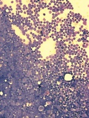
a patient with ALL
Investigators have identified the genetic events leading to leukemic transformation in ETV6-RUNX1 acute lymphoblastic leukemia (ALL), according to a paper published in Nature Genetics.
Previous studies have shown that, for 1 in 4 ALL patients, a key factor driving the disease is a chromosomal translocation that creates the ETV6-RUNX1 fusion gene.
However, the gene cannot cause overt leukemia on its own. Additional mutations are required for ALL to develop.
In this study, researchers found that RAG proteins—which rearrange the genome in normal immune cells to generate antibody diversity—can also rearrange the DNA of genes involved in cancer.
And this leads to ALL in individuals with the ETV6-RUNX1 fusion gene.
“For the first time, we see the combined events that are driving this treatable but highly devastating disease,” said lead study author Elli Papaemmanuil, PhD, of the Wellcome Trust Sanger Institute in Hinxton, UK.
“We now have a better understanding of the natural history of this disease and the critical events—from the initial acquisition of the fusion ETV6-RUNX1 to the sequential acquisition of RAG-mediated genome alterations—that ultimately result in this childhood leukemia.”
To unearth this discovery, the investigators sequenced the genomes of 57 ALL patients with the fusion gene. The team found that genomic rearrangements, and deletions in particular, were the predominant drivers of leukemia.
All samples showed evidence of events involving the RAG proteins. The proteins use a unique sequence of DNA letters as a signpost to direct them to antibody regions.
The researchers discovered that remnants of this sequence lay close to more than 50% of the cancer-driving genetic rearrangements. And this process often prompted the loss of the very genes required for normal immune cell development.
It is the deletion of these genes that, in combination with the fusion gene, leads to ALL, the investigators said. And the genetic signature linking the RAG proteins to genomic instability is not found in other types of leukemia or other common cancers.
“In this childhood leukemia, we see that the very process required to make normal antibodies is co-opted by the leukemia cells to knock out other genes with unprecedented specificity,” said Peter Campbell, PhD, also of the Wellcome Trust Sanger Institute.
To better understand the events that led to ALL development, the researchers used single-cell genomics to analyze samples from 2 patients. The team found that the cancer-causing process they identified occurs many times and results in continuous diversification of the leukemia.
“It may seem surprising that evolution should have provided a mechanism for diversifying antibodies that can collaterally damage genes that then contribute to cancer,” said Mel Greaves, PhD, of The Institute of Cancer Research in London, UK.
“But this only happens because the fusion gene that initiates the disease ‘traps’ cells in a normally very transient window of cell development where the RAG enzymes are active, teasing out their imperfect specificity.”
The researchers are now planning to investigate how the RAG-mediated genomic instability accrues in cells with the ETV6-RUNX1 fusion gene and what role this process plays in patients who relapse.
a patient with ALL
Investigators have identified the genetic events leading to leukemic transformation in ETV6-RUNX1 acute lymphoblastic leukemia (ALL), according to a paper published in Nature Genetics.
Previous studies have shown that, for 1 in 4 ALL patients, a key factor driving the disease is a chromosomal translocation that creates the ETV6-RUNX1 fusion gene.
However, the gene cannot cause overt leukemia on its own. Additional mutations are required for ALL to develop.
In this study, researchers found that RAG proteins—which rearrange the genome in normal immune cells to generate antibody diversity—can also rearrange the DNA of genes involved in cancer.
And this leads to ALL in individuals with the ETV6-RUNX1 fusion gene.
“For the first time, we see the combined events that are driving this treatable but highly devastating disease,” said lead study author Elli Papaemmanuil, PhD, of the Wellcome Trust Sanger Institute in Hinxton, UK.
“We now have a better understanding of the natural history of this disease and the critical events—from the initial acquisition of the fusion ETV6-RUNX1 to the sequential acquisition of RAG-mediated genome alterations—that ultimately result in this childhood leukemia.”
To unearth this discovery, the investigators sequenced the genomes of 57 ALL patients with the fusion gene. The team found that genomic rearrangements, and deletions in particular, were the predominant drivers of leukemia.
All samples showed evidence of events involving the RAG proteins. The proteins use a unique sequence of DNA letters as a signpost to direct them to antibody regions.
The researchers discovered that remnants of this sequence lay close to more than 50% of the cancer-driving genetic rearrangements. And this process often prompted the loss of the very genes required for normal immune cell development.
It is the deletion of these genes that, in combination with the fusion gene, leads to ALL, the investigators said. And the genetic signature linking the RAG proteins to genomic instability is not found in other types of leukemia or other common cancers.
“In this childhood leukemia, we see that the very process required to make normal antibodies is co-opted by the leukemia cells to knock out other genes with unprecedented specificity,” said Peter Campbell, PhD, also of the Wellcome Trust Sanger Institute.
To better understand the events that led to ALL development, the researchers used single-cell genomics to analyze samples from 2 patients. The team found that the cancer-causing process they identified occurs many times and results in continuous diversification of the leukemia.
“It may seem surprising that evolution should have provided a mechanism for diversifying antibodies that can collaterally damage genes that then contribute to cancer,” said Mel Greaves, PhD, of The Institute of Cancer Research in London, UK.
“But this only happens because the fusion gene that initiates the disease ‘traps’ cells in a normally very transient window of cell development where the RAG enzymes are active, teasing out their imperfect specificity.”
The researchers are now planning to investigate how the RAG-mediated genomic instability accrues in cells with the ETV6-RUNX1 fusion gene and what role this process plays in patients who relapse.
a patient with ALL
Investigators have identified the genetic events leading to leukemic transformation in ETV6-RUNX1 acute lymphoblastic leukemia (ALL), according to a paper published in Nature Genetics.
Previous studies have shown that, for 1 in 4 ALL patients, a key factor driving the disease is a chromosomal translocation that creates the ETV6-RUNX1 fusion gene.
However, the gene cannot cause overt leukemia on its own. Additional mutations are required for ALL to develop.
In this study, researchers found that RAG proteins—which rearrange the genome in normal immune cells to generate antibody diversity—can also rearrange the DNA of genes involved in cancer.
And this leads to ALL in individuals with the ETV6-RUNX1 fusion gene.
“For the first time, we see the combined events that are driving this treatable but highly devastating disease,” said lead study author Elli Papaemmanuil, PhD, of the Wellcome Trust Sanger Institute in Hinxton, UK.
“We now have a better understanding of the natural history of this disease and the critical events—from the initial acquisition of the fusion ETV6-RUNX1 to the sequential acquisition of RAG-mediated genome alterations—that ultimately result in this childhood leukemia.”
To unearth this discovery, the investigators sequenced the genomes of 57 ALL patients with the fusion gene. The team found that genomic rearrangements, and deletions in particular, were the predominant drivers of leukemia.
All samples showed evidence of events involving the RAG proteins. The proteins use a unique sequence of DNA letters as a signpost to direct them to antibody regions.
The researchers discovered that remnants of this sequence lay close to more than 50% of the cancer-driving genetic rearrangements. And this process often prompted the loss of the very genes required for normal immune cell development.
It is the deletion of these genes that, in combination with the fusion gene, leads to ALL, the investigators said. And the genetic signature linking the RAG proteins to genomic instability is not found in other types of leukemia or other common cancers.
“In this childhood leukemia, we see that the very process required to make normal antibodies is co-opted by the leukemia cells to knock out other genes with unprecedented specificity,” said Peter Campbell, PhD, also of the Wellcome Trust Sanger Institute.
To better understand the events that led to ALL development, the researchers used single-cell genomics to analyze samples from 2 patients. The team found that the cancer-causing process they identified occurs many times and results in continuous diversification of the leukemia.
“It may seem surprising that evolution should have provided a mechanism for diversifying antibodies that can collaterally damage genes that then contribute to cancer,” said Mel Greaves, PhD, of The Institute of Cancer Research in London, UK.
“But this only happens because the fusion gene that initiates the disease ‘traps’ cells in a normally very transient window of cell development where the RAG enzymes are active, teasing out their imperfect specificity.”
The researchers are now planning to investigate how the RAG-mediated genomic instability accrues in cells with the ETV6-RUNX1 fusion gene and what role this process plays in patients who relapse.
Antipsychotic drug is active against T-ALL

Experiments in zebrafish have shown that a 50-year-old antipsychotic medication called perphenazine can actively combat T-cell acute lymphoblastic leukemia (T-ALL).
The drug works by turning on a cancer-suppressing enzyme called PP2A and causing malignant tumor cells to self-destruct.
The findings suggest that developing medications that activate PP2A, while avoiding perphenazine’s psychotropic effects, could help clinicians make much-needed headway against T-ALL and perhaps other tumors as well.
Alejandro Gutierrez, MD, of the Dana-Farber Cancer Institute in Boston, and his colleagues detailed this research in The Journal of Clinical Investigation.
The researchers screened a library of 4880 compounds—including FDA-approved drugs whose patents had expired, small molecules, and natural products—in a model of T-ALL engineered using zebrafish.
One of the strongest hits in the zebrafish screen was perphenazine. The drug is a member of the phenothiazines family of antipsychotic medications, which can block dopamine receptors.
The investigators verified perphenazine’s anti-leukemic potential in vitro in several mouse and human T-ALL cell lines. Biochemical studies indicated that perphenazine’s anti-tumor activity is independent of its psychotropic activity and that it attacks T-ALL cells by turning on PP2A.
The fact that perphenazine works by reactivating a protein shut down in cancer cells is novel in the drug development field.
“We rarely find potential drug molecules that activate an enzyme,” Dr Gutierrez explained. “Most new drugs deactivate some protein or signal that the cancer cell requires to survive. But, here, perphenazine is restoring the activity of PP2A in the T-ALL cell.”
The researchers are now working to better understand the interactions between PP2A and perphenazine. They also want to search for or develop molecules that bind to and activate the enzyme more tightly and specifically to avoid perphenazine’s psychiatric effects.
“The challenge is to use medicinal chemistry to develop new PP2A inhibitors similar to perphenazine and the other phenothiazines, but to dial down dopamine interactions and accentuate those with PP2A,” said study author A. Thomas Look, MD, also of Dana-Farber.
He added that future PP2A inhibitors could be important additions to the oncologist’s arsenal. When used in combination with other drugs, the inhibitors might “make a real difference” for patients with T-ALL.
The investigators also believe the benefits of PP2A-activating drugs could extend beyond T-ALL.
“The proteins that PP2A suppresses, such as Myc and Akt, are involved in many tumors,” Dr Look noted. “We are optimistic that PP2A activators will have quite broad activity against different kinds of cancer, and we’re anxious to study the pathway in other malignancies as well.”
Experiments in zebrafish have shown that a 50-year-old antipsychotic medication called perphenazine can actively combat T-cell acute lymphoblastic leukemia (T-ALL).
The drug works by turning on a cancer-suppressing enzyme called PP2A and causing malignant tumor cells to self-destruct.
The findings suggest that developing medications that activate PP2A, while avoiding perphenazine’s psychotropic effects, could help clinicians make much-needed headway against T-ALL and perhaps other tumors as well.
Alejandro Gutierrez, MD, of the Dana-Farber Cancer Institute in Boston, and his colleagues detailed this research in The Journal of Clinical Investigation.
The researchers screened a library of 4880 compounds—including FDA-approved drugs whose patents had expired, small molecules, and natural products—in a model of T-ALL engineered using zebrafish.
One of the strongest hits in the zebrafish screen was perphenazine. The drug is a member of the phenothiazines family of antipsychotic medications, which can block dopamine receptors.
The investigators verified perphenazine’s anti-leukemic potential in vitro in several mouse and human T-ALL cell lines. Biochemical studies indicated that perphenazine’s anti-tumor activity is independent of its psychotropic activity and that it attacks T-ALL cells by turning on PP2A.
The fact that perphenazine works by reactivating a protein shut down in cancer cells is novel in the drug development field.
“We rarely find potential drug molecules that activate an enzyme,” Dr Gutierrez explained. “Most new drugs deactivate some protein or signal that the cancer cell requires to survive. But, here, perphenazine is restoring the activity of PP2A in the T-ALL cell.”
The researchers are now working to better understand the interactions between PP2A and perphenazine. They also want to search for or develop molecules that bind to and activate the enzyme more tightly and specifically to avoid perphenazine’s psychiatric effects.
“The challenge is to use medicinal chemistry to develop new PP2A inhibitors similar to perphenazine and the other phenothiazines, but to dial down dopamine interactions and accentuate those with PP2A,” said study author A. Thomas Look, MD, also of Dana-Farber.
He added that future PP2A inhibitors could be important additions to the oncologist’s arsenal. When used in combination with other drugs, the inhibitors might “make a real difference” for patients with T-ALL.
The investigators also believe the benefits of PP2A-activating drugs could extend beyond T-ALL.
“The proteins that PP2A suppresses, such as Myc and Akt, are involved in many tumors,” Dr Look noted. “We are optimistic that PP2A activators will have quite broad activity against different kinds of cancer, and we’re anxious to study the pathway in other malignancies as well.”
Experiments in zebrafish have shown that a 50-year-old antipsychotic medication called perphenazine can actively combat T-cell acute lymphoblastic leukemia (T-ALL).
The drug works by turning on a cancer-suppressing enzyme called PP2A and causing malignant tumor cells to self-destruct.
The findings suggest that developing medications that activate PP2A, while avoiding perphenazine’s psychotropic effects, could help clinicians make much-needed headway against T-ALL and perhaps other tumors as well.
Alejandro Gutierrez, MD, of the Dana-Farber Cancer Institute in Boston, and his colleagues detailed this research in The Journal of Clinical Investigation.
The researchers screened a library of 4880 compounds—including FDA-approved drugs whose patents had expired, small molecules, and natural products—in a model of T-ALL engineered using zebrafish.
One of the strongest hits in the zebrafish screen was perphenazine. The drug is a member of the phenothiazines family of antipsychotic medications, which can block dopamine receptors.
The investigators verified perphenazine’s anti-leukemic potential in vitro in several mouse and human T-ALL cell lines. Biochemical studies indicated that perphenazine’s anti-tumor activity is independent of its psychotropic activity and that it attacks T-ALL cells by turning on PP2A.
The fact that perphenazine works by reactivating a protein shut down in cancer cells is novel in the drug development field.
“We rarely find potential drug molecules that activate an enzyme,” Dr Gutierrez explained. “Most new drugs deactivate some protein or signal that the cancer cell requires to survive. But, here, perphenazine is restoring the activity of PP2A in the T-ALL cell.”
The researchers are now working to better understand the interactions between PP2A and perphenazine. They also want to search for or develop molecules that bind to and activate the enzyme more tightly and specifically to avoid perphenazine’s psychiatric effects.
“The challenge is to use medicinal chemistry to develop new PP2A inhibitors similar to perphenazine and the other phenothiazines, but to dial down dopamine interactions and accentuate those with PP2A,” said study author A. Thomas Look, MD, also of Dana-Farber.
He added that future PP2A inhibitors could be important additions to the oncologist’s arsenal. When used in combination with other drugs, the inhibitors might “make a real difference” for patients with T-ALL.
The investigators also believe the benefits of PP2A-activating drugs could extend beyond T-ALL.
“The proteins that PP2A suppresses, such as Myc and Akt, are involved in many tumors,” Dr Look noted. “We are optimistic that PP2A activators will have quite broad activity against different kinds of cancer, and we’re anxious to study the pathway in other malignancies as well.”
Deaths from leukemia, NHL declining in the UK

Credit: National Cancer
Institute-Mathews Media Group
Deaths from leukemia and non-Hodgkin lymphoma (NHL) are on the decline in the UK, but these malignancies are still among the leading causes of cancer death, a new analysis suggests.
Leukemia and NHL are among the 10 most common causes of cancer death for men and women in the UK, according to data from 2011.
But deaths from these malignancies have decreased from the number of deaths seen in the early 2000s.
These findings, published on the Cancer Research UK website, are similar to the results of a recent report on cancer deaths in the US.
The Cancer Research UK analysis showed that the death rate from cancer has dropped by more than a fifth since the 1990s.
In 1990, 220 in every 100,000 people died of cancer. But by 2011, the death rate had fallen 22%—to 170 per 100,000 people. The cancer mortality rate fell by 20% for women and 26% for men.
“Today, cancer is not the death sentence people once believed it to be,” said Harpal Kumar, Cancer Research UK chief executive.
“As these new figures show, mortality rates from this much-feared disease are dropping significantly . . . . But while we’re heading in the right direction, too many lives are still being lost to the disease, highlighting how much more work there is to do.”
NHL and leukemia stats
The analysis showed that, in men, the 3-year mortality rate for NHL decreased by 16% from 2000-2002 to 2009-2012. And the 3-year mortality rate for leukemia decreased by 6%.
In women, the 3-year mortality rate for NHL decreased by 18% from 2000-2002 to 2009-2012. And the 3-year mortality rate for leukemia decreased by 9%.
But the 2011 data showed that both types of cancer are among the 10 most common causes of cancer death in both men and women.
Among women, 2156 patients died of NHL (7th leading cause of cancer death), and 1994 patients died of leukemia (8th leading cause).
Among men, 2609 patients died of leukemia (8th leading cause of cancer death), and 2490 died of NHL (10th leading cause).
For more details on cancer mortality, including projections up to the year 2030, visit the Cancer Research UK website.
Credit: National Cancer
Institute-Mathews Media Group
Deaths from leukemia and non-Hodgkin lymphoma (NHL) are on the decline in the UK, but these malignancies are still among the leading causes of cancer death, a new analysis suggests.
Leukemia and NHL are among the 10 most common causes of cancer death for men and women in the UK, according to data from 2011.
But deaths from these malignancies have decreased from the number of deaths seen in the early 2000s.
These findings, published on the Cancer Research UK website, are similar to the results of a recent report on cancer deaths in the US.
The Cancer Research UK analysis showed that the death rate from cancer has dropped by more than a fifth since the 1990s.
In 1990, 220 in every 100,000 people died of cancer. But by 2011, the death rate had fallen 22%—to 170 per 100,000 people. The cancer mortality rate fell by 20% for women and 26% for men.
“Today, cancer is not the death sentence people once believed it to be,” said Harpal Kumar, Cancer Research UK chief executive.
“As these new figures show, mortality rates from this much-feared disease are dropping significantly . . . . But while we’re heading in the right direction, too many lives are still being lost to the disease, highlighting how much more work there is to do.”
NHL and leukemia stats
The analysis showed that, in men, the 3-year mortality rate for NHL decreased by 16% from 2000-2002 to 2009-2012. And the 3-year mortality rate for leukemia decreased by 6%.
In women, the 3-year mortality rate for NHL decreased by 18% from 2000-2002 to 2009-2012. And the 3-year mortality rate for leukemia decreased by 9%.
But the 2011 data showed that both types of cancer are among the 10 most common causes of cancer death in both men and women.
Among women, 2156 patients died of NHL (7th leading cause of cancer death), and 1994 patients died of leukemia (8th leading cause).
Among men, 2609 patients died of leukemia (8th leading cause of cancer death), and 2490 died of NHL (10th leading cause).
For more details on cancer mortality, including projections up to the year 2030, visit the Cancer Research UK website.
Credit: National Cancer
Institute-Mathews Media Group
Deaths from leukemia and non-Hodgkin lymphoma (NHL) are on the decline in the UK, but these malignancies are still among the leading causes of cancer death, a new analysis suggests.
Leukemia and NHL are among the 10 most common causes of cancer death for men and women in the UK, according to data from 2011.
But deaths from these malignancies have decreased from the number of deaths seen in the early 2000s.
These findings, published on the Cancer Research UK website, are similar to the results of a recent report on cancer deaths in the US.
The Cancer Research UK analysis showed that the death rate from cancer has dropped by more than a fifth since the 1990s.
In 1990, 220 in every 100,000 people died of cancer. But by 2011, the death rate had fallen 22%—to 170 per 100,000 people. The cancer mortality rate fell by 20% for women and 26% for men.
“Today, cancer is not the death sentence people once believed it to be,” said Harpal Kumar, Cancer Research UK chief executive.
“As these new figures show, mortality rates from this much-feared disease are dropping significantly . . . . But while we’re heading in the right direction, too many lives are still being lost to the disease, highlighting how much more work there is to do.”
NHL and leukemia stats
The analysis showed that, in men, the 3-year mortality rate for NHL decreased by 16% from 2000-2002 to 2009-2012. And the 3-year mortality rate for leukemia decreased by 6%.
In women, the 3-year mortality rate for NHL decreased by 18% from 2000-2002 to 2009-2012. And the 3-year mortality rate for leukemia decreased by 9%.
But the 2011 data showed that both types of cancer are among the 10 most common causes of cancer death in both men and women.
Among women, 2156 patients died of NHL (7th leading cause of cancer death), and 1994 patients died of leukemia (8th leading cause).
Among men, 2609 patients died of leukemia (8th leading cause of cancer death), and 2490 died of NHL (10th leading cause).
For more details on cancer mortality, including projections up to the year 2030, visit the Cancer Research UK website.
Discovery may aid vaccine design for P vivax malaria

attached to syncytiotrophoblast
Credit: Fabio T.M. Costa
Plasmodium vivax malaria attacks red blood cells by clamping down on the cells with a pair of proteins, researchers have found.
Earlier studies suggested that a single P vivax protein binds to a protein on the surface of red blood cells.
But the new study showed that binding is a 2-step process that involves 2 copies of a parasite protein coming together like tongs around 2 copies of a host protein.
The researchers believe this discovery, detailed in PLOS Pathogens, could help scientists design better vaccines and treatments for P vivax, which is common in India, Southeast Asia, and South America.
“More people live at risk of infection by this strain of malaria than any other,” said senior study author Niraj Tolia, PhD, of the Washington University School of Medicine in St Louis, Missouri.
“We now are using what we have learned to create vaccines tailored to stop the infectious process by preventing the parasite from attaching to red blood cells.”
Dr Tolia and his colleagues knew that P vivax Duffy binding protein (DBP) recognizes the receptor Duffy antigen/receptor for chemokines (DARC) during the parasite’s invasion of red blood cells. But the team wanted to identify binding contacts during invasion and determine the molecular basis of DBP receptor recognition.
So they conducted structural studies on the minimal binding domain of DBP in complex with the minimal region from DARC. And they found that 2 DBP molecules bind 2 DARC molecules.
The researchers also performed erythrocyte binding assays with binding site mutants and identified essential receptor contacts.
“It’s a very intricate and chemically strong interaction that was not easily understood before,” Dr Tolia said. “We have had hints that other forms of malaria, including the African strain, may be binding in a similar fashion to host cells, but this is one of the first definitive proofs of this kind of attack.”
Dr Tolia suspects that blocking any of the proteins with drugs or vaccines will stop the infectious process.
“For example, some people have a mutation that eliminates the protein on red blood cell surfaces that P vivax binds to, and they tend to be resistant to the parasite,” he said. “This is why this strain isn’t prevalent in Africa. Evolutionary pressure has caused most of the populations there to stop making this protein.”
Dr Tolia and his colleagues also found evidence that other people with immunity to P vivax have developed naturally occurring antibodies that attach to a key part of the parasite’s binding protein, preventing infection.
“The parasite protein is very large, and human antibodies bind to it at many different points along its length,” Dr Tolia explained. “We have observed that the ones that are most effective, so far, are the antibodies that bind to the protein at the region highlighted by our new research.”
attached to syncytiotrophoblast
Credit: Fabio T.M. Costa
Plasmodium vivax malaria attacks red blood cells by clamping down on the cells with a pair of proteins, researchers have found.
Earlier studies suggested that a single P vivax protein binds to a protein on the surface of red blood cells.
But the new study showed that binding is a 2-step process that involves 2 copies of a parasite protein coming together like tongs around 2 copies of a host protein.
The researchers believe this discovery, detailed in PLOS Pathogens, could help scientists design better vaccines and treatments for P vivax, which is common in India, Southeast Asia, and South America.
“More people live at risk of infection by this strain of malaria than any other,” said senior study author Niraj Tolia, PhD, of the Washington University School of Medicine in St Louis, Missouri.
“We now are using what we have learned to create vaccines tailored to stop the infectious process by preventing the parasite from attaching to red blood cells.”
Dr Tolia and his colleagues knew that P vivax Duffy binding protein (DBP) recognizes the receptor Duffy antigen/receptor for chemokines (DARC) during the parasite’s invasion of red blood cells. But the team wanted to identify binding contacts during invasion and determine the molecular basis of DBP receptor recognition.
So they conducted structural studies on the minimal binding domain of DBP in complex with the minimal region from DARC. And they found that 2 DBP molecules bind 2 DARC molecules.
The researchers also performed erythrocyte binding assays with binding site mutants and identified essential receptor contacts.
“It’s a very intricate and chemically strong interaction that was not easily understood before,” Dr Tolia said. “We have had hints that other forms of malaria, including the African strain, may be binding in a similar fashion to host cells, but this is one of the first definitive proofs of this kind of attack.”
Dr Tolia suspects that blocking any of the proteins with drugs or vaccines will stop the infectious process.
“For example, some people have a mutation that eliminates the protein on red blood cell surfaces that P vivax binds to, and they tend to be resistant to the parasite,” he said. “This is why this strain isn’t prevalent in Africa. Evolutionary pressure has caused most of the populations there to stop making this protein.”
Dr Tolia and his colleagues also found evidence that other people with immunity to P vivax have developed naturally occurring antibodies that attach to a key part of the parasite’s binding protein, preventing infection.
“The parasite protein is very large, and human antibodies bind to it at many different points along its length,” Dr Tolia explained. “We have observed that the ones that are most effective, so far, are the antibodies that bind to the protein at the region highlighted by our new research.”
attached to syncytiotrophoblast
Credit: Fabio T.M. Costa
Plasmodium vivax malaria attacks red blood cells by clamping down on the cells with a pair of proteins, researchers have found.
Earlier studies suggested that a single P vivax protein binds to a protein on the surface of red blood cells.
But the new study showed that binding is a 2-step process that involves 2 copies of a parasite protein coming together like tongs around 2 copies of a host protein.
The researchers believe this discovery, detailed in PLOS Pathogens, could help scientists design better vaccines and treatments for P vivax, which is common in India, Southeast Asia, and South America.
“More people live at risk of infection by this strain of malaria than any other,” said senior study author Niraj Tolia, PhD, of the Washington University School of Medicine in St Louis, Missouri.
“We now are using what we have learned to create vaccines tailored to stop the infectious process by preventing the parasite from attaching to red blood cells.”
Dr Tolia and his colleagues knew that P vivax Duffy binding protein (DBP) recognizes the receptor Duffy antigen/receptor for chemokines (DARC) during the parasite’s invasion of red blood cells. But the team wanted to identify binding contacts during invasion and determine the molecular basis of DBP receptor recognition.
So they conducted structural studies on the minimal binding domain of DBP in complex with the minimal region from DARC. And they found that 2 DBP molecules bind 2 DARC molecules.
The researchers also performed erythrocyte binding assays with binding site mutants and identified essential receptor contacts.
“It’s a very intricate and chemically strong interaction that was not easily understood before,” Dr Tolia said. “We have had hints that other forms of malaria, including the African strain, may be binding in a similar fashion to host cells, but this is one of the first definitive proofs of this kind of attack.”
Dr Tolia suspects that blocking any of the proteins with drugs or vaccines will stop the infectious process.
“For example, some people have a mutation that eliminates the protein on red blood cell surfaces that P vivax binds to, and they tend to be resistant to the parasite,” he said. “This is why this strain isn’t prevalent in Africa. Evolutionary pressure has caused most of the populations there to stop making this protein.”
Dr Tolia and his colleagues also found evidence that other people with immunity to P vivax have developed naturally occurring antibodies that attach to a key part of the parasite’s binding protein, preventing infection.
“The parasite protein is very large, and human antibodies bind to it at many different points along its length,” Dr Tolia explained. “We have observed that the ones that are most effective, so far, are the antibodies that bind to the protein at the region highlighted by our new research.”
Team identifies mutations that may drive FL
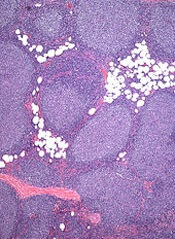
Genetic profiling has provided a clearer picture of follicular lymphoma (FL) development and progression, according to research published in Nature Genetics.
Investigators performed whole-genome and whole-exome sequencing of samples from FL patients and found a number of mutations that appeared to be responsible for disease onset.
The team also identified mutations that seemed to drive FL toward a more aggressive form.
They said these findings provide a number of new therapeutic targets that may stop FL from becoming aggressive or developing resistance to treatment.
“Resistance to treatment is a major problem for follicular lymphoma patients, as they often respond well to treatment and later relapse,” said study author Jude Fitzgibbon, PhD, of Barts Cancer Institute in London, England.
“[This] gives the cancer multiple opportunities to evolve into a more aggressive and more difficult-to-treat form of the disease. We’ve been able to chronicle the chain of genetic events that leads to aggressive forms of the disease. If we can develop treatments to prevent some of these changes from taking place, we should be able to stop the cancer in its tracks.”
Dr Fitzgibbon and his colleagues performed whole-genome or whole-exome sequencing of sequential FL and transformed FL pairs and matched germline samples from 10 FL cases with deep-targeted sequencing of 28 genes in an extension cohort.
Among the 10 cases, the researchers identified 1560 protein-altering variants affecting 908 genes, including missense changes (84.8%), short indels (8.9%), and nonsense mutations (6.3%).
Patterns of evolution
The investigators constructed phylogenetic trees for the 10 FL cases and discovered a common progenitor clone (CPC), as well as 2 patterns of evolution.
Eight of the cases exhibited evolution through a “rich” ancestral CPC, showing high clonal semblance between the FL and transformed-FL tumors. The other 2 cases showed evolution through a “sparse” CPC, with only 4 nonsynonymous mutations shared by the FL and transformed-FL samples.
These 2 patterns of evolution shared mutations in 3 genes—KMT2D, TNFRSF14, and CREBBP. According to the researchers, this suggests tumor dependency on these alterations during lymphomagenesis and progression.
Mutation prevalence, timing
The investigators then set out to determine the prevalence of the mutations they identified in the 10 cases. They performed deep-targeted resequencing of 28 candidate genes in an extension cohort of 100 independent FL biopsies and 32 paired FL-transformed FL cases (including the 10 index cases).
More than 70% of cases had concurrent mutations in at least 2 of the histone-modifying enzymes screened (CREBBP, EZH2, MEF2B, and KMT2D).
Twenty-eight percent of cases had mutations affecting at least one histone H1 gene. HIST1H1C and HIST1H1E were the most frequently mutated.
The researchers also saw frequent mutations in components of the JAK-STAT signaling pathway, including STAT6 (12%) and SOCS1 (8%).
They found mutually exclusive mutations in the NF-κB signaling pathway in a third of FLs, including CARD11 (11%) and TNFAIP3 (11%).
And 17% of cases had mutations in genes important for B-cell development, including Ebf1.
Finally, the investigators set out to differentiate early genetic events from late ones. They found that mutations in histone-modifying genes—KMT2D, CREBBP, and EZH2—as well as mutations in STAT6 and TNFRSF14 were predominantly clonal events.
On the other hand, mutations in EBF1 and regulators of NF-κB signaling—MYD88 and TNFAIP3—were gained at transformation.
“This study has uncovered some of the key molecular changes taking place [in FL] and offers new targets for treating the disease,” said Nell Barrie, of Cancer Research UK, the organization that funded this study.
“Research into the genetics that underpin cancer is helping us to better know the enemy and find new ways in which we might beat it.”
Genetic profiling has provided a clearer picture of follicular lymphoma (FL) development and progression, according to research published in Nature Genetics.
Investigators performed whole-genome and whole-exome sequencing of samples from FL patients and found a number of mutations that appeared to be responsible for disease onset.
The team also identified mutations that seemed to drive FL toward a more aggressive form.
They said these findings provide a number of new therapeutic targets that may stop FL from becoming aggressive or developing resistance to treatment.
“Resistance to treatment is a major problem for follicular lymphoma patients, as they often respond well to treatment and later relapse,” said study author Jude Fitzgibbon, PhD, of Barts Cancer Institute in London, England.
“[This] gives the cancer multiple opportunities to evolve into a more aggressive and more difficult-to-treat form of the disease. We’ve been able to chronicle the chain of genetic events that leads to aggressive forms of the disease. If we can develop treatments to prevent some of these changes from taking place, we should be able to stop the cancer in its tracks.”
Dr Fitzgibbon and his colleagues performed whole-genome or whole-exome sequencing of sequential FL and transformed FL pairs and matched germline samples from 10 FL cases with deep-targeted sequencing of 28 genes in an extension cohort.
Among the 10 cases, the researchers identified 1560 protein-altering variants affecting 908 genes, including missense changes (84.8%), short indels (8.9%), and nonsense mutations (6.3%).
Patterns of evolution
The investigators constructed phylogenetic trees for the 10 FL cases and discovered a common progenitor clone (CPC), as well as 2 patterns of evolution.
Eight of the cases exhibited evolution through a “rich” ancestral CPC, showing high clonal semblance between the FL and transformed-FL tumors. The other 2 cases showed evolution through a “sparse” CPC, with only 4 nonsynonymous mutations shared by the FL and transformed-FL samples.
These 2 patterns of evolution shared mutations in 3 genes—KMT2D, TNFRSF14, and CREBBP. According to the researchers, this suggests tumor dependency on these alterations during lymphomagenesis and progression.
Mutation prevalence, timing
The investigators then set out to determine the prevalence of the mutations they identified in the 10 cases. They performed deep-targeted resequencing of 28 candidate genes in an extension cohort of 100 independent FL biopsies and 32 paired FL-transformed FL cases (including the 10 index cases).
More than 70% of cases had concurrent mutations in at least 2 of the histone-modifying enzymes screened (CREBBP, EZH2, MEF2B, and KMT2D).
Twenty-eight percent of cases had mutations affecting at least one histone H1 gene. HIST1H1C and HIST1H1E were the most frequently mutated.
The researchers also saw frequent mutations in components of the JAK-STAT signaling pathway, including STAT6 (12%) and SOCS1 (8%).
They found mutually exclusive mutations in the NF-κB signaling pathway in a third of FLs, including CARD11 (11%) and TNFAIP3 (11%).
And 17% of cases had mutations in genes important for B-cell development, including Ebf1.
Finally, the investigators set out to differentiate early genetic events from late ones. They found that mutations in histone-modifying genes—KMT2D, CREBBP, and EZH2—as well as mutations in STAT6 and TNFRSF14 were predominantly clonal events.
On the other hand, mutations in EBF1 and regulators of NF-κB signaling—MYD88 and TNFAIP3—were gained at transformation.
“This study has uncovered some of the key molecular changes taking place [in FL] and offers new targets for treating the disease,” said Nell Barrie, of Cancer Research UK, the organization that funded this study.
“Research into the genetics that underpin cancer is helping us to better know the enemy and find new ways in which we might beat it.”
Genetic profiling has provided a clearer picture of follicular lymphoma (FL) development and progression, according to research published in Nature Genetics.
Investigators performed whole-genome and whole-exome sequencing of samples from FL patients and found a number of mutations that appeared to be responsible for disease onset.
The team also identified mutations that seemed to drive FL toward a more aggressive form.
They said these findings provide a number of new therapeutic targets that may stop FL from becoming aggressive or developing resistance to treatment.
“Resistance to treatment is a major problem for follicular lymphoma patients, as they often respond well to treatment and later relapse,” said study author Jude Fitzgibbon, PhD, of Barts Cancer Institute in London, England.
“[This] gives the cancer multiple opportunities to evolve into a more aggressive and more difficult-to-treat form of the disease. We’ve been able to chronicle the chain of genetic events that leads to aggressive forms of the disease. If we can develop treatments to prevent some of these changes from taking place, we should be able to stop the cancer in its tracks.”
Dr Fitzgibbon and his colleagues performed whole-genome or whole-exome sequencing of sequential FL and transformed FL pairs and matched germline samples from 10 FL cases with deep-targeted sequencing of 28 genes in an extension cohort.
Among the 10 cases, the researchers identified 1560 protein-altering variants affecting 908 genes, including missense changes (84.8%), short indels (8.9%), and nonsense mutations (6.3%).
Patterns of evolution
The investigators constructed phylogenetic trees for the 10 FL cases and discovered a common progenitor clone (CPC), as well as 2 patterns of evolution.
Eight of the cases exhibited evolution through a “rich” ancestral CPC, showing high clonal semblance between the FL and transformed-FL tumors. The other 2 cases showed evolution through a “sparse” CPC, with only 4 nonsynonymous mutations shared by the FL and transformed-FL samples.
These 2 patterns of evolution shared mutations in 3 genes—KMT2D, TNFRSF14, and CREBBP. According to the researchers, this suggests tumor dependency on these alterations during lymphomagenesis and progression.
Mutation prevalence, timing
The investigators then set out to determine the prevalence of the mutations they identified in the 10 cases. They performed deep-targeted resequencing of 28 candidate genes in an extension cohort of 100 independent FL biopsies and 32 paired FL-transformed FL cases (including the 10 index cases).
More than 70% of cases had concurrent mutations in at least 2 of the histone-modifying enzymes screened (CREBBP, EZH2, MEF2B, and KMT2D).
Twenty-eight percent of cases had mutations affecting at least one histone H1 gene. HIST1H1C and HIST1H1E were the most frequently mutated.
The researchers also saw frequent mutations in components of the JAK-STAT signaling pathway, including STAT6 (12%) and SOCS1 (8%).
They found mutually exclusive mutations in the NF-κB signaling pathway in a third of FLs, including CARD11 (11%) and TNFAIP3 (11%).
And 17% of cases had mutations in genes important for B-cell development, including Ebf1.
Finally, the investigators set out to differentiate early genetic events from late ones. They found that mutations in histone-modifying genes—KMT2D, CREBBP, and EZH2—as well as mutations in STAT6 and TNFRSF14 were predominantly clonal events.
On the other hand, mutations in EBF1 and regulators of NF-κB signaling—MYD88 and TNFAIP3—were gained at transformation.
“This study has uncovered some of the key molecular changes taking place [in FL] and offers new targets for treating the disease,” said Nell Barrie, of Cancer Research UK, the organization that funded this study.
“Research into the genetics that underpin cancer is helping us to better know the enemy and find new ways in which we might beat it.”
Study reveals RBC function in clot contraction
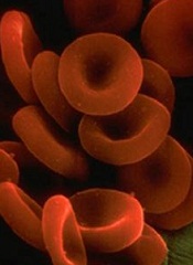
Red blood cells (RBCs) take on a new shape and perform important functions in contracted blood clots, a new study suggests.
Researchers found that, during clot contraction, RBCs can be compressed into many-sided, closely packed, polyhedral structures.
These polyhedral RBCs form an impermeable seal within the clot to stem bleeding and help prevent vascular obstruction. And the cells may be the reason fibrinolysis is hampered after clot contraction.
John Weisel, PhD, of the University of Pennsylvania in Philadelphia, and his colleagues described these findings in Blood.
The researchers knew that, after a blood clot forms, the actin and myosin in platelets start the contraction process and cause the clot to shrink to about one-third of its original size. RBCs are caught up in the contraction process and get pulled by platelets toward the interior of the clot.
But little was known about the structure of contracted clots or the role RBCs play in the contraction process. So Dr Weisel and his colleagues decided to study clot contraction using a novel magnetic resonance technology.
“We found that contracted blood clots develop a remarkable structure, with a meshwork of fibrin and platelet aggregates on the exterior of the clot and a close-packed, tessellated array of compressed polyhedral erythrocytes within,” Dr Weisel said.
The team saw the same morphology in clots created from human blood reconstituted with its cellular and plasma components, as well as clots made with mouse blood.
The polyhedral erythrocytes, or polyhedrocytes as the researchers named them, were also present in human arterial thrombi taken from patients with myocardial infarctions.
The researchers believe the RBCs take on the polyhedral shape so as to decrease volume, surface energy, or bending energy.
The team said their findings might have clinical implications. It is well known that, with time, thrombi develop resistance to being broken up by thrombolytic agents.
And the nearly impermeable barrier formed by RBCs within the contracted clots may help to explain why. Clot contraction could be a target of intervention to prevent the formation of the closely packed polyhedrocytes.
Red blood cells (RBCs) take on a new shape and perform important functions in contracted blood clots, a new study suggests.
Researchers found that, during clot contraction, RBCs can be compressed into many-sided, closely packed, polyhedral structures.
These polyhedral RBCs form an impermeable seal within the clot to stem bleeding and help prevent vascular obstruction. And the cells may be the reason fibrinolysis is hampered after clot contraction.
John Weisel, PhD, of the University of Pennsylvania in Philadelphia, and his colleagues described these findings in Blood.
The researchers knew that, after a blood clot forms, the actin and myosin in platelets start the contraction process and cause the clot to shrink to about one-third of its original size. RBCs are caught up in the contraction process and get pulled by platelets toward the interior of the clot.
But little was known about the structure of contracted clots or the role RBCs play in the contraction process. So Dr Weisel and his colleagues decided to study clot contraction using a novel magnetic resonance technology.
“We found that contracted blood clots develop a remarkable structure, with a meshwork of fibrin and platelet aggregates on the exterior of the clot and a close-packed, tessellated array of compressed polyhedral erythrocytes within,” Dr Weisel said.
The team saw the same morphology in clots created from human blood reconstituted with its cellular and plasma components, as well as clots made with mouse blood.
The polyhedral erythrocytes, or polyhedrocytes as the researchers named them, were also present in human arterial thrombi taken from patients with myocardial infarctions.
The researchers believe the RBCs take on the polyhedral shape so as to decrease volume, surface energy, or bending energy.
The team said their findings might have clinical implications. It is well known that, with time, thrombi develop resistance to being broken up by thrombolytic agents.
And the nearly impermeable barrier formed by RBCs within the contracted clots may help to explain why. Clot contraction could be a target of intervention to prevent the formation of the closely packed polyhedrocytes.
Red blood cells (RBCs) take on a new shape and perform important functions in contracted blood clots, a new study suggests.
Researchers found that, during clot contraction, RBCs can be compressed into many-sided, closely packed, polyhedral structures.
These polyhedral RBCs form an impermeable seal within the clot to stem bleeding and help prevent vascular obstruction. And the cells may be the reason fibrinolysis is hampered after clot contraction.
John Weisel, PhD, of the University of Pennsylvania in Philadelphia, and his colleagues described these findings in Blood.
The researchers knew that, after a blood clot forms, the actin and myosin in platelets start the contraction process and cause the clot to shrink to about one-third of its original size. RBCs are caught up in the contraction process and get pulled by platelets toward the interior of the clot.
But little was known about the structure of contracted clots or the role RBCs play in the contraction process. So Dr Weisel and his colleagues decided to study clot contraction using a novel magnetic resonance technology.
“We found that contracted blood clots develop a remarkable structure, with a meshwork of fibrin and platelet aggregates on the exterior of the clot and a close-packed, tessellated array of compressed polyhedral erythrocytes within,” Dr Weisel said.
The team saw the same morphology in clots created from human blood reconstituted with its cellular and plasma components, as well as clots made with mouse blood.
The polyhedral erythrocytes, or polyhedrocytes as the researchers named them, were also present in human arterial thrombi taken from patients with myocardial infarctions.
The researchers believe the RBCs take on the polyhedral shape so as to decrease volume, surface energy, or bending energy.
The team said their findings might have clinical implications. It is well known that, with time, thrombi develop resistance to being broken up by thrombolytic agents.
And the nearly impermeable barrier formed by RBCs within the contracted clots may help to explain why. Clot contraction could be a target of intervention to prevent the formation of the closely packed polyhedrocytes.
AML scoring system could optimize treatment

Credit: NIGMS
A scoring system that combines genetic and epigenetic changes could help guide therapy for acute myeloid leukemia (AML), according to a study published in the Journal of Clinical Oncology.
The score is based on the presence of 7 mutated genes and DNA methylation.
For each of these genes, lower expression and higher DNA methylation were associated with better patient outcomes.
The investigators therefore believe this scoring system could guide treatment by identifying novel subsets of patients.
“To date, disease classification and prognostication for AML patients have been based largely on chromosomal and genetic markers,” said principal investigator Clara D. Bloomfield, MD, of The Ohio State University in Columbus.
“Epigenetic changes that affect gene expression have not been considered. Here, we show that epigenetic changes in previously recognized and prognostically important mutated genes can identify novel patient subgroups, which might better help guide therapy.”
Creating the score
Dr Bloomfield and her colleagues identified the 7-gene panel in 134 patients who were 60 and older, had cytogenetically normal AML (CN-AML), and had been treated on Cancer and Leukemia Group B/Alliance clinical trials.
The investigators used next-generation sequencing to analyze regions of methylated DNA associated with prognostically important genetic mutations. The 7 genes they identified are CD34, RHOC, SCRN1, F2RL1, FAM92A1, MIR155HG, and VWA8.
The team then developed a summary score based on the number of genes in the panel showing high expression.
And they applied the unweighted score to 126 of the aforementioned patients. Individuals with 1 or no highly expressed genes had a 96% complete remission (CR) rate, a 3-year disease-free survival (DFS) rate of 32%, and a 3-year overall survival (OS) rate of 39%.
Patients with 6 to 7 highly expressed genes, on the other hand, had a 25% CR rate, a 3-year DFS of 0%, and a 3-year OS of 4%.
Validating the system
The investigators also tested the score in 4 validation cohorts: older patients (age 60 and up) with primary AML (n=72), younger patients (59 and under) with primary AML (n=134), older patients with CN-AML (n=65), and younger patients with CN-AML (n=84).
“In both younger and older patients, those who had no highly expressed genes, or had one highly expressed gene, had the best outcomes,” said study author Guido Marcucci, MD, of The Ohio State University Comprehensive Cancer Center.
For the younger patients (with primary or CN-AML), individuals with 1 or no highly expressed genes had a 91% to 100% CR rate, a 3-year DFS of 60% to 65%, and a 3-year OS of 76% to 82%.
But younger patients with 6 to 7 highly expressed genes had a 53% to 71% CR rate, a 3-year DFS of 13% to 17%, and a 3-year OS of 7% to 24%.
For the older patients, individuals with 1 or no highly expressed genes had a 69% to 89% CR rate, a 3-year DFS of 42% (CN-AML only), and a 3-year OS of 44% to 46%.
Older patients with 6 to 7 highly expressed genes had a 50% CR rate (both types of AML), a 3-year DFS of 0% (CN-AML only), and a 3-year OS of 10% to 12%. DFS data were not evaluable for the older patients with primary AML due to the small sample size.
“Overall, our findings suggest that the unweighted summary score is a better model compared with all other prognostic markers and previously reported gene-expression profiles,” Dr Bloomfield concluded.
Credit: NIGMS
A scoring system that combines genetic and epigenetic changes could help guide therapy for acute myeloid leukemia (AML), according to a study published in the Journal of Clinical Oncology.
The score is based on the presence of 7 mutated genes and DNA methylation.
For each of these genes, lower expression and higher DNA methylation were associated with better patient outcomes.
The investigators therefore believe this scoring system could guide treatment by identifying novel subsets of patients.
“To date, disease classification and prognostication for AML patients have been based largely on chromosomal and genetic markers,” said principal investigator Clara D. Bloomfield, MD, of The Ohio State University in Columbus.
“Epigenetic changes that affect gene expression have not been considered. Here, we show that epigenetic changes in previously recognized and prognostically important mutated genes can identify novel patient subgroups, which might better help guide therapy.”
Creating the score
Dr Bloomfield and her colleagues identified the 7-gene panel in 134 patients who were 60 and older, had cytogenetically normal AML (CN-AML), and had been treated on Cancer and Leukemia Group B/Alliance clinical trials.
The investigators used next-generation sequencing to analyze regions of methylated DNA associated with prognostically important genetic mutations. The 7 genes they identified are CD34, RHOC, SCRN1, F2RL1, FAM92A1, MIR155HG, and VWA8.
The team then developed a summary score based on the number of genes in the panel showing high expression.
And they applied the unweighted score to 126 of the aforementioned patients. Individuals with 1 or no highly expressed genes had a 96% complete remission (CR) rate, a 3-year disease-free survival (DFS) rate of 32%, and a 3-year overall survival (OS) rate of 39%.
Patients with 6 to 7 highly expressed genes, on the other hand, had a 25% CR rate, a 3-year DFS of 0%, and a 3-year OS of 4%.
Validating the system
The investigators also tested the score in 4 validation cohorts: older patients (age 60 and up) with primary AML (n=72), younger patients (59 and under) with primary AML (n=134), older patients with CN-AML (n=65), and younger patients with CN-AML (n=84).
“In both younger and older patients, those who had no highly expressed genes, or had one highly expressed gene, had the best outcomes,” said study author Guido Marcucci, MD, of The Ohio State University Comprehensive Cancer Center.
For the younger patients (with primary or CN-AML), individuals with 1 or no highly expressed genes had a 91% to 100% CR rate, a 3-year DFS of 60% to 65%, and a 3-year OS of 76% to 82%.
But younger patients with 6 to 7 highly expressed genes had a 53% to 71% CR rate, a 3-year DFS of 13% to 17%, and a 3-year OS of 7% to 24%.
For the older patients, individuals with 1 or no highly expressed genes had a 69% to 89% CR rate, a 3-year DFS of 42% (CN-AML only), and a 3-year OS of 44% to 46%.
Older patients with 6 to 7 highly expressed genes had a 50% CR rate (both types of AML), a 3-year DFS of 0% (CN-AML only), and a 3-year OS of 10% to 12%. DFS data were not evaluable for the older patients with primary AML due to the small sample size.
“Overall, our findings suggest that the unweighted summary score is a better model compared with all other prognostic markers and previously reported gene-expression profiles,” Dr Bloomfield concluded.
Credit: NIGMS
A scoring system that combines genetic and epigenetic changes could help guide therapy for acute myeloid leukemia (AML), according to a study published in the Journal of Clinical Oncology.
The score is based on the presence of 7 mutated genes and DNA methylation.
For each of these genes, lower expression and higher DNA methylation were associated with better patient outcomes.
The investigators therefore believe this scoring system could guide treatment by identifying novel subsets of patients.
“To date, disease classification and prognostication for AML patients have been based largely on chromosomal and genetic markers,” said principal investigator Clara D. Bloomfield, MD, of The Ohio State University in Columbus.
“Epigenetic changes that affect gene expression have not been considered. Here, we show that epigenetic changes in previously recognized and prognostically important mutated genes can identify novel patient subgroups, which might better help guide therapy.”
Creating the score
Dr Bloomfield and her colleagues identified the 7-gene panel in 134 patients who were 60 and older, had cytogenetically normal AML (CN-AML), and had been treated on Cancer and Leukemia Group B/Alliance clinical trials.
The investigators used next-generation sequencing to analyze regions of methylated DNA associated with prognostically important genetic mutations. The 7 genes they identified are CD34, RHOC, SCRN1, F2RL1, FAM92A1, MIR155HG, and VWA8.
The team then developed a summary score based on the number of genes in the panel showing high expression.
And they applied the unweighted score to 126 of the aforementioned patients. Individuals with 1 or no highly expressed genes had a 96% complete remission (CR) rate, a 3-year disease-free survival (DFS) rate of 32%, and a 3-year overall survival (OS) rate of 39%.
Patients with 6 to 7 highly expressed genes, on the other hand, had a 25% CR rate, a 3-year DFS of 0%, and a 3-year OS of 4%.
Validating the system
The investigators also tested the score in 4 validation cohorts: older patients (age 60 and up) with primary AML (n=72), younger patients (59 and under) with primary AML (n=134), older patients with CN-AML (n=65), and younger patients with CN-AML (n=84).
“In both younger and older patients, those who had no highly expressed genes, or had one highly expressed gene, had the best outcomes,” said study author Guido Marcucci, MD, of The Ohio State University Comprehensive Cancer Center.
For the younger patients (with primary or CN-AML), individuals with 1 or no highly expressed genes had a 91% to 100% CR rate, a 3-year DFS of 60% to 65%, and a 3-year OS of 76% to 82%.
But younger patients with 6 to 7 highly expressed genes had a 53% to 71% CR rate, a 3-year DFS of 13% to 17%, and a 3-year OS of 7% to 24%.
For the older patients, individuals with 1 or no highly expressed genes had a 69% to 89% CR rate, a 3-year DFS of 42% (CN-AML only), and a 3-year OS of 44% to 46%.
Older patients with 6 to 7 highly expressed genes had a 50% CR rate (both types of AML), a 3-year DFS of 0% (CN-AML only), and a 3-year OS of 10% to 12%. DFS data were not evaluable for the older patients with primary AML due to the small sample size.
“Overall, our findings suggest that the unweighted summary score is a better model compared with all other prognostic markers and previously reported gene-expression profiles,” Dr Bloomfield concluded.