User login
Baxter issues Class I recall of infusion pumps
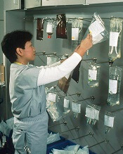
of chemotherapy drugs
Credit: Bill Branson
Baxter Healthcare Corporation is recalling some of its infusion pumps after receiving more than 3500 reports of the pumps malfunctioning.
According to the US Food and Drug Administration (FDA), the malfunctioning pumps have resulted in 9 severe adverse events but no deaths.
This Class I recall includes Sigma Spectrum Infusion Pumps with Master Drug Library Model No. 35700BAX and 35700ABB.
The pumps were made between July 1, 2005, and January 15, 2014. They were distributed between February 20, 2013, and January 15, 2014.
The Sigma Spectrum infusion pumps are intended to deliver controlled amounts of medicines, blood, blood products, and other intravenous fluids.
The FDA said there have been more than 3500 reports of these pumps malfunctioning—specifically, reports of System Error 322 “Link Switch Error (low).” This error occurs when the pump detects that the door is open even though it is closed. A System Error 322 may lead to an interruption or delay in therapy.
When this error occurs, the Sigma Spectrum infusion pump stops the infusion, an alarm sounds, and a light flashes (a visual “322” alarm). This requires a clinician to reset the alarm, reprogram the pump, and confirm the infusion is running properly.
The use of affected pumps may cause serious adverse health consequences, including death; hence, the Class I recall.
Customers who encounter a System Error 322 should turn off the pump by pressing the ON/OFF key, then turn the pump back on by pressing the ON/OFF key to clear the alarm.
Clinicians will need to reprogram the infusion after the pump is turned back on. If the alarm cannot be cleared using these instructions, the device should be removed from use and sent to the facility’s biomedical engineering department.
If the System Error 322 reoccurs, the pump may need to be inspected and serviced by Baxter Healthcare. To contact Baxter, call 1-800-356-3454 (choose option 1) Monday through Friday, 7 am to 7 pm, Eastern Time.
Adverse reactions or quality problems related to these pumps can be reported to the FDA’s MedWatch Program. 
of chemotherapy drugs
Credit: Bill Branson
Baxter Healthcare Corporation is recalling some of its infusion pumps after receiving more than 3500 reports of the pumps malfunctioning.
According to the US Food and Drug Administration (FDA), the malfunctioning pumps have resulted in 9 severe adverse events but no deaths.
This Class I recall includes Sigma Spectrum Infusion Pumps with Master Drug Library Model No. 35700BAX and 35700ABB.
The pumps were made between July 1, 2005, and January 15, 2014. They were distributed between February 20, 2013, and January 15, 2014.
The Sigma Spectrum infusion pumps are intended to deliver controlled amounts of medicines, blood, blood products, and other intravenous fluids.
The FDA said there have been more than 3500 reports of these pumps malfunctioning—specifically, reports of System Error 322 “Link Switch Error (low).” This error occurs when the pump detects that the door is open even though it is closed. A System Error 322 may lead to an interruption or delay in therapy.
When this error occurs, the Sigma Spectrum infusion pump stops the infusion, an alarm sounds, and a light flashes (a visual “322” alarm). This requires a clinician to reset the alarm, reprogram the pump, and confirm the infusion is running properly.
The use of affected pumps may cause serious adverse health consequences, including death; hence, the Class I recall.
Customers who encounter a System Error 322 should turn off the pump by pressing the ON/OFF key, then turn the pump back on by pressing the ON/OFF key to clear the alarm.
Clinicians will need to reprogram the infusion after the pump is turned back on. If the alarm cannot be cleared using these instructions, the device should be removed from use and sent to the facility’s biomedical engineering department.
If the System Error 322 reoccurs, the pump may need to be inspected and serviced by Baxter Healthcare. To contact Baxter, call 1-800-356-3454 (choose option 1) Monday through Friday, 7 am to 7 pm, Eastern Time.
Adverse reactions or quality problems related to these pumps can be reported to the FDA’s MedWatch Program.
of chemotherapy drugs
Credit: Bill Branson
Baxter Healthcare Corporation is recalling some of its infusion pumps after receiving more than 3500 reports of the pumps malfunctioning.
According to the US Food and Drug Administration (FDA), the malfunctioning pumps have resulted in 9 severe adverse events but no deaths.
This Class I recall includes Sigma Spectrum Infusion Pumps with Master Drug Library Model No. 35700BAX and 35700ABB.
The pumps were made between July 1, 2005, and January 15, 2014. They were distributed between February 20, 2013, and January 15, 2014.
The Sigma Spectrum infusion pumps are intended to deliver controlled amounts of medicines, blood, blood products, and other intravenous fluids.
The FDA said there have been more than 3500 reports of these pumps malfunctioning—specifically, reports of System Error 322 “Link Switch Error (low).” This error occurs when the pump detects that the door is open even though it is closed. A System Error 322 may lead to an interruption or delay in therapy.
When this error occurs, the Sigma Spectrum infusion pump stops the infusion, an alarm sounds, and a light flashes (a visual “322” alarm). This requires a clinician to reset the alarm, reprogram the pump, and confirm the infusion is running properly.
The use of affected pumps may cause serious adverse health consequences, including death; hence, the Class I recall.
Customers who encounter a System Error 322 should turn off the pump by pressing the ON/OFF key, then turn the pump back on by pressing the ON/OFF key to clear the alarm.
Clinicians will need to reprogram the infusion after the pump is turned back on. If the alarm cannot be cleared using these instructions, the device should be removed from use and sent to the facility’s biomedical engineering department.
If the System Error 322 reoccurs, the pump may need to be inspected and serviced by Baxter Healthcare. To contact Baxter, call 1-800-356-3454 (choose option 1) Monday through Friday, 7 am to 7 pm, Eastern Time.
Adverse reactions or quality problems related to these pumps can be reported to the FDA’s MedWatch Program.
Using chromatin conformation data to classify leukemia
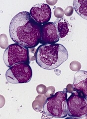
Chromatin conformation can guide the classification of leukemia, according to research published in Genome Biology.
Investigators mapped the conformation of the homeobox A (HOXA) gene cluster—11 genes encoding proteins that are highly relevant to many cancers—in a panel of leukemia cell lines.
And the team found they could use this information to distinguish subtypes of leukemia from one another.
“Previous studies have shown that looking at gene expression—the specific proteins produced by the genes—is a good predictor of whether patients have leukemia,” said study author Mathieu Blanchette, PhD, of McGill University in Montréal, Québec, Canada.
“We found that different types of leukemia cells also have a distinctive chromatin interaction—how the chromatin that makes up the genome is folded.”
The investigators used 5C chromosome conformation capture technology to analyze the HOXA gene cluster and then used the data to train and test a support vector machine classifier called 3D-SP.
They found 3D-SP could distinguish leukemias expressing MLL-fusion proteins from those expressing wild-type MLL. It could also classify leukemia subtypes according to MLL fusion partner.
The team noted that it is not clear whether the genome shape plays a role in causing leukemia or whether the leukemia causes the genome to change shape. And additional studies are needed to determine whether genome shape is as useful for classifying other types of cancer.
“Our study validates a new research avenue: the application of 3D genomics for developing medical diagnostics or treatments that could be explored for diseases where current technologies, including gene expression data, have failed to improve patient care,” said Josée Dostie, PhD, also of McGill University.
“While the use of 3D genomics in the clinic is still remote when considering the technical challenges required for translating the information to the bedside, we discovered a new approach for classifying human disease that must be explored further, if only for what it can reveal about how the human genome works.”
Chromatin conformation can guide the classification of leukemia, according to research published in Genome Biology.
Investigators mapped the conformation of the homeobox A (HOXA) gene cluster—11 genes encoding proteins that are highly relevant to many cancers—in a panel of leukemia cell lines.
And the team found they could use this information to distinguish subtypes of leukemia from one another.
“Previous studies have shown that looking at gene expression—the specific proteins produced by the genes—is a good predictor of whether patients have leukemia,” said study author Mathieu Blanchette, PhD, of McGill University in Montréal, Québec, Canada.
“We found that different types of leukemia cells also have a distinctive chromatin interaction—how the chromatin that makes up the genome is folded.”
The investigators used 5C chromosome conformation capture technology to analyze the HOXA gene cluster and then used the data to train and test a support vector machine classifier called 3D-SP.
They found 3D-SP could distinguish leukemias expressing MLL-fusion proteins from those expressing wild-type MLL. It could also classify leukemia subtypes according to MLL fusion partner.
The team noted that it is not clear whether the genome shape plays a role in causing leukemia or whether the leukemia causes the genome to change shape. And additional studies are needed to determine whether genome shape is as useful for classifying other types of cancer.
“Our study validates a new research avenue: the application of 3D genomics for developing medical diagnostics or treatments that could be explored for diseases where current technologies, including gene expression data, have failed to improve patient care,” said Josée Dostie, PhD, also of McGill University.
“While the use of 3D genomics in the clinic is still remote when considering the technical challenges required for translating the information to the bedside, we discovered a new approach for classifying human disease that must be explored further, if only for what it can reveal about how the human genome works.”
Chromatin conformation can guide the classification of leukemia, according to research published in Genome Biology.
Investigators mapped the conformation of the homeobox A (HOXA) gene cluster—11 genes encoding proteins that are highly relevant to many cancers—in a panel of leukemia cell lines.
And the team found they could use this information to distinguish subtypes of leukemia from one another.
“Previous studies have shown that looking at gene expression—the specific proteins produced by the genes—is a good predictor of whether patients have leukemia,” said study author Mathieu Blanchette, PhD, of McGill University in Montréal, Québec, Canada.
“We found that different types of leukemia cells also have a distinctive chromatin interaction—how the chromatin that makes up the genome is folded.”
The investigators used 5C chromosome conformation capture technology to analyze the HOXA gene cluster and then used the data to train and test a support vector machine classifier called 3D-SP.
They found 3D-SP could distinguish leukemias expressing MLL-fusion proteins from those expressing wild-type MLL. It could also classify leukemia subtypes according to MLL fusion partner.
The team noted that it is not clear whether the genome shape plays a role in causing leukemia or whether the leukemia causes the genome to change shape. And additional studies are needed to determine whether genome shape is as useful for classifying other types of cancer.
“Our study validates a new research avenue: the application of 3D genomics for developing medical diagnostics or treatments that could be explored for diseases where current technologies, including gene expression data, have failed to improve patient care,” said Josée Dostie, PhD, also of McGill University.
“While the use of 3D genomics in the clinic is still remote when considering the technical challenges required for translating the information to the bedside, we discovered a new approach for classifying human disease that must be explored further, if only for what it can reveal about how the human genome works.”
FDA wants more information on cangrelor
Credit: Kevin MacKenzie
The US Food and Drug Administration (FDA) has issued a Complete Response Letter to The Medicines Company regarding its new drug application for the antiplatelet agent cangrelor.
The company applied for approval of cangrelor to treat patients undergoing percutaneous coronary intervention (PCI) and those who require bridging from oral antiplatelet therapy to surgery.
The new drug application filing was based on the results of a development program that included 4 randomized trials.
These trials—BRIDGE, CHAMPION PHOENIX, CHAMPION PLATFORM, and CHAMPION PCI—included 25,567 patients with coronary artery disease.
In the Complete Response Letter, the FDA said it cannot approve cangrelor for the PCI indication without additional information.
The agency suggested The Medicines Company conduct a series of clinical data analyses of the CHAMPION PHOENIX study, review certain processes regarding data management, and provide bioequivalence information on the clopidogrel clinical supplies for the CHAMPION trials.
For the bridge indication, the FDA said a prospective, adequate, and well-controlled trial, in which outcomes such as bleeding are studied, is needed. Such a trial could provide the clinical data necessary to assess the benefit-risk relationship of cangrelor in this indication.
The FDA provided additional comments for the company to address, which could affect product labeling, but the company did not disclose them.
“We are grateful for the agency’s review, comments, and suggestions,” said Clive Meanwell, Chairman and Chief Executive Officer of The Medicines Company. “The next steps of review will focus on additional analyses in response to the FDA.”
Cangrelor is an investigational agent not approved for commercial use in any market. The product is a bioavailable, quickly reversible, intravenous antiplatelet agent. It is in development to prevent platelet activation and aggregation that leads to thrombosis in the acute care setting.
Credit: Kevin MacKenzie
The US Food and Drug Administration (FDA) has issued a Complete Response Letter to The Medicines Company regarding its new drug application for the antiplatelet agent cangrelor.
The company applied for approval of cangrelor to treat patients undergoing percutaneous coronary intervention (PCI) and those who require bridging from oral antiplatelet therapy to surgery.
The new drug application filing was based on the results of a development program that included 4 randomized trials.
These trials—BRIDGE, CHAMPION PHOENIX, CHAMPION PLATFORM, and CHAMPION PCI—included 25,567 patients with coronary artery disease.
In the Complete Response Letter, the FDA said it cannot approve cangrelor for the PCI indication without additional information.
The agency suggested The Medicines Company conduct a series of clinical data analyses of the CHAMPION PHOENIX study, review certain processes regarding data management, and provide bioequivalence information on the clopidogrel clinical supplies for the CHAMPION trials.
For the bridge indication, the FDA said a prospective, adequate, and well-controlled trial, in which outcomes such as bleeding are studied, is needed. Such a trial could provide the clinical data necessary to assess the benefit-risk relationship of cangrelor in this indication.
The FDA provided additional comments for the company to address, which could affect product labeling, but the company did not disclose them.
“We are grateful for the agency’s review, comments, and suggestions,” said Clive Meanwell, Chairman and Chief Executive Officer of The Medicines Company. “The next steps of review will focus on additional analyses in response to the FDA.”
Cangrelor is an investigational agent not approved for commercial use in any market. The product is a bioavailable, quickly reversible, intravenous antiplatelet agent. It is in development to prevent platelet activation and aggregation that leads to thrombosis in the acute care setting.
Credit: Kevin MacKenzie
The US Food and Drug Administration (FDA) has issued a Complete Response Letter to The Medicines Company regarding its new drug application for the antiplatelet agent cangrelor.
The company applied for approval of cangrelor to treat patients undergoing percutaneous coronary intervention (PCI) and those who require bridging from oral antiplatelet therapy to surgery.
The new drug application filing was based on the results of a development program that included 4 randomized trials.
These trials—BRIDGE, CHAMPION PHOENIX, CHAMPION PLATFORM, and CHAMPION PCI—included 25,567 patients with coronary artery disease.
In the Complete Response Letter, the FDA said it cannot approve cangrelor for the PCI indication without additional information.
The agency suggested The Medicines Company conduct a series of clinical data analyses of the CHAMPION PHOENIX study, review certain processes regarding data management, and provide bioequivalence information on the clopidogrel clinical supplies for the CHAMPION trials.
For the bridge indication, the FDA said a prospective, adequate, and well-controlled trial, in which outcomes such as bleeding are studied, is needed. Such a trial could provide the clinical data necessary to assess the benefit-risk relationship of cangrelor in this indication.
The FDA provided additional comments for the company to address, which could affect product labeling, but the company did not disclose them.
“We are grateful for the agency’s review, comments, and suggestions,” said Clive Meanwell, Chairman and Chief Executive Officer of The Medicines Company. “The next steps of review will focus on additional analyses in response to the FDA.”
Cangrelor is an investigational agent not approved for commercial use in any market. The product is a bioavailable, quickly reversible, intravenous antiplatelet agent. It is in development to prevent platelet activation and aggregation that leads to thrombosis in the acute care setting.
Finding could aid treatment of Fanconi anemia
Credit: Tom Ellenberger
Understanding the interaction between 2 genes may be the key to better treatment of Fanconi anemia, according to a paper published in Cell Cycle.
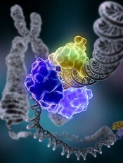
Researchers investigated the relationship between FANCD2 and DNA2, 2 genes known to play roles in DNA repair.
A defective version of FANCD2 can result in Fanconi anemia. And although DNA2 has not been associated with a Fanconi anemia family
yet, genetic studies have implicated DNA2 in the Fanconi anemia DNA repair pathway.
With the current study, the researchers found that deleting either FANCD2 or DNA2 alone makes cells susceptible to DNA damage. But the deletion of both genes enables DNA repair.
“A key implication of this finding is the potential to manipulate DNA2 to improve the survival of FANCD2-deficient cells, and hopefully, by extension, the survival of [Fanconi anemia] patients,” said study author Kenneth Karanja, PhD, a former postdoctoral scholar at the California Institute of Technology in Pasadena.
To uncover the relationship between the genes, Dr Karanja and his colleagues applied DNA-damaging substances—formaldehyde and cisplatin—to 3 types of cells: those lacking FANCD2, those lacking DNA2, and cells lacking both genes.
The groups of cells in which only 1 of the 2 genes had been deleted quickly succumbed to the substance-induced DNA damage. However, the cells lacking both FANCD2 and DNA2 were able to repair the DNA damage and survive.
So the researchers concluded that depletion of DNA2 in FANCD2-deficient cells reverses the cells’ sensitivity to DNA-damaging substances. And this finding may have implications for Fanconi anemia treatment.
“DNA2 is a well-studied gene, and this recent discovery could potentially become the basis for ameliorating the symptoms of this incurable disorder,” said study author Judith Campbell, PhD, of the California Institute of Technology.
“Since much is known about the mechanism of action of DNA2, it is an attractive target for future drug treatments—like small-molecule inhibitors that could reduce [a Fanconi anemia] patient’s cancer predisposition—as well as a possible gene therapy for aiding a patient’s blood cell development.”
Credit: Tom Ellenberger
Understanding the interaction between 2 genes may be the key to better treatment of Fanconi anemia, according to a paper published in Cell Cycle.
Researchers investigated the relationship between FANCD2 and DNA2, 2 genes known to play roles in DNA repair.
A defective version of FANCD2 can result in Fanconi anemia. And although DNA2 has not been associated with a Fanconi anemia family
yet, genetic studies have implicated DNA2 in the Fanconi anemia DNA repair pathway.
With the current study, the researchers found that deleting either FANCD2 or DNA2 alone makes cells susceptible to DNA damage. But the deletion of both genes enables DNA repair.
“A key implication of this finding is the potential to manipulate DNA2 to improve the survival of FANCD2-deficient cells, and hopefully, by extension, the survival of [Fanconi anemia] patients,” said study author Kenneth Karanja, PhD, a former postdoctoral scholar at the California Institute of Technology in Pasadena.
To uncover the relationship between the genes, Dr Karanja and his colleagues applied DNA-damaging substances—formaldehyde and cisplatin—to 3 types of cells: those lacking FANCD2, those lacking DNA2, and cells lacking both genes.
The groups of cells in which only 1 of the 2 genes had been deleted quickly succumbed to the substance-induced DNA damage. However, the cells lacking both FANCD2 and DNA2 were able to repair the DNA damage and survive.
So the researchers concluded that depletion of DNA2 in FANCD2-deficient cells reverses the cells’ sensitivity to DNA-damaging substances. And this finding may have implications for Fanconi anemia treatment.
“DNA2 is a well-studied gene, and this recent discovery could potentially become the basis for ameliorating the symptoms of this incurable disorder,” said study author Judith Campbell, PhD, of the California Institute of Technology.
“Since much is known about the mechanism of action of DNA2, it is an attractive target for future drug treatments—like small-molecule inhibitors that could reduce [a Fanconi anemia] patient’s cancer predisposition—as well as a possible gene therapy for aiding a patient’s blood cell development.”
Credit: Tom Ellenberger
Understanding the interaction between 2 genes may be the key to better treatment of Fanconi anemia, according to a paper published in Cell Cycle.
Researchers investigated the relationship between FANCD2 and DNA2, 2 genes known to play roles in DNA repair.
A defective version of FANCD2 can result in Fanconi anemia. And although DNA2 has not been associated with a Fanconi anemia family
yet, genetic studies have implicated DNA2 in the Fanconi anemia DNA repair pathway.
With the current study, the researchers found that deleting either FANCD2 or DNA2 alone makes cells susceptible to DNA damage. But the deletion of both genes enables DNA repair.
“A key implication of this finding is the potential to manipulate DNA2 to improve the survival of FANCD2-deficient cells, and hopefully, by extension, the survival of [Fanconi anemia] patients,” said study author Kenneth Karanja, PhD, a former postdoctoral scholar at the California Institute of Technology in Pasadena.
To uncover the relationship between the genes, Dr Karanja and his colleagues applied DNA-damaging substances—formaldehyde and cisplatin—to 3 types of cells: those lacking FANCD2, those lacking DNA2, and cells lacking both genes.
The groups of cells in which only 1 of the 2 genes had been deleted quickly succumbed to the substance-induced DNA damage. However, the cells lacking both FANCD2 and DNA2 were able to repair the DNA damage and survive.
So the researchers concluded that depletion of DNA2 in FANCD2-deficient cells reverses the cells’ sensitivity to DNA-damaging substances. And this finding may have implications for Fanconi anemia treatment.
“DNA2 is a well-studied gene, and this recent discovery could potentially become the basis for ameliorating the symptoms of this incurable disorder,” said study author Judith Campbell, PhD, of the California Institute of Technology.
“Since much is known about the mechanism of action of DNA2, it is an attractive target for future drug treatments—like small-molecule inhibitors that could reduce [a Fanconi anemia] patient’s cancer predisposition—as well as a possible gene therapy for aiding a patient’s blood cell development.”
How bleomycin cuts cancer to pieces

Credit: Bill Branson
The antitumor agent bleomycin can treat a range of cancers, but its disease-fighting properties have been poorly understood.
Now, a pair of researchers have characterized bleomycin’s ability to cut through double-stranded DNA in cancerous cells.
The duo believe their research could help inform efforts to fine-tune the drug, improving its cancer-killing properties and limiting toxicity to healthy cells.
The research appears in the Journal of the American Chemical Society.
Bleomycin is part of a family of structurally related antibiotics produced by the bacterium Streptomyces verticillus. Three potent versions of the drug—labeled A2, A5, and B2—are the primary forms in clinical use against cancers.
Previous research has shown that bleomycin can cause death in aberrant cells by migrating to the cell nucleus, binding with DNA, and subsequently causing breaks in the DNA sequence. Following a binding event, a molecule of bleomycin can effectively slice through one or both strands of DNA.
Cleavage of DNA is believed to be the primary mechanism by which bleomycin kills cancer cells, particularly through double-strand cleavages, which are more challenging for the cellular machinery to repair.
“There are several mechanisms for repairing both single-strand and double-strand breaks in DNA, but double-strand breaks are a more potent form of DNA lesion,” explained study author Basab Roy, a graduate student at Arizona State University in Tempe.
For this study, Roy and Sidney Hecht, PhD, used bleomycin A5, which has similar DNA binding and cleaving properties as bleomycin A2 and B2.
Previous research revealed that bleomycin binds with highly specific regions of the DNA strand, typically G-C sites, where a guanosine base pairs with a cytidine. The strength of this binding is closely associated with the degree of double-strand DNA cleavage.
From a pool of random DNA sequences, the researchers selected a library of 10 hairpin DNAs, based on their strong binding affinity for bleomycin A5. Hairpin DNAs are looped structures that form when a segment of a DNA strand base-pairs with another portion of the same strand. These hairpin DNAs were used to investigate double-strand cleavage.
Each of the 10 DNA samples underwent double-strand cleavage at more than one site. All of the observed cleavage sites were found within or in close proximity to an 8-base-pair-variable region.
Examination of the 10 DNA samples exposed to bleomycin revealed a total of 31 double-strand cleavage sites. Earlier research had described the form of double-strand DNA cleavage bleomycin induced at 14 of these sites. But the remaining 17 cases of double-stranded cleavage occurred through a different mechanism, described for the first time by Roy and Dr Hecht.
The pair used iron (FeII) as a cofactor for bleomycin in the binding events, and they observed 2 types of bleomycin binding and cleavage activity.
In the first, bleomycin and its iron cofactor (Fe.BLM) bind with hairpin DNA at a primary site. Typically, this is a site with a particular sequence: 5´-G-Py-B-3´. (Here, 5´ refers to one end of the DNA hairpin, G refers to the base guanosine, Py refers to a pyrimidinic base—either cytidine or thymidine, B refers to any nucleobase, and 3´ refers to the other DNA end.)
The result of this binding is the abstraction of a hydrogen atom at the primary site. Two results are possible following the primary binding event, one causing a single-strand break in the primary site and the other, failing to produce full cleavage of the strand, producing instead a site lacking either a purine or pyrimidine base. This is known as an AP site.
In the first case—where bleomycin achieves single strand cleavage—the bleomycin molecule can then become reactivated, once more abstracting a hydrogen atom from the opposing DNA strand.
The opposite strand can again follow 1 of 2 pathways, (a) full cleavage of the opposing strand, yielding a double-strand cleavage or (b) formation of an AP site. The researchers noted that this AP site can lead to strand cleavage through the opposing DNA strand with the addition of a mild base like n-butylamine.
The results of this study emphasize the correlation between the strength of bleomycin binding to DNA and the frequency of double-strand cleavage. Of the 10 sample hairpin DNAs, the 2 most tightly bound to bleomycin each showed 5 double-strand cleavages, whereas the least tightly bound samples exhibited just 2 double-strand cleavages.
This suggests a plausible mechanism for DNA cleavage by bleomycin that may lead to tumor cell killing, as well as identifying the most common sequences involved in DNA site binding and subsequent strand breakage.
Roy noted, however, that more research is needed to elucidate the biochemical causes of tight binding by bleomycin. Furthermore, bleomycin’s specificity for cancer cells remains enigmatic.
“Cancer is still a black hole,” Roy said. “We’re trying to make this particular molecule better. There is still so much to learn.”
Credit: Bill Branson
The antitumor agent bleomycin can treat a range of cancers, but its disease-fighting properties have been poorly understood.
Now, a pair of researchers have characterized bleomycin’s ability to cut through double-stranded DNA in cancerous cells.
The duo believe their research could help inform efforts to fine-tune the drug, improving its cancer-killing properties and limiting toxicity to healthy cells.
The research appears in the Journal of the American Chemical Society.
Bleomycin is part of a family of structurally related antibiotics produced by the bacterium Streptomyces verticillus. Three potent versions of the drug—labeled A2, A5, and B2—are the primary forms in clinical use against cancers.
Previous research has shown that bleomycin can cause death in aberrant cells by migrating to the cell nucleus, binding with DNA, and subsequently causing breaks in the DNA sequence. Following a binding event, a molecule of bleomycin can effectively slice through one or both strands of DNA.
Cleavage of DNA is believed to be the primary mechanism by which bleomycin kills cancer cells, particularly through double-strand cleavages, which are more challenging for the cellular machinery to repair.
“There are several mechanisms for repairing both single-strand and double-strand breaks in DNA, but double-strand breaks are a more potent form of DNA lesion,” explained study author Basab Roy, a graduate student at Arizona State University in Tempe.
For this study, Roy and Sidney Hecht, PhD, used bleomycin A5, which has similar DNA binding and cleaving properties as bleomycin A2 and B2.
Previous research revealed that bleomycin binds with highly specific regions of the DNA strand, typically G-C sites, where a guanosine base pairs with a cytidine. The strength of this binding is closely associated with the degree of double-strand DNA cleavage.
From a pool of random DNA sequences, the researchers selected a library of 10 hairpin DNAs, based on their strong binding affinity for bleomycin A5. Hairpin DNAs are looped structures that form when a segment of a DNA strand base-pairs with another portion of the same strand. These hairpin DNAs were used to investigate double-strand cleavage.
Each of the 10 DNA samples underwent double-strand cleavage at more than one site. All of the observed cleavage sites were found within or in close proximity to an 8-base-pair-variable region.
Examination of the 10 DNA samples exposed to bleomycin revealed a total of 31 double-strand cleavage sites. Earlier research had described the form of double-strand DNA cleavage bleomycin induced at 14 of these sites. But the remaining 17 cases of double-stranded cleavage occurred through a different mechanism, described for the first time by Roy and Dr Hecht.
The pair used iron (FeII) as a cofactor for bleomycin in the binding events, and they observed 2 types of bleomycin binding and cleavage activity.
In the first, bleomycin and its iron cofactor (Fe.BLM) bind with hairpin DNA at a primary site. Typically, this is a site with a particular sequence: 5´-G-Py-B-3´. (Here, 5´ refers to one end of the DNA hairpin, G refers to the base guanosine, Py refers to a pyrimidinic base—either cytidine or thymidine, B refers to any nucleobase, and 3´ refers to the other DNA end.)
The result of this binding is the abstraction of a hydrogen atom at the primary site. Two results are possible following the primary binding event, one causing a single-strand break in the primary site and the other, failing to produce full cleavage of the strand, producing instead a site lacking either a purine or pyrimidine base. This is known as an AP site.
In the first case—where bleomycin achieves single strand cleavage—the bleomycin molecule can then become reactivated, once more abstracting a hydrogen atom from the opposing DNA strand.
The opposite strand can again follow 1 of 2 pathways, (a) full cleavage of the opposing strand, yielding a double-strand cleavage or (b) formation of an AP site. The researchers noted that this AP site can lead to strand cleavage through the opposing DNA strand with the addition of a mild base like n-butylamine.
The results of this study emphasize the correlation between the strength of bleomycin binding to DNA and the frequency of double-strand cleavage. Of the 10 sample hairpin DNAs, the 2 most tightly bound to bleomycin each showed 5 double-strand cleavages, whereas the least tightly bound samples exhibited just 2 double-strand cleavages.
This suggests a plausible mechanism for DNA cleavage by bleomycin that may lead to tumor cell killing, as well as identifying the most common sequences involved in DNA site binding and subsequent strand breakage.
Roy noted, however, that more research is needed to elucidate the biochemical causes of tight binding by bleomycin. Furthermore, bleomycin’s specificity for cancer cells remains enigmatic.
“Cancer is still a black hole,” Roy said. “We’re trying to make this particular molecule better. There is still so much to learn.”
Credit: Bill Branson
The antitumor agent bleomycin can treat a range of cancers, but its disease-fighting properties have been poorly understood.
Now, a pair of researchers have characterized bleomycin’s ability to cut through double-stranded DNA in cancerous cells.
The duo believe their research could help inform efforts to fine-tune the drug, improving its cancer-killing properties and limiting toxicity to healthy cells.
The research appears in the Journal of the American Chemical Society.
Bleomycin is part of a family of structurally related antibiotics produced by the bacterium Streptomyces verticillus. Three potent versions of the drug—labeled A2, A5, and B2—are the primary forms in clinical use against cancers.
Previous research has shown that bleomycin can cause death in aberrant cells by migrating to the cell nucleus, binding with DNA, and subsequently causing breaks in the DNA sequence. Following a binding event, a molecule of bleomycin can effectively slice through one or both strands of DNA.
Cleavage of DNA is believed to be the primary mechanism by which bleomycin kills cancer cells, particularly through double-strand cleavages, which are more challenging for the cellular machinery to repair.
“There are several mechanisms for repairing both single-strand and double-strand breaks in DNA, but double-strand breaks are a more potent form of DNA lesion,” explained study author Basab Roy, a graduate student at Arizona State University in Tempe.
For this study, Roy and Sidney Hecht, PhD, used bleomycin A5, which has similar DNA binding and cleaving properties as bleomycin A2 and B2.
Previous research revealed that bleomycin binds with highly specific regions of the DNA strand, typically G-C sites, where a guanosine base pairs with a cytidine. The strength of this binding is closely associated with the degree of double-strand DNA cleavage.
From a pool of random DNA sequences, the researchers selected a library of 10 hairpin DNAs, based on their strong binding affinity for bleomycin A5. Hairpin DNAs are looped structures that form when a segment of a DNA strand base-pairs with another portion of the same strand. These hairpin DNAs were used to investigate double-strand cleavage.
Each of the 10 DNA samples underwent double-strand cleavage at more than one site. All of the observed cleavage sites were found within or in close proximity to an 8-base-pair-variable region.
Examination of the 10 DNA samples exposed to bleomycin revealed a total of 31 double-strand cleavage sites. Earlier research had described the form of double-strand DNA cleavage bleomycin induced at 14 of these sites. But the remaining 17 cases of double-stranded cleavage occurred through a different mechanism, described for the first time by Roy and Dr Hecht.
The pair used iron (FeII) as a cofactor for bleomycin in the binding events, and they observed 2 types of bleomycin binding and cleavage activity.
In the first, bleomycin and its iron cofactor (Fe.BLM) bind with hairpin DNA at a primary site. Typically, this is a site with a particular sequence: 5´-G-Py-B-3´. (Here, 5´ refers to one end of the DNA hairpin, G refers to the base guanosine, Py refers to a pyrimidinic base—either cytidine or thymidine, B refers to any nucleobase, and 3´ refers to the other DNA end.)
The result of this binding is the abstraction of a hydrogen atom at the primary site. Two results are possible following the primary binding event, one causing a single-strand break in the primary site and the other, failing to produce full cleavage of the strand, producing instead a site lacking either a purine or pyrimidine base. This is known as an AP site.
In the first case—where bleomycin achieves single strand cleavage—the bleomycin molecule can then become reactivated, once more abstracting a hydrogen atom from the opposing DNA strand.
The opposite strand can again follow 1 of 2 pathways, (a) full cleavage of the opposing strand, yielding a double-strand cleavage or (b) formation of an AP site. The researchers noted that this AP site can lead to strand cleavage through the opposing DNA strand with the addition of a mild base like n-butylamine.
The results of this study emphasize the correlation between the strength of bleomycin binding to DNA and the frequency of double-strand cleavage. Of the 10 sample hairpin DNAs, the 2 most tightly bound to bleomycin each showed 5 double-strand cleavages, whereas the least tightly bound samples exhibited just 2 double-strand cleavages.
This suggests a plausible mechanism for DNA cleavage by bleomycin that may lead to tumor cell killing, as well as identifying the most common sequences involved in DNA site binding and subsequent strand breakage.
Roy noted, however, that more research is needed to elucidate the biochemical causes of tight binding by bleomycin. Furthermore, bleomycin’s specificity for cancer cells remains enigmatic.
“Cancer is still a black hole,” Roy said. “We’re trying to make this particular molecule better. There is still so much to learn.”
Team overcomes problem with new lab on a chip

Credit: Rhoda Baer
Chemists say they have overcome one of the main obstacles in creating effective lab-on-a-chip devices, and the device they’ve invented has many potential applications, such as screening biological molecules.
To create their device, the team developed a technique that involves printing droplets of a special solvent onto a gold-coated or glass surface.
“We use a class of ‘green’ solvents called ionic liquids, which are salts that are liquid at room temperature,” said study author Chuan Zhao, PhD, of The University of New South Wales in Sydney, Australia.
“They are non-volatile, so this overcomes one of the main problems in making useful miniaturized devices—rapid evaporation of the solvents on the chip.”
Dr Zhao and his colleagues described this research in Nature Communications.
Lab-on-a-chip devices, where chemical reactions are carried out on a miniature scale, are under intensive development because they offer the promise of faster reaction times, reduced use of materials, and high yields of product.
However, the evaporation of solvents on the chip is a problem because it can affect the concentration of substances and disrupt the reactions.
Researchers have attempted to overcome the problem by containing the solvents within tiny channels, or “walls,” and having reservoirs to store extra solvent on the chip.
Dr Zhao and his colleagues said their “wall-less” design—using non-volatile ionic liquids as solvents to fabricate a microarray of droplets chemically anchored to the chip—has several advantages.
“Ionic liquids are designer solvents and have wide application,” Dr Zhao said. “We can now carry out many reactions or analytical procedures in ionic liquids at the micro-scale on a chip with enhanced yields and efficiency.”
“These microarray chips can be easily produced in high numbers and are very stable. They can survive being turned upside down and heated to 50 degrees, and some can even survive being immersed in another liquid. These properties will be important for commercial applications, including storage and transportation of microchips.”
The droplets of ionic liquid are about 50 µm across and 10 µm high. The researchers showed these tiny droplets can act as rapid, sensitive monitors of the presence of a gas, due to their small volume.
Metal salts dissolved in the droplets could be electrically deposited as microstructures, a technique that could be of use in the fabrication of integrated circuits.
And some biological molecules added to the droplets remained stable and active, opening up the possibility of using the microarrays for diagnostic purposes.
“The versatility of our chips means they could have a wide range of prospective functions,” Dr Zhao said, “such as for use in fast and accurate hand-held sensors for environmental monitoring, medical diagnosis, and process control in manufacturing.”
Credit: Rhoda Baer
Chemists say they have overcome one of the main obstacles in creating effective lab-on-a-chip devices, and the device they’ve invented has many potential applications, such as screening biological molecules.
To create their device, the team developed a technique that involves printing droplets of a special solvent onto a gold-coated or glass surface.
“We use a class of ‘green’ solvents called ionic liquids, which are salts that are liquid at room temperature,” said study author Chuan Zhao, PhD, of The University of New South Wales in Sydney, Australia.
“They are non-volatile, so this overcomes one of the main problems in making useful miniaturized devices—rapid evaporation of the solvents on the chip.”
Dr Zhao and his colleagues described this research in Nature Communications.
Lab-on-a-chip devices, where chemical reactions are carried out on a miniature scale, are under intensive development because they offer the promise of faster reaction times, reduced use of materials, and high yields of product.
However, the evaporation of solvents on the chip is a problem because it can affect the concentration of substances and disrupt the reactions.
Researchers have attempted to overcome the problem by containing the solvents within tiny channels, or “walls,” and having reservoirs to store extra solvent on the chip.
Dr Zhao and his colleagues said their “wall-less” design—using non-volatile ionic liquids as solvents to fabricate a microarray of droplets chemically anchored to the chip—has several advantages.
“Ionic liquids are designer solvents and have wide application,” Dr Zhao said. “We can now carry out many reactions or analytical procedures in ionic liquids at the micro-scale on a chip with enhanced yields and efficiency.”
“These microarray chips can be easily produced in high numbers and are very stable. They can survive being turned upside down and heated to 50 degrees, and some can even survive being immersed in another liquid. These properties will be important for commercial applications, including storage and transportation of microchips.”
The droplets of ionic liquid are about 50 µm across and 10 µm high. The researchers showed these tiny droplets can act as rapid, sensitive monitors of the presence of a gas, due to their small volume.
Metal salts dissolved in the droplets could be electrically deposited as microstructures, a technique that could be of use in the fabrication of integrated circuits.
And some biological molecules added to the droplets remained stable and active, opening up the possibility of using the microarrays for diagnostic purposes.
“The versatility of our chips means they could have a wide range of prospective functions,” Dr Zhao said, “such as for use in fast and accurate hand-held sensors for environmental monitoring, medical diagnosis, and process control in manufacturing.”
Credit: Rhoda Baer
Chemists say they have overcome one of the main obstacles in creating effective lab-on-a-chip devices, and the device they’ve invented has many potential applications, such as screening biological molecules.
To create their device, the team developed a technique that involves printing droplets of a special solvent onto a gold-coated or glass surface.
“We use a class of ‘green’ solvents called ionic liquids, which are salts that are liquid at room temperature,” said study author Chuan Zhao, PhD, of The University of New South Wales in Sydney, Australia.
“They are non-volatile, so this overcomes one of the main problems in making useful miniaturized devices—rapid evaporation of the solvents on the chip.”
Dr Zhao and his colleagues described this research in Nature Communications.
Lab-on-a-chip devices, where chemical reactions are carried out on a miniature scale, are under intensive development because they offer the promise of faster reaction times, reduced use of materials, and high yields of product.
However, the evaporation of solvents on the chip is a problem because it can affect the concentration of substances and disrupt the reactions.
Researchers have attempted to overcome the problem by containing the solvents within tiny channels, or “walls,” and having reservoirs to store extra solvent on the chip.
Dr Zhao and his colleagues said their “wall-less” design—using non-volatile ionic liquids as solvents to fabricate a microarray of droplets chemically anchored to the chip—has several advantages.
“Ionic liquids are designer solvents and have wide application,” Dr Zhao said. “We can now carry out many reactions or analytical procedures in ionic liquids at the micro-scale on a chip with enhanced yields and efficiency.”
“These microarray chips can be easily produced in high numbers and are very stable. They can survive being turned upside down and heated to 50 degrees, and some can even survive being immersed in another liquid. These properties will be important for commercial applications, including storage and transportation of microchips.”
The droplets of ionic liquid are about 50 µm across and 10 µm high. The researchers showed these tiny droplets can act as rapid, sensitive monitors of the presence of a gas, due to their small volume.
Metal salts dissolved in the droplets could be electrically deposited as microstructures, a technique that could be of use in the fabrication of integrated circuits.
And some biological molecules added to the droplets remained stable and active, opening up the possibility of using the microarrays for diagnostic purposes.
“The versatility of our chips means they could have a wide range of prospective functions,” Dr Zhao said, “such as for use in fast and accurate hand-held sensors for environmental monitoring, medical diagnosis, and process control in manufacturing.”
FDA allows importation of more saline
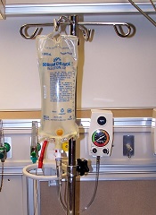
In response to the ongoing shortage of normal saline (0.9% sodium chloride injection), the US Food and Drug Administration (FDA) is allowing a company to import product manufactured in Spain.
Baxter Healthcare Corp. will be allowed to temporarily distribute sodium chloride 0.9% injection solution for intravenous infusion in VIAFLO
non-polyvinyl chloride containers, which is manufactured at the company’s Bieffe Medital, Sabinanigo, Spain facility.
This is the second time in recent months that the FDA has allowed the importation of saline. Last month, the agency announced it would allow Fresenius Kabi USA to import saline products manufactured in Norway.
As with the Norway plant, the FDA has inspected Baxter’s Spain facility to ensure it meets FDA standards.
Baxter will import sodium chloride 0.9% intravenous infusion in VIAFLO containers in the following volumes and quantities: 250 mL (30 bags per carton), 500 mL (20 bags per carton), and 1 L (10 bags per carton).
The FDA is asking that healthcare professionals contact Baxter directly to obtain the product. To place an order, contact Baxter’s Center for Service at 1-888-229-0001.
For more information on the products, see Baxter’s “Dear Healthcare Professional” letter, visit the company’s website, or contact Baxter’s Medical Information Service at 1-800-933-0303.
In addition to the imported saline, US-based manufacturers—B.Braun Medical Inc., Hospira Inc., and Baxter Healthcare Corp.—are producing and releasing normal saline. (Baxter’s saline product from Spain will be distributed in addition to Baxter’s FDA-approved version that is manufactured in the US.)
The FDA noted that, although the aforementioned shipments will help reduce current disruptions, they will not resolve the shortage of 0.9% sodium chloride injection. So the agency will continue working to alleviate the shortage.
In response to the ongoing shortage of normal saline (0.9% sodium chloride injection), the US Food and Drug Administration (FDA) is allowing a company to import product manufactured in Spain.
Baxter Healthcare Corp. will be allowed to temporarily distribute sodium chloride 0.9% injection solution for intravenous infusion in VIAFLO
non-polyvinyl chloride containers, which is manufactured at the company’s Bieffe Medital, Sabinanigo, Spain facility.
This is the second time in recent months that the FDA has allowed the importation of saline. Last month, the agency announced it would allow Fresenius Kabi USA to import saline products manufactured in Norway.
As with the Norway plant, the FDA has inspected Baxter’s Spain facility to ensure it meets FDA standards.
Baxter will import sodium chloride 0.9% intravenous infusion in VIAFLO containers in the following volumes and quantities: 250 mL (30 bags per carton), 500 mL (20 bags per carton), and 1 L (10 bags per carton).
The FDA is asking that healthcare professionals contact Baxter directly to obtain the product. To place an order, contact Baxter’s Center for Service at 1-888-229-0001.
For more information on the products, see Baxter’s “Dear Healthcare Professional” letter, visit the company’s website, or contact Baxter’s Medical Information Service at 1-800-933-0303.
In addition to the imported saline, US-based manufacturers—B.Braun Medical Inc., Hospira Inc., and Baxter Healthcare Corp.—are producing and releasing normal saline. (Baxter’s saline product from Spain will be distributed in addition to Baxter’s FDA-approved version that is manufactured in the US.)
The FDA noted that, although the aforementioned shipments will help reduce current disruptions, they will not resolve the shortage of 0.9% sodium chloride injection. So the agency will continue working to alleviate the shortage.
In response to the ongoing shortage of normal saline (0.9% sodium chloride injection), the US Food and Drug Administration (FDA) is allowing a company to import product manufactured in Spain.
Baxter Healthcare Corp. will be allowed to temporarily distribute sodium chloride 0.9% injection solution for intravenous infusion in VIAFLO
non-polyvinyl chloride containers, which is manufactured at the company’s Bieffe Medital, Sabinanigo, Spain facility.
This is the second time in recent months that the FDA has allowed the importation of saline. Last month, the agency announced it would allow Fresenius Kabi USA to import saline products manufactured in Norway.
As with the Norway plant, the FDA has inspected Baxter’s Spain facility to ensure it meets FDA standards.
Baxter will import sodium chloride 0.9% intravenous infusion in VIAFLO containers in the following volumes and quantities: 250 mL (30 bags per carton), 500 mL (20 bags per carton), and 1 L (10 bags per carton).
The FDA is asking that healthcare professionals contact Baxter directly to obtain the product. To place an order, contact Baxter’s Center for Service at 1-888-229-0001.
For more information on the products, see Baxter’s “Dear Healthcare Professional” letter, visit the company’s website, or contact Baxter’s Medical Information Service at 1-800-933-0303.
In addition to the imported saline, US-based manufacturers—B.Braun Medical Inc., Hospira Inc., and Baxter Healthcare Corp.—are producing and releasing normal saline. (Baxter’s saline product from Spain will be distributed in addition to Baxter’s FDA-approved version that is manufactured in the US.)
The FDA noted that, although the aforementioned shipments will help reduce current disruptions, they will not resolve the shortage of 0.9% sodium chloride injection. So the agency will continue working to alleviate the shortage.
Protein is key to HSC recovery after chemo, radiation

in the bone marrow
The protein beta-catenin plays a critical role in promoting the recovery of hematopoietic stem cells (HSCs) after exposure to radiation or chemotherapy, according to preclinical research published in Genes & Development.
The study provides new insight into the impact of radiation and chemotherapy on cellular and molecular processes.
And it points to possibilities for improving HSC regeneration in cancer patients who have undergone these treatments.
Study investigators first used mouse models to show that exposure to radiation triggers activation of the Wnt signaling pathway in hematopoietic stem and progenitor cells.
“The Wnt pathway and its key mediator, beta catenin, are critical for embryonic development and establishment of the body plan,” explained Tannishtha Reya, PhD, of the University of California, San Diego.
“In addition, the Wnt pathway is activated in stem cells from many tissues and is needed for their continued maintenance.”
Dr Reya and her colleagues then found that mice deficient in beta-catenin lacked the ability to activate canonical Wnt signaling. They also suffered from impaired HSC regeneration and bone marrow recovery after radiation or chemotherapy.
Mouse HSCs without beta-catenin could not suppress the production of reactive oxygen species or resolve DNA double-strand breaks. As a result, they could not recover effectively after radiation exposure or treatment with the chemotherapeutic agent fluorouracil.
“Our work shows that Wnt signaling is important in the mammalian hematopoietic system and is critical for recovery from chemotherapy and radiation,” Dr Reya said. “There are 2 major reasons why accelerating regeneration is important clinically.”
“One is that, after cancer patients are irradiated and transplanted with stem cells, the rate and extent of recovery is often not sufficient to protect the patient from anemia or infections . . . . Identifying signals that can boost regeneration after the bone marrow is severely damaged may help improve outcomes after transplantation.”
“Second, doses of chemotherapy and radiation used to treat cancer are often limited by the collateral damage they cause to normal tissues. If we can improve and accelerate recovery, we might be able to use higher doses of radiation or chemotherapy and reduce the risk of cancer relapse.”
Dr Reya added that this research suggests HSC regeneration could be accelerated by modulating the Wnt pathway, either by delivering additional Wnt proteins directly to patients or through drugs that activate the pathway.
in the bone marrow
The protein beta-catenin plays a critical role in promoting the recovery of hematopoietic stem cells (HSCs) after exposure to radiation or chemotherapy, according to preclinical research published in Genes & Development.
The study provides new insight into the impact of radiation and chemotherapy on cellular and molecular processes.
And it points to possibilities for improving HSC regeneration in cancer patients who have undergone these treatments.
Study investigators first used mouse models to show that exposure to radiation triggers activation of the Wnt signaling pathway in hematopoietic stem and progenitor cells.
“The Wnt pathway and its key mediator, beta catenin, are critical for embryonic development and establishment of the body plan,” explained Tannishtha Reya, PhD, of the University of California, San Diego.
“In addition, the Wnt pathway is activated in stem cells from many tissues and is needed for their continued maintenance.”
Dr Reya and her colleagues then found that mice deficient in beta-catenin lacked the ability to activate canonical Wnt signaling. They also suffered from impaired HSC regeneration and bone marrow recovery after radiation or chemotherapy.
Mouse HSCs without beta-catenin could not suppress the production of reactive oxygen species or resolve DNA double-strand breaks. As a result, they could not recover effectively after radiation exposure or treatment with the chemotherapeutic agent fluorouracil.
“Our work shows that Wnt signaling is important in the mammalian hematopoietic system and is critical for recovery from chemotherapy and radiation,” Dr Reya said. “There are 2 major reasons why accelerating regeneration is important clinically.”
“One is that, after cancer patients are irradiated and transplanted with stem cells, the rate and extent of recovery is often not sufficient to protect the patient from anemia or infections . . . . Identifying signals that can boost regeneration after the bone marrow is severely damaged may help improve outcomes after transplantation.”
“Second, doses of chemotherapy and radiation used to treat cancer are often limited by the collateral damage they cause to normal tissues. If we can improve and accelerate recovery, we might be able to use higher doses of radiation or chemotherapy and reduce the risk of cancer relapse.”
Dr Reya added that this research suggests HSC regeneration could be accelerated by modulating the Wnt pathway, either by delivering additional Wnt proteins directly to patients or through drugs that activate the pathway.
in the bone marrow
The protein beta-catenin plays a critical role in promoting the recovery of hematopoietic stem cells (HSCs) after exposure to radiation or chemotherapy, according to preclinical research published in Genes & Development.
The study provides new insight into the impact of radiation and chemotherapy on cellular and molecular processes.
And it points to possibilities for improving HSC regeneration in cancer patients who have undergone these treatments.
Study investigators first used mouse models to show that exposure to radiation triggers activation of the Wnt signaling pathway in hematopoietic stem and progenitor cells.
“The Wnt pathway and its key mediator, beta catenin, are critical for embryonic development and establishment of the body plan,” explained Tannishtha Reya, PhD, of the University of California, San Diego.
“In addition, the Wnt pathway is activated in stem cells from many tissues and is needed for their continued maintenance.”
Dr Reya and her colleagues then found that mice deficient in beta-catenin lacked the ability to activate canonical Wnt signaling. They also suffered from impaired HSC regeneration and bone marrow recovery after radiation or chemotherapy.
Mouse HSCs without beta-catenin could not suppress the production of reactive oxygen species or resolve DNA double-strand breaks. As a result, they could not recover effectively after radiation exposure or treatment with the chemotherapeutic agent fluorouracil.
“Our work shows that Wnt signaling is important in the mammalian hematopoietic system and is critical for recovery from chemotherapy and radiation,” Dr Reya said. “There are 2 major reasons why accelerating regeneration is important clinically.”
“One is that, after cancer patients are irradiated and transplanted with stem cells, the rate and extent of recovery is often not sufficient to protect the patient from anemia or infections . . . . Identifying signals that can boost regeneration after the bone marrow is severely damaged may help improve outcomes after transplantation.”
“Second, doses of chemotherapy and radiation used to treat cancer are often limited by the collateral damage they cause to normal tissues. If we can improve and accelerate recovery, we might be able to use higher doses of radiation or chemotherapy and reduce the risk of cancer relapse.”
Dr Reya added that this research suggests HSC regeneration could be accelerated by modulating the Wnt pathway, either by delivering additional Wnt proteins directly to patients or through drugs that activate the pathway.
Stats show increase in cancer survival rates

Credit: NIH
New statistics suggest 10-year survival rates for cancer patients in England and Wales have more than doubled over a 40-year period.
And rates increased substantially for those with hematologic malignancies.
From 1971 to 2011, 10-year survival rates increased nearly 7-fold for patients with multiple myeloma and almost 6-fold for leukemia patients.
Rates nearly tripled for non-Hodgkin lymphoma patients and almost doubled for those with Hodgkin lymphoma.
These statistics were released by Cancer Research UK.
“These results come from detailed analysis of the survival of more than 7 million cancer patients diagnosed in England and Wales since the 1970s,” said Michel Coleman, BM BCh, head of Cancer Research UK’s Cancer Survival Group at the London School of Hygiene and Tropical Medicine.
“They show just how far we’ve come in improving cancer survival, but they also shine a spotlight on areas where much more needs to be done.”
The statistics include all adults (aged 15 to 99) diagnosed with cancer in England and Wales.
An analysis of the figures showed that, in 1971-1972, 24% of all cancer patients survived 10 years. By 2010-2011, that figure had increased to 50%.
For leukemia patients, 10-year survival increased from 8% in 1970-1971 to 46% in 2010-2011. For patients with multiple myeloma, it rose from 5% to 33%.
For patients with Hodgkin lymphoma, 10-year survival increased from 49% to 80%. And for non-Hodgkin lymphoma patients, it increased from 22% to 63%.
There were substantial increases in shorter-term survival rates (1-year and 5-year) as well. For details, see the Cancer Research UK website.
Credit: NIH
New statistics suggest 10-year survival rates for cancer patients in England and Wales have more than doubled over a 40-year period.
And rates increased substantially for those with hematologic malignancies.
From 1971 to 2011, 10-year survival rates increased nearly 7-fold for patients with multiple myeloma and almost 6-fold for leukemia patients.
Rates nearly tripled for non-Hodgkin lymphoma patients and almost doubled for those with Hodgkin lymphoma.
These statistics were released by Cancer Research UK.
“These results come from detailed analysis of the survival of more than 7 million cancer patients diagnosed in England and Wales since the 1970s,” said Michel Coleman, BM BCh, head of Cancer Research UK’s Cancer Survival Group at the London School of Hygiene and Tropical Medicine.
“They show just how far we’ve come in improving cancer survival, but they also shine a spotlight on areas where much more needs to be done.”
The statistics include all adults (aged 15 to 99) diagnosed with cancer in England and Wales.
An analysis of the figures showed that, in 1971-1972, 24% of all cancer patients survived 10 years. By 2010-2011, that figure had increased to 50%.
For leukemia patients, 10-year survival increased from 8% in 1970-1971 to 46% in 2010-2011. For patients with multiple myeloma, it rose from 5% to 33%.
For patients with Hodgkin lymphoma, 10-year survival increased from 49% to 80%. And for non-Hodgkin lymphoma patients, it increased from 22% to 63%.
There were substantial increases in shorter-term survival rates (1-year and 5-year) as well. For details, see the Cancer Research UK website.
Credit: NIH
New statistics suggest 10-year survival rates for cancer patients in England and Wales have more than doubled over a 40-year period.
And rates increased substantially for those with hematologic malignancies.
From 1971 to 2011, 10-year survival rates increased nearly 7-fold for patients with multiple myeloma and almost 6-fold for leukemia patients.
Rates nearly tripled for non-Hodgkin lymphoma patients and almost doubled for those with Hodgkin lymphoma.
These statistics were released by Cancer Research UK.
“These results come from detailed analysis of the survival of more than 7 million cancer patients diagnosed in England and Wales since the 1970s,” said Michel Coleman, BM BCh, head of Cancer Research UK’s Cancer Survival Group at the London School of Hygiene and Tropical Medicine.
“They show just how far we’ve come in improving cancer survival, but they also shine a spotlight on areas where much more needs to be done.”
The statistics include all adults (aged 15 to 99) diagnosed with cancer in England and Wales.
An analysis of the figures showed that, in 1971-1972, 24% of all cancer patients survived 10 years. By 2010-2011, that figure had increased to 50%.
For leukemia patients, 10-year survival increased from 8% in 1970-1971 to 46% in 2010-2011. For patients with multiple myeloma, it rose from 5% to 33%.
For patients with Hodgkin lymphoma, 10-year survival increased from 49% to 80%. And for non-Hodgkin lymphoma patients, it increased from 22% to 63%.
There were substantial increases in shorter-term survival rates (1-year and 5-year) as well. For details, see the Cancer Research UK website.
Mutations implicated in hematologic disorders
Credit: Jeremy L. Grisham
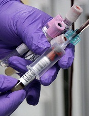
An analysis of more than 30,000 individuals has revealed several genetic mutations that appear to play roles in hematologic disorders.
Investigators discovered variants that showed correlations with platelet counts, white blood cell (WBC) counts, hemoglobin concentration, and hematocrit levels.
The group believes these findings could have implications for a range of conditions, including cytopenias, myeloproliferative neoplasms, and stroke.
Guillaume Lettre, PhD, of Université de Montréal and the Montreal Heart Institute in Canada, and his colleagues recounted their discoveries in a letter to Nature Genetics.
The investigators analyzed hemoglobin concentration, hematocrit levels, WBC counts, and platelet counts in 31,340 individuals genotyped on an exome array.
This revealed several missense variants in CXCR2 that were associated with a decreased WBC count. And in a resequencing study, the team identified a CXCR2 frameshift mutation that was associated with congenital neutropenia.
The group also discovered several missense and splice-site variants in genes known to regulate hematopoiesis—TFR2, HBB, TUBB1, SH2B3, and EPO.
A TFR2 mutation (rs139178017) was independently associated with higher hematocrit levels and hemoglobin concentration.
An HBB variant (rs33971440) and an EPO variant (rs62483572), on the other hand, were associated with lower hematocrit levels and hemoglobin concentrations. Further analyses confirmed that having these mutations increased a person’s risk of anemia, with odds ratios of 36.1 and 1.7, respectively.
A TUBB1 missense variant (rs41303899) was associated with decreased platelet count, while 2 missense variants of SH2B3 (rs148636776 and rs72650673) were associated with increased platelet counts.
Lastly, a mutation in JAK2 (rs77375493) was associated with increases in platelets, WBCs, hemoglobin, and hematocrit. And further analyses suggested that individuals with this variant had early stage myeloproliferative neoplasms.
“[T]hese donors also had a higher risk of having a stroke during their lifetime,” said study author Jean-Claude Tardif, MD, of Université de Montréal and the Montreal Heart Institute.
He and his colleagues believe these findings are encouraging, as they provide additional insight into hematologic disorders. But the results also suggest the experimental approach used in this study can be applied to other diseases as well.
Credit: Jeremy L. Grisham
An analysis of more than 30,000 individuals has revealed several genetic mutations that appear to play roles in hematologic disorders.
Investigators discovered variants that showed correlations with platelet counts, white blood cell (WBC) counts, hemoglobin concentration, and hematocrit levels.
The group believes these findings could have implications for a range of conditions, including cytopenias, myeloproliferative neoplasms, and stroke.
Guillaume Lettre, PhD, of Université de Montréal and the Montreal Heart Institute in Canada, and his colleagues recounted their discoveries in a letter to Nature Genetics.
The investigators analyzed hemoglobin concentration, hematocrit levels, WBC counts, and platelet counts in 31,340 individuals genotyped on an exome array.
This revealed several missense variants in CXCR2 that were associated with a decreased WBC count. And in a resequencing study, the team identified a CXCR2 frameshift mutation that was associated with congenital neutropenia.
The group also discovered several missense and splice-site variants in genes known to regulate hematopoiesis—TFR2, HBB, TUBB1, SH2B3, and EPO.
A TFR2 mutation (rs139178017) was independently associated with higher hematocrit levels and hemoglobin concentration.
An HBB variant (rs33971440) and an EPO variant (rs62483572), on the other hand, were associated with lower hematocrit levels and hemoglobin concentrations. Further analyses confirmed that having these mutations increased a person’s risk of anemia, with odds ratios of 36.1 and 1.7, respectively.
A TUBB1 missense variant (rs41303899) was associated with decreased platelet count, while 2 missense variants of SH2B3 (rs148636776 and rs72650673) were associated with increased platelet counts.
Lastly, a mutation in JAK2 (rs77375493) was associated with increases in platelets, WBCs, hemoglobin, and hematocrit. And further analyses suggested that individuals with this variant had early stage myeloproliferative neoplasms.
“[T]hese donors also had a higher risk of having a stroke during their lifetime,” said study author Jean-Claude Tardif, MD, of Université de Montréal and the Montreal Heart Institute.
He and his colleagues believe these findings are encouraging, as they provide additional insight into hematologic disorders. But the results also suggest the experimental approach used in this study can be applied to other diseases as well.
Credit: Jeremy L. Grisham
An analysis of more than 30,000 individuals has revealed several genetic mutations that appear to play roles in hematologic disorders.
Investigators discovered variants that showed correlations with platelet counts, white blood cell (WBC) counts, hemoglobin concentration, and hematocrit levels.
The group believes these findings could have implications for a range of conditions, including cytopenias, myeloproliferative neoplasms, and stroke.
Guillaume Lettre, PhD, of Université de Montréal and the Montreal Heart Institute in Canada, and his colleagues recounted their discoveries in a letter to Nature Genetics.
The investigators analyzed hemoglobin concentration, hematocrit levels, WBC counts, and platelet counts in 31,340 individuals genotyped on an exome array.
This revealed several missense variants in CXCR2 that were associated with a decreased WBC count. And in a resequencing study, the team identified a CXCR2 frameshift mutation that was associated with congenital neutropenia.
The group also discovered several missense and splice-site variants in genes known to regulate hematopoiesis—TFR2, HBB, TUBB1, SH2B3, and EPO.
A TFR2 mutation (rs139178017) was independently associated with higher hematocrit levels and hemoglobin concentration.
An HBB variant (rs33971440) and an EPO variant (rs62483572), on the other hand, were associated with lower hematocrit levels and hemoglobin concentrations. Further analyses confirmed that having these mutations increased a person’s risk of anemia, with odds ratios of 36.1 and 1.7, respectively.
A TUBB1 missense variant (rs41303899) was associated with decreased platelet count, while 2 missense variants of SH2B3 (rs148636776 and rs72650673) were associated with increased platelet counts.
Lastly, a mutation in JAK2 (rs77375493) was associated with increases in platelets, WBCs, hemoglobin, and hematocrit. And further analyses suggested that individuals with this variant had early stage myeloproliferative neoplasms.
“[T]hese donors also had a higher risk of having a stroke during their lifetime,” said study author Jean-Claude Tardif, MD, of Université de Montréal and the Montreal Heart Institute.
He and his colleagues believe these findings are encouraging, as they provide additional insight into hematologic disorders. But the results also suggest the experimental approach used in this study can be applied to other diseases as well.