User login
Vitamin D may affect outcome in cancer patients

Cancer patients with higher levels of vitamin D at diagnosis tend to have better outcomes than patients who are vitamin D-deficient, a meta-analysis suggests.
Researchers found the association between vitamin D and prognosis was most pronounced in patients with breast cancer, colorectal cancer, and lymphomas.
The team observed an association between vitamin D and prognosis in other cancers as well, but the correlations were not significant.
Hui Wang, MD, PhD, of the Chinese Academy of Sciences in Shanghai, China, and colleagues conducted this research and reported their results in the Journal of Clinical Endocrinology & Metabolism.
The team analyzed data from 25 studies including 17,332 cancer patients. In most of the studies, patients had their vitamin D levels tested before they underwent any cancer treatment.
The analysis showed that, overall, a 10 nmol/L increase in vitamin D levels was associated with a 4% increase in survival.
The strongest links between vitamin D levels and survival were seen in breast cancer, colorectal cancer, and lymphomas. There was less evidence of a connection in patients with leukemias, lung cancer, gastric cancer, prostate cancer, melanoma, or Merkel cell carcinoma, but the available data were positive.
There were 2 studies evaluating the association between survival and vitamin D levels in leukemia patients.
Vitamin D insufficiency was associated with poor overall survival in patients with newly diagnosed chronic lymphocytic leukemia, although this was not statistically significant. The hazard ratio (HR) was 0.68 (P=0.07).
For acute myeloid leukemia, patients with vitamin D levels that were considered insufficient (20-31.9 ng/mL) or deficient (<20 ng/mL) had worse overall survival (HR=0.65) and disease-free survival (HR=0.65) than patients with normal vitamin D levels.
There were also 2 studies evaluating the association between survival and vitamin D levels in lymphoma patients. The results showed that patients with the highest vitamin D levels had better overall survival than those with the lowest vitamin D levels (HR=0.48, P<0.001).
Lymphoma patients in the highest quartile of vitamin D levels also had a lower risk of cancer-specific mortality (HR=0.50, P<0.001) and better disease-free survival (HR=0.80, P=0.04).
Similarly, higher vitamin D levels were significantly associated with reduced cancer-specific mortality for patients with colorectal cancer (P=0.005) and improved disease-free survival for patients with breast cancer (P<0.001).
For overall survival, the HR for the highest vs lowest quartile of vitamin D levels was 0.55 for colorectal cancer patients and 0.63 for breast cancer patients.
Based on these results, Dr Wang concluded, “Physicians need to pay close attention to vitamin D levels in people who have been diagnosed with cancer.” 
Cancer patients with higher levels of vitamin D at diagnosis tend to have better outcomes than patients who are vitamin D-deficient, a meta-analysis suggests.
Researchers found the association between vitamin D and prognosis was most pronounced in patients with breast cancer, colorectal cancer, and lymphomas.
The team observed an association between vitamin D and prognosis in other cancers as well, but the correlations were not significant.
Hui Wang, MD, PhD, of the Chinese Academy of Sciences in Shanghai, China, and colleagues conducted this research and reported their results in the Journal of Clinical Endocrinology & Metabolism.
The team analyzed data from 25 studies including 17,332 cancer patients. In most of the studies, patients had their vitamin D levels tested before they underwent any cancer treatment.
The analysis showed that, overall, a 10 nmol/L increase in vitamin D levels was associated with a 4% increase in survival.
The strongest links between vitamin D levels and survival were seen in breast cancer, colorectal cancer, and lymphomas. There was less evidence of a connection in patients with leukemias, lung cancer, gastric cancer, prostate cancer, melanoma, or Merkel cell carcinoma, but the available data were positive.
There were 2 studies evaluating the association between survival and vitamin D levels in leukemia patients.
Vitamin D insufficiency was associated with poor overall survival in patients with newly diagnosed chronic lymphocytic leukemia, although this was not statistically significant. The hazard ratio (HR) was 0.68 (P=0.07).
For acute myeloid leukemia, patients with vitamin D levels that were considered insufficient (20-31.9 ng/mL) or deficient (<20 ng/mL) had worse overall survival (HR=0.65) and disease-free survival (HR=0.65) than patients with normal vitamin D levels.
There were also 2 studies evaluating the association between survival and vitamin D levels in lymphoma patients. The results showed that patients with the highest vitamin D levels had better overall survival than those with the lowest vitamin D levels (HR=0.48, P<0.001).
Lymphoma patients in the highest quartile of vitamin D levels also had a lower risk of cancer-specific mortality (HR=0.50, P<0.001) and better disease-free survival (HR=0.80, P=0.04).
Similarly, higher vitamin D levels were significantly associated with reduced cancer-specific mortality for patients with colorectal cancer (P=0.005) and improved disease-free survival for patients with breast cancer (P<0.001).
For overall survival, the HR for the highest vs lowest quartile of vitamin D levels was 0.55 for colorectal cancer patients and 0.63 for breast cancer patients.
Based on these results, Dr Wang concluded, “Physicians need to pay close attention to vitamin D levels in people who have been diagnosed with cancer.”
Cancer patients with higher levels of vitamin D at diagnosis tend to have better outcomes than patients who are vitamin D-deficient, a meta-analysis suggests.
Researchers found the association between vitamin D and prognosis was most pronounced in patients with breast cancer, colorectal cancer, and lymphomas.
The team observed an association between vitamin D and prognosis in other cancers as well, but the correlations were not significant.
Hui Wang, MD, PhD, of the Chinese Academy of Sciences in Shanghai, China, and colleagues conducted this research and reported their results in the Journal of Clinical Endocrinology & Metabolism.
The team analyzed data from 25 studies including 17,332 cancer patients. In most of the studies, patients had their vitamin D levels tested before they underwent any cancer treatment.
The analysis showed that, overall, a 10 nmol/L increase in vitamin D levels was associated with a 4% increase in survival.
The strongest links between vitamin D levels and survival were seen in breast cancer, colorectal cancer, and lymphomas. There was less evidence of a connection in patients with leukemias, lung cancer, gastric cancer, prostate cancer, melanoma, or Merkel cell carcinoma, but the available data were positive.
There were 2 studies evaluating the association between survival and vitamin D levels in leukemia patients.
Vitamin D insufficiency was associated with poor overall survival in patients with newly diagnosed chronic lymphocytic leukemia, although this was not statistically significant. The hazard ratio (HR) was 0.68 (P=0.07).
For acute myeloid leukemia, patients with vitamin D levels that were considered insufficient (20-31.9 ng/mL) or deficient (<20 ng/mL) had worse overall survival (HR=0.65) and disease-free survival (HR=0.65) than patients with normal vitamin D levels.
There were also 2 studies evaluating the association between survival and vitamin D levels in lymphoma patients. The results showed that patients with the highest vitamin D levels had better overall survival than those with the lowest vitamin D levels (HR=0.48, P<0.001).
Lymphoma patients in the highest quartile of vitamin D levels also had a lower risk of cancer-specific mortality (HR=0.50, P<0.001) and better disease-free survival (HR=0.80, P=0.04).
Similarly, higher vitamin D levels were significantly associated with reduced cancer-specific mortality for patients with colorectal cancer (P=0.005) and improved disease-free survival for patients with breast cancer (P<0.001).
For overall survival, the HR for the highest vs lowest quartile of vitamin D levels was 0.55 for colorectal cancer patients and 0.63 for breast cancer patients.
Based on these results, Dr Wang concluded, “Physicians need to pay close attention to vitamin D levels in people who have been diagnosed with cancer.”
Loss of chromosome Y linked to cancer risk, survival

Credit: Rhoda Baer
New research indicates that loss of the Y chromosome in the peripheral blood may be associated with decreased survival—both higher mortality from cancer and a shorter life span in general.
Study investigators say their findings may explain why cancer incidence and mortality are both higher in men than in women, and why men have shorter average life spans than women.
Lars Forsberg, PhD, of Uppsala University in Sweden, and his colleagues recounted their findings in Nature Genetics.
The team analyzed blood samples from 1153 elderly men and found the most common genetic alteration was a loss of the Y chromosome in a proportion of leukocytes.
Further analyses revealed that loss of chromosome Y was associated with an increase in cancer diagnosis, cancer mortality, and all-cause mortality.
Specifically, the hazard ratio (HR) was 1.91 (P=0.010) for all-cause mortality and 3.29 (P=0.003) for cancer mortality. The HRs were 3.62 (P=0.003) for non-hematologic cancer mortality and 2.19 (P=0.450) for hematologic cancer mortality.
The HR for a diagnosis of any cancer was 2.47 (P=0.014). And the HR for diagnosis of a non-hematologic malignancy was 2.68 (P=0.008).
The investigators said these results suggest that chromosome Y is important in processes beyond sex determination.
“You have probably heard before that the Y chromosome is small, insignificant, and contains very little genetic information; this is not true,” said study author Jan Dumanski, PhD, of Uppsala University.
“Our results indicate that the Y chromosome has a role in tumor suppression, and they might explain why men get cancer more often than women. We believe that analyses of the Y chromosome could, in the future, become a useful general marker to predict the risk for men to develop cancer.”
Credit: Rhoda Baer
New research indicates that loss of the Y chromosome in the peripheral blood may be associated with decreased survival—both higher mortality from cancer and a shorter life span in general.
Study investigators say their findings may explain why cancer incidence and mortality are both higher in men than in women, and why men have shorter average life spans than women.
Lars Forsberg, PhD, of Uppsala University in Sweden, and his colleagues recounted their findings in Nature Genetics.
The team analyzed blood samples from 1153 elderly men and found the most common genetic alteration was a loss of the Y chromosome in a proportion of leukocytes.
Further analyses revealed that loss of chromosome Y was associated with an increase in cancer diagnosis, cancer mortality, and all-cause mortality.
Specifically, the hazard ratio (HR) was 1.91 (P=0.010) for all-cause mortality and 3.29 (P=0.003) for cancer mortality. The HRs were 3.62 (P=0.003) for non-hematologic cancer mortality and 2.19 (P=0.450) for hematologic cancer mortality.
The HR for a diagnosis of any cancer was 2.47 (P=0.014). And the HR for diagnosis of a non-hematologic malignancy was 2.68 (P=0.008).
The investigators said these results suggest that chromosome Y is important in processes beyond sex determination.
“You have probably heard before that the Y chromosome is small, insignificant, and contains very little genetic information; this is not true,” said study author Jan Dumanski, PhD, of Uppsala University.
“Our results indicate that the Y chromosome has a role in tumor suppression, and they might explain why men get cancer more often than women. We believe that analyses of the Y chromosome could, in the future, become a useful general marker to predict the risk for men to develop cancer.”
Credit: Rhoda Baer
New research indicates that loss of the Y chromosome in the peripheral blood may be associated with decreased survival—both higher mortality from cancer and a shorter life span in general.
Study investigators say their findings may explain why cancer incidence and mortality are both higher in men than in women, and why men have shorter average life spans than women.
Lars Forsberg, PhD, of Uppsala University in Sweden, and his colleagues recounted their findings in Nature Genetics.
The team analyzed blood samples from 1153 elderly men and found the most common genetic alteration was a loss of the Y chromosome in a proportion of leukocytes.
Further analyses revealed that loss of chromosome Y was associated with an increase in cancer diagnosis, cancer mortality, and all-cause mortality.
Specifically, the hazard ratio (HR) was 1.91 (P=0.010) for all-cause mortality and 3.29 (P=0.003) for cancer mortality. The HRs were 3.62 (P=0.003) for non-hematologic cancer mortality and 2.19 (P=0.450) for hematologic cancer mortality.
The HR for a diagnosis of any cancer was 2.47 (P=0.014). And the HR for diagnosis of a non-hematologic malignancy was 2.68 (P=0.008).
The investigators said these results suggest that chromosome Y is important in processes beyond sex determination.
“You have probably heard before that the Y chromosome is small, insignificant, and contains very little genetic information; this is not true,” said study author Jan Dumanski, PhD, of Uppsala University.
“Our results indicate that the Y chromosome has a role in tumor suppression, and they might explain why men get cancer more often than women. We believe that analyses of the Y chromosome could, in the future, become a useful general marker to predict the risk for men to develop cancer.”
FDA approves new formulation of mercaptopurine

Credit: Bill Branson
The US Food and Drug Administration (FDA) has approved an oral suspension of mercaptopurine (Purixan), a drug used in combination therapy to treat patients with acute lymphoblastic leukemia (ALL).
Mercaptopurine will now be available as a 20 mg/mL oral suspension.
The drug was originally approved as a 50 mg tablet in 1953 and, since that time, has only been commercially available in this form.
Unfortunately, a 50 mg tablet is not ideal because of the age and weight range of children with ALL.
The tablet does not allow for easy body-surface-area dosing and dose adjustments. In addition, tablets can be difficult to administer to children younger than 6 years of age.
To overcome these issues, physicians have used ad hoc local formulations of mercaptopurine compounded in pharmacies or recommended splitting tablets to provide children with the desired dose.
According to the FDA, offering mercaptopurine as a suspension will allow for more accurate delivery of the desired dose to children with a wide range of weights. And a commercially produced suspension is likely to provide a more consistent dose of 6-mercaptopurine than ad hoc compounded formulations.
The FDA’s approval of mercaptopurine as a 20 mg/mL oral suspension is based on results of a clinical pharmacology study. The goal of the study was to assess the bioequivalence of mercaptopurine from a tablet with that of the mercaptopurine oral suspension in a healthy adult population.
The starting dose of mercaptopurine in multi-agent combination chemotherapy maintenance regimens is 1.5 mg/kg to 2.5 mg/kg (50 mg/m2 to 75 mg/m2) as a single, daily dose.
After initiating mercaptopurine, continuation of appropriate dosing requires periodic monitoring of absolute neutrophil counts and platelet counts to assure sufficient drug exposure and to adjust for excessive hematologic toxicity.
Mercaptopurine is distributed by Rare Disease Therapeutics, Inc., in Nashville, Tennessee. For more details on the drug, see the prescribing information.
Credit: Bill Branson
The US Food and Drug Administration (FDA) has approved an oral suspension of mercaptopurine (Purixan), a drug used in combination therapy to treat patients with acute lymphoblastic leukemia (ALL).
Mercaptopurine will now be available as a 20 mg/mL oral suspension.
The drug was originally approved as a 50 mg tablet in 1953 and, since that time, has only been commercially available in this form.
Unfortunately, a 50 mg tablet is not ideal because of the age and weight range of children with ALL.
The tablet does not allow for easy body-surface-area dosing and dose adjustments. In addition, tablets can be difficult to administer to children younger than 6 years of age.
To overcome these issues, physicians have used ad hoc local formulations of mercaptopurine compounded in pharmacies or recommended splitting tablets to provide children with the desired dose.
According to the FDA, offering mercaptopurine as a suspension will allow for more accurate delivery of the desired dose to children with a wide range of weights. And a commercially produced suspension is likely to provide a more consistent dose of 6-mercaptopurine than ad hoc compounded formulations.
The FDA’s approval of mercaptopurine as a 20 mg/mL oral suspension is based on results of a clinical pharmacology study. The goal of the study was to assess the bioequivalence of mercaptopurine from a tablet with that of the mercaptopurine oral suspension in a healthy adult population.
The starting dose of mercaptopurine in multi-agent combination chemotherapy maintenance regimens is 1.5 mg/kg to 2.5 mg/kg (50 mg/m2 to 75 mg/m2) as a single, daily dose.
After initiating mercaptopurine, continuation of appropriate dosing requires periodic monitoring of absolute neutrophil counts and platelet counts to assure sufficient drug exposure and to adjust for excessive hematologic toxicity.
Mercaptopurine is distributed by Rare Disease Therapeutics, Inc., in Nashville, Tennessee. For more details on the drug, see the prescribing information.
Credit: Bill Branson
The US Food and Drug Administration (FDA) has approved an oral suspension of mercaptopurine (Purixan), a drug used in combination therapy to treat patients with acute lymphoblastic leukemia (ALL).
Mercaptopurine will now be available as a 20 mg/mL oral suspension.
The drug was originally approved as a 50 mg tablet in 1953 and, since that time, has only been commercially available in this form.
Unfortunately, a 50 mg tablet is not ideal because of the age and weight range of children with ALL.
The tablet does not allow for easy body-surface-area dosing and dose adjustments. In addition, tablets can be difficult to administer to children younger than 6 years of age.
To overcome these issues, physicians have used ad hoc local formulations of mercaptopurine compounded in pharmacies or recommended splitting tablets to provide children with the desired dose.
According to the FDA, offering mercaptopurine as a suspension will allow for more accurate delivery of the desired dose to children with a wide range of weights. And a commercially produced suspension is likely to provide a more consistent dose of 6-mercaptopurine than ad hoc compounded formulations.
The FDA’s approval of mercaptopurine as a 20 mg/mL oral suspension is based on results of a clinical pharmacology study. The goal of the study was to assess the bioequivalence of mercaptopurine from a tablet with that of the mercaptopurine oral suspension in a healthy adult population.
The starting dose of mercaptopurine in multi-agent combination chemotherapy maintenance regimens is 1.5 mg/kg to 2.5 mg/kg (50 mg/m2 to 75 mg/m2) as a single, daily dose.
After initiating mercaptopurine, continuation of appropriate dosing requires periodic monitoring of absolute neutrophil counts and platelet counts to assure sufficient drug exposure and to adjust for excessive hematologic toxicity.
Mercaptopurine is distributed by Rare Disease Therapeutics, Inc., in Nashville, Tennessee. For more details on the drug, see the prescribing information.
Maintaining stem cell pluripotency

Salk Institute
Retrotransposons—viral elements incorporated into the human genome—may play a key role in maintaining stem cell pluripotency, a new study suggests.
Previous research indicated that an important fraction of mammalian transcriptomes consists of transcripts derived from retrotransposon elements, but the biological function of these transcripts was unknown.
Now, experiments in induced pluripotent stem cells (iPSCs) and embryonic stem cells (ESCs) have provided some insight into the transcripts’ function.
Piero Carninci, PhD, of RIKEN Center for Life Science Technologies in Yokohama, Japan, and his colleagues described these findings in Nature Genetics.
The researchers found that thousands of transcripts in stem cells that have not yet been annotated are transcribed from retrotransposons, presumably to elicit nuclear functions. These transcripts were expressed in iPSCs and ESCs but not in differentiated cells.
Furthermore, several of the transcripts were shown to be involved in the maintenance of pluripotency. Degrading them using RNA interference caused iPSCs to lose their pluripotency and differentiate.
The researchers said these transcripts appear to have been recruited, both in the human and mouse genome, where they are used to maintain the pluripotency of stem cells. But more research is needed to determine exactly how and why this occurs.
“Our work has just begun to unravel the scale of unexpected functions carried out by retrotransposons and their derived transcripts in stem cell biology,” Dr Carninci said.
“We were extremely surprised to learn from our data that what was once considered genetic junk—namely, ancient retroviruses that were thought to just parasite the genome—are, in reality, symbiotic elements that work closely with other genes to maintain [iPSCs and ESCs] in their undifferentiated state.”
Salk Institute
Retrotransposons—viral elements incorporated into the human genome—may play a key role in maintaining stem cell pluripotency, a new study suggests.
Previous research indicated that an important fraction of mammalian transcriptomes consists of transcripts derived from retrotransposon elements, but the biological function of these transcripts was unknown.
Now, experiments in induced pluripotent stem cells (iPSCs) and embryonic stem cells (ESCs) have provided some insight into the transcripts’ function.
Piero Carninci, PhD, of RIKEN Center for Life Science Technologies in Yokohama, Japan, and his colleagues described these findings in Nature Genetics.
The researchers found that thousands of transcripts in stem cells that have not yet been annotated are transcribed from retrotransposons, presumably to elicit nuclear functions. These transcripts were expressed in iPSCs and ESCs but not in differentiated cells.
Furthermore, several of the transcripts were shown to be involved in the maintenance of pluripotency. Degrading them using RNA interference caused iPSCs to lose their pluripotency and differentiate.
The researchers said these transcripts appear to have been recruited, both in the human and mouse genome, where they are used to maintain the pluripotency of stem cells. But more research is needed to determine exactly how and why this occurs.
“Our work has just begun to unravel the scale of unexpected functions carried out by retrotransposons and their derived transcripts in stem cell biology,” Dr Carninci said.
“We were extremely surprised to learn from our data that what was once considered genetic junk—namely, ancient retroviruses that were thought to just parasite the genome—are, in reality, symbiotic elements that work closely with other genes to maintain [iPSCs and ESCs] in their undifferentiated state.”
Salk Institute
Retrotransposons—viral elements incorporated into the human genome—may play a key role in maintaining stem cell pluripotency, a new study suggests.
Previous research indicated that an important fraction of mammalian transcriptomes consists of transcripts derived from retrotransposon elements, but the biological function of these transcripts was unknown.
Now, experiments in induced pluripotent stem cells (iPSCs) and embryonic stem cells (ESCs) have provided some insight into the transcripts’ function.
Piero Carninci, PhD, of RIKEN Center for Life Science Technologies in Yokohama, Japan, and his colleagues described these findings in Nature Genetics.
The researchers found that thousands of transcripts in stem cells that have not yet been annotated are transcribed from retrotransposons, presumably to elicit nuclear functions. These transcripts were expressed in iPSCs and ESCs but not in differentiated cells.
Furthermore, several of the transcripts were shown to be involved in the maintenance of pluripotency. Degrading them using RNA interference caused iPSCs to lose their pluripotency and differentiate.
The researchers said these transcripts appear to have been recruited, both in the human and mouse genome, where they are used to maintain the pluripotency of stem cells. But more research is needed to determine exactly how and why this occurs.
“Our work has just begun to unravel the scale of unexpected functions carried out by retrotransposons and their derived transcripts in stem cell biology,” Dr Carninci said.
“We were extremely surprised to learn from our data that what was once considered genetic junk—namely, ancient retroviruses that were thought to just parasite the genome—are, in reality, symbiotic elements that work closely with other genes to maintain [iPSCs and ESCs] in their undifferentiated state.”
Study links serum phosphorous levels and anemia risk
Graham Colm
LAS VEGAS—New research suggests a link between serum phosphorous levels and anemia in patients without chronic kidney disease (CKD).
Previous studies have shown that elevations in serum phosphorous are associated with anemia in patients with end-stage renal disease, but whether the link exists in patients without CKD has been unclear.
Now, results of a large study indicate that patients without CKD who have elevated serum phosphorus also have an increased risk of anemia.
John J. Sim, MD, of the Kaiser Permanente Los Angeles Medical Center, and his colleagues presented these findings at the National Kidney Foundation’s 2014 Spring Clinical Meetings (abstract 1708).
The researchers evaluated 32,907 patients with documented serum phosphorus levels, hemoglobin values, and estimated glomerular filtration rates of 60 mL/min or greater. Anemia was defined as having a hemoglobin level below 11 g/dL.
The mean age was 52 years, and 62% of patients were female. The majority of patients were classified as white, 26% as Hispanic, 15% as black, and 7% as Asian.
Serum phosphorus levels ranged from 1.9 mg/dL to 5.7 mg/dL. And 13% of subjects met the criteria for anemia.
Multivariable analysis revealed that each 0.5 mg/dL increase in serum phosphorus level was associated with a 7% increase in the risk of anemia.
The researchers also divided subjects into quartiles according to serum phosphorous levels and calculated the odds ratios (ORs) for anemia.
For the 3.1 mg/dL to 3.5 mg/dL quartile, the OR was 0.85. For the 3.5 mg/dL to 3.9 mg/dL quartile, the OR was 0.90. And for the 3.9 mg/dL to 5.7 mg/dL quartile, the OR was 1.05.
The researchers noted that the link was “more pronounced” in men, but the results suggest that elevated serum phosphorous levels are associated with anemia in non-CKD patients of both sexes.
Graham Colm
LAS VEGAS—New research suggests a link between serum phosphorous levels and anemia in patients without chronic kidney disease (CKD).
Previous studies have shown that elevations in serum phosphorous are associated with anemia in patients with end-stage renal disease, but whether the link exists in patients without CKD has been unclear.
Now, results of a large study indicate that patients without CKD who have elevated serum phosphorus also have an increased risk of anemia.
John J. Sim, MD, of the Kaiser Permanente Los Angeles Medical Center, and his colleagues presented these findings at the National Kidney Foundation’s 2014 Spring Clinical Meetings (abstract 1708).
The researchers evaluated 32,907 patients with documented serum phosphorus levels, hemoglobin values, and estimated glomerular filtration rates of 60 mL/min or greater. Anemia was defined as having a hemoglobin level below 11 g/dL.
The mean age was 52 years, and 62% of patients were female. The majority of patients were classified as white, 26% as Hispanic, 15% as black, and 7% as Asian.
Serum phosphorus levels ranged from 1.9 mg/dL to 5.7 mg/dL. And 13% of subjects met the criteria for anemia.
Multivariable analysis revealed that each 0.5 mg/dL increase in serum phosphorus level was associated with a 7% increase in the risk of anemia.
The researchers also divided subjects into quartiles according to serum phosphorous levels and calculated the odds ratios (ORs) for anemia.
For the 3.1 mg/dL to 3.5 mg/dL quartile, the OR was 0.85. For the 3.5 mg/dL to 3.9 mg/dL quartile, the OR was 0.90. And for the 3.9 mg/dL to 5.7 mg/dL quartile, the OR was 1.05.
The researchers noted that the link was “more pronounced” in men, but the results suggest that elevated serum phosphorous levels are associated with anemia in non-CKD patients of both sexes.
Graham Colm
LAS VEGAS—New research suggests a link between serum phosphorous levels and anemia in patients without chronic kidney disease (CKD).
Previous studies have shown that elevations in serum phosphorous are associated with anemia in patients with end-stage renal disease, but whether the link exists in patients without CKD has been unclear.
Now, results of a large study indicate that patients without CKD who have elevated serum phosphorus also have an increased risk of anemia.
John J. Sim, MD, of the Kaiser Permanente Los Angeles Medical Center, and his colleagues presented these findings at the National Kidney Foundation’s 2014 Spring Clinical Meetings (abstract 1708).
The researchers evaluated 32,907 patients with documented serum phosphorus levels, hemoglobin values, and estimated glomerular filtration rates of 60 mL/min or greater. Anemia was defined as having a hemoglobin level below 11 g/dL.
The mean age was 52 years, and 62% of patients were female. The majority of patients were classified as white, 26% as Hispanic, 15% as black, and 7% as Asian.
Serum phosphorus levels ranged from 1.9 mg/dL to 5.7 mg/dL. And 13% of subjects met the criteria for anemia.
Multivariable analysis revealed that each 0.5 mg/dL increase in serum phosphorus level was associated with a 7% increase in the risk of anemia.
The researchers also divided subjects into quartiles according to serum phosphorous levels and calculated the odds ratios (ORs) for anemia.
For the 3.1 mg/dL to 3.5 mg/dL quartile, the OR was 0.85. For the 3.5 mg/dL to 3.9 mg/dL quartile, the OR was 0.90. And for the 3.9 mg/dL to 5.7 mg/dL quartile, the OR was 1.05.
The researchers noted that the link was “more pronounced” in men, but the results suggest that elevated serum phosphorous levels are associated with anemia in non-CKD patients of both sexes.
New guidelines for NOAC use in surgical patients

Andre E.X. Brown
After reviewing relevant studies, researchers have developed guidelines for the use of new oral anticoagulants (NOACs) in patients undergoing surgery.
The team analyzed 14 years’ worth of data and devised recommendations pertaining to apixaban, dabigatran, and rivaroxaban.
Their guidelines include recommendations for coagulation monitoring, reversing the effects of NOACs, stopping NOACs before surgery, resuming treatment after surgery, and managing bleeding complications.
Aida Lai, MBChB, of the North Bristol NHS Trust in the UK, and her colleagues detailed these recommendations in the British Journal of Surgery.
The researchers looked at studies published between January 2000 and January 2014 that reported on the use of apixaban, dabigatran, and rivaroxaban.
“As these drugs are still relatively new in the market, knowledge about how they work and their associated bleeding risks are still limited in the medical and surgical community,” Dr Lai said. “Our review covers recommendation for the discontinuation of new oral anticoagulant drugs before surgical procedures and resuming of these drugs after procedures.”
Monitoring coagulation
Dr Lai and her colleagues noted that routine coagulation monitoring is not required in patients on NOACs. However, physicians can use these tests to estimate the drugs’ anticoagulation effect in the event of bleeding, suspected overdose, or the need for emergency surgery.
For dabigatran, thrombin clotting time and ecarin clotting time can be used to test the anticoagulation effect. But prothrombin time (PT) is relatively insensitive to the drug, and sensitivity is variable with the activated partial thromboplastin time (APTT) assay. The diluted thrombin time (dTT) provides a direct assessment of thrombin activity, but the assay is not always available.
For rivaroxaban, PT has higher sensitivity than APTT, but there is variability among PT reagents. The anti-Xa chromogenic assay can help estimate the anticoagulation effect of rivaroxaban and apixaban, but this requires calibration with drug-specific reagents.
Reversibility of NOACs
The researchers pointed out that, as NOACs have short half-lives, drug concentrations will decline rapidly in patients with normal renal function. There are few options for reversing the effects of NOACs, but studies are underway investigating the use of antifibrinolytic agents and monoclonal antibodies.
Activated charcoal can decrease the absorption of dabigatran, and hemodialysis can eliminate it from the system, to a large extent. Research has shown that prothrombin complex concentrate (PCC) can reverse the anticoagulation effect of rivaroxaban, although only in healthy subjects thus far.
Discontinuing NOACs before surgery
The decision of when to discontinue NOACs before elective surgery depends on the procedure and the drug in question, Dr Lai and her colleagues said. Physicians must also take into account the individual patient’s risk of bleeding. Recommendations for discontinuation range from 18 hours to 5 days before surgery.
As for emergency and trauma surgery, withholding NOAC doses and initiating supportive care may be sufficient for most patients, due to NOACs’ relatively short half-lives. However, if possible, surgery should be deferred at least 12 hours, ideally 24 hours, from the last dose of a NOAC.
If a patient has taken dabigatran within 2 hours, oral activated charcoal can be used to decrease absorption. Physicians should also consider hemodialysis in patients on dabigatran who have impaired renal function and will require more time for drug clearance. But dialysis will likely be ineffective for clearing rivaroxaban or apixaban.
The researchers recommend the use of PCC or fresh-frozen plasma only in the event of severe hemorrhage. They recommend hemodynamic support in the presence of major bleeding and note that massively transfused patients may require plasma or platelets in addition to red blood cells. Activated PCC or factor VIIa should be considered a last resort.
Restarting NOACs after surgery
The researchers said NOACs can be restarted at a therapeutic dose 24 hours after procedures that confer a low bleeding risk and 48 to 72 hours after procedures that confer a high bleeding risk, as long as adequate hemostasis has been achieved.
If patients have undergone procedures associated with immobilization, they should be given low-molecular-weight heparins 6 to 8 hours after surgery, once hemostasis has been achieved. Then, they can receive NOACs 48 to 72 hours after the procedure.
Managing bleeding complications
NOACs pose a lower risk of intracranial bleeding than warfarin, but they also confer an increased risk of gastrointestinal bleeding. If any bleeding occurs, physicians should enquire about the exact time and amount of the patient’s last NOAC dose.
“As NOACs have short elimination half-lives, time is the most important antidote,” Dr Lai and her colleagues noted.
If the bleeding is not life-threatening, withholding the NOAC and initiating standard supportive measures, such as fluid resuscitation and hemostatic measures, may be sufficient. Patients may receive red cell and platelet transfusions if necessary. And fresh-frozen plasma is appropriate as a plasma expander but not as a reversal agent.
If a patient is experiencing severe or life-threatening bleeding, physicians should withhold the NOAC and initiate standard supportive measures. But they should also try to reverse the anticoagulant effect with activated PCC (50 units/kg) or PCC (25 units/kg) in the case of rivaroxaban and hemodialysis in the case of dabigatran.
Andre E.X. Brown
After reviewing relevant studies, researchers have developed guidelines for the use of new oral anticoagulants (NOACs) in patients undergoing surgery.
The team analyzed 14 years’ worth of data and devised recommendations pertaining to apixaban, dabigatran, and rivaroxaban.
Their guidelines include recommendations for coagulation monitoring, reversing the effects of NOACs, stopping NOACs before surgery, resuming treatment after surgery, and managing bleeding complications.
Aida Lai, MBChB, of the North Bristol NHS Trust in the UK, and her colleagues detailed these recommendations in the British Journal of Surgery.
The researchers looked at studies published between January 2000 and January 2014 that reported on the use of apixaban, dabigatran, and rivaroxaban.
“As these drugs are still relatively new in the market, knowledge about how they work and their associated bleeding risks are still limited in the medical and surgical community,” Dr Lai said. “Our review covers recommendation for the discontinuation of new oral anticoagulant drugs before surgical procedures and resuming of these drugs after procedures.”
Monitoring coagulation
Dr Lai and her colleagues noted that routine coagulation monitoring is not required in patients on NOACs. However, physicians can use these tests to estimate the drugs’ anticoagulation effect in the event of bleeding, suspected overdose, or the need for emergency surgery.
For dabigatran, thrombin clotting time and ecarin clotting time can be used to test the anticoagulation effect. But prothrombin time (PT) is relatively insensitive to the drug, and sensitivity is variable with the activated partial thromboplastin time (APTT) assay. The diluted thrombin time (dTT) provides a direct assessment of thrombin activity, but the assay is not always available.
For rivaroxaban, PT has higher sensitivity than APTT, but there is variability among PT reagents. The anti-Xa chromogenic assay can help estimate the anticoagulation effect of rivaroxaban and apixaban, but this requires calibration with drug-specific reagents.
Reversibility of NOACs
The researchers pointed out that, as NOACs have short half-lives, drug concentrations will decline rapidly in patients with normal renal function. There are few options for reversing the effects of NOACs, but studies are underway investigating the use of antifibrinolytic agents and monoclonal antibodies.
Activated charcoal can decrease the absorption of dabigatran, and hemodialysis can eliminate it from the system, to a large extent. Research has shown that prothrombin complex concentrate (PCC) can reverse the anticoagulation effect of rivaroxaban, although only in healthy subjects thus far.
Discontinuing NOACs before surgery
The decision of when to discontinue NOACs before elective surgery depends on the procedure and the drug in question, Dr Lai and her colleagues said. Physicians must also take into account the individual patient’s risk of bleeding. Recommendations for discontinuation range from 18 hours to 5 days before surgery.
As for emergency and trauma surgery, withholding NOAC doses and initiating supportive care may be sufficient for most patients, due to NOACs’ relatively short half-lives. However, if possible, surgery should be deferred at least 12 hours, ideally 24 hours, from the last dose of a NOAC.
If a patient has taken dabigatran within 2 hours, oral activated charcoal can be used to decrease absorption. Physicians should also consider hemodialysis in patients on dabigatran who have impaired renal function and will require more time for drug clearance. But dialysis will likely be ineffective for clearing rivaroxaban or apixaban.
The researchers recommend the use of PCC or fresh-frozen plasma only in the event of severe hemorrhage. They recommend hemodynamic support in the presence of major bleeding and note that massively transfused patients may require plasma or platelets in addition to red blood cells. Activated PCC or factor VIIa should be considered a last resort.
Restarting NOACs after surgery
The researchers said NOACs can be restarted at a therapeutic dose 24 hours after procedures that confer a low bleeding risk and 48 to 72 hours after procedures that confer a high bleeding risk, as long as adequate hemostasis has been achieved.
If patients have undergone procedures associated with immobilization, they should be given low-molecular-weight heparins 6 to 8 hours after surgery, once hemostasis has been achieved. Then, they can receive NOACs 48 to 72 hours after the procedure.
Managing bleeding complications
NOACs pose a lower risk of intracranial bleeding than warfarin, but they also confer an increased risk of gastrointestinal bleeding. If any bleeding occurs, physicians should enquire about the exact time and amount of the patient’s last NOAC dose.
“As NOACs have short elimination half-lives, time is the most important antidote,” Dr Lai and her colleagues noted.
If the bleeding is not life-threatening, withholding the NOAC and initiating standard supportive measures, such as fluid resuscitation and hemostatic measures, may be sufficient. Patients may receive red cell and platelet transfusions if necessary. And fresh-frozen plasma is appropriate as a plasma expander but not as a reversal agent.
If a patient is experiencing severe or life-threatening bleeding, physicians should withhold the NOAC and initiate standard supportive measures. But they should also try to reverse the anticoagulant effect with activated PCC (50 units/kg) or PCC (25 units/kg) in the case of rivaroxaban and hemodialysis in the case of dabigatran.
Andre E.X. Brown
After reviewing relevant studies, researchers have developed guidelines for the use of new oral anticoagulants (NOACs) in patients undergoing surgery.
The team analyzed 14 years’ worth of data and devised recommendations pertaining to apixaban, dabigatran, and rivaroxaban.
Their guidelines include recommendations for coagulation monitoring, reversing the effects of NOACs, stopping NOACs before surgery, resuming treatment after surgery, and managing bleeding complications.
Aida Lai, MBChB, of the North Bristol NHS Trust in the UK, and her colleagues detailed these recommendations in the British Journal of Surgery.
The researchers looked at studies published between January 2000 and January 2014 that reported on the use of apixaban, dabigatran, and rivaroxaban.
“As these drugs are still relatively new in the market, knowledge about how they work and their associated bleeding risks are still limited in the medical and surgical community,” Dr Lai said. “Our review covers recommendation for the discontinuation of new oral anticoagulant drugs before surgical procedures and resuming of these drugs after procedures.”
Monitoring coagulation
Dr Lai and her colleagues noted that routine coagulation monitoring is not required in patients on NOACs. However, physicians can use these tests to estimate the drugs’ anticoagulation effect in the event of bleeding, suspected overdose, or the need for emergency surgery.
For dabigatran, thrombin clotting time and ecarin clotting time can be used to test the anticoagulation effect. But prothrombin time (PT) is relatively insensitive to the drug, and sensitivity is variable with the activated partial thromboplastin time (APTT) assay. The diluted thrombin time (dTT) provides a direct assessment of thrombin activity, but the assay is not always available.
For rivaroxaban, PT has higher sensitivity than APTT, but there is variability among PT reagents. The anti-Xa chromogenic assay can help estimate the anticoagulation effect of rivaroxaban and apixaban, but this requires calibration with drug-specific reagents.
Reversibility of NOACs
The researchers pointed out that, as NOACs have short half-lives, drug concentrations will decline rapidly in patients with normal renal function. There are few options for reversing the effects of NOACs, but studies are underway investigating the use of antifibrinolytic agents and monoclonal antibodies.
Activated charcoal can decrease the absorption of dabigatran, and hemodialysis can eliminate it from the system, to a large extent. Research has shown that prothrombin complex concentrate (PCC) can reverse the anticoagulation effect of rivaroxaban, although only in healthy subjects thus far.
Discontinuing NOACs before surgery
The decision of when to discontinue NOACs before elective surgery depends on the procedure and the drug in question, Dr Lai and her colleagues said. Physicians must also take into account the individual patient’s risk of bleeding. Recommendations for discontinuation range from 18 hours to 5 days before surgery.
As for emergency and trauma surgery, withholding NOAC doses and initiating supportive care may be sufficient for most patients, due to NOACs’ relatively short half-lives. However, if possible, surgery should be deferred at least 12 hours, ideally 24 hours, from the last dose of a NOAC.
If a patient has taken dabigatran within 2 hours, oral activated charcoal can be used to decrease absorption. Physicians should also consider hemodialysis in patients on dabigatran who have impaired renal function and will require more time for drug clearance. But dialysis will likely be ineffective for clearing rivaroxaban or apixaban.
The researchers recommend the use of PCC or fresh-frozen plasma only in the event of severe hemorrhage. They recommend hemodynamic support in the presence of major bleeding and note that massively transfused patients may require plasma or platelets in addition to red blood cells. Activated PCC or factor VIIa should be considered a last resort.
Restarting NOACs after surgery
The researchers said NOACs can be restarted at a therapeutic dose 24 hours after procedures that confer a low bleeding risk and 48 to 72 hours after procedures that confer a high bleeding risk, as long as adequate hemostasis has been achieved.
If patients have undergone procedures associated with immobilization, they should be given low-molecular-weight heparins 6 to 8 hours after surgery, once hemostasis has been achieved. Then, they can receive NOACs 48 to 72 hours after the procedure.
Managing bleeding complications
NOACs pose a lower risk of intracranial bleeding than warfarin, but they also confer an increased risk of gastrointestinal bleeding. If any bleeding occurs, physicians should enquire about the exact time and amount of the patient’s last NOAC dose.
“As NOACs have short elimination half-lives, time is the most important antidote,” Dr Lai and her colleagues noted.
If the bleeding is not life-threatening, withholding the NOAC and initiating standard supportive measures, such as fluid resuscitation and hemostatic measures, may be sufficient. Patients may receive red cell and platelet transfusions if necessary. And fresh-frozen plasma is appropriate as a plasma expander but not as a reversal agent.
If a patient is experiencing severe or life-threatening bleeding, physicians should withhold the NOAC and initiate standard supportive measures. But they should also try to reverse the anticoagulant effect with activated PCC (50 units/kg) or PCC (25 units/kg) in the case of rivaroxaban and hemodialysis in the case of dabigatran.
Team uncovers novel function of p53

Andrei Thomas Tikhonenko
Investigators have uncovered a novel role for the tumor suppressor p53, according to a paper published in Nature Cell Biology.
The research showed that loss of p53 function caused overproduction of the Aurora A kinase, an enzyme involved in cell division.
That overproduction led to mitotic spindle malformation and aberrant separation of duplicated chromosomes over daughter cells, a phenomenon that predicts tumor metastasis and poor patient outcomes.
“Attempts to identify which genetic defects drive chromosome reshuffling in human cancer led us to focus on cyclin B1 and B2, two key regulators of the stage in the cell cycle where duplicated chromosomes normally separate,” said principal investigator Jan van Deursen, PhD, of the Mayo Clinic in Rochester, Minnesota.
Dr van Deursen and his colleague, Hyun-Ja Nam, PhD, used mouse models to mimic the cyclin B1 and B2 gene defects observed in treatment-resistant human cancers. And the pair discovered that both cyclin B1 and B2 induce chromosome reshuffling and tumor formation.
Subsequent experiments investigating cyclin B2’s mechanism of action pinpointed Aurora A kinase hyperactivity as the main culprit and showed that damage or loss of p53 is a mimetic of cyclin B2 gene defects.
The investigators said the next step for this research will be testing whether anticancer drugs that inhibit Aurora A kinase can be effective in treating cancer patients whose tumors have defects in p53.
Andrei Thomas Tikhonenko
Investigators have uncovered a novel role for the tumor suppressor p53, according to a paper published in Nature Cell Biology.
The research showed that loss of p53 function caused overproduction of the Aurora A kinase, an enzyme involved in cell division.
That overproduction led to mitotic spindle malformation and aberrant separation of duplicated chromosomes over daughter cells, a phenomenon that predicts tumor metastasis and poor patient outcomes.
“Attempts to identify which genetic defects drive chromosome reshuffling in human cancer led us to focus on cyclin B1 and B2, two key regulators of the stage in the cell cycle where duplicated chromosomes normally separate,” said principal investigator Jan van Deursen, PhD, of the Mayo Clinic in Rochester, Minnesota.
Dr van Deursen and his colleague, Hyun-Ja Nam, PhD, used mouse models to mimic the cyclin B1 and B2 gene defects observed in treatment-resistant human cancers. And the pair discovered that both cyclin B1 and B2 induce chromosome reshuffling and tumor formation.
Subsequent experiments investigating cyclin B2’s mechanism of action pinpointed Aurora A kinase hyperactivity as the main culprit and showed that damage or loss of p53 is a mimetic of cyclin B2 gene defects.
The investigators said the next step for this research will be testing whether anticancer drugs that inhibit Aurora A kinase can be effective in treating cancer patients whose tumors have defects in p53.
Andrei Thomas Tikhonenko
Investigators have uncovered a novel role for the tumor suppressor p53, according to a paper published in Nature Cell Biology.
The research showed that loss of p53 function caused overproduction of the Aurora A kinase, an enzyme involved in cell division.
That overproduction led to mitotic spindle malformation and aberrant separation of duplicated chromosomes over daughter cells, a phenomenon that predicts tumor metastasis and poor patient outcomes.
“Attempts to identify which genetic defects drive chromosome reshuffling in human cancer led us to focus on cyclin B1 and B2, two key regulators of the stage in the cell cycle where duplicated chromosomes normally separate,” said principal investigator Jan van Deursen, PhD, of the Mayo Clinic in Rochester, Minnesota.
Dr van Deursen and his colleague, Hyun-Ja Nam, PhD, used mouse models to mimic the cyclin B1 and B2 gene defects observed in treatment-resistant human cancers. And the pair discovered that both cyclin B1 and B2 induce chromosome reshuffling and tumor formation.
Subsequent experiments investigating cyclin B2’s mechanism of action pinpointed Aurora A kinase hyperactivity as the main culprit and showed that damage or loss of p53 is a mimetic of cyclin B2 gene defects.
The investigators said the next step for this research will be testing whether anticancer drugs that inhibit Aurora A kinase can be effective in treating cancer patients whose tumors have defects in p53.
HDAC inhibitors aid expansion of HSCs from cord blood
Credit: NHS
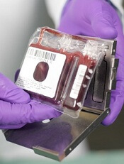
New research suggests that histone deacetylase (HDAC) inhibitors can be used to expand hematopoietic stem cells (HSCs) isolated from cord blood.
Investigators found that valproic acid (VPA), in particular, could greatly expand HSCs from cord blood.
These HSCs expressed markers of pluripotency and were more efficient than conventionally expanded HSCs in repopulating the bone marrow and establishing hematopoiesis in immune-deficient mice.
Pratima Chaurasia, PhD, of the Mount Sinai School of Medicine in New York, and colleagues reported these results in The Journal of Clinical Investigation.
For several decades, investigators have used a variety of strategies to expand the numbers of HSCs isolated from cord blood, with limited success. Evidence has suggested the accumulation of epigenetic modifications influences preservation of stem-cell characteristics in HSC daughter cells.
So Dr Chaurasia’s group tested the effects of HDAC inhibitors on CD34+ cells derived from cord blood. The team primed the cells for 16 hours with cytokines, then treated the cells for 7 days, with or without additional cytokines and in the presence or absence of HDAC inhibitors.
The inhibitors included VPA, scriptaid (SCR), trichostatin A, suberoylanilide hydroxamic acid, CAY10433 (C433), CAY10398, and CAY10603 .
VPA, SCR, and C433 were the most active inhibitors. Treatment with these agents led to a similar percentage of CD34+CD90+ cells—75.2% ± 10.7%, 73.4% ± 13.9%, and 70.1% ± 18.4%, respectively—which was significantly higher than control conditions—16.2% ± 9.2% (P<0.0001).
VPA, SCR, and C433 also generated a greater absolute number of CD34+ and CD34+CD90+ cells per cord blood collection, when compared to control conditions (P≤0.0007). And the inhibitors generated a greater absolute number of CD34+CD90+CD184+ cells (P<0.0001)
The investigators conducted subsequent experiments with VPA only. And they found that VPA was more effective under serum-free conditions and in the presence of cytokines.
Additional experiments revealed that VPA influences the expression of pluripotency genes—SOX2, OCT4, and NANOG. And these genes proved essential for the expansion of CD34+ CD90+ cells.
Lastly, the investigators tested VPA-expanded cells by transplanting them into immune-deficient mice. The cells were more efficient than conventionally expanded cord blood HSCs in repopulating the bone marrow and establishing hematopoietic populations.
Specifically, at 13 to 14 weeks after transplant, VPA-treated cord blood CD34+ cells resulted in a greater degree of human CD45+ cell chimerism—32.2% ± 11.3%—when compared to primary cord blood CD34+ cells—19.4% ± 4.9%—and to cells from control cultures—13.2% ± 6.4% (P=0.006 and P=0.0008, respectively).
The investigators evaluated the self-renewal potential of the expanded grafts by transplanting donor-derived cells from primary recipients into secondary recipients.
After 15 to 16 weeks, the secondary recipients transplanted with VPA-treated cord blood CD34+ cells had achieved the greatest degree of human CD45+ cell chimerism, compared to primary cord blood CD34+ cells and cells from control cultures (P<0.0001).
The team also noted that the VPA-treated cells belonged to multiple hematopoietic lineages. And this pattern was distinct from that observed in mice that had received primary cord blood CD34+ cells or cells from control cultures (P<0.0001).
In addition, the VPA-treated HSCs did not cause hematologic malignancies or teratomas in the mice.
The investigators said these results suggest that cord blood cells can be epigenetically reprogrammed by VPA to generate greater numbers of functional HSCs for transplantation.
In a related commentary, Hal Broxmeyer, PhD, of the Indiana University School of Medicine in Indianapolis, discussed how these findings enhance our understanding of HSC function and could potentially provide clinical benefit.
Credit: NHS
New research suggests that histone deacetylase (HDAC) inhibitors can be used to expand hematopoietic stem cells (HSCs) isolated from cord blood.
Investigators found that valproic acid (VPA), in particular, could greatly expand HSCs from cord blood.
These HSCs expressed markers of pluripotency and were more efficient than conventionally expanded HSCs in repopulating the bone marrow and establishing hematopoiesis in immune-deficient mice.
Pratima Chaurasia, PhD, of the Mount Sinai School of Medicine in New York, and colleagues reported these results in The Journal of Clinical Investigation.
For several decades, investigators have used a variety of strategies to expand the numbers of HSCs isolated from cord blood, with limited success. Evidence has suggested the accumulation of epigenetic modifications influences preservation of stem-cell characteristics in HSC daughter cells.
So Dr Chaurasia’s group tested the effects of HDAC inhibitors on CD34+ cells derived from cord blood. The team primed the cells for 16 hours with cytokines, then treated the cells for 7 days, with or without additional cytokines and in the presence or absence of HDAC inhibitors.
The inhibitors included VPA, scriptaid (SCR), trichostatin A, suberoylanilide hydroxamic acid, CAY10433 (C433), CAY10398, and CAY10603 .
VPA, SCR, and C433 were the most active inhibitors. Treatment with these agents led to a similar percentage of CD34+CD90+ cells—75.2% ± 10.7%, 73.4% ± 13.9%, and 70.1% ± 18.4%, respectively—which was significantly higher than control conditions—16.2% ± 9.2% (P<0.0001).
VPA, SCR, and C433 also generated a greater absolute number of CD34+ and CD34+CD90+ cells per cord blood collection, when compared to control conditions (P≤0.0007). And the inhibitors generated a greater absolute number of CD34+CD90+CD184+ cells (P<0.0001)
The investigators conducted subsequent experiments with VPA only. And they found that VPA was more effective under serum-free conditions and in the presence of cytokines.
Additional experiments revealed that VPA influences the expression of pluripotency genes—SOX2, OCT4, and NANOG. And these genes proved essential for the expansion of CD34+ CD90+ cells.
Lastly, the investigators tested VPA-expanded cells by transplanting them into immune-deficient mice. The cells were more efficient than conventionally expanded cord blood HSCs in repopulating the bone marrow and establishing hematopoietic populations.
Specifically, at 13 to 14 weeks after transplant, VPA-treated cord blood CD34+ cells resulted in a greater degree of human CD45+ cell chimerism—32.2% ± 11.3%—when compared to primary cord blood CD34+ cells—19.4% ± 4.9%—and to cells from control cultures—13.2% ± 6.4% (P=0.006 and P=0.0008, respectively).
The investigators evaluated the self-renewal potential of the expanded grafts by transplanting donor-derived cells from primary recipients into secondary recipients.
After 15 to 16 weeks, the secondary recipients transplanted with VPA-treated cord blood CD34+ cells had achieved the greatest degree of human CD45+ cell chimerism, compared to primary cord blood CD34+ cells and cells from control cultures (P<0.0001).
The team also noted that the VPA-treated cells belonged to multiple hematopoietic lineages. And this pattern was distinct from that observed in mice that had received primary cord blood CD34+ cells or cells from control cultures (P<0.0001).
In addition, the VPA-treated HSCs did not cause hematologic malignancies or teratomas in the mice.
The investigators said these results suggest that cord blood cells can be epigenetically reprogrammed by VPA to generate greater numbers of functional HSCs for transplantation.
In a related commentary, Hal Broxmeyer, PhD, of the Indiana University School of Medicine in Indianapolis, discussed how these findings enhance our understanding of HSC function and could potentially provide clinical benefit.
Credit: NHS
New research suggests that histone deacetylase (HDAC) inhibitors can be used to expand hematopoietic stem cells (HSCs) isolated from cord blood.
Investigators found that valproic acid (VPA), in particular, could greatly expand HSCs from cord blood.
These HSCs expressed markers of pluripotency and were more efficient than conventionally expanded HSCs in repopulating the bone marrow and establishing hematopoiesis in immune-deficient mice.
Pratima Chaurasia, PhD, of the Mount Sinai School of Medicine in New York, and colleagues reported these results in The Journal of Clinical Investigation.
For several decades, investigators have used a variety of strategies to expand the numbers of HSCs isolated from cord blood, with limited success. Evidence has suggested the accumulation of epigenetic modifications influences preservation of stem-cell characteristics in HSC daughter cells.
So Dr Chaurasia’s group tested the effects of HDAC inhibitors on CD34+ cells derived from cord blood. The team primed the cells for 16 hours with cytokines, then treated the cells for 7 days, with or without additional cytokines and in the presence or absence of HDAC inhibitors.
The inhibitors included VPA, scriptaid (SCR), trichostatin A, suberoylanilide hydroxamic acid, CAY10433 (C433), CAY10398, and CAY10603 .
VPA, SCR, and C433 were the most active inhibitors. Treatment with these agents led to a similar percentage of CD34+CD90+ cells—75.2% ± 10.7%, 73.4% ± 13.9%, and 70.1% ± 18.4%, respectively—which was significantly higher than control conditions—16.2% ± 9.2% (P<0.0001).
VPA, SCR, and C433 also generated a greater absolute number of CD34+ and CD34+CD90+ cells per cord blood collection, when compared to control conditions (P≤0.0007). And the inhibitors generated a greater absolute number of CD34+CD90+CD184+ cells (P<0.0001)
The investigators conducted subsequent experiments with VPA only. And they found that VPA was more effective under serum-free conditions and in the presence of cytokines.
Additional experiments revealed that VPA influences the expression of pluripotency genes—SOX2, OCT4, and NANOG. And these genes proved essential for the expansion of CD34+ CD90+ cells.
Lastly, the investigators tested VPA-expanded cells by transplanting them into immune-deficient mice. The cells were more efficient than conventionally expanded cord blood HSCs in repopulating the bone marrow and establishing hematopoietic populations.
Specifically, at 13 to 14 weeks after transplant, VPA-treated cord blood CD34+ cells resulted in a greater degree of human CD45+ cell chimerism—32.2% ± 11.3%—when compared to primary cord blood CD34+ cells—19.4% ± 4.9%—and to cells from control cultures—13.2% ± 6.4% (P=0.006 and P=0.0008, respectively).
The investigators evaluated the self-renewal potential of the expanded grafts by transplanting donor-derived cells from primary recipients into secondary recipients.
After 15 to 16 weeks, the secondary recipients transplanted with VPA-treated cord blood CD34+ cells had achieved the greatest degree of human CD45+ cell chimerism, compared to primary cord blood CD34+ cells and cells from control cultures (P<0.0001).
The team also noted that the VPA-treated cells belonged to multiple hematopoietic lineages. And this pattern was distinct from that observed in mice that had received primary cord blood CD34+ cells or cells from control cultures (P<0.0001).
In addition, the VPA-treated HSCs did not cause hematologic malignancies or teratomas in the mice.
The investigators said these results suggest that cord blood cells can be epigenetically reprogrammed by VPA to generate greater numbers of functional HSCs for transplantation.
In a related commentary, Hal Broxmeyer, PhD, of the Indiana University School of Medicine in Indianapolis, discussed how these findings enhance our understanding of HSC function and could potentially provide clinical benefit.
Tracking protein movement to improve patient monitoring, drug development

Credit: Virginia Tech
A novel technique that can detect the subcellular location of a protein may help improve the study of therapies for cancer and other diseases, according to a paper published in Chemical Science.
“Modulation of protein transport inside a cell is practiced as an important therapeutic approach for cancer treatment,” explained Chang Lu, PhD, of Virginia Tech in Blacksburg.
“The subcellular location of a target protein can also serve as a useful read-out for high-content screening of cancer drugs.”
With that in mind, Dr Lu and his colleagues set out to develop a simple and accessible protein detection method that can rapidly screen a large cell population and offers single-cell resolution.
Dr Lu noted that such techniques have been seriously lacking. For instance, fluorescence microscopy can only be used to analyze a limited number of cells.
And data collected by subcellular fractionation only reflects the average properties of the cell populations without revealing the heterogeneity that often exists among cells that seem identical.
Dr Lu and his colleagues had previously made some progress in screening cell populations using an electroporation-based technique, but it did not allow for the examination of native proteins and primary cells isolated from animals and from patients.
Their new work uses a method that “incorporates selective chemical release of cytosolic proteins with a standard procedure for fluorescent labeling of the protein,” Dr Lu said.
This simple tweak to the conventional cell-staining process allowed the researchers to pinpoint the subcellular location of the protein by measuring the amount of the residual protein after release. Using a flow cytometer, the speed of such measurement could reach 10,000 to 100,000 cells per second.
A key ingredient for the team’s process is saponin, an amphipathic glycoside. Saponin dissolves cholesterol and permeates the plasma membrane to allow protein release. And it “shows minimal effects on the state of the cell,” Dr Lu said.
Credit: Virginia Tech
A novel technique that can detect the subcellular location of a protein may help improve the study of therapies for cancer and other diseases, according to a paper published in Chemical Science.
“Modulation of protein transport inside a cell is practiced as an important therapeutic approach for cancer treatment,” explained Chang Lu, PhD, of Virginia Tech in Blacksburg.
“The subcellular location of a target protein can also serve as a useful read-out for high-content screening of cancer drugs.”
With that in mind, Dr Lu and his colleagues set out to develop a simple and accessible protein detection method that can rapidly screen a large cell population and offers single-cell resolution.
Dr Lu noted that such techniques have been seriously lacking. For instance, fluorescence microscopy can only be used to analyze a limited number of cells.
And data collected by subcellular fractionation only reflects the average properties of the cell populations without revealing the heterogeneity that often exists among cells that seem identical.
Dr Lu and his colleagues had previously made some progress in screening cell populations using an electroporation-based technique, but it did not allow for the examination of native proteins and primary cells isolated from animals and from patients.
Their new work uses a method that “incorporates selective chemical release of cytosolic proteins with a standard procedure for fluorescent labeling of the protein,” Dr Lu said.
This simple tweak to the conventional cell-staining process allowed the researchers to pinpoint the subcellular location of the protein by measuring the amount of the residual protein after release. Using a flow cytometer, the speed of such measurement could reach 10,000 to 100,000 cells per second.
A key ingredient for the team’s process is saponin, an amphipathic glycoside. Saponin dissolves cholesterol and permeates the plasma membrane to allow protein release. And it “shows minimal effects on the state of the cell,” Dr Lu said.
Credit: Virginia Tech
A novel technique that can detect the subcellular location of a protein may help improve the study of therapies for cancer and other diseases, according to a paper published in Chemical Science.
“Modulation of protein transport inside a cell is practiced as an important therapeutic approach for cancer treatment,” explained Chang Lu, PhD, of Virginia Tech in Blacksburg.
“The subcellular location of a target protein can also serve as a useful read-out for high-content screening of cancer drugs.”
With that in mind, Dr Lu and his colleagues set out to develop a simple and accessible protein detection method that can rapidly screen a large cell population and offers single-cell resolution.
Dr Lu noted that such techniques have been seriously lacking. For instance, fluorescence microscopy can only be used to analyze a limited number of cells.
And data collected by subcellular fractionation only reflects the average properties of the cell populations without revealing the heterogeneity that often exists among cells that seem identical.
Dr Lu and his colleagues had previously made some progress in screening cell populations using an electroporation-based technique, but it did not allow for the examination of native proteins and primary cells isolated from animals and from patients.
Their new work uses a method that “incorporates selective chemical release of cytosolic proteins with a standard procedure for fluorescent labeling of the protein,” Dr Lu said.
This simple tweak to the conventional cell-staining process allowed the researchers to pinpoint the subcellular location of the protein by measuring the amount of the residual protein after release. Using a flow cytometer, the speed of such measurement could reach 10,000 to 100,000 cells per second.
A key ingredient for the team’s process is saponin, an amphipathic glycoside. Saponin dissolves cholesterol and permeates the plasma membrane to allow protein release. And it “shows minimal effects on the state of the cell,” Dr Lu said.
New insight into PTEN’s role in cancers
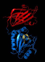
Researchers say they’ve uncovered new details that help explain how the PTEN gene exerts its anticancer effects and how PTEN loss or alteration can set cells on a cancerous course.
The team’s study, published in Cell, reveals that PTEN loss and PTEN mutations are not synonymous.
This discovery provides additional insight into basic tumor biology and offers a potential new direction in the pursuit of cancer therapies, according to the researchers.
“By characterizing the ways that 2 specific PTEN mutations regulate the tumor suppressor function of the normal PTEN protein, our findings suggest that different PTEN mutations contribute to tumorigenesis by regulating different aspects of PTEN biology,” said study author Pier Paolo Pandolfi, MD, PhD, of Beth Israel Deaconess Medical Center in Boston.
“It has been suggested that cancer patients harboring mutations in PTEN had poorer outcomes than cancer patients with PTEN loss. Now, using mouse modeling, we are able to demonstrate that this is indeed the case. Because PTEN mutations are extremely frequent in various types of tumors, this discovery could help pave the way for a new level of personalized cancer treatment.”
Several of the proteins that PTEN acts upon, both lipids and proteins, are known to promote cancer when bound to a phosphate. Consequently, when PTEN removes their phosphates, it is acting as a tumor suppressor to prevent cancer. When PTEN is mutated, it loses this suppressive ability, and the cancer-promoting proteins are left intact and uninhibited.
This new study showed that the PTEN mutant protein is not only functionally impaired, it also acquires the ability to affect the function of normal PTEN proteins, thereby gaining a pro-tumorigenic function. But the researchers also wanted to determine the difference between PTEN mutation and PTEN loss.
“We wanted to know, would outcomes differ in cases when PTEN was not expressed, compared with cases when PTEN was expressed but encoded a mutation within its sequence?” said study author Antonella Papa, PhD, an investigator in the Pandolfi lab.
To find out, the team created several genetically modified strains of mice to mimic the PTEN mutations found in human cancer patients.
“All mice [and humans] have 2 copies of the PTEN gene,” Dr Papa explained. “The genetically modified mice in our study had 1 copy of the PTEN gene that contained a cancer-associated mutation [either PTENC124S or PTENG129E] and 1 normal copy of PTEN. Other mice in the study had only 1 copy of the normal PTEN gene, and the second copy was removed.”
The researchers found that the mice with a single mutated copy of PTEN were more tumor-prone than the mice with a deleted copy of PTEN. They also discovered that the mutated protein that was produced by PTENC124S or PTENG129E was binding to and inhibiting the PTEN protein made from the normal copy of the PTEN gene.
“This was very surprising, as we were expecting a reduction in tumorigenesis,” Dr Papa said. “Instead, mechanistically, we found that PTEN exists as a dimer, and, in this new conformation, the mutated protein prevents the normal protein from functioning.”
At the molecular level, this generates an increased activation of a PTEN target—the protein Akt—which is what leads to the augmented tumorigenesis in the mice. Akt is part of a signaling pathway that regulates cell growth, division and metabolism.
When PTEN is prevented from inhibiting Akt, the pathway becomes overactive. As a result, targeting Akt and its pathway may be an effective treatment strategy for patients with PTEN mutations, the researchers said, adding that inhibitors to affect this pathway are currently being tested and developed.
“This defines a new working model for the function and regulation of PTEN and tells us that PTEN mutational status can be used to determine which cancer patients might benefit from earlier and more radical therapeutic interventions and, ultimately, better prognosis,” Dr Pandolfi said.
“Our findings may help to better identify and stratify patients and their response to treatment based on the different genetic alterations found in the PTEN gene. Importantly, our study shows that cancer therapy should be tailored on the basis of the very specific type of mutations that the tumor harbors.”
“This adds a new layer of complexity but also a new opportunity for precision medicine. I would say that, based on these thorough genetic analyses, this story represents the ultimate example of why personalized cancer medicine is so urgently needed.”
Researchers say they’ve uncovered new details that help explain how the PTEN gene exerts its anticancer effects and how PTEN loss or alteration can set cells on a cancerous course.
The team’s study, published in Cell, reveals that PTEN loss and PTEN mutations are not synonymous.
This discovery provides additional insight into basic tumor biology and offers a potential new direction in the pursuit of cancer therapies, according to the researchers.
“By characterizing the ways that 2 specific PTEN mutations regulate the tumor suppressor function of the normal PTEN protein, our findings suggest that different PTEN mutations contribute to tumorigenesis by regulating different aspects of PTEN biology,” said study author Pier Paolo Pandolfi, MD, PhD, of Beth Israel Deaconess Medical Center in Boston.
“It has been suggested that cancer patients harboring mutations in PTEN had poorer outcomes than cancer patients with PTEN loss. Now, using mouse modeling, we are able to demonstrate that this is indeed the case. Because PTEN mutations are extremely frequent in various types of tumors, this discovery could help pave the way for a new level of personalized cancer treatment.”
Several of the proteins that PTEN acts upon, both lipids and proteins, are known to promote cancer when bound to a phosphate. Consequently, when PTEN removes their phosphates, it is acting as a tumor suppressor to prevent cancer. When PTEN is mutated, it loses this suppressive ability, and the cancer-promoting proteins are left intact and uninhibited.
This new study showed that the PTEN mutant protein is not only functionally impaired, it also acquires the ability to affect the function of normal PTEN proteins, thereby gaining a pro-tumorigenic function. But the researchers also wanted to determine the difference between PTEN mutation and PTEN loss.
“We wanted to know, would outcomes differ in cases when PTEN was not expressed, compared with cases when PTEN was expressed but encoded a mutation within its sequence?” said study author Antonella Papa, PhD, an investigator in the Pandolfi lab.
To find out, the team created several genetically modified strains of mice to mimic the PTEN mutations found in human cancer patients.
“All mice [and humans] have 2 copies of the PTEN gene,” Dr Papa explained. “The genetically modified mice in our study had 1 copy of the PTEN gene that contained a cancer-associated mutation [either PTENC124S or PTENG129E] and 1 normal copy of PTEN. Other mice in the study had only 1 copy of the normal PTEN gene, and the second copy was removed.”
The researchers found that the mice with a single mutated copy of PTEN were more tumor-prone than the mice with a deleted copy of PTEN. They also discovered that the mutated protein that was produced by PTENC124S or PTENG129E was binding to and inhibiting the PTEN protein made from the normal copy of the PTEN gene.
“This was very surprising, as we were expecting a reduction in tumorigenesis,” Dr Papa said. “Instead, mechanistically, we found that PTEN exists as a dimer, and, in this new conformation, the mutated protein prevents the normal protein from functioning.”
At the molecular level, this generates an increased activation of a PTEN target—the protein Akt—which is what leads to the augmented tumorigenesis in the mice. Akt is part of a signaling pathway that regulates cell growth, division and metabolism.
When PTEN is prevented from inhibiting Akt, the pathway becomes overactive. As a result, targeting Akt and its pathway may be an effective treatment strategy for patients with PTEN mutations, the researchers said, adding that inhibitors to affect this pathway are currently being tested and developed.
“This defines a new working model for the function and regulation of PTEN and tells us that PTEN mutational status can be used to determine which cancer patients might benefit from earlier and more radical therapeutic interventions and, ultimately, better prognosis,” Dr Pandolfi said.
“Our findings may help to better identify and stratify patients and their response to treatment based on the different genetic alterations found in the PTEN gene. Importantly, our study shows that cancer therapy should be tailored on the basis of the very specific type of mutations that the tumor harbors.”
“This adds a new layer of complexity but also a new opportunity for precision medicine. I would say that, based on these thorough genetic analyses, this story represents the ultimate example of why personalized cancer medicine is so urgently needed.”
Researchers say they’ve uncovered new details that help explain how the PTEN gene exerts its anticancer effects and how PTEN loss or alteration can set cells on a cancerous course.
The team’s study, published in Cell, reveals that PTEN loss and PTEN mutations are not synonymous.
This discovery provides additional insight into basic tumor biology and offers a potential new direction in the pursuit of cancer therapies, according to the researchers.
“By characterizing the ways that 2 specific PTEN mutations regulate the tumor suppressor function of the normal PTEN protein, our findings suggest that different PTEN mutations contribute to tumorigenesis by regulating different aspects of PTEN biology,” said study author Pier Paolo Pandolfi, MD, PhD, of Beth Israel Deaconess Medical Center in Boston.
“It has been suggested that cancer patients harboring mutations in PTEN had poorer outcomes than cancer patients with PTEN loss. Now, using mouse modeling, we are able to demonstrate that this is indeed the case. Because PTEN mutations are extremely frequent in various types of tumors, this discovery could help pave the way for a new level of personalized cancer treatment.”
Several of the proteins that PTEN acts upon, both lipids and proteins, are known to promote cancer when bound to a phosphate. Consequently, when PTEN removes their phosphates, it is acting as a tumor suppressor to prevent cancer. When PTEN is mutated, it loses this suppressive ability, and the cancer-promoting proteins are left intact and uninhibited.
This new study showed that the PTEN mutant protein is not only functionally impaired, it also acquires the ability to affect the function of normal PTEN proteins, thereby gaining a pro-tumorigenic function. But the researchers also wanted to determine the difference between PTEN mutation and PTEN loss.
“We wanted to know, would outcomes differ in cases when PTEN was not expressed, compared with cases when PTEN was expressed but encoded a mutation within its sequence?” said study author Antonella Papa, PhD, an investigator in the Pandolfi lab.
To find out, the team created several genetically modified strains of mice to mimic the PTEN mutations found in human cancer patients.
“All mice [and humans] have 2 copies of the PTEN gene,” Dr Papa explained. “The genetically modified mice in our study had 1 copy of the PTEN gene that contained a cancer-associated mutation [either PTENC124S or PTENG129E] and 1 normal copy of PTEN. Other mice in the study had only 1 copy of the normal PTEN gene, and the second copy was removed.”
The researchers found that the mice with a single mutated copy of PTEN were more tumor-prone than the mice with a deleted copy of PTEN. They also discovered that the mutated protein that was produced by PTENC124S or PTENG129E was binding to and inhibiting the PTEN protein made from the normal copy of the PTEN gene.
“This was very surprising, as we were expecting a reduction in tumorigenesis,” Dr Papa said. “Instead, mechanistically, we found that PTEN exists as a dimer, and, in this new conformation, the mutated protein prevents the normal protein from functioning.”
At the molecular level, this generates an increased activation of a PTEN target—the protein Akt—which is what leads to the augmented tumorigenesis in the mice. Akt is part of a signaling pathway that regulates cell growth, division and metabolism.
When PTEN is prevented from inhibiting Akt, the pathway becomes overactive. As a result, targeting Akt and its pathway may be an effective treatment strategy for patients with PTEN mutations, the researchers said, adding that inhibitors to affect this pathway are currently being tested and developed.
“This defines a new working model for the function and regulation of PTEN and tells us that PTEN mutational status can be used to determine which cancer patients might benefit from earlier and more radical therapeutic interventions and, ultimately, better prognosis,” Dr Pandolfi said.
“Our findings may help to better identify and stratify patients and their response to treatment based on the different genetic alterations found in the PTEN gene. Importantly, our study shows that cancer therapy should be tailored on the basis of the very specific type of mutations that the tumor harbors.”
“This adds a new layer of complexity but also a new opportunity for precision medicine. I would say that, based on these thorough genetic analyses, this story represents the ultimate example of why personalized cancer medicine is so urgently needed.”