User login
INR test strips linked to bleeding, deaths

Alere Inc., has initiated a Class 1 recall in the US of the Alere INRatio2 PT/INR Professional Test Strips (PN 99008G2).
The strips are used by healthcare professionals to determine the international normalized ratio (INR) in fresh capillary whole blood to monitor the effect of warfarin on clotting time.
Alere has received reports of patients who had a therapeutic or near therapeutic INR with the test strips but a significantly higher INR in tests performed by a central lab.
This error has been linked to 9 reports of serious adverse events, 3 of which described bleeding associated with patient deaths.
So the company has issued a recall of the Alere INRatio2 PT/INR Professional Test Strips (PN 99008G2). This recall does not include the Alere INRatio PT/INR Test Strip (PN 100071), which is used by patients for home INR monitoring.
Alere has not determined the root cause of the error but is concerned that the test strips may continue to report inaccurately low INR results. In the reports, the test strip results were between 3.1 and 12.2 INR units lower than the lab results.
Customers should stop using the Alere INRatio2 PT/INR Professional Test Strips immediately and return unused product to the company.
Alere will transition customers from the current Alere INRatio2 PT/INR Professional Test Strip to the Alere INRatio PT/INR Test Strip (PN 100139).
Alere said it has reported this issue to the US Food and Drug Administration and is conducting a thorough investigation into the adverse events.
Customers with questions about this recall and those who require replacement product can contact Alere at 844-292-5373. For additional information on the recall, visit www.inr-care.com.
Any adverse events or quality problems related to use of the test strips can be reported to the FDA’s MedWatch Adverse Event Reporting Program. 
Alere Inc., has initiated a Class 1 recall in the US of the Alere INRatio2 PT/INR Professional Test Strips (PN 99008G2).
The strips are used by healthcare professionals to determine the international normalized ratio (INR) in fresh capillary whole blood to monitor the effect of warfarin on clotting time.
Alere has received reports of patients who had a therapeutic or near therapeutic INR with the test strips but a significantly higher INR in tests performed by a central lab.
This error has been linked to 9 reports of serious adverse events, 3 of which described bleeding associated with patient deaths.
So the company has issued a recall of the Alere INRatio2 PT/INR Professional Test Strips (PN 99008G2). This recall does not include the Alere INRatio PT/INR Test Strip (PN 100071), which is used by patients for home INR monitoring.
Alere has not determined the root cause of the error but is concerned that the test strips may continue to report inaccurately low INR results. In the reports, the test strip results were between 3.1 and 12.2 INR units lower than the lab results.
Customers should stop using the Alere INRatio2 PT/INR Professional Test Strips immediately and return unused product to the company.
Alere will transition customers from the current Alere INRatio2 PT/INR Professional Test Strip to the Alere INRatio PT/INR Test Strip (PN 100139).
Alere said it has reported this issue to the US Food and Drug Administration and is conducting a thorough investigation into the adverse events.
Customers with questions about this recall and those who require replacement product can contact Alere at 844-292-5373. For additional information on the recall, visit www.inr-care.com.
Any adverse events or quality problems related to use of the test strips can be reported to the FDA’s MedWatch Adverse Event Reporting Program.
Alere Inc., has initiated a Class 1 recall in the US of the Alere INRatio2 PT/INR Professional Test Strips (PN 99008G2).
The strips are used by healthcare professionals to determine the international normalized ratio (INR) in fresh capillary whole blood to monitor the effect of warfarin on clotting time.
Alere has received reports of patients who had a therapeutic or near therapeutic INR with the test strips but a significantly higher INR in tests performed by a central lab.
This error has been linked to 9 reports of serious adverse events, 3 of which described bleeding associated with patient deaths.
So the company has issued a recall of the Alere INRatio2 PT/INR Professional Test Strips (PN 99008G2). This recall does not include the Alere INRatio PT/INR Test Strip (PN 100071), which is used by patients for home INR monitoring.
Alere has not determined the root cause of the error but is concerned that the test strips may continue to report inaccurately low INR results. In the reports, the test strip results were between 3.1 and 12.2 INR units lower than the lab results.
Customers should stop using the Alere INRatio2 PT/INR Professional Test Strips immediately and return unused product to the company.
Alere will transition customers from the current Alere INRatio2 PT/INR Professional Test Strip to the Alere INRatio PT/INR Test Strip (PN 100139).
Alere said it has reported this issue to the US Food and Drug Administration and is conducting a thorough investigation into the adverse events.
Customers with questions about this recall and those who require replacement product can contact Alere at 844-292-5373. For additional information on the recall, visit www.inr-care.com.
Any adverse events or quality problems related to use of the test strips can be reported to the FDA’s MedWatch Adverse Event Reporting Program.
Eculizumab gets full FDA approval for aHUS

Credit: Globovision
The US Food and Drug Administration (FDA) has granted full approval for eculizumab (Soliris) to treat adult and pediatric patients with atypical
hemolytic uremic syndrome (aHUS).
The drug received accelerated approval for this indication in 2011.
Now, eculizumab has received full FDA approval based on the fulfillment of post-marketing requirements, including the submission of data from 2 additional prospective trials of eculizumab in patients with aHUS.
The revised eculizumab label now includes results with 2 years of ongoing treatment in aHUS patients and data on the use of eculizumab prior to supportive care with plasma or plasma exchange in prospective clinical trials.
The drug’s label also includes a boxed warning informing readers that life-threatening and fatal meningococcal infections have occurred in patients treated with eculizumab.
About aHUS and eculizumab
aHUS is a chronic, life-threatening disease in which a genetic deficiency in one or more complement regulatory genes causes chronic, uncontrolled complement activation. This results in complement-mediated thrombotic microangiopathy (TMA), the formation of blood clots in small blood vessels throughout the body.
Permanent, uncontrolled complement activation in aHUS causes a life-long risk for TMA, which leads to sudden and life-threatening damage to the kidney, brain, heart, and other vital organs, as well as premature death. Complement-mediated TMA also causes thrombocytopenia and hemolysis.
Eculizumab is a first-in-class terminal complement inhibitor indicated to inhibit complement-mediated TMA. The drug’s effectiveness in aHUS is based on its effects on TMA and renal function.
Eculizumab received accelerated FDA approval to treat aHUS in September 2011. The FDA granted this approval based on the results of 2 trials that suggested the drug was likely to provide a clinical benefit.
To achieve traditional FDA approval, the drug’s developer, Alexion Pharmaceuticals, was required to submit additional data that confirm the drug provides a clinical benefit.
To that end, the Clinical Studies section (Section 14.2) of the revised eculizumab prescribing information now contains results from 4 prospective, single-arm studies in patients with aHUS.
This includes updated data from the first 2 trials, as well as data from 2 new trials, 1 in pediatric patients with aHUS and the other in adolescents and adults with aHUS.
For details on these trials, see the full prescribing information.
Credit: Globovision
The US Food and Drug Administration (FDA) has granted full approval for eculizumab (Soliris) to treat adult and pediatric patients with atypical
hemolytic uremic syndrome (aHUS).
The drug received accelerated approval for this indication in 2011.
Now, eculizumab has received full FDA approval based on the fulfillment of post-marketing requirements, including the submission of data from 2 additional prospective trials of eculizumab in patients with aHUS.
The revised eculizumab label now includes results with 2 years of ongoing treatment in aHUS patients and data on the use of eculizumab prior to supportive care with plasma or plasma exchange in prospective clinical trials.
The drug’s label also includes a boxed warning informing readers that life-threatening and fatal meningococcal infections have occurred in patients treated with eculizumab.
About aHUS and eculizumab
aHUS is a chronic, life-threatening disease in which a genetic deficiency in one or more complement regulatory genes causes chronic, uncontrolled complement activation. This results in complement-mediated thrombotic microangiopathy (TMA), the formation of blood clots in small blood vessels throughout the body.
Permanent, uncontrolled complement activation in aHUS causes a life-long risk for TMA, which leads to sudden and life-threatening damage to the kidney, brain, heart, and other vital organs, as well as premature death. Complement-mediated TMA also causes thrombocytopenia and hemolysis.
Eculizumab is a first-in-class terminal complement inhibitor indicated to inhibit complement-mediated TMA. The drug’s effectiveness in aHUS is based on its effects on TMA and renal function.
Eculizumab received accelerated FDA approval to treat aHUS in September 2011. The FDA granted this approval based on the results of 2 trials that suggested the drug was likely to provide a clinical benefit.
To achieve traditional FDA approval, the drug’s developer, Alexion Pharmaceuticals, was required to submit additional data that confirm the drug provides a clinical benefit.
To that end, the Clinical Studies section (Section 14.2) of the revised eculizumab prescribing information now contains results from 4 prospective, single-arm studies in patients with aHUS.
This includes updated data from the first 2 trials, as well as data from 2 new trials, 1 in pediatric patients with aHUS and the other in adolescents and adults with aHUS.
For details on these trials, see the full prescribing information.
Credit: Globovision
The US Food and Drug Administration (FDA) has granted full approval for eculizumab (Soliris) to treat adult and pediatric patients with atypical
hemolytic uremic syndrome (aHUS).
The drug received accelerated approval for this indication in 2011.
Now, eculizumab has received full FDA approval based on the fulfillment of post-marketing requirements, including the submission of data from 2 additional prospective trials of eculizumab in patients with aHUS.
The revised eculizumab label now includes results with 2 years of ongoing treatment in aHUS patients and data on the use of eculizumab prior to supportive care with plasma or plasma exchange in prospective clinical trials.
The drug’s label also includes a boxed warning informing readers that life-threatening and fatal meningococcal infections have occurred in patients treated with eculizumab.
About aHUS and eculizumab
aHUS is a chronic, life-threatening disease in which a genetic deficiency in one or more complement regulatory genes causes chronic, uncontrolled complement activation. This results in complement-mediated thrombotic microangiopathy (TMA), the formation of blood clots in small blood vessels throughout the body.
Permanent, uncontrolled complement activation in aHUS causes a life-long risk for TMA, which leads to sudden and life-threatening damage to the kidney, brain, heart, and other vital organs, as well as premature death. Complement-mediated TMA also causes thrombocytopenia and hemolysis.
Eculizumab is a first-in-class terminal complement inhibitor indicated to inhibit complement-mediated TMA. The drug’s effectiveness in aHUS is based on its effects on TMA and renal function.
Eculizumab received accelerated FDA approval to treat aHUS in September 2011. The FDA granted this approval based on the results of 2 trials that suggested the drug was likely to provide a clinical benefit.
To achieve traditional FDA approval, the drug’s developer, Alexion Pharmaceuticals, was required to submit additional data that confirm the drug provides a clinical benefit.
To that end, the Clinical Studies section (Section 14.2) of the revised eculizumab prescribing information now contains results from 4 prospective, single-arm studies in patients with aHUS.
This includes updated data from the first 2 trials, as well as data from 2 new trials, 1 in pediatric patients with aHUS and the other in adolescents and adults with aHUS.
For details on these trials, see the full prescribing information.
Artificial bone marrow seems just like the real thing

Credit: James Weaver,
Harvard’s Wyss Institute
Researchers have created a device that reproduces the structure, function, and cellular make-up of bone marrow, according to a paper published
in Nature Methods.
The device, dubbed “bone marrow on a chip,” could serve as a new tool for testing the effects of radiation and other toxic agents on whole bone marrow.
The researchers believe this bone marrow on a chip could provide an alternative to animal testing, although the device itself was generated in mice.
The team also thinks that, in the future, the engineered bone marrow could be used to maintain a cancer patient’s own marrow temporarily during radiation or high-dose chemotherapy.
In an initial test, the engineered bone marrow withered in response to radiation, just as natural bone marrow does. And, as in natural marrow, granulocyte colony-stimulating factor conferred a protective effect on the engineered marrow.
“Bone marrow is an incredibly complex organ that is responsible for producing all of the blood cell types in our body, and our bone marrow chips are able to recapitulate this complexity in its entirety and maintain it in a functional form in vitro,” said study author Donald Ingber, MD, PhD, of the Wyss Institute for Biologically Inspired Engineering at Harvard University in Boston.
Dr Ingber leads an effort to develop human organs on chips—small microfluidic devices that mimic the physiology of living organs. To build these devices, researchers combine multiple types of cells from an organ on a plastic microfluidic device, while steadily supplying nutrients, removing waste, and applying mechanical forces the tissues would face in the body.
But bone marrow is so complex that Dr Ingber and his colleagues needed a new approach to mimic organ function. Rather than trying to reproduce such a complex structure cell by cell, the team used mice.
Specifically, the researchers packed dried bone powder into an open, ring-shaped mold the size of a coin battery and implanted the mold under the skin on the animal’s back.
After 8 weeks, the team surgically removed the disk-shaped bone that had formed in the mold and examined it with a specialized CAT scanner. The scan showed a honeycomb-like structure that looked identical to natural trabecular bone.
The marrow looked like the real thing as well. When the researchers stained the tissue and examined it under a microscope, the marrow was packed with blood cells, just like marrow from a living mouse.
And when the team sorted the bone marrow cells by type and tallied their numbers, the mix of different types of blood and immune cells in the engineered bone marrow was identical to that in a mouse thighbone.
To sustain the engineered bone marrow outside of a living animal, the researchers surgically removed the engineered bone from mice, then placed it in a microfluidic device that mimics the circulation the tissue would experience in the body.
Marrow in the device remained healthy for up to 1 week. This is typically long enough to test the toxicity and effectiveness of a new drug, the team said.
The device also passed an initial test of its drug-testing capabilities. Like marrow from live mice, this engineered marrow was susceptible to radiation, but granulocyte colony-stimulating factor conferred a protective effect.
The researchers believe that, in the future, they could potentially grow human bone marrow in immune-deficient mice. And their bone marrow on a chip could generate blood cells, which could circulate in an artificial circulatory system to supply a network of other organs on chips.
Credit: James Weaver,
Harvard’s Wyss Institute
Researchers have created a device that reproduces the structure, function, and cellular make-up of bone marrow, according to a paper published
in Nature Methods.
The device, dubbed “bone marrow on a chip,” could serve as a new tool for testing the effects of radiation and other toxic agents on whole bone marrow.
The researchers believe this bone marrow on a chip could provide an alternative to animal testing, although the device itself was generated in mice.
The team also thinks that, in the future, the engineered bone marrow could be used to maintain a cancer patient’s own marrow temporarily during radiation or high-dose chemotherapy.
In an initial test, the engineered bone marrow withered in response to radiation, just as natural bone marrow does. And, as in natural marrow, granulocyte colony-stimulating factor conferred a protective effect on the engineered marrow.
“Bone marrow is an incredibly complex organ that is responsible for producing all of the blood cell types in our body, and our bone marrow chips are able to recapitulate this complexity in its entirety and maintain it in a functional form in vitro,” said study author Donald Ingber, MD, PhD, of the Wyss Institute for Biologically Inspired Engineering at Harvard University in Boston.
Dr Ingber leads an effort to develop human organs on chips—small microfluidic devices that mimic the physiology of living organs. To build these devices, researchers combine multiple types of cells from an organ on a plastic microfluidic device, while steadily supplying nutrients, removing waste, and applying mechanical forces the tissues would face in the body.
But bone marrow is so complex that Dr Ingber and his colleagues needed a new approach to mimic organ function. Rather than trying to reproduce such a complex structure cell by cell, the team used mice.
Specifically, the researchers packed dried bone powder into an open, ring-shaped mold the size of a coin battery and implanted the mold under the skin on the animal’s back.
After 8 weeks, the team surgically removed the disk-shaped bone that had formed in the mold and examined it with a specialized CAT scanner. The scan showed a honeycomb-like structure that looked identical to natural trabecular bone.
The marrow looked like the real thing as well. When the researchers stained the tissue and examined it under a microscope, the marrow was packed with blood cells, just like marrow from a living mouse.
And when the team sorted the bone marrow cells by type and tallied their numbers, the mix of different types of blood and immune cells in the engineered bone marrow was identical to that in a mouse thighbone.
To sustain the engineered bone marrow outside of a living animal, the researchers surgically removed the engineered bone from mice, then placed it in a microfluidic device that mimics the circulation the tissue would experience in the body.
Marrow in the device remained healthy for up to 1 week. This is typically long enough to test the toxicity and effectiveness of a new drug, the team said.
The device also passed an initial test of its drug-testing capabilities. Like marrow from live mice, this engineered marrow was susceptible to radiation, but granulocyte colony-stimulating factor conferred a protective effect.
The researchers believe that, in the future, they could potentially grow human bone marrow in immune-deficient mice. And their bone marrow on a chip could generate blood cells, which could circulate in an artificial circulatory system to supply a network of other organs on chips.
Credit: James Weaver,
Harvard’s Wyss Institute
Researchers have created a device that reproduces the structure, function, and cellular make-up of bone marrow, according to a paper published
in Nature Methods.
The device, dubbed “bone marrow on a chip,” could serve as a new tool for testing the effects of radiation and other toxic agents on whole bone marrow.
The researchers believe this bone marrow on a chip could provide an alternative to animal testing, although the device itself was generated in mice.
The team also thinks that, in the future, the engineered bone marrow could be used to maintain a cancer patient’s own marrow temporarily during radiation or high-dose chemotherapy.
In an initial test, the engineered bone marrow withered in response to radiation, just as natural bone marrow does. And, as in natural marrow, granulocyte colony-stimulating factor conferred a protective effect on the engineered marrow.
“Bone marrow is an incredibly complex organ that is responsible for producing all of the blood cell types in our body, and our bone marrow chips are able to recapitulate this complexity in its entirety and maintain it in a functional form in vitro,” said study author Donald Ingber, MD, PhD, of the Wyss Institute for Biologically Inspired Engineering at Harvard University in Boston.
Dr Ingber leads an effort to develop human organs on chips—small microfluidic devices that mimic the physiology of living organs. To build these devices, researchers combine multiple types of cells from an organ on a plastic microfluidic device, while steadily supplying nutrients, removing waste, and applying mechanical forces the tissues would face in the body.
But bone marrow is so complex that Dr Ingber and his colleagues needed a new approach to mimic organ function. Rather than trying to reproduce such a complex structure cell by cell, the team used mice.
Specifically, the researchers packed dried bone powder into an open, ring-shaped mold the size of a coin battery and implanted the mold under the skin on the animal’s back.
After 8 weeks, the team surgically removed the disk-shaped bone that had formed in the mold and examined it with a specialized CAT scanner. The scan showed a honeycomb-like structure that looked identical to natural trabecular bone.
The marrow looked like the real thing as well. When the researchers stained the tissue and examined it under a microscope, the marrow was packed with blood cells, just like marrow from a living mouse.
And when the team sorted the bone marrow cells by type and tallied their numbers, the mix of different types of blood and immune cells in the engineered bone marrow was identical to that in a mouse thighbone.
To sustain the engineered bone marrow outside of a living animal, the researchers surgically removed the engineered bone from mice, then placed it in a microfluidic device that mimics the circulation the tissue would experience in the body.
Marrow in the device remained healthy for up to 1 week. This is typically long enough to test the toxicity and effectiveness of a new drug, the team said.
The device also passed an initial test of its drug-testing capabilities. Like marrow from live mice, this engineered marrow was susceptible to radiation, but granulocyte colony-stimulating factor conferred a protective effect.
The researchers believe that, in the future, they could potentially grow human bone marrow in immune-deficient mice. And their bone marrow on a chip could generate blood cells, which could circulate in an artificial circulatory system to supply a network of other organs on chips.
Team describes protein’s antimyeloma effects

Researchers say they’ve discovered how a lack of the protein adiponectin promotes disease progression in patients with multiple myeloma (MM).
Obesity is known to increase the risk of developing MM, and obese individuals have lower-than-normal levels of adiponectin.
So the researchers set out to characterize the relationship between adiponectin and MM.
They found evidence to suggest the protein induces apoptosis in MM cells by suppressing lipogenesis. And this points to new treatment avenues for MM.
Edward Medina, MD, PhD, of University of Texas Health Science Center at San Antonio, and his colleagues described this research in Leukemia.
The team noted that adiponectin plays several roles in preserving health, including killing cancer cells. And, as levels of adiponectin are reduced in obese individuals, the protein has been implicated in MM progression.
Furthermore, adipocytes in obese individuals produce more fatty acids, and it’s likely that MM cells feed on these fatty acids.
“Synthesizing fatty acids is important for myeloma cells to build vital structures, including cell membranes, that enable them to keep on growing,” Dr Medina said.
With this in mind, he and his colleagues set out to determine how prolonged exposure to adiponectin affects MM cell survival and to describe exactly how the protein works.
Their experiments revealed that adiponectin activates protein kinase A (PKA). This leads to a decrease in AKT activity and an increase in AMP-activated protein kinase (AMPK) activation. Then, AMPK induces cell-cycle arrest and apoptosis.
The researchers said this apoptosis may be mediated, at least partly, by a decline in the expression of acetyl-CoA-carboxylase (ACC), which is essential to lipogenesis.
So the team introduced palmitic acid, the preliminary end product of fatty acid synthesis, to the MM cells and found it prevented adiponectin-induced apoptosis.
In addition, the ACC inhibitor 5-(tetradecyloxy)-2-furancarboxylic acid had an antiproliferative effect on MM cells. But an increase in fatty acids inhibited that effect.
The researchers said this new understanding of the pathways adiponectin uses to kill MM cells might lead to the development of drugs that would function in the same way.
“If we could pharmacologically suppress these fatty acid levels in obese myeloma patients, we could boost the effects of the chemotherapy that targets PKA or fatty acid synthesis and potentially decrease the chemotherapeutic dose,” Dr Medina said. “Also, it would give your own body’s protective measures more of a chance to work against the cancer.”
Researchers say they’ve discovered how a lack of the protein adiponectin promotes disease progression in patients with multiple myeloma (MM).
Obesity is known to increase the risk of developing MM, and obese individuals have lower-than-normal levels of adiponectin.
So the researchers set out to characterize the relationship between adiponectin and MM.
They found evidence to suggest the protein induces apoptosis in MM cells by suppressing lipogenesis. And this points to new treatment avenues for MM.
Edward Medina, MD, PhD, of University of Texas Health Science Center at San Antonio, and his colleagues described this research in Leukemia.
The team noted that adiponectin plays several roles in preserving health, including killing cancer cells. And, as levels of adiponectin are reduced in obese individuals, the protein has been implicated in MM progression.
Furthermore, adipocytes in obese individuals produce more fatty acids, and it’s likely that MM cells feed on these fatty acids.
“Synthesizing fatty acids is important for myeloma cells to build vital structures, including cell membranes, that enable them to keep on growing,” Dr Medina said.
With this in mind, he and his colleagues set out to determine how prolonged exposure to adiponectin affects MM cell survival and to describe exactly how the protein works.
Their experiments revealed that adiponectin activates protein kinase A (PKA). This leads to a decrease in AKT activity and an increase in AMP-activated protein kinase (AMPK) activation. Then, AMPK induces cell-cycle arrest and apoptosis.
The researchers said this apoptosis may be mediated, at least partly, by a decline in the expression of acetyl-CoA-carboxylase (ACC), which is essential to lipogenesis.
So the team introduced palmitic acid, the preliminary end product of fatty acid synthesis, to the MM cells and found it prevented adiponectin-induced apoptosis.
In addition, the ACC inhibitor 5-(tetradecyloxy)-2-furancarboxylic acid had an antiproliferative effect on MM cells. But an increase in fatty acids inhibited that effect.
The researchers said this new understanding of the pathways adiponectin uses to kill MM cells might lead to the development of drugs that would function in the same way.
“If we could pharmacologically suppress these fatty acid levels in obese myeloma patients, we could boost the effects of the chemotherapy that targets PKA or fatty acid synthesis and potentially decrease the chemotherapeutic dose,” Dr Medina said. “Also, it would give your own body’s protective measures more of a chance to work against the cancer.”
Researchers say they’ve discovered how a lack of the protein adiponectin promotes disease progression in patients with multiple myeloma (MM).
Obesity is known to increase the risk of developing MM, and obese individuals have lower-than-normal levels of adiponectin.
So the researchers set out to characterize the relationship between adiponectin and MM.
They found evidence to suggest the protein induces apoptosis in MM cells by suppressing lipogenesis. And this points to new treatment avenues for MM.
Edward Medina, MD, PhD, of University of Texas Health Science Center at San Antonio, and his colleagues described this research in Leukemia.
The team noted that adiponectin plays several roles in preserving health, including killing cancer cells. And, as levels of adiponectin are reduced in obese individuals, the protein has been implicated in MM progression.
Furthermore, adipocytes in obese individuals produce more fatty acids, and it’s likely that MM cells feed on these fatty acids.
“Synthesizing fatty acids is important for myeloma cells to build vital structures, including cell membranes, that enable them to keep on growing,” Dr Medina said.
With this in mind, he and his colleagues set out to determine how prolonged exposure to adiponectin affects MM cell survival and to describe exactly how the protein works.
Their experiments revealed that adiponectin activates protein kinase A (PKA). This leads to a decrease in AKT activity and an increase in AMP-activated protein kinase (AMPK) activation. Then, AMPK induces cell-cycle arrest and apoptosis.
The researchers said this apoptosis may be mediated, at least partly, by a decline in the expression of acetyl-CoA-carboxylase (ACC), which is essential to lipogenesis.
So the team introduced palmitic acid, the preliminary end product of fatty acid synthesis, to the MM cells and found it prevented adiponectin-induced apoptosis.
In addition, the ACC inhibitor 5-(tetradecyloxy)-2-furancarboxylic acid had an antiproliferative effect on MM cells. But an increase in fatty acids inhibited that effect.
The researchers said this new understanding of the pathways adiponectin uses to kill MM cells might lead to the development of drugs that would function in the same way.
“If we could pharmacologically suppress these fatty acid levels in obese myeloma patients, we could boost the effects of the chemotherapy that targets PKA or fatty acid synthesis and potentially decrease the chemotherapeutic dose,” Dr Medina said. “Also, it would give your own body’s protective measures more of a chance to work against the cancer.”
Portable spectrometers can detect malaria early, group says
Credit: St Jude Children’s
Research Hospital
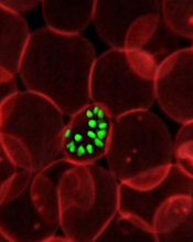
An infrared spectroscopy technique can detect malaria parasites at early stages of development, according to preclinical research published in Analytical Chemistry.
A group of biochemists found this method could detect Plasmodium falciparum in red blood cells by picking up on a fatty acid signature.
This allowed the researchers to identify and quantify parasites at various stages of development, including the ring and gametocyte stages.
The team also pointed out that the spectrometer they used is portable, inexpensive, and does not require highly trained staff for operation. It could therefore prove useful in the field.
“Current tests for malaria suffer from serious limitations,” said study author Bayden Wood, PhD, of Monash University in Victoria, Australia.
“Many are expensive [and] require specialist instruments and highly trained staff to judge whether blood samples contain the parasite. What’s been holding us back is the lack of an accurate and inexpensive test to detect malaria early and stop it in its tracks. We believe we’ve found it.”
Dr Wood and his colleagues already knew that fatty acids were a marker for malaria from previous studies conducted at the Australian Synchrotron. The Synchrotron allowed the team to see the different life stages of the parasite and the variation in its fatty acids.
The researchers thought they might be able to use this information for diagnosis, but they needed a more portable detection method.
So they decided to test whether attenuated total reflectance Fourier transform infrared spectroscopy (ATR-FT-IR) could detect the fatty acid signature. The technique utilizes infrared light to pick up on the vibrations of molecules.
The researchers spiked red blood cells with parasites of different numbers and life stages and observed them using ATR-FT-IR.
Dr Wood said the method produced results within minutes. And it gave a clear indication of malaria at a much earlier stage of infection than current tests on the market—at the ring and gametocyte stages.
The absolute detection limit was 0.00001% parasitemia (<1 parasite/μL of blood) for cultured early ring-stage parasites in a suspension of normal red blood cells.
“Now that we can detect the early stages of a parasite’s life in the bloodstream, the disease will be much easier to test and treat,” Dr Wood said. “The big advantage of our test is that it doesn’t need scientists and expensive equipment. This has the potential to dramatically reduce the number of people dying from this disease in remote communities.”
The method also shows the potential to detect a number of other blood-borne diseases, according to the researchers. Dr Wood and his colleagues are now planning a clinical trial of ATR-FT-IR in Thailand.
Credit: St Jude Children’s
Research Hospital
An infrared spectroscopy technique can detect malaria parasites at early stages of development, according to preclinical research published in Analytical Chemistry.
A group of biochemists found this method could detect Plasmodium falciparum in red blood cells by picking up on a fatty acid signature.
This allowed the researchers to identify and quantify parasites at various stages of development, including the ring and gametocyte stages.
The team also pointed out that the spectrometer they used is portable, inexpensive, and does not require highly trained staff for operation. It could therefore prove useful in the field.
“Current tests for malaria suffer from serious limitations,” said study author Bayden Wood, PhD, of Monash University in Victoria, Australia.
“Many are expensive [and] require specialist instruments and highly trained staff to judge whether blood samples contain the parasite. What’s been holding us back is the lack of an accurate and inexpensive test to detect malaria early and stop it in its tracks. We believe we’ve found it.”
Dr Wood and his colleagues already knew that fatty acids were a marker for malaria from previous studies conducted at the Australian Synchrotron. The Synchrotron allowed the team to see the different life stages of the parasite and the variation in its fatty acids.
The researchers thought they might be able to use this information for diagnosis, but they needed a more portable detection method.
So they decided to test whether attenuated total reflectance Fourier transform infrared spectroscopy (ATR-FT-IR) could detect the fatty acid signature. The technique utilizes infrared light to pick up on the vibrations of molecules.
The researchers spiked red blood cells with parasites of different numbers and life stages and observed them using ATR-FT-IR.
Dr Wood said the method produced results within minutes. And it gave a clear indication of malaria at a much earlier stage of infection than current tests on the market—at the ring and gametocyte stages.
The absolute detection limit was 0.00001% parasitemia (<1 parasite/μL of blood) for cultured early ring-stage parasites in a suspension of normal red blood cells.
“Now that we can detect the early stages of a parasite’s life in the bloodstream, the disease will be much easier to test and treat,” Dr Wood said. “The big advantage of our test is that it doesn’t need scientists and expensive equipment. This has the potential to dramatically reduce the number of people dying from this disease in remote communities.”
The method also shows the potential to detect a number of other blood-borne diseases, according to the researchers. Dr Wood and his colleagues are now planning a clinical trial of ATR-FT-IR in Thailand.
Credit: St Jude Children’s
Research Hospital
An infrared spectroscopy technique can detect malaria parasites at early stages of development, according to preclinical research published in Analytical Chemistry.
A group of biochemists found this method could detect Plasmodium falciparum in red blood cells by picking up on a fatty acid signature.
This allowed the researchers to identify and quantify parasites at various stages of development, including the ring and gametocyte stages.
The team also pointed out that the spectrometer they used is portable, inexpensive, and does not require highly trained staff for operation. It could therefore prove useful in the field.
“Current tests for malaria suffer from serious limitations,” said study author Bayden Wood, PhD, of Monash University in Victoria, Australia.
“Many are expensive [and] require specialist instruments and highly trained staff to judge whether blood samples contain the parasite. What’s been holding us back is the lack of an accurate and inexpensive test to detect malaria early and stop it in its tracks. We believe we’ve found it.”
Dr Wood and his colleagues already knew that fatty acids were a marker for malaria from previous studies conducted at the Australian Synchrotron. The Synchrotron allowed the team to see the different life stages of the parasite and the variation in its fatty acids.
The researchers thought they might be able to use this information for diagnosis, but they needed a more portable detection method.
So they decided to test whether attenuated total reflectance Fourier transform infrared spectroscopy (ATR-FT-IR) could detect the fatty acid signature. The technique utilizes infrared light to pick up on the vibrations of molecules.
The researchers spiked red blood cells with parasites of different numbers and life stages and observed them using ATR-FT-IR.
Dr Wood said the method produced results within minutes. And it gave a clear indication of malaria at a much earlier stage of infection than current tests on the market—at the ring and gametocyte stages.
The absolute detection limit was 0.00001% parasitemia (<1 parasite/μL of blood) for cultured early ring-stage parasites in a suspension of normal red blood cells.
“Now that we can detect the early stages of a parasite’s life in the bloodstream, the disease will be much easier to test and treat,” Dr Wood said. “The big advantage of our test is that it doesn’t need scientists and expensive equipment. This has the potential to dramatically reduce the number of people dying from this disease in remote communities.”
The method also shows the potential to detect a number of other blood-borne diseases, according to the researchers. Dr Wood and his colleagues are now planning a clinical trial of ATR-FT-IR in Thailand.
Hospira issues Class I recall of infusion pumps

Credit: Daniel Gay
Hospira, Inc., has issued a Class I recall of Abbott Acclaim infusion pumps and Hospira Acclaim Encore infusion pumps, after receiving reports of broken door assemblies on these products.
If a door assembly breaks, the door may not close properly and an over-infusion or a delay of therapy may occur.
If the door cannot be closed, the pump cannot be used, and this can result in a delay of therapy.
Use of these products may cause serious adverse events, including death.
These pumps are used to deliver hydration fluids, drugs, blood and blood fractions, intravenous nutritionals, and enteral nutritionals.
The affected Abbott Acclaim Infusion Pumps, list Number 12032, were manufactured from February 1998 to November 1998 and distributed from September 1998 through February 2004.
The affected Hospira Acclaim Encore infusion pumps, list Number 12237, were manufactured from February 1997 to February 2010 and distributed from July 1999 through November 2013.
Hospira is recommending that users inspect each Hospira/Abbott Acclaim Encore infusion pump for door handle cracks prior to programming a therapy, by taking the following steps:
1. After inserting the tubing (with the roller clamp closed) and closing the door handle against the infusion pump, check that the door is fully closed.
If a pump has a door that does not close properly and a gap or separation exists between the completely closed door and the pump itself, remove the pump from clinical service, and call Hospira at 1-800-441-4100 (M-F, 8am-5pm, CT). If the door closes correctly, proceed to Step 2.
2. If the door closes correctly and a gap or separation does not exist between the completely closed door and the pump itself, check that there is no free flow activity in the drip chamber of the administration set by opening the roller clamp.
If free flow is detected, close the roller clamp, remove the pump from clinical service, and call Hospira at 1-800-441-4100 (M-F, 8am-5pm, CT).
3. If no issues are found through steps 1 and 2, the pump is acceptable for use. However, healthcare professionals should still ensure that anyone in their facility who might use these products is made aware of this safety notification and the recommended actions.
Healthcare professionals and patients can report adverse events or side effects related to the use of these products to the FDA’s MedWatch Program.
Credit: Daniel Gay
Hospira, Inc., has issued a Class I recall of Abbott Acclaim infusion pumps and Hospira Acclaim Encore infusion pumps, after receiving reports of broken door assemblies on these products.
If a door assembly breaks, the door may not close properly and an over-infusion or a delay of therapy may occur.
If the door cannot be closed, the pump cannot be used, and this can result in a delay of therapy.
Use of these products may cause serious adverse events, including death.
These pumps are used to deliver hydration fluids, drugs, blood and blood fractions, intravenous nutritionals, and enteral nutritionals.
The affected Abbott Acclaim Infusion Pumps, list Number 12032, were manufactured from February 1998 to November 1998 and distributed from September 1998 through February 2004.
The affected Hospira Acclaim Encore infusion pumps, list Number 12237, were manufactured from February 1997 to February 2010 and distributed from July 1999 through November 2013.
Hospira is recommending that users inspect each Hospira/Abbott Acclaim Encore infusion pump for door handle cracks prior to programming a therapy, by taking the following steps:
1. After inserting the tubing (with the roller clamp closed) and closing the door handle against the infusion pump, check that the door is fully closed.
If a pump has a door that does not close properly and a gap or separation exists between the completely closed door and the pump itself, remove the pump from clinical service, and call Hospira at 1-800-441-4100 (M-F, 8am-5pm, CT). If the door closes correctly, proceed to Step 2.
2. If the door closes correctly and a gap or separation does not exist between the completely closed door and the pump itself, check that there is no free flow activity in the drip chamber of the administration set by opening the roller clamp.
If free flow is detected, close the roller clamp, remove the pump from clinical service, and call Hospira at 1-800-441-4100 (M-F, 8am-5pm, CT).
3. If no issues are found through steps 1 and 2, the pump is acceptable for use. However, healthcare professionals should still ensure that anyone in their facility who might use these products is made aware of this safety notification and the recommended actions.
Healthcare professionals and patients can report adverse events or side effects related to the use of these products to the FDA’s MedWatch Program.
Credit: Daniel Gay
Hospira, Inc., has issued a Class I recall of Abbott Acclaim infusion pumps and Hospira Acclaim Encore infusion pumps, after receiving reports of broken door assemblies on these products.
If a door assembly breaks, the door may not close properly and an over-infusion or a delay of therapy may occur.
If the door cannot be closed, the pump cannot be used, and this can result in a delay of therapy.
Use of these products may cause serious adverse events, including death.
These pumps are used to deliver hydration fluids, drugs, blood and blood fractions, intravenous nutritionals, and enteral nutritionals.
The affected Abbott Acclaim Infusion Pumps, list Number 12032, were manufactured from February 1998 to November 1998 and distributed from September 1998 through February 2004.
The affected Hospira Acclaim Encore infusion pumps, list Number 12237, were manufactured from February 1997 to February 2010 and distributed from July 1999 through November 2013.
Hospira is recommending that users inspect each Hospira/Abbott Acclaim Encore infusion pump for door handle cracks prior to programming a therapy, by taking the following steps:
1. After inserting the tubing (with the roller clamp closed) and closing the door handle against the infusion pump, check that the door is fully closed.
If a pump has a door that does not close properly and a gap or separation exists between the completely closed door and the pump itself, remove the pump from clinical service, and call Hospira at 1-800-441-4100 (M-F, 8am-5pm, CT). If the door closes correctly, proceed to Step 2.
2. If the door closes correctly and a gap or separation does not exist between the completely closed door and the pump itself, check that there is no free flow activity in the drip chamber of the administration set by opening the roller clamp.
If free flow is detected, close the roller clamp, remove the pump from clinical service, and call Hospira at 1-800-441-4100 (M-F, 8am-5pm, CT).
3. If no issues are found through steps 1 and 2, the pump is acceptable for use. However, healthcare professionals should still ensure that anyone in their facility who might use these products is made aware of this safety notification and the recommended actions.
Healthcare professionals and patients can report adverse events or side effects related to the use of these products to the FDA’s MedWatch Program.
FDA approves CML drug for home administration

The US Food and Drug Administration (FDA) has expanded the approval of omacetaxine mepesuccinate (Synribo) to include home administration.
The drug is already FDA-approved to treat adults with chronic or accelerated phase chronic myeloid leukemia (CML) who do not respond to or cannot tolerate 2 or more tyrosine kinase inhibitors.
The new approval allows CML patients to self-administer subcutaneous injections of omacetaxine mepesuccinate at home.
“It had been necessary for adults living with chronic or accelerated phase CML who are prescribed Synribo to travel to their doctor’s office twice a day for 2 weeks, which can be extremely burdensome and inconvenient to both patients and their caregivers,” said Meir Wetzler, MD, FACP, Chief of the Leukemia Section at Roswell Park Cancer Institute in Buffalo, New York.
“Now, physicians can decide if their patients are candidates for self-administration and, if so, provide their patients with guidance on how to properly administer reconstituted Synribo in the home.”
The drug’s maker, Teva Pharmaceutical Industries, Ltd., is working to finalize a pharmacy support program that will help facilitate successful home administration of omacetaxine mepesuccinate. The program is expected to “go live” this month or next.
About omacetaxine mepesuccinate
Omacetaxine mepesuccinate is a protein synthesis inhibitor. Although the drug’s mechanism of action is not fully understood, it is known to prevent the production of Bcr-Abl and Mcl-1, which help drive CML.
In October 2012, the FDA granted omacetaxine mepesuccinate accelerated approval for the treatment of adult patients with chronic or accelerated phase CML with resistance and/or intolerance to 2 or more tyrosine kinase inhibitors. Omacetaxine mepesuccinate gained full FDA approval in February.
The drug has been associated with severe and fatal myelosuppression, including thrombocytopenia, neutropenia, and anemia in some patients. So healthcare professionals should monitor patients’ complete blood counts weekly during induction and initial maintenance cycles and every 2 weeks during later maintenance cycles, as clinically indicated.
Omacetaxine mepesuccinate has been known to cause severe thrombocytopenia, which increases the risk of hemorrhage. Fatalities from cerebral hemorrhage have occurred. And severe, non-fatal gastrointestinal hemorrhages have occurred.
So healthcare professionals should monitor platelet counts as part of the complete blood count as recommended. Patients should not receive anticoagulants, aspirin, or non-steroidal anti-inflammatory drugs when their platelet counts are <50,000/μL, as these drugs may increase the risk of bleeding.
Omacetaxine mepesuccinate can induce glucose intolerance as well. So healthcare professionals should monitor blood glucose levels frequently, especially in patients with diabetes or risk factors for diabetes. Patients with poorly controlled diabetes mellitus should not receive omacetaxine mepesuccinate until good glycemic control has been established.
Omacetaxine mepesuccinate can cause fetal harm when administered to a pregnant woman. So women should be advised to avoid becoming pregnant while using the drug.
For more details on omacetaxine mepesuccinate, see the full prescribing information.
The US Food and Drug Administration (FDA) has expanded the approval of omacetaxine mepesuccinate (Synribo) to include home administration.
The drug is already FDA-approved to treat adults with chronic or accelerated phase chronic myeloid leukemia (CML) who do not respond to or cannot tolerate 2 or more tyrosine kinase inhibitors.
The new approval allows CML patients to self-administer subcutaneous injections of omacetaxine mepesuccinate at home.
“It had been necessary for adults living with chronic or accelerated phase CML who are prescribed Synribo to travel to their doctor’s office twice a day for 2 weeks, which can be extremely burdensome and inconvenient to both patients and their caregivers,” said Meir Wetzler, MD, FACP, Chief of the Leukemia Section at Roswell Park Cancer Institute in Buffalo, New York.
“Now, physicians can decide if their patients are candidates for self-administration and, if so, provide their patients with guidance on how to properly administer reconstituted Synribo in the home.”
The drug’s maker, Teva Pharmaceutical Industries, Ltd., is working to finalize a pharmacy support program that will help facilitate successful home administration of omacetaxine mepesuccinate. The program is expected to “go live” this month or next.
About omacetaxine mepesuccinate
Omacetaxine mepesuccinate is a protein synthesis inhibitor. Although the drug’s mechanism of action is not fully understood, it is known to prevent the production of Bcr-Abl and Mcl-1, which help drive CML.
In October 2012, the FDA granted omacetaxine mepesuccinate accelerated approval for the treatment of adult patients with chronic or accelerated phase CML with resistance and/or intolerance to 2 or more tyrosine kinase inhibitors. Omacetaxine mepesuccinate gained full FDA approval in February.
The drug has been associated with severe and fatal myelosuppression, including thrombocytopenia, neutropenia, and anemia in some patients. So healthcare professionals should monitor patients’ complete blood counts weekly during induction and initial maintenance cycles and every 2 weeks during later maintenance cycles, as clinically indicated.
Omacetaxine mepesuccinate has been known to cause severe thrombocytopenia, which increases the risk of hemorrhage. Fatalities from cerebral hemorrhage have occurred. And severe, non-fatal gastrointestinal hemorrhages have occurred.
So healthcare professionals should monitor platelet counts as part of the complete blood count as recommended. Patients should not receive anticoagulants, aspirin, or non-steroidal anti-inflammatory drugs when their platelet counts are <50,000/μL, as these drugs may increase the risk of bleeding.
Omacetaxine mepesuccinate can induce glucose intolerance as well. So healthcare professionals should monitor blood glucose levels frequently, especially in patients with diabetes or risk factors for diabetes. Patients with poorly controlled diabetes mellitus should not receive omacetaxine mepesuccinate until good glycemic control has been established.
Omacetaxine mepesuccinate can cause fetal harm when administered to a pregnant woman. So women should be advised to avoid becoming pregnant while using the drug.
For more details on omacetaxine mepesuccinate, see the full prescribing information.
The US Food and Drug Administration (FDA) has expanded the approval of omacetaxine mepesuccinate (Synribo) to include home administration.
The drug is already FDA-approved to treat adults with chronic or accelerated phase chronic myeloid leukemia (CML) who do not respond to or cannot tolerate 2 or more tyrosine kinase inhibitors.
The new approval allows CML patients to self-administer subcutaneous injections of omacetaxine mepesuccinate at home.
“It had been necessary for adults living with chronic or accelerated phase CML who are prescribed Synribo to travel to their doctor’s office twice a day for 2 weeks, which can be extremely burdensome and inconvenient to both patients and their caregivers,” said Meir Wetzler, MD, FACP, Chief of the Leukemia Section at Roswell Park Cancer Institute in Buffalo, New York.
“Now, physicians can decide if their patients are candidates for self-administration and, if so, provide their patients with guidance on how to properly administer reconstituted Synribo in the home.”
The drug’s maker, Teva Pharmaceutical Industries, Ltd., is working to finalize a pharmacy support program that will help facilitate successful home administration of omacetaxine mepesuccinate. The program is expected to “go live” this month or next.
About omacetaxine mepesuccinate
Omacetaxine mepesuccinate is a protein synthesis inhibitor. Although the drug’s mechanism of action is not fully understood, it is known to prevent the production of Bcr-Abl and Mcl-1, which help drive CML.
In October 2012, the FDA granted omacetaxine mepesuccinate accelerated approval for the treatment of adult patients with chronic or accelerated phase CML with resistance and/or intolerance to 2 or more tyrosine kinase inhibitors. Omacetaxine mepesuccinate gained full FDA approval in February.
The drug has been associated with severe and fatal myelosuppression, including thrombocytopenia, neutropenia, and anemia in some patients. So healthcare professionals should monitor patients’ complete blood counts weekly during induction and initial maintenance cycles and every 2 weeks during later maintenance cycles, as clinically indicated.
Omacetaxine mepesuccinate has been known to cause severe thrombocytopenia, which increases the risk of hemorrhage. Fatalities from cerebral hemorrhage have occurred. And severe, non-fatal gastrointestinal hemorrhages have occurred.
So healthcare professionals should monitor platelet counts as part of the complete blood count as recommended. Patients should not receive anticoagulants, aspirin, or non-steroidal anti-inflammatory drugs when their platelet counts are <50,000/μL, as these drugs may increase the risk of bleeding.
Omacetaxine mepesuccinate can induce glucose intolerance as well. So healthcare professionals should monitor blood glucose levels frequently, especially in patients with diabetes or risk factors for diabetes. Patients with poorly controlled diabetes mellitus should not receive omacetaxine mepesuccinate until good glycemic control has been established.
Omacetaxine mepesuccinate can cause fetal harm when administered to a pregnant woman. So women should be advised to avoid becoming pregnant while using the drug.
For more details on omacetaxine mepesuccinate, see the full prescribing information.
Liquid droplets help explain cell migration

Scientists have discovered an unexpected link between the shape of a cell and its migration efficiency, and they’ve explained its physics using a model of a liquid droplet.
Cell migration is achieved through the movement of the cell’s membrane, which is powered by the action of a protein network inside the cell.
This interaction is affected by the cell’s overall shape, but exactly how this takes place has been unclear.
Research published in Current Biology provides some insight.
The first step in cell migration occurs when the cell extends its front edge—a process called protrusion. This is driven by the growth of actin filaments, which push the cell membrane from inside. At the same time, membrane tension controls protrusion by providing resistance and protecting the cell from over-extending.
But physical laws dictate that the shape of the cell membrane must play a role in the balance between force exerted by actin and the resisting membrane tension. This was not taken into account in previous studies, which used 2D models of migrating cells.
Now, Chiara Gabella, PhD, of Ecole Polytechnique Fédérale de Lausanne in Switzerland, and her colleagues have used a 3D model to better describe the relationship between cell protrusion, shape, and membrane tension.
The scientists developed a way to evaluate the 3D shape of migrating fish epidermal keratocytes by observing the cells in a chamber filled with a fluorescent solution.
The team applied various treatments to swell, shrink, or stretch the cells. And they were able to observe the treatment’s impact on membrane tension, shape, and protrusion velocity.
The treatments only affected the cells’ shape and migration speed, not membrane tension. The results also showed that the more spherical a cell is, the faster it moves.
To interpret these unexpected findings, the scientists modeled a migrating cell as a liquid droplet spreading on a surface.
“It is well known that a droplet’s shape and, in particular, the contact angle that it makes with the surface are determined by the tension forces between the droplet, its environmental medium (eg, air or a different liquid), and the surface on which it moves,” Dr Gabella said.
Results of the modeling experiment suggested that the leading edge could be considered a triple interface between the substrate, membrane, and extracellular medium. And the contact angle between the membrane and the substrate determines the load on actin polymerization and, therefore, the protrusion rate.
“From this point of view, a more spherical cell means less load for actin filaments to overcome and, therefore, faster actin growth and migration,” said Alexander Verkhovsky, PhD, also of Ecole Polytechnique Fédérale de Lausanne.
In support of this idea, the scientists found the cells were sensitive to the surface characteristics, just as droplets would be, by slowing down or being pinned at ridges.
“The emphasis of many studies has been on discovering and characterizing individual cellular components,” Dr Verkhovsky said. “This is rooted in the common belief that a cell’s behavior is determined by intricate networks of genes and proteins.”
In contrast, this work shows that, despite their molecular complexity, cells can be described as physical objects. The findings point to a new relationship between a cell’s shape and its dynamics and may help us to understand how cell migration is guided by the cell’s 3D environment.
Scientists have discovered an unexpected link between the shape of a cell and its migration efficiency, and they’ve explained its physics using a model of a liquid droplet.
Cell migration is achieved through the movement of the cell’s membrane, which is powered by the action of a protein network inside the cell.
This interaction is affected by the cell’s overall shape, but exactly how this takes place has been unclear.
Research published in Current Biology provides some insight.
The first step in cell migration occurs when the cell extends its front edge—a process called protrusion. This is driven by the growth of actin filaments, which push the cell membrane from inside. At the same time, membrane tension controls protrusion by providing resistance and protecting the cell from over-extending.
But physical laws dictate that the shape of the cell membrane must play a role in the balance between force exerted by actin and the resisting membrane tension. This was not taken into account in previous studies, which used 2D models of migrating cells.
Now, Chiara Gabella, PhD, of Ecole Polytechnique Fédérale de Lausanne in Switzerland, and her colleagues have used a 3D model to better describe the relationship between cell protrusion, shape, and membrane tension.
The scientists developed a way to evaluate the 3D shape of migrating fish epidermal keratocytes by observing the cells in a chamber filled with a fluorescent solution.
The team applied various treatments to swell, shrink, or stretch the cells. And they were able to observe the treatment’s impact on membrane tension, shape, and protrusion velocity.
The treatments only affected the cells’ shape and migration speed, not membrane tension. The results also showed that the more spherical a cell is, the faster it moves.
To interpret these unexpected findings, the scientists modeled a migrating cell as a liquid droplet spreading on a surface.
“It is well known that a droplet’s shape and, in particular, the contact angle that it makes with the surface are determined by the tension forces between the droplet, its environmental medium (eg, air or a different liquid), and the surface on which it moves,” Dr Gabella said.
Results of the modeling experiment suggested that the leading edge could be considered a triple interface between the substrate, membrane, and extracellular medium. And the contact angle between the membrane and the substrate determines the load on actin polymerization and, therefore, the protrusion rate.
“From this point of view, a more spherical cell means less load for actin filaments to overcome and, therefore, faster actin growth and migration,” said Alexander Verkhovsky, PhD, also of Ecole Polytechnique Fédérale de Lausanne.
In support of this idea, the scientists found the cells were sensitive to the surface characteristics, just as droplets would be, by slowing down or being pinned at ridges.
“The emphasis of many studies has been on discovering and characterizing individual cellular components,” Dr Verkhovsky said. “This is rooted in the common belief that a cell’s behavior is determined by intricate networks of genes and proteins.”
In contrast, this work shows that, despite their molecular complexity, cells can be described as physical objects. The findings point to a new relationship between a cell’s shape and its dynamics and may help us to understand how cell migration is guided by the cell’s 3D environment.
Scientists have discovered an unexpected link between the shape of a cell and its migration efficiency, and they’ve explained its physics using a model of a liquid droplet.
Cell migration is achieved through the movement of the cell’s membrane, which is powered by the action of a protein network inside the cell.
This interaction is affected by the cell’s overall shape, but exactly how this takes place has been unclear.
Research published in Current Biology provides some insight.
The first step in cell migration occurs when the cell extends its front edge—a process called protrusion. This is driven by the growth of actin filaments, which push the cell membrane from inside. At the same time, membrane tension controls protrusion by providing resistance and protecting the cell from over-extending.
But physical laws dictate that the shape of the cell membrane must play a role in the balance between force exerted by actin and the resisting membrane tension. This was not taken into account in previous studies, which used 2D models of migrating cells.
Now, Chiara Gabella, PhD, of Ecole Polytechnique Fédérale de Lausanne in Switzerland, and her colleagues have used a 3D model to better describe the relationship between cell protrusion, shape, and membrane tension.
The scientists developed a way to evaluate the 3D shape of migrating fish epidermal keratocytes by observing the cells in a chamber filled with a fluorescent solution.
The team applied various treatments to swell, shrink, or stretch the cells. And they were able to observe the treatment’s impact on membrane tension, shape, and protrusion velocity.
The treatments only affected the cells’ shape and migration speed, not membrane tension. The results also showed that the more spherical a cell is, the faster it moves.
To interpret these unexpected findings, the scientists modeled a migrating cell as a liquid droplet spreading on a surface.
“It is well known that a droplet’s shape and, in particular, the contact angle that it makes with the surface are determined by the tension forces between the droplet, its environmental medium (eg, air or a different liquid), and the surface on which it moves,” Dr Gabella said.
Results of the modeling experiment suggested that the leading edge could be considered a triple interface between the substrate, membrane, and extracellular medium. And the contact angle between the membrane and the substrate determines the load on actin polymerization and, therefore, the protrusion rate.
“From this point of view, a more spherical cell means less load for actin filaments to overcome and, therefore, faster actin growth and migration,” said Alexander Verkhovsky, PhD, also of Ecole Polytechnique Fédérale de Lausanne.
In support of this idea, the scientists found the cells were sensitive to the surface characteristics, just as droplets would be, by slowing down or being pinned at ridges.
“The emphasis of many studies has been on discovering and characterizing individual cellular components,” Dr Verkhovsky said. “This is rooted in the common belief that a cell’s behavior is determined by intricate networks of genes and proteins.”
In contrast, this work shows that, despite their molecular complexity, cells can be described as physical objects. The findings point to a new relationship between a cell’s shape and its dynamics and may help us to understand how cell migration is guided by the cell’s 3D environment.
Combo can overcome resistance in MM

Credit: PNAS
A 2-drug combination can overcome Mcl-1-dependent treatment resistance in multiple myeloma (MM), preclinical research suggests.
The therapy consists of the Chk1 inhibitor CEP3891 and the MEK1/2 inhibitor PD184352.
Chk1 inhibitors prevent cells from arresting in stages of the cell cycle that facilitate DNA repair. And MEK inhibitors prevent cells from activating proteins that regulate DNA repair, while promoting the accumulation of pro-apoptotic proteins.
Researchers recounted their results with the 2 inhibitors in PLOS ONE.
The team noted that, although several drugs are effective against MM, the cancer cells can often survive treatment by increasing production of Mcl-1. This protein regulates processes that promote cell survival and has been implicated in resistance to bortezomib and other anti-myeloma drugs that were initially effective.
With their experiments, the researchers discovered that CEP3891 and PD184352 can reduce Mcl-1 expression and disrupt its interactions with other proteins to effectively kill MM cells.
“This research builds on our previous studies that showed exposing multiple myeloma and leukemia cells to Chk1 inhibitors activated a protective response through the Ras/MEK/ERK signaling pathway,” said Xin-Yan Pei, MD, PhD, of Virginia Commonwealth University and the Massey Cancer Center in Richmond.
“By combining a Chk1 inhibitor with a MEK inhibitor, we have developed one of only a limited number of strategies shown to circumvent therapeutic resistance caused by high expressions of Mcl-1.”
The team began this research by forcing overexpression of Mcl-1 in human MM cells. This caused the cells to become highly resistant to bortezomib, but it failed to protect them from CEP3891 and PD184352.
Furthermore, CEP3891 and PD184352 completely overcame resistance due to microenvironmental factors associated with increased expression of Mcl-1.
“Not only was the combination therapy effective against multiple myeloma cells, it notably did not harm normal bone marrow cells, raising the possibility of therapeutic selectivity,” said study author Steven Grant, MD, also of Virginia Commonwealth University and the Massey Cancer Center.
“We are hopeful that this research will lead to better therapies for multiple myeloma and help make current therapies more effective by overcoming resistance caused by Mcl-1.”
The researchers have started initial discussions with clinical investigators and drug manufacturers about a clinical trial testing a combination of Chk1 and MEK inhibitors in patients with refractory MM.
Credit: PNAS
A 2-drug combination can overcome Mcl-1-dependent treatment resistance in multiple myeloma (MM), preclinical research suggests.
The therapy consists of the Chk1 inhibitor CEP3891 and the MEK1/2 inhibitor PD184352.
Chk1 inhibitors prevent cells from arresting in stages of the cell cycle that facilitate DNA repair. And MEK inhibitors prevent cells from activating proteins that regulate DNA repair, while promoting the accumulation of pro-apoptotic proteins.
Researchers recounted their results with the 2 inhibitors in PLOS ONE.
The team noted that, although several drugs are effective against MM, the cancer cells can often survive treatment by increasing production of Mcl-1. This protein regulates processes that promote cell survival and has been implicated in resistance to bortezomib and other anti-myeloma drugs that were initially effective.
With their experiments, the researchers discovered that CEP3891 and PD184352 can reduce Mcl-1 expression and disrupt its interactions with other proteins to effectively kill MM cells.
“This research builds on our previous studies that showed exposing multiple myeloma and leukemia cells to Chk1 inhibitors activated a protective response through the Ras/MEK/ERK signaling pathway,” said Xin-Yan Pei, MD, PhD, of Virginia Commonwealth University and the Massey Cancer Center in Richmond.
“By combining a Chk1 inhibitor with a MEK inhibitor, we have developed one of only a limited number of strategies shown to circumvent therapeutic resistance caused by high expressions of Mcl-1.”
The team began this research by forcing overexpression of Mcl-1 in human MM cells. This caused the cells to become highly resistant to bortezomib, but it failed to protect them from CEP3891 and PD184352.
Furthermore, CEP3891 and PD184352 completely overcame resistance due to microenvironmental factors associated with increased expression of Mcl-1.
“Not only was the combination therapy effective against multiple myeloma cells, it notably did not harm normal bone marrow cells, raising the possibility of therapeutic selectivity,” said study author Steven Grant, MD, also of Virginia Commonwealth University and the Massey Cancer Center.
“We are hopeful that this research will lead to better therapies for multiple myeloma and help make current therapies more effective by overcoming resistance caused by Mcl-1.”
The researchers have started initial discussions with clinical investigators and drug manufacturers about a clinical trial testing a combination of Chk1 and MEK inhibitors in patients with refractory MM.
Credit: PNAS
A 2-drug combination can overcome Mcl-1-dependent treatment resistance in multiple myeloma (MM), preclinical research suggests.
The therapy consists of the Chk1 inhibitor CEP3891 and the MEK1/2 inhibitor PD184352.
Chk1 inhibitors prevent cells from arresting in stages of the cell cycle that facilitate DNA repair. And MEK inhibitors prevent cells from activating proteins that regulate DNA repair, while promoting the accumulation of pro-apoptotic proteins.
Researchers recounted their results with the 2 inhibitors in PLOS ONE.
The team noted that, although several drugs are effective against MM, the cancer cells can often survive treatment by increasing production of Mcl-1. This protein regulates processes that promote cell survival and has been implicated in resistance to bortezomib and other anti-myeloma drugs that were initially effective.
With their experiments, the researchers discovered that CEP3891 and PD184352 can reduce Mcl-1 expression and disrupt its interactions with other proteins to effectively kill MM cells.
“This research builds on our previous studies that showed exposing multiple myeloma and leukemia cells to Chk1 inhibitors activated a protective response through the Ras/MEK/ERK signaling pathway,” said Xin-Yan Pei, MD, PhD, of Virginia Commonwealth University and the Massey Cancer Center in Richmond.
“By combining a Chk1 inhibitor with a MEK inhibitor, we have developed one of only a limited number of strategies shown to circumvent therapeutic resistance caused by high expressions of Mcl-1.”
The team began this research by forcing overexpression of Mcl-1 in human MM cells. This caused the cells to become highly resistant to bortezomib, but it failed to protect them from CEP3891 and PD184352.
Furthermore, CEP3891 and PD184352 completely overcame resistance due to microenvironmental factors associated with increased expression of Mcl-1.
“Not only was the combination therapy effective against multiple myeloma cells, it notably did not harm normal bone marrow cells, raising the possibility of therapeutic selectivity,” said study author Steven Grant, MD, also of Virginia Commonwealth University and the Massey Cancer Center.
“We are hopeful that this research will lead to better therapies for multiple myeloma and help make current therapies more effective by overcoming resistance caused by Mcl-1.”
The researchers have started initial discussions with clinical investigators and drug manufacturers about a clinical trial testing a combination of Chk1 and MEK inhibitors in patients with refractory MM.
Hospira announces device correction for infusion pump docking station

Credit: CDC
Hospira, Inc., has announced a medical device correction for the GemStar Docking Station (list number 13075), used in conjunction with the GemStar infusion pump.
The correction follows customer reports of 2 malfunctions that may occur with the docking station.
The company is not recalling the product but is notifying US customers of the potential malfunctions and providing instructions for overriding these errors.
The errors could potentially cause delays or interruptions in therapy. And this might result in serious adverse events or death, but there have been no such events reported to date.
Potential malfunctions
The GemStar Docking Station is an accessory to the GemStar infusion pump (sold separately) and provides an alternate power source to the GemStar pump.
When the docking station is used in conjunction with a GemStar Phase 3 pump (List 13000, 13100 or 13150), there is a risk that the GemStar Phase 3 pump may fail to power up while connected to the docking station.
When a GemStar Phase 3 (List 13000, 13100 or 13150) or GemStar Phase 4 pump (List 13086, 13087 or 13088) is used in conjunction with both a docking station and an external battery pack accessory (List 13073), the GemStar pump may display error code 11/003 and give an audible alarm, indicating excessive input voltage from the external sources.
If the GemStar pump detects what is perceived to be more than 3.6 volts, as measured on the external voltage input, the pump will stop the infusion. This will trigger an audible alarm, and the device will display alarm code 11/003.
If a GemStar fails to power up or the 11/003 error code stops an infusion, a patient’s therapy might be delayed or interrupted. This could result in significant injury or death, although there have been no reports of death or serious injury associated with these malfunctions to date.
The products impacted by these issues have been in distribution since February 2002.
Responding to/preventing malfunctions
Hospira is advising that healthcare professionals weigh the risk/benefit to patients associated with the use of the docking station when administering critical therapies. Clinicians should consider the use of an alternative pump, particularly in patients for whom a delay or interruption of therapy could result in serious injury or death.
However, the company says there is no need to return the GemStar Docking Station at this time. Instead, Hospira recommends that users take the following actions.
To avoid a failure to power up, turn on the pump before connecting it with the docking station. This will prevent the failure to power up.
To mitigate the potential for an 11/003 error code, remove the external battery pack accessory (List 13073) from the docking station and pump prior to installing the pump in the docking station.
In addition, clinicians should stop using a docking station in conjunction with an external battery pack accessory (List 13073). Contact Hospira to discuss an appropriate alternative option.
Docking station users who experience a failure to power up or an 11/003 error code should report the issue to Hospira by calling 1-800-441-4100 (M-F, 8am-5pm CT) or emailing ProductComplaintsPP@hospira.com.
For additional assistance or to obtain a copy of the Urgent Medical Device Correction letter and/or a reply form, contact Stericycle at 1-866-792-5451 (M-F, 8am-5pm ET).
On May 1, 2013, Hospira announced that it would begin the process of retiring the GemStar family of infusion devices in accordance with the company’s global device strategy. As of July 31, 2015, Hospira will consider the products within the GemStar Infusion System family retired and will no longer support them.
Adverse reactions or quality problems related to the GemStar Docking Station can be reported to the US Food and Drug Administration’s MedWatch Program.
Credit: CDC
Hospira, Inc., has announced a medical device correction for the GemStar Docking Station (list number 13075), used in conjunction with the GemStar infusion pump.
The correction follows customer reports of 2 malfunctions that may occur with the docking station.
The company is not recalling the product but is notifying US customers of the potential malfunctions and providing instructions for overriding these errors.
The errors could potentially cause delays or interruptions in therapy. And this might result in serious adverse events or death, but there have been no such events reported to date.
Potential malfunctions
The GemStar Docking Station is an accessory to the GemStar infusion pump (sold separately) and provides an alternate power source to the GemStar pump.
When the docking station is used in conjunction with a GemStar Phase 3 pump (List 13000, 13100 or 13150), there is a risk that the GemStar Phase 3 pump may fail to power up while connected to the docking station.
When a GemStar Phase 3 (List 13000, 13100 or 13150) or GemStar Phase 4 pump (List 13086, 13087 or 13088) is used in conjunction with both a docking station and an external battery pack accessory (List 13073), the GemStar pump may display error code 11/003 and give an audible alarm, indicating excessive input voltage from the external sources.
If the GemStar pump detects what is perceived to be more than 3.6 volts, as measured on the external voltage input, the pump will stop the infusion. This will trigger an audible alarm, and the device will display alarm code 11/003.
If a GemStar fails to power up or the 11/003 error code stops an infusion, a patient’s therapy might be delayed or interrupted. This could result in significant injury or death, although there have been no reports of death or serious injury associated with these malfunctions to date.
The products impacted by these issues have been in distribution since February 2002.
Responding to/preventing malfunctions
Hospira is advising that healthcare professionals weigh the risk/benefit to patients associated with the use of the docking station when administering critical therapies. Clinicians should consider the use of an alternative pump, particularly in patients for whom a delay or interruption of therapy could result in serious injury or death.
However, the company says there is no need to return the GemStar Docking Station at this time. Instead, Hospira recommends that users take the following actions.
To avoid a failure to power up, turn on the pump before connecting it with the docking station. This will prevent the failure to power up.
To mitigate the potential for an 11/003 error code, remove the external battery pack accessory (List 13073) from the docking station and pump prior to installing the pump in the docking station.
In addition, clinicians should stop using a docking station in conjunction with an external battery pack accessory (List 13073). Contact Hospira to discuss an appropriate alternative option.
Docking station users who experience a failure to power up or an 11/003 error code should report the issue to Hospira by calling 1-800-441-4100 (M-F, 8am-5pm CT) or emailing ProductComplaintsPP@hospira.com.
For additional assistance or to obtain a copy of the Urgent Medical Device Correction letter and/or a reply form, contact Stericycle at 1-866-792-5451 (M-F, 8am-5pm ET).
On May 1, 2013, Hospira announced that it would begin the process of retiring the GemStar family of infusion devices in accordance with the company’s global device strategy. As of July 31, 2015, Hospira will consider the products within the GemStar Infusion System family retired and will no longer support them.
Adverse reactions or quality problems related to the GemStar Docking Station can be reported to the US Food and Drug Administration’s MedWatch Program.
Credit: CDC
Hospira, Inc., has announced a medical device correction for the GemStar Docking Station (list number 13075), used in conjunction with the GemStar infusion pump.
The correction follows customer reports of 2 malfunctions that may occur with the docking station.
The company is not recalling the product but is notifying US customers of the potential malfunctions and providing instructions for overriding these errors.
The errors could potentially cause delays or interruptions in therapy. And this might result in serious adverse events or death, but there have been no such events reported to date.
Potential malfunctions
The GemStar Docking Station is an accessory to the GemStar infusion pump (sold separately) and provides an alternate power source to the GemStar pump.
When the docking station is used in conjunction with a GemStar Phase 3 pump (List 13000, 13100 or 13150), there is a risk that the GemStar Phase 3 pump may fail to power up while connected to the docking station.
When a GemStar Phase 3 (List 13000, 13100 or 13150) or GemStar Phase 4 pump (List 13086, 13087 or 13088) is used in conjunction with both a docking station and an external battery pack accessory (List 13073), the GemStar pump may display error code 11/003 and give an audible alarm, indicating excessive input voltage from the external sources.
If the GemStar pump detects what is perceived to be more than 3.6 volts, as measured on the external voltage input, the pump will stop the infusion. This will trigger an audible alarm, and the device will display alarm code 11/003.
If a GemStar fails to power up or the 11/003 error code stops an infusion, a patient’s therapy might be delayed or interrupted. This could result in significant injury or death, although there have been no reports of death or serious injury associated with these malfunctions to date.
The products impacted by these issues have been in distribution since February 2002.
Responding to/preventing malfunctions
Hospira is advising that healthcare professionals weigh the risk/benefit to patients associated with the use of the docking station when administering critical therapies. Clinicians should consider the use of an alternative pump, particularly in patients for whom a delay or interruption of therapy could result in serious injury or death.
However, the company says there is no need to return the GemStar Docking Station at this time. Instead, Hospira recommends that users take the following actions.
To avoid a failure to power up, turn on the pump before connecting it with the docking station. This will prevent the failure to power up.
To mitigate the potential for an 11/003 error code, remove the external battery pack accessory (List 13073) from the docking station and pump prior to installing the pump in the docking station.
In addition, clinicians should stop using a docking station in conjunction with an external battery pack accessory (List 13073). Contact Hospira to discuss an appropriate alternative option.
Docking station users who experience a failure to power up or an 11/003 error code should report the issue to Hospira by calling 1-800-441-4100 (M-F, 8am-5pm CT) or emailing ProductComplaintsPP@hospira.com.
For additional assistance or to obtain a copy of the Urgent Medical Device Correction letter and/or a reply form, contact Stericycle at 1-866-792-5451 (M-F, 8am-5pm ET).
On May 1, 2013, Hospira announced that it would begin the process of retiring the GemStar family of infusion devices in accordance with the company’s global device strategy. As of July 31, 2015, Hospira will consider the products within the GemStar Infusion System family retired and will no longer support them.
Adverse reactions or quality problems related to the GemStar Docking Station can be reported to the US Food and Drug Administration’s MedWatch Program.