User login
RIKEN upholds misconduct allegations, calls for paper’s retraction

Credit: RIKEN
The Japanese research institute RIKEN has announced that it will not re-investigate the allegations of misconduct levelled against the researcher who claimed to have discovered a new method for inducing pluripotency in somatic cells.
In January, Haruko Obokata, PhD, and her colleagues reported the creation of stimulus-triggered acquisition of pluripotency (STAP) cells.
The team said they could induce pluripotency by exposing somatic cells to a low-pH environment.
But members of the scientific community voiced concerns about the research, so RIKEN launched an investigation.
In April, the investigative committee concluded that Dr Obokata and some of her colleagues were guilty of misconduct and/or negligence.
Dr Obokata appealed the findings, but the committee has decided another investigation is not warranted. RIKEN has also called for a retraction of the Nature paper in which the committee found evidence of misconduct.
Another RIKEN committee has already met to discuss possible disciplinary action for Dr Obokata and some of her colleagues. Potential punishments range from pay cuts to temporary suspension to disciplinary discharge.
In the meantime, a group headed by Shinichi Aizawa, PhD, special advisor to RIKEN, is attempting to verify the results of the STAP experiments and determine if the STAP phenomenon is, in fact, real.
As the events of this case unfolded, another possible case of misconduct surfaced at RIKEN.
The institute recently announced that Shunsuke Ishii, PhD, chairperson of the committee investigating misconduct in the STAP papers, had resigned from the committee amid concerns about one of his own papers.
RIKEN replaced Dr Ishii with Jun Watanabe, a lawyer already on the committee. And the institute has launched an investigation into Dr Ishii’s work.
Furthermore, RIKEN’s office for internal reform, headed by President Ryoji Noyori, PhD, has commissioned a committee of outside experts to deliberate and recommend measures to prevent research misconduct in the future. 
Credit: RIKEN
The Japanese research institute RIKEN has announced that it will not re-investigate the allegations of misconduct levelled against the researcher who claimed to have discovered a new method for inducing pluripotency in somatic cells.
In January, Haruko Obokata, PhD, and her colleagues reported the creation of stimulus-triggered acquisition of pluripotency (STAP) cells.
The team said they could induce pluripotency by exposing somatic cells to a low-pH environment.
But members of the scientific community voiced concerns about the research, so RIKEN launched an investigation.
In April, the investigative committee concluded that Dr Obokata and some of her colleagues were guilty of misconduct and/or negligence.
Dr Obokata appealed the findings, but the committee has decided another investigation is not warranted. RIKEN has also called for a retraction of the Nature paper in which the committee found evidence of misconduct.
Another RIKEN committee has already met to discuss possible disciplinary action for Dr Obokata and some of her colleagues. Potential punishments range from pay cuts to temporary suspension to disciplinary discharge.
In the meantime, a group headed by Shinichi Aizawa, PhD, special advisor to RIKEN, is attempting to verify the results of the STAP experiments and determine if the STAP phenomenon is, in fact, real.
As the events of this case unfolded, another possible case of misconduct surfaced at RIKEN.
The institute recently announced that Shunsuke Ishii, PhD, chairperson of the committee investigating misconduct in the STAP papers, had resigned from the committee amid concerns about one of his own papers.
RIKEN replaced Dr Ishii with Jun Watanabe, a lawyer already on the committee. And the institute has launched an investigation into Dr Ishii’s work.
Furthermore, RIKEN’s office for internal reform, headed by President Ryoji Noyori, PhD, has commissioned a committee of outside experts to deliberate and recommend measures to prevent research misconduct in the future.
Credit: RIKEN
The Japanese research institute RIKEN has announced that it will not re-investigate the allegations of misconduct levelled against the researcher who claimed to have discovered a new method for inducing pluripotency in somatic cells.
In January, Haruko Obokata, PhD, and her colleagues reported the creation of stimulus-triggered acquisition of pluripotency (STAP) cells.
The team said they could induce pluripotency by exposing somatic cells to a low-pH environment.
But members of the scientific community voiced concerns about the research, so RIKEN launched an investigation.
In April, the investigative committee concluded that Dr Obokata and some of her colleagues were guilty of misconduct and/or negligence.
Dr Obokata appealed the findings, but the committee has decided another investigation is not warranted. RIKEN has also called for a retraction of the Nature paper in which the committee found evidence of misconduct.
Another RIKEN committee has already met to discuss possible disciplinary action for Dr Obokata and some of her colleagues. Potential punishments range from pay cuts to temporary suspension to disciplinary discharge.
In the meantime, a group headed by Shinichi Aizawa, PhD, special advisor to RIKEN, is attempting to verify the results of the STAP experiments and determine if the STAP phenomenon is, in fact, real.
As the events of this case unfolded, another possible case of misconduct surfaced at RIKEN.
The institute recently announced that Shunsuke Ishii, PhD, chairperson of the committee investigating misconduct in the STAP papers, had resigned from the committee amid concerns about one of his own papers.
RIKEN replaced Dr Ishii with Jun Watanabe, a lawyer already on the committee. And the institute has launched an investigation into Dr Ishii’s work.
Furthermore, RIKEN’s office for internal reform, headed by President Ryoji Noyori, PhD, has commissioned a committee of outside experts to deliberate and recommend measures to prevent research misconduct in the future.
Team applies single-cell genomics to malaria
Credit: Peter H. Seeberger
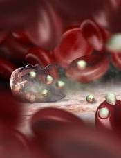
Researchers have devised a way to perform genome sequencing on individual cells infected with malaria parasites.
The team found this single-cell approach could generate parasite genome sequences directly from the blood of patients infected with Plasmodium vivax or Plasmodium falciparum.
It provided new insight into the biology of the parasites, revealing their virulence and capacity for drug resistance.
The team described this work in Genome Research.
They noted that malaria infections commonly contain complex mixtures of Plasmodium parasites. These multiple-genotype infections (MGIs) can alter the impact of the infection and drive the spread of drug resistance. MGIs are extremely common in regions with high levels of malaria infection, but their biology is poorly understood.
“Up to 70% of infections in sub-Saharan Africa are MGIs, and we currently don’t know how many genotypes are present and whether parasites come from a single mosquito bite or multiple mosquito bites,” said study author Shalini Nair, of Texas Biomedical Research Institute in San Antonio.
“Current sequencing techniques really limit our understanding of malaria parasite biology,” added study author Ian Cheeseman, PhD, also of Texas Biomed.
“It’s like trying to understand human genetics by taking DNA from everyone in a village at once. The data is all jumbled up, but what we really want is information from individuals.”
To achieve a better understanding of MGIs and malaria parasites in general, the researchers developed a method for isolating an individual parasite cell and sequencing its genome. Although single-cell genomics approaches are already used in cancer research, it has been difficult to adapt the approach to other organisms.
“One of the real challenges was learning how to cope with the tiny amounts of DNA involved,” Nair said. “In a single cell, we have a thousand-million-millionth of a gram of DNA. It took a lot of effort before we developed a method where we simply didn’t lose this.”
But the researchers eventually found they could use methods of single-cell sorting and whole-genome amplification to separate out individual cells and amplify their DNA for sequencing. The team sequenced the DNA from red blood cells infected with P falciparum or P vivax.
They discovered this technique can reveal the composition of MGIs and provide information on the strength of an infection and the development of drug resistance.
“One of the major surprises we found when we started looking at individual parasites instead of whole infections was the level of variation in drug-resistance genes,” Nair said. “The patterns we saw suggested that different parasites within a single malaria infection would react very differently to drug treatment.”
Unfortunately, this technology is currently too expensive and demanding for routine use in the clinic. But the potential applications are significant, according to the researchers.
“We’re now able to look at malaria infections with incredible detail,” Dr Cheeseman said. “This will help us understand how to best design drugs and vaccines to tackle this major global killer.”
Credit: Peter H. Seeberger
Researchers have devised a way to perform genome sequencing on individual cells infected with malaria parasites.
The team found this single-cell approach could generate parasite genome sequences directly from the blood of patients infected with Plasmodium vivax or Plasmodium falciparum.
It provided new insight into the biology of the parasites, revealing their virulence and capacity for drug resistance.
The team described this work in Genome Research.
They noted that malaria infections commonly contain complex mixtures of Plasmodium parasites. These multiple-genotype infections (MGIs) can alter the impact of the infection and drive the spread of drug resistance. MGIs are extremely common in regions with high levels of malaria infection, but their biology is poorly understood.
“Up to 70% of infections in sub-Saharan Africa are MGIs, and we currently don’t know how many genotypes are present and whether parasites come from a single mosquito bite or multiple mosquito bites,” said study author Shalini Nair, of Texas Biomedical Research Institute in San Antonio.
“Current sequencing techniques really limit our understanding of malaria parasite biology,” added study author Ian Cheeseman, PhD, also of Texas Biomed.
“It’s like trying to understand human genetics by taking DNA from everyone in a village at once. The data is all jumbled up, but what we really want is information from individuals.”
To achieve a better understanding of MGIs and malaria parasites in general, the researchers developed a method for isolating an individual parasite cell and sequencing its genome. Although single-cell genomics approaches are already used in cancer research, it has been difficult to adapt the approach to other organisms.
“One of the real challenges was learning how to cope with the tiny amounts of DNA involved,” Nair said. “In a single cell, we have a thousand-million-millionth of a gram of DNA. It took a lot of effort before we developed a method where we simply didn’t lose this.”
But the researchers eventually found they could use methods of single-cell sorting and whole-genome amplification to separate out individual cells and amplify their DNA for sequencing. The team sequenced the DNA from red blood cells infected with P falciparum or P vivax.
They discovered this technique can reveal the composition of MGIs and provide information on the strength of an infection and the development of drug resistance.
“One of the major surprises we found when we started looking at individual parasites instead of whole infections was the level of variation in drug-resistance genes,” Nair said. “The patterns we saw suggested that different parasites within a single malaria infection would react very differently to drug treatment.”
Unfortunately, this technology is currently too expensive and demanding for routine use in the clinic. But the potential applications are significant, according to the researchers.
“We’re now able to look at malaria infections with incredible detail,” Dr Cheeseman said. “This will help us understand how to best design drugs and vaccines to tackle this major global killer.”
Credit: Peter H. Seeberger
Researchers have devised a way to perform genome sequencing on individual cells infected with malaria parasites.
The team found this single-cell approach could generate parasite genome sequences directly from the blood of patients infected with Plasmodium vivax or Plasmodium falciparum.
It provided new insight into the biology of the parasites, revealing their virulence and capacity for drug resistance.
The team described this work in Genome Research.
They noted that malaria infections commonly contain complex mixtures of Plasmodium parasites. These multiple-genotype infections (MGIs) can alter the impact of the infection and drive the spread of drug resistance. MGIs are extremely common in regions with high levels of malaria infection, but their biology is poorly understood.
“Up to 70% of infections in sub-Saharan Africa are MGIs, and we currently don’t know how many genotypes are present and whether parasites come from a single mosquito bite or multiple mosquito bites,” said study author Shalini Nair, of Texas Biomedical Research Institute in San Antonio.
“Current sequencing techniques really limit our understanding of malaria parasite biology,” added study author Ian Cheeseman, PhD, also of Texas Biomed.
“It’s like trying to understand human genetics by taking DNA from everyone in a village at once. The data is all jumbled up, but what we really want is information from individuals.”
To achieve a better understanding of MGIs and malaria parasites in general, the researchers developed a method for isolating an individual parasite cell and sequencing its genome. Although single-cell genomics approaches are already used in cancer research, it has been difficult to adapt the approach to other organisms.
“One of the real challenges was learning how to cope with the tiny amounts of DNA involved,” Nair said. “In a single cell, we have a thousand-million-millionth of a gram of DNA. It took a lot of effort before we developed a method where we simply didn’t lose this.”
But the researchers eventually found they could use methods of single-cell sorting and whole-genome amplification to separate out individual cells and amplify their DNA for sequencing. The team sequenced the DNA from red blood cells infected with P falciparum or P vivax.
They discovered this technique can reveal the composition of MGIs and provide information on the strength of an infection and the development of drug resistance.
“One of the major surprises we found when we started looking at individual parasites instead of whole infections was the level of variation in drug-resistance genes,” Nair said. “The patterns we saw suggested that different parasites within a single malaria infection would react very differently to drug treatment.”
Unfortunately, this technology is currently too expensive and demanding for routine use in the clinic. But the potential applications are significant, according to the researchers.
“We’re now able to look at malaria infections with incredible detail,” Dr Cheeseman said. “This will help us understand how to best design drugs and vaccines to tackle this major global killer.”
Antiviral agent may prevent CMV infection
An antiviral agent can reduce the incidence of cytomegalovirus (CMV) infection in patients receiving an allogeneic hematopoietic stem cell
transplant, according to a study published in The New England Journal of Medicine.
The agent, letermovir, proved more effective than placebo in preventing CMV, and the highest dose tested, 240 mg/day, was most effective.
The most common adverse events were gastrointestinal disorders and infections.
However, the incidence of events was similar among treated patients and those in the placebo arm.
Roy F. Chemaly, MD, of the University of Texas MD Anderson Cancer Center in Houston, and his colleagues conducted this randomized, double-blind, phase 2 trial. It was funded by AiCuris, the company that was developing letermovir before Merck purchased worldwide rights to develop and commercialize the drug in 2012.
The researchers evaluated the effect of letermovir on the incidence and time-to-onset of CMV prophylaxis failure in CMV-seropositive, matched transplant recipients.
The study included 131 patients. For 12 weeks after engraftment, they received placebo (n=33) or letermovir at 60 mg/day (n=33), 120 mg/day (n=31), or 240 mg/day (n=34).
Efficacy analysis
The primary endpoint was all-cause prophylaxis failure, which was defined as discontinuation of the study drug due to CMV antigen or CMV DNA detection, end-organ disease, or any other causes unrelated to CMV.
The primary efficacy analysis population was a modified intention-to-treat population, which included all patients who received at least 1 dose of the study drug and had at least 1 measurement of the CMV viral load during the study.
The incidence of all-cause prophylaxis failure was significantly lower in the groups that received letermovir at doses of 120 mg/day or 240 mg/day, when compared with the placebo group—32% and 29% vs 64%; P=0.01 and P=0.007, respectively.
The time to the onset of prophylaxis failure was significantly shorter in the 240-mg group (range, 1 to 8 days) than in the placebo group (range, 1 to 21 days; P=0.002).
However, comparisons with the placebo group were not significant for the 60-mg group (range, 1 to 42 days; P=0.15) or the 120-mg group (range, 1 to 15 days; P=0.13).
The incidence of virologic failure was lower in the 240-mg group (6%) than in the 120-mg group (19%), the 60-mg group (21%), or the placebo group (36%).
Virologic failure was defined as either detectable CMV antigen or DNA in the blood at 2 consecutive time points (with at least 1 instance confirmed by the central lab), leading to discontinuation of the study drug and the administration of rescue medication or the development of CMV end-organ disease.
Safety analysis
Nearly all of the patients had at least 1 adverse event during treatment—94% in the 60-mg and 120-mg groups and 100% in the 240-mg and placebo groups. Most events were mild or moderate.
However, 24% of letermovir-treated patients and 30% of those who received placebo experienced severe adverse events during treatment. Investigators, who were blinded to treatment, considered 17% of the severe events in the letermovir group to be drug-related and 33% of severe events in the placebo group to be drug-related.
Most adverse events were gastrointestinal disorders—diarrhea, nausea, and vomiting—which occurred in 66% of letermovir-treated patients and 61% of patients in the placebo group. Infections—mostly CMV—were also common, occurring in 59% of letermovir-treated patients and 76% of patients in the placebo group.
Five patients died during the trial. None of the deaths were thought to be related to treatment or to CMV.
An antiviral agent can reduce the incidence of cytomegalovirus (CMV) infection in patients receiving an allogeneic hematopoietic stem cell
transplant, according to a study published in The New England Journal of Medicine.
The agent, letermovir, proved more effective than placebo in preventing CMV, and the highest dose tested, 240 mg/day, was most effective.
The most common adverse events were gastrointestinal disorders and infections.
However, the incidence of events was similar among treated patients and those in the placebo arm.
Roy F. Chemaly, MD, of the University of Texas MD Anderson Cancer Center in Houston, and his colleagues conducted this randomized, double-blind, phase 2 trial. It was funded by AiCuris, the company that was developing letermovir before Merck purchased worldwide rights to develop and commercialize the drug in 2012.
The researchers evaluated the effect of letermovir on the incidence and time-to-onset of CMV prophylaxis failure in CMV-seropositive, matched transplant recipients.
The study included 131 patients. For 12 weeks after engraftment, they received placebo (n=33) or letermovir at 60 mg/day (n=33), 120 mg/day (n=31), or 240 mg/day (n=34).
Efficacy analysis
The primary endpoint was all-cause prophylaxis failure, which was defined as discontinuation of the study drug due to CMV antigen or CMV DNA detection, end-organ disease, or any other causes unrelated to CMV.
The primary efficacy analysis population was a modified intention-to-treat population, which included all patients who received at least 1 dose of the study drug and had at least 1 measurement of the CMV viral load during the study.
The incidence of all-cause prophylaxis failure was significantly lower in the groups that received letermovir at doses of 120 mg/day or 240 mg/day, when compared with the placebo group—32% and 29% vs 64%; P=0.01 and P=0.007, respectively.
The time to the onset of prophylaxis failure was significantly shorter in the 240-mg group (range, 1 to 8 days) than in the placebo group (range, 1 to 21 days; P=0.002).
However, comparisons with the placebo group were not significant for the 60-mg group (range, 1 to 42 days; P=0.15) or the 120-mg group (range, 1 to 15 days; P=0.13).
The incidence of virologic failure was lower in the 240-mg group (6%) than in the 120-mg group (19%), the 60-mg group (21%), or the placebo group (36%).
Virologic failure was defined as either detectable CMV antigen or DNA in the blood at 2 consecutive time points (with at least 1 instance confirmed by the central lab), leading to discontinuation of the study drug and the administration of rescue medication or the development of CMV end-organ disease.
Safety analysis
Nearly all of the patients had at least 1 adverse event during treatment—94% in the 60-mg and 120-mg groups and 100% in the 240-mg and placebo groups. Most events were mild or moderate.
However, 24% of letermovir-treated patients and 30% of those who received placebo experienced severe adverse events during treatment. Investigators, who were blinded to treatment, considered 17% of the severe events in the letermovir group to be drug-related and 33% of severe events in the placebo group to be drug-related.
Most adverse events were gastrointestinal disorders—diarrhea, nausea, and vomiting—which occurred in 66% of letermovir-treated patients and 61% of patients in the placebo group. Infections—mostly CMV—were also common, occurring in 59% of letermovir-treated patients and 76% of patients in the placebo group.
Five patients died during the trial. None of the deaths were thought to be related to treatment or to CMV.
An antiviral agent can reduce the incidence of cytomegalovirus (CMV) infection in patients receiving an allogeneic hematopoietic stem cell
transplant, according to a study published in The New England Journal of Medicine.
The agent, letermovir, proved more effective than placebo in preventing CMV, and the highest dose tested, 240 mg/day, was most effective.
The most common adverse events were gastrointestinal disorders and infections.
However, the incidence of events was similar among treated patients and those in the placebo arm.
Roy F. Chemaly, MD, of the University of Texas MD Anderson Cancer Center in Houston, and his colleagues conducted this randomized, double-blind, phase 2 trial. It was funded by AiCuris, the company that was developing letermovir before Merck purchased worldwide rights to develop and commercialize the drug in 2012.
The researchers evaluated the effect of letermovir on the incidence and time-to-onset of CMV prophylaxis failure in CMV-seropositive, matched transplant recipients.
The study included 131 patients. For 12 weeks after engraftment, they received placebo (n=33) or letermovir at 60 mg/day (n=33), 120 mg/day (n=31), or 240 mg/day (n=34).
Efficacy analysis
The primary endpoint was all-cause prophylaxis failure, which was defined as discontinuation of the study drug due to CMV antigen or CMV DNA detection, end-organ disease, or any other causes unrelated to CMV.
The primary efficacy analysis population was a modified intention-to-treat population, which included all patients who received at least 1 dose of the study drug and had at least 1 measurement of the CMV viral load during the study.
The incidence of all-cause prophylaxis failure was significantly lower in the groups that received letermovir at doses of 120 mg/day or 240 mg/day, when compared with the placebo group—32% and 29% vs 64%; P=0.01 and P=0.007, respectively.
The time to the onset of prophylaxis failure was significantly shorter in the 240-mg group (range, 1 to 8 days) than in the placebo group (range, 1 to 21 days; P=0.002).
However, comparisons with the placebo group were not significant for the 60-mg group (range, 1 to 42 days; P=0.15) or the 120-mg group (range, 1 to 15 days; P=0.13).
The incidence of virologic failure was lower in the 240-mg group (6%) than in the 120-mg group (19%), the 60-mg group (21%), or the placebo group (36%).
Virologic failure was defined as either detectable CMV antigen or DNA in the blood at 2 consecutive time points (with at least 1 instance confirmed by the central lab), leading to discontinuation of the study drug and the administration of rescue medication or the development of CMV end-organ disease.
Safety analysis
Nearly all of the patients had at least 1 adverse event during treatment—94% in the 60-mg and 120-mg groups and 100% in the 240-mg and placebo groups. Most events were mild or moderate.
However, 24% of letermovir-treated patients and 30% of those who received placebo experienced severe adverse events during treatment. Investigators, who were blinded to treatment, considered 17% of the severe events in the letermovir group to be drug-related and 33% of severe events in the placebo group to be drug-related.
Most adverse events were gastrointestinal disorders—diarrhea, nausea, and vomiting—which occurred in 66% of letermovir-treated patients and 61% of patients in the placebo group. Infections—mostly CMV—were also common, occurring in 59% of letermovir-treated patients and 76% of patients in the placebo group.
Five patients died during the trial. None of the deaths were thought to be related to treatment or to CMV.
How IL-27 promotes tumor growth
Credit: Kathryn T. Iacono
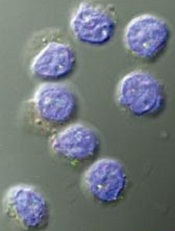
Research in mice has revealed a potential approach to cancer treatment—a way to inhibit the recruitment of regulatory T cells (Tregs) to tumors.
The study showed that dendritic-cell-derived interleukin-27 (IL-27) promotes Treg recruitment in models of lymphoma, melanoma, and fibrosarcoma.
This suggests that if cancer therapies can inhibit IL-27’s immunosuppressive function, they could more effectively activate other T cells to attack and destroy tumors.
Researchers described this discovery in the Journal of Leukocyte Biology.
“Our study not only provides a new insight into the effects of interleukin-27 in regulatory T-cell biology but also greatly improves our understanding of the physiological functions of interleukin-27, especially in tumor immunology,” said study author Siyuan Xia, of Nankai University in Tianjin, China.
“We hope our study could shed new light on developing novel interventional therapies by targeting regulatory T cells in cancer patients.”
The researchers made their discovery by using mice deficient in a specific subunit of IL-27 called p28. They compared tumor-infiltrating lymphocytes between IL-27p28 knockout mice and wild-type mice.
This revealed that Tregs were significantly decreased in knockout mice transplanted with EL-4 lymphoma, B16 melanoma, and MCA-induced fibrosarcoma.
The team also found that IL-27 promotes the expression of CCL22, which is known to mediate Treg recruitment to tumors. And tumor-associated dendritic cells were the major source of CCL22.
When the researchers restored CCL22 or IL-27 in the knockout mice, they observed significant restoration of the tumor-infiltrating Tregs.
Furthermore, tumor-infiltrating CD4 T cells produced much more IFN-γ in the IL-27p28 knockout mice than in wild-type mice. According to the researchers, this reinforces the physiological importance of Tregs in suppressing an antitumor immune response.
“Suppressive and regulatory pathways in the immune system are incredibly important for normal health and preventing autoimmunity,” said John Wherry, PhD, Deputy Editor of the Journal of Leukocyte Biology.
“However, these pathways also get exploited by cancer to prevent immune responses leading to cancer progression. The current studies point to an important regulatory network centered on interleukin-27 that could be targeted to improve immunity to cancer in humans.”
Credit: Kathryn T. Iacono
Research in mice has revealed a potential approach to cancer treatment—a way to inhibit the recruitment of regulatory T cells (Tregs) to tumors.
The study showed that dendritic-cell-derived interleukin-27 (IL-27) promotes Treg recruitment in models of lymphoma, melanoma, and fibrosarcoma.
This suggests that if cancer therapies can inhibit IL-27’s immunosuppressive function, they could more effectively activate other T cells to attack and destroy tumors.
Researchers described this discovery in the Journal of Leukocyte Biology.
“Our study not only provides a new insight into the effects of interleukin-27 in regulatory T-cell biology but also greatly improves our understanding of the physiological functions of interleukin-27, especially in tumor immunology,” said study author Siyuan Xia, of Nankai University in Tianjin, China.
“We hope our study could shed new light on developing novel interventional therapies by targeting regulatory T cells in cancer patients.”
The researchers made their discovery by using mice deficient in a specific subunit of IL-27 called p28. They compared tumor-infiltrating lymphocytes between IL-27p28 knockout mice and wild-type mice.
This revealed that Tregs were significantly decreased in knockout mice transplanted with EL-4 lymphoma, B16 melanoma, and MCA-induced fibrosarcoma.
The team also found that IL-27 promotes the expression of CCL22, which is known to mediate Treg recruitment to tumors. And tumor-associated dendritic cells were the major source of CCL22.
When the researchers restored CCL22 or IL-27 in the knockout mice, they observed significant restoration of the tumor-infiltrating Tregs.
Furthermore, tumor-infiltrating CD4 T cells produced much more IFN-γ in the IL-27p28 knockout mice than in wild-type mice. According to the researchers, this reinforces the physiological importance of Tregs in suppressing an antitumor immune response.
“Suppressive and regulatory pathways in the immune system are incredibly important for normal health and preventing autoimmunity,” said John Wherry, PhD, Deputy Editor of the Journal of Leukocyte Biology.
“However, these pathways also get exploited by cancer to prevent immune responses leading to cancer progression. The current studies point to an important regulatory network centered on interleukin-27 that could be targeted to improve immunity to cancer in humans.”
Credit: Kathryn T. Iacono
Research in mice has revealed a potential approach to cancer treatment—a way to inhibit the recruitment of regulatory T cells (Tregs) to tumors.
The study showed that dendritic-cell-derived interleukin-27 (IL-27) promotes Treg recruitment in models of lymphoma, melanoma, and fibrosarcoma.
This suggests that if cancer therapies can inhibit IL-27’s immunosuppressive function, they could more effectively activate other T cells to attack and destroy tumors.
Researchers described this discovery in the Journal of Leukocyte Biology.
“Our study not only provides a new insight into the effects of interleukin-27 in regulatory T-cell biology but also greatly improves our understanding of the physiological functions of interleukin-27, especially in tumor immunology,” said study author Siyuan Xia, of Nankai University in Tianjin, China.
“We hope our study could shed new light on developing novel interventional therapies by targeting regulatory T cells in cancer patients.”
The researchers made their discovery by using mice deficient in a specific subunit of IL-27 called p28. They compared tumor-infiltrating lymphocytes between IL-27p28 knockout mice and wild-type mice.
This revealed that Tregs were significantly decreased in knockout mice transplanted with EL-4 lymphoma, B16 melanoma, and MCA-induced fibrosarcoma.
The team also found that IL-27 promotes the expression of CCL22, which is known to mediate Treg recruitment to tumors. And tumor-associated dendritic cells were the major source of CCL22.
When the researchers restored CCL22 or IL-27 in the knockout mice, they observed significant restoration of the tumor-infiltrating Tregs.
Furthermore, tumor-infiltrating CD4 T cells produced much more IFN-γ in the IL-27p28 knockout mice than in wild-type mice. According to the researchers, this reinforces the physiological importance of Tregs in suppressing an antitumor immune response.
“Suppressive and regulatory pathways in the immune system are incredibly important for normal health and preventing autoimmunity,” said John Wherry, PhD, Deputy Editor of the Journal of Leukocyte Biology.
“However, these pathways also get exploited by cancer to prevent immune responses leading to cancer progression. The current studies point to an important regulatory network centered on interleukin-27 that could be targeted to improve immunity to cancer in humans.”
Hepcidin levels vary according to cause of anemia
showing anemia
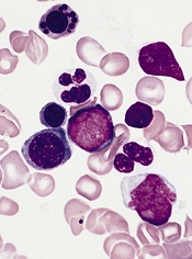
Measuring levels of the hormone hepcidin can help us distinguish anemia caused by iron deficiency from anemia caused by other conditions, a new study suggests.
Investigators say the findings, published in Science Translational Medicine, could help public officials make more informed decisions about distributing iron supplements.
Giving iron supplements to individuals who don’t need them can promote or exacerbate malaria and other infections.
With this research, the investigators linked low levels of hepcidin to iron-deficiency anemia in preschool children living in Africa.
Sant-Rayn Pasricha, PhD, of the University of Oxford in the UK, and his colleagues tested hepcidin levels in 1313 samples taken in 2001 and 2008 from children in The Gambia and Tanzania, respectively.
The team also looked at retrospective data from 25 Gambian children with either postmalarial or nonmalarial anemia.
The mean hepcidin level was significantly lower in children with iron-deficiency anemia than in children with anemia due to inflammation/infection (1.8 ng/mL and 21.7 ng/mL, respectively; P<0.0001).
To expand upon this finding, the investigators modeled the potential impact of screening children for iron supplementation needs based on hepcidin levels rather than anemia status.
If a screen was based on the presence of anemia (hemoglobin <11 g/dL), 77% of iron-deficient children would have received iron supplements, but so would 73% of children with Plasmodium falciparum parasitemia and all of the children with anemia due to inflammation.
On the other hand, if a screen was based on a hepcidin cutoff of <5.5 ng/mL, iron supplements would be given to 77% of children with iron deficiency, 80% of children with iron-deficiency anemia, 20% of children with P falciparum infection, and 14% of children with anemia due to inflammation.
The investigators noted that a lower hepcidin cutoff would have reduced the proportion of individuals with infection and/or inflammation receiving iron, but it would have increased the risk that more children with iron-deficiency anemia would not receive iron. And of course, the reverse is true of a higher hepcidin cutoff.
Nevertheless, the team believes these results are promising. They are now conducting clinical trials to test whether hepcidin levels can be used as a marker to guide iron supplementation decisions.
showing anemia
Measuring levels of the hormone hepcidin can help us distinguish anemia caused by iron deficiency from anemia caused by other conditions, a new study suggests.
Investigators say the findings, published in Science Translational Medicine, could help public officials make more informed decisions about distributing iron supplements.
Giving iron supplements to individuals who don’t need them can promote or exacerbate malaria and other infections.
With this research, the investigators linked low levels of hepcidin to iron-deficiency anemia in preschool children living in Africa.
Sant-Rayn Pasricha, PhD, of the University of Oxford in the UK, and his colleagues tested hepcidin levels in 1313 samples taken in 2001 and 2008 from children in The Gambia and Tanzania, respectively.
The team also looked at retrospective data from 25 Gambian children with either postmalarial or nonmalarial anemia.
The mean hepcidin level was significantly lower in children with iron-deficiency anemia than in children with anemia due to inflammation/infection (1.8 ng/mL and 21.7 ng/mL, respectively; P<0.0001).
To expand upon this finding, the investigators modeled the potential impact of screening children for iron supplementation needs based on hepcidin levels rather than anemia status.
If a screen was based on the presence of anemia (hemoglobin <11 g/dL), 77% of iron-deficient children would have received iron supplements, but so would 73% of children with Plasmodium falciparum parasitemia and all of the children with anemia due to inflammation.
On the other hand, if a screen was based on a hepcidin cutoff of <5.5 ng/mL, iron supplements would be given to 77% of children with iron deficiency, 80% of children with iron-deficiency anemia, 20% of children with P falciparum infection, and 14% of children with anemia due to inflammation.
The investigators noted that a lower hepcidin cutoff would have reduced the proportion of individuals with infection and/or inflammation receiving iron, but it would have increased the risk that more children with iron-deficiency anemia would not receive iron. And of course, the reverse is true of a higher hepcidin cutoff.
Nevertheless, the team believes these results are promising. They are now conducting clinical trials to test whether hepcidin levels can be used as a marker to guide iron supplementation decisions.
showing anemia
Measuring levels of the hormone hepcidin can help us distinguish anemia caused by iron deficiency from anemia caused by other conditions, a new study suggests.
Investigators say the findings, published in Science Translational Medicine, could help public officials make more informed decisions about distributing iron supplements.
Giving iron supplements to individuals who don’t need them can promote or exacerbate malaria and other infections.
With this research, the investigators linked low levels of hepcidin to iron-deficiency anemia in preschool children living in Africa.
Sant-Rayn Pasricha, PhD, of the University of Oxford in the UK, and his colleagues tested hepcidin levels in 1313 samples taken in 2001 and 2008 from children in The Gambia and Tanzania, respectively.
The team also looked at retrospective data from 25 Gambian children with either postmalarial or nonmalarial anemia.
The mean hepcidin level was significantly lower in children with iron-deficiency anemia than in children with anemia due to inflammation/infection (1.8 ng/mL and 21.7 ng/mL, respectively; P<0.0001).
To expand upon this finding, the investigators modeled the potential impact of screening children for iron supplementation needs based on hepcidin levels rather than anemia status.
If a screen was based on the presence of anemia (hemoglobin <11 g/dL), 77% of iron-deficient children would have received iron supplements, but so would 73% of children with Plasmodium falciparum parasitemia and all of the children with anemia due to inflammation.
On the other hand, if a screen was based on a hepcidin cutoff of <5.5 ng/mL, iron supplements would be given to 77% of children with iron deficiency, 80% of children with iron-deficiency anemia, 20% of children with P falciparum infection, and 14% of children with anemia due to inflammation.
The investigators noted that a lower hepcidin cutoff would have reduced the proportion of individuals with infection and/or inflammation receiving iron, but it would have increased the risk that more children with iron-deficiency anemia would not receive iron. And of course, the reverse is true of a higher hepcidin cutoff.
Nevertheless, the team believes these results are promising. They are now conducting clinical trials to test whether hepcidin levels can be used as a marker to guide iron supplementation decisions.
A more precise method of delivering gene therapy

Jeff Fitlow/Rice University
Researchers have developed an adeno-associated virus (AAV) that releases its payload only in the presence of 2 selected proteases.
Because certain proteases are elevated at tumor sites, the virus can be designed to target and destroy cancer cells.
Junghae Suh, PhD, of Rice University in Houston, Texas, and her colleagues engineered the virus and described their work in ACS Nano.
AAVs have become the object of study as delivery vehicles for gene therapy.
Researchers often try to target AAVs to cellular receptors that may be slightly overexpressed on diseased cells, but Dr Suh’s team took a different approach.
“We were looking for other types of biomarkers beyond cellular receptors present at disease sites,” she said. “In breast cancer, for example, it’s known the tumor cells oversecrete extracellular proteases, but perhaps more important are the infiltrating immune cells that migrate into the tumor microenvironment and start dumping out a whole bunch of proteases as well.”
“So that’s what we’re going after to do targeted delivery. Our basic idea is to create viruses that, in the locked configuration, can’t do anything.”
But when the programmed AAVs encounter the right proteases at sites of disease, they unlock and bind to the cells. The AAVs then deliver payloads that will either kill the cells, in the case of cancer therapy, or deliver genes that can repair the cells.
Dr Suh and her colleagues genetically insert peptides into the self-assembling AAVs to lock the capsids, the hard shells that protect genes contained within. The target proteases recognize the peptides and “chew off the locks,” effectively unlocking the virus and allowing it to bind to the diseased cells.
“If we were just looking for 1 protease, it might be at the cancer site, but it could also be somewhere else in your body where you have inflammation,” Dr Suh said. “This could lead to undesirable side effects.”
“By requiring 2 different proteases—let’s say protease A and protease B—to open the locked virus, we may achieve higher delivery specificity since the chance of having both proteases elevated at a site becomes smaller.”
The ultimate vision of this technology is to design viruses that can carry out a combination of steps for targeting.
“To increase the specificity of virus unlocking, you can imagine creating viruses that require many more keys to open,” Dr Suh said. “For example, you may need both proteases A and B, as well as a cellular receptor, to unlock the virus. The work reported here is a good first step toward this goal.”
Jeff Fitlow/Rice University
Researchers have developed an adeno-associated virus (AAV) that releases its payload only in the presence of 2 selected proteases.
Because certain proteases are elevated at tumor sites, the virus can be designed to target and destroy cancer cells.
Junghae Suh, PhD, of Rice University in Houston, Texas, and her colleagues engineered the virus and described their work in ACS Nano.
AAVs have become the object of study as delivery vehicles for gene therapy.
Researchers often try to target AAVs to cellular receptors that may be slightly overexpressed on diseased cells, but Dr Suh’s team took a different approach.
“We were looking for other types of biomarkers beyond cellular receptors present at disease sites,” she said. “In breast cancer, for example, it’s known the tumor cells oversecrete extracellular proteases, but perhaps more important are the infiltrating immune cells that migrate into the tumor microenvironment and start dumping out a whole bunch of proteases as well.”
“So that’s what we’re going after to do targeted delivery. Our basic idea is to create viruses that, in the locked configuration, can’t do anything.”
But when the programmed AAVs encounter the right proteases at sites of disease, they unlock and bind to the cells. The AAVs then deliver payloads that will either kill the cells, in the case of cancer therapy, or deliver genes that can repair the cells.
Dr Suh and her colleagues genetically insert peptides into the self-assembling AAVs to lock the capsids, the hard shells that protect genes contained within. The target proteases recognize the peptides and “chew off the locks,” effectively unlocking the virus and allowing it to bind to the diseased cells.
“If we were just looking for 1 protease, it might be at the cancer site, but it could also be somewhere else in your body where you have inflammation,” Dr Suh said. “This could lead to undesirable side effects.”
“By requiring 2 different proteases—let’s say protease A and protease B—to open the locked virus, we may achieve higher delivery specificity since the chance of having both proteases elevated at a site becomes smaller.”
The ultimate vision of this technology is to design viruses that can carry out a combination of steps for targeting.
“To increase the specificity of virus unlocking, you can imagine creating viruses that require many more keys to open,” Dr Suh said. “For example, you may need both proteases A and B, as well as a cellular receptor, to unlock the virus. The work reported here is a good first step toward this goal.”
Jeff Fitlow/Rice University
Researchers have developed an adeno-associated virus (AAV) that releases its payload only in the presence of 2 selected proteases.
Because certain proteases are elevated at tumor sites, the virus can be designed to target and destroy cancer cells.
Junghae Suh, PhD, of Rice University in Houston, Texas, and her colleagues engineered the virus and described their work in ACS Nano.
AAVs have become the object of study as delivery vehicles for gene therapy.
Researchers often try to target AAVs to cellular receptors that may be slightly overexpressed on diseased cells, but Dr Suh’s team took a different approach.
“We were looking for other types of biomarkers beyond cellular receptors present at disease sites,” she said. “In breast cancer, for example, it’s known the tumor cells oversecrete extracellular proteases, but perhaps more important are the infiltrating immune cells that migrate into the tumor microenvironment and start dumping out a whole bunch of proteases as well.”
“So that’s what we’re going after to do targeted delivery. Our basic idea is to create viruses that, in the locked configuration, can’t do anything.”
But when the programmed AAVs encounter the right proteases at sites of disease, they unlock and bind to the cells. The AAVs then deliver payloads that will either kill the cells, in the case of cancer therapy, or deliver genes that can repair the cells.
Dr Suh and her colleagues genetically insert peptides into the self-assembling AAVs to lock the capsids, the hard shells that protect genes contained within. The target proteases recognize the peptides and “chew off the locks,” effectively unlocking the virus and allowing it to bind to the diseased cells.
“If we were just looking for 1 protease, it might be at the cancer site, but it could also be somewhere else in your body where you have inflammation,” Dr Suh said. “This could lead to undesirable side effects.”
“By requiring 2 different proteases—let’s say protease A and protease B—to open the locked virus, we may achieve higher delivery specificity since the chance of having both proteases elevated at a site becomes smaller.”
The ultimate vision of this technology is to design viruses that can carry out a combination of steps for targeting.
“To increase the specificity of virus unlocking, you can imagine creating viruses that require many more keys to open,” Dr Suh said. “For example, you may need both proteases A and B, as well as a cellular receptor, to unlock the virus. The work reported here is a good first step toward this goal.”
Agricultural chemicals and the risk of NHL

EPA/John Messina
After reviewing nearly 30 years’ worth of data, investigators have compiled a list of agricultural chemicals that appear to increase a person’s risk of developing non-Hodgkin lymphoma (NHL).
Meta-analyses suggested that occupational exposure to phenoxy herbicides, carbamate insecticides, organochlorine insecticides, and organophosphorus insecticides/herbicides can increase the risk of NHL.
The research also revealed associations between certain chemicals and specific NHL subtypes.
Leah Schinasi, PhD, and Maria E. Leon, PhD, of the International Agency for Research on Cancer in Lyon, France, described the analysis and its results in the International Journal of Environmental Research and Public Health.
The investigators reviewed epidemiological research spanning nearly 30 years and identified 44 relevant papers. The papers recounted studies conducted in the US, Canada, Europe, Australia, and New Zealand.
Drs Schinasi and Leon used these data to assess occupational exposure to 80 active ingredients and 21 chemical groups and clarify their role in the development of NHL. Most, but not all, of the studies looked at lifetime exposure to the chemicals in question.
The investigators performed a meta-analysis of the data and found associations between NHL and a range of insecticides and herbicides. But the strongest risk ratios (RRs) were for subtypes of NHL.
There was a positive association between exposure to the organophosphorus herbicide glyphosate and any NHL (RR=1.5), but the link was stronger for B-cell lymphoma in particular (RR=2.0).
Phenoxy herbicide exposure was associated with an increased risk of NHL in general (RR=1.4), B-cell lymphoma (RR=1.8), lymphocytic lymphoma (RR=1.8), and diffuse large B-cell lymphoma (RR=2.0). As for specific phenoxy herbicides, both MCPA (RR=1.5) and 2,4-D (RR=1.4) were associated with NHL.
Carbamate insecticides, as a group, appeared to confer an increased risk of NHL (RR=1.7). The individual insecticides carbaryl and carbofuran showed positive associations with NHL as well (RRs of 1.7 and 1.6, respectively).
There was a positive association with NHL for organophosphorus insecticides as a group (RR=1.6), as well as the individual insecticides chlorpyrifos (RR=1.6), diazinon (RR=1.6), dimethoate (RR=1.4), and malathion (RR=1.8).
Lastly, organochlorine insecticides appeared to confer an increased risk of NHL (RR=1.3). DDT was associated with NHL (RR=1.3), B-cell lymphoma (RR=1.4), diffuse large B-cell lymphoma (RR=1.2), and follicular lymphoma (RR=1.5). And lindane was associated with NHL in general (RR=1.6).
The investigators said this analysis represents one of the most comprehensive reviews on the topic of occupational exposure to agricultural chemicals in the scientific literature.
But it also suggests a need to study a wider variety of chemicals in more geographic areas, especially in low- and middle-income countries, as they were missing from the literature.
EPA/John Messina
After reviewing nearly 30 years’ worth of data, investigators have compiled a list of agricultural chemicals that appear to increase a person’s risk of developing non-Hodgkin lymphoma (NHL).
Meta-analyses suggested that occupational exposure to phenoxy herbicides, carbamate insecticides, organochlorine insecticides, and organophosphorus insecticides/herbicides can increase the risk of NHL.
The research also revealed associations between certain chemicals and specific NHL subtypes.
Leah Schinasi, PhD, and Maria E. Leon, PhD, of the International Agency for Research on Cancer in Lyon, France, described the analysis and its results in the International Journal of Environmental Research and Public Health.
The investigators reviewed epidemiological research spanning nearly 30 years and identified 44 relevant papers. The papers recounted studies conducted in the US, Canada, Europe, Australia, and New Zealand.
Drs Schinasi and Leon used these data to assess occupational exposure to 80 active ingredients and 21 chemical groups and clarify their role in the development of NHL. Most, but not all, of the studies looked at lifetime exposure to the chemicals in question.
The investigators performed a meta-analysis of the data and found associations between NHL and a range of insecticides and herbicides. But the strongest risk ratios (RRs) were for subtypes of NHL.
There was a positive association between exposure to the organophosphorus herbicide glyphosate and any NHL (RR=1.5), but the link was stronger for B-cell lymphoma in particular (RR=2.0).
Phenoxy herbicide exposure was associated with an increased risk of NHL in general (RR=1.4), B-cell lymphoma (RR=1.8), lymphocytic lymphoma (RR=1.8), and diffuse large B-cell lymphoma (RR=2.0). As for specific phenoxy herbicides, both MCPA (RR=1.5) and 2,4-D (RR=1.4) were associated with NHL.
Carbamate insecticides, as a group, appeared to confer an increased risk of NHL (RR=1.7). The individual insecticides carbaryl and carbofuran showed positive associations with NHL as well (RRs of 1.7 and 1.6, respectively).
There was a positive association with NHL for organophosphorus insecticides as a group (RR=1.6), as well as the individual insecticides chlorpyrifos (RR=1.6), diazinon (RR=1.6), dimethoate (RR=1.4), and malathion (RR=1.8).
Lastly, organochlorine insecticides appeared to confer an increased risk of NHL (RR=1.3). DDT was associated with NHL (RR=1.3), B-cell lymphoma (RR=1.4), diffuse large B-cell lymphoma (RR=1.2), and follicular lymphoma (RR=1.5). And lindane was associated with NHL in general (RR=1.6).
The investigators said this analysis represents one of the most comprehensive reviews on the topic of occupational exposure to agricultural chemicals in the scientific literature.
But it also suggests a need to study a wider variety of chemicals in more geographic areas, especially in low- and middle-income countries, as they were missing from the literature.
EPA/John Messina
After reviewing nearly 30 years’ worth of data, investigators have compiled a list of agricultural chemicals that appear to increase a person’s risk of developing non-Hodgkin lymphoma (NHL).
Meta-analyses suggested that occupational exposure to phenoxy herbicides, carbamate insecticides, organochlorine insecticides, and organophosphorus insecticides/herbicides can increase the risk of NHL.
The research also revealed associations between certain chemicals and specific NHL subtypes.
Leah Schinasi, PhD, and Maria E. Leon, PhD, of the International Agency for Research on Cancer in Lyon, France, described the analysis and its results in the International Journal of Environmental Research and Public Health.
The investigators reviewed epidemiological research spanning nearly 30 years and identified 44 relevant papers. The papers recounted studies conducted in the US, Canada, Europe, Australia, and New Zealand.
Drs Schinasi and Leon used these data to assess occupational exposure to 80 active ingredients and 21 chemical groups and clarify their role in the development of NHL. Most, but not all, of the studies looked at lifetime exposure to the chemicals in question.
The investigators performed a meta-analysis of the data and found associations between NHL and a range of insecticides and herbicides. But the strongest risk ratios (RRs) were for subtypes of NHL.
There was a positive association between exposure to the organophosphorus herbicide glyphosate and any NHL (RR=1.5), but the link was stronger for B-cell lymphoma in particular (RR=2.0).
Phenoxy herbicide exposure was associated with an increased risk of NHL in general (RR=1.4), B-cell lymphoma (RR=1.8), lymphocytic lymphoma (RR=1.8), and diffuse large B-cell lymphoma (RR=2.0). As for specific phenoxy herbicides, both MCPA (RR=1.5) and 2,4-D (RR=1.4) were associated with NHL.
Carbamate insecticides, as a group, appeared to confer an increased risk of NHL (RR=1.7). The individual insecticides carbaryl and carbofuran showed positive associations with NHL as well (RRs of 1.7 and 1.6, respectively).
There was a positive association with NHL for organophosphorus insecticides as a group (RR=1.6), as well as the individual insecticides chlorpyrifos (RR=1.6), diazinon (RR=1.6), dimethoate (RR=1.4), and malathion (RR=1.8).
Lastly, organochlorine insecticides appeared to confer an increased risk of NHL (RR=1.3). DDT was associated with NHL (RR=1.3), B-cell lymphoma (RR=1.4), diffuse large B-cell lymphoma (RR=1.2), and follicular lymphoma (RR=1.5). And lindane was associated with NHL in general (RR=1.6).
The investigators said this analysis represents one of the most comprehensive reviews on the topic of occupational exposure to agricultural chemicals in the scientific literature.
But it also suggests a need to study a wider variety of chemicals in more geographic areas, especially in low- and middle-income countries, as they were missing from the literature.
Protein interaction may be therapeutic target for AML

University of Queensland
Inhibiting the interaction of 2 proteins can prevent the development of acute myeloid leukemia (AML), according to a study published in Blood.
Researchers found evidence to suggest the “docking” of one protein, Myb, with another, p300, is essential for AML development.
“Our data identifies the critical role of this Myb-p300 interaction and shows that the disruption of this interaction could lead to a potential therapeutic strategy,” said Tom Gonda, PhD, of the University of Queensland’s School of Pharmacy in Woolloongabba, Queensland, Australia.
“This finding could lead to our team developing a drug to block this interaction and stop the growth of not only acute myeloid leukemia cells but probably the cells of other types of leukemia as well.”
Dr Gonda and his colleagues conducted this research using cells from Booreana mice, which carry a mutant allele of Myb, as well as cells from wild-type mice.
Experiments showed that the Myb-p300 interaction was necessary for in vitro transformation by the oncogenes AML1-ETO, AML1-ETO9a, MLL-ENL, and MLL-AF9.
The researchers also transduced cells from Booreana mice and wild-type mice with either AML1-ETO9a or MLL-AF9 retroviruses and transplanted the cells into irradiated mice. The cells from wild-type mice generated leukemia in the recipients, but the Booreana cells did not.
Lastly, the team performed gene expression analyses to gain more insight into the Myb-p300 relationship. They found that several genes already implicated in myeloid leukemogenesis and hematopoietic stem cell function are regulated in an Myb-p300-dependent manner.
The researchers therefore concluded that the Myb-p300 interaction is important to myeloid leukemogenesis. And disrupting this interaction could prove useful in the fight against AML.
Dr Gonda pointed out, however, that the Myb protein is produced by the MYB oncogene. And although this oncogene is required for the continued growth of leukemia cells, it is also essential for normal blood cell formation.
“[S]o we need an approach for targeting it that won’t completely disrupt normal blood cell production,” he said. “Our research shows that normal blood cells can continue to form even when the Myb-p300 interaction is unable to occur, suggesting that a drug that blocks the interaction could be safe for use in patients.”
Dr Gonda and his colleagues are also planning to examine the possibility of targeting genes and proteins that work downstream of MYB.
University of Queensland
Inhibiting the interaction of 2 proteins can prevent the development of acute myeloid leukemia (AML), according to a study published in Blood.
Researchers found evidence to suggest the “docking” of one protein, Myb, with another, p300, is essential for AML development.
“Our data identifies the critical role of this Myb-p300 interaction and shows that the disruption of this interaction could lead to a potential therapeutic strategy,” said Tom Gonda, PhD, of the University of Queensland’s School of Pharmacy in Woolloongabba, Queensland, Australia.
“This finding could lead to our team developing a drug to block this interaction and stop the growth of not only acute myeloid leukemia cells but probably the cells of other types of leukemia as well.”
Dr Gonda and his colleagues conducted this research using cells from Booreana mice, which carry a mutant allele of Myb, as well as cells from wild-type mice.
Experiments showed that the Myb-p300 interaction was necessary for in vitro transformation by the oncogenes AML1-ETO, AML1-ETO9a, MLL-ENL, and MLL-AF9.
The researchers also transduced cells from Booreana mice and wild-type mice with either AML1-ETO9a or MLL-AF9 retroviruses and transplanted the cells into irradiated mice. The cells from wild-type mice generated leukemia in the recipients, but the Booreana cells did not.
Lastly, the team performed gene expression analyses to gain more insight into the Myb-p300 relationship. They found that several genes already implicated in myeloid leukemogenesis and hematopoietic stem cell function are regulated in an Myb-p300-dependent manner.
The researchers therefore concluded that the Myb-p300 interaction is important to myeloid leukemogenesis. And disrupting this interaction could prove useful in the fight against AML.
Dr Gonda pointed out, however, that the Myb protein is produced by the MYB oncogene. And although this oncogene is required for the continued growth of leukemia cells, it is also essential for normal blood cell formation.
“[S]o we need an approach for targeting it that won’t completely disrupt normal blood cell production,” he said. “Our research shows that normal blood cells can continue to form even when the Myb-p300 interaction is unable to occur, suggesting that a drug that blocks the interaction could be safe for use in patients.”
Dr Gonda and his colleagues are also planning to examine the possibility of targeting genes and proteins that work downstream of MYB.
University of Queensland
Inhibiting the interaction of 2 proteins can prevent the development of acute myeloid leukemia (AML), according to a study published in Blood.
Researchers found evidence to suggest the “docking” of one protein, Myb, with another, p300, is essential for AML development.
“Our data identifies the critical role of this Myb-p300 interaction and shows that the disruption of this interaction could lead to a potential therapeutic strategy,” said Tom Gonda, PhD, of the University of Queensland’s School of Pharmacy in Woolloongabba, Queensland, Australia.
“This finding could lead to our team developing a drug to block this interaction and stop the growth of not only acute myeloid leukemia cells but probably the cells of other types of leukemia as well.”
Dr Gonda and his colleagues conducted this research using cells from Booreana mice, which carry a mutant allele of Myb, as well as cells from wild-type mice.
Experiments showed that the Myb-p300 interaction was necessary for in vitro transformation by the oncogenes AML1-ETO, AML1-ETO9a, MLL-ENL, and MLL-AF9.
The researchers also transduced cells from Booreana mice and wild-type mice with either AML1-ETO9a or MLL-AF9 retroviruses and transplanted the cells into irradiated mice. The cells from wild-type mice generated leukemia in the recipients, but the Booreana cells did not.
Lastly, the team performed gene expression analyses to gain more insight into the Myb-p300 relationship. They found that several genes already implicated in myeloid leukemogenesis and hematopoietic stem cell function are regulated in an Myb-p300-dependent manner.
The researchers therefore concluded that the Myb-p300 interaction is important to myeloid leukemogenesis. And disrupting this interaction could prove useful in the fight against AML.
Dr Gonda pointed out, however, that the Myb protein is produced by the MYB oncogene. And although this oncogene is required for the continued growth of leukemia cells, it is also essential for normal blood cell formation.
“[S]o we need an approach for targeting it that won’t completely disrupt normal blood cell production,” he said. “Our research shows that normal blood cells can continue to form even when the Myb-p300 interaction is unable to occur, suggesting that a drug that blocks the interaction could be safe for use in patients.”
Dr Gonda and his colleagues are also planning to examine the possibility of targeting genes and proteins that work downstream of MYB.
Wnt pathway appears key to cell reprogramming

Salk Institute
The Wnt signaling pathway plays a key role in the generation of induced pluripotent stem cells (iPSCs), according to a study published in Stem Cell Reports.
Researchers found they could increase the efficiency of the cell reprogramming process by inhibiting the Wnt pathway.
“[U]ntil now, this was a very inefficient process,” said study author Ilda Theka, a PhD student at the Centre for Genomic Regulation in Barcelona, Spain.
“There are many groups trying to understand the mechanism by which adult cells become pluripotent and what blocks that process and makes only a small percentage of cells end up being reprogrammed. We are providing information on why it happens.”
The researchers studied how the Wnt pathway behaves throughout the process of transforming mature cells into iPSCs, which usually lasts 2 weeks. It’s a dynamic process that produces oscillations from the pathway, which is not active all the time.
“We have seen that there are two phases and that, in each one of them, Wnt fulfils a different function,” Theka said. “And we have shown that, by inhibiting it at the beginning of the process and activating it at the end, we can increase the efficiency of reprogramming and obtain a larger number of pluripotent cells.”
The team also discovered that the exact moment when the Wnt pathway is activated is crucial. Activating the pathway too early makes the cells begin to differentiate, and they are not reprogrammed.
To artificially control the pathway, the researchers used a molecule called Iwp2, a Wnt-secretion inhibitor that does not permanently alter the cells.
Salk Institute
The Wnt signaling pathway plays a key role in the generation of induced pluripotent stem cells (iPSCs), according to a study published in Stem Cell Reports.
Researchers found they could increase the efficiency of the cell reprogramming process by inhibiting the Wnt pathway.
“[U]ntil now, this was a very inefficient process,” said study author Ilda Theka, a PhD student at the Centre for Genomic Regulation in Barcelona, Spain.
“There are many groups trying to understand the mechanism by which adult cells become pluripotent and what blocks that process and makes only a small percentage of cells end up being reprogrammed. We are providing information on why it happens.”
The researchers studied how the Wnt pathway behaves throughout the process of transforming mature cells into iPSCs, which usually lasts 2 weeks. It’s a dynamic process that produces oscillations from the pathway, which is not active all the time.
“We have seen that there are two phases and that, in each one of them, Wnt fulfils a different function,” Theka said. “And we have shown that, by inhibiting it at the beginning of the process and activating it at the end, we can increase the efficiency of reprogramming and obtain a larger number of pluripotent cells.”
The team also discovered that the exact moment when the Wnt pathway is activated is crucial. Activating the pathway too early makes the cells begin to differentiate, and they are not reprogrammed.
To artificially control the pathway, the researchers used a molecule called Iwp2, a Wnt-secretion inhibitor that does not permanently alter the cells.
Salk Institute
The Wnt signaling pathway plays a key role in the generation of induced pluripotent stem cells (iPSCs), according to a study published in Stem Cell Reports.
Researchers found they could increase the efficiency of the cell reprogramming process by inhibiting the Wnt pathway.
“[U]ntil now, this was a very inefficient process,” said study author Ilda Theka, a PhD student at the Centre for Genomic Regulation in Barcelona, Spain.
“There are many groups trying to understand the mechanism by which adult cells become pluripotent and what blocks that process and makes only a small percentage of cells end up being reprogrammed. We are providing information on why it happens.”
The researchers studied how the Wnt pathway behaves throughout the process of transforming mature cells into iPSCs, which usually lasts 2 weeks. It’s a dynamic process that produces oscillations from the pathway, which is not active all the time.
“We have seen that there are two phases and that, in each one of them, Wnt fulfils a different function,” Theka said. “And we have shown that, by inhibiting it at the beginning of the process and activating it at the end, we can increase the efficiency of reprogramming and obtain a larger number of pluripotent cells.”
The team also discovered that the exact moment when the Wnt pathway is activated is crucial. Activating the pathway too early makes the cells begin to differentiate, and they are not reprogrammed.
To artificially control the pathway, the researchers used a molecule called Iwp2, a Wnt-secretion inhibitor that does not permanently alter the cells.
Rise in VKDB cases prompts call for a tracking system

Credit: Petr Kratochvil
Physicians at a Tennessee hospital have seen a rise in late-onset vitamin K deficiency bleeding (VKDB) in young infants, due to parents declining a vitamin K shot at birth.
Over a period of 8 months, 7 infants were diagnosed with vitamin K deficiency, and 5 of them had VKDB.
Four of the infants experienced intracranial hemorrhaging, and 2 required urgent neurosurgical intervention.
These cases were diagnosed at Monroe Carell Jr. Children’s Hospital at Vanderbilt in Nashville.
And they were described in Pediatric Neurology.
Now, the authors are calling for a state and national tracking system to help them determine how many infants are not receiving a vitamin K shot at birth.
“There is no national tracking of this in the US, unfortunately, and cases are rarely reported,” said Robert Sidonio Jr, MD, of Vanderbilt University School of Medicine.
“We are probably just seeing the tip of the iceberg, and I worry that people are missing these cases often and not considering this diagnosis when presented with a sick infant.”
He and his colleagues are also calling for better education on this issue for healthcare providers and families. The group believes misinformation—that the shot causes leukemia, is a toxin, or is unnecessary in uncomplicated births—may be leading some families to decline the shot.
The American Academy of Pediatrics has recommended the single-dose shot of vitamin K at birth since 1961. Most cases of VKDB seen today occur in infants in the first 6 months of life who did not get the shot and are exclusively breastfed or who have an undiagnosed liver disorder.
Following a recent rise in cases in Tennessee, the US Centers for Disease Control and Prevention discovered that 28% (61/218) of parents of children born at private birthing centers in the state declined the shot.
Mark and Melissa Knotowicz declined the shot for their twins, Silas and Abel, following their birth last July at a Nashville hospital.
“From the information we had, we heard the main side effect was a preservative in the shot that could lead to childhood leukemia,” said Mark Knotowicz. “We thought, ‘We don’t want our kids to have childhood leukemia,’ so we declined it without really hearing any of the benefits.”
At about 6 weeks old, Silas was noticeably fussy. He vomited overnight, woke up extremely pale, and wouldn’t nurse.
The twins had a checkup scheduled that day. The pediatrician suspected sepsis and told the Knotowiczes to take Silas to the Emergency Department at Monroe Carell Jr. Children’s Hospital.
Because of a rash of VKBD cases, the staff there had received additional education. After Silas’s parents confirmed that he had not received the vitamin K shot, CT scans and blood work revealed he had suffered multiple brain bleeds.
Silas received a double dose of vitamin K to get the bleeding under control. His twin, Abel, was diagnosed with asymptomatic vitamin K deficiency and received the shot.
Silas spent a week in the hospital. Today, he undergoes physical therapy for neuromuscular development issues. Any effects on cognitive development aren’t yet known.
“The twins’ cases highlight our inability to determine which infants will go on to develop vitamin K deficiency bleeding, as they both had prolonged bleeding times at presentation,” Dr Sidonio said.
Mark Knotowicz wishes medical staff at the birthing hospital had more clearly defined the risks of declining the shot.
“Why didn’t they say, ‘There have been 4 other cases in Nashville, and we’re trying to prevent a lethal brain hemorrhage in your child?’”
Anna Morad, MD, of the Vanderbilt University School of Medicine, has worked to raise awareness about the vitamin K shot and the risk of VKDB, with some success.
“After our educational outreach, we have seen a decrease in our refusal rates [with about 3.4% of parents declining vitamin K],” she said. “Our goal is to fully educate parents on the risk of declining so they can make an informed choice for their baby.”
Credit: Petr Kratochvil
Physicians at a Tennessee hospital have seen a rise in late-onset vitamin K deficiency bleeding (VKDB) in young infants, due to parents declining a vitamin K shot at birth.
Over a period of 8 months, 7 infants were diagnosed with vitamin K deficiency, and 5 of them had VKDB.
Four of the infants experienced intracranial hemorrhaging, and 2 required urgent neurosurgical intervention.
These cases were diagnosed at Monroe Carell Jr. Children’s Hospital at Vanderbilt in Nashville.
And they were described in Pediatric Neurology.
Now, the authors are calling for a state and national tracking system to help them determine how many infants are not receiving a vitamin K shot at birth.
“There is no national tracking of this in the US, unfortunately, and cases are rarely reported,” said Robert Sidonio Jr, MD, of Vanderbilt University School of Medicine.
“We are probably just seeing the tip of the iceberg, and I worry that people are missing these cases often and not considering this diagnosis when presented with a sick infant.”
He and his colleagues are also calling for better education on this issue for healthcare providers and families. The group believes misinformation—that the shot causes leukemia, is a toxin, or is unnecessary in uncomplicated births—may be leading some families to decline the shot.
The American Academy of Pediatrics has recommended the single-dose shot of vitamin K at birth since 1961. Most cases of VKDB seen today occur in infants in the first 6 months of life who did not get the shot and are exclusively breastfed or who have an undiagnosed liver disorder.
Following a recent rise in cases in Tennessee, the US Centers for Disease Control and Prevention discovered that 28% (61/218) of parents of children born at private birthing centers in the state declined the shot.
Mark and Melissa Knotowicz declined the shot for their twins, Silas and Abel, following their birth last July at a Nashville hospital.
“From the information we had, we heard the main side effect was a preservative in the shot that could lead to childhood leukemia,” said Mark Knotowicz. “We thought, ‘We don’t want our kids to have childhood leukemia,’ so we declined it without really hearing any of the benefits.”
At about 6 weeks old, Silas was noticeably fussy. He vomited overnight, woke up extremely pale, and wouldn’t nurse.
The twins had a checkup scheduled that day. The pediatrician suspected sepsis and told the Knotowiczes to take Silas to the Emergency Department at Monroe Carell Jr. Children’s Hospital.
Because of a rash of VKBD cases, the staff there had received additional education. After Silas’s parents confirmed that he had not received the vitamin K shot, CT scans and blood work revealed he had suffered multiple brain bleeds.
Silas received a double dose of vitamin K to get the bleeding under control. His twin, Abel, was diagnosed with asymptomatic vitamin K deficiency and received the shot.
Silas spent a week in the hospital. Today, he undergoes physical therapy for neuromuscular development issues. Any effects on cognitive development aren’t yet known.
“The twins’ cases highlight our inability to determine which infants will go on to develop vitamin K deficiency bleeding, as they both had prolonged bleeding times at presentation,” Dr Sidonio said.
Mark Knotowicz wishes medical staff at the birthing hospital had more clearly defined the risks of declining the shot.
“Why didn’t they say, ‘There have been 4 other cases in Nashville, and we’re trying to prevent a lethal brain hemorrhage in your child?’”
Anna Morad, MD, of the Vanderbilt University School of Medicine, has worked to raise awareness about the vitamin K shot and the risk of VKDB, with some success.
“After our educational outreach, we have seen a decrease in our refusal rates [with about 3.4% of parents declining vitamin K],” she said. “Our goal is to fully educate parents on the risk of declining so they can make an informed choice for their baby.”
Credit: Petr Kratochvil
Physicians at a Tennessee hospital have seen a rise in late-onset vitamin K deficiency bleeding (VKDB) in young infants, due to parents declining a vitamin K shot at birth.
Over a period of 8 months, 7 infants were diagnosed with vitamin K deficiency, and 5 of them had VKDB.
Four of the infants experienced intracranial hemorrhaging, and 2 required urgent neurosurgical intervention.
These cases were diagnosed at Monroe Carell Jr. Children’s Hospital at Vanderbilt in Nashville.
And they were described in Pediatric Neurology.
Now, the authors are calling for a state and national tracking system to help them determine how many infants are not receiving a vitamin K shot at birth.
“There is no national tracking of this in the US, unfortunately, and cases are rarely reported,” said Robert Sidonio Jr, MD, of Vanderbilt University School of Medicine.
“We are probably just seeing the tip of the iceberg, and I worry that people are missing these cases often and not considering this diagnosis when presented with a sick infant.”
He and his colleagues are also calling for better education on this issue for healthcare providers and families. The group believes misinformation—that the shot causes leukemia, is a toxin, or is unnecessary in uncomplicated births—may be leading some families to decline the shot.
The American Academy of Pediatrics has recommended the single-dose shot of vitamin K at birth since 1961. Most cases of VKDB seen today occur in infants in the first 6 months of life who did not get the shot and are exclusively breastfed or who have an undiagnosed liver disorder.
Following a recent rise in cases in Tennessee, the US Centers for Disease Control and Prevention discovered that 28% (61/218) of parents of children born at private birthing centers in the state declined the shot.
Mark and Melissa Knotowicz declined the shot for their twins, Silas and Abel, following their birth last July at a Nashville hospital.
“From the information we had, we heard the main side effect was a preservative in the shot that could lead to childhood leukemia,” said Mark Knotowicz. “We thought, ‘We don’t want our kids to have childhood leukemia,’ so we declined it without really hearing any of the benefits.”
At about 6 weeks old, Silas was noticeably fussy. He vomited overnight, woke up extremely pale, and wouldn’t nurse.
The twins had a checkup scheduled that day. The pediatrician suspected sepsis and told the Knotowiczes to take Silas to the Emergency Department at Monroe Carell Jr. Children’s Hospital.
Because of a rash of VKBD cases, the staff there had received additional education. After Silas’s parents confirmed that he had not received the vitamin K shot, CT scans and blood work revealed he had suffered multiple brain bleeds.
Silas received a double dose of vitamin K to get the bleeding under control. His twin, Abel, was diagnosed with asymptomatic vitamin K deficiency and received the shot.
Silas spent a week in the hospital. Today, he undergoes physical therapy for neuromuscular development issues. Any effects on cognitive development aren’t yet known.
“The twins’ cases highlight our inability to determine which infants will go on to develop vitamin K deficiency bleeding, as they both had prolonged bleeding times at presentation,” Dr Sidonio said.
Mark Knotowicz wishes medical staff at the birthing hospital had more clearly defined the risks of declining the shot.
“Why didn’t they say, ‘There have been 4 other cases in Nashville, and we’re trying to prevent a lethal brain hemorrhage in your child?’”
Anna Morad, MD, of the Vanderbilt University School of Medicine, has worked to raise awareness about the vitamin K shot and the risk of VKDB, with some success.
“After our educational outreach, we have seen a decrease in our refusal rates [with about 3.4% of parents declining vitamin K],” she said. “Our goal is to fully educate parents on the risk of declining so they can make an informed choice for their baby.”