User login
Group finds CAR T-cell therapy can fight MM
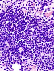
showing multiple myeloma
Results of preclinical research indicate chimeric antigen receptor (CAR) T cells are effective against multiple myeloma (MM).
Researchers generated CAR T cells that target CS1, a molecule found on more than 95% of MM cells.
When injected in mice, these T cells could locate and destroy MM cells.
Jianhua Yu, PhD, of The Ohio State University Comprehensive Cancer Center in Columbus, and his colleagues reported these results in Clinical Cancer Research.
“[O]ur study shows that we can modify T lymphocytes to target CS1 and that these cells efficiently destroy human multiple myeloma cells,” Dr Yu said.
“An important possible advantage to this approach is that these therapeutic T cells have the potential to replicate in the body, and, therefore, they might suppress tumor growth and prevent relapse for a prolonged period.”
Dr Yu and his colleagues used cell lines and cells from MM patients to produce CAR T cells targeting CS1. These cells could be expanded in the lab, and they efficiently recognized and eradicated MM cells in vitro and in vivo.
Compared to control T cells, the CAR T cells better recognized MM cells that overexpressed CS1, and they became more activated following the recognition.
In mice, the CAR T cells greatly reduced the tumor burden and prolonged overall survival. All mice that received CAR T cells were alive 44 days after treatment, compared to 29% and 17% of the mice in the study’s 2 control groups.
“Despite current drugs and use of bone marrow transplantation, multiple myeloma is still incurable, and almost all patients eventually relapse,” said study author Craig Hofmeister, MD, MPH, also of The Ohio State University Comprehensive Cancer Center.
“This study presents a novel strategy for treating multiple myeloma, and we hope to bring it to patients as part of a phase 1 clinical trial as soon as possible.” 
showing multiple myeloma
Results of preclinical research indicate chimeric antigen receptor (CAR) T cells are effective against multiple myeloma (MM).
Researchers generated CAR T cells that target CS1, a molecule found on more than 95% of MM cells.
When injected in mice, these T cells could locate and destroy MM cells.
Jianhua Yu, PhD, of The Ohio State University Comprehensive Cancer Center in Columbus, and his colleagues reported these results in Clinical Cancer Research.
“[O]ur study shows that we can modify T lymphocytes to target CS1 and that these cells efficiently destroy human multiple myeloma cells,” Dr Yu said.
“An important possible advantage to this approach is that these therapeutic T cells have the potential to replicate in the body, and, therefore, they might suppress tumor growth and prevent relapse for a prolonged period.”
Dr Yu and his colleagues used cell lines and cells from MM patients to produce CAR T cells targeting CS1. These cells could be expanded in the lab, and they efficiently recognized and eradicated MM cells in vitro and in vivo.
Compared to control T cells, the CAR T cells better recognized MM cells that overexpressed CS1, and they became more activated following the recognition.
In mice, the CAR T cells greatly reduced the tumor burden and prolonged overall survival. All mice that received CAR T cells were alive 44 days after treatment, compared to 29% and 17% of the mice in the study’s 2 control groups.
“Despite current drugs and use of bone marrow transplantation, multiple myeloma is still incurable, and almost all patients eventually relapse,” said study author Craig Hofmeister, MD, MPH, also of The Ohio State University Comprehensive Cancer Center.
“This study presents a novel strategy for treating multiple myeloma, and we hope to bring it to patients as part of a phase 1 clinical trial as soon as possible.”
showing multiple myeloma
Results of preclinical research indicate chimeric antigen receptor (CAR) T cells are effective against multiple myeloma (MM).
Researchers generated CAR T cells that target CS1, a molecule found on more than 95% of MM cells.
When injected in mice, these T cells could locate and destroy MM cells.
Jianhua Yu, PhD, of The Ohio State University Comprehensive Cancer Center in Columbus, and his colleagues reported these results in Clinical Cancer Research.
“[O]ur study shows that we can modify T lymphocytes to target CS1 and that these cells efficiently destroy human multiple myeloma cells,” Dr Yu said.
“An important possible advantage to this approach is that these therapeutic T cells have the potential to replicate in the body, and, therefore, they might suppress tumor growth and prevent relapse for a prolonged period.”
Dr Yu and his colleagues used cell lines and cells from MM patients to produce CAR T cells targeting CS1. These cells could be expanded in the lab, and they efficiently recognized and eradicated MM cells in vitro and in vivo.
Compared to control T cells, the CAR T cells better recognized MM cells that overexpressed CS1, and they became more activated following the recognition.
In mice, the CAR T cells greatly reduced the tumor burden and prolonged overall survival. All mice that received CAR T cells were alive 44 days after treatment, compared to 29% and 17% of the mice in the study’s 2 control groups.
“Despite current drugs and use of bone marrow transplantation, multiple myeloma is still incurable, and almost all patients eventually relapse,” said study author Craig Hofmeister, MD, MPH, also of The Ohio State University Comprehensive Cancer Center.
“This study presents a novel strategy for treating multiple myeloma, and we hope to bring it to patients as part of a phase 1 clinical trial as soon as possible.”
FDA study suggests dabigatran’s pros outweigh cons

Credit: Darren Baker
After reviewing data from more than 134,000 patients, researchers at the US Food and Drug Administration (FDA) have concluded that dabigatran has a favorable risk-benefit profile.
Their research showed that, compared to warfarin, dabigatran decreased the risk of stroke, death, and intracranial hemorrhage in patients with atrial fibrillation.
On the other hand, dabigatran increased the risk of gastrointestinal bleeding, and the risk of myocardial infarction was similar for the 2 drugs.
This study was part of the FDA’s ongoing review of dabigatran. The agency has been investigating the safety of dabigatran since 2011, following reports of serious bleeding events associated with the drug.
A previous FDA study, announced in 2012, suggested dabigatran does not pose an increased risk of serious bleeding when compared to warfarin. But other studies have provided conflicting results, so the FDA decided to conduct additional research.
This study included information from more than 134,000 Medicare patients, aged 65 years or older. The researchers compared dabigatran and warfarin, evaluating the risk of ischemic stroke, intracranial hemorrhage, major gastrointestinal bleeding, myocardial infarction, and death.
The FDA said this study is based on a much larger and older patient population than those used in the agency’s earlier review of post-market data, and researchers employed a more sophisticated analytical method to capture and analyze the events of concern.
The data showed that, among new users of anticoagulants, dabigatran was associated with a lower risk of ischemic stroke, intracranial hemorrhage, and death, when compared to warfarin.
The incidence rate of ischemic stroke per 1000 person-years was 11.3 among dabigatran users and 13.9 among warfarin users (hazard ratio [HR]=0.80).
The incidence of intracranial hemorrhage was 3.3 and 9.6, respectively (HR=0.34). And the incidence of death was 32.6 and 37.8, respectively (HR=0.86).
The study also showed an increased risk of major gastrointestinal bleeding with the use of dabigatran compared to warfarin. The incidence rate per 1000 person-years was 34.2 and 26.5, respectively (HR=1.28).
The risk of myocardial infarction was similar for the 2 drugs. The incidence rate per 1000 person-years was 15.7 for dabigatran users and 16.9 for warfarin users (HR=0.92).
These results, except with regard to myocardial infarction, are consistent with the clinical trial results that provided the basis for dabigatran’s approval.
The FDA said these findings suggest dabigatran has a favorable risk-benefit profile, so the agency has made no changes to the drug’s current label or recommendations for use.
The FDA is planning to publish detailed data from this study. Until then, some information is available on the agency’s website.
The FDA said it will continue to investigate the reasons for differences in major gastrointestinal bleeding rates for dabigatran and warfarin. And it will continue reviewing anticoagulant use and the risk of bleeding.
Credit: Darren Baker
After reviewing data from more than 134,000 patients, researchers at the US Food and Drug Administration (FDA) have concluded that dabigatran has a favorable risk-benefit profile.
Their research showed that, compared to warfarin, dabigatran decreased the risk of stroke, death, and intracranial hemorrhage in patients with atrial fibrillation.
On the other hand, dabigatran increased the risk of gastrointestinal bleeding, and the risk of myocardial infarction was similar for the 2 drugs.
This study was part of the FDA’s ongoing review of dabigatran. The agency has been investigating the safety of dabigatran since 2011, following reports of serious bleeding events associated with the drug.
A previous FDA study, announced in 2012, suggested dabigatran does not pose an increased risk of serious bleeding when compared to warfarin. But other studies have provided conflicting results, so the FDA decided to conduct additional research.
This study included information from more than 134,000 Medicare patients, aged 65 years or older. The researchers compared dabigatran and warfarin, evaluating the risk of ischemic stroke, intracranial hemorrhage, major gastrointestinal bleeding, myocardial infarction, and death.
The FDA said this study is based on a much larger and older patient population than those used in the agency’s earlier review of post-market data, and researchers employed a more sophisticated analytical method to capture and analyze the events of concern.
The data showed that, among new users of anticoagulants, dabigatran was associated with a lower risk of ischemic stroke, intracranial hemorrhage, and death, when compared to warfarin.
The incidence rate of ischemic stroke per 1000 person-years was 11.3 among dabigatran users and 13.9 among warfarin users (hazard ratio [HR]=0.80).
The incidence of intracranial hemorrhage was 3.3 and 9.6, respectively (HR=0.34). And the incidence of death was 32.6 and 37.8, respectively (HR=0.86).
The study also showed an increased risk of major gastrointestinal bleeding with the use of dabigatran compared to warfarin. The incidence rate per 1000 person-years was 34.2 and 26.5, respectively (HR=1.28).
The risk of myocardial infarction was similar for the 2 drugs. The incidence rate per 1000 person-years was 15.7 for dabigatran users and 16.9 for warfarin users (HR=0.92).
These results, except with regard to myocardial infarction, are consistent with the clinical trial results that provided the basis for dabigatran’s approval.
The FDA said these findings suggest dabigatran has a favorable risk-benefit profile, so the agency has made no changes to the drug’s current label or recommendations for use.
The FDA is planning to publish detailed data from this study. Until then, some information is available on the agency’s website.
The FDA said it will continue to investigate the reasons for differences in major gastrointestinal bleeding rates for dabigatran and warfarin. And it will continue reviewing anticoagulant use and the risk of bleeding.
Credit: Darren Baker
After reviewing data from more than 134,000 patients, researchers at the US Food and Drug Administration (FDA) have concluded that dabigatran has a favorable risk-benefit profile.
Their research showed that, compared to warfarin, dabigatran decreased the risk of stroke, death, and intracranial hemorrhage in patients with atrial fibrillation.
On the other hand, dabigatran increased the risk of gastrointestinal bleeding, and the risk of myocardial infarction was similar for the 2 drugs.
This study was part of the FDA’s ongoing review of dabigatran. The agency has been investigating the safety of dabigatran since 2011, following reports of serious bleeding events associated with the drug.
A previous FDA study, announced in 2012, suggested dabigatran does not pose an increased risk of serious bleeding when compared to warfarin. But other studies have provided conflicting results, so the FDA decided to conduct additional research.
This study included information from more than 134,000 Medicare patients, aged 65 years or older. The researchers compared dabigatran and warfarin, evaluating the risk of ischemic stroke, intracranial hemorrhage, major gastrointestinal bleeding, myocardial infarction, and death.
The FDA said this study is based on a much larger and older patient population than those used in the agency’s earlier review of post-market data, and researchers employed a more sophisticated analytical method to capture and analyze the events of concern.
The data showed that, among new users of anticoagulants, dabigatran was associated with a lower risk of ischemic stroke, intracranial hemorrhage, and death, when compared to warfarin.
The incidence rate of ischemic stroke per 1000 person-years was 11.3 among dabigatran users and 13.9 among warfarin users (hazard ratio [HR]=0.80).
The incidence of intracranial hemorrhage was 3.3 and 9.6, respectively (HR=0.34). And the incidence of death was 32.6 and 37.8, respectively (HR=0.86).
The study also showed an increased risk of major gastrointestinal bleeding with the use of dabigatran compared to warfarin. The incidence rate per 1000 person-years was 34.2 and 26.5, respectively (HR=1.28).
The risk of myocardial infarction was similar for the 2 drugs. The incidence rate per 1000 person-years was 15.7 for dabigatran users and 16.9 for warfarin users (HR=0.92).
These results, except with regard to myocardial infarction, are consistent with the clinical trial results that provided the basis for dabigatran’s approval.
The FDA said these findings suggest dabigatran has a favorable risk-benefit profile, so the agency has made no changes to the drug’s current label or recommendations for use.
The FDA is planning to publish detailed data from this study. Until then, some information is available on the agency’s website.
The FDA said it will continue to investigate the reasons for differences in major gastrointestinal bleeding rates for dabigatran and warfarin. And it will continue reviewing anticoagulant use and the risk of bleeding.
Study links warfarin dosing and dementia
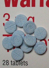
SAN FRANSICO—New research suggests bleeds and thrombosis are not the only adverse effects of improper warfarin dosing.
The study showed an increased risk of dementia among patients with atrial fibrillation whose warfarin doses were outside the therapeutic range for an extended period of time.
Jared Bunch, MD, of the Intermountain Medical Center Heart Institute in Murray, Utah, and his colleagues presented this finding at the 2014 Annual Heart Rhythm Society Scientific Session (PO05-169).
Previous research suggested that patients with atrial fibrillation had an increased risk of developing dementia. But the cause of that association
was unknown.
“[W]e now know that if warfarin doses are consistently too high or too low, one of the long-term consequences can be brain damage,” Dr Bunch said. “This points to the possibility that dementia in atrial fibrillation patients is partly due to small, repetitive clots and/or bleeds in the brain.”
To make this connection, the researchers analyzed data from 2693 atrial fibrillation patients receiving warfarin. The team evaluated the relationship between dementia and the percentage of time that patients’ warfarin doses were within the therapeutic range (international normalized ratio of 2 to 3).
In all, 4.1% of patients (n=111) were diagnosed with dementia. This included senile dementia (n=37, 1.4%), vascular dementia (n=8, 0.3%), and Alzheimer’s dementia (n=66, 2.4%).
The researchers’ analysis (adjusted for the risk of stroke and bleeding) revealed that the more time a patient’s warfarin dosages were outside the therapeutic range, the greater his risk of developing dementia.
Specifically, patients within the therapeutic range less than 25% of the time were 4.6 times more likely to develop dementia than patients within range more than 75% of the time.
Patients within the therapeutic range 25% to 50% of the time were 4.1 times more likely to develop dementia. And patients within the therapeutic range 51% to 75% of the time were 2.5 times more likely to develop dementia.
“Our results from the study tell us 2 things,” Dr Bunch said. “With careful use of anticoagulation medications, the dementia risk can be reduced.
Patients on warfarin need very close follow-up in specialized anticoagulation centers, if possible, to ensure their blood levels are within the recommended levels more often.”
“Second, these results also point to a potential new long-term consequence of dependency on long-term anticoagulation medications. In this regard, stroke prevention therapies do not require long-term anticoagulation medications, and reducing the use of these drugs will hopefully lower
dementia risk.”
SAN FRANSICO—New research suggests bleeds and thrombosis are not the only adverse effects of improper warfarin dosing.
The study showed an increased risk of dementia among patients with atrial fibrillation whose warfarin doses were outside the therapeutic range for an extended period of time.
Jared Bunch, MD, of the Intermountain Medical Center Heart Institute in Murray, Utah, and his colleagues presented this finding at the 2014 Annual Heart Rhythm Society Scientific Session (PO05-169).
Previous research suggested that patients with atrial fibrillation had an increased risk of developing dementia. But the cause of that association
was unknown.
“[W]e now know that if warfarin doses are consistently too high or too low, one of the long-term consequences can be brain damage,” Dr Bunch said. “This points to the possibility that dementia in atrial fibrillation patients is partly due to small, repetitive clots and/or bleeds in the brain.”
To make this connection, the researchers analyzed data from 2693 atrial fibrillation patients receiving warfarin. The team evaluated the relationship between dementia and the percentage of time that patients’ warfarin doses were within the therapeutic range (international normalized ratio of 2 to 3).
In all, 4.1% of patients (n=111) were diagnosed with dementia. This included senile dementia (n=37, 1.4%), vascular dementia (n=8, 0.3%), and Alzheimer’s dementia (n=66, 2.4%).
The researchers’ analysis (adjusted for the risk of stroke and bleeding) revealed that the more time a patient’s warfarin dosages were outside the therapeutic range, the greater his risk of developing dementia.
Specifically, patients within the therapeutic range less than 25% of the time were 4.6 times more likely to develop dementia than patients within range more than 75% of the time.
Patients within the therapeutic range 25% to 50% of the time were 4.1 times more likely to develop dementia. And patients within the therapeutic range 51% to 75% of the time were 2.5 times more likely to develop dementia.
“Our results from the study tell us 2 things,” Dr Bunch said. “With careful use of anticoagulation medications, the dementia risk can be reduced.
Patients on warfarin need very close follow-up in specialized anticoagulation centers, if possible, to ensure their blood levels are within the recommended levels more often.”
“Second, these results also point to a potential new long-term consequence of dependency on long-term anticoagulation medications. In this regard, stroke prevention therapies do not require long-term anticoagulation medications, and reducing the use of these drugs will hopefully lower
dementia risk.”
SAN FRANSICO—New research suggests bleeds and thrombosis are not the only adverse effects of improper warfarin dosing.
The study showed an increased risk of dementia among patients with atrial fibrillation whose warfarin doses were outside the therapeutic range for an extended period of time.
Jared Bunch, MD, of the Intermountain Medical Center Heart Institute in Murray, Utah, and his colleagues presented this finding at the 2014 Annual Heart Rhythm Society Scientific Session (PO05-169).
Previous research suggested that patients with atrial fibrillation had an increased risk of developing dementia. But the cause of that association
was unknown.
“[W]e now know that if warfarin doses are consistently too high or too low, one of the long-term consequences can be brain damage,” Dr Bunch said. “This points to the possibility that dementia in atrial fibrillation patients is partly due to small, repetitive clots and/or bleeds in the brain.”
To make this connection, the researchers analyzed data from 2693 atrial fibrillation patients receiving warfarin. The team evaluated the relationship between dementia and the percentage of time that patients’ warfarin doses were within the therapeutic range (international normalized ratio of 2 to 3).
In all, 4.1% of patients (n=111) were diagnosed with dementia. This included senile dementia (n=37, 1.4%), vascular dementia (n=8, 0.3%), and Alzheimer’s dementia (n=66, 2.4%).
The researchers’ analysis (adjusted for the risk of stroke and bleeding) revealed that the more time a patient’s warfarin dosages were outside the therapeutic range, the greater his risk of developing dementia.
Specifically, patients within the therapeutic range less than 25% of the time were 4.6 times more likely to develop dementia than patients within range more than 75% of the time.
Patients within the therapeutic range 25% to 50% of the time were 4.1 times more likely to develop dementia. And patients within the therapeutic range 51% to 75% of the time were 2.5 times more likely to develop dementia.
“Our results from the study tell us 2 things,” Dr Bunch said. “With careful use of anticoagulation medications, the dementia risk can be reduced.
Patients on warfarin need very close follow-up in specialized anticoagulation centers, if possible, to ensure their blood levels are within the recommended levels more often.”
“Second, these results also point to a potential new long-term consequence of dependency on long-term anticoagulation medications. In this regard, stroke prevention therapies do not require long-term anticoagulation medications, and reducing the use of these drugs will hopefully lower
dementia risk.”
FDA approves octocog alfa for adults with hemophilia A
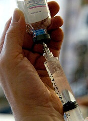
The US Food and Drug Administration (FDA) has approved a new indication for the recombinant antihemophilic factor VIII product octocog alfa (Kogenate).
It is now approved for routine prophylaxis to prevent or reduce the frequency of bleeding episodes in adults with hemophilia A.
Octocog alfa was already FDA-approved for the control and prevention of bleeding episodes in adults and children with hemophilia A, for perioperative management in adults and children with hemophilia A, as routine prophylaxis in children with hemophilia A, and to reduce the risk of joint damage in children without pre-existing joint damage.
The FDA’s latest approval is based on data from the SPINART study, in which patients were randomized to receive prophylactic octocog alfa or on-demand treatment. The study was sponsored by Bayer Healthcare, the company developing octocog alfa.
“In Bayer’s SPINART study, adult patients with hemophilia A on the prophylactic regimen experienced significantly fewer bleeding events than those using on-demand treatment,” said Marilyn Manco-Johnson, MD, principal investigator of the study and director at of the Mountain States Regional Hemophilia and Thrombosis Center at the University of Colorado at Denver and Health Sciences Center.
Results of the SPINART study were presented at the World Federation of Hemophilia 2012 World Congress and published in The Journal of Thrombosis and Haemostasis.
The study included 84 patients, ages 15 to 50, with hemophilia A. They were randomized to a prophylaxis regimen of 25 IU/kg 3 times per week (n=42) or on-demand treatment (n=42). Escalation by 5 IU/kg (to 30 or 35 IU/kg maximum) was allowed for subjects with 12 or more annual bleeds after 1 and 2 years.
Patients were stratified based on target joints (presence/absence) and the number of bleeding events in the previous 6 months (≥15 vs <15 annualized bleeds).
Patients who received prophylaxis experienced significantly fewer bleeds (P<0.0001) than patients treated on demand, regardless of factors such as age, bleeding history, and the presence or absence of target joints.
The ratio of the mean bleeding frequency was 15.2 (P<0.0001) for on-demand vs prophylaxis, indicating that patients who received on-demand treatment experienced, on average, 15.2 times as many bleeds as patients treated prophylactically.
The mean annualized bleed rates (bleeds/subject/year) were 37 in the on-demand group versus 2 in the prophylaxis group. The median annualized bleed rate in the on-demand group was 33 versus 0 in the prophylaxis group.
Fifty-two percent (22/42) of prophylaxis subjects experienced no bleeding, and 29% (12/42) of prophylaxis subjects experienced 1 to 2 bleeds during the follow-up period.
Adverse events were consistent with the existing safety profile for octocog alfa. The most common adverse reactions (≥4%) were inhibitor formation in previously untreated and minimally treated patients, skin-related hypersensitivity reactions (eg, rash, pruritus), infusion-site reactions (eg, inflammation, pain), and infections associated with a central venous access device.
Serious adverse reactions associated with octocog alfa include systemic hypersensitivity reactions—bronchospastic reactions and/or hypotension and anaphylaxis—and the development of high-titer inhibitors necessitating alternative treatments to factor VIII.
For more details on octocog alfa, see the full prescribing information.
The US Food and Drug Administration (FDA) has approved a new indication for the recombinant antihemophilic factor VIII product octocog alfa (Kogenate).
It is now approved for routine prophylaxis to prevent or reduce the frequency of bleeding episodes in adults with hemophilia A.
Octocog alfa was already FDA-approved for the control and prevention of bleeding episodes in adults and children with hemophilia A, for perioperative management in adults and children with hemophilia A, as routine prophylaxis in children with hemophilia A, and to reduce the risk of joint damage in children without pre-existing joint damage.
The FDA’s latest approval is based on data from the SPINART study, in which patients were randomized to receive prophylactic octocog alfa or on-demand treatment. The study was sponsored by Bayer Healthcare, the company developing octocog alfa.
“In Bayer’s SPINART study, adult patients with hemophilia A on the prophylactic regimen experienced significantly fewer bleeding events than those using on-demand treatment,” said Marilyn Manco-Johnson, MD, principal investigator of the study and director at of the Mountain States Regional Hemophilia and Thrombosis Center at the University of Colorado at Denver and Health Sciences Center.
Results of the SPINART study were presented at the World Federation of Hemophilia 2012 World Congress and published in The Journal of Thrombosis and Haemostasis.
The study included 84 patients, ages 15 to 50, with hemophilia A. They were randomized to a prophylaxis regimen of 25 IU/kg 3 times per week (n=42) or on-demand treatment (n=42). Escalation by 5 IU/kg (to 30 or 35 IU/kg maximum) was allowed for subjects with 12 or more annual bleeds after 1 and 2 years.
Patients were stratified based on target joints (presence/absence) and the number of bleeding events in the previous 6 months (≥15 vs <15 annualized bleeds).
Patients who received prophylaxis experienced significantly fewer bleeds (P<0.0001) than patients treated on demand, regardless of factors such as age, bleeding history, and the presence or absence of target joints.
The ratio of the mean bleeding frequency was 15.2 (P<0.0001) for on-demand vs prophylaxis, indicating that patients who received on-demand treatment experienced, on average, 15.2 times as many bleeds as patients treated prophylactically.
The mean annualized bleed rates (bleeds/subject/year) were 37 in the on-demand group versus 2 in the prophylaxis group. The median annualized bleed rate in the on-demand group was 33 versus 0 in the prophylaxis group.
Fifty-two percent (22/42) of prophylaxis subjects experienced no bleeding, and 29% (12/42) of prophylaxis subjects experienced 1 to 2 bleeds during the follow-up period.
Adverse events were consistent with the existing safety profile for octocog alfa. The most common adverse reactions (≥4%) were inhibitor formation in previously untreated and minimally treated patients, skin-related hypersensitivity reactions (eg, rash, pruritus), infusion-site reactions (eg, inflammation, pain), and infections associated with a central venous access device.
Serious adverse reactions associated with octocog alfa include systemic hypersensitivity reactions—bronchospastic reactions and/or hypotension and anaphylaxis—and the development of high-titer inhibitors necessitating alternative treatments to factor VIII.
For more details on octocog alfa, see the full prescribing information.
The US Food and Drug Administration (FDA) has approved a new indication for the recombinant antihemophilic factor VIII product octocog alfa (Kogenate).
It is now approved for routine prophylaxis to prevent or reduce the frequency of bleeding episodes in adults with hemophilia A.
Octocog alfa was already FDA-approved for the control and prevention of bleeding episodes in adults and children with hemophilia A, for perioperative management in adults and children with hemophilia A, as routine prophylaxis in children with hemophilia A, and to reduce the risk of joint damage in children without pre-existing joint damage.
The FDA’s latest approval is based on data from the SPINART study, in which patients were randomized to receive prophylactic octocog alfa or on-demand treatment. The study was sponsored by Bayer Healthcare, the company developing octocog alfa.
“In Bayer’s SPINART study, adult patients with hemophilia A on the prophylactic regimen experienced significantly fewer bleeding events than those using on-demand treatment,” said Marilyn Manco-Johnson, MD, principal investigator of the study and director at of the Mountain States Regional Hemophilia and Thrombosis Center at the University of Colorado at Denver and Health Sciences Center.
Results of the SPINART study were presented at the World Federation of Hemophilia 2012 World Congress and published in The Journal of Thrombosis and Haemostasis.
The study included 84 patients, ages 15 to 50, with hemophilia A. They were randomized to a prophylaxis regimen of 25 IU/kg 3 times per week (n=42) or on-demand treatment (n=42). Escalation by 5 IU/kg (to 30 or 35 IU/kg maximum) was allowed for subjects with 12 or more annual bleeds after 1 and 2 years.
Patients were stratified based on target joints (presence/absence) and the number of bleeding events in the previous 6 months (≥15 vs <15 annualized bleeds).
Patients who received prophylaxis experienced significantly fewer bleeds (P<0.0001) than patients treated on demand, regardless of factors such as age, bleeding history, and the presence or absence of target joints.
The ratio of the mean bleeding frequency was 15.2 (P<0.0001) for on-demand vs prophylaxis, indicating that patients who received on-demand treatment experienced, on average, 15.2 times as many bleeds as patients treated prophylactically.
The mean annualized bleed rates (bleeds/subject/year) were 37 in the on-demand group versus 2 in the prophylaxis group. The median annualized bleed rate in the on-demand group was 33 versus 0 in the prophylaxis group.
Fifty-two percent (22/42) of prophylaxis subjects experienced no bleeding, and 29% (12/42) of prophylaxis subjects experienced 1 to 2 bleeds during the follow-up period.
Adverse events were consistent with the existing safety profile for octocog alfa. The most common adverse reactions (≥4%) were inhibitor formation in previously untreated and minimally treated patients, skin-related hypersensitivity reactions (eg, rash, pruritus), infusion-site reactions (eg, inflammation, pain), and infections associated with a central venous access device.
Serious adverse reactions associated with octocog alfa include systemic hypersensitivity reactions—bronchospastic reactions and/or hypotension and anaphylaxis—and the development of high-titer inhibitors necessitating alternative treatments to factor VIII.
For more details on octocog alfa, see the full prescribing information.
Drug delivery vehicle has antimyeloma effects
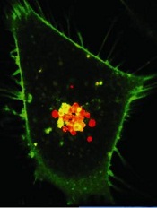
Credit: PNAS
Researchers have discovered that a solvent used as a drug delivery vehicle has antimyeloma properties.
The team found that N-methyl-2-pyrrolidone (NMP)—long-regarded a basic, stable, and inactive solvent—affects the growth and survival of multiple myeloma (MM) cells and stimulates the immune system to kill these tumors.
Jake Shortt, MBChB, PhD, of the Peter MacCallum Cancer Centre in Melbourne, Australia, described these findings in Cell Reports.
“[W]e found NMP effectively ‘reprograms’ myeloma cells by targeting a class of gene-regulating proteins,” Dr Shortt said. “This reprogramming reawakens thousands of genes that have been silenced in the cancer cells, immediately stopping the myeloma cells from growing, while activating the immune system to respond to the cancer.”
The researchers were surprised by this discovery because they had been using NMP as a vehicle to deliver drugs in mouse models of MM.
“In a routine experiment in 2010, Dr Shortt noticed our preclinical models of myeloma were responding to the control dose of NMP, which was surprising, as this control dose contained none of the novel cancer agents we were actually testing,” said study author Ricky Johnstone, PhD, also of the Peter MacCallum Cancer Centre.
Specifically, the researchers were using NMP as a drug delivery vehicle in mice transplanted with Vk*MYC MM tumors. And they consistently observed antitumor activity in vehicle-only control mice.
The team saw responses to NMP in mice transplanted with multiple, independently derived myelomas; namely, delayed paraprotein progression and improved survival in aggressive clones and sustained regressions in more indolent disease.
Further investigation revealed that NMP is an acetylated lysine mimetic and bromodomain ligand. And NMP shares molecular targets with lenalidomide, including cMYC and IRF4.
Unlike lenalidomide, NMP showed CRBN-independent activity. NMP induced similar growth arrest in CRBN-knockdown MM cells and lenalidomide-resistant MM cells.
Based on these findings, the researchers are planning a phase 1 trial of NMP, which is due to start later this year. The team noted that, because safe levels of NMP in humans are already well-established, the study is in the advanced planning stage.
“We’re at an advantage with this trial because we can immediately start at dosage levels within those recommended under occupational health and safety guidelines,” said David Ritchie, MBChB, PhD, a professor at the Peter MacCallum Cancer Centre and chief investigator of the phase 1 trial.
“It is extremely exciting to have this new insight into NMP, which is comparatively cost-effective and plentiful, compared to novel treatments developed by pharmaceutical companies, and hopefully holds promise for new or improved treatments in other cancer types.”
Credit: PNAS
Researchers have discovered that a solvent used as a drug delivery vehicle has antimyeloma properties.
The team found that N-methyl-2-pyrrolidone (NMP)—long-regarded a basic, stable, and inactive solvent—affects the growth and survival of multiple myeloma (MM) cells and stimulates the immune system to kill these tumors.
Jake Shortt, MBChB, PhD, of the Peter MacCallum Cancer Centre in Melbourne, Australia, described these findings in Cell Reports.
“[W]e found NMP effectively ‘reprograms’ myeloma cells by targeting a class of gene-regulating proteins,” Dr Shortt said. “This reprogramming reawakens thousands of genes that have been silenced in the cancer cells, immediately stopping the myeloma cells from growing, while activating the immune system to respond to the cancer.”
The researchers were surprised by this discovery because they had been using NMP as a vehicle to deliver drugs in mouse models of MM.
“In a routine experiment in 2010, Dr Shortt noticed our preclinical models of myeloma were responding to the control dose of NMP, which was surprising, as this control dose contained none of the novel cancer agents we were actually testing,” said study author Ricky Johnstone, PhD, also of the Peter MacCallum Cancer Centre.
Specifically, the researchers were using NMP as a drug delivery vehicle in mice transplanted with Vk*MYC MM tumors. And they consistently observed antitumor activity in vehicle-only control mice.
The team saw responses to NMP in mice transplanted with multiple, independently derived myelomas; namely, delayed paraprotein progression and improved survival in aggressive clones and sustained regressions in more indolent disease.
Further investigation revealed that NMP is an acetylated lysine mimetic and bromodomain ligand. And NMP shares molecular targets with lenalidomide, including cMYC and IRF4.
Unlike lenalidomide, NMP showed CRBN-independent activity. NMP induced similar growth arrest in CRBN-knockdown MM cells and lenalidomide-resistant MM cells.
Based on these findings, the researchers are planning a phase 1 trial of NMP, which is due to start later this year. The team noted that, because safe levels of NMP in humans are already well-established, the study is in the advanced planning stage.
“We’re at an advantage with this trial because we can immediately start at dosage levels within those recommended under occupational health and safety guidelines,” said David Ritchie, MBChB, PhD, a professor at the Peter MacCallum Cancer Centre and chief investigator of the phase 1 trial.
“It is extremely exciting to have this new insight into NMP, which is comparatively cost-effective and plentiful, compared to novel treatments developed by pharmaceutical companies, and hopefully holds promise for new or improved treatments in other cancer types.”
Credit: PNAS
Researchers have discovered that a solvent used as a drug delivery vehicle has antimyeloma properties.
The team found that N-methyl-2-pyrrolidone (NMP)—long-regarded a basic, stable, and inactive solvent—affects the growth and survival of multiple myeloma (MM) cells and stimulates the immune system to kill these tumors.
Jake Shortt, MBChB, PhD, of the Peter MacCallum Cancer Centre in Melbourne, Australia, described these findings in Cell Reports.
“[W]e found NMP effectively ‘reprograms’ myeloma cells by targeting a class of gene-regulating proteins,” Dr Shortt said. “This reprogramming reawakens thousands of genes that have been silenced in the cancer cells, immediately stopping the myeloma cells from growing, while activating the immune system to respond to the cancer.”
The researchers were surprised by this discovery because they had been using NMP as a vehicle to deliver drugs in mouse models of MM.
“In a routine experiment in 2010, Dr Shortt noticed our preclinical models of myeloma were responding to the control dose of NMP, which was surprising, as this control dose contained none of the novel cancer agents we were actually testing,” said study author Ricky Johnstone, PhD, also of the Peter MacCallum Cancer Centre.
Specifically, the researchers were using NMP as a drug delivery vehicle in mice transplanted with Vk*MYC MM tumors. And they consistently observed antitumor activity in vehicle-only control mice.
The team saw responses to NMP in mice transplanted with multiple, independently derived myelomas; namely, delayed paraprotein progression and improved survival in aggressive clones and sustained regressions in more indolent disease.
Further investigation revealed that NMP is an acetylated lysine mimetic and bromodomain ligand. And NMP shares molecular targets with lenalidomide, including cMYC and IRF4.
Unlike lenalidomide, NMP showed CRBN-independent activity. NMP induced similar growth arrest in CRBN-knockdown MM cells and lenalidomide-resistant MM cells.
Based on these findings, the researchers are planning a phase 1 trial of NMP, which is due to start later this year. The team noted that, because safe levels of NMP in humans are already well-established, the study is in the advanced planning stage.
“We’re at an advantage with this trial because we can immediately start at dosage levels within those recommended under occupational health and safety guidelines,” said David Ritchie, MBChB, PhD, a professor at the Peter MacCallum Cancer Centre and chief investigator of the phase 1 trial.
“It is extremely exciting to have this new insight into NMP, which is comparatively cost-effective and plentiful, compared to novel treatments developed by pharmaceutical companies, and hopefully holds promise for new or improved treatments in other cancer types.”
Method can track circulating cancer cells

Investigators have developed a technique that allowed them to track single tumor circulating in the blood of mice.
The method, described in Chemistry & Biology, involves photoswitchable fluorescent proteins that change color in response to light.
When one laser light hits the circulating tumor cells, they appear to be fluorescent green. A second laser makes the cells appear fluorescent red.
To label cells, the investigators use a violet laser beam aimed at small blood vessels.
The fluorescence from each cell is collected, detected, and reproduced on a computer monitor as real-time signal traces, allowing the team to count and track individual cells in the bloodstream.
“This technology allows for the labeling of just one circulating pathological cell among billions of other normal blood cells by ultrafast changing color of photosensitive proteins inside the cell in response to laser light,” said study author Ekaterina Galanzha, PhD, of the University of Arkansas for Medical Sciences in Little Rock.
In tumor-bearing mice, the investigators could monitor the real-time dynamics of circulating cancer cells released from a primary tumor.
They could also image the various final destinations of individual circulating cells and observe how these cells travel through circulation and colonize healthy tissue, existing sites of metastasis, or the site of the primary tumor.
“Therefore, the approach may give oncologists knowledge on how to intervene and stop circulating cancer cell dissemination that might prevent the development of metastasis,” Dr Galanzha said.
The investigators believe the approach might also prove useful for other areas of medicine—for example, tracking bacteria during infections or immune-related cells during the development of autoimmune disease.
Investigators have developed a technique that allowed them to track single tumor circulating in the blood of mice.
The method, described in Chemistry & Biology, involves photoswitchable fluorescent proteins that change color in response to light.
When one laser light hits the circulating tumor cells, they appear to be fluorescent green. A second laser makes the cells appear fluorescent red.
To label cells, the investigators use a violet laser beam aimed at small blood vessels.
The fluorescence from each cell is collected, detected, and reproduced on a computer monitor as real-time signal traces, allowing the team to count and track individual cells in the bloodstream.
“This technology allows for the labeling of just one circulating pathological cell among billions of other normal blood cells by ultrafast changing color of photosensitive proteins inside the cell in response to laser light,” said study author Ekaterina Galanzha, PhD, of the University of Arkansas for Medical Sciences in Little Rock.
In tumor-bearing mice, the investigators could monitor the real-time dynamics of circulating cancer cells released from a primary tumor.
They could also image the various final destinations of individual circulating cells and observe how these cells travel through circulation and colonize healthy tissue, existing sites of metastasis, or the site of the primary tumor.
“Therefore, the approach may give oncologists knowledge on how to intervene and stop circulating cancer cell dissemination that might prevent the development of metastasis,” Dr Galanzha said.
The investigators believe the approach might also prove useful for other areas of medicine—for example, tracking bacteria during infections or immune-related cells during the development of autoimmune disease.
Investigators have developed a technique that allowed them to track single tumor circulating in the blood of mice.
The method, described in Chemistry & Biology, involves photoswitchable fluorescent proteins that change color in response to light.
When one laser light hits the circulating tumor cells, they appear to be fluorescent green. A second laser makes the cells appear fluorescent red.
To label cells, the investigators use a violet laser beam aimed at small blood vessels.
The fluorescence from each cell is collected, detected, and reproduced on a computer monitor as real-time signal traces, allowing the team to count and track individual cells in the bloodstream.
“This technology allows for the labeling of just one circulating pathological cell among billions of other normal blood cells by ultrafast changing color of photosensitive proteins inside the cell in response to laser light,” said study author Ekaterina Galanzha, PhD, of the University of Arkansas for Medical Sciences in Little Rock.
In tumor-bearing mice, the investigators could monitor the real-time dynamics of circulating cancer cells released from a primary tumor.
They could also image the various final destinations of individual circulating cells and observe how these cells travel through circulation and colonize healthy tissue, existing sites of metastasis, or the site of the primary tumor.
“Therefore, the approach may give oncologists knowledge on how to intervene and stop circulating cancer cell dissemination that might prevent the development of metastasis,” Dr Galanzha said.
The investigators believe the approach might also prove useful for other areas of medicine—for example, tracking bacteria during infections or immune-related cells during the development of autoimmune disease.
Study reveals racial disparity in perioperative transfusion practices
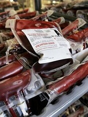
Credit: Elise Amendola
Results of a large study showed that black patients were more likely than white patients to receive perioperative blood transfusions for 2 of 3 common surgical procedures.
Researchers evaluated transfusion practices in these 2 racial groups for coronary artery bypass surgery (CABG), total hip replacement (THR), and colectomy.
And they found that black patients undergoing CABG or THR had a significantly higher incidence of transfusion than white patients undergoing these procedures.
Feng Qian, PhD, of the University at Albany School of Public Health, and his colleagues reported these findings in BMC Health Services Research.
The team examined the use of perioperative red blood cell transfusion using patient data from the University Health System Consortium, a network of academic medical centers and affiliated hospitals. The data included hospitalizations occurring from 2009 to 2011.
The researchers’ final sample included 42,933 patients who underwent THR (37,888 white and 5045 black), 25,849 patients who underwent CABG (23,113 white and 2736 black), and 8255 patients who underwent colectomy (6861 white and 1394 black).
Black patients tended to be younger than white patients, with the overall age ranging from 48 to 73 years. Blacks were also less well-insured than whites and more likely to have comorbidities such as diabetes, renal failure, and anemia.
Dr Qian and his colleagues adjusted for these differences in their analysis, as well as for patient gender, admission status, and severity of illness.
The analysis revealed that black patients undergoing CABG had a 41% higher incidence of perioperative transfusion than white patients (P=0.002).
For THR, the incidence of transfusion was 39% higher among blacks than whites (P<0.001). And for colectomy, the incidence was 8% higher among blacks than whites (P=0.40).
The researchers then performed an analysis adjusted for the aforementioned factors as well as for hospital-fixed effects.
This revealed that black patients undergoing CABG had a 42% higher incidence of transfusion than whites (P<0.001). Blacks undergoing THR had a 43% higher incidence of transfusion (P<0.001). And blacks undergoing colectomy had a 1% higher incidence of transfusion (P=0.92)
The researchers noted that, although blood transfusion is widely employed in surgery, the practice is associated with adverse outcomes. So overuse of transfusions may pose serious health risks, specifically in black patients undergoing CABG and THR.
Dr Qian added that recognizing racial disparities related to the use of perioperative red blood cell transfusion may help reduce potentially unnecessary transfusions in minority patients.
Credit: Elise Amendola
Results of a large study showed that black patients were more likely than white patients to receive perioperative blood transfusions for 2 of 3 common surgical procedures.
Researchers evaluated transfusion practices in these 2 racial groups for coronary artery bypass surgery (CABG), total hip replacement (THR), and colectomy.
And they found that black patients undergoing CABG or THR had a significantly higher incidence of transfusion than white patients undergoing these procedures.
Feng Qian, PhD, of the University at Albany School of Public Health, and his colleagues reported these findings in BMC Health Services Research.
The team examined the use of perioperative red blood cell transfusion using patient data from the University Health System Consortium, a network of academic medical centers and affiliated hospitals. The data included hospitalizations occurring from 2009 to 2011.
The researchers’ final sample included 42,933 patients who underwent THR (37,888 white and 5045 black), 25,849 patients who underwent CABG (23,113 white and 2736 black), and 8255 patients who underwent colectomy (6861 white and 1394 black).
Black patients tended to be younger than white patients, with the overall age ranging from 48 to 73 years. Blacks were also less well-insured than whites and more likely to have comorbidities such as diabetes, renal failure, and anemia.
Dr Qian and his colleagues adjusted for these differences in their analysis, as well as for patient gender, admission status, and severity of illness.
The analysis revealed that black patients undergoing CABG had a 41% higher incidence of perioperative transfusion than white patients (P=0.002).
For THR, the incidence of transfusion was 39% higher among blacks than whites (P<0.001). And for colectomy, the incidence was 8% higher among blacks than whites (P=0.40).
The researchers then performed an analysis adjusted for the aforementioned factors as well as for hospital-fixed effects.
This revealed that black patients undergoing CABG had a 42% higher incidence of transfusion than whites (P<0.001). Blacks undergoing THR had a 43% higher incidence of transfusion (P<0.001). And blacks undergoing colectomy had a 1% higher incidence of transfusion (P=0.92)
The researchers noted that, although blood transfusion is widely employed in surgery, the practice is associated with adverse outcomes. So overuse of transfusions may pose serious health risks, specifically in black patients undergoing CABG and THR.
Dr Qian added that recognizing racial disparities related to the use of perioperative red blood cell transfusion may help reduce potentially unnecessary transfusions in minority patients.
Credit: Elise Amendola
Results of a large study showed that black patients were more likely than white patients to receive perioperative blood transfusions for 2 of 3 common surgical procedures.
Researchers evaluated transfusion practices in these 2 racial groups for coronary artery bypass surgery (CABG), total hip replacement (THR), and colectomy.
And they found that black patients undergoing CABG or THR had a significantly higher incidence of transfusion than white patients undergoing these procedures.
Feng Qian, PhD, of the University at Albany School of Public Health, and his colleagues reported these findings in BMC Health Services Research.
The team examined the use of perioperative red blood cell transfusion using patient data from the University Health System Consortium, a network of academic medical centers and affiliated hospitals. The data included hospitalizations occurring from 2009 to 2011.
The researchers’ final sample included 42,933 patients who underwent THR (37,888 white and 5045 black), 25,849 patients who underwent CABG (23,113 white and 2736 black), and 8255 patients who underwent colectomy (6861 white and 1394 black).
Black patients tended to be younger than white patients, with the overall age ranging from 48 to 73 years. Blacks were also less well-insured than whites and more likely to have comorbidities such as diabetes, renal failure, and anemia.
Dr Qian and his colleagues adjusted for these differences in their analysis, as well as for patient gender, admission status, and severity of illness.
The analysis revealed that black patients undergoing CABG had a 41% higher incidence of perioperative transfusion than white patients (P=0.002).
For THR, the incidence of transfusion was 39% higher among blacks than whites (P<0.001). And for colectomy, the incidence was 8% higher among blacks than whites (P=0.40).
The researchers then performed an analysis adjusted for the aforementioned factors as well as for hospital-fixed effects.
This revealed that black patients undergoing CABG had a 42% higher incidence of transfusion than whites (P<0.001). Blacks undergoing THR had a 43% higher incidence of transfusion (P<0.001). And blacks undergoing colectomy had a 1% higher incidence of transfusion (P=0.92)
The researchers noted that, although blood transfusion is widely employed in surgery, the practice is associated with adverse outcomes. So overuse of transfusions may pose serious health risks, specifically in black patients undergoing CABG and THR.
Dr Qian added that recognizing racial disparities related to the use of perioperative red blood cell transfusion may help reduce potentially unnecessary transfusions in minority patients.
Recycled RBCs prove more functional than banked ones
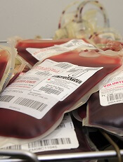
Credit: UAB Hospital
Reinfusing the blood a patient loses during cardiopulmonary bypass surgery confers benefits over transfusing the patient with banked blood, results of a small study suggest.
Investigators noted that both the surgery and red blood cell (RBC) storage are associated with changes in RBCs that can adversely affect oxygen delivery.
However, their study revealed minimal effects on RBC structure and function among patients who received their own recycled blood during surgery.
On the other hand, patients who received banked RBCs along with their own blood experienced a dose-dependent decrease in RBC cell membrane deformability that could persist beyond 3 days.
Steven Frank, MD, of the Johns Hopkins University School of Medicine in Baltimore, Maryland, and his colleagues reported these findings in Anesthesia & Analgesia.
The team studied 32 patients undergoing cardiopulmonary bypass, categorizing them by their transfusion status: those who received their own RBCs (n=12), those who received their own blood plus fewer than 5 units of banked blood (n=10), and those who received their own RBCs plus 5 or more units of stored blood (n=10).
All patients had blood samples drawn before, during, and for 3 days after surgery. The investigators examined samples for blood cell membrane stiffness and flexibility.
In patients who received only their own recycled blood, their cells behaved normally right away, as if they had never been outside the body.
But the more banked blood a patient received, the less flexible their entire population of RBCs. Three days after surgery, RBCs in the group that received the largest number of transfused units still had not recovered their full function.
“We now have more evidence that fresh blood cells are of a higher quality than what comes from a blood bank,” Dr Frank said.
“If banked blood, which is stored for up to 6 weeks, is now shown to be of a lower quality, it makes more sense to use recycled blood that has only been outside the body for 1 or 2 hours. It’s always been the case that patients feel better about getting their own blood, and recycling is also more cost-effective.”
The investigators used a cell saver machine to collect the material a patient lost during surgery. They then rinsed away the unneeded fat and tissue, centrifuged and separated the red cells, and returned them to the patient.
Dr Frank and his colleagues noted that disposable parts of the cell saver, which can be used to process multiple units of blood, cost around $120, compared to $240 for each unit of banked blood. Additionally, recycling blood reduces a patient’s risk of contracting infections and experiencing transfusion-related adverse reactions.
Dr Frank pointed out, however, that cell saver machines are not appropriate for all operations, and not all hospitals have access to round-the-clock perfusionists to run them. For heart surgeries, a perfusionist is already in the operating room to run the heart-lung bypass machine.
Dr Frank also noted that many operations are considered to be a low risk for blood loss, in which case, the cell saver is unnecessary. Nevertheless, he advocates wider use of recycled blood.
“In any patient where you expect to give 1 unit of red blood cells or more, it’s cost-effective and beneficial to recycle,” he said.
Credit: UAB Hospital
Reinfusing the blood a patient loses during cardiopulmonary bypass surgery confers benefits over transfusing the patient with banked blood, results of a small study suggest.
Investigators noted that both the surgery and red blood cell (RBC) storage are associated with changes in RBCs that can adversely affect oxygen delivery.
However, their study revealed minimal effects on RBC structure and function among patients who received their own recycled blood during surgery.
On the other hand, patients who received banked RBCs along with their own blood experienced a dose-dependent decrease in RBC cell membrane deformability that could persist beyond 3 days.
Steven Frank, MD, of the Johns Hopkins University School of Medicine in Baltimore, Maryland, and his colleagues reported these findings in Anesthesia & Analgesia.
The team studied 32 patients undergoing cardiopulmonary bypass, categorizing them by their transfusion status: those who received their own RBCs (n=12), those who received their own blood plus fewer than 5 units of banked blood (n=10), and those who received their own RBCs plus 5 or more units of stored blood (n=10).
All patients had blood samples drawn before, during, and for 3 days after surgery. The investigators examined samples for blood cell membrane stiffness and flexibility.
In patients who received only their own recycled blood, their cells behaved normally right away, as if they had never been outside the body.
But the more banked blood a patient received, the less flexible their entire population of RBCs. Three days after surgery, RBCs in the group that received the largest number of transfused units still had not recovered their full function.
“We now have more evidence that fresh blood cells are of a higher quality than what comes from a blood bank,” Dr Frank said.
“If banked blood, which is stored for up to 6 weeks, is now shown to be of a lower quality, it makes more sense to use recycled blood that has only been outside the body for 1 or 2 hours. It’s always been the case that patients feel better about getting their own blood, and recycling is also more cost-effective.”
The investigators used a cell saver machine to collect the material a patient lost during surgery. They then rinsed away the unneeded fat and tissue, centrifuged and separated the red cells, and returned them to the patient.
Dr Frank and his colleagues noted that disposable parts of the cell saver, which can be used to process multiple units of blood, cost around $120, compared to $240 for each unit of banked blood. Additionally, recycling blood reduces a patient’s risk of contracting infections and experiencing transfusion-related adverse reactions.
Dr Frank pointed out, however, that cell saver machines are not appropriate for all operations, and not all hospitals have access to round-the-clock perfusionists to run them. For heart surgeries, a perfusionist is already in the operating room to run the heart-lung bypass machine.
Dr Frank also noted that many operations are considered to be a low risk for blood loss, in which case, the cell saver is unnecessary. Nevertheless, he advocates wider use of recycled blood.
“In any patient where you expect to give 1 unit of red blood cells or more, it’s cost-effective and beneficial to recycle,” he said.
Credit: UAB Hospital
Reinfusing the blood a patient loses during cardiopulmonary bypass surgery confers benefits over transfusing the patient with banked blood, results of a small study suggest.
Investigators noted that both the surgery and red blood cell (RBC) storage are associated with changes in RBCs that can adversely affect oxygen delivery.
However, their study revealed minimal effects on RBC structure and function among patients who received their own recycled blood during surgery.
On the other hand, patients who received banked RBCs along with their own blood experienced a dose-dependent decrease in RBC cell membrane deformability that could persist beyond 3 days.
Steven Frank, MD, of the Johns Hopkins University School of Medicine in Baltimore, Maryland, and his colleagues reported these findings in Anesthesia & Analgesia.
The team studied 32 patients undergoing cardiopulmonary bypass, categorizing them by their transfusion status: those who received their own RBCs (n=12), those who received their own blood plus fewer than 5 units of banked blood (n=10), and those who received their own RBCs plus 5 or more units of stored blood (n=10).
All patients had blood samples drawn before, during, and for 3 days after surgery. The investigators examined samples for blood cell membrane stiffness and flexibility.
In patients who received only their own recycled blood, their cells behaved normally right away, as if they had never been outside the body.
But the more banked blood a patient received, the less flexible their entire population of RBCs. Three days after surgery, RBCs in the group that received the largest number of transfused units still had not recovered their full function.
“We now have more evidence that fresh blood cells are of a higher quality than what comes from a blood bank,” Dr Frank said.
“If banked blood, which is stored for up to 6 weeks, is now shown to be of a lower quality, it makes more sense to use recycled blood that has only been outside the body for 1 or 2 hours. It’s always been the case that patients feel better about getting their own blood, and recycling is also more cost-effective.”
The investigators used a cell saver machine to collect the material a patient lost during surgery. They then rinsed away the unneeded fat and tissue, centrifuged and separated the red cells, and returned them to the patient.
Dr Frank and his colleagues noted that disposable parts of the cell saver, which can be used to process multiple units of blood, cost around $120, compared to $240 for each unit of banked blood. Additionally, recycling blood reduces a patient’s risk of contracting infections and experiencing transfusion-related adverse reactions.
Dr Frank pointed out, however, that cell saver machines are not appropriate for all operations, and not all hospitals have access to round-the-clock perfusionists to run them. For heart surgeries, a perfusionist is already in the operating room to run the heart-lung bypass machine.
Dr Frank also noted that many operations are considered to be a low risk for blood loss, in which case, the cell saver is unnecessary. Nevertheless, he advocates wider use of recycled blood.
“In any patient where you expect to give 1 unit of red blood cells or more, it’s cost-effective and beneficial to recycle,” he said.
FDA approves antiplatelet agent despite bleeding risk

Credit: Andre E.X. Brown
The US Food and Drug Administration (FDA) has approved the antiplatelet agent vorapaxar (Zontivity) to reduce the risk of thrombotic cardiovascular events in patients with a history of myocardial infarction or peripheral arterial disease.
Results of a large study suggested the drug can be effective as prophylaxis but may also increase the risk of bleeding.
Vorapaxar’s label has a black box warning describing this risk, which includes intracranial hemorrhage and fatal bleeding.
The warning also states that vorapaxar should not be given to patients with a history of stroke, transient ischemic attack, intracranial hemorrhage, or active pathological bleeding.
The drug will be dispensed with an FDA-approved patient medication guide that provides instructions for its use and important safety information.
Vorapaxar tablets are intended to be given once daily, along with aspirin and/or clopidogrel, according to their indications or the standard of care.
“In patients who have had a heart attack or who have peripheral arterial disease, this drug will lower the risk of heart attack, stroke, and cardiovascular death,” said Ellis Unger, MD, director of the Office of Drug Evaluation in the FDA’s Center for Drug Evaluation and Research.
“In the study that supported the drug’s approval, [vorapaxar] lowered this risk from 9.5% to 7.9% over a 3-year period—about 0.5% per year.”
The study, which was published in NEJM, included 26,449 patients with a history of myocardial infarction (n=17,779), ischemic stroke (n=4883), or peripheral arterial disease (n=3787).
The patients were randomized to receive vorapaxar at 2.5 mg daily or matching placebo, in addition to standard antiplatelet therapy.
The study’s primary efficacy endpoint was the composite of death from cardiovascular causes, myocardial infarction, or stroke. At 3 years, 1028 patients (9.3%) in the vorapaxar group and 1176 patients (10.5%) in the placebo group reached the primary endpoint.
Both moderate and severe bleeding events were significantly higher in patients on vorapaxar than in the placebo group. Bleeding occurred in 4.2% and 2.5% of patients, respectively. And intracranial hemorrhage occurred in 1.0% and 0.5%, respectively.
The risk of intracranial bleeding was highest among patients with a history of stroke, so these patients were taken off of vorapaxar early.
Vorapaxar is made by Merck Sharp & Dohme Corp., a subsidiary of Merck & Co., Inc., of Whitehouse Station, New Jersey. For more information on the drug, see the full prescribing information.
Credit: Andre E.X. Brown
The US Food and Drug Administration (FDA) has approved the antiplatelet agent vorapaxar (Zontivity) to reduce the risk of thrombotic cardiovascular events in patients with a history of myocardial infarction or peripheral arterial disease.
Results of a large study suggested the drug can be effective as prophylaxis but may also increase the risk of bleeding.
Vorapaxar’s label has a black box warning describing this risk, which includes intracranial hemorrhage and fatal bleeding.
The warning also states that vorapaxar should not be given to patients with a history of stroke, transient ischemic attack, intracranial hemorrhage, or active pathological bleeding.
The drug will be dispensed with an FDA-approved patient medication guide that provides instructions for its use and important safety information.
Vorapaxar tablets are intended to be given once daily, along with aspirin and/or clopidogrel, according to their indications or the standard of care.
“In patients who have had a heart attack or who have peripheral arterial disease, this drug will lower the risk of heart attack, stroke, and cardiovascular death,” said Ellis Unger, MD, director of the Office of Drug Evaluation in the FDA’s Center for Drug Evaluation and Research.
“In the study that supported the drug’s approval, [vorapaxar] lowered this risk from 9.5% to 7.9% over a 3-year period—about 0.5% per year.”
The study, which was published in NEJM, included 26,449 patients with a history of myocardial infarction (n=17,779), ischemic stroke (n=4883), or peripheral arterial disease (n=3787).
The patients were randomized to receive vorapaxar at 2.5 mg daily or matching placebo, in addition to standard antiplatelet therapy.
The study’s primary efficacy endpoint was the composite of death from cardiovascular causes, myocardial infarction, or stroke. At 3 years, 1028 patients (9.3%) in the vorapaxar group and 1176 patients (10.5%) in the placebo group reached the primary endpoint.
Both moderate and severe bleeding events were significantly higher in patients on vorapaxar than in the placebo group. Bleeding occurred in 4.2% and 2.5% of patients, respectively. And intracranial hemorrhage occurred in 1.0% and 0.5%, respectively.
The risk of intracranial bleeding was highest among patients with a history of stroke, so these patients were taken off of vorapaxar early.
Vorapaxar is made by Merck Sharp & Dohme Corp., a subsidiary of Merck & Co., Inc., of Whitehouse Station, New Jersey. For more information on the drug, see the full prescribing information.
Credit: Andre E.X. Brown
The US Food and Drug Administration (FDA) has approved the antiplatelet agent vorapaxar (Zontivity) to reduce the risk of thrombotic cardiovascular events in patients with a history of myocardial infarction or peripheral arterial disease.
Results of a large study suggested the drug can be effective as prophylaxis but may also increase the risk of bleeding.
Vorapaxar’s label has a black box warning describing this risk, which includes intracranial hemorrhage and fatal bleeding.
The warning also states that vorapaxar should not be given to patients with a history of stroke, transient ischemic attack, intracranial hemorrhage, or active pathological bleeding.
The drug will be dispensed with an FDA-approved patient medication guide that provides instructions for its use and important safety information.
Vorapaxar tablets are intended to be given once daily, along with aspirin and/or clopidogrel, according to their indications or the standard of care.
“In patients who have had a heart attack or who have peripheral arterial disease, this drug will lower the risk of heart attack, stroke, and cardiovascular death,” said Ellis Unger, MD, director of the Office of Drug Evaluation in the FDA’s Center for Drug Evaluation and Research.
“In the study that supported the drug’s approval, [vorapaxar] lowered this risk from 9.5% to 7.9% over a 3-year period—about 0.5% per year.”
The study, which was published in NEJM, included 26,449 patients with a history of myocardial infarction (n=17,779), ischemic stroke (n=4883), or peripheral arterial disease (n=3787).
The patients were randomized to receive vorapaxar at 2.5 mg daily or matching placebo, in addition to standard antiplatelet therapy.
The study’s primary efficacy endpoint was the composite of death from cardiovascular causes, myocardial infarction, or stroke. At 3 years, 1028 patients (9.3%) in the vorapaxar group and 1176 patients (10.5%) in the placebo group reached the primary endpoint.
Both moderate and severe bleeding events were significantly higher in patients on vorapaxar than in the placebo group. Bleeding occurred in 4.2% and 2.5% of patients, respectively. And intracranial hemorrhage occurred in 1.0% and 0.5%, respectively.
The risk of intracranial bleeding was highest among patients with a history of stroke, so these patients were taken off of vorapaxar early.
Vorapaxar is made by Merck Sharp & Dohme Corp., a subsidiary of Merck & Co., Inc., of Whitehouse Station, New Jersey. For more information on the drug, see the full prescribing information.
Paper recounts failed attempt to create STAP cells
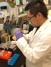
Credit: NIH
A group of researchers who tried, and failed, to replicate the STAP cell phenomenon have detailed their work in F1000Research.
Kenneth Ka Ho Lee, PhD, of the Chinese University of Hong Kong, and his colleagues attempted to create STAP (stimulus-triggered acquisition of pluripotency) cells using the method described in a recent Nature paper.
The paper’s authors had reported they could induce pluripotency in somatic cells by bathing them in acid.
However, not long after the paper and a related letter were published, members of the scientific community voiced concerns about the publications—such as suspicions of plagiarism and the possibility of doctored images—and began to question the findings.
Then, an investigation by the Japanese research institute RIKEN (where many of the researchers are employed) suggested that at least some of the paper’s authors were guilty of misconduct and/or negligence.
However, the study’s lead author, Haruko Obokata, PhD, has maintained that, despite errors in the paper, STAP cells can be created.
Dr Lee’s experiments suggest otherwise—or at least that the cells cannot be created using the methods outlined in the Nature paper.
Dr Lee and his colleagues reported that carefully replicating the original acid-treatment method does not induce pluripotency in 2 types of mouse somatic cells.
Using both white blood cells isolated from the spleen of neonatal mice—the same cells used in the original study—and lung fibroblasts, the team was unable to replicate the original findings.
They’ve published a full account of this work in F1000Research. The journal also employs open peer review by invited experts, which occurs after publication and is published in full online alongside the paper.
Dr Lee’s study will now undergo this process, and readers interested to see referees’ views as they come in can follow the paper by clicking “Track” on the published article.
Credit: NIH
A group of researchers who tried, and failed, to replicate the STAP cell phenomenon have detailed their work in F1000Research.
Kenneth Ka Ho Lee, PhD, of the Chinese University of Hong Kong, and his colleagues attempted to create STAP (stimulus-triggered acquisition of pluripotency) cells using the method described in a recent Nature paper.
The paper’s authors had reported they could induce pluripotency in somatic cells by bathing them in acid.
However, not long after the paper and a related letter were published, members of the scientific community voiced concerns about the publications—such as suspicions of plagiarism and the possibility of doctored images—and began to question the findings.
Then, an investigation by the Japanese research institute RIKEN (where many of the researchers are employed) suggested that at least some of the paper’s authors were guilty of misconduct and/or negligence.
However, the study’s lead author, Haruko Obokata, PhD, has maintained that, despite errors in the paper, STAP cells can be created.
Dr Lee’s experiments suggest otherwise—or at least that the cells cannot be created using the methods outlined in the Nature paper.
Dr Lee and his colleagues reported that carefully replicating the original acid-treatment method does not induce pluripotency in 2 types of mouse somatic cells.
Using both white blood cells isolated from the spleen of neonatal mice—the same cells used in the original study—and lung fibroblasts, the team was unable to replicate the original findings.
They’ve published a full account of this work in F1000Research. The journal also employs open peer review by invited experts, which occurs after publication and is published in full online alongside the paper.
Dr Lee’s study will now undergo this process, and readers interested to see referees’ views as they come in can follow the paper by clicking “Track” on the published article.
Credit: NIH
A group of researchers who tried, and failed, to replicate the STAP cell phenomenon have detailed their work in F1000Research.
Kenneth Ka Ho Lee, PhD, of the Chinese University of Hong Kong, and his colleagues attempted to create STAP (stimulus-triggered acquisition of pluripotency) cells using the method described in a recent Nature paper.
The paper’s authors had reported they could induce pluripotency in somatic cells by bathing them in acid.
However, not long after the paper and a related letter were published, members of the scientific community voiced concerns about the publications—such as suspicions of plagiarism and the possibility of doctored images—and began to question the findings.
Then, an investigation by the Japanese research institute RIKEN (where many of the researchers are employed) suggested that at least some of the paper’s authors were guilty of misconduct and/or negligence.
However, the study’s lead author, Haruko Obokata, PhD, has maintained that, despite errors in the paper, STAP cells can be created.
Dr Lee’s experiments suggest otherwise—or at least that the cells cannot be created using the methods outlined in the Nature paper.
Dr Lee and his colleagues reported that carefully replicating the original acid-treatment method does not induce pluripotency in 2 types of mouse somatic cells.
Using both white blood cells isolated from the spleen of neonatal mice—the same cells used in the original study—and lung fibroblasts, the team was unable to replicate the original findings.
They’ve published a full account of this work in F1000Research. The journal also employs open peer review by invited experts, which occurs after publication and is published in full online alongside the paper.
Dr Lee’s study will now undergo this process, and readers interested to see referees’ views as they come in can follow the paper by clicking “Track” on the published article.