User login
Blocking STAT3 in NK cells to fight leukemia

Credit: Bjorn Onfelt/Dan Davis
Inhibiting STAT3 in natural killer (NK) cells can kill leukemia in two ways, according to research published in Blood.
In mouse models lacking STAT3, NK cells were still able to develop normally, and the loss of STAT3 prompted an increase in antileukemic activity.
Mice whose NK cells lacked STAT3 showed a reduction in tumor growth and an increase in survival compared to controls.
Similar results were observed in mouse models of melanoma.
“We were expecting the loss of STAT3 to make the NK cells less efficient,” said study author Dagmar Gotthardt, of the University of Veterinary Medicine, Vienna (Vetmeduni Vienna).
“Instead, it makes them even more potent killers. Inhibiting STAT3 could thus help cancer patients in two ways: both stopping the cancer cells from dividing and helping the patients’ NK cells to fight them more efficiently.”
Researchers are already attempting to develop STAT3 inhibitors for cancer therapy, but their effect on NK cells was not known.
So Gotthardt and her colleagues assessed the function of STAT3 in NK cells using Stat3Δ/ΔNcr1-iCreTg mice, whose NK cells lack STAT3.
The team discovered that NK cells lacking STAT3 still develop and mature normally, but they do have alterations in the kinetics of interferon-γ production. In addition, there is a consistent increase in levels of perforin, granzyme B, and DNAM-1 in the absence of STAT3.
The investigators also found the loss of STAT3 in NK cells improved tumor surveillance against leukemia.
They injected v-abl1 leukemic cell lines into Stat3fl/fl and Stat3Δ/ΔNcr1-iCreTg mice. After 12 days, the Stat3fl/fl mice had large tumors, but there was “a pronounced reduction of tumor mass” in the Stat3Δ/ΔNcr1-iCreTg mice.
The researchers then injected 2 individually derived leukemic cell lines into Stat3fl/fl and Stat3Δ/ΔNcr1-iCreTg mice. And the results were similar to those of the previous experiment. Stat3Δ/ΔNcr1-iCreTg mice survived significantly longer than Stat3fl/fl mice (P=0.0002).
Next, the investigators injected newborn mice with a replication-incompetent ecotropic retrovirus encoding for v-abl, as this model more closely mimics the development of human disease.
Again, there was a significant difference in survival between Stat3fl/fl mice and Stat3Δ/ΔNcr1-iCreTg mice (P=0.007). All Stat3fl/fl mice died, but 20% of Stat3Δ/ΔNcr1-iCreTg mice were still alive at 150 days post-injection, and their disease latency was significantly delayed.
The researchers also found that loss of STAT3 in NK cells led to an increase in killing activity against melanoma cells. They said the decrease in metastasis caused by melanoma cells was especially dramatic and confirmed that NK cells lacking STAT3 are extremely efficient killers of tumor cells. 
Credit: Bjorn Onfelt/Dan Davis
Inhibiting STAT3 in natural killer (NK) cells can kill leukemia in two ways, according to research published in Blood.
In mouse models lacking STAT3, NK cells were still able to develop normally, and the loss of STAT3 prompted an increase in antileukemic activity.
Mice whose NK cells lacked STAT3 showed a reduction in tumor growth and an increase in survival compared to controls.
Similar results were observed in mouse models of melanoma.
“We were expecting the loss of STAT3 to make the NK cells less efficient,” said study author Dagmar Gotthardt, of the University of Veterinary Medicine, Vienna (Vetmeduni Vienna).
“Instead, it makes them even more potent killers. Inhibiting STAT3 could thus help cancer patients in two ways: both stopping the cancer cells from dividing and helping the patients’ NK cells to fight them more efficiently.”
Researchers are already attempting to develop STAT3 inhibitors for cancer therapy, but their effect on NK cells was not known.
So Gotthardt and her colleagues assessed the function of STAT3 in NK cells using Stat3Δ/ΔNcr1-iCreTg mice, whose NK cells lack STAT3.
The team discovered that NK cells lacking STAT3 still develop and mature normally, but they do have alterations in the kinetics of interferon-γ production. In addition, there is a consistent increase in levels of perforin, granzyme B, and DNAM-1 in the absence of STAT3.
The investigators also found the loss of STAT3 in NK cells improved tumor surveillance against leukemia.
They injected v-abl1 leukemic cell lines into Stat3fl/fl and Stat3Δ/ΔNcr1-iCreTg mice. After 12 days, the Stat3fl/fl mice had large tumors, but there was “a pronounced reduction of tumor mass” in the Stat3Δ/ΔNcr1-iCreTg mice.
The researchers then injected 2 individually derived leukemic cell lines into Stat3fl/fl and Stat3Δ/ΔNcr1-iCreTg mice. And the results were similar to those of the previous experiment. Stat3Δ/ΔNcr1-iCreTg mice survived significantly longer than Stat3fl/fl mice (P=0.0002).
Next, the investigators injected newborn mice with a replication-incompetent ecotropic retrovirus encoding for v-abl, as this model more closely mimics the development of human disease.
Again, there was a significant difference in survival between Stat3fl/fl mice and Stat3Δ/ΔNcr1-iCreTg mice (P=0.007). All Stat3fl/fl mice died, but 20% of Stat3Δ/ΔNcr1-iCreTg mice were still alive at 150 days post-injection, and their disease latency was significantly delayed.
The researchers also found that loss of STAT3 in NK cells led to an increase in killing activity against melanoma cells. They said the decrease in metastasis caused by melanoma cells was especially dramatic and confirmed that NK cells lacking STAT3 are extremely efficient killers of tumor cells.
Credit: Bjorn Onfelt/Dan Davis
Inhibiting STAT3 in natural killer (NK) cells can kill leukemia in two ways, according to research published in Blood.
In mouse models lacking STAT3, NK cells were still able to develop normally, and the loss of STAT3 prompted an increase in antileukemic activity.
Mice whose NK cells lacked STAT3 showed a reduction in tumor growth and an increase in survival compared to controls.
Similar results were observed in mouse models of melanoma.
“We were expecting the loss of STAT3 to make the NK cells less efficient,” said study author Dagmar Gotthardt, of the University of Veterinary Medicine, Vienna (Vetmeduni Vienna).
“Instead, it makes them even more potent killers. Inhibiting STAT3 could thus help cancer patients in two ways: both stopping the cancer cells from dividing and helping the patients’ NK cells to fight them more efficiently.”
Researchers are already attempting to develop STAT3 inhibitors for cancer therapy, but their effect on NK cells was not known.
So Gotthardt and her colleagues assessed the function of STAT3 in NK cells using Stat3Δ/ΔNcr1-iCreTg mice, whose NK cells lack STAT3.
The team discovered that NK cells lacking STAT3 still develop and mature normally, but they do have alterations in the kinetics of interferon-γ production. In addition, there is a consistent increase in levels of perforin, granzyme B, and DNAM-1 in the absence of STAT3.
The investigators also found the loss of STAT3 in NK cells improved tumor surveillance against leukemia.
They injected v-abl1 leukemic cell lines into Stat3fl/fl and Stat3Δ/ΔNcr1-iCreTg mice. After 12 days, the Stat3fl/fl mice had large tumors, but there was “a pronounced reduction of tumor mass” in the Stat3Δ/ΔNcr1-iCreTg mice.
The researchers then injected 2 individually derived leukemic cell lines into Stat3fl/fl and Stat3Δ/ΔNcr1-iCreTg mice. And the results were similar to those of the previous experiment. Stat3Δ/ΔNcr1-iCreTg mice survived significantly longer than Stat3fl/fl mice (P=0.0002).
Next, the investigators injected newborn mice with a replication-incompetent ecotropic retrovirus encoding for v-abl, as this model more closely mimics the development of human disease.
Again, there was a significant difference in survival between Stat3fl/fl mice and Stat3Δ/ΔNcr1-iCreTg mice (P=0.007). All Stat3fl/fl mice died, but 20% of Stat3Δ/ΔNcr1-iCreTg mice were still alive at 150 days post-injection, and their disease latency was significantly delayed.
The researchers also found that loss of STAT3 in NK cells led to an increase in killing activity against melanoma cells. They said the decrease in metastasis caused by melanoma cells was especially dramatic and confirmed that NK cells lacking STAT3 are extremely efficient killers of tumor cells.
Stem cells can help form blood vessels
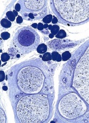
A novel technique can jump-start the creation of blood vessels, researchers have reported in Nature Biotechnology.
The team used human embryonic stem cells (hESCs) and induced pluripotent stem cells (hiPSCs) to create endothelial colony-forming cells (ECFCs).
These lab-generated ECFCs successfully produced human blood vessels in mice and restored blood flow to damaged retinas and limbs, without posing a risk of teratoma formation.
ECFCs can lose their ability to proliferate into new blood vessels as patients age or develop diseases such as peripheral arterial disease, said Mervin C. Yoder Jr, MD, of the Indiana University School of Medicine in Indianapolis.
He and his colleagues theorized that injecting patients with “younger,” more “enthusiastic” ECFCs might jump-start the process of creating new blood vessels. Such cells are difficult to find in adults but are common in umbilical cord blood.
With that in mind, the researchers developed a novel method to mature hiPSCs or hESCs into cells with the characteristics of ECFCs found in cord blood.
Both hiPSC-ECFCs and hESC-ECFCs exhibited properties of cord blood-derived ECFCs, including a homogenous monolayer with a cobblestone appearance and high clonal proliferative potential.
In addition, hiPSC-ECFCs and hESC-ECFCs formed capillary structures when cultured on Matrigel, and they generated in vivo inosculated vessels when implanted in immune-deficient mice.
In other murine experiments, hiPSC-ECFCs contributed to vascular repair of experimentally induced ischemic limbs and injured retinas. The cells’ effects were similar to those observed with cord blood-derived ECFCs and superior to effects observed with endothelial cells isolated using other published protocols.
Furthermore, hiPSC-ECFCs did not transition to nonendothelial cells over prolonged culture, and they could expand to more than 1 trillion endothelial cells in less than 3 months.
“This is one of the first studies using induced pluripotent stem cells that has been able to produce new cells in clinically relevant numbers, enough to enable a clinical trial,” Dr Yoder said.
He added that one of the next steps for this research is reaching an agreement with a facility approved to produce cells for use in human testing.
A novel technique can jump-start the creation of blood vessels, researchers have reported in Nature Biotechnology.
The team used human embryonic stem cells (hESCs) and induced pluripotent stem cells (hiPSCs) to create endothelial colony-forming cells (ECFCs).
These lab-generated ECFCs successfully produced human blood vessels in mice and restored blood flow to damaged retinas and limbs, without posing a risk of teratoma formation.
ECFCs can lose their ability to proliferate into new blood vessels as patients age or develop diseases such as peripheral arterial disease, said Mervin C. Yoder Jr, MD, of the Indiana University School of Medicine in Indianapolis.
He and his colleagues theorized that injecting patients with “younger,” more “enthusiastic” ECFCs might jump-start the process of creating new blood vessels. Such cells are difficult to find in adults but are common in umbilical cord blood.
With that in mind, the researchers developed a novel method to mature hiPSCs or hESCs into cells with the characteristics of ECFCs found in cord blood.
Both hiPSC-ECFCs and hESC-ECFCs exhibited properties of cord blood-derived ECFCs, including a homogenous monolayer with a cobblestone appearance and high clonal proliferative potential.
In addition, hiPSC-ECFCs and hESC-ECFCs formed capillary structures when cultured on Matrigel, and they generated in vivo inosculated vessels when implanted in immune-deficient mice.
In other murine experiments, hiPSC-ECFCs contributed to vascular repair of experimentally induced ischemic limbs and injured retinas. The cells’ effects were similar to those observed with cord blood-derived ECFCs and superior to effects observed with endothelial cells isolated using other published protocols.
Furthermore, hiPSC-ECFCs did not transition to nonendothelial cells over prolonged culture, and they could expand to more than 1 trillion endothelial cells in less than 3 months.
“This is one of the first studies using induced pluripotent stem cells that has been able to produce new cells in clinically relevant numbers, enough to enable a clinical trial,” Dr Yoder said.
He added that one of the next steps for this research is reaching an agreement with a facility approved to produce cells for use in human testing.
A novel technique can jump-start the creation of blood vessels, researchers have reported in Nature Biotechnology.
The team used human embryonic stem cells (hESCs) and induced pluripotent stem cells (hiPSCs) to create endothelial colony-forming cells (ECFCs).
These lab-generated ECFCs successfully produced human blood vessels in mice and restored blood flow to damaged retinas and limbs, without posing a risk of teratoma formation.
ECFCs can lose their ability to proliferate into new blood vessels as patients age or develop diseases such as peripheral arterial disease, said Mervin C. Yoder Jr, MD, of the Indiana University School of Medicine in Indianapolis.
He and his colleagues theorized that injecting patients with “younger,” more “enthusiastic” ECFCs might jump-start the process of creating new blood vessels. Such cells are difficult to find in adults but are common in umbilical cord blood.
With that in mind, the researchers developed a novel method to mature hiPSCs or hESCs into cells with the characteristics of ECFCs found in cord blood.
Both hiPSC-ECFCs and hESC-ECFCs exhibited properties of cord blood-derived ECFCs, including a homogenous monolayer with a cobblestone appearance and high clonal proliferative potential.
In addition, hiPSC-ECFCs and hESC-ECFCs formed capillary structures when cultured on Matrigel, and they generated in vivo inosculated vessels when implanted in immune-deficient mice.
In other murine experiments, hiPSC-ECFCs contributed to vascular repair of experimentally induced ischemic limbs and injured retinas. The cells’ effects were similar to those observed with cord blood-derived ECFCs and superior to effects observed with endothelial cells isolated using other published protocols.
Furthermore, hiPSC-ECFCs did not transition to nonendothelial cells over prolonged culture, and they could expand to more than 1 trillion endothelial cells in less than 3 months.
“This is one of the first studies using induced pluripotent stem cells that has been able to produce new cells in clinically relevant numbers, enough to enable a clinical trial,” Dr Yoder said.
He added that one of the next steps for this research is reaching an agreement with a facility approved to produce cells for use in human testing.
Invention allows for precise gene control

Credit: NIGMS
A new way to control genes may provide a better understanding of cancers and aid the development of new therapies.
The key to the method is an invention called SunTag, a series of molecular hooks for hanging multiple copies of biologically active molecules onto a single protein scaffold used to target genes or other molecules.
Compared to molecules assembled without these hooks, those incorporating SunTag can greatly amplify biological activity.
SunTag was developed by researchers in the lab of Ron Vale, PhD, of the University of California, San Francisco (UCSF), and described in two papers published in Cell.
In one paper, the authors recount how they used SunTag to greatly amplify the light-emitting signal from the green fluorescent protein commonly used to label molecules within cells.
In another paper, the researchers explain how they used SunTag to supercharge a variation of a biochemical approach known as CRISPR.
CRISPR is a technique that emerged a few years ago as a way to edit DNA anywhere within the genome.
The UCSF researchers adapted CRISPR to activate genes or interfere with their activity in a reversible way without altering DNA. The team believes this capability might make previous methods for probing poorly understood cellular functions obsolete.
“With these techniques, we can fine-tune the activity of genes within cells, and this has broad implications for the reprogramming of cells,” said Jonathan Weissman, PhD, also of UCSF.
Scientists previously reported ways to activate or interfere with genes using CRISPR, but Dr Vale said these methods were inefficient, especially for activating genes.
“It depends on the gene, but this new approach appears to amplify gene-switching by as much as 50-fold,” he said. “It’s a much more robust way of activating genes.”
CRISPR with SunTag sheds light on cancers
CRISPR is a natural system that bacteria use to defend themselves against viruses. The basis for CRISPR applications in the lab is a protein called Cas9, a chassis into which scientists can insert any specific RNA partner molecule.
The selected RNA serves as an adaptor that determines the target anywhere within the genome. The researchers attached SunTag to this chassis, enabling one Cas9 to recruit many copies of a protein to a specific DNA sequence.
The adaptation of SunTag for CRISPR activation makes it possible to systematically probe the biological roles of all genes within the genome in a single experiment, the team said.
They used CRISPR activation to identify a number of tumor suppressor genes that inhibit the growth of cancer cells. In future studies, they plan to use CRISPR activation to reveal mechanisms by which cancer cells develop resistance to anticancer drugs, a process that typically involves gene activation.
Will RNA interference become obsolete?
CRISPR interference has the potential to render RNA interference obsolete, according to Dr Weissman.
Unlike conventional RNA interference techniques, CRISPR interference allows any number of individual genes to be silenced at the same time. In addition, there is little risk of turning off untargeted genes the way RNA interference techniques do.
RNA interference blocks the messenger RNA that drives protein protection based on the blueprint contained within a gene’s DNA sequence. By preventing protein production, RNA interference may be used to get around the problem of difficult-to-target proteins, a frequent challenge in drug development.
But CRISPR interference acts one step earlier in the cell’s protein-manufacturing process.
“The horse has already left the barn with RNA interference, in the sense that the RNA message already has been transcribed from DNA,” Dr Weissman said. “With CRISPR interference, we can prevent the message from being written.”
Credit: NIGMS
A new way to control genes may provide a better understanding of cancers and aid the development of new therapies.
The key to the method is an invention called SunTag, a series of molecular hooks for hanging multiple copies of biologically active molecules onto a single protein scaffold used to target genes or other molecules.
Compared to molecules assembled without these hooks, those incorporating SunTag can greatly amplify biological activity.
SunTag was developed by researchers in the lab of Ron Vale, PhD, of the University of California, San Francisco (UCSF), and described in two papers published in Cell.
In one paper, the authors recount how they used SunTag to greatly amplify the light-emitting signal from the green fluorescent protein commonly used to label molecules within cells.
In another paper, the researchers explain how they used SunTag to supercharge a variation of a biochemical approach known as CRISPR.
CRISPR is a technique that emerged a few years ago as a way to edit DNA anywhere within the genome.
The UCSF researchers adapted CRISPR to activate genes or interfere with their activity in a reversible way without altering DNA. The team believes this capability might make previous methods for probing poorly understood cellular functions obsolete.
“With these techniques, we can fine-tune the activity of genes within cells, and this has broad implications for the reprogramming of cells,” said Jonathan Weissman, PhD, also of UCSF.
Scientists previously reported ways to activate or interfere with genes using CRISPR, but Dr Vale said these methods were inefficient, especially for activating genes.
“It depends on the gene, but this new approach appears to amplify gene-switching by as much as 50-fold,” he said. “It’s a much more robust way of activating genes.”
CRISPR with SunTag sheds light on cancers
CRISPR is a natural system that bacteria use to defend themselves against viruses. The basis for CRISPR applications in the lab is a protein called Cas9, a chassis into which scientists can insert any specific RNA partner molecule.
The selected RNA serves as an adaptor that determines the target anywhere within the genome. The researchers attached SunTag to this chassis, enabling one Cas9 to recruit many copies of a protein to a specific DNA sequence.
The adaptation of SunTag for CRISPR activation makes it possible to systematically probe the biological roles of all genes within the genome in a single experiment, the team said.
They used CRISPR activation to identify a number of tumor suppressor genes that inhibit the growth of cancer cells. In future studies, they plan to use CRISPR activation to reveal mechanisms by which cancer cells develop resistance to anticancer drugs, a process that typically involves gene activation.
Will RNA interference become obsolete?
CRISPR interference has the potential to render RNA interference obsolete, according to Dr Weissman.
Unlike conventional RNA interference techniques, CRISPR interference allows any number of individual genes to be silenced at the same time. In addition, there is little risk of turning off untargeted genes the way RNA interference techniques do.
RNA interference blocks the messenger RNA that drives protein protection based on the blueprint contained within a gene’s DNA sequence. By preventing protein production, RNA interference may be used to get around the problem of difficult-to-target proteins, a frequent challenge in drug development.
But CRISPR interference acts one step earlier in the cell’s protein-manufacturing process.
“The horse has already left the barn with RNA interference, in the sense that the RNA message already has been transcribed from DNA,” Dr Weissman said. “With CRISPR interference, we can prevent the message from being written.”
Credit: NIGMS
A new way to control genes may provide a better understanding of cancers and aid the development of new therapies.
The key to the method is an invention called SunTag, a series of molecular hooks for hanging multiple copies of biologically active molecules onto a single protein scaffold used to target genes or other molecules.
Compared to molecules assembled without these hooks, those incorporating SunTag can greatly amplify biological activity.
SunTag was developed by researchers in the lab of Ron Vale, PhD, of the University of California, San Francisco (UCSF), and described in two papers published in Cell.
In one paper, the authors recount how they used SunTag to greatly amplify the light-emitting signal from the green fluorescent protein commonly used to label molecules within cells.
In another paper, the researchers explain how they used SunTag to supercharge a variation of a biochemical approach known as CRISPR.
CRISPR is a technique that emerged a few years ago as a way to edit DNA anywhere within the genome.
The UCSF researchers adapted CRISPR to activate genes or interfere with their activity in a reversible way without altering DNA. The team believes this capability might make previous methods for probing poorly understood cellular functions obsolete.
“With these techniques, we can fine-tune the activity of genes within cells, and this has broad implications for the reprogramming of cells,” said Jonathan Weissman, PhD, also of UCSF.
Scientists previously reported ways to activate or interfere with genes using CRISPR, but Dr Vale said these methods were inefficient, especially for activating genes.
“It depends on the gene, but this new approach appears to amplify gene-switching by as much as 50-fold,” he said. “It’s a much more robust way of activating genes.”
CRISPR with SunTag sheds light on cancers
CRISPR is a natural system that bacteria use to defend themselves against viruses. The basis for CRISPR applications in the lab is a protein called Cas9, a chassis into which scientists can insert any specific RNA partner molecule.
The selected RNA serves as an adaptor that determines the target anywhere within the genome. The researchers attached SunTag to this chassis, enabling one Cas9 to recruit many copies of a protein to a specific DNA sequence.
The adaptation of SunTag for CRISPR activation makes it possible to systematically probe the biological roles of all genes within the genome in a single experiment, the team said.
They used CRISPR activation to identify a number of tumor suppressor genes that inhibit the growth of cancer cells. In future studies, they plan to use CRISPR activation to reveal mechanisms by which cancer cells develop resistance to anticancer drugs, a process that typically involves gene activation.
Will RNA interference become obsolete?
CRISPR interference has the potential to render RNA interference obsolete, according to Dr Weissman.
Unlike conventional RNA interference techniques, CRISPR interference allows any number of individual genes to be silenced at the same time. In addition, there is little risk of turning off untargeted genes the way RNA interference techniques do.
RNA interference blocks the messenger RNA that drives protein protection based on the blueprint contained within a gene’s DNA sequence. By preventing protein production, RNA interference may be used to get around the problem of difficult-to-target proteins, a frequent challenge in drug development.
But CRISPR interference acts one step earlier in the cell’s protein-manufacturing process.
“The horse has already left the barn with RNA interference, in the sense that the RNA message already has been transcribed from DNA,” Dr Weissman said. “With CRISPR interference, we can prevent the message from being written.”
‘Nano-cocoons’ offer targeted drug delivery
Photo courtesy of the Gu lab
Biomedical engineers have developed a drug delivery system in which cancer cells are “tricked” into absorbing nanoscale “cocoons” before
they unleash anticancer drugs.
Each cocoon is made of a deoxyribonuclease (DNase)-degradable DNA nanoclew embedded with an acid-responsive DNase I nanocapsule.
A cancer cell’s acidic environment prompts the DNase to degrade the cocoon and release the drug encapsulated in the endolysosomal compartment.
The engineers described the creation of these nano-cocoons and in vitro experiments testing the delivery of doxorubicin in the Journal of the American Chemical Society.
“This drug delivery system is DNA-based, which means it is biocompatible and less toxic to patients than systems that use synthetic materials,” said study author Zhen Gu, PhD, of the University of North Carolina Chapel Hill.
“This technique also specifically targets cancer cells, can carry a large drug load, and releases the drugs very quickly once inside the cancer cell.”
“In addition, because we used self-assembling DNA techniques, it is relatively easy to manufacture,” added Wujin Sun, a PhD student in Dr Gu’s lab.
Each nano-cocoon is made of a single strand of DNA that self-assembles into a structure measuring 150 nanometers across.
The core of the nano-cocoon contains the anticancer drug doxorubicin and DNase. The DNase, an enzyme that would normally dismantle the DNA cocoon, is contained by a thin polymer coating.
The surface of the nano-cocoon is studded with folic acid ligands. When the nano-cocoon encounters a cancer cell, the ligands bind the nano-cocoon to receptors on the surface of the cell, causing the cell to engulf the nano-cocoon.
Once the cocoon is inside the cancer cell, the cell’s acidic environment destroys the polymer sheath containing the DNase. The DNase then slices through the DNA cocoon, spilling doxorubicin into the cancer cell and killing it.
“We’re preparing to launch preclinical testing now,” Dr Gu said. “We’re very excited about this system and think it holds promise for delivering a variety of drugs targeting cancer and other diseases.”
Photo courtesy of the Gu lab
Biomedical engineers have developed a drug delivery system in which cancer cells are “tricked” into absorbing nanoscale “cocoons” before
they unleash anticancer drugs.
Each cocoon is made of a deoxyribonuclease (DNase)-degradable DNA nanoclew embedded with an acid-responsive DNase I nanocapsule.
A cancer cell’s acidic environment prompts the DNase to degrade the cocoon and release the drug encapsulated in the endolysosomal compartment.
The engineers described the creation of these nano-cocoons and in vitro experiments testing the delivery of doxorubicin in the Journal of the American Chemical Society.
“This drug delivery system is DNA-based, which means it is biocompatible and less toxic to patients than systems that use synthetic materials,” said study author Zhen Gu, PhD, of the University of North Carolina Chapel Hill.
“This technique also specifically targets cancer cells, can carry a large drug load, and releases the drugs very quickly once inside the cancer cell.”
“In addition, because we used self-assembling DNA techniques, it is relatively easy to manufacture,” added Wujin Sun, a PhD student in Dr Gu’s lab.
Each nano-cocoon is made of a single strand of DNA that self-assembles into a structure measuring 150 nanometers across.
The core of the nano-cocoon contains the anticancer drug doxorubicin and DNase. The DNase, an enzyme that would normally dismantle the DNA cocoon, is contained by a thin polymer coating.
The surface of the nano-cocoon is studded with folic acid ligands. When the nano-cocoon encounters a cancer cell, the ligands bind the nano-cocoon to receptors on the surface of the cell, causing the cell to engulf the nano-cocoon.
Once the cocoon is inside the cancer cell, the cell’s acidic environment destroys the polymer sheath containing the DNase. The DNase then slices through the DNA cocoon, spilling doxorubicin into the cancer cell and killing it.
“We’re preparing to launch preclinical testing now,” Dr Gu said. “We’re very excited about this system and think it holds promise for delivering a variety of drugs targeting cancer and other diseases.”
Photo courtesy of the Gu lab
Biomedical engineers have developed a drug delivery system in which cancer cells are “tricked” into absorbing nanoscale “cocoons” before
they unleash anticancer drugs.
Each cocoon is made of a deoxyribonuclease (DNase)-degradable DNA nanoclew embedded with an acid-responsive DNase I nanocapsule.
A cancer cell’s acidic environment prompts the DNase to degrade the cocoon and release the drug encapsulated in the endolysosomal compartment.
The engineers described the creation of these nano-cocoons and in vitro experiments testing the delivery of doxorubicin in the Journal of the American Chemical Society.
“This drug delivery system is DNA-based, which means it is biocompatible and less toxic to patients than systems that use synthetic materials,” said study author Zhen Gu, PhD, of the University of North Carolina Chapel Hill.
“This technique also specifically targets cancer cells, can carry a large drug load, and releases the drugs very quickly once inside the cancer cell.”
“In addition, because we used self-assembling DNA techniques, it is relatively easy to manufacture,” added Wujin Sun, a PhD student in Dr Gu’s lab.
Each nano-cocoon is made of a single strand of DNA that self-assembles into a structure measuring 150 nanometers across.
The core of the nano-cocoon contains the anticancer drug doxorubicin and DNase. The DNase, an enzyme that would normally dismantle the DNA cocoon, is contained by a thin polymer coating.
The surface of the nano-cocoon is studded with folic acid ligands. When the nano-cocoon encounters a cancer cell, the ligands bind the nano-cocoon to receptors on the surface of the cell, causing the cell to engulf the nano-cocoon.
Once the cocoon is inside the cancer cell, the cell’s acidic environment destroys the polymer sheath containing the DNase. The DNase then slices through the DNA cocoon, spilling doxorubicin into the cancer cell and killing it.
“We’re preparing to launch preclinical testing now,” Dr Gu said. “We’re very excited about this system and think it holds promise for delivering a variety of drugs targeting cancer and other diseases.”
CAR T cells can produce durable remissions
Credit: Penn Medicine
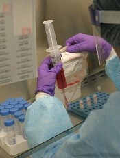
In a small study, 90% of children and adults with relapsed or refractory acute lymphoblastic leukemia (ALL) achieved remission after receiving CTL019, a chimeric antigen receptor (CAR) T-cell therapy.
Seven of these 27 patients ultimately relapsed, and 5 went on to receive additional therapy, including stem cell transplant.
Still, 15 of these heavily pretreated patients remained in remission at a median of 7 months of follow-up and did not require further treatment.
“[Patients] had relapsed as many as 4 times, including 60% whose cancers came back even after stem cell transplants,” said Stephan Grupp, MD, PhD, of the Children’s Hospital of Philadelphia in Pennsylvania.
“Their cancers were so aggressive they had no treatment options left. The durable responses we have observed with CTL019 therapy are unprecedented.”
Dr Grupp and his colleagues reported these results in NEJM. The new data build on preliminary findings presented at the 2013 ASH Annual Meeting and the 2012 ASH Annual Meeting.
CTL019 cells are a patient’s own T cells genetically engineered to express an anti-CD19 scFv coupled to CD3ζ signaling and 4-1BB co-stimulatory domains. The cells are activated and expanded ex vivo with anti-CD3 and anti-CD28 beads, then infused into patients.
In all, 30 patients received CTL019, including 25 children and young adults (ages 5 to 22) and 5 adults (ages 26 to 60). Three patients had primary refractory disease, 5 had relapsed once, and 22 patients had relapsed 2 or more times. Eighteen patients had received an allogeneic stem cell transplant.
Twenty-seven patients achieved a complete remission after an infusion of CTL019. Nineteen patients remain in remission, 15 of whom received CTL019 alone. Follow-up ranged from 1.4 months to 24 months.
Tests in patients who experienced complete remissions showed that their normal B cells had been eliminated along with their tumors. The researchers noted that persistent absence of normal B cells following CTL019 treatment indicates continued activity of the CAR T cells.
“Our results support that CTL019 can produce long-lasting remissions for certain heavily pretreated ALL patients without further therapy,” said Noelle Frey, MD, of the University of Pennsylvania in Philadelphia.
However, 5 patients did seek additional therapy, 3 of whom proceeded to allogeneic stem cell transplants while in remission.
Seven patients relapsed, between 6 weeks and 8.5 months after their infusions, including 3 whose cancers returned as CD19-negative leukemia that would not have been targeted by CTL019.
At 6 months, the overall survival rate was 78%, and the event-free survival rate was 67%.
All of the patients experienced cytokine release syndrome (CRS) within a few days of receiving CTL019, but they all fully recovered.
Twenty-two patients experienced mild to moderate CRS, which included varying degrees of flu-like symptoms, with high fevers, nausea, and muscle pain. Eight patients developed severe CRS, which required treatment for low blood pressure and breathing difficulties.
Nine patients were treated with tocilizumab, an immunosuppressant that inhibits the effects of the inflammatory cytokine IL-6, which have been found to spike during the most robust phase of the CAR T cells’ expansion in the body. Six patients also received short courses of steroids to combat CRS symptoms.
CTL019 was invented at The University of Pennsylvania but has been licensed to Novartis. In July, the US Food and Drug Administration granted CTL019 breakthrough therapy designation for the treatment of relapsed and refractory adult and pediatric ALL.
The first multicenter trial of CTL019 recently opened in the US, and additional multisite trials are expected to begin by the end of the year.
Credit: Penn Medicine
In a small study, 90% of children and adults with relapsed or refractory acute lymphoblastic leukemia (ALL) achieved remission after receiving CTL019, a chimeric antigen receptor (CAR) T-cell therapy.
Seven of these 27 patients ultimately relapsed, and 5 went on to receive additional therapy, including stem cell transplant.
Still, 15 of these heavily pretreated patients remained in remission at a median of 7 months of follow-up and did not require further treatment.
“[Patients] had relapsed as many as 4 times, including 60% whose cancers came back even after stem cell transplants,” said Stephan Grupp, MD, PhD, of the Children’s Hospital of Philadelphia in Pennsylvania.
“Their cancers were so aggressive they had no treatment options left. The durable responses we have observed with CTL019 therapy are unprecedented.”
Dr Grupp and his colleagues reported these results in NEJM. The new data build on preliminary findings presented at the 2013 ASH Annual Meeting and the 2012 ASH Annual Meeting.
CTL019 cells are a patient’s own T cells genetically engineered to express an anti-CD19 scFv coupled to CD3ζ signaling and 4-1BB co-stimulatory domains. The cells are activated and expanded ex vivo with anti-CD3 and anti-CD28 beads, then infused into patients.
In all, 30 patients received CTL019, including 25 children and young adults (ages 5 to 22) and 5 adults (ages 26 to 60). Three patients had primary refractory disease, 5 had relapsed once, and 22 patients had relapsed 2 or more times. Eighteen patients had received an allogeneic stem cell transplant.
Twenty-seven patients achieved a complete remission after an infusion of CTL019. Nineteen patients remain in remission, 15 of whom received CTL019 alone. Follow-up ranged from 1.4 months to 24 months.
Tests in patients who experienced complete remissions showed that their normal B cells had been eliminated along with their tumors. The researchers noted that persistent absence of normal B cells following CTL019 treatment indicates continued activity of the CAR T cells.
“Our results support that CTL019 can produce long-lasting remissions for certain heavily pretreated ALL patients without further therapy,” said Noelle Frey, MD, of the University of Pennsylvania in Philadelphia.
However, 5 patients did seek additional therapy, 3 of whom proceeded to allogeneic stem cell transplants while in remission.
Seven patients relapsed, between 6 weeks and 8.5 months after their infusions, including 3 whose cancers returned as CD19-negative leukemia that would not have been targeted by CTL019.
At 6 months, the overall survival rate was 78%, and the event-free survival rate was 67%.
All of the patients experienced cytokine release syndrome (CRS) within a few days of receiving CTL019, but they all fully recovered.
Twenty-two patients experienced mild to moderate CRS, which included varying degrees of flu-like symptoms, with high fevers, nausea, and muscle pain. Eight patients developed severe CRS, which required treatment for low blood pressure and breathing difficulties.
Nine patients were treated with tocilizumab, an immunosuppressant that inhibits the effects of the inflammatory cytokine IL-6, which have been found to spike during the most robust phase of the CAR T cells’ expansion in the body. Six patients also received short courses of steroids to combat CRS symptoms.
CTL019 was invented at The University of Pennsylvania but has been licensed to Novartis. In July, the US Food and Drug Administration granted CTL019 breakthrough therapy designation for the treatment of relapsed and refractory adult and pediatric ALL.
The first multicenter trial of CTL019 recently opened in the US, and additional multisite trials are expected to begin by the end of the year.
Credit: Penn Medicine
In a small study, 90% of children and adults with relapsed or refractory acute lymphoblastic leukemia (ALL) achieved remission after receiving CTL019, a chimeric antigen receptor (CAR) T-cell therapy.
Seven of these 27 patients ultimately relapsed, and 5 went on to receive additional therapy, including stem cell transplant.
Still, 15 of these heavily pretreated patients remained in remission at a median of 7 months of follow-up and did not require further treatment.
“[Patients] had relapsed as many as 4 times, including 60% whose cancers came back even after stem cell transplants,” said Stephan Grupp, MD, PhD, of the Children’s Hospital of Philadelphia in Pennsylvania.
“Their cancers were so aggressive they had no treatment options left. The durable responses we have observed with CTL019 therapy are unprecedented.”
Dr Grupp and his colleagues reported these results in NEJM. The new data build on preliminary findings presented at the 2013 ASH Annual Meeting and the 2012 ASH Annual Meeting.
CTL019 cells are a patient’s own T cells genetically engineered to express an anti-CD19 scFv coupled to CD3ζ signaling and 4-1BB co-stimulatory domains. The cells are activated and expanded ex vivo with anti-CD3 and anti-CD28 beads, then infused into patients.
In all, 30 patients received CTL019, including 25 children and young adults (ages 5 to 22) and 5 adults (ages 26 to 60). Three patients had primary refractory disease, 5 had relapsed once, and 22 patients had relapsed 2 or more times. Eighteen patients had received an allogeneic stem cell transplant.
Twenty-seven patients achieved a complete remission after an infusion of CTL019. Nineteen patients remain in remission, 15 of whom received CTL019 alone. Follow-up ranged from 1.4 months to 24 months.
Tests in patients who experienced complete remissions showed that their normal B cells had been eliminated along with their tumors. The researchers noted that persistent absence of normal B cells following CTL019 treatment indicates continued activity of the CAR T cells.
“Our results support that CTL019 can produce long-lasting remissions for certain heavily pretreated ALL patients without further therapy,” said Noelle Frey, MD, of the University of Pennsylvania in Philadelphia.
However, 5 patients did seek additional therapy, 3 of whom proceeded to allogeneic stem cell transplants while in remission.
Seven patients relapsed, between 6 weeks and 8.5 months after their infusions, including 3 whose cancers returned as CD19-negative leukemia that would not have been targeted by CTL019.
At 6 months, the overall survival rate was 78%, and the event-free survival rate was 67%.
All of the patients experienced cytokine release syndrome (CRS) within a few days of receiving CTL019, but they all fully recovered.
Twenty-two patients experienced mild to moderate CRS, which included varying degrees of flu-like symptoms, with high fevers, nausea, and muscle pain. Eight patients developed severe CRS, which required treatment for low blood pressure and breathing difficulties.
Nine patients were treated with tocilizumab, an immunosuppressant that inhibits the effects of the inflammatory cytokine IL-6, which have been found to spike during the most robust phase of the CAR T cells’ expansion in the body. Six patients also received short courses of steroids to combat CRS symptoms.
CTL019 was invented at The University of Pennsylvania but has been licensed to Novartis. In July, the US Food and Drug Administration granted CTL019 breakthrough therapy designation for the treatment of relapsed and refractory adult and pediatric ALL.
The first multicenter trial of CTL019 recently opened in the US, and additional multisite trials are expected to begin by the end of the year.
NICE doesn’t support pomalidomide for MM

Credit: CDC
The UK’s National Institute for Health and Care Excellence (NICE) has issued a preliminary draft guidance rejecting the use of pomalidomide
(Imnovid) to treat patients with relapsed or refractory multiple myeloma (MM).
NICE said the drug’s maker, Celgene, did not provide sufficient evidence of pomalidomide’s effectiveness as compared to current treatment.
Furthermore, the drug did not offer enough of a benefit to justify its high price.
“We are disappointed not to be able to recommend pomalidomide in this preliminary guidance, but the analyses submitted by Celgene, the company that makes the drug, did not show how well the drug works compared to the other treatments available,” said Sir Andrew Dillon, NICE chief executive.
Specifically, NICE said it cannot recommend pomalidomide in combination with dexamethasone for treating relapsed and refractory MM in adults who have had at least 2 prior treatments, including lenalidomide and bortezomib, and whose disease has progressed on their last therapy.
A committee advising NICE concluded that, because of the design of the MM-003 study, the extent of the benefits associated with pomalidomide was uncertain. In addition, the MM-003 results were of limited value in comparing pomalidomide with “established practice without pomalidomide.”
The recommended dose of pomalidomide is 4 mg once daily, taken on days 1 to 21 of repeated 28-day cycles. Treatment should continue until disease progression.
The price of a pack (21 tablets) of 1 mg, 2 mg, 3 mg, or 4 mg tablets is £8884 (excluding value-added tax). Costs may vary in different settings because of negotiated procurement discounts.
The cost per quality-adjusted life-year (QALY) gained presented by Celgene was over £50,000 compared with bortezomib.
The committee heard from a clinical expert that, although there is no standard of care for people with relapsed or refractory MM, bendamustine was likely to be the most commonly used therapy in this setting in England.
When comparing pomalidomide with bendamustine plus thalidomide and dexamethasone, all costs per QALYs presented were over £70,000.
The committee was not persuaded that the estimates of the extension to life were robust, objective, or plausible based on the company’s economic modeling. It therefore concluded that pomalidomide did not fulfill the criteria for being a life-extending, end-of-life treatment.
The committee further concluded that, even if pomalidomide fulfilled these criteria, the weight that would have to be placed on the QALYs would be too high for pomalidomide to be considered a cost-effective use of National Health Service resources.
Consultees, including the manufacturer, healthcare professionals, and members of the public are now able to comment on these preliminary recommendations.
Until a final guidance is issued, National Health Service bodies should make decisions locally on the funding of specific treatments. Once NICE issues its final guidance on a technology, it replaces local recommendations.
Credit: CDC
The UK’s National Institute for Health and Care Excellence (NICE) has issued a preliminary draft guidance rejecting the use of pomalidomide
(Imnovid) to treat patients with relapsed or refractory multiple myeloma (MM).
NICE said the drug’s maker, Celgene, did not provide sufficient evidence of pomalidomide’s effectiveness as compared to current treatment.
Furthermore, the drug did not offer enough of a benefit to justify its high price.
“We are disappointed not to be able to recommend pomalidomide in this preliminary guidance, but the analyses submitted by Celgene, the company that makes the drug, did not show how well the drug works compared to the other treatments available,” said Sir Andrew Dillon, NICE chief executive.
Specifically, NICE said it cannot recommend pomalidomide in combination with dexamethasone for treating relapsed and refractory MM in adults who have had at least 2 prior treatments, including lenalidomide and bortezomib, and whose disease has progressed on their last therapy.
A committee advising NICE concluded that, because of the design of the MM-003 study, the extent of the benefits associated with pomalidomide was uncertain. In addition, the MM-003 results were of limited value in comparing pomalidomide with “established practice without pomalidomide.”
The recommended dose of pomalidomide is 4 mg once daily, taken on days 1 to 21 of repeated 28-day cycles. Treatment should continue until disease progression.
The price of a pack (21 tablets) of 1 mg, 2 mg, 3 mg, or 4 mg tablets is £8884 (excluding value-added tax). Costs may vary in different settings because of negotiated procurement discounts.
The cost per quality-adjusted life-year (QALY) gained presented by Celgene was over £50,000 compared with bortezomib.
The committee heard from a clinical expert that, although there is no standard of care for people with relapsed or refractory MM, bendamustine was likely to be the most commonly used therapy in this setting in England.
When comparing pomalidomide with bendamustine plus thalidomide and dexamethasone, all costs per QALYs presented were over £70,000.
The committee was not persuaded that the estimates of the extension to life were robust, objective, or plausible based on the company’s economic modeling. It therefore concluded that pomalidomide did not fulfill the criteria for being a life-extending, end-of-life treatment.
The committee further concluded that, even if pomalidomide fulfilled these criteria, the weight that would have to be placed on the QALYs would be too high for pomalidomide to be considered a cost-effective use of National Health Service resources.
Consultees, including the manufacturer, healthcare professionals, and members of the public are now able to comment on these preliminary recommendations.
Until a final guidance is issued, National Health Service bodies should make decisions locally on the funding of specific treatments. Once NICE issues its final guidance on a technology, it replaces local recommendations.
Credit: CDC
The UK’s National Institute for Health and Care Excellence (NICE) has issued a preliminary draft guidance rejecting the use of pomalidomide
(Imnovid) to treat patients with relapsed or refractory multiple myeloma (MM).
NICE said the drug’s maker, Celgene, did not provide sufficient evidence of pomalidomide’s effectiveness as compared to current treatment.
Furthermore, the drug did not offer enough of a benefit to justify its high price.
“We are disappointed not to be able to recommend pomalidomide in this preliminary guidance, but the analyses submitted by Celgene, the company that makes the drug, did not show how well the drug works compared to the other treatments available,” said Sir Andrew Dillon, NICE chief executive.
Specifically, NICE said it cannot recommend pomalidomide in combination with dexamethasone for treating relapsed and refractory MM in adults who have had at least 2 prior treatments, including lenalidomide and bortezomib, and whose disease has progressed on their last therapy.
A committee advising NICE concluded that, because of the design of the MM-003 study, the extent of the benefits associated with pomalidomide was uncertain. In addition, the MM-003 results were of limited value in comparing pomalidomide with “established practice without pomalidomide.”
The recommended dose of pomalidomide is 4 mg once daily, taken on days 1 to 21 of repeated 28-day cycles. Treatment should continue until disease progression.
The price of a pack (21 tablets) of 1 mg, 2 mg, 3 mg, or 4 mg tablets is £8884 (excluding value-added tax). Costs may vary in different settings because of negotiated procurement discounts.
The cost per quality-adjusted life-year (QALY) gained presented by Celgene was over £50,000 compared with bortezomib.
The committee heard from a clinical expert that, although there is no standard of care for people with relapsed or refractory MM, bendamustine was likely to be the most commonly used therapy in this setting in England.
When comparing pomalidomide with bendamustine plus thalidomide and dexamethasone, all costs per QALYs presented were over £70,000.
The committee was not persuaded that the estimates of the extension to life were robust, objective, or plausible based on the company’s economic modeling. It therefore concluded that pomalidomide did not fulfill the criteria for being a life-extending, end-of-life treatment.
The committee further concluded that, even if pomalidomide fulfilled these criteria, the weight that would have to be placed on the QALYs would be too high for pomalidomide to be considered a cost-effective use of National Health Service resources.
Consultees, including the manufacturer, healthcare professionals, and members of the public are now able to comment on these preliminary recommendations.
Until a final guidance is issued, National Health Service bodies should make decisions locally on the funding of specific treatments. Once NICE issues its final guidance on a technology, it replaces local recommendations.
Amoeba could help fight cancers

Experiments in a soil-dwelling amoeba have provided insight that could help us treat cancers characterized by PTEN mutations, researchers have reported in PLOS ONE.
The team discovered that this amoeba has two genes that function like the human tumor suppressor PTEN.
And increasing expression of one of these genes compensated for a mutation in the other gene.
If the same method works in humans with mutated PTEN, this finding could have implications for a range of cancers.
PTEN mutations are thought to be involved in nearly half of all leukemia cases, 40% of breast cancer cases, and up to 70% of prostate cancer cases.
“If you look at tumors across the board . . . , you find that PTEN is the most generally mutated gene, and, when you mutate PTEN in mice, you cause tumors,” said study author David Soll, PhD, of the University of Iowa in Iowa City.
He and his colleagues found that the amoeba Dictyostelium discoideum has the gene ptenA, which mutates similarly to the human PTEN gene and causes behavioral defects in the cell.
They also found a close relative of ptenA in the amoeba, called lpten, that performs the same functions of ptenA but to a lesser degree.
The researchers hypothesized that ramping up the presence of lpten could compensate for the mutated ptenA.
They tested this theory by placing lpten in a plasmid behind a powerful promoter designed to overexpress the gene. They then introduced the super-charged lpten into a cell with the mutated ptenA gene.
The team found that the overexpressed lpten gene fully compensated for all of the defects in the ptenA mutant.
If this method works in human cells, it could lead to a new way to treat cancers, the researchers said. They are now aiming to identify a drug that would activate the promoter for one of PTEN’s close relatives.
Once a patient is diagnosed with cancer caused by a PTEN mutation, the patient could take the drug, overexpress the PTEN replacement gene, and potentially stop cancer in its tracks, Dr Soll said.
This research has also led Dr Soll and his colleagues to study other human genes that may be able to step in for the mutated PTEN gene and perform the same tumor-suppressing role. The team is currently studying 2 close relatives of PTEN.
“And nature might have put them there just for that; that’s the curious thing,” Dr Soll said. “Somewhere, there may be a backup system, what we call ‘redundancy,’ that might be the basis for better identifying tumors and possibly creating cancer-fighting drugs. You have another gene which might be able to step in for the broken gene to keep things normal, and that’s what we’re playing with here. It’s very sophisticated.”
Experiments in a soil-dwelling amoeba have provided insight that could help us treat cancers characterized by PTEN mutations, researchers have reported in PLOS ONE.
The team discovered that this amoeba has two genes that function like the human tumor suppressor PTEN.
And increasing expression of one of these genes compensated for a mutation in the other gene.
If the same method works in humans with mutated PTEN, this finding could have implications for a range of cancers.
PTEN mutations are thought to be involved in nearly half of all leukemia cases, 40% of breast cancer cases, and up to 70% of prostate cancer cases.
“If you look at tumors across the board . . . , you find that PTEN is the most generally mutated gene, and, when you mutate PTEN in mice, you cause tumors,” said study author David Soll, PhD, of the University of Iowa in Iowa City.
He and his colleagues found that the amoeba Dictyostelium discoideum has the gene ptenA, which mutates similarly to the human PTEN gene and causes behavioral defects in the cell.
They also found a close relative of ptenA in the amoeba, called lpten, that performs the same functions of ptenA but to a lesser degree.
The researchers hypothesized that ramping up the presence of lpten could compensate for the mutated ptenA.
They tested this theory by placing lpten in a plasmid behind a powerful promoter designed to overexpress the gene. They then introduced the super-charged lpten into a cell with the mutated ptenA gene.
The team found that the overexpressed lpten gene fully compensated for all of the defects in the ptenA mutant.
If this method works in human cells, it could lead to a new way to treat cancers, the researchers said. They are now aiming to identify a drug that would activate the promoter for one of PTEN’s close relatives.
Once a patient is diagnosed with cancer caused by a PTEN mutation, the patient could take the drug, overexpress the PTEN replacement gene, and potentially stop cancer in its tracks, Dr Soll said.
This research has also led Dr Soll and his colleagues to study other human genes that may be able to step in for the mutated PTEN gene and perform the same tumor-suppressing role. The team is currently studying 2 close relatives of PTEN.
“And nature might have put them there just for that; that’s the curious thing,” Dr Soll said. “Somewhere, there may be a backup system, what we call ‘redundancy,’ that might be the basis for better identifying tumors and possibly creating cancer-fighting drugs. You have another gene which might be able to step in for the broken gene to keep things normal, and that’s what we’re playing with here. It’s very sophisticated.”
Experiments in a soil-dwelling amoeba have provided insight that could help us treat cancers characterized by PTEN mutations, researchers have reported in PLOS ONE.
The team discovered that this amoeba has two genes that function like the human tumor suppressor PTEN.
And increasing expression of one of these genes compensated for a mutation in the other gene.
If the same method works in humans with mutated PTEN, this finding could have implications for a range of cancers.
PTEN mutations are thought to be involved in nearly half of all leukemia cases, 40% of breast cancer cases, and up to 70% of prostate cancer cases.
“If you look at tumors across the board . . . , you find that PTEN is the most generally mutated gene, and, when you mutate PTEN in mice, you cause tumors,” said study author David Soll, PhD, of the University of Iowa in Iowa City.
He and his colleagues found that the amoeba Dictyostelium discoideum has the gene ptenA, which mutates similarly to the human PTEN gene and causes behavioral defects in the cell.
They also found a close relative of ptenA in the amoeba, called lpten, that performs the same functions of ptenA but to a lesser degree.
The researchers hypothesized that ramping up the presence of lpten could compensate for the mutated ptenA.
They tested this theory by placing lpten in a plasmid behind a powerful promoter designed to overexpress the gene. They then introduced the super-charged lpten into a cell with the mutated ptenA gene.
The team found that the overexpressed lpten gene fully compensated for all of the defects in the ptenA mutant.
If this method works in human cells, it could lead to a new way to treat cancers, the researchers said. They are now aiming to identify a drug that would activate the promoter for one of PTEN’s close relatives.
Once a patient is diagnosed with cancer caused by a PTEN mutation, the patient could take the drug, overexpress the PTEN replacement gene, and potentially stop cancer in its tracks, Dr Soll said.
This research has also led Dr Soll and his colleagues to study other human genes that may be able to step in for the mutated PTEN gene and perform the same tumor-suppressing role. The team is currently studying 2 close relatives of PTEN.
“And nature might have put them there just for that; that’s the curious thing,” Dr Soll said. “Somewhere, there may be a backup system, what we call ‘redundancy,’ that might be the basis for better identifying tumors and possibly creating cancer-fighting drugs. You have another gene which might be able to step in for the broken gene to keep things normal, and that’s what we’re playing with here. It’s very sophisticated.”
Malpractice reform may not reduce ‘defensive medicine’

Credit: CDC
Making it more difficult for patients to sue physicians for medical malpractice may not reduce the amount of “defensive medicine” physicians
practice, new research suggests.
Investigators studied patient records in 3 states that raised the standard for malpractice in the emergency room to gross negligence.
And they found that strong new legal protections did not significantly reduce the use of common defensive medicine practices or the cost of care.
The team detailed these findings in NEJM.
“Our findings suggest that malpractice reform may have less effect on costs than has been projected by conventional wisdom,” said lead study author Daniel A. Waxman, MD, PhD, of RAND Health in Santa Monica, California.
“Physicians say they order unnecessary tests strictly out of fear of being sued, but our results suggest the story is more complicated.”
The investigators evaluated the results of malpractice reform in 3 states—Georgia, Texas, and South Carolina.
About a decade ago, these states changed the legal malpractice standard for emergency care to gross negligence. The higher standard means plaintiffs must prove that doctors consciously disregarded the need to use reasonable care, knowing full well that their actions were likely to cause serious injury.
“These malpractice reforms have been said to provide virtual immunity against lawsuits,” Dr Waxman noted.
He and his colleagues examined 3.8 million Medicare patient records from 1166 hospital emergency departments spanning the period from 1997 to 2011. They compared care in the 3 reform states, before and after the statutes took effect, to care in neighboring states that did not pass malpractice reform.
The team assessed whether physicians ordered an advanced imaging study (CT or MRI scan), whether the patient was hospitalized after the emergency visit, and total charges for the visit.
Advanced imaging and hospitalization are among the most costly consequences of an emergency room visit, and physicians have identified them as common defensive medicine practices.
The malpractice reform laws had no effect on the use of imaging or the rate of hospitalization following emergency visits.
For 2 of the states, Texas and South Carolina, the law did not appear to cause any reduction in charges. Relative to neighboring states, Georgia saw a drop of 3.6% in average emergency room charges following its 2005 reform.
“This study suggests that, even when the risk of being sued for malpractice decreases, the path of least resistance still may favor resource-intensive care, at least in hospital emergency departments,” Dr Waxman said.
Credit: CDC
Making it more difficult for patients to sue physicians for medical malpractice may not reduce the amount of “defensive medicine” physicians
practice, new research suggests.
Investigators studied patient records in 3 states that raised the standard for malpractice in the emergency room to gross negligence.
And they found that strong new legal protections did not significantly reduce the use of common defensive medicine practices or the cost of care.
The team detailed these findings in NEJM.
“Our findings suggest that malpractice reform may have less effect on costs than has been projected by conventional wisdom,” said lead study author Daniel A. Waxman, MD, PhD, of RAND Health in Santa Monica, California.
“Physicians say they order unnecessary tests strictly out of fear of being sued, but our results suggest the story is more complicated.”
The investigators evaluated the results of malpractice reform in 3 states—Georgia, Texas, and South Carolina.
About a decade ago, these states changed the legal malpractice standard for emergency care to gross negligence. The higher standard means plaintiffs must prove that doctors consciously disregarded the need to use reasonable care, knowing full well that their actions were likely to cause serious injury.
“These malpractice reforms have been said to provide virtual immunity against lawsuits,” Dr Waxman noted.
He and his colleagues examined 3.8 million Medicare patient records from 1166 hospital emergency departments spanning the period from 1997 to 2011. They compared care in the 3 reform states, before and after the statutes took effect, to care in neighboring states that did not pass malpractice reform.
The team assessed whether physicians ordered an advanced imaging study (CT or MRI scan), whether the patient was hospitalized after the emergency visit, and total charges for the visit.
Advanced imaging and hospitalization are among the most costly consequences of an emergency room visit, and physicians have identified them as common defensive medicine practices.
The malpractice reform laws had no effect on the use of imaging or the rate of hospitalization following emergency visits.
For 2 of the states, Texas and South Carolina, the law did not appear to cause any reduction in charges. Relative to neighboring states, Georgia saw a drop of 3.6% in average emergency room charges following its 2005 reform.
“This study suggests that, even when the risk of being sued for malpractice decreases, the path of least resistance still may favor resource-intensive care, at least in hospital emergency departments,” Dr Waxman said.
Credit: CDC
Making it more difficult for patients to sue physicians for medical malpractice may not reduce the amount of “defensive medicine” physicians
practice, new research suggests.
Investigators studied patient records in 3 states that raised the standard for malpractice in the emergency room to gross negligence.
And they found that strong new legal protections did not significantly reduce the use of common defensive medicine practices or the cost of care.
The team detailed these findings in NEJM.
“Our findings suggest that malpractice reform may have less effect on costs than has been projected by conventional wisdom,” said lead study author Daniel A. Waxman, MD, PhD, of RAND Health in Santa Monica, California.
“Physicians say they order unnecessary tests strictly out of fear of being sued, but our results suggest the story is more complicated.”
The investigators evaluated the results of malpractice reform in 3 states—Georgia, Texas, and South Carolina.
About a decade ago, these states changed the legal malpractice standard for emergency care to gross negligence. The higher standard means plaintiffs must prove that doctors consciously disregarded the need to use reasonable care, knowing full well that their actions were likely to cause serious injury.
“These malpractice reforms have been said to provide virtual immunity against lawsuits,” Dr Waxman noted.
He and his colleagues examined 3.8 million Medicare patient records from 1166 hospital emergency departments spanning the period from 1997 to 2011. They compared care in the 3 reform states, before and after the statutes took effect, to care in neighboring states that did not pass malpractice reform.
The team assessed whether physicians ordered an advanced imaging study (CT or MRI scan), whether the patient was hospitalized after the emergency visit, and total charges for the visit.
Advanced imaging and hospitalization are among the most costly consequences of an emergency room visit, and physicians have identified them as common defensive medicine practices.
The malpractice reform laws had no effect on the use of imaging or the rate of hospitalization following emergency visits.
For 2 of the states, Texas and South Carolina, the law did not appear to cause any reduction in charges. Relative to neighboring states, Georgia saw a drop of 3.6% in average emergency room charges following its 2005 reform.
“This study suggests that, even when the risk of being sued for malpractice decreases, the path of least resistance still may favor resource-intensive care, at least in hospital emergency departments,” Dr Waxman said.
Drug can safely target NF-κB pathway in MM

Credit: Rhoda Baer
A new drug can safely target the NF-κB pathway in multiple myeloma (MM), according to research published in Cancer Cell.
The investigators identified an interaction between the NF-κB-regulated antiapoptotic factor GADD45β and the JNK kinase MKK7 as a therapeutic target in MM.
They then developed a drug known as DTP3, which disrupts the GADD45β/MKK7 complex, thereby killing MM cells in vitro and in vivo, without harming normal cells.
“Lab studies suggest that DTP3 could have therapeutic benefit for patients with multiple myeloma and potentially several other types of cancer, but we will need to confirm this in our clinical trials, the first of which will start next year,” said study author Guido Franzoso, MD, PhD, of Imperial College London in the UK.
Dr Franzoso and his colleagues knew that NF-κB is overactive in MM and other malignancies, but targeting NF-κB can have detrimental effects on healthy cells. So they looked for target genes downstream of NF-kB that might be responsible for its role in cancers.
By studying cells from MM patients, the investigators identified the protein complex GADD45β/MKK7, which appeared to play a critical role in allowing MM cells to survive.
Searching for a safe way to target the NF-kB pathway, the team screened more than 20,000 molecules and found 2 that disrupted GADD45β/MKK7. Further refinements led to the development of DTP3.
In human MM cells, DTP3 had a similar anticancer potency to that of bortezomib, but with a more than 100-fold higher cancer-cell specificity. DTP3 also retained full therapeutic efficacy in cell lines that were resistant to standard MM treatments.
In mice, DTP3 eradicated MM, with no apparent side effects at the effective doses.
In an orthotopic xenograft model of MM, control mice had a median overall survival of 26 days. But mice treated with DTP3 had a median overall survival that extended past the experimental endpoint on day 161.
“We had known for many years that NF-kB is very important for cancer cells, but because it is also needed by healthy cells, we did not know how to block it specifically,” Dr Franzoso said.
“The discovery that blocking the GADD45β/MKK7 segment of the NF-kB pathway with our DTP3 peptide therapeutic selectively kills myeloma cells could offer a completely new approach to treating patients with certain cancers, such as multiple myeloma.”
A spinout company, Kesios Therapeutics, was formed to commercialize DTP3 and other drug candidates based on Dr Franzoso’s research, with support from Imperial Innovations, a technology commercialization company focused on developing academic research in the UK.
Credit: Rhoda Baer
A new drug can safely target the NF-κB pathway in multiple myeloma (MM), according to research published in Cancer Cell.
The investigators identified an interaction between the NF-κB-regulated antiapoptotic factor GADD45β and the JNK kinase MKK7 as a therapeutic target in MM.
They then developed a drug known as DTP3, which disrupts the GADD45β/MKK7 complex, thereby killing MM cells in vitro and in vivo, without harming normal cells.
“Lab studies suggest that DTP3 could have therapeutic benefit for patients with multiple myeloma and potentially several other types of cancer, but we will need to confirm this in our clinical trials, the first of which will start next year,” said study author Guido Franzoso, MD, PhD, of Imperial College London in the UK.
Dr Franzoso and his colleagues knew that NF-κB is overactive in MM and other malignancies, but targeting NF-κB can have detrimental effects on healthy cells. So they looked for target genes downstream of NF-kB that might be responsible for its role in cancers.
By studying cells from MM patients, the investigators identified the protein complex GADD45β/MKK7, which appeared to play a critical role in allowing MM cells to survive.
Searching for a safe way to target the NF-kB pathway, the team screened more than 20,000 molecules and found 2 that disrupted GADD45β/MKK7. Further refinements led to the development of DTP3.
In human MM cells, DTP3 had a similar anticancer potency to that of bortezomib, but with a more than 100-fold higher cancer-cell specificity. DTP3 also retained full therapeutic efficacy in cell lines that were resistant to standard MM treatments.
In mice, DTP3 eradicated MM, with no apparent side effects at the effective doses.
In an orthotopic xenograft model of MM, control mice had a median overall survival of 26 days. But mice treated with DTP3 had a median overall survival that extended past the experimental endpoint on day 161.
“We had known for many years that NF-kB is very important for cancer cells, but because it is also needed by healthy cells, we did not know how to block it specifically,” Dr Franzoso said.
“The discovery that blocking the GADD45β/MKK7 segment of the NF-kB pathway with our DTP3 peptide therapeutic selectively kills myeloma cells could offer a completely new approach to treating patients with certain cancers, such as multiple myeloma.”
A spinout company, Kesios Therapeutics, was formed to commercialize DTP3 and other drug candidates based on Dr Franzoso’s research, with support from Imperial Innovations, a technology commercialization company focused on developing academic research in the UK.
Credit: Rhoda Baer
A new drug can safely target the NF-κB pathway in multiple myeloma (MM), according to research published in Cancer Cell.
The investigators identified an interaction between the NF-κB-regulated antiapoptotic factor GADD45β and the JNK kinase MKK7 as a therapeutic target in MM.
They then developed a drug known as DTP3, which disrupts the GADD45β/MKK7 complex, thereby killing MM cells in vitro and in vivo, without harming normal cells.
“Lab studies suggest that DTP3 could have therapeutic benefit for patients with multiple myeloma and potentially several other types of cancer, but we will need to confirm this in our clinical trials, the first of which will start next year,” said study author Guido Franzoso, MD, PhD, of Imperial College London in the UK.
Dr Franzoso and his colleagues knew that NF-κB is overactive in MM and other malignancies, but targeting NF-κB can have detrimental effects on healthy cells. So they looked for target genes downstream of NF-kB that might be responsible for its role in cancers.
By studying cells from MM patients, the investigators identified the protein complex GADD45β/MKK7, which appeared to play a critical role in allowing MM cells to survive.
Searching for a safe way to target the NF-kB pathway, the team screened more than 20,000 molecules and found 2 that disrupted GADD45β/MKK7. Further refinements led to the development of DTP3.
In human MM cells, DTP3 had a similar anticancer potency to that of bortezomib, but with a more than 100-fold higher cancer-cell specificity. DTP3 also retained full therapeutic efficacy in cell lines that were resistant to standard MM treatments.
In mice, DTP3 eradicated MM, with no apparent side effects at the effective doses.
In an orthotopic xenograft model of MM, control mice had a median overall survival of 26 days. But mice treated with DTP3 had a median overall survival that extended past the experimental endpoint on day 161.
“We had known for many years that NF-kB is very important for cancer cells, but because it is also needed by healthy cells, we did not know how to block it specifically,” Dr Franzoso said.
“The discovery that blocking the GADD45β/MKK7 segment of the NF-kB pathway with our DTP3 peptide therapeutic selectively kills myeloma cells could offer a completely new approach to treating patients with certain cancers, such as multiple myeloma.”
A spinout company, Kesios Therapeutics, was formed to commercialize DTP3 and other drug candidates based on Dr Franzoso’s research, with support from Imperial Innovations, a technology commercialization company focused on developing academic research in the UK.
VTE is ‘major contributor’ to global disease burden

Credit: Andre E.X. Brown
Results of a systematic review suggest venous thromboembolism (VTE) has a high disease burden throughout the world, regardless of a country’s income.
Researchers reviewed the literature to quantify the global disease burden of VTE.
And they found that VTE associated with hospitalization was the leading cause of disability-adjusted life-years (DALYs) lost in low- and middle-income countries. It was the second most common cause of DALYs lost in high-income countries.
“Venous thromboembolism in hospitalized patients was responsible for more years lost due to ill-health than hospital-acquired pneumonia, catheter-related blood stream infections, and side effects from drugs,” said study author Gary Raskob, PhD, of the University of Oklahoma Health Sciences Center in Oklahoma City.
Dr Raskob and his colleagues reported these results in the Journal of Thrombosis & Haemostasis.
The team reviewed 8702 studies on VTE and found that studies from western Europe, North America, Australia, and southern Latin America (Argentina) yielded consistent results.
The annual incidence of VTE ranged from 0.75 to 2.69 per 1000 individuals in the population. The incidence increased to between 2 and 7 VTEs per 1000 people among those 70 years of age or older.
Only two studies were designed to evaluate the prevalence of VTE, and both were conducted using US data. In one study, researchers investigated the prevalence of VTE from 2002 to 2006 using a health insurance claims database of 12.7 million individuals.
VTE occurred in 3.2 enrollees per 1000 in 2002 and 4.2 per 1000 in 2006. In 2006, the prevalence of VTE was 13.8 per 1000 in enrollees age 65 and older and 2.3 per 1000 in those younger than 65 years of age.
The other study designed to assess VTE prevalence in the US showed that prevalence was highest in African American males, followed by Caucasian males, Caucasian females, and African American females. Hispanic individuals of both sexes had the lowest prevalence rates.
Two studies were designed to evaluate VTE disease burden in terms of DALYs. The strongest of these included data from a literature review and epidemiologic studies commissioned by the WHO.
The study showed that VTE occurred in 3.3 of 100 hospitalizations in high-income countries and 3.0 of 100 hospitalizations in low-income and middle-income countries. The estimated annual cases of VTE were 3.9 million and 6.0 million, respectively.
VTE was the leading cause of hospital-related DALYs lost overall. It was responsible for a third (7681) of the 22,644 DALYs.
The researchers concluded that VTE causes a major burden of disease across low-income, middle-income, and high-income countries. However, more detailed data on the global burden of VTE is needed.
Credit: Andre E.X. Brown
Results of a systematic review suggest venous thromboembolism (VTE) has a high disease burden throughout the world, regardless of a country’s income.
Researchers reviewed the literature to quantify the global disease burden of VTE.
And they found that VTE associated with hospitalization was the leading cause of disability-adjusted life-years (DALYs) lost in low- and middle-income countries. It was the second most common cause of DALYs lost in high-income countries.
“Venous thromboembolism in hospitalized patients was responsible for more years lost due to ill-health than hospital-acquired pneumonia, catheter-related blood stream infections, and side effects from drugs,” said study author Gary Raskob, PhD, of the University of Oklahoma Health Sciences Center in Oklahoma City.
Dr Raskob and his colleagues reported these results in the Journal of Thrombosis & Haemostasis.
The team reviewed 8702 studies on VTE and found that studies from western Europe, North America, Australia, and southern Latin America (Argentina) yielded consistent results.
The annual incidence of VTE ranged from 0.75 to 2.69 per 1000 individuals in the population. The incidence increased to between 2 and 7 VTEs per 1000 people among those 70 years of age or older.
Only two studies were designed to evaluate the prevalence of VTE, and both were conducted using US data. In one study, researchers investigated the prevalence of VTE from 2002 to 2006 using a health insurance claims database of 12.7 million individuals.
VTE occurred in 3.2 enrollees per 1000 in 2002 and 4.2 per 1000 in 2006. In 2006, the prevalence of VTE was 13.8 per 1000 in enrollees age 65 and older and 2.3 per 1000 in those younger than 65 years of age.
The other study designed to assess VTE prevalence in the US showed that prevalence was highest in African American males, followed by Caucasian males, Caucasian females, and African American females. Hispanic individuals of both sexes had the lowest prevalence rates.
Two studies were designed to evaluate VTE disease burden in terms of DALYs. The strongest of these included data from a literature review and epidemiologic studies commissioned by the WHO.
The study showed that VTE occurred in 3.3 of 100 hospitalizations in high-income countries and 3.0 of 100 hospitalizations in low-income and middle-income countries. The estimated annual cases of VTE were 3.9 million and 6.0 million, respectively.
VTE was the leading cause of hospital-related DALYs lost overall. It was responsible for a third (7681) of the 22,644 DALYs.
The researchers concluded that VTE causes a major burden of disease across low-income, middle-income, and high-income countries. However, more detailed data on the global burden of VTE is needed.
Credit: Andre E.X. Brown
Results of a systematic review suggest venous thromboembolism (VTE) has a high disease burden throughout the world, regardless of a country’s income.
Researchers reviewed the literature to quantify the global disease burden of VTE.
And they found that VTE associated with hospitalization was the leading cause of disability-adjusted life-years (DALYs) lost in low- and middle-income countries. It was the second most common cause of DALYs lost in high-income countries.
“Venous thromboembolism in hospitalized patients was responsible for more years lost due to ill-health than hospital-acquired pneumonia, catheter-related blood stream infections, and side effects from drugs,” said study author Gary Raskob, PhD, of the University of Oklahoma Health Sciences Center in Oklahoma City.
Dr Raskob and his colleagues reported these results in the Journal of Thrombosis & Haemostasis.
The team reviewed 8702 studies on VTE and found that studies from western Europe, North America, Australia, and southern Latin America (Argentina) yielded consistent results.
The annual incidence of VTE ranged from 0.75 to 2.69 per 1000 individuals in the population. The incidence increased to between 2 and 7 VTEs per 1000 people among those 70 years of age or older.
Only two studies were designed to evaluate the prevalence of VTE, and both were conducted using US data. In one study, researchers investigated the prevalence of VTE from 2002 to 2006 using a health insurance claims database of 12.7 million individuals.
VTE occurred in 3.2 enrollees per 1000 in 2002 and 4.2 per 1000 in 2006. In 2006, the prevalence of VTE was 13.8 per 1000 in enrollees age 65 and older and 2.3 per 1000 in those younger than 65 years of age.
The other study designed to assess VTE prevalence in the US showed that prevalence was highest in African American males, followed by Caucasian males, Caucasian females, and African American females. Hispanic individuals of both sexes had the lowest prevalence rates.
Two studies were designed to evaluate VTE disease burden in terms of DALYs. The strongest of these included data from a literature review and epidemiologic studies commissioned by the WHO.
The study showed that VTE occurred in 3.3 of 100 hospitalizations in high-income countries and 3.0 of 100 hospitalizations in low-income and middle-income countries. The estimated annual cases of VTE were 3.9 million and 6.0 million, respectively.
VTE was the leading cause of hospital-related DALYs lost overall. It was responsible for a third (7681) of the 22,644 DALYs.
The researchers concluded that VTE causes a major burden of disease across low-income, middle-income, and high-income countries. However, more detailed data on the global burden of VTE is needed.