User login
Study reveals potential strategy for treating leukemia

The protein kinases Cdk4 and Cdk6 may determine the incidence and aggressiveness of leukemia, according to preclinical research.
The study showed that Cdk4 and Cdk6 cooperate to promote hematopoietic tumor development in mice.
And inhibiting Cdk4 and Cdk6 simultaneously proved a more effective method of fighting leukemia than inhibiting either protein alone.
The researchers recounted these findings in Blood.
“Cdk4/6 inhibitors used in cancer treatment don’t differentiate between the two molecules,” said study author Marcos Malumbres, PhD, of the Spanish National Cancer Research Centre (CNIO).
“The effectiveness of blocking both proteins at once has not been demonstrated to date.”
To get a wider perspective on this issue, Dr Malumbres and his colleagues designed genetically modified mice carrying active Cdk4, active Cdk6, or both versions of the active proteins.
The researchers found that simultaneous activation of both proteins promoted tumor growth in the mice, leading to more aggressive tumors and an increased risk of developing leukemia.
The team also found an explanation for why the simultaneous activation of Cdk4 and Cdk6 leads to such aggressive tumors.
Under normal conditions, Cdk4 and Cdk6 are inhibited by p16INK4A proteins. But when both Cdk4 and Cdk6 are present at high levels, p16INK4A proteins are unable to act as a retaining wall, leading to uncontrolled tumor growth.
“The assumption to date has been that these molecules act independently of each other,” Dr Malumbres said. “However, our recent findings now suggest that the combined inhibitors could be a more effective cancer treatment.”
“The clinical success of these compounds depends on the appropriate selection of patients. Our findings could help us to understand the molecular basis underpinning the success of these inhibitors, thereby contributing to the development of novel and more effective drugs.” 
The protein kinases Cdk4 and Cdk6 may determine the incidence and aggressiveness of leukemia, according to preclinical research.
The study showed that Cdk4 and Cdk6 cooperate to promote hematopoietic tumor development in mice.
And inhibiting Cdk4 and Cdk6 simultaneously proved a more effective method of fighting leukemia than inhibiting either protein alone.
The researchers recounted these findings in Blood.
“Cdk4/6 inhibitors used in cancer treatment don’t differentiate between the two molecules,” said study author Marcos Malumbres, PhD, of the Spanish National Cancer Research Centre (CNIO).
“The effectiveness of blocking both proteins at once has not been demonstrated to date.”
To get a wider perspective on this issue, Dr Malumbres and his colleagues designed genetically modified mice carrying active Cdk4, active Cdk6, or both versions of the active proteins.
The researchers found that simultaneous activation of both proteins promoted tumor growth in the mice, leading to more aggressive tumors and an increased risk of developing leukemia.
The team also found an explanation for why the simultaneous activation of Cdk4 and Cdk6 leads to such aggressive tumors.
Under normal conditions, Cdk4 and Cdk6 are inhibited by p16INK4A proteins. But when both Cdk4 and Cdk6 are present at high levels, p16INK4A proteins are unable to act as a retaining wall, leading to uncontrolled tumor growth.
“The assumption to date has been that these molecules act independently of each other,” Dr Malumbres said. “However, our recent findings now suggest that the combined inhibitors could be a more effective cancer treatment.”
“The clinical success of these compounds depends on the appropriate selection of patients. Our findings could help us to understand the molecular basis underpinning the success of these inhibitors, thereby contributing to the development of novel and more effective drugs.”
The protein kinases Cdk4 and Cdk6 may determine the incidence and aggressiveness of leukemia, according to preclinical research.
The study showed that Cdk4 and Cdk6 cooperate to promote hematopoietic tumor development in mice.
And inhibiting Cdk4 and Cdk6 simultaneously proved a more effective method of fighting leukemia than inhibiting either protein alone.
The researchers recounted these findings in Blood.
“Cdk4/6 inhibitors used in cancer treatment don’t differentiate between the two molecules,” said study author Marcos Malumbres, PhD, of the Spanish National Cancer Research Centre (CNIO).
“The effectiveness of blocking both proteins at once has not been demonstrated to date.”
To get a wider perspective on this issue, Dr Malumbres and his colleagues designed genetically modified mice carrying active Cdk4, active Cdk6, or both versions of the active proteins.
The researchers found that simultaneous activation of both proteins promoted tumor growth in the mice, leading to more aggressive tumors and an increased risk of developing leukemia.
The team also found an explanation for why the simultaneous activation of Cdk4 and Cdk6 leads to such aggressive tumors.
Under normal conditions, Cdk4 and Cdk6 are inhibited by p16INK4A proteins. But when both Cdk4 and Cdk6 are present at high levels, p16INK4A proteins are unable to act as a retaining wall, leading to uncontrolled tumor growth.
“The assumption to date has been that these molecules act independently of each other,” Dr Malumbres said. “However, our recent findings now suggest that the combined inhibitors could be a more effective cancer treatment.”
“The clinical success of these compounds depends on the appropriate selection of patients. Our findings could help us to understand the molecular basis underpinning the success of these inhibitors, thereby contributing to the development of novel and more effective drugs.”
How being a parent affects advanced cancer patients

Credit: Vera Kratochvil
BOSTON—Having dependent children may motivate advanced cancer patients to pursue more aggressive treatment, a pilot study indicates.
Parental status was an important factor in treatment decision-making for most of the 42 patients studied.
A majority of patients said being a parent motivated them to pursue life-extending treatments, although some patients mentioned the importance of staying close to their families to receive treatment and retaining the ability to function as a parent.
This research was released in a presscast in advance of ASCO’s 2014 Quality Care Symposium, which is scheduled to take place October 17-18 at the Boston Marriott Copley Place. The research is set to be presented as abstract 65.*
Researchers interviewed 42 patients with metastatic cancer who have children younger than 18 years of age. The patients had an average age of 44.
When asked how having children influenced their treatment decisions, 64% of patients said being a parent motivated them to pursue life-extending treatments, largely out of a desire to have more time with their children. However, 24% of patients said being a parent did not influence their treatment decisions.
Twenty-four percent of respondents identified preserving parental functioning as a treatment priority. They didn’t want side effects interfering with their or their children’s daily lives.
And 12% of patients mentioned the importance of receiving treatment close to their families, rather than traveling for a second opinion or pursuing treatment that might require long hospital stays.
Fifty-two percent of patients indicated an interest in using hospice services. Others were specifically interested in institutional rather than home hospice care, due to a desire to protect their children from the dying experience.
Fifty-nine percent of patients reported an interest in receiving palliative care concurrent with their cancer treatment, although several patients seemed to conflate palliative care with end-of-life care.
“Numerous psychosocial factors influence patients’ decisions about cancer treatment,” said lead study author Devon Check, a PhD student at the University of North Carolina at Chapel Hill.
“It’s important for patients with dependent children to discuss their treatment priorities with their oncologist, who may not know, for example, how important it is for a patient with children to preserve their functioning at home.”
“We hope that our study can help oncologists engage patients with children in shared decision-making and promote alignment of the treatment plan with the patients’ priorities, including family responsibilities.”
Although this study included patients with a range of physical functioning and a variety of cancer types, the findings may not generalize to other patient groups.
The researchers are planning a larger study to explore the associations between parental status, parenting concerns, and decision-making about treatment for advanced cancer.
*Some of the data presented differ from data in the abstract.
Credit: Vera Kratochvil
BOSTON—Having dependent children may motivate advanced cancer patients to pursue more aggressive treatment, a pilot study indicates.
Parental status was an important factor in treatment decision-making for most of the 42 patients studied.
A majority of patients said being a parent motivated them to pursue life-extending treatments, although some patients mentioned the importance of staying close to their families to receive treatment and retaining the ability to function as a parent.
This research was released in a presscast in advance of ASCO’s 2014 Quality Care Symposium, which is scheduled to take place October 17-18 at the Boston Marriott Copley Place. The research is set to be presented as abstract 65.*
Researchers interviewed 42 patients with metastatic cancer who have children younger than 18 years of age. The patients had an average age of 44.
When asked how having children influenced their treatment decisions, 64% of patients said being a parent motivated them to pursue life-extending treatments, largely out of a desire to have more time with their children. However, 24% of patients said being a parent did not influence their treatment decisions.
Twenty-four percent of respondents identified preserving parental functioning as a treatment priority. They didn’t want side effects interfering with their or their children’s daily lives.
And 12% of patients mentioned the importance of receiving treatment close to their families, rather than traveling for a second opinion or pursuing treatment that might require long hospital stays.
Fifty-two percent of patients indicated an interest in using hospice services. Others were specifically interested in institutional rather than home hospice care, due to a desire to protect their children from the dying experience.
Fifty-nine percent of patients reported an interest in receiving palliative care concurrent with their cancer treatment, although several patients seemed to conflate palliative care with end-of-life care.
“Numerous psychosocial factors influence patients’ decisions about cancer treatment,” said lead study author Devon Check, a PhD student at the University of North Carolina at Chapel Hill.
“It’s important for patients with dependent children to discuss their treatment priorities with their oncologist, who may not know, for example, how important it is for a patient with children to preserve their functioning at home.”
“We hope that our study can help oncologists engage patients with children in shared decision-making and promote alignment of the treatment plan with the patients’ priorities, including family responsibilities.”
Although this study included patients with a range of physical functioning and a variety of cancer types, the findings may not generalize to other patient groups.
The researchers are planning a larger study to explore the associations between parental status, parenting concerns, and decision-making about treatment for advanced cancer.
*Some of the data presented differ from data in the abstract.
Credit: Vera Kratochvil
BOSTON—Having dependent children may motivate advanced cancer patients to pursue more aggressive treatment, a pilot study indicates.
Parental status was an important factor in treatment decision-making for most of the 42 patients studied.
A majority of patients said being a parent motivated them to pursue life-extending treatments, although some patients mentioned the importance of staying close to their families to receive treatment and retaining the ability to function as a parent.
This research was released in a presscast in advance of ASCO’s 2014 Quality Care Symposium, which is scheduled to take place October 17-18 at the Boston Marriott Copley Place. The research is set to be presented as abstract 65.*
Researchers interviewed 42 patients with metastatic cancer who have children younger than 18 years of age. The patients had an average age of 44.
When asked how having children influenced their treatment decisions, 64% of patients said being a parent motivated them to pursue life-extending treatments, largely out of a desire to have more time with their children. However, 24% of patients said being a parent did not influence their treatment decisions.
Twenty-four percent of respondents identified preserving parental functioning as a treatment priority. They didn’t want side effects interfering with their or their children’s daily lives.
And 12% of patients mentioned the importance of receiving treatment close to their families, rather than traveling for a second opinion or pursuing treatment that might require long hospital stays.
Fifty-two percent of patients indicated an interest in using hospice services. Others were specifically interested in institutional rather than home hospice care, due to a desire to protect their children from the dying experience.
Fifty-nine percent of patients reported an interest in receiving palliative care concurrent with their cancer treatment, although several patients seemed to conflate palliative care with end-of-life care.
“Numerous psychosocial factors influence patients’ decisions about cancer treatment,” said lead study author Devon Check, a PhD student at the University of North Carolina at Chapel Hill.
“It’s important for patients with dependent children to discuss their treatment priorities with their oncologist, who may not know, for example, how important it is for a patient with children to preserve their functioning at home.”
“We hope that our study can help oncologists engage patients with children in shared decision-making and promote alignment of the treatment plan with the patients’ priorities, including family responsibilities.”
Although this study included patients with a range of physical functioning and a variety of cancer types, the findings may not generalize to other patient groups.
The researchers are planning a larger study to explore the associations between parental status, parenting concerns, and decision-making about treatment for advanced cancer.
*Some of the data presented differ from data in the abstract.
CAR T cells serve as bridge to HSCT in ALL
Credit: Charles Haymond
An anti-CD19 chimeric antigen receptor (CAR) T-cell therapy can elicit complete responses (CRs) in heavily pretreated patients with acute lymphoblastic leukemia (ALL), results of a phase 1 trial suggest.
More than half of the 21 patients enrolled achieved a CR, and most of those patients went on to hematopoietic stem cell transplant (HSCT).
All 10 patients who underwent HSCT remain leukemia-free at a median follow-up of 10 months.
The CAR T cells did prompt some serious adverse effects, but all effects were fully reversible.
Crystal L. Mackall, MD, of the National Cancer Institute in Bethesda, Maryland, and her colleagues reported these results in The Lancet.
The phase 1 study enrolled patients ages 1 to 30 years who had relapsed or refractory ALL or non-Hodgkin lymphoma. Twenty patients had B-cell ALL, and 1 had diffuse large B-cell lymphoma (DLBCL). All of the patients had been heavily pretreated, and 8 had received a prior HSCT.
Patients received a conditioning regimen of cyclophosphamide and fludarabine, followed by a single infusion of CAR T cells: either 1×10⁶ CAR-transduced T cells per kg or 3×10⁶ CAR-transduced T cells per kg.
The CAR T cells were produced from each patient’s own peripheral blood mononuclear cells, modified using a gammaretroviral vector encoding the CAR, as well as a CD28 costimulatory moiety. After the dose-escalation phase, an expansion cohort was treated at the maximum-tolerated dose.
Twenty-one patients were enrolled and infused, but 2 of them did not receive the prescribed dose of CAR T cells, as the assigned dose could not be generated. The maximum-tolerated dose was 1×10⁶ CAR T cells per kg.
All toxicities were fully reversible. The most common non-hematologic grade 3 adverse events were fever (n=9), hypokalemia (n=9), fever and neutropenia (n=8), and cytokine release syndrome (n=3). Grade 4 cytokine release syndrome occurred in 3 patients.
At day 28, 12 patients had achieved a minimal residual disease (MRD)-negative CR. One patient had an MRD-positive CR, 1 had an MRD-positive CR with incomplete count recovery, and 3 patients had stable disease. Four patients, including the one with DLBCL, progressed.
Ten of the patients with an MRD-negative CR subsequently underwent HSCT, and all 10 remained disease-free with a median follow-up of 10 months.
“The results show that this treatment is feasible in many patients with ALL and can eradicate chemoresistant disease with an acceptable toxicity profile,” said Gary Schiller, MD, of the University of California, Los Angeles, who was not involved in this study.
“Further, the findings demonstrate substantially higher response rates than seen in the literature for the most recently approved agent for refractory ALL. CD19-CAR therapy represents a potentially important new tool to address the urgent need for new treatment modalities in these patients.”
Researchers previously reported positive results with this therapy in patients with chemotherapy-refractory DLBCL.
Credit: Charles Haymond
An anti-CD19 chimeric antigen receptor (CAR) T-cell therapy can elicit complete responses (CRs) in heavily pretreated patients with acute lymphoblastic leukemia (ALL), results of a phase 1 trial suggest.
More than half of the 21 patients enrolled achieved a CR, and most of those patients went on to hematopoietic stem cell transplant (HSCT).
All 10 patients who underwent HSCT remain leukemia-free at a median follow-up of 10 months.
The CAR T cells did prompt some serious adverse effects, but all effects were fully reversible.
Crystal L. Mackall, MD, of the National Cancer Institute in Bethesda, Maryland, and her colleagues reported these results in The Lancet.
The phase 1 study enrolled patients ages 1 to 30 years who had relapsed or refractory ALL or non-Hodgkin lymphoma. Twenty patients had B-cell ALL, and 1 had diffuse large B-cell lymphoma (DLBCL). All of the patients had been heavily pretreated, and 8 had received a prior HSCT.
Patients received a conditioning regimen of cyclophosphamide and fludarabine, followed by a single infusion of CAR T cells: either 1×10⁶ CAR-transduced T cells per kg or 3×10⁶ CAR-transduced T cells per kg.
The CAR T cells were produced from each patient’s own peripheral blood mononuclear cells, modified using a gammaretroviral vector encoding the CAR, as well as a CD28 costimulatory moiety. After the dose-escalation phase, an expansion cohort was treated at the maximum-tolerated dose.
Twenty-one patients were enrolled and infused, but 2 of them did not receive the prescribed dose of CAR T cells, as the assigned dose could not be generated. The maximum-tolerated dose was 1×10⁶ CAR T cells per kg.
All toxicities were fully reversible. The most common non-hematologic grade 3 adverse events were fever (n=9), hypokalemia (n=9), fever and neutropenia (n=8), and cytokine release syndrome (n=3). Grade 4 cytokine release syndrome occurred in 3 patients.
At day 28, 12 patients had achieved a minimal residual disease (MRD)-negative CR. One patient had an MRD-positive CR, 1 had an MRD-positive CR with incomplete count recovery, and 3 patients had stable disease. Four patients, including the one with DLBCL, progressed.
Ten of the patients with an MRD-negative CR subsequently underwent HSCT, and all 10 remained disease-free with a median follow-up of 10 months.
“The results show that this treatment is feasible in many patients with ALL and can eradicate chemoresistant disease with an acceptable toxicity profile,” said Gary Schiller, MD, of the University of California, Los Angeles, who was not involved in this study.
“Further, the findings demonstrate substantially higher response rates than seen in the literature for the most recently approved agent for refractory ALL. CD19-CAR therapy represents a potentially important new tool to address the urgent need for new treatment modalities in these patients.”
Researchers previously reported positive results with this therapy in patients with chemotherapy-refractory DLBCL.
Credit: Charles Haymond
An anti-CD19 chimeric antigen receptor (CAR) T-cell therapy can elicit complete responses (CRs) in heavily pretreated patients with acute lymphoblastic leukemia (ALL), results of a phase 1 trial suggest.
More than half of the 21 patients enrolled achieved a CR, and most of those patients went on to hematopoietic stem cell transplant (HSCT).
All 10 patients who underwent HSCT remain leukemia-free at a median follow-up of 10 months.
The CAR T cells did prompt some serious adverse effects, but all effects were fully reversible.
Crystal L. Mackall, MD, of the National Cancer Institute in Bethesda, Maryland, and her colleagues reported these results in The Lancet.
The phase 1 study enrolled patients ages 1 to 30 years who had relapsed or refractory ALL or non-Hodgkin lymphoma. Twenty patients had B-cell ALL, and 1 had diffuse large B-cell lymphoma (DLBCL). All of the patients had been heavily pretreated, and 8 had received a prior HSCT.
Patients received a conditioning regimen of cyclophosphamide and fludarabine, followed by a single infusion of CAR T cells: either 1×10⁶ CAR-transduced T cells per kg or 3×10⁶ CAR-transduced T cells per kg.
The CAR T cells were produced from each patient’s own peripheral blood mononuclear cells, modified using a gammaretroviral vector encoding the CAR, as well as a CD28 costimulatory moiety. After the dose-escalation phase, an expansion cohort was treated at the maximum-tolerated dose.
Twenty-one patients were enrolled and infused, but 2 of them did not receive the prescribed dose of CAR T cells, as the assigned dose could not be generated. The maximum-tolerated dose was 1×10⁶ CAR T cells per kg.
All toxicities were fully reversible. The most common non-hematologic grade 3 adverse events were fever (n=9), hypokalemia (n=9), fever and neutropenia (n=8), and cytokine release syndrome (n=3). Grade 4 cytokine release syndrome occurred in 3 patients.
At day 28, 12 patients had achieved a minimal residual disease (MRD)-negative CR. One patient had an MRD-positive CR, 1 had an MRD-positive CR with incomplete count recovery, and 3 patients had stable disease. Four patients, including the one with DLBCL, progressed.
Ten of the patients with an MRD-negative CR subsequently underwent HSCT, and all 10 remained disease-free with a median follow-up of 10 months.
“The results show that this treatment is feasible in many patients with ALL and can eradicate chemoresistant disease with an acceptable toxicity profile,” said Gary Schiller, MD, of the University of California, Los Angeles, who was not involved in this study.
“Further, the findings demonstrate substantially higher response rates than seen in the literature for the most recently approved agent for refractory ALL. CD19-CAR therapy represents a potentially important new tool to address the urgent need for new treatment modalities in these patients.”
Researchers previously reported positive results with this therapy in patients with chemotherapy-refractory DLBCL.
Coating repels blood and bacteria

to form a blood clot
Credit: James Weaver
Researchers have developed a coating that can prevent blood and bacteria from adhering to the surface of medical devices.
The coating repelled blood from more than 20 medically relevant substrates, suppressed biofilm formation, and prevented coagulation in an animal model for at least 8 hours.
The researchers believe this technology could reduce the use of anticoagulants and help prevent thrombotic occlusion and biofouling of medical devices.
The idea for the coating evolved from a surface technology known as “slippery liquid-infused porous surfaces (SLIPS),” which was developed by Joanna Aizenberg, PhD, of Harvard University’s Wyss Institute for Biologically Inspired Engineering.
Inspired by the slippery surface of the carnivorous pitcher plant, which enables the plant to capture insects, SLIPS can repel nearly any material. The liquid layer on the surface provides a barrier to everything from ice to crude oil and blood.
“Traditional SLIPS uses porous, textured surface substrates to immobilize the liquid layer, whereas medical surfaces are mostly flat and smooth,” Dr Aizenberg said. “So we further adapted our approach by capitalizing on the natural roughness of chemically modified surfaces of medical devices.”
She and her colleagues described this work in Nature Biotechnology.
The researchers developed a super-repellent coating that can be adhered to existing, approved medical devices. In a 2-step surface-coating process, they chemically attached a monolayer of perfluorocarbon, which is similar to Teflon.
Then, they added a layer of liquid perfluorocarbon, which is widely used in medicine for applications such as liquid ventilation for infants with breathing challenges, blood substitution, and eye surgery. The team calls the tethered perfluorocarbon plus the liquid layer a “tethered-liquid perfluorocarbon surface (TLP)”.
TLP worked seamlessly when coated on more than 20 different medical surfaces, including plastics, glasses, and metals.
The researchers also implanted medical-grade tubing and catheters coated with TLP in large blood vessels in pigs, and the coating prevented blood from clotting for at least 8 hours without the use of anticoagulants.
TLP-treated medical tubing was stored for more than a year under normal temperature and humidity conditions and still prevented clot formation, repelling fibrin and platelets.
The TLP surface remained stable under the full range of clinically relevant physiological shear stresses, or rates of blood flow seen in catheters and central lines, all the way up to dialysis machines.
When the bacteria Pseudomonas aeruginosa were grown in TLP-coated medical tubing for more than 6 weeks, less than 1 in a billion bacteria were able to adhere. Central lines coated with TLP significantly reduced sepsis from central-line associated bloodstream infections.
Out of curiosity, the researchers even tested a TLP-coated surface with a gecko, whose footpads contain hair-like structures with tremendous adhesive strength. And the gecko was unable to hold on.
“We were wonderfully surprised by how well the TLP coating worked, particularly in vivo without heparin,” said Anna Waterhouse, PhD, of the Wyss Institute.
“Usually, the blood will start to clot within an hour in the extracorporeal circuit, so our experiments really demonstrate the clinical relevance of this new coating.”
to form a blood clot
Credit: James Weaver
Researchers have developed a coating that can prevent blood and bacteria from adhering to the surface of medical devices.
The coating repelled blood from more than 20 medically relevant substrates, suppressed biofilm formation, and prevented coagulation in an animal model for at least 8 hours.
The researchers believe this technology could reduce the use of anticoagulants and help prevent thrombotic occlusion and biofouling of medical devices.
The idea for the coating evolved from a surface technology known as “slippery liquid-infused porous surfaces (SLIPS),” which was developed by Joanna Aizenberg, PhD, of Harvard University’s Wyss Institute for Biologically Inspired Engineering.
Inspired by the slippery surface of the carnivorous pitcher plant, which enables the plant to capture insects, SLIPS can repel nearly any material. The liquid layer on the surface provides a barrier to everything from ice to crude oil and blood.
“Traditional SLIPS uses porous, textured surface substrates to immobilize the liquid layer, whereas medical surfaces are mostly flat and smooth,” Dr Aizenberg said. “So we further adapted our approach by capitalizing on the natural roughness of chemically modified surfaces of medical devices.”
She and her colleagues described this work in Nature Biotechnology.
The researchers developed a super-repellent coating that can be adhered to existing, approved medical devices. In a 2-step surface-coating process, they chemically attached a monolayer of perfluorocarbon, which is similar to Teflon.
Then, they added a layer of liquid perfluorocarbon, which is widely used in medicine for applications such as liquid ventilation for infants with breathing challenges, blood substitution, and eye surgery. The team calls the tethered perfluorocarbon plus the liquid layer a “tethered-liquid perfluorocarbon surface (TLP)”.
TLP worked seamlessly when coated on more than 20 different medical surfaces, including plastics, glasses, and metals.
The researchers also implanted medical-grade tubing and catheters coated with TLP in large blood vessels in pigs, and the coating prevented blood from clotting for at least 8 hours without the use of anticoagulants.
TLP-treated medical tubing was stored for more than a year under normal temperature and humidity conditions and still prevented clot formation, repelling fibrin and platelets.
The TLP surface remained stable under the full range of clinically relevant physiological shear stresses, or rates of blood flow seen in catheters and central lines, all the way up to dialysis machines.
When the bacteria Pseudomonas aeruginosa were grown in TLP-coated medical tubing for more than 6 weeks, less than 1 in a billion bacteria were able to adhere. Central lines coated with TLP significantly reduced sepsis from central-line associated bloodstream infections.
Out of curiosity, the researchers even tested a TLP-coated surface with a gecko, whose footpads contain hair-like structures with tremendous adhesive strength. And the gecko was unable to hold on.
“We were wonderfully surprised by how well the TLP coating worked, particularly in vivo without heparin,” said Anna Waterhouse, PhD, of the Wyss Institute.
“Usually, the blood will start to clot within an hour in the extracorporeal circuit, so our experiments really demonstrate the clinical relevance of this new coating.”
to form a blood clot
Credit: James Weaver
Researchers have developed a coating that can prevent blood and bacteria from adhering to the surface of medical devices.
The coating repelled blood from more than 20 medically relevant substrates, suppressed biofilm formation, and prevented coagulation in an animal model for at least 8 hours.
The researchers believe this technology could reduce the use of anticoagulants and help prevent thrombotic occlusion and biofouling of medical devices.
The idea for the coating evolved from a surface technology known as “slippery liquid-infused porous surfaces (SLIPS),” which was developed by Joanna Aizenberg, PhD, of Harvard University’s Wyss Institute for Biologically Inspired Engineering.
Inspired by the slippery surface of the carnivorous pitcher plant, which enables the plant to capture insects, SLIPS can repel nearly any material. The liquid layer on the surface provides a barrier to everything from ice to crude oil and blood.
“Traditional SLIPS uses porous, textured surface substrates to immobilize the liquid layer, whereas medical surfaces are mostly flat and smooth,” Dr Aizenberg said. “So we further adapted our approach by capitalizing on the natural roughness of chemically modified surfaces of medical devices.”
She and her colleagues described this work in Nature Biotechnology.
The researchers developed a super-repellent coating that can be adhered to existing, approved medical devices. In a 2-step surface-coating process, they chemically attached a monolayer of perfluorocarbon, which is similar to Teflon.
Then, they added a layer of liquid perfluorocarbon, which is widely used in medicine for applications such as liquid ventilation for infants with breathing challenges, blood substitution, and eye surgery. The team calls the tethered perfluorocarbon plus the liquid layer a “tethered-liquid perfluorocarbon surface (TLP)”.
TLP worked seamlessly when coated on more than 20 different medical surfaces, including plastics, glasses, and metals.
The researchers also implanted medical-grade tubing and catheters coated with TLP in large blood vessels in pigs, and the coating prevented blood from clotting for at least 8 hours without the use of anticoagulants.
TLP-treated medical tubing was stored for more than a year under normal temperature and humidity conditions and still prevented clot formation, repelling fibrin and platelets.
The TLP surface remained stable under the full range of clinically relevant physiological shear stresses, or rates of blood flow seen in catheters and central lines, all the way up to dialysis machines.
When the bacteria Pseudomonas aeruginosa were grown in TLP-coated medical tubing for more than 6 weeks, less than 1 in a billion bacteria were able to adhere. Central lines coated with TLP significantly reduced sepsis from central-line associated bloodstream infections.
Out of curiosity, the researchers even tested a TLP-coated surface with a gecko, whose footpads contain hair-like structures with tremendous adhesive strength. And the gecko was unable to hold on.
“We were wonderfully surprised by how well the TLP coating worked, particularly in vivo without heparin,” said Anna Waterhouse, PhD, of the Wyss Institute.
“Usually, the blood will start to clot within an hour in the extracorporeal circuit, so our experiments really demonstrate the clinical relevance of this new coating.”
System can detect sepsis early

Credit: CDC
An early warning and response system can predict a patient’s likelihood of developing sepsis, according to research published in the Journal of Hospital Medicine.
The system uses lab and vital-sign data in the electronic health record of hospital inpatients to identify those at risk for sepsis.
In a multi-hospital study, the system allowed for a marked increase in sepsis identification and care, transfer to the intensive care unit (ICU), and an indication of fewer deaths due to sepsis.
Craig A. Umscheid, MD, of Penn Medicine in Philadelphia, and his colleagues developed the system using 4575 patients admitted to the University of Pennsylvania Health System in October 2011.
The system monitored lab values and vital signs in real time. If a patient had 4 or more predefined abnormalities at any single time, an electronic communication was sent to the provider, nurse, and rapid response coordinator, who performed an immediate bedside patient evaluation.
The researchers validated the effectiveness of the system during a pre-implementation period from June to September 2012, when data on admitted patients was evaluated and alerts triggered in a database, but no notifications were sent to providers on the ground.
Outcomes in that control period were then compared to a post-implementation period from June to September 2013. The total number of patients included in both periods was 31,093.
In the pre- and post-implementation periods, 4% of patient visits triggered the alert. Analysis revealed that 90% of those patients received bedside evaluations by the care team within 30 minutes of the alert being issued.
The system resulted in a 2- to 3-fold increase in orders for tests that could help identify the presence of sepsis and a 1.5- to 2-fold increase in the administration of antibiotics and intravenous fluids.
The system prompted an increase of more than 50% in the proportion of patients quickly transferred to the ICU and a 50% increase in documentation of sepsis in the patients’ electronic health record.
There was a lower death rate from sepsis and an increase in the number of patients successfully discharged home in the post-implementation period. But these rates were not significantly different from those in the pre-implementation period.
“Our study is the first we’re aware of that was implemented throughout a multihospital health system,” Dr Umscheid said.
“Previous studies that have examined the impact of sepsis prediction tools at other institutions have only taken place on a limited number of inpatient wards. The varied patient populations, clinical staffing, practice models, and practice cultures across our health system increases the generalizability of our findings to other healthcare settings.”
Dr Umscheid also noted that the system could help triage patients for suitability of ICU transfer.
“By better identifying those with sepsis requiring advanced care,” he said, “the tool can help screen out patients not needing the inevitably limited number of ICU beds.”
Credit: CDC
An early warning and response system can predict a patient’s likelihood of developing sepsis, according to research published in the Journal of Hospital Medicine.
The system uses lab and vital-sign data in the electronic health record of hospital inpatients to identify those at risk for sepsis.
In a multi-hospital study, the system allowed for a marked increase in sepsis identification and care, transfer to the intensive care unit (ICU), and an indication of fewer deaths due to sepsis.
Craig A. Umscheid, MD, of Penn Medicine in Philadelphia, and his colleagues developed the system using 4575 patients admitted to the University of Pennsylvania Health System in October 2011.
The system monitored lab values and vital signs in real time. If a patient had 4 or more predefined abnormalities at any single time, an electronic communication was sent to the provider, nurse, and rapid response coordinator, who performed an immediate bedside patient evaluation.
The researchers validated the effectiveness of the system during a pre-implementation period from June to September 2012, when data on admitted patients was evaluated and alerts triggered in a database, but no notifications were sent to providers on the ground.
Outcomes in that control period were then compared to a post-implementation period from June to September 2013. The total number of patients included in both periods was 31,093.
In the pre- and post-implementation periods, 4% of patient visits triggered the alert. Analysis revealed that 90% of those patients received bedside evaluations by the care team within 30 minutes of the alert being issued.
The system resulted in a 2- to 3-fold increase in orders for tests that could help identify the presence of sepsis and a 1.5- to 2-fold increase in the administration of antibiotics and intravenous fluids.
The system prompted an increase of more than 50% in the proportion of patients quickly transferred to the ICU and a 50% increase in documentation of sepsis in the patients’ electronic health record.
There was a lower death rate from sepsis and an increase in the number of patients successfully discharged home in the post-implementation period. But these rates were not significantly different from those in the pre-implementation period.
“Our study is the first we’re aware of that was implemented throughout a multihospital health system,” Dr Umscheid said.
“Previous studies that have examined the impact of sepsis prediction tools at other institutions have only taken place on a limited number of inpatient wards. The varied patient populations, clinical staffing, practice models, and practice cultures across our health system increases the generalizability of our findings to other healthcare settings.”
Dr Umscheid also noted that the system could help triage patients for suitability of ICU transfer.
“By better identifying those with sepsis requiring advanced care,” he said, “the tool can help screen out patients not needing the inevitably limited number of ICU beds.”
Credit: CDC
An early warning and response system can predict a patient’s likelihood of developing sepsis, according to research published in the Journal of Hospital Medicine.
The system uses lab and vital-sign data in the electronic health record of hospital inpatients to identify those at risk for sepsis.
In a multi-hospital study, the system allowed for a marked increase in sepsis identification and care, transfer to the intensive care unit (ICU), and an indication of fewer deaths due to sepsis.
Craig A. Umscheid, MD, of Penn Medicine in Philadelphia, and his colleagues developed the system using 4575 patients admitted to the University of Pennsylvania Health System in October 2011.
The system monitored lab values and vital signs in real time. If a patient had 4 or more predefined abnormalities at any single time, an electronic communication was sent to the provider, nurse, and rapid response coordinator, who performed an immediate bedside patient evaluation.
The researchers validated the effectiveness of the system during a pre-implementation period from June to September 2012, when data on admitted patients was evaluated and alerts triggered in a database, but no notifications were sent to providers on the ground.
Outcomes in that control period were then compared to a post-implementation period from June to September 2013. The total number of patients included in both periods was 31,093.
In the pre- and post-implementation periods, 4% of patient visits triggered the alert. Analysis revealed that 90% of those patients received bedside evaluations by the care team within 30 minutes of the alert being issued.
The system resulted in a 2- to 3-fold increase in orders for tests that could help identify the presence of sepsis and a 1.5- to 2-fold increase in the administration of antibiotics and intravenous fluids.
The system prompted an increase of more than 50% in the proportion of patients quickly transferred to the ICU and a 50% increase in documentation of sepsis in the patients’ electronic health record.
There was a lower death rate from sepsis and an increase in the number of patients successfully discharged home in the post-implementation period. But these rates were not significantly different from those in the pre-implementation period.
“Our study is the first we’re aware of that was implemented throughout a multihospital health system,” Dr Umscheid said.
“Previous studies that have examined the impact of sepsis prediction tools at other institutions have only taken place on a limited number of inpatient wards. The varied patient populations, clinical staffing, practice models, and practice cultures across our health system increases the generalizability of our findings to other healthcare settings.”
Dr Umscheid also noted that the system could help triage patients for suitability of ICU transfer.
“By better identifying those with sepsis requiring advanced care,” he said, “the tool can help screen out patients not needing the inevitably limited number of ICU beds.”
FDA approves drug for nausea, vomiting
The US Food and Drug Administration (FDA) has approved Akynzeo to treat nausea and vomiting in cancer patients undergoing chemotherapy. Akynzeo is a capsule consisting of two drugs, netupitant and palonosetron.
Palonosetron prevents nausea and vomiting in the acute phase—within the first 24 hours of chemotherapy initiation.
Netupitant prevents nausea and vomiting in the acute phase and the delayed phase—25 to 120 hours after chemotherapy began.
Akynzeo’s effectiveness was established in two clinical trials of 1720 cancer patients receiving chemotherapy. Patients were randomized to receive Akynzeo or oral palonosetron.
The trials were designed to measure whether the drugs prevented any vomiting episodes in the acute, delayed, and overall phases after the start of chemotherapy.
Most Akynzeo-treated patients did not experience any vomiting or require rescue medication for nausea during the acute (98.5%), delayed (90.4%), and overall phases (89.6%).
The same was true for patients who received palonosetron, although percentages were lower—89.7%, 80.1%, and 76.5%, respectively.
The second trial showed similar results.
Common side effects of Akynzeo in the clinical trials were headache, asthenia, fatigue, dyspepsia, and constipation.
Akynzeo is distributed and marketed by Eisai Inc., under license from Helsinn Healthcare S.A.
The US Food and Drug Administration (FDA) has approved Akynzeo to treat nausea and vomiting in cancer patients undergoing chemotherapy. Akynzeo is a capsule consisting of two drugs, netupitant and palonosetron.
Palonosetron prevents nausea and vomiting in the acute phase—within the first 24 hours of chemotherapy initiation.
Netupitant prevents nausea and vomiting in the acute phase and the delayed phase—25 to 120 hours after chemotherapy began.
Akynzeo’s effectiveness was established in two clinical trials of 1720 cancer patients receiving chemotherapy. Patients were randomized to receive Akynzeo or oral palonosetron.
The trials were designed to measure whether the drugs prevented any vomiting episodes in the acute, delayed, and overall phases after the start of chemotherapy.
Most Akynzeo-treated patients did not experience any vomiting or require rescue medication for nausea during the acute (98.5%), delayed (90.4%), and overall phases (89.6%).
The same was true for patients who received palonosetron, although percentages were lower—89.7%, 80.1%, and 76.5%, respectively.
The second trial showed similar results.
Common side effects of Akynzeo in the clinical trials were headache, asthenia, fatigue, dyspepsia, and constipation.
Akynzeo is distributed and marketed by Eisai Inc., under license from Helsinn Healthcare S.A.
The US Food and Drug Administration (FDA) has approved Akynzeo to treat nausea and vomiting in cancer patients undergoing chemotherapy. Akynzeo is a capsule consisting of two drugs, netupitant and palonosetron.
Palonosetron prevents nausea and vomiting in the acute phase—within the first 24 hours of chemotherapy initiation.
Netupitant prevents nausea and vomiting in the acute phase and the delayed phase—25 to 120 hours after chemotherapy began.
Akynzeo’s effectiveness was established in two clinical trials of 1720 cancer patients receiving chemotherapy. Patients were randomized to receive Akynzeo or oral palonosetron.
The trials were designed to measure whether the drugs prevented any vomiting episodes in the acute, delayed, and overall phases after the start of chemotherapy.
Most Akynzeo-treated patients did not experience any vomiting or require rescue medication for nausea during the acute (98.5%), delayed (90.4%), and overall phases (89.6%).
The same was true for patients who received palonosetron, although percentages were lower—89.7%, 80.1%, and 76.5%, respectively.
The second trial showed similar results.
Common side effects of Akynzeo in the clinical trials were headache, asthenia, fatigue, dyspepsia, and constipation.
Akynzeo is distributed and marketed by Eisai Inc., under license from Helsinn Healthcare S.A.
Physical changes in RBCs remain a mystery
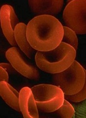
During their approximately 100-day lifespan in the bloodstream, red blood cells (RBCs) lose membrane surface area, volume, and hemoglobin content.
A study published in PLOS Computational Biology shows that, of these 3 changes, only surface-area loss can be explained by RBCs shedding small, hemoglobin-containing vesicles budding off their cells’ membrane.
Therefore, an unknown process must be primarily responsible for loss of RBC volume and hemoglobin reduction.
Roy Malka, PhD, of Massachusetts General Hospital in Boston, and his colleagues noted that variations in RBCs’ mean volume and hemoglobin content are associated with important clinical conditions, but the mechanisms controlling these physical characteristics are not well understood.
Vesicle shedding was thought to be the most important mechanism, but the researchers found evidence to suggest that a dominant role for vesicle shedding would violate empirical geometric and biophysical constraints.
So they concluded that an unknown mechanism must control loss of RBC volume and hemoglobin reduction. And this mechanism is likely responsible for 60% to 90% of volume loss and hemoglobin reduction.
The group’s work combined mathematical modeling of the mechanism that changes the physical properties of RBCs, clinical measurements of both cellular volume and hemoglobin content, and data from a new system for characterizing the non-water cellular mass of individual cells.
The researchers said the quantitative characterization of RBC loss processes will help focus future research into the molecular mechanisms of RBC maturation. And it may ultimately help in the early detection of clinical conditions where the RBC maturation pattern is altered.
During their approximately 100-day lifespan in the bloodstream, red blood cells (RBCs) lose membrane surface area, volume, and hemoglobin content.
A study published in PLOS Computational Biology shows that, of these 3 changes, only surface-area loss can be explained by RBCs shedding small, hemoglobin-containing vesicles budding off their cells’ membrane.
Therefore, an unknown process must be primarily responsible for loss of RBC volume and hemoglobin reduction.
Roy Malka, PhD, of Massachusetts General Hospital in Boston, and his colleagues noted that variations in RBCs’ mean volume and hemoglobin content are associated with important clinical conditions, but the mechanisms controlling these physical characteristics are not well understood.
Vesicle shedding was thought to be the most important mechanism, but the researchers found evidence to suggest that a dominant role for vesicle shedding would violate empirical geometric and biophysical constraints.
So they concluded that an unknown mechanism must control loss of RBC volume and hemoglobin reduction. And this mechanism is likely responsible for 60% to 90% of volume loss and hemoglobin reduction.
The group’s work combined mathematical modeling of the mechanism that changes the physical properties of RBCs, clinical measurements of both cellular volume and hemoglobin content, and data from a new system for characterizing the non-water cellular mass of individual cells.
The researchers said the quantitative characterization of RBC loss processes will help focus future research into the molecular mechanisms of RBC maturation. And it may ultimately help in the early detection of clinical conditions where the RBC maturation pattern is altered.
During their approximately 100-day lifespan in the bloodstream, red blood cells (RBCs) lose membrane surface area, volume, and hemoglobin content.
A study published in PLOS Computational Biology shows that, of these 3 changes, only surface-area loss can be explained by RBCs shedding small, hemoglobin-containing vesicles budding off their cells’ membrane.
Therefore, an unknown process must be primarily responsible for loss of RBC volume and hemoglobin reduction.
Roy Malka, PhD, of Massachusetts General Hospital in Boston, and his colleagues noted that variations in RBCs’ mean volume and hemoglobin content are associated with important clinical conditions, but the mechanisms controlling these physical characteristics are not well understood.
Vesicle shedding was thought to be the most important mechanism, but the researchers found evidence to suggest that a dominant role for vesicle shedding would violate empirical geometric and biophysical constraints.
So they concluded that an unknown mechanism must control loss of RBC volume and hemoglobin reduction. And this mechanism is likely responsible for 60% to 90% of volume loss and hemoglobin reduction.
The group’s work combined mathematical modeling of the mechanism that changes the physical properties of RBCs, clinical measurements of both cellular volume and hemoglobin content, and data from a new system for characterizing the non-water cellular mass of individual cells.
The researchers said the quantitative characterization of RBC loss processes will help focus future research into the molecular mechanisms of RBC maturation. And it may ultimately help in the early detection of clinical conditions where the RBC maturation pattern is altered.
FDA approves drug for untreated MCL

The US Food and Drug Administration (FDA) has approved bortezomib (Velcade) for use in previously untreated patients with mantle cell
lymphoma (MCL).
This is the first drug to be approved in the US for previously untreated patients with MCL.
The approval extends the utility of bortezomib beyond relapsed or refractory MCL, for which it has been approved since 2006.
The new approval is based on results of a phase 3 trial.
The study was a comparison of VcR-CAP (bortezomib, rituximab, cyclophosphamide, doxorubicin, and prednisone) and R-CHOP (rituximab, cyclophosphamide, doxorubicin, vincristine, and prednisone) in 487 patients newly diagnosed with stage II, III, or IV MCL.
Survival and response
VcR-CAP demonstrated a 59% relative improvement in the study’s primary endpoint of progression-free survival. At a median follow-up of 40 months, the median progression-free survival was 25 months in the VcR-CAP arm and 14 months in the R-CHOP arm (hazard ratio [HR]=0.63, P<0.001).
However, there was no significant improvement in overall survival. The median overall survival was not reached in the VcR-CAP arm and was 56.3 months in the R-CHOP arm (HR=0.80; P=0.173).
Patients in the VcR-CAP arm had a higher rate of complete response/unconfirmed complete response than those in the R-CHOP arm—53% and 42%, respectively (P=0.007). But there was no significant difference in overall response—92% and 90%, respectively (P=0.275).
The time to progression was significantly longer in the VcR-CAP arm—30.5 months, compared to 16.1 months in the R-CHOP arm (HR=0.58; P<0.001). And the median time to subsequent treatment was significantly longer in the VcR-CAP arm—44.5 months vs 24.8 months (HR 0.50; P<0.001).
Adverse events
VcR-CAP was associated with additional but manageable toxicity compared to R-CHOP.
Serious adverse events were reported in 38% of patients in the VcR-CAP arm and 30% in the R-CHOP arm. Grade 3 or higher adverse events were reported in 93% and 85%, respectively.
There were similar rates of all-grade peripheral neuropathy between the VcR-CAP arm and the R-CHOP arm—30% and 29%, respectively. But the rate of grade 3 or higher peripheral neuropathy was significantly higher in the VcR-CAP arm—7.5% vs 4.1%.
The incidence of all-grade thrombocytopenia was substantially higher in the VcR-CAP arm than the R-CHOP arm—72% and 19%, respectively. But there was no significant difference in bleeding events—6% and 5%, respectively.
The incidence of all-grade neutropenia was 88% in the VcR-CAP arm and 74% in the R-CHOP arm. The rate of grade 3 or higher febrile neutropenia was 14% and 15%, respectively, and the rate of infection was 60% and 46%, respectively.
These data were presented at ASCO 2014 as abstract 8500.
Bortezomib is marketed as Velcade by Millennium/Takeda and Janssen Pharmaceutical Companies. Millennium is responsible for commercialization in the US, and Janssen Pharmaceutical Companies are responsible for commercialization in the rest of the world.
For more details on the drug, visit www.velcade.com.
The US Food and Drug Administration (FDA) has approved bortezomib (Velcade) for use in previously untreated patients with mantle cell
lymphoma (MCL).
This is the first drug to be approved in the US for previously untreated patients with MCL.
The approval extends the utility of bortezomib beyond relapsed or refractory MCL, for which it has been approved since 2006.
The new approval is based on results of a phase 3 trial.
The study was a comparison of VcR-CAP (bortezomib, rituximab, cyclophosphamide, doxorubicin, and prednisone) and R-CHOP (rituximab, cyclophosphamide, doxorubicin, vincristine, and prednisone) in 487 patients newly diagnosed with stage II, III, or IV MCL.
Survival and response
VcR-CAP demonstrated a 59% relative improvement in the study’s primary endpoint of progression-free survival. At a median follow-up of 40 months, the median progression-free survival was 25 months in the VcR-CAP arm and 14 months in the R-CHOP arm (hazard ratio [HR]=0.63, P<0.001).
However, there was no significant improvement in overall survival. The median overall survival was not reached in the VcR-CAP arm and was 56.3 months in the R-CHOP arm (HR=0.80; P=0.173).
Patients in the VcR-CAP arm had a higher rate of complete response/unconfirmed complete response than those in the R-CHOP arm—53% and 42%, respectively (P=0.007). But there was no significant difference in overall response—92% and 90%, respectively (P=0.275).
The time to progression was significantly longer in the VcR-CAP arm—30.5 months, compared to 16.1 months in the R-CHOP arm (HR=0.58; P<0.001). And the median time to subsequent treatment was significantly longer in the VcR-CAP arm—44.5 months vs 24.8 months (HR 0.50; P<0.001).
Adverse events
VcR-CAP was associated with additional but manageable toxicity compared to R-CHOP.
Serious adverse events were reported in 38% of patients in the VcR-CAP arm and 30% in the R-CHOP arm. Grade 3 or higher adverse events were reported in 93% and 85%, respectively.
There were similar rates of all-grade peripheral neuropathy between the VcR-CAP arm and the R-CHOP arm—30% and 29%, respectively. But the rate of grade 3 or higher peripheral neuropathy was significantly higher in the VcR-CAP arm—7.5% vs 4.1%.
The incidence of all-grade thrombocytopenia was substantially higher in the VcR-CAP arm than the R-CHOP arm—72% and 19%, respectively. But there was no significant difference in bleeding events—6% and 5%, respectively.
The incidence of all-grade neutropenia was 88% in the VcR-CAP arm and 74% in the R-CHOP arm. The rate of grade 3 or higher febrile neutropenia was 14% and 15%, respectively, and the rate of infection was 60% and 46%, respectively.
These data were presented at ASCO 2014 as abstract 8500.
Bortezomib is marketed as Velcade by Millennium/Takeda and Janssen Pharmaceutical Companies. Millennium is responsible for commercialization in the US, and Janssen Pharmaceutical Companies are responsible for commercialization in the rest of the world.
For more details on the drug, visit www.velcade.com.
The US Food and Drug Administration (FDA) has approved bortezomib (Velcade) for use in previously untreated patients with mantle cell
lymphoma (MCL).
This is the first drug to be approved in the US for previously untreated patients with MCL.
The approval extends the utility of bortezomib beyond relapsed or refractory MCL, for which it has been approved since 2006.
The new approval is based on results of a phase 3 trial.
The study was a comparison of VcR-CAP (bortezomib, rituximab, cyclophosphamide, doxorubicin, and prednisone) and R-CHOP (rituximab, cyclophosphamide, doxorubicin, vincristine, and prednisone) in 487 patients newly diagnosed with stage II, III, or IV MCL.
Survival and response
VcR-CAP demonstrated a 59% relative improvement in the study’s primary endpoint of progression-free survival. At a median follow-up of 40 months, the median progression-free survival was 25 months in the VcR-CAP arm and 14 months in the R-CHOP arm (hazard ratio [HR]=0.63, P<0.001).
However, there was no significant improvement in overall survival. The median overall survival was not reached in the VcR-CAP arm and was 56.3 months in the R-CHOP arm (HR=0.80; P=0.173).
Patients in the VcR-CAP arm had a higher rate of complete response/unconfirmed complete response than those in the R-CHOP arm—53% and 42%, respectively (P=0.007). But there was no significant difference in overall response—92% and 90%, respectively (P=0.275).
The time to progression was significantly longer in the VcR-CAP arm—30.5 months, compared to 16.1 months in the R-CHOP arm (HR=0.58; P<0.001). And the median time to subsequent treatment was significantly longer in the VcR-CAP arm—44.5 months vs 24.8 months (HR 0.50; P<0.001).
Adverse events
VcR-CAP was associated with additional but manageable toxicity compared to R-CHOP.
Serious adverse events were reported in 38% of patients in the VcR-CAP arm and 30% in the R-CHOP arm. Grade 3 or higher adverse events were reported in 93% and 85%, respectively.
There were similar rates of all-grade peripheral neuropathy between the VcR-CAP arm and the R-CHOP arm—30% and 29%, respectively. But the rate of grade 3 or higher peripheral neuropathy was significantly higher in the VcR-CAP arm—7.5% vs 4.1%.
The incidence of all-grade thrombocytopenia was substantially higher in the VcR-CAP arm than the R-CHOP arm—72% and 19%, respectively. But there was no significant difference in bleeding events—6% and 5%, respectively.
The incidence of all-grade neutropenia was 88% in the VcR-CAP arm and 74% in the R-CHOP arm. The rate of grade 3 or higher febrile neutropenia was 14% and 15%, respectively, and the rate of infection was 60% and 46%, respectively.
These data were presented at ASCO 2014 as abstract 8500.
Bortezomib is marketed as Velcade by Millennium/Takeda and Janssen Pharmaceutical Companies. Millennium is responsible for commercialization in the US, and Janssen Pharmaceutical Companies are responsible for commercialization in the rest of the world.
For more details on the drug, visit www.velcade.com.
No need to switch antibiotics, study shows

Staphylococcus
infectionCredit: Bill Branson
New research suggests the antibiotic vancomycin is still effective in treating Staphylococcus aureus bloodstream (SAB) infections, despite increases in minimum inhibitory concentration (MIC) values.
Researchers found no difference in mortality between patients with low-vancomycin MIC and those with high-vancomycin MIC.
So it seems physicians can continue using vancomycin when MIC values are elevated but within the susceptible range, rather than
switching to newer antibiotics.
Andre Kalil, MD, of the University of Nebraska Medical Center in Omaha, and his colleagues described this research in JAMA.
In recent years, physicians treating Staphylococcus infections with vancomycin have seen an increase in the MIC, the lowest concentration of an antimicrobial agent that inhibits the growth of a microorganism.
MIC values lower than 4 mg/L suggest Staphylococcus is susceptible to vancomycin. However, when the MIC value exceeds 1.5 mg/L, some physicians have taken it as an indication that vancomycin may not be working at maximum effectiveness.
Some reports have suggested that elevations in vancomycin MIC values may be associated with increased treatment failure and death.
To determine the effectiveness of vancomycin, Dr Kalil and his colleagues analyzed data from 38 studies covering 8291 episodes of SAB infection.
The team evaluated the association between vancomycin MIC elevation and mortality. Among all SAB infections studied, the overall mortality was 26.1%.
The adjusted absolute risk of mortality did not differ significantly between patients with high-vancomycin MIC and those with low-vancomycin MIC—26.8% and 25.8%, respectively.
In studies that included only methicillin-resistant Staphylococcus aureus infections, the mortality among SAB episodes in patients with high-vancomycin MIC was 27.6%, compared with a mortality of 27.4% among patients with low-vancomycin MIC.
“The study provides strong evidence that vancomycin remains highly useful,” Dr Kalil said. “Even though vancomycin is an older drug, it is still killing staph very efficiently. There are newer antibiotics available to treat Staphylococcus aureus infections, but this study demonstrates that physicians don’t necessarily need to switch to these new drugs when the MIC is increased but still within the susceptible range.”
“The prevention of a rapid switch to newer drugs has another great benefit to our patients—less unnecessary exposure to these drugs, which will translate into less development of antibiotic resistance.”
Dr Kalil said the study may have implications for clinical practice and public health.
The results suggest standards for vancomycin MIC likely do not need to be lowered, routine differentiation of MIC values between 1 mg/L and 2 mg/L appears unnecessary, and the use of alternative drugs may not be required for Staphylococcus aureus isolates with elevated but susceptible vancomycin MIC values.
Staphylococcus
infectionCredit: Bill Branson
New research suggests the antibiotic vancomycin is still effective in treating Staphylococcus aureus bloodstream (SAB) infections, despite increases in minimum inhibitory concentration (MIC) values.
Researchers found no difference in mortality between patients with low-vancomycin MIC and those with high-vancomycin MIC.
So it seems physicians can continue using vancomycin when MIC values are elevated but within the susceptible range, rather than
switching to newer antibiotics.
Andre Kalil, MD, of the University of Nebraska Medical Center in Omaha, and his colleagues described this research in JAMA.
In recent years, physicians treating Staphylococcus infections with vancomycin have seen an increase in the MIC, the lowest concentration of an antimicrobial agent that inhibits the growth of a microorganism.
MIC values lower than 4 mg/L suggest Staphylococcus is susceptible to vancomycin. However, when the MIC value exceeds 1.5 mg/L, some physicians have taken it as an indication that vancomycin may not be working at maximum effectiveness.
Some reports have suggested that elevations in vancomycin MIC values may be associated with increased treatment failure and death.
To determine the effectiveness of vancomycin, Dr Kalil and his colleagues analyzed data from 38 studies covering 8291 episodes of SAB infection.
The team evaluated the association between vancomycin MIC elevation and mortality. Among all SAB infections studied, the overall mortality was 26.1%.
The adjusted absolute risk of mortality did not differ significantly between patients with high-vancomycin MIC and those with low-vancomycin MIC—26.8% and 25.8%, respectively.
In studies that included only methicillin-resistant Staphylococcus aureus infections, the mortality among SAB episodes in patients with high-vancomycin MIC was 27.6%, compared with a mortality of 27.4% among patients with low-vancomycin MIC.
“The study provides strong evidence that vancomycin remains highly useful,” Dr Kalil said. “Even though vancomycin is an older drug, it is still killing staph very efficiently. There are newer antibiotics available to treat Staphylococcus aureus infections, but this study demonstrates that physicians don’t necessarily need to switch to these new drugs when the MIC is increased but still within the susceptible range.”
“The prevention of a rapid switch to newer drugs has another great benefit to our patients—less unnecessary exposure to these drugs, which will translate into less development of antibiotic resistance.”
Dr Kalil said the study may have implications for clinical practice and public health.
The results suggest standards for vancomycin MIC likely do not need to be lowered, routine differentiation of MIC values between 1 mg/L and 2 mg/L appears unnecessary, and the use of alternative drugs may not be required for Staphylococcus aureus isolates with elevated but susceptible vancomycin MIC values.
Staphylococcus
infectionCredit: Bill Branson
New research suggests the antibiotic vancomycin is still effective in treating Staphylococcus aureus bloodstream (SAB) infections, despite increases in minimum inhibitory concentration (MIC) values.
Researchers found no difference in mortality between patients with low-vancomycin MIC and those with high-vancomycin MIC.
So it seems physicians can continue using vancomycin when MIC values are elevated but within the susceptible range, rather than
switching to newer antibiotics.
Andre Kalil, MD, of the University of Nebraska Medical Center in Omaha, and his colleagues described this research in JAMA.
In recent years, physicians treating Staphylococcus infections with vancomycin have seen an increase in the MIC, the lowest concentration of an antimicrobial agent that inhibits the growth of a microorganism.
MIC values lower than 4 mg/L suggest Staphylococcus is susceptible to vancomycin. However, when the MIC value exceeds 1.5 mg/L, some physicians have taken it as an indication that vancomycin may not be working at maximum effectiveness.
Some reports have suggested that elevations in vancomycin MIC values may be associated with increased treatment failure and death.
To determine the effectiveness of vancomycin, Dr Kalil and his colleagues analyzed data from 38 studies covering 8291 episodes of SAB infection.
The team evaluated the association between vancomycin MIC elevation and mortality. Among all SAB infections studied, the overall mortality was 26.1%.
The adjusted absolute risk of mortality did not differ significantly between patients with high-vancomycin MIC and those with low-vancomycin MIC—26.8% and 25.8%, respectively.
In studies that included only methicillin-resistant Staphylococcus aureus infections, the mortality among SAB episodes in patients with high-vancomycin MIC was 27.6%, compared with a mortality of 27.4% among patients with low-vancomycin MIC.
“The study provides strong evidence that vancomycin remains highly useful,” Dr Kalil said. “Even though vancomycin is an older drug, it is still killing staph very efficiently. There are newer antibiotics available to treat Staphylococcus aureus infections, but this study demonstrates that physicians don’t necessarily need to switch to these new drugs when the MIC is increased but still within the susceptible range.”
“The prevention of a rapid switch to newer drugs has another great benefit to our patients—less unnecessary exposure to these drugs, which will translate into less development of antibiotic resistance.”
Dr Kalil said the study may have implications for clinical practice and public health.
The results suggest standards for vancomycin MIC likely do not need to be lowered, routine differentiation of MIC values between 1 mg/L and 2 mg/L appears unnecessary, and the use of alternative drugs may not be required for Staphylococcus aureus isolates with elevated but susceptible vancomycin MIC values.
New insight into T-cell development
Credit: NIAID
A fundamental theory about how the thymus “educates” T cells appears to be wrong, according to research published in Nature Communications.
It’s known that stem cells leave the bone marrow and travel to the thymus to become one of two CD4 T-cell types: effector cells or regulatory T cells (Tregs).
One widely held concept of why the cells become one type or the other is that they are exposed to different ligands in the thymus, said Leszek Ignatowicz, PhD, of Georgia Regents University in Augusta.
But when he and his colleagues limited the cells’ exposure to only one ligand, the same mix of T cells still emerged.
“We asked a simple question, ‘Is it going to affect their development,’ and the answer was ‘no,’” Dr Ignatowicz said. “The cells still mature in the thymus, so something else must be determining it.”
The finding provides more insight into immunity that could one day enable a new approach to vaccines that steer the thymus to produce more of whatever T-cell type a patient needs: more effector cells if they have an infection or cancer, more Tregs if they have autoimmune diseases such as arthritis and multiple sclerosis.
“We could help steer the education process in the desired direction,” Dr Ignatowicz said.
To uncover their findings, he and his colleagues studied two types of mice, each expressing a single ligand in the thymus. The researchers thought one would prompt strong ligand binding and favor Treg development, and the other would favor a weaker ligand bond and effector cell development.
The mix of resulting T cells was the same as if both were exposed to the usual thousands of ligands, although the experiment did reveal one difference.
Ligands—and, eventually, bacteria and other invaders—get the attention of T cells by activating their receptors. Both CD4 T-cell types generally have the same receptors, but they are organized differently.
The researchers found that as long as the binding was weak, as it was in the first mouse, there was a lot of overlap in the receptors the ligand bound to in both T-cell types. However, in the second mouse, where the ligand prompted strong binding, there was far less overlap.
“We are now trying to find what causes that difference,” Dr Ignatowicz said.
Credit: NIAID
A fundamental theory about how the thymus “educates” T cells appears to be wrong, according to research published in Nature Communications.
It’s known that stem cells leave the bone marrow and travel to the thymus to become one of two CD4 T-cell types: effector cells or regulatory T cells (Tregs).
One widely held concept of why the cells become one type or the other is that they are exposed to different ligands in the thymus, said Leszek Ignatowicz, PhD, of Georgia Regents University in Augusta.
But when he and his colleagues limited the cells’ exposure to only one ligand, the same mix of T cells still emerged.
“We asked a simple question, ‘Is it going to affect their development,’ and the answer was ‘no,’” Dr Ignatowicz said. “The cells still mature in the thymus, so something else must be determining it.”
The finding provides more insight into immunity that could one day enable a new approach to vaccines that steer the thymus to produce more of whatever T-cell type a patient needs: more effector cells if they have an infection or cancer, more Tregs if they have autoimmune diseases such as arthritis and multiple sclerosis.
“We could help steer the education process in the desired direction,” Dr Ignatowicz said.
To uncover their findings, he and his colleagues studied two types of mice, each expressing a single ligand in the thymus. The researchers thought one would prompt strong ligand binding and favor Treg development, and the other would favor a weaker ligand bond and effector cell development.
The mix of resulting T cells was the same as if both were exposed to the usual thousands of ligands, although the experiment did reveal one difference.
Ligands—and, eventually, bacteria and other invaders—get the attention of T cells by activating their receptors. Both CD4 T-cell types generally have the same receptors, but they are organized differently.
The researchers found that as long as the binding was weak, as it was in the first mouse, there was a lot of overlap in the receptors the ligand bound to in both T-cell types. However, in the second mouse, where the ligand prompted strong binding, there was far less overlap.
“We are now trying to find what causes that difference,” Dr Ignatowicz said.
Credit: NIAID
A fundamental theory about how the thymus “educates” T cells appears to be wrong, according to research published in Nature Communications.
It’s known that stem cells leave the bone marrow and travel to the thymus to become one of two CD4 T-cell types: effector cells or regulatory T cells (Tregs).
One widely held concept of why the cells become one type or the other is that they are exposed to different ligands in the thymus, said Leszek Ignatowicz, PhD, of Georgia Regents University in Augusta.
But when he and his colleagues limited the cells’ exposure to only one ligand, the same mix of T cells still emerged.
“We asked a simple question, ‘Is it going to affect their development,’ and the answer was ‘no,’” Dr Ignatowicz said. “The cells still mature in the thymus, so something else must be determining it.”
The finding provides more insight into immunity that could one day enable a new approach to vaccines that steer the thymus to produce more of whatever T-cell type a patient needs: more effector cells if they have an infection or cancer, more Tregs if they have autoimmune diseases such as arthritis and multiple sclerosis.
“We could help steer the education process in the desired direction,” Dr Ignatowicz said.
To uncover their findings, he and his colleagues studied two types of mice, each expressing a single ligand in the thymus. The researchers thought one would prompt strong ligand binding and favor Treg development, and the other would favor a weaker ligand bond and effector cell development.
The mix of resulting T cells was the same as if both were exposed to the usual thousands of ligands, although the experiment did reveal one difference.
Ligands—and, eventually, bacteria and other invaders—get the attention of T cells by activating their receptors. Both CD4 T-cell types generally have the same receptors, but they are organized differently.
The researchers found that as long as the binding was weak, as it was in the first mouse, there was a lot of overlap in the receptors the ligand bound to in both T-cell types. However, in the second mouse, where the ligand prompted strong binding, there was far less overlap.
“We are now trying to find what causes that difference,” Dr Ignatowicz said.