User login
How ‘urban’ mosquitoes transmit malaria
Credit: CDC
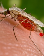
Researchers believe that by analyzing the genome of a mosquito species, they have discovered how that mosquito evolves to withstand a variety of environmental conditions.
The results, published in Genome Biology, provide a better understanding of Anopheles stephensi, a common carrier of malaria in urban environments.
“Anopheles stephensi is emerging as a model mosquito species for genetic and molecular studies,” said Zhijian Jake Tu, PhD, of Virginia Tech in Blacksburg.
He and his colleagues believe their genome map of An stephensi will be an important tool for scientists to identify potential targets for mosquito control. In addition, studies of immunity genes can provide insight into mosquito biology and mosquito-parasite interactions.
“Genome mapping of Anopheles stephensi revealed genetic differences between it and a species especially dangerous for transmitting malaria in Africa, Anopheles gambiae,” said Igor Sharakhov, PhD, also of Virginia Tech.
“This tells us that the sex chromosome is especially prone to mutations that flip chromosomal segments, which, in turn, may promote new, evolved species.”
The researchers assembled more than 92% of the An stephensi genome, and physical mapping assigned 62% of the genome onto chromosomes.
When the team compared An stephensi and An gambiae, they discovered the rate of gene order reshuffling on the Anopheles X chromosome was 3 times higher than that on the autosomes.
An stephensi had more repeat-rich heterochromatin in pericentric regions than An gambiae but fewer repetitive sequences in chromosome arms.
The researchers also identified Y-chromosome contigs and BACs, which represent the most abundant set of Y sequences in any Anopheles species.
Lastly, the team noted that RNA-sequencing and studies of immunity genes provided new insights into mosquito biology and mosquito-parasite interactions.
For instance, FKBP12, a protein that interacts with TOR and TGF-β signaling pathways, showed abundant mRNA expression in a wide range of tissues. This information could help improve our understanding of TOR and TGF-β signaling in mosquitoes. 
Credit: CDC
Researchers believe that by analyzing the genome of a mosquito species, they have discovered how that mosquito evolves to withstand a variety of environmental conditions.
The results, published in Genome Biology, provide a better understanding of Anopheles stephensi, a common carrier of malaria in urban environments.
“Anopheles stephensi is emerging as a model mosquito species for genetic and molecular studies,” said Zhijian Jake Tu, PhD, of Virginia Tech in Blacksburg.
He and his colleagues believe their genome map of An stephensi will be an important tool for scientists to identify potential targets for mosquito control. In addition, studies of immunity genes can provide insight into mosquito biology and mosquito-parasite interactions.
“Genome mapping of Anopheles stephensi revealed genetic differences between it and a species especially dangerous for transmitting malaria in Africa, Anopheles gambiae,” said Igor Sharakhov, PhD, also of Virginia Tech.
“This tells us that the sex chromosome is especially prone to mutations that flip chromosomal segments, which, in turn, may promote new, evolved species.”
The researchers assembled more than 92% of the An stephensi genome, and physical mapping assigned 62% of the genome onto chromosomes.
When the team compared An stephensi and An gambiae, they discovered the rate of gene order reshuffling on the Anopheles X chromosome was 3 times higher than that on the autosomes.
An stephensi had more repeat-rich heterochromatin in pericentric regions than An gambiae but fewer repetitive sequences in chromosome arms.
The researchers also identified Y-chromosome contigs and BACs, which represent the most abundant set of Y sequences in any Anopheles species.
Lastly, the team noted that RNA-sequencing and studies of immunity genes provided new insights into mosquito biology and mosquito-parasite interactions.
For instance, FKBP12, a protein that interacts with TOR and TGF-β signaling pathways, showed abundant mRNA expression in a wide range of tissues. This information could help improve our understanding of TOR and TGF-β signaling in mosquitoes.
Credit: CDC
Researchers believe that by analyzing the genome of a mosquito species, they have discovered how that mosquito evolves to withstand a variety of environmental conditions.
The results, published in Genome Biology, provide a better understanding of Anopheles stephensi, a common carrier of malaria in urban environments.
“Anopheles stephensi is emerging as a model mosquito species for genetic and molecular studies,” said Zhijian Jake Tu, PhD, of Virginia Tech in Blacksburg.
He and his colleagues believe their genome map of An stephensi will be an important tool for scientists to identify potential targets for mosquito control. In addition, studies of immunity genes can provide insight into mosquito biology and mosquito-parasite interactions.
“Genome mapping of Anopheles stephensi revealed genetic differences between it and a species especially dangerous for transmitting malaria in Africa, Anopheles gambiae,” said Igor Sharakhov, PhD, also of Virginia Tech.
“This tells us that the sex chromosome is especially prone to mutations that flip chromosomal segments, which, in turn, may promote new, evolved species.”
The researchers assembled more than 92% of the An stephensi genome, and physical mapping assigned 62% of the genome onto chromosomes.
When the team compared An stephensi and An gambiae, they discovered the rate of gene order reshuffling on the Anopheles X chromosome was 3 times higher than that on the autosomes.
An stephensi had more repeat-rich heterochromatin in pericentric regions than An gambiae but fewer repetitive sequences in chromosome arms.
The researchers also identified Y-chromosome contigs and BACs, which represent the most abundant set of Y sequences in any Anopheles species.
Lastly, the team noted that RNA-sequencing and studies of immunity genes provided new insights into mosquito biology and mosquito-parasite interactions.
For instance, FKBP12, a protein that interacts with TOR and TGF-β signaling pathways, showed abundant mRNA expression in a wide range of tissues. This information could help improve our understanding of TOR and TGF-β signaling in mosquitoes.
Blood sample storage may hinder leukemia research
Credit: Graham Colm
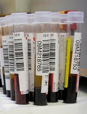
Storing blood samples at room temperature can induce changes that may cloud research findings, according to a group of investigators.
The team initially found that blood samples from leukemia patients had high levels of malformed RNA, a discovery they believed could explain leukemia’s origins.
But additional research showed this abnormality was a result of storing blood samples at room temperature for hours, or even days, prior to processing.
Heidi Dvinge, PhD, of the Fred Hutchinson Cancer Research Center in Seattle, and her colleagues reported these findings in PNAS.
The investigators had searched databases to collect genomic information on leukemia patients and healthy control subjects. In all but one of the datasets they analyzed, the team found high levels of abnormal RNA in leukemia cells.
Another finding was that samples from pediatric leukemia patients had the highest levels of abnormal RNA. And this led the researchers to speculate about the cause.
They realized that pediatric leukemias are rare, so blood samples can be difficult to obtain. Therefore, the samples in the databases were collected at facilities throughout the world and shipped to where they were needed. So the samples could be stored at room temperature for days at a time.
To confirm that this practice can affect blood samples, the investigators conducted an experiment. They collected samples from 4 healthy subjects (2 men and 2 women) and looked for differences between samples that were processed immediately and samples that sat in the lab at room temperature for up to 48 hours.
Sure enough, the team observed changes in the stored samples, even if they were only stored for 4 hours. Stored samples exhibited biased activation of biological pathways and upregulation of pseudogenes, antisense RNAs, and unannotated coding isoforms.
Storage affected a number of genes that play roles in biological pathways relevant to leukemia, including cytokine production, NF-κB signaling, chromatin modification, and RNA splicing. Additionally, storage inhibited RNA surveillance, leading to the genome-wide expression of normally degraded RNAs.
The researchers said these findings coincide with the database findings, as they observed inhibited RNA surveillance in all but one of the datasets they analyzed.
The samples from that dataset were processed immediately after collection. And samples from healthy subjects were processed immediately, which explains why those samples were normal as well.
The team also noted that they did not observe inhibited RNA surveillance in lymphoma or solid tumor datasets.
These results suggest previous research utilizing these types of databases may contain errors, and future work making use of these databases could be affected as well.
Fortunately, the investigators found that putting blood samples on ice can prevent the negative effects they observed. The group also identified biomarkers that indicate prolonged storage, so researchers can look for those biomarkers if chilling blood samples is not an option.
In addition to describing these findings in PNAS, Dr Dvinge and her colleagues are planning to present their work at the upcoming ASH Annual Meeting.
Credit: Graham Colm
Storing blood samples at room temperature can induce changes that may cloud research findings, according to a group of investigators.
The team initially found that blood samples from leukemia patients had high levels of malformed RNA, a discovery they believed could explain leukemia’s origins.
But additional research showed this abnormality was a result of storing blood samples at room temperature for hours, or even days, prior to processing.
Heidi Dvinge, PhD, of the Fred Hutchinson Cancer Research Center in Seattle, and her colleagues reported these findings in PNAS.
The investigators had searched databases to collect genomic information on leukemia patients and healthy control subjects. In all but one of the datasets they analyzed, the team found high levels of abnormal RNA in leukemia cells.
Another finding was that samples from pediatric leukemia patients had the highest levels of abnormal RNA. And this led the researchers to speculate about the cause.
They realized that pediatric leukemias are rare, so blood samples can be difficult to obtain. Therefore, the samples in the databases were collected at facilities throughout the world and shipped to where they were needed. So the samples could be stored at room temperature for days at a time.
To confirm that this practice can affect blood samples, the investigators conducted an experiment. They collected samples from 4 healthy subjects (2 men and 2 women) and looked for differences between samples that were processed immediately and samples that sat in the lab at room temperature for up to 48 hours.
Sure enough, the team observed changes in the stored samples, even if they were only stored for 4 hours. Stored samples exhibited biased activation of biological pathways and upregulation of pseudogenes, antisense RNAs, and unannotated coding isoforms.
Storage affected a number of genes that play roles in biological pathways relevant to leukemia, including cytokine production, NF-κB signaling, chromatin modification, and RNA splicing. Additionally, storage inhibited RNA surveillance, leading to the genome-wide expression of normally degraded RNAs.
The researchers said these findings coincide with the database findings, as they observed inhibited RNA surveillance in all but one of the datasets they analyzed.
The samples from that dataset were processed immediately after collection. And samples from healthy subjects were processed immediately, which explains why those samples were normal as well.
The team also noted that they did not observe inhibited RNA surveillance in lymphoma or solid tumor datasets.
These results suggest previous research utilizing these types of databases may contain errors, and future work making use of these databases could be affected as well.
Fortunately, the investigators found that putting blood samples on ice can prevent the negative effects they observed. The group also identified biomarkers that indicate prolonged storage, so researchers can look for those biomarkers if chilling blood samples is not an option.
In addition to describing these findings in PNAS, Dr Dvinge and her colleagues are planning to present their work at the upcoming ASH Annual Meeting.
Credit: Graham Colm
Storing blood samples at room temperature can induce changes that may cloud research findings, according to a group of investigators.
The team initially found that blood samples from leukemia patients had high levels of malformed RNA, a discovery they believed could explain leukemia’s origins.
But additional research showed this abnormality was a result of storing blood samples at room temperature for hours, or even days, prior to processing.
Heidi Dvinge, PhD, of the Fred Hutchinson Cancer Research Center in Seattle, and her colleagues reported these findings in PNAS.
The investigators had searched databases to collect genomic information on leukemia patients and healthy control subjects. In all but one of the datasets they analyzed, the team found high levels of abnormal RNA in leukemia cells.
Another finding was that samples from pediatric leukemia patients had the highest levels of abnormal RNA. And this led the researchers to speculate about the cause.
They realized that pediatric leukemias are rare, so blood samples can be difficult to obtain. Therefore, the samples in the databases were collected at facilities throughout the world and shipped to where they were needed. So the samples could be stored at room temperature for days at a time.
To confirm that this practice can affect blood samples, the investigators conducted an experiment. They collected samples from 4 healthy subjects (2 men and 2 women) and looked for differences between samples that were processed immediately and samples that sat in the lab at room temperature for up to 48 hours.
Sure enough, the team observed changes in the stored samples, even if they were only stored for 4 hours. Stored samples exhibited biased activation of biological pathways and upregulation of pseudogenes, antisense RNAs, and unannotated coding isoforms.
Storage affected a number of genes that play roles in biological pathways relevant to leukemia, including cytokine production, NF-κB signaling, chromatin modification, and RNA splicing. Additionally, storage inhibited RNA surveillance, leading to the genome-wide expression of normally degraded RNAs.
The researchers said these findings coincide with the database findings, as they observed inhibited RNA surveillance in all but one of the datasets they analyzed.
The samples from that dataset were processed immediately after collection. And samples from healthy subjects were processed immediately, which explains why those samples were normal as well.
The team also noted that they did not observe inhibited RNA surveillance in lymphoma or solid tumor datasets.
These results suggest previous research utilizing these types of databases may contain errors, and future work making use of these databases could be affected as well.
Fortunately, the investigators found that putting blood samples on ice can prevent the negative effects they observed. The group also identified biomarkers that indicate prolonged storage, so researchers can look for those biomarkers if chilling blood samples is not an option.
In addition to describing these findings in PNAS, Dr Dvinge and her colleagues are planning to present their work at the upcoming ASH Annual Meeting.
Sharing research with public prompts more citations
Credit: Rhoda Baer
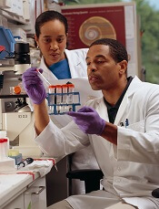
Academic researchers who talk to the press and use social media are more likely than their less communicative peers to have their work cited, a new study suggests.
The research revealed a connection between h-index—a measure of the quality of a researcher’s work and influence—and whether the scientists interact with reporters and get mentioned on Twitter.
The results appear in Journalism & Mass Communications Quarterly.
“I’ve been in science communication for a while now, and I am really seeing a change—especially among the younger scientists—in their willingness to share their work,” said study author Dominique Brossard, PhD, of the University of Wisconsin-Madison.
Attention from reporters is good news for h-index, Dr Brossard noted. But couple that with attention on Twitter, and you see a more pronounced spike in reputation.
“If you talk to reporters and you tweet about your research, your work is more likely to be cited than people who do one or the other,” she said.
That sort of activity hasn’t always been encouraged, Dr Brossard pointed out. Any distraction from a researcher’s work can draw criticism as a waste of a precious resource. But Dr Brossard hopes a new understanding of the relationship between research and communicating with the public can change that.
“What this shows us is that sharing your science with the public is not hurting the science by stealing time,” she said. “If the goal is to encourage people, ultimately, to be productive scientists, and if directors of labs are discouraging people from engaging in this activity, they’re actually hurting the science itself. Because people who do this are cited more often in scientific journals, [and] they’re making science accessible to broader audiences at the same time.”
Social media use is rising in other professional circles as well, according to Michael Xenos, PhD, also of the University of Wisconsin-Madison.
“As in other areas, such as politics for example, social media was once met with skepticism but is increasingly part of the culture,” he said. “Just like it became the norm there, our research shows it may one day become the norm in science.”
Even if you flip the connection between social media attention and h-index on its head, it’s still worth taking to heart, according to the researchers.
“The counter argument is that it may be just the other way around—that it may just be the big names that get mentions,” said study author Dietram A. Scheufele, PhD, also of the University of Wisconsin-Madison.
“But then, the lesson should be that the most successful people in your field are also the ones that are good at getting outside the ivory tower. That should be something to emulate.”
Credit: Rhoda Baer
Academic researchers who talk to the press and use social media are more likely than their less communicative peers to have their work cited, a new study suggests.
The research revealed a connection between h-index—a measure of the quality of a researcher’s work and influence—and whether the scientists interact with reporters and get mentioned on Twitter.
The results appear in Journalism & Mass Communications Quarterly.
“I’ve been in science communication for a while now, and I am really seeing a change—especially among the younger scientists—in their willingness to share their work,” said study author Dominique Brossard, PhD, of the University of Wisconsin-Madison.
Attention from reporters is good news for h-index, Dr Brossard noted. But couple that with attention on Twitter, and you see a more pronounced spike in reputation.
“If you talk to reporters and you tweet about your research, your work is more likely to be cited than people who do one or the other,” she said.
That sort of activity hasn’t always been encouraged, Dr Brossard pointed out. Any distraction from a researcher’s work can draw criticism as a waste of a precious resource. But Dr Brossard hopes a new understanding of the relationship between research and communicating with the public can change that.
“What this shows us is that sharing your science with the public is not hurting the science by stealing time,” she said. “If the goal is to encourage people, ultimately, to be productive scientists, and if directors of labs are discouraging people from engaging in this activity, they’re actually hurting the science itself. Because people who do this are cited more often in scientific journals, [and] they’re making science accessible to broader audiences at the same time.”
Social media use is rising in other professional circles as well, according to Michael Xenos, PhD, also of the University of Wisconsin-Madison.
“As in other areas, such as politics for example, social media was once met with skepticism but is increasingly part of the culture,” he said. “Just like it became the norm there, our research shows it may one day become the norm in science.”
Even if you flip the connection between social media attention and h-index on its head, it’s still worth taking to heart, according to the researchers.
“The counter argument is that it may be just the other way around—that it may just be the big names that get mentions,” said study author Dietram A. Scheufele, PhD, also of the University of Wisconsin-Madison.
“But then, the lesson should be that the most successful people in your field are also the ones that are good at getting outside the ivory tower. That should be something to emulate.”
Credit: Rhoda Baer
Academic researchers who talk to the press and use social media are more likely than their less communicative peers to have their work cited, a new study suggests.
The research revealed a connection between h-index—a measure of the quality of a researcher’s work and influence—and whether the scientists interact with reporters and get mentioned on Twitter.
The results appear in Journalism & Mass Communications Quarterly.
“I’ve been in science communication for a while now, and I am really seeing a change—especially among the younger scientists—in their willingness to share their work,” said study author Dominique Brossard, PhD, of the University of Wisconsin-Madison.
Attention from reporters is good news for h-index, Dr Brossard noted. But couple that with attention on Twitter, and you see a more pronounced spike in reputation.
“If you talk to reporters and you tweet about your research, your work is more likely to be cited than people who do one or the other,” she said.
That sort of activity hasn’t always been encouraged, Dr Brossard pointed out. Any distraction from a researcher’s work can draw criticism as a waste of a precious resource. But Dr Brossard hopes a new understanding of the relationship between research and communicating with the public can change that.
“What this shows us is that sharing your science with the public is not hurting the science by stealing time,” she said. “If the goal is to encourage people, ultimately, to be productive scientists, and if directors of labs are discouraging people from engaging in this activity, they’re actually hurting the science itself. Because people who do this are cited more often in scientific journals, [and] they’re making science accessible to broader audiences at the same time.”
Social media use is rising in other professional circles as well, according to Michael Xenos, PhD, also of the University of Wisconsin-Madison.
“As in other areas, such as politics for example, social media was once met with skepticism but is increasingly part of the culture,” he said. “Just like it became the norm there, our research shows it may one day become the norm in science.”
Even if you flip the connection between social media attention and h-index on its head, it’s still worth taking to heart, according to the researchers.
“The counter argument is that it may be just the other way around—that it may just be the big names that get mentions,” said study author Dietram A. Scheufele, PhD, also of the University of Wisconsin-Madison.
“But then, the lesson should be that the most successful people in your field are also the ones that are good at getting outside the ivory tower. That should be something to emulate.”
Hospice cuts cost and use of care for cancer patients

Credit: CDC
Patients with advanced cancer receive less aggressive care and have lower healthcare costs during their last year of life if they use hospice care, according to research published in JAMA.
Patients who entered hospice had significantly lower rates of hospitalization, intensive care unit admissions, and invasive procedures, compared to patients who did not enter hospice.
Furthermore, patients who chose hospice were about 5 times less likely to die in hospitals and nursing homes.
“Our study shows very clearly that hospice matters,” said Ziad Obermeyer, MD, of Brigham and Women’s Hospital in Boston.
“Hospice and non-hospice patients had very similar patterns of healthcare utilization right up until the week of hospice enrollment. Then, the care started to look very different. Patients who didn’t enroll in hospice ended up with far more aggressive care in their last year of life, most of it related to acute complications like infections and organ failure, and not directly related to their cancer diagnosis.”
To conduct this study, Dr Obermeyer and his colleagues used data from Medicare beneficiaries with poor-prognosis cancers, including hematologic malignancies. The study included a nationally representative 20% sample of Medicare fee-for-service beneficiaries who died in 2011.
Among 86,851 patients, 51,924 (60%) entered hospice before death. Matching patients based on various criteria produced hospice and non-hospice groups, each with 18,165 patients. The median hospice duration was 11 days.
The researchers found that non-hospice patients had significantly greater healthcare utilization, largely for acute conditions not directly related to cancer.
This included rates of hospitalization (65% vs 42%), intensive care unit admissions (36% vs 15%), invasive procedures (51% vs 27%), and death in a hospital or nursing facility (74% vs 14%).
The costs of care for hospice and non-hospice patients were not significantly different before hospice care began. But they diverged sharply thereafter, contributing to a significant difference in total costs of $8697 over the last year of life—$71,517 for non-hospice patients and $62,819 for hospice patients.
“These findings highlight the importance of honest discussions between doctors and patients about our patients’ goals of their care at the end of life, relating to treatment decisions and quality of life,” Dr Obermeyer said. “This is of particular importance now, in light of the ongoing policy discussions around reimbursing providers for advance-care planning.”
Credit: CDC
Patients with advanced cancer receive less aggressive care and have lower healthcare costs during their last year of life if they use hospice care, according to research published in JAMA.
Patients who entered hospice had significantly lower rates of hospitalization, intensive care unit admissions, and invasive procedures, compared to patients who did not enter hospice.
Furthermore, patients who chose hospice were about 5 times less likely to die in hospitals and nursing homes.
“Our study shows very clearly that hospice matters,” said Ziad Obermeyer, MD, of Brigham and Women’s Hospital in Boston.
“Hospice and non-hospice patients had very similar patterns of healthcare utilization right up until the week of hospice enrollment. Then, the care started to look very different. Patients who didn’t enroll in hospice ended up with far more aggressive care in their last year of life, most of it related to acute complications like infections and organ failure, and not directly related to their cancer diagnosis.”
To conduct this study, Dr Obermeyer and his colleagues used data from Medicare beneficiaries with poor-prognosis cancers, including hematologic malignancies. The study included a nationally representative 20% sample of Medicare fee-for-service beneficiaries who died in 2011.
Among 86,851 patients, 51,924 (60%) entered hospice before death. Matching patients based on various criteria produced hospice and non-hospice groups, each with 18,165 patients. The median hospice duration was 11 days.
The researchers found that non-hospice patients had significantly greater healthcare utilization, largely for acute conditions not directly related to cancer.
This included rates of hospitalization (65% vs 42%), intensive care unit admissions (36% vs 15%), invasive procedures (51% vs 27%), and death in a hospital or nursing facility (74% vs 14%).
The costs of care for hospice and non-hospice patients were not significantly different before hospice care began. But they diverged sharply thereafter, contributing to a significant difference in total costs of $8697 over the last year of life—$71,517 for non-hospice patients and $62,819 for hospice patients.
“These findings highlight the importance of honest discussions between doctors and patients about our patients’ goals of their care at the end of life, relating to treatment decisions and quality of life,” Dr Obermeyer said. “This is of particular importance now, in light of the ongoing policy discussions around reimbursing providers for advance-care planning.”
Credit: CDC
Patients with advanced cancer receive less aggressive care and have lower healthcare costs during their last year of life if they use hospice care, according to research published in JAMA.
Patients who entered hospice had significantly lower rates of hospitalization, intensive care unit admissions, and invasive procedures, compared to patients who did not enter hospice.
Furthermore, patients who chose hospice were about 5 times less likely to die in hospitals and nursing homes.
“Our study shows very clearly that hospice matters,” said Ziad Obermeyer, MD, of Brigham and Women’s Hospital in Boston.
“Hospice and non-hospice patients had very similar patterns of healthcare utilization right up until the week of hospice enrollment. Then, the care started to look very different. Patients who didn’t enroll in hospice ended up with far more aggressive care in their last year of life, most of it related to acute complications like infections and organ failure, and not directly related to their cancer diagnosis.”
To conduct this study, Dr Obermeyer and his colleagues used data from Medicare beneficiaries with poor-prognosis cancers, including hematologic malignancies. The study included a nationally representative 20% sample of Medicare fee-for-service beneficiaries who died in 2011.
Among 86,851 patients, 51,924 (60%) entered hospice before death. Matching patients based on various criteria produced hospice and non-hospice groups, each with 18,165 patients. The median hospice duration was 11 days.
The researchers found that non-hospice patients had significantly greater healthcare utilization, largely for acute conditions not directly related to cancer.
This included rates of hospitalization (65% vs 42%), intensive care unit admissions (36% vs 15%), invasive procedures (51% vs 27%), and death in a hospital or nursing facility (74% vs 14%).
The costs of care for hospice and non-hospice patients were not significantly different before hospice care began. But they diverged sharply thereafter, contributing to a significant difference in total costs of $8697 over the last year of life—$71,517 for non-hospice patients and $62,819 for hospice patients.
“These findings highlight the importance of honest discussions between doctors and patients about our patients’ goals of their care at the end of life, relating to treatment decisions and quality of life,” Dr Obermeyer said. “This is of particular importance now, in light of the ongoing policy discussions around reimbursing providers for advance-care planning.”
NICE supports use of catheter-positioning device

The UK’s National Institute for Health and Care Excellence (NICE) has opened consultation on a draft guidance about a device designed to help healthcare professionals correctly place peripherally inserted central catheters
(PICCs).
The draft guidance supports using the Sherlock 3CG Tip Confirmation System for placing PICCs.
The standard procedure for placing PICCs is blind insertion, followed by a chest X-ray to check the catheter’s position.
In some cases, fluoroscopy is used instead of standard X-ray to assist with positioning the PICC when placing it proves difficult.
The Sherlock system, on the other hand, uses magnetic and electrocardiographic real-time tracking of a PICC to enable the person placing the catheter to detect and correct any error in how the tip is positioned.
The device’s manufacturer, C.R. Bard, says the Sherlock System eliminates the need for the patient to have an X-ray, thus preventing delays in treatment or monitoring.
“Using the technology also increases staff and patient confidence of the accuracy of the procedure during catheter insertion,” said Professor Carole Longson, director of the NICE centre for health technology evaluation.
The cost of the Sherlock 3CG TCS is stated in the manufacturer’s submission as £9990 (excluding value-added tax). The cost of consumables associated with each insertion is £189.91. Maintenance costs associated with the technology are £595 per year.
Across the whole population in which PICCs are placed, the cost of using the Sherlock system is similar to blind insertion followed by X-ray, but it can save up to £106 per patient in specific clinical situations.
In intensive care, where PICCs are more likely to be placed incorrectly using blind insertion, the savings from using the Sherlock system and a confirmatory X-ray are estimated at around £41 per patient, compared with blind insertion and standard X-ray. (In intensive care settings, staff members sometimes initially use Sherlock with confirmatory X-ray while they are becoming accustomed to the system.)
For more information on the system, see the draft guidance consultation. It is open for public comment until December 9. {HT_DN}
The UK’s National Institute for Health and Care Excellence (NICE) has opened consultation on a draft guidance about a device designed to help healthcare professionals correctly place peripherally inserted central catheters
(PICCs).
The draft guidance supports using the Sherlock 3CG Tip Confirmation System for placing PICCs.
The standard procedure for placing PICCs is blind insertion, followed by a chest X-ray to check the catheter’s position.
In some cases, fluoroscopy is used instead of standard X-ray to assist with positioning the PICC when placing it proves difficult.
The Sherlock system, on the other hand, uses magnetic and electrocardiographic real-time tracking of a PICC to enable the person placing the catheter to detect and correct any error in how the tip is positioned.
The device’s manufacturer, C.R. Bard, says the Sherlock System eliminates the need for the patient to have an X-ray, thus preventing delays in treatment or monitoring.
“Using the technology also increases staff and patient confidence of the accuracy of the procedure during catheter insertion,” said Professor Carole Longson, director of the NICE centre for health technology evaluation.
The cost of the Sherlock 3CG TCS is stated in the manufacturer’s submission as £9990 (excluding value-added tax). The cost of consumables associated with each insertion is £189.91. Maintenance costs associated with the technology are £595 per year.
Across the whole population in which PICCs are placed, the cost of using the Sherlock system is similar to blind insertion followed by X-ray, but it can save up to £106 per patient in specific clinical situations.
In intensive care, where PICCs are more likely to be placed incorrectly using blind insertion, the savings from using the Sherlock system and a confirmatory X-ray are estimated at around £41 per patient, compared with blind insertion and standard X-ray. (In intensive care settings, staff members sometimes initially use Sherlock with confirmatory X-ray while they are becoming accustomed to the system.)
For more information on the system, see the draft guidance consultation. It is open for public comment until December 9. {HT_DN}
The UK’s National Institute for Health and Care Excellence (NICE) has opened consultation on a draft guidance about a device designed to help healthcare professionals correctly place peripherally inserted central catheters
(PICCs).
The draft guidance supports using the Sherlock 3CG Tip Confirmation System for placing PICCs.
The standard procedure for placing PICCs is blind insertion, followed by a chest X-ray to check the catheter’s position.
In some cases, fluoroscopy is used instead of standard X-ray to assist with positioning the PICC when placing it proves difficult.
The Sherlock system, on the other hand, uses magnetic and electrocardiographic real-time tracking of a PICC to enable the person placing the catheter to detect and correct any error in how the tip is positioned.
The device’s manufacturer, C.R. Bard, says the Sherlock System eliminates the need for the patient to have an X-ray, thus preventing delays in treatment or monitoring.
“Using the technology also increases staff and patient confidence of the accuracy of the procedure during catheter insertion,” said Professor Carole Longson, director of the NICE centre for health technology evaluation.
The cost of the Sherlock 3CG TCS is stated in the manufacturer’s submission as £9990 (excluding value-added tax). The cost of consumables associated with each insertion is £189.91. Maintenance costs associated with the technology are £595 per year.
Across the whole population in which PICCs are placed, the cost of using the Sherlock system is similar to blind insertion followed by X-ray, but it can save up to £106 per patient in specific clinical situations.
In intensive care, where PICCs are more likely to be placed incorrectly using blind insertion, the savings from using the Sherlock system and a confirmatory X-ray are estimated at around £41 per patient, compared with blind insertion and standard X-ray. (In intensive care settings, staff members sometimes initially use Sherlock with confirmatory X-ray while they are becoming accustomed to the system.)
For more information on the system, see the draft guidance consultation. It is open for public comment until December 9. {HT_DN}
Compounds could treat CML more effectively

Credit: Darren Baker
A handful of newly identified compounds may be able to treat chronic myelogenous leukemia (CML) more effectively than the tyrosine kinase inhibitors (TKIs) currently on the market, new research suggests.
Investigators identified 7 molecules that exhibited the potential to be more potent than approved TKIs.
Experiments suggested that 5 of the compounds—DB07107, DB06977, ST013616, DB04200, and ST007180—were more effective at inhibiting T315I-mutant BCR-ABL than wild-type BCR-ABL.
And 2 of them—ST019342 and DB01172—were effective only against mutant BCR-ABL.
Hemanth Naick Banavath, a PhD student at Pondicherry University-India, and his colleagues detailed these results in Nature: Scientific Reports.
To identify compounds to treat CML, the investigators screened several small molecule databases and docked against wild-type and drug-resistant T315I-mutant BCR-ABL.
The team also docked the TKIs ponatinib, bosutinib, bafetinib, dasatinib, nilotinib, and imatinib.
They identified 7 lead molecules—DB07107, DB06977, ST013616, DB04200, ST007180, ST019342, and DB01172—with better binding affinity and higher binding free energy than the TKIs.
Molecular dynamics simulations showed the dynamic behavior of protein-ligand complexes.
The protein backbone and ligand backbone of DB07107, DB06977, ST013616, DB04200, ST007180, and DB01172 were stable throughout the simulation period.
But the backbone of ST019342 showed anomalous fluctuations, and DB01172 showed instability with wild-type BCR-ABL.
A hydrogen bond analysis revealed that DB01172 has 6 hydrogens on average. However, in wild-type, it has 2 to 3 hydrogens on average. The investigators said this suggests low efficacy toward wild-type BCR-ABL.
Taking these findings together, the team said they cannot recommend ST019342 and DB01172 as potential treatments for CML.
On the other hand, they can endorse DB07107, DB06977, ST013616, DB04200, and ST007180, which showed “remarkable results.”
Credit: Darren Baker
A handful of newly identified compounds may be able to treat chronic myelogenous leukemia (CML) more effectively than the tyrosine kinase inhibitors (TKIs) currently on the market, new research suggests.
Investigators identified 7 molecules that exhibited the potential to be more potent than approved TKIs.
Experiments suggested that 5 of the compounds—DB07107, DB06977, ST013616, DB04200, and ST007180—were more effective at inhibiting T315I-mutant BCR-ABL than wild-type BCR-ABL.
And 2 of them—ST019342 and DB01172—were effective only against mutant BCR-ABL.
Hemanth Naick Banavath, a PhD student at Pondicherry University-India, and his colleagues detailed these results in Nature: Scientific Reports.
To identify compounds to treat CML, the investigators screened several small molecule databases and docked against wild-type and drug-resistant T315I-mutant BCR-ABL.
The team also docked the TKIs ponatinib, bosutinib, bafetinib, dasatinib, nilotinib, and imatinib.
They identified 7 lead molecules—DB07107, DB06977, ST013616, DB04200, ST007180, ST019342, and DB01172—with better binding affinity and higher binding free energy than the TKIs.
Molecular dynamics simulations showed the dynamic behavior of protein-ligand complexes.
The protein backbone and ligand backbone of DB07107, DB06977, ST013616, DB04200, ST007180, and DB01172 were stable throughout the simulation period.
But the backbone of ST019342 showed anomalous fluctuations, and DB01172 showed instability with wild-type BCR-ABL.
A hydrogen bond analysis revealed that DB01172 has 6 hydrogens on average. However, in wild-type, it has 2 to 3 hydrogens on average. The investigators said this suggests low efficacy toward wild-type BCR-ABL.
Taking these findings together, the team said they cannot recommend ST019342 and DB01172 as potential treatments for CML.
On the other hand, they can endorse DB07107, DB06977, ST013616, DB04200, and ST007180, which showed “remarkable results.”
Credit: Darren Baker
A handful of newly identified compounds may be able to treat chronic myelogenous leukemia (CML) more effectively than the tyrosine kinase inhibitors (TKIs) currently on the market, new research suggests.
Investigators identified 7 molecules that exhibited the potential to be more potent than approved TKIs.
Experiments suggested that 5 of the compounds—DB07107, DB06977, ST013616, DB04200, and ST007180—were more effective at inhibiting T315I-mutant BCR-ABL than wild-type BCR-ABL.
And 2 of them—ST019342 and DB01172—were effective only against mutant BCR-ABL.
Hemanth Naick Banavath, a PhD student at Pondicherry University-India, and his colleagues detailed these results in Nature: Scientific Reports.
To identify compounds to treat CML, the investigators screened several small molecule databases and docked against wild-type and drug-resistant T315I-mutant BCR-ABL.
The team also docked the TKIs ponatinib, bosutinib, bafetinib, dasatinib, nilotinib, and imatinib.
They identified 7 lead molecules—DB07107, DB06977, ST013616, DB04200, ST007180, ST019342, and DB01172—with better binding affinity and higher binding free energy than the TKIs.
Molecular dynamics simulations showed the dynamic behavior of protein-ligand complexes.
The protein backbone and ligand backbone of DB07107, DB06977, ST013616, DB04200, ST007180, and DB01172 were stable throughout the simulation period.
But the backbone of ST019342 showed anomalous fluctuations, and DB01172 showed instability with wild-type BCR-ABL.
A hydrogen bond analysis revealed that DB01172 has 6 hydrogens on average. However, in wild-type, it has 2 to 3 hydrogens on average. The investigators said this suggests low efficacy toward wild-type BCR-ABL.
Taking these findings together, the team said they cannot recommend ST019342 and DB01172 as potential treatments for CML.
On the other hand, they can endorse DB07107, DB06977, ST013616, DB04200, and ST007180, which showed “remarkable results.”
Group develops cancer health literacy tool

patient and her father
Credit: Rhoda Baer
A new tool can identify patients with limited cancer health literacy, according to research published in the Journal of Health Communications.
The study authors noted that cancer patients are expected to play an active role in their care by adhering to medication regimens, distinguishing between scientifically credible medical evidence and misconceptions, and communicating with their providers about treatment decisions, risks, and survival rates.
However, patients with limited cancer health literacy can struggle with these responsibilities and potentially jeopardize their health by making uninformed decisions.
So Levent Dumenci, PhD, of Virginia Commonwealth University in Richmond, and his colleagues developed a tool to assess health literacy among cancer patients.
“Before now, there were only instruments that measured a particular aspect of general health literacy,” Dr Dumenci said. “It is important to have a tool that is specific to cancer because of the complex treatment options that cancer patients face, along with the increased demand for self-care.”
To meet that need, he and his colleagues developed the Cancer Health Literacy Test (CHLT)-30. The test, which is given via a touch-screen device, includes 30
questions about cancer treatment, medication side effects, and other cancer-related issues.
Six of the questions (CHLT-6) were specifically designed to quickly identify individuals with limited cancer health literacy.
The researchers tested the CHLT-30 in 1306 African American and Caucasian patients who were treated at oncology clinics in Virginia.
The team said the test accurately measured literacy and quickly identified patients with limited cancer health literacy. Eighteen percent of cancer patients had limited literacy, with an overrepresentation of African American, undereducated, and low-income patients.
“Using this tool, it takes just 1 to 2 minutes in the doctor’s office waiting room to identify patients with limited [cancer health literacy],” Dr Dumenci said. “Then, this information can be digitally communicated to the doctors prior to seeing the patients, so that they are prepared to talk with the patients in terms they can understand. This simple change could lead to big improvements in health outcomes.”
In future studies, the researchers hope to evaluate the cancer health literacy of Hispanic patients because prior research has pointed to disparities in this population’s health literacy.
patient and her father
Credit: Rhoda Baer
A new tool can identify patients with limited cancer health literacy, according to research published in the Journal of Health Communications.
The study authors noted that cancer patients are expected to play an active role in their care by adhering to medication regimens, distinguishing between scientifically credible medical evidence and misconceptions, and communicating with their providers about treatment decisions, risks, and survival rates.
However, patients with limited cancer health literacy can struggle with these responsibilities and potentially jeopardize their health by making uninformed decisions.
So Levent Dumenci, PhD, of Virginia Commonwealth University in Richmond, and his colleagues developed a tool to assess health literacy among cancer patients.
“Before now, there were only instruments that measured a particular aspect of general health literacy,” Dr Dumenci said. “It is important to have a tool that is specific to cancer because of the complex treatment options that cancer patients face, along with the increased demand for self-care.”
To meet that need, he and his colleagues developed the Cancer Health Literacy Test (CHLT)-30. The test, which is given via a touch-screen device, includes 30
questions about cancer treatment, medication side effects, and other cancer-related issues.
Six of the questions (CHLT-6) were specifically designed to quickly identify individuals with limited cancer health literacy.
The researchers tested the CHLT-30 in 1306 African American and Caucasian patients who were treated at oncology clinics in Virginia.
The team said the test accurately measured literacy and quickly identified patients with limited cancer health literacy. Eighteen percent of cancer patients had limited literacy, with an overrepresentation of African American, undereducated, and low-income patients.
“Using this tool, it takes just 1 to 2 minutes in the doctor’s office waiting room to identify patients with limited [cancer health literacy],” Dr Dumenci said. “Then, this information can be digitally communicated to the doctors prior to seeing the patients, so that they are prepared to talk with the patients in terms they can understand. This simple change could lead to big improvements in health outcomes.”
In future studies, the researchers hope to evaluate the cancer health literacy of Hispanic patients because prior research has pointed to disparities in this population’s health literacy.
patient and her father
Credit: Rhoda Baer
A new tool can identify patients with limited cancer health literacy, according to research published in the Journal of Health Communications.
The study authors noted that cancer patients are expected to play an active role in their care by adhering to medication regimens, distinguishing between scientifically credible medical evidence and misconceptions, and communicating with their providers about treatment decisions, risks, and survival rates.
However, patients with limited cancer health literacy can struggle with these responsibilities and potentially jeopardize their health by making uninformed decisions.
So Levent Dumenci, PhD, of Virginia Commonwealth University in Richmond, and his colleagues developed a tool to assess health literacy among cancer patients.
“Before now, there were only instruments that measured a particular aspect of general health literacy,” Dr Dumenci said. “It is important to have a tool that is specific to cancer because of the complex treatment options that cancer patients face, along with the increased demand for self-care.”
To meet that need, he and his colleagues developed the Cancer Health Literacy Test (CHLT)-30. The test, which is given via a touch-screen device, includes 30
questions about cancer treatment, medication side effects, and other cancer-related issues.
Six of the questions (CHLT-6) were specifically designed to quickly identify individuals with limited cancer health literacy.
The researchers tested the CHLT-30 in 1306 African American and Caucasian patients who were treated at oncology clinics in Virginia.
The team said the test accurately measured literacy and quickly identified patients with limited cancer health literacy. Eighteen percent of cancer patients had limited literacy, with an overrepresentation of African American, undereducated, and low-income patients.
“Using this tool, it takes just 1 to 2 minutes in the doctor’s office waiting room to identify patients with limited [cancer health literacy],” Dr Dumenci said. “Then, this information can be digitally communicated to the doctors prior to seeing the patients, so that they are prepared to talk with the patients in terms they can understand. This simple change could lead to big improvements in health outcomes.”
In future studies, the researchers hope to evaluate the cancer health literacy of Hispanic patients because prior research has pointed to disparities in this population’s health literacy.
AABB creates new guidelines for platelet transfusion
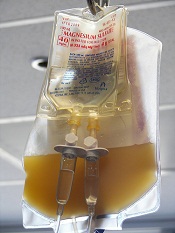
The AABB has developed new guidelines that specify situations
in which adults should receive platelet transfusions.
The group’s
recommendations are based on results of a systematic review.
The
suggestions cover several clinical situations in which platelet
transfusions might be an option, such as therapy-induced hypoproliferative thrombocytopenia, central venous catheter placement, diagnostic lumbar puncture, and cardiac surgery.
To formulate the guidelines, researchers conducted a review of randomized clinical trials and observational studies published from 1900 through September 2014. All studies reported clinical outcomes in patients receiving prophylactic or therapeutic platelet transfusions.
Based on the collected data, the AABB now strongly recommends prophylactic platelet transfusions to reduce the risk for spontaneous bleeding in hospitalized adults with therapy-induced hypoproliferative thrombocytopenia and a platelet count of 10 x 109 cells/L or less. The recommendation states that transfusing 0.5 to 1 apheresis unit is sufficient in these patients.
A weaker recommendation is that prophylactic platelet transfusions be given to patients undergoing elective central venous catheter placement who have a platelet count of less than 20 x 109 cells/L.
Likewise, the AABB says prophylactic transfusions should be given to patients having elective diagnostic lumbar puncture or major elective nonneuraxial surgery who have a platelet count less than 50 x 109 cells/L.
Another weak recommendation advises against routine prophylactic platelet transfusions in patients who are nonthrombocytopenic and have cardiac surgery with cardiopulmonary bypass.
The AABB also suggests (without recommending) that patients with perioperative bleeding and thrombocytopenia or platelet dysfunction may benefit from transfusions.
Finally, the AABB said it cannot recommend for or against platelet transfusions in patients receiving antiplatelet therapy who have intracranial hemorrhage (traumatic or spontaneous).
For more details, see the complete guidelines in Annals of Internal Medicine.
The AABB has developed new guidelines that specify situations
in which adults should receive platelet transfusions.
The group’s
recommendations are based on results of a systematic review.
The
suggestions cover several clinical situations in which platelet
transfusions might be an option, such as therapy-induced hypoproliferative thrombocytopenia, central venous catheter placement, diagnostic lumbar puncture, and cardiac surgery.
To formulate the guidelines, researchers conducted a review of randomized clinical trials and observational studies published from 1900 through September 2014. All studies reported clinical outcomes in patients receiving prophylactic or therapeutic platelet transfusions.
Based on the collected data, the AABB now strongly recommends prophylactic platelet transfusions to reduce the risk for spontaneous bleeding in hospitalized adults with therapy-induced hypoproliferative thrombocytopenia and a platelet count of 10 x 109 cells/L or less. The recommendation states that transfusing 0.5 to 1 apheresis unit is sufficient in these patients.
A weaker recommendation is that prophylactic platelet transfusions be given to patients undergoing elective central venous catheter placement who have a platelet count of less than 20 x 109 cells/L.
Likewise, the AABB says prophylactic transfusions should be given to patients having elective diagnostic lumbar puncture or major elective nonneuraxial surgery who have a platelet count less than 50 x 109 cells/L.
Another weak recommendation advises against routine prophylactic platelet transfusions in patients who are nonthrombocytopenic and have cardiac surgery with cardiopulmonary bypass.
The AABB also suggests (without recommending) that patients with perioperative bleeding and thrombocytopenia or platelet dysfunction may benefit from transfusions.
Finally, the AABB said it cannot recommend for or against platelet transfusions in patients receiving antiplatelet therapy who have intracranial hemorrhage (traumatic or spontaneous).
For more details, see the complete guidelines in Annals of Internal Medicine.
The AABB has developed new guidelines that specify situations
in which adults should receive platelet transfusions.
The group’s
recommendations are based on results of a systematic review.
The
suggestions cover several clinical situations in which platelet
transfusions might be an option, such as therapy-induced hypoproliferative thrombocytopenia, central venous catheter placement, diagnostic lumbar puncture, and cardiac surgery.
To formulate the guidelines, researchers conducted a review of randomized clinical trials and observational studies published from 1900 through September 2014. All studies reported clinical outcomes in patients receiving prophylactic or therapeutic platelet transfusions.
Based on the collected data, the AABB now strongly recommends prophylactic platelet transfusions to reduce the risk for spontaneous bleeding in hospitalized adults with therapy-induced hypoproliferative thrombocytopenia and a platelet count of 10 x 109 cells/L or less. The recommendation states that transfusing 0.5 to 1 apheresis unit is sufficient in these patients.
A weaker recommendation is that prophylactic platelet transfusions be given to patients undergoing elective central venous catheter placement who have a platelet count of less than 20 x 109 cells/L.
Likewise, the AABB says prophylactic transfusions should be given to patients having elective diagnostic lumbar puncture or major elective nonneuraxial surgery who have a platelet count less than 50 x 109 cells/L.
Another weak recommendation advises against routine prophylactic platelet transfusions in patients who are nonthrombocytopenic and have cardiac surgery with cardiopulmonary bypass.
The AABB also suggests (without recommending) that patients with perioperative bleeding and thrombocytopenia or platelet dysfunction may benefit from transfusions.
Finally, the AABB said it cannot recommend for or against platelet transfusions in patients receiving antiplatelet therapy who have intracranial hemorrhage (traumatic or spontaneous).
For more details, see the complete guidelines in Annals of Internal Medicine.
Sugar beets can be used to create hemoglobin

holding a sugar beet
Credit: Lund University
Biochemists have found evidence to suggest that sugar beets can be used to create a substitute for human hemoglobin.
While studying the genome of the sugar beet, the researchers identified 4 hemoglobin genes—3 non-symbiotic genes (BvHb1.1, BvHb1.2, and BvHb2) and 1 truncated gene (BvHb3).
The team then discovered they could extract hemoglobin from sugar beets using a process that’s about as simple as the one used to extract sugar.
The researchers cloned the hemoglobin genes and inserted them into bacteria, which facilitates their expression and purification.
The group described the gene discovery in Plant and Cell Physiology. Study author Nélida Leiva-Eriksson, a doctoral student at Lund University in Sweden, disclosed additional details in her dissertation. And a short video on the research is available on YouTube.
The researchers discovered that the hemoglobin extracted from sugar beets is almost identical to human hemoglobin, especially the form of hemoglobin in the brain.
“There is a difference in a small detail on the surface of the protein, but this simply appears to extend the lifespan of the hemoglobin from sugar beet, which is good news,” Leiva-Eriksson said.
On the other hand, sugar beet hemoglobin has a completely different function from human hemoglobin.
“We have found that the hemoglobin in the plant binds nitric oxide,” Leiva-Eriksson said. “It is probably needed to keep certain processes in check, for example, so that the nitric oxide doesn’t become toxic, and to ward off bacteria.”
The researchers are planning to start testing the sugar beet hemoglobin in animal experiments in just over a year.
The team said there is good reason to think hemoglobin derived from sugar beets and other crops could become a realistic alternative to human hemoglobin.
“From 1 hectare, we could produce 1 to 2 tons of hemoglobin,” said Leif Bülow, PhD, of Lund University, “which could save thousands of lives.”
holding a sugar beet
Credit: Lund University
Biochemists have found evidence to suggest that sugar beets can be used to create a substitute for human hemoglobin.
While studying the genome of the sugar beet, the researchers identified 4 hemoglobin genes—3 non-symbiotic genes (BvHb1.1, BvHb1.2, and BvHb2) and 1 truncated gene (BvHb3).
The team then discovered they could extract hemoglobin from sugar beets using a process that’s about as simple as the one used to extract sugar.
The researchers cloned the hemoglobin genes and inserted them into bacteria, which facilitates their expression and purification.
The group described the gene discovery in Plant and Cell Physiology. Study author Nélida Leiva-Eriksson, a doctoral student at Lund University in Sweden, disclosed additional details in her dissertation. And a short video on the research is available on YouTube.
The researchers discovered that the hemoglobin extracted from sugar beets is almost identical to human hemoglobin, especially the form of hemoglobin in the brain.
“There is a difference in a small detail on the surface of the protein, but this simply appears to extend the lifespan of the hemoglobin from sugar beet, which is good news,” Leiva-Eriksson said.
On the other hand, sugar beet hemoglobin has a completely different function from human hemoglobin.
“We have found that the hemoglobin in the plant binds nitric oxide,” Leiva-Eriksson said. “It is probably needed to keep certain processes in check, for example, so that the nitric oxide doesn’t become toxic, and to ward off bacteria.”
The researchers are planning to start testing the sugar beet hemoglobin in animal experiments in just over a year.
The team said there is good reason to think hemoglobin derived from sugar beets and other crops could become a realistic alternative to human hemoglobin.
“From 1 hectare, we could produce 1 to 2 tons of hemoglobin,” said Leif Bülow, PhD, of Lund University, “which could save thousands of lives.”
holding a sugar beet
Credit: Lund University
Biochemists have found evidence to suggest that sugar beets can be used to create a substitute for human hemoglobin.
While studying the genome of the sugar beet, the researchers identified 4 hemoglobin genes—3 non-symbiotic genes (BvHb1.1, BvHb1.2, and BvHb2) and 1 truncated gene (BvHb3).
The team then discovered they could extract hemoglobin from sugar beets using a process that’s about as simple as the one used to extract sugar.
The researchers cloned the hemoglobin genes and inserted them into bacteria, which facilitates their expression and purification.
The group described the gene discovery in Plant and Cell Physiology. Study author Nélida Leiva-Eriksson, a doctoral student at Lund University in Sweden, disclosed additional details in her dissertation. And a short video on the research is available on YouTube.
The researchers discovered that the hemoglobin extracted from sugar beets is almost identical to human hemoglobin, especially the form of hemoglobin in the brain.
“There is a difference in a small detail on the surface of the protein, but this simply appears to extend the lifespan of the hemoglobin from sugar beet, which is good news,” Leiva-Eriksson said.
On the other hand, sugar beet hemoglobin has a completely different function from human hemoglobin.
“We have found that the hemoglobin in the plant binds nitric oxide,” Leiva-Eriksson said. “It is probably needed to keep certain processes in check, for example, so that the nitric oxide doesn’t become toxic, and to ward off bacteria.”
The researchers are planning to start testing the sugar beet hemoglobin in animal experiments in just over a year.
The team said there is good reason to think hemoglobin derived from sugar beets and other crops could become a realistic alternative to human hemoglobin.
“From 1 hectare, we could produce 1 to 2 tons of hemoglobin,” said Leif Bülow, PhD, of Lund University, “which could save thousands of lives.”
Team pinpoints new target for MM therapy
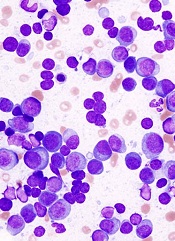
Researchers say they’ve identified a promising therapeutic target for multiple myeloma (MM).
The group first discovered that MM patients have higher expression of proline-rich tyrosine kinase 2 (PYK2), a member of the focal adhesion kinase (FAK) family, compared to healthy individuals.
Then, preclinical experiments revealed that PYK2 plays a role in tumor progression, and inhibiting this kinase can impede MM cell growth.
Yu Zhang, PhD, of the Dana-Farber Cancer Institute in Boston, and colleagues reported these findings in Blood.
Comparing samples from MM patients and healthy controls, the researchers found that PYK2 is highly expressed in MM. The team even detected higher levels of PYK2 in patients with monoclonal gammopathy of unknown significance and smoldering myeloma.
Additional experiments helped the group elucidate PYK2’s role in MM, showing that the kinase promotes tumor progression by modulating the Wnt/β-catenin signaling pathway.
When the researchers inhibited PYK2, they observed a reduction in MM tumor growth in vivo as well as decreased cell proliferation, cell cycle progression, and adhesion ability in vitro.
In contrast, overexpression of PYK2 promoted the malignant phenotype, as evidenced by enhanced tumor growth and reduced survival in mouse models.
Finally, the researchers showed that a FAK/PYK2 inhibitor, VS-4718, inhibited MM cell growth both in vitro and in vivo.
“In addition to this work recently published by our collaborators at the Dana-Farber Cancer Institute, we have been conducting further research and collaborating with scientific leaders to understand the potential of cancer stem cell inhibitors in hematological malignancies,” said Jonathan Pachter, PhD, head of research at Verastem, Inc., the company developing VS-4781.
“We believe that inhibitors of FAK and PYK2, such as VS-4718, may be useful in the treatment of many types of cancer, particularly where there is minimal residual disease with enrichment of cancer stem cells following chemotherapy. We and our collaborators will be presenting additional research at the upcoming American Society of Hematology in December.”
VS-4718 is being evaluated in a phase 1 dose-escalation trial in patients with advanced solid tumors. Another phase 1 trial in hematological malignancies is set to begin in the first quarter of 2015.
Researchers say they’ve identified a promising therapeutic target for multiple myeloma (MM).
The group first discovered that MM patients have higher expression of proline-rich tyrosine kinase 2 (PYK2), a member of the focal adhesion kinase (FAK) family, compared to healthy individuals.
Then, preclinical experiments revealed that PYK2 plays a role in tumor progression, and inhibiting this kinase can impede MM cell growth.
Yu Zhang, PhD, of the Dana-Farber Cancer Institute in Boston, and colleagues reported these findings in Blood.
Comparing samples from MM patients and healthy controls, the researchers found that PYK2 is highly expressed in MM. The team even detected higher levels of PYK2 in patients with monoclonal gammopathy of unknown significance and smoldering myeloma.
Additional experiments helped the group elucidate PYK2’s role in MM, showing that the kinase promotes tumor progression by modulating the Wnt/β-catenin signaling pathway.
When the researchers inhibited PYK2, they observed a reduction in MM tumor growth in vivo as well as decreased cell proliferation, cell cycle progression, and adhesion ability in vitro.
In contrast, overexpression of PYK2 promoted the malignant phenotype, as evidenced by enhanced tumor growth and reduced survival in mouse models.
Finally, the researchers showed that a FAK/PYK2 inhibitor, VS-4718, inhibited MM cell growth both in vitro and in vivo.
“In addition to this work recently published by our collaborators at the Dana-Farber Cancer Institute, we have been conducting further research and collaborating with scientific leaders to understand the potential of cancer stem cell inhibitors in hematological malignancies,” said Jonathan Pachter, PhD, head of research at Verastem, Inc., the company developing VS-4781.
“We believe that inhibitors of FAK and PYK2, such as VS-4718, may be useful in the treatment of many types of cancer, particularly where there is minimal residual disease with enrichment of cancer stem cells following chemotherapy. We and our collaborators will be presenting additional research at the upcoming American Society of Hematology in December.”
VS-4718 is being evaluated in a phase 1 dose-escalation trial in patients with advanced solid tumors. Another phase 1 trial in hematological malignancies is set to begin in the first quarter of 2015.
Researchers say they’ve identified a promising therapeutic target for multiple myeloma (MM).
The group first discovered that MM patients have higher expression of proline-rich tyrosine kinase 2 (PYK2), a member of the focal adhesion kinase (FAK) family, compared to healthy individuals.
Then, preclinical experiments revealed that PYK2 plays a role in tumor progression, and inhibiting this kinase can impede MM cell growth.
Yu Zhang, PhD, of the Dana-Farber Cancer Institute in Boston, and colleagues reported these findings in Blood.
Comparing samples from MM patients and healthy controls, the researchers found that PYK2 is highly expressed in MM. The team even detected higher levels of PYK2 in patients with monoclonal gammopathy of unknown significance and smoldering myeloma.
Additional experiments helped the group elucidate PYK2’s role in MM, showing that the kinase promotes tumor progression by modulating the Wnt/β-catenin signaling pathway.
When the researchers inhibited PYK2, they observed a reduction in MM tumor growth in vivo as well as decreased cell proliferation, cell cycle progression, and adhesion ability in vitro.
In contrast, overexpression of PYK2 promoted the malignant phenotype, as evidenced by enhanced tumor growth and reduced survival in mouse models.
Finally, the researchers showed that a FAK/PYK2 inhibitor, VS-4718, inhibited MM cell growth both in vitro and in vivo.
“In addition to this work recently published by our collaborators at the Dana-Farber Cancer Institute, we have been conducting further research and collaborating with scientific leaders to understand the potential of cancer stem cell inhibitors in hematological malignancies,” said Jonathan Pachter, PhD, head of research at Verastem, Inc., the company developing VS-4781.
“We believe that inhibitors of FAK and PYK2, such as VS-4718, may be useful in the treatment of many types of cancer, particularly where there is minimal residual disease with enrichment of cancer stem cells following chemotherapy. We and our collaborators will be presenting additional research at the upcoming American Society of Hematology in December.”
VS-4718 is being evaluated in a phase 1 dose-escalation trial in patients with advanced solid tumors. Another phase 1 trial in hematological malignancies is set to begin in the first quarter of 2015.