User login
Test detects bloodstream infections faster
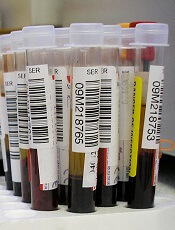
Credit: Graham Colm
A new test can detect bloodstream infections with “unprecedented” speed and accuracy, according to researchers.
The group developed a test called Integrated Comprehensive Droplet Digital Detection (IC 3D).
And experiments showed that IC 3D can detect bacteria in milliliters of blood with single-cell sensitivity in 1.5 to 4 hours, with no cell culture necessary.
The researchers described this test in Nature Communications.
“We are extremely excited about this technology because it addresses a long-standing unmet medical need in the field,” said study author Weian Zhao, PhD, of the University of California Irvine (UCI).
“As a platform technology, it may have many applications in detecting extremely low-abundance biomarkers in other areas, such as cancers, HIV, and, most notably, Ebola.”
Worldwide, more than 2 million people a year get antibiotic-resistant blood infections, and 23,000 of them die, according to the US Centers for Disease Control & Prevention. This high mortality rate is due, in part, to the inability to diagnose and treat patients in the early stages.
Recent molecular diagnostic methods can reduce the assay time to hours but may not be sensitive enough to detect bacteria that occur at low concentrations in blood.
The IC 3D technology differs from other diagnostic techniques in that it converts blood samples directly into billions of very small droplets. Fluorescent DNA sensor solution infused into the droplets detects those with bacterial markers, lighting them up with an intense fluorescent signal.
Dr Zhao said that separating the samples into so many small droplets minimizes the interference of other components in blood, making it possible to directly detect target bacteria without the purification typically required in conventional assays.
To identify bacteria-containing droplets among billions of others in a timely fashion, Dr Zhao and his colleagues incorporated a 3-dimensional particle counter developed by UCI biomedical engineer Enrico Gratton, PhD, and his colleagues that tags fluorescent particles within several minutes.
“The IC 3D instrument is designed to read a large volume in each measurement, to speed up diagnosis,” Dr Gratton said. “Importantly, using this technique, we can detect a positive hit with very high confidence.”
The researchers reported that IC 3D provided absolute quantification of stock and clinical isolates of Escherichia coli at a wide range of concentrations, from 1 to 10,000 bacteria per mL, in 1.5 to 4 hours.
A UCI spinoff, Velox Biosystems, is now developing the IC 3D technology. 
Credit: Graham Colm
A new test can detect bloodstream infections with “unprecedented” speed and accuracy, according to researchers.
The group developed a test called Integrated Comprehensive Droplet Digital Detection (IC 3D).
And experiments showed that IC 3D can detect bacteria in milliliters of blood with single-cell sensitivity in 1.5 to 4 hours, with no cell culture necessary.
The researchers described this test in Nature Communications.
“We are extremely excited about this technology because it addresses a long-standing unmet medical need in the field,” said study author Weian Zhao, PhD, of the University of California Irvine (UCI).
“As a platform technology, it may have many applications in detecting extremely low-abundance biomarkers in other areas, such as cancers, HIV, and, most notably, Ebola.”
Worldwide, more than 2 million people a year get antibiotic-resistant blood infections, and 23,000 of them die, according to the US Centers for Disease Control & Prevention. This high mortality rate is due, in part, to the inability to diagnose and treat patients in the early stages.
Recent molecular diagnostic methods can reduce the assay time to hours but may not be sensitive enough to detect bacteria that occur at low concentrations in blood.
The IC 3D technology differs from other diagnostic techniques in that it converts blood samples directly into billions of very small droplets. Fluorescent DNA sensor solution infused into the droplets detects those with bacterial markers, lighting them up with an intense fluorescent signal.
Dr Zhao said that separating the samples into so many small droplets minimizes the interference of other components in blood, making it possible to directly detect target bacteria without the purification typically required in conventional assays.
To identify bacteria-containing droplets among billions of others in a timely fashion, Dr Zhao and his colleagues incorporated a 3-dimensional particle counter developed by UCI biomedical engineer Enrico Gratton, PhD, and his colleagues that tags fluorescent particles within several minutes.
“The IC 3D instrument is designed to read a large volume in each measurement, to speed up diagnosis,” Dr Gratton said. “Importantly, using this technique, we can detect a positive hit with very high confidence.”
The researchers reported that IC 3D provided absolute quantification of stock and clinical isolates of Escherichia coli at a wide range of concentrations, from 1 to 10,000 bacteria per mL, in 1.5 to 4 hours.
A UCI spinoff, Velox Biosystems, is now developing the IC 3D technology.
Credit: Graham Colm
A new test can detect bloodstream infections with “unprecedented” speed and accuracy, according to researchers.
The group developed a test called Integrated Comprehensive Droplet Digital Detection (IC 3D).
And experiments showed that IC 3D can detect bacteria in milliliters of blood with single-cell sensitivity in 1.5 to 4 hours, with no cell culture necessary.
The researchers described this test in Nature Communications.
“We are extremely excited about this technology because it addresses a long-standing unmet medical need in the field,” said study author Weian Zhao, PhD, of the University of California Irvine (UCI).
“As a platform technology, it may have many applications in detecting extremely low-abundance biomarkers in other areas, such as cancers, HIV, and, most notably, Ebola.”
Worldwide, more than 2 million people a year get antibiotic-resistant blood infections, and 23,000 of them die, according to the US Centers for Disease Control & Prevention. This high mortality rate is due, in part, to the inability to diagnose and treat patients in the early stages.
Recent molecular diagnostic methods can reduce the assay time to hours but may not be sensitive enough to detect bacteria that occur at low concentrations in blood.
The IC 3D technology differs from other diagnostic techniques in that it converts blood samples directly into billions of very small droplets. Fluorescent DNA sensor solution infused into the droplets detects those with bacterial markers, lighting them up with an intense fluorescent signal.
Dr Zhao said that separating the samples into so many small droplets minimizes the interference of other components in blood, making it possible to directly detect target bacteria without the purification typically required in conventional assays.
To identify bacteria-containing droplets among billions of others in a timely fashion, Dr Zhao and his colleagues incorporated a 3-dimensional particle counter developed by UCI biomedical engineer Enrico Gratton, PhD, and his colleagues that tags fluorescent particles within several minutes.
“The IC 3D instrument is designed to read a large volume in each measurement, to speed up diagnosis,” Dr Gratton said. “Importantly, using this technique, we can detect a positive hit with very high confidence.”
The researchers reported that IC 3D provided absolute quantification of stock and clinical isolates of Escherichia coli at a wide range of concentrations, from 1 to 10,000 bacteria per mL, in 1.5 to 4 hours.
A UCI spinoff, Velox Biosystems, is now developing the IC 3D technology.
FDA investigating risk of death with DAPT

Credit: CDC
The US Food and Drug Administration (FDA) is evaluating trial data suggesting that 30 months of dual antiplatelet therapy (DAPT), compared to 12 months of the therapy, can increase the overall risk of death among patients who have a drug-eluting coronary stent.
The trial, which was published in NEJM, also indicates that longer-term DAPT can decrease the risk of stent thrombosis and major cardiovascular and cerebrovascular events.
For this trial, researchers compared 12 months and 30 months of DAPT—aspirin plus either clopidogrel or prasugrel—in 9961 patients who had a drug-eluting stent. The team also enrolled patients with bare-metal stents, but those patients were not included in this publication.
All 9961 patients received aspirin and clopidogrel/prasugrel for 12 months. Then, they were randomized to continue DAPT for another 18 months or to receive aspirin and placebo during that period.
In the 30-month group, 65.2% of patients received clopidogrel, and 34.8% received prasugrel. In the 12-month group, 65.4% of patients received clopidogrel, and 34.6% received prasugrel.
The investigators followed all patients for 33 months. At that point, 4732 patients in the 30-month group and 4658 patients in the 12-month group were evaluable.
The researchers observed a significant reduction in stent thrombosis for patients who received DAPT for 30 months compared to 12 months—0.4% vs 1.4% (hazard ratio [HR]=0.29, P<0.001).
Patients who received DAPT for 30 months also saw a reduction in major cardiovascular and cerebrovascular events, a combined endpoint of all-cause death, myocardial infarction, and stroke. The rate was 4.3% for the 30-month group and 5.9% for the 12-month group (HR 0.71, P<0.001).
The reduction in the risk of major cardiovascular and cerebrovascular events was driven by a decrease in the rate of non-fatal myocardial infarction. There were no differences in the rates of cardiovascular death or stroke.
The investigators also found that the overall death rate was higher among patients receiving DAPT for a longer period. The rate was 2.0% in the 30-month group and 1.5% in the 12-month group (HR=1.36, P=0.05).
The researchers said this can be explained by an increase in non-cardiovascular deaths (1% vs 0.5%, HR 2.2, P=0.002). The most frequent causes of non-cardiovascular death were cancer (34 deaths vs 17 deaths) and trauma (8 deaths vs 2 deaths) for 30 months and 12 months of DAPT, respectively.
The increase in overall mortality was present in clopidogrel-treated patients (2.2% vs 1.5%, respectively) but not prasugrel-treated patients (1.6% vs 1.6%). The FDA noted that other large clopidogrel trials have not shown an increase in the risk of non-cardiovascular death.
The FDA said it plans to evaluate the results from this trial and other relevant data, then announce its conclusions and recommendations.
For now, the agency said healthcare professionals should not change the way they administer DAPT. And patients should not stop taking these drugs, as this may result in an increased risk of heart attacks, thrombosis, strokes, and other major cardiovascular problems.
Healthcare professionals and patients can report adverse events or side effects related to the aforementioned drugs to the FDA’s MedWatch Safety Information and Adverse Event Reporting Program.
Credit: CDC
The US Food and Drug Administration (FDA) is evaluating trial data suggesting that 30 months of dual antiplatelet therapy (DAPT), compared to 12 months of the therapy, can increase the overall risk of death among patients who have a drug-eluting coronary stent.
The trial, which was published in NEJM, also indicates that longer-term DAPT can decrease the risk of stent thrombosis and major cardiovascular and cerebrovascular events.
For this trial, researchers compared 12 months and 30 months of DAPT—aspirin plus either clopidogrel or prasugrel—in 9961 patients who had a drug-eluting stent. The team also enrolled patients with bare-metal stents, but those patients were not included in this publication.
All 9961 patients received aspirin and clopidogrel/prasugrel for 12 months. Then, they were randomized to continue DAPT for another 18 months or to receive aspirin and placebo during that period.
In the 30-month group, 65.2% of patients received clopidogrel, and 34.8% received prasugrel. In the 12-month group, 65.4% of patients received clopidogrel, and 34.6% received prasugrel.
The investigators followed all patients for 33 months. At that point, 4732 patients in the 30-month group and 4658 patients in the 12-month group were evaluable.
The researchers observed a significant reduction in stent thrombosis for patients who received DAPT for 30 months compared to 12 months—0.4% vs 1.4% (hazard ratio [HR]=0.29, P<0.001).
Patients who received DAPT for 30 months also saw a reduction in major cardiovascular and cerebrovascular events, a combined endpoint of all-cause death, myocardial infarction, and stroke. The rate was 4.3% for the 30-month group and 5.9% for the 12-month group (HR 0.71, P<0.001).
The reduction in the risk of major cardiovascular and cerebrovascular events was driven by a decrease in the rate of non-fatal myocardial infarction. There were no differences in the rates of cardiovascular death or stroke.
The investigators also found that the overall death rate was higher among patients receiving DAPT for a longer period. The rate was 2.0% in the 30-month group and 1.5% in the 12-month group (HR=1.36, P=0.05).
The researchers said this can be explained by an increase in non-cardiovascular deaths (1% vs 0.5%, HR 2.2, P=0.002). The most frequent causes of non-cardiovascular death were cancer (34 deaths vs 17 deaths) and trauma (8 deaths vs 2 deaths) for 30 months and 12 months of DAPT, respectively.
The increase in overall mortality was present in clopidogrel-treated patients (2.2% vs 1.5%, respectively) but not prasugrel-treated patients (1.6% vs 1.6%). The FDA noted that other large clopidogrel trials have not shown an increase in the risk of non-cardiovascular death.
The FDA said it plans to evaluate the results from this trial and other relevant data, then announce its conclusions and recommendations.
For now, the agency said healthcare professionals should not change the way they administer DAPT. And patients should not stop taking these drugs, as this may result in an increased risk of heart attacks, thrombosis, strokes, and other major cardiovascular problems.
Healthcare professionals and patients can report adverse events or side effects related to the aforementioned drugs to the FDA’s MedWatch Safety Information and Adverse Event Reporting Program.
Credit: CDC
The US Food and Drug Administration (FDA) is evaluating trial data suggesting that 30 months of dual antiplatelet therapy (DAPT), compared to 12 months of the therapy, can increase the overall risk of death among patients who have a drug-eluting coronary stent.
The trial, which was published in NEJM, also indicates that longer-term DAPT can decrease the risk of stent thrombosis and major cardiovascular and cerebrovascular events.
For this trial, researchers compared 12 months and 30 months of DAPT—aspirin plus either clopidogrel or prasugrel—in 9961 patients who had a drug-eluting stent. The team also enrolled patients with bare-metal stents, but those patients were not included in this publication.
All 9961 patients received aspirin and clopidogrel/prasugrel for 12 months. Then, they were randomized to continue DAPT for another 18 months or to receive aspirin and placebo during that period.
In the 30-month group, 65.2% of patients received clopidogrel, and 34.8% received prasugrel. In the 12-month group, 65.4% of patients received clopidogrel, and 34.6% received prasugrel.
The investigators followed all patients for 33 months. At that point, 4732 patients in the 30-month group and 4658 patients in the 12-month group were evaluable.
The researchers observed a significant reduction in stent thrombosis for patients who received DAPT for 30 months compared to 12 months—0.4% vs 1.4% (hazard ratio [HR]=0.29, P<0.001).
Patients who received DAPT for 30 months also saw a reduction in major cardiovascular and cerebrovascular events, a combined endpoint of all-cause death, myocardial infarction, and stroke. The rate was 4.3% for the 30-month group and 5.9% for the 12-month group (HR 0.71, P<0.001).
The reduction in the risk of major cardiovascular and cerebrovascular events was driven by a decrease in the rate of non-fatal myocardial infarction. There were no differences in the rates of cardiovascular death or stroke.
The investigators also found that the overall death rate was higher among patients receiving DAPT for a longer period. The rate was 2.0% in the 30-month group and 1.5% in the 12-month group (HR=1.36, P=0.05).
The researchers said this can be explained by an increase in non-cardiovascular deaths (1% vs 0.5%, HR 2.2, P=0.002). The most frequent causes of non-cardiovascular death were cancer (34 deaths vs 17 deaths) and trauma (8 deaths vs 2 deaths) for 30 months and 12 months of DAPT, respectively.
The increase in overall mortality was present in clopidogrel-treated patients (2.2% vs 1.5%, respectively) but not prasugrel-treated patients (1.6% vs 1.6%). The FDA noted that other large clopidogrel trials have not shown an increase in the risk of non-cardiovascular death.
The FDA said it plans to evaluate the results from this trial and other relevant data, then announce its conclusions and recommendations.
For now, the agency said healthcare professionals should not change the way they administer DAPT. And patients should not stop taking these drugs, as this may result in an increased risk of heart attacks, thrombosis, strokes, and other major cardiovascular problems.
Healthcare professionals and patients can report adverse events or side effects related to the aforementioned drugs to the FDA’s MedWatch Safety Information and Adverse Event Reporting Program.
Failure to control INR may increase risk of dementia

CHICAGO—Long-term overtreatment with warfarin given in combination with antiplatelet therapy may increase the risk of dementia in patients with atrial fibrillation, a new study suggests.
Researchers retrospectively analyzed patients who received warfarin plus aspirin or clopidogrel for up to 10 years.
And patients who had an international normalized ratio (INR) above the acceptable range more than 25% of the time had an increased risk of dementia.
T. Jared Bunch, MD, of the Intermountain Medical Center Heart Institute in Murray, Utah, presented these findings at the American Heart Association’s Scientific Sessions 2014 (abstract 13426).
“[W]e have to consider that long-term exposure to anticlotting drugs such as warfarin, if not well controlled, can significantly increase bleeding risk,” Dr Bunch said. “This may result in micro bleeds in the brain that don’t cause symptoms right away but accumulate over time, raising the risk of dementia.”
Dr Bunch and his colleagues studied 1031 patients with no previous history of stroke or dementia for up to 10 years while they received warfarin plus aspirin or clopidogrel.
Among patients who had an INR higher than 3 more than 25% of the time, 2.7% developed dementia.
The researchers adjusted their analysis for traditional stroke and bleeding risk factors and found that patients with supratherapeutic INR levels more than 25% of the time had more than double the risk of dementia compared to patients with supratherapeutic INR levels less than 10% of the time. The hazard ratio was 2.40 (P=0.04).
This increase is higher than what researchers found in a previous study of warfarin alone.
Previous research also suggested that patients with atrial fibrillation taking warfarin were more likely to develop dementia if their INRs were too high or too low.
From those results, the researchers concluded that brain injury from both small bleeds and blood clots is likely important in the development of dementia in patients with atrial fibrillation.
“Even at skilled centers, it’s very common to have INR outside the ideal range up to 40% of the time,” Dr Bunch noted. “And, over the years, there may be an accumulative negative impact on cognitive ability.”
CHICAGO—Long-term overtreatment with warfarin given in combination with antiplatelet therapy may increase the risk of dementia in patients with atrial fibrillation, a new study suggests.
Researchers retrospectively analyzed patients who received warfarin plus aspirin or clopidogrel for up to 10 years.
And patients who had an international normalized ratio (INR) above the acceptable range more than 25% of the time had an increased risk of dementia.
T. Jared Bunch, MD, of the Intermountain Medical Center Heart Institute in Murray, Utah, presented these findings at the American Heart Association’s Scientific Sessions 2014 (abstract 13426).
“[W]e have to consider that long-term exposure to anticlotting drugs such as warfarin, if not well controlled, can significantly increase bleeding risk,” Dr Bunch said. “This may result in micro bleeds in the brain that don’t cause symptoms right away but accumulate over time, raising the risk of dementia.”
Dr Bunch and his colleagues studied 1031 patients with no previous history of stroke or dementia for up to 10 years while they received warfarin plus aspirin or clopidogrel.
Among patients who had an INR higher than 3 more than 25% of the time, 2.7% developed dementia.
The researchers adjusted their analysis for traditional stroke and bleeding risk factors and found that patients with supratherapeutic INR levels more than 25% of the time had more than double the risk of dementia compared to patients with supratherapeutic INR levels less than 10% of the time. The hazard ratio was 2.40 (P=0.04).
This increase is higher than what researchers found in a previous study of warfarin alone.
Previous research also suggested that patients with atrial fibrillation taking warfarin were more likely to develop dementia if their INRs were too high or too low.
From those results, the researchers concluded that brain injury from both small bleeds and blood clots is likely important in the development of dementia in patients with atrial fibrillation.
“Even at skilled centers, it’s very common to have INR outside the ideal range up to 40% of the time,” Dr Bunch noted. “And, over the years, there may be an accumulative negative impact on cognitive ability.”
CHICAGO—Long-term overtreatment with warfarin given in combination with antiplatelet therapy may increase the risk of dementia in patients with atrial fibrillation, a new study suggests.
Researchers retrospectively analyzed patients who received warfarin plus aspirin or clopidogrel for up to 10 years.
And patients who had an international normalized ratio (INR) above the acceptable range more than 25% of the time had an increased risk of dementia.
T. Jared Bunch, MD, of the Intermountain Medical Center Heart Institute in Murray, Utah, presented these findings at the American Heart Association’s Scientific Sessions 2014 (abstract 13426).
“[W]e have to consider that long-term exposure to anticlotting drugs such as warfarin, if not well controlled, can significantly increase bleeding risk,” Dr Bunch said. “This may result in micro bleeds in the brain that don’t cause symptoms right away but accumulate over time, raising the risk of dementia.”
Dr Bunch and his colleagues studied 1031 patients with no previous history of stroke or dementia for up to 10 years while they received warfarin plus aspirin or clopidogrel.
Among patients who had an INR higher than 3 more than 25% of the time, 2.7% developed dementia.
The researchers adjusted their analysis for traditional stroke and bleeding risk factors and found that patients with supratherapeutic INR levels more than 25% of the time had more than double the risk of dementia compared to patients with supratherapeutic INR levels less than 10% of the time. The hazard ratio was 2.40 (P=0.04).
This increase is higher than what researchers found in a previous study of warfarin alone.
Previous research also suggested that patients with atrial fibrillation taking warfarin were more likely to develop dementia if their INRs were too high or too low.
From those results, the researchers concluded that brain injury from both small bleeds and blood clots is likely important in the development of dementia in patients with atrial fibrillation.
“Even at skilled centers, it’s very common to have INR outside the ideal range up to 40% of the time,” Dr Bunch noted. “And, over the years, there may be an accumulative negative impact on cognitive ability.”
IL-6 inhibitor helps prevent GVHD

Credit: Chad McNeeley
Adding the interleukin 6 (IL-6) inhibitor tocilizumab can improve standard prophylaxis for graft-vs-host disease (GVHD), researchers have reported in The Lancet Oncology.
IL-6 is the main detectable and dysregulated cytokine secreted after allogeneic stem cell transplant (allo-SCT).
So the researchers theorized that inhibiting IL-6 with tocilizumab might protect patients from acute GVHD despite robust immune reconstitution.
In their phase 1/2 study, the team saw acute GVHD drop from the usual 50% observed in patients receiving standard GVHD prophylaxis to 12% in patients receiving tocilizumab.
“Severe cases—which often result in death—were reduced from 21% to 4%,” said Geoff Hill, MD, of QIMR Berghofer Medical Research Institute in Brisbane, Queensland, Australia.
He and his colleagues enrolled 48 patients in this study. They ranged in age from 18 to 65 and underwent T-replete, HLA-matched, allo-SCT from unrelated or sibling donors. Patients received either total-body-irradiation-based myeloablative conditioning or reduced-intensity conditioning.
As GVHD prophylaxis, patients received a single intravenous dose of tocilizumab (8 mg/kg, capped at 800 mg, over a 60-minute infusion) the day before transplant.
They also received standard GVHD prophylaxis—cyclosporin (5 mg/kg per day on days −1 to +1, then 3 mg/kg per day to maintain therapeutic levels [trough levels of 140-300 ng/mL] for 100 days) and methotrexate (15 mg/m2 on day 1, then 10 mg/m2 on days 3, 6, and 11).
The primary endpoint was the incidence of grade 2-4 acute GVHD at day 100, which was 12%. The incidence of grade 3-4 acute GVHD was 4%.
Five patients (10%) had grade 2-4 acute GVHD involving the skin, and 4 (8%) had grade 2-4 acute GVHD involving the gastrointestinal tract. None of the patients had GVHD involving the liver.
The researchers noted that the rate of grade 2-4 acute GVHD was low regardless of the conditioning regimen a patient received. The rate was 12% for both myeloablative and reduced-intensity conditioning.
In addition, patients’ immune reconstitution was preserved after receiving tocilizumab, but the researchers did observe suppression of known pathogenic STAT3-dependent pathways.
These results represent a significant advance in allo-SCT, according to study author Glen Kennedy, MBBS, also of QIMR Berghofer Medical Research Institute.
“The new therapy has the potential to make transplant safer,” he said, “and applicable to a larger group of patients.”
A phase 3 study of tocilizumab as GVHD prophylaxis is now underway. The drug is currently approved to treat rheumatoid arthritis.
Credit: Chad McNeeley
Adding the interleukin 6 (IL-6) inhibitor tocilizumab can improve standard prophylaxis for graft-vs-host disease (GVHD), researchers have reported in The Lancet Oncology.
IL-6 is the main detectable and dysregulated cytokine secreted after allogeneic stem cell transplant (allo-SCT).
So the researchers theorized that inhibiting IL-6 with tocilizumab might protect patients from acute GVHD despite robust immune reconstitution.
In their phase 1/2 study, the team saw acute GVHD drop from the usual 50% observed in patients receiving standard GVHD prophylaxis to 12% in patients receiving tocilizumab.
“Severe cases—which often result in death—were reduced from 21% to 4%,” said Geoff Hill, MD, of QIMR Berghofer Medical Research Institute in Brisbane, Queensland, Australia.
He and his colleagues enrolled 48 patients in this study. They ranged in age from 18 to 65 and underwent T-replete, HLA-matched, allo-SCT from unrelated or sibling donors. Patients received either total-body-irradiation-based myeloablative conditioning or reduced-intensity conditioning.
As GVHD prophylaxis, patients received a single intravenous dose of tocilizumab (8 mg/kg, capped at 800 mg, over a 60-minute infusion) the day before transplant.
They also received standard GVHD prophylaxis—cyclosporin (5 mg/kg per day on days −1 to +1, then 3 mg/kg per day to maintain therapeutic levels [trough levels of 140-300 ng/mL] for 100 days) and methotrexate (15 mg/m2 on day 1, then 10 mg/m2 on days 3, 6, and 11).
The primary endpoint was the incidence of grade 2-4 acute GVHD at day 100, which was 12%. The incidence of grade 3-4 acute GVHD was 4%.
Five patients (10%) had grade 2-4 acute GVHD involving the skin, and 4 (8%) had grade 2-4 acute GVHD involving the gastrointestinal tract. None of the patients had GVHD involving the liver.
The researchers noted that the rate of grade 2-4 acute GVHD was low regardless of the conditioning regimen a patient received. The rate was 12% for both myeloablative and reduced-intensity conditioning.
In addition, patients’ immune reconstitution was preserved after receiving tocilizumab, but the researchers did observe suppression of known pathogenic STAT3-dependent pathways.
These results represent a significant advance in allo-SCT, according to study author Glen Kennedy, MBBS, also of QIMR Berghofer Medical Research Institute.
“The new therapy has the potential to make transplant safer,” he said, “and applicable to a larger group of patients.”
A phase 3 study of tocilizumab as GVHD prophylaxis is now underway. The drug is currently approved to treat rheumatoid arthritis.
Credit: Chad McNeeley
Adding the interleukin 6 (IL-6) inhibitor tocilizumab can improve standard prophylaxis for graft-vs-host disease (GVHD), researchers have reported in The Lancet Oncology.
IL-6 is the main detectable and dysregulated cytokine secreted after allogeneic stem cell transplant (allo-SCT).
So the researchers theorized that inhibiting IL-6 with tocilizumab might protect patients from acute GVHD despite robust immune reconstitution.
In their phase 1/2 study, the team saw acute GVHD drop from the usual 50% observed in patients receiving standard GVHD prophylaxis to 12% in patients receiving tocilizumab.
“Severe cases—which often result in death—were reduced from 21% to 4%,” said Geoff Hill, MD, of QIMR Berghofer Medical Research Institute in Brisbane, Queensland, Australia.
He and his colleagues enrolled 48 patients in this study. They ranged in age from 18 to 65 and underwent T-replete, HLA-matched, allo-SCT from unrelated or sibling donors. Patients received either total-body-irradiation-based myeloablative conditioning or reduced-intensity conditioning.
As GVHD prophylaxis, patients received a single intravenous dose of tocilizumab (8 mg/kg, capped at 800 mg, over a 60-minute infusion) the day before transplant.
They also received standard GVHD prophylaxis—cyclosporin (5 mg/kg per day on days −1 to +1, then 3 mg/kg per day to maintain therapeutic levels [trough levels of 140-300 ng/mL] for 100 days) and methotrexate (15 mg/m2 on day 1, then 10 mg/m2 on days 3, 6, and 11).
The primary endpoint was the incidence of grade 2-4 acute GVHD at day 100, which was 12%. The incidence of grade 3-4 acute GVHD was 4%.
Five patients (10%) had grade 2-4 acute GVHD involving the skin, and 4 (8%) had grade 2-4 acute GVHD involving the gastrointestinal tract. None of the patients had GVHD involving the liver.
The researchers noted that the rate of grade 2-4 acute GVHD was low regardless of the conditioning regimen a patient received. The rate was 12% for both myeloablative and reduced-intensity conditioning.
In addition, patients’ immune reconstitution was preserved after receiving tocilizumab, but the researchers did observe suppression of known pathogenic STAT3-dependent pathways.
These results represent a significant advance in allo-SCT, according to study author Glen Kennedy, MBBS, also of QIMR Berghofer Medical Research Institute.
“The new therapy has the potential to make transplant safer,” he said, “and applicable to a larger group of patients.”
A phase 3 study of tocilizumab as GVHD prophylaxis is now underway. The drug is currently approved to treat rheumatoid arthritis.
Will US lift ban on MSM blood donation?
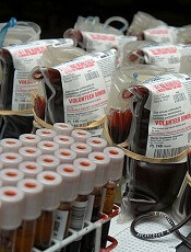
Credit: Daniel Gay
A committee that advises the US Department of Health and Human Services (HHS) has recommended changing the policy that prevents men who have sex with men (MSM) from donating blood.
The Advisory Committee on Blood and Tissue Safety and Availability decided MSM should be allowed to donate blood if they have abstained from sex for a year.
A group of advisers to the US Food and Drug Administration (FDA) will consider this recommendation in a meeting on December 2.
The HHS advisory committee plans to meet on December 5 to discuss establishing a donor transfusion-transmissible infection-monitoring system. The HHS has said such a system should be put in place before lifting the lifetime ban on MSM blood donation.
Should the FDA decide to change its policy, the US would follow other countries that have lifted the lifetime ban in recent years.
For instance, MSM in the UK and Australia are now allowed to donate blood if they have been celibate for a year, MSM in Canada must be celibate for 5 years, and MSM in South Africa can donate if they have been celibate or in a monogamous relationship for 6 months.
The safety of the blood supply
Lifting the lifetime ban on MSM blood donors may raise concerns about the safety of the blood supply, with transfusion recipients worrying they will have a greater risk of contracting HIV.
Although donated blood is tested for HIV, there is an 11-day window in which current tests cannot detect the virus in people who just contracted it. And MSM are more severely affected by HIV than any other group in the US, according to the Centers for Disease Control and Prevention.
Of course, deferring MSM donation for a year would allow enough time for HIV to be strong enough for tests to detect the virus. However, that assumes that donors are telling the truth about their sexual practices.
A study of more than 1000 MSM in Britain showed that 11% had donated blood after having penetrative sex with a man, and 3% had done so in the past 12 months, despite the lifetime ban on MSM blood donation. The study was conducted before the UK lifted the ban.
Still, study investigators said the results supported lifting the lifetime ban on MSM because men who did not comply with the ban generally said they would comply with a 1-year deferral period.
The AABB, America’s Blood Centers, and the American Red Cross have said they support a 1-year deferral period for MSM who want to donate blood in the US.
“This change in policy would align the donor deferral period for MSM with criteria for other activities that may pose a similar risk of transfusion-transmissible infections,” the groups said.
“We believe the current FDA indefinite blood donation deferral for a man who has sex with another man since 1977 is medically and scientifically unwarranted. The blood banking community strongly supports the use of rational, scientifically based deferral periods that are applied fairly and consistently among blood donors who engage in similar-risk activities.”
A report by the Williams Institute suggested that, if the FDA were to lift the ban on MSM completely, an additional 360,600 men could donate 615,300 additional pints of blood each year.
On the other hand, an HHS report suggested that the US supply of blood units is already surpassing demand. The report showed that 15.7 million units of whole blood and red blood cells were collected in 2011, and the total number of units transfused was 13.8 million.
Credit: Daniel Gay
A committee that advises the US Department of Health and Human Services (HHS) has recommended changing the policy that prevents men who have sex with men (MSM) from donating blood.
The Advisory Committee on Blood and Tissue Safety and Availability decided MSM should be allowed to donate blood if they have abstained from sex for a year.
A group of advisers to the US Food and Drug Administration (FDA) will consider this recommendation in a meeting on December 2.
The HHS advisory committee plans to meet on December 5 to discuss establishing a donor transfusion-transmissible infection-monitoring system. The HHS has said such a system should be put in place before lifting the lifetime ban on MSM blood donation.
Should the FDA decide to change its policy, the US would follow other countries that have lifted the lifetime ban in recent years.
For instance, MSM in the UK and Australia are now allowed to donate blood if they have been celibate for a year, MSM in Canada must be celibate for 5 years, and MSM in South Africa can donate if they have been celibate or in a monogamous relationship for 6 months.
The safety of the blood supply
Lifting the lifetime ban on MSM blood donors may raise concerns about the safety of the blood supply, with transfusion recipients worrying they will have a greater risk of contracting HIV.
Although donated blood is tested for HIV, there is an 11-day window in which current tests cannot detect the virus in people who just contracted it. And MSM are more severely affected by HIV than any other group in the US, according to the Centers for Disease Control and Prevention.
Of course, deferring MSM donation for a year would allow enough time for HIV to be strong enough for tests to detect the virus. However, that assumes that donors are telling the truth about their sexual practices.
A study of more than 1000 MSM in Britain showed that 11% had donated blood after having penetrative sex with a man, and 3% had done so in the past 12 months, despite the lifetime ban on MSM blood donation. The study was conducted before the UK lifted the ban.
Still, study investigators said the results supported lifting the lifetime ban on MSM because men who did not comply with the ban generally said they would comply with a 1-year deferral period.
The AABB, America’s Blood Centers, and the American Red Cross have said they support a 1-year deferral period for MSM who want to donate blood in the US.
“This change in policy would align the donor deferral period for MSM with criteria for other activities that may pose a similar risk of transfusion-transmissible infections,” the groups said.
“We believe the current FDA indefinite blood donation deferral for a man who has sex with another man since 1977 is medically and scientifically unwarranted. The blood banking community strongly supports the use of rational, scientifically based deferral periods that are applied fairly and consistently among blood donors who engage in similar-risk activities.”
A report by the Williams Institute suggested that, if the FDA were to lift the ban on MSM completely, an additional 360,600 men could donate 615,300 additional pints of blood each year.
On the other hand, an HHS report suggested that the US supply of blood units is already surpassing demand. The report showed that 15.7 million units of whole blood and red blood cells were collected in 2011, and the total number of units transfused was 13.8 million.
Credit: Daniel Gay
A committee that advises the US Department of Health and Human Services (HHS) has recommended changing the policy that prevents men who have sex with men (MSM) from donating blood.
The Advisory Committee on Blood and Tissue Safety and Availability decided MSM should be allowed to donate blood if they have abstained from sex for a year.
A group of advisers to the US Food and Drug Administration (FDA) will consider this recommendation in a meeting on December 2.
The HHS advisory committee plans to meet on December 5 to discuss establishing a donor transfusion-transmissible infection-monitoring system. The HHS has said such a system should be put in place before lifting the lifetime ban on MSM blood donation.
Should the FDA decide to change its policy, the US would follow other countries that have lifted the lifetime ban in recent years.
For instance, MSM in the UK and Australia are now allowed to donate blood if they have been celibate for a year, MSM in Canada must be celibate for 5 years, and MSM in South Africa can donate if they have been celibate or in a monogamous relationship for 6 months.
The safety of the blood supply
Lifting the lifetime ban on MSM blood donors may raise concerns about the safety of the blood supply, with transfusion recipients worrying they will have a greater risk of contracting HIV.
Although donated blood is tested for HIV, there is an 11-day window in which current tests cannot detect the virus in people who just contracted it. And MSM are more severely affected by HIV than any other group in the US, according to the Centers for Disease Control and Prevention.
Of course, deferring MSM donation for a year would allow enough time for HIV to be strong enough for tests to detect the virus. However, that assumes that donors are telling the truth about their sexual practices.
A study of more than 1000 MSM in Britain showed that 11% had donated blood after having penetrative sex with a man, and 3% had done so in the past 12 months, despite the lifetime ban on MSM blood donation. The study was conducted before the UK lifted the ban.
Still, study investigators said the results supported lifting the lifetime ban on MSM because men who did not comply with the ban generally said they would comply with a 1-year deferral period.
The AABB, America’s Blood Centers, and the American Red Cross have said they support a 1-year deferral period for MSM who want to donate blood in the US.
“This change in policy would align the donor deferral period for MSM with criteria for other activities that may pose a similar risk of transfusion-transmissible infections,” the groups said.
“We believe the current FDA indefinite blood donation deferral for a man who has sex with another man since 1977 is medically and scientifically unwarranted. The blood banking community strongly supports the use of rational, scientifically based deferral periods that are applied fairly and consistently among blood donors who engage in similar-risk activities.”
A report by the Williams Institute suggested that, if the FDA were to lift the ban on MSM completely, an additional 360,600 men could donate 615,300 additional pints of blood each year.
On the other hand, an HHS report suggested that the US supply of blood units is already surpassing demand. The report showed that 15.7 million units of whole blood and red blood cells were collected in 2011, and the total number of units transfused was 13.8 million.
Protein discovery paves way for patient-specific HSCs
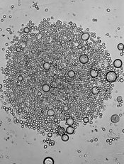
Credit: John Perry
A protein known as GPI-80 is integral to the self-renewal of hematopoietic stem cells (HSCs) during human development, investigators have reported in Cell Stem Cell.
The team says this discovery lays the groundwork for researchers to generate HSCs in the lab that better mirror HSCs in their natural environment.
This could lead to improved therapies for hematologic disorders by enabling the creation of patient-specific HSCs for transplantation.
In a 5-year study, Hanna Katri Annikki Mikkola, MD, PhD, of the University of California, Los Angeles, and her colleagues investigated a unique HSC surface protein called GPI-80.
They found that GPI-80 is produced by a subpopulation of human fetal hematopoietic stem/progenitor cells (HSPCs)—the only group of cells that could self-renew and differentiate into various blood cell types.
The investigators also found that this subpopulation—CD34+CD38lo/-CD90+GPI-80+ HSPCs—was the sole population able to permanently integrate into and thrive within the blood system of a recipient mouse.
Dr Mikkola and her colleagues further discovered that GPI-80 identifies human HSPCs during multiple phases of development and migration.
These include the early first trimester of fetal development, when newly generated HSCs can be found in the placenta, and the second trimester, when HSCs are actively replicating in the fetal liver and the fetal bone marrow.
“We found that whatever HSC niche we investigated, we could use GPI-80 as the best determinant to find the stem cell as it was being generated or colonized different hematopoietic tissues,” Dr Mikkola said.
“Moreover, loss of GPI-80 caused the stem cells to differentiate. This essentially tells us that GPI-80 must be present to make HSCs. We now have a very unique marker for investigating how human hematopoietic cells develop, migrate, and function.”
Dr Mikkola’s team is exploring different stages of human HSC development and pluripotent stem cell differentiation based on the GPI-80 marker and comparing how HSCs are being generated in vitro and in vivo.
The group says this paves the way for scientists to redirect pluripotent stem cells into patient-specific HSCs for transplantation into a patient without the need to find a suitable donor.
“Now that we can use GPI-80 as a marker to isolate the human hematopoietic stem cell at different stages of development, this can serve as a guide for identifying and overcoming the barriers to making human HSCs in vitro, which has never been done successfully,” Dr Mikkola said.
“We can now better understand the missing molecular elements that in vitro-derived cells don’t have, which is critical to fulfilling the functional and safety criteria for transplantation to patients.”
Credit: John Perry
A protein known as GPI-80 is integral to the self-renewal of hematopoietic stem cells (HSCs) during human development, investigators have reported in Cell Stem Cell.
The team says this discovery lays the groundwork for researchers to generate HSCs in the lab that better mirror HSCs in their natural environment.
This could lead to improved therapies for hematologic disorders by enabling the creation of patient-specific HSCs for transplantation.
In a 5-year study, Hanna Katri Annikki Mikkola, MD, PhD, of the University of California, Los Angeles, and her colleagues investigated a unique HSC surface protein called GPI-80.
They found that GPI-80 is produced by a subpopulation of human fetal hematopoietic stem/progenitor cells (HSPCs)—the only group of cells that could self-renew and differentiate into various blood cell types.
The investigators also found that this subpopulation—CD34+CD38lo/-CD90+GPI-80+ HSPCs—was the sole population able to permanently integrate into and thrive within the blood system of a recipient mouse.
Dr Mikkola and her colleagues further discovered that GPI-80 identifies human HSPCs during multiple phases of development and migration.
These include the early first trimester of fetal development, when newly generated HSCs can be found in the placenta, and the second trimester, when HSCs are actively replicating in the fetal liver and the fetal bone marrow.
“We found that whatever HSC niche we investigated, we could use GPI-80 as the best determinant to find the stem cell as it was being generated or colonized different hematopoietic tissues,” Dr Mikkola said.
“Moreover, loss of GPI-80 caused the stem cells to differentiate. This essentially tells us that GPI-80 must be present to make HSCs. We now have a very unique marker for investigating how human hematopoietic cells develop, migrate, and function.”
Dr Mikkola’s team is exploring different stages of human HSC development and pluripotent stem cell differentiation based on the GPI-80 marker and comparing how HSCs are being generated in vitro and in vivo.
The group says this paves the way for scientists to redirect pluripotent stem cells into patient-specific HSCs for transplantation into a patient without the need to find a suitable donor.
“Now that we can use GPI-80 as a marker to isolate the human hematopoietic stem cell at different stages of development, this can serve as a guide for identifying and overcoming the barriers to making human HSCs in vitro, which has never been done successfully,” Dr Mikkola said.
“We can now better understand the missing molecular elements that in vitro-derived cells don’t have, which is critical to fulfilling the functional and safety criteria for transplantation to patients.”
Credit: John Perry
A protein known as GPI-80 is integral to the self-renewal of hematopoietic stem cells (HSCs) during human development, investigators have reported in Cell Stem Cell.
The team says this discovery lays the groundwork for researchers to generate HSCs in the lab that better mirror HSCs in their natural environment.
This could lead to improved therapies for hematologic disorders by enabling the creation of patient-specific HSCs for transplantation.
In a 5-year study, Hanna Katri Annikki Mikkola, MD, PhD, of the University of California, Los Angeles, and her colleagues investigated a unique HSC surface protein called GPI-80.
They found that GPI-80 is produced by a subpopulation of human fetal hematopoietic stem/progenitor cells (HSPCs)—the only group of cells that could self-renew and differentiate into various blood cell types.
The investigators also found that this subpopulation—CD34+CD38lo/-CD90+GPI-80+ HSPCs—was the sole population able to permanently integrate into and thrive within the blood system of a recipient mouse.
Dr Mikkola and her colleagues further discovered that GPI-80 identifies human HSPCs during multiple phases of development and migration.
These include the early first trimester of fetal development, when newly generated HSCs can be found in the placenta, and the second trimester, when HSCs are actively replicating in the fetal liver and the fetal bone marrow.
“We found that whatever HSC niche we investigated, we could use GPI-80 as the best determinant to find the stem cell as it was being generated or colonized different hematopoietic tissues,” Dr Mikkola said.
“Moreover, loss of GPI-80 caused the stem cells to differentiate. This essentially tells us that GPI-80 must be present to make HSCs. We now have a very unique marker for investigating how human hematopoietic cells develop, migrate, and function.”
Dr Mikkola’s team is exploring different stages of human HSC development and pluripotent stem cell differentiation based on the GPI-80 marker and comparing how HSCs are being generated in vitro and in vivo.
The group says this paves the way for scientists to redirect pluripotent stem cells into patient-specific HSCs for transplantation into a patient without the need to find a suitable donor.
“Now that we can use GPI-80 as a marker to isolate the human hematopoietic stem cell at different stages of development, this can serve as a guide for identifying and overcoming the barriers to making human HSCs in vitro, which has never been done successfully,” Dr Mikkola said.
“We can now better understand the missing molecular elements that in vitro-derived cells don’t have, which is critical to fulfilling the functional and safety criteria for transplantation to patients.”
DNA finding has implications for MPNs

Credit: NIGMS
A new study suggests the timing of DNA replication—including where the origin points are and in what order DNA segments are copied—varies from person to person.
The research also revealed the first genetic variants that orchestrate replication timing.
And researchers found evidence suggesting that differences in replication timing may explain why some people are more prone than others to developing myeloproliferative neoplasms (MPNs).
“Everyone’s cells have a plan for copying the genome,” said study author Steven McCarroll, PhD, of Harvard Medical School in Boston. “The idea that we don’t all have the same plan is surprising and interesting.”
Dr McCarroll and his colleagues described this research in Cell.
Replication timing and MPNs
The researchers noted that DNA replication is one of the most fundamental cellular processes, and any variation among people is likely to affect genetic inheritance, including individual disease risk as well as human evolution.
Replication timing is known to affect mutation rates. DNA segments that are copied too late or too early tend to have more errors.
The new study indicates that people with different timing programs therefore have different patterns of mutation risk across their genomes. For example, differences in replication timing could explain predisposition to MPNs.
Researchers previously showed that acquired mutations in JAK2 lead to MPNs. They also noticed that people with JAK2 mutations tend to have a distinctive set of inherited genetic variants nearby, but they weren’t sure how the inherited variants and the new mutations were connected.
Dr McCarroll’s team found that the inherited variants are associated with an “unusually early” replication origin point and proposed that JAK2 is more likely to develop mutations in people with that very early origin point.
“Replication timing may be a way that inherited variation contributes to the risk of later mutations and diseases that we usually think of as arising by chance,” Dr McCarroll said.
A new method of study
Dr McCarroll and his colleagues were able to make these discoveries, in large part, because they invented a new way to obtain DNA replication timing data. They turned to the 1000 Genomes Project, which maintains an online database of sequencing data collected from hundreds of people around the world.
Because much of the DNA in the 1000 Genomes Project had been extracted from actively dividing cells, the team hypothesized that information about replication timing lurked within, and they were right.
They counted the number of copies of individual genes in each genome. Because early replication origins had created more segment copies at the time the sample was taken than late replication origins had, the researchers were able to create a personalized replication timing map for each person.
“People had seen these patterns before but just dismissed them as artifacts of sequencing technology,” Dr McCarroll said. After conducting numerous tests to rule out that possibility, “we found that they reflect real biology.”
The researchers then compared each person’s copy number information with his or her genetic sequence data to see if they could match specific genetic variants to replication timing differences. From 161 samples, the team identified 16 variants. The variants were short, and most were common.
“I think this is the first time we can pinpoint genetic influences on replication timing in any organism,” said study author Amnon Koren, PhD, also of Harvard Medical School.
The variants were located near replication origin points, leading the researchers to wonder if they affect replication timing by altering where a person’s origin points are. The team also suspects the variants work by altering chromatin structure, exposing local sequences to replication machinery.
The group intends to find out. They also want to search for additional variants that control replication timing.
“These 16 variants are almost certainly just the tip of the iceberg,” Dr Koren said.
He and his colleagues believe that, as more variants come to light in future studies, researchers should be better able to manipulate replication timing in the lab and learn more about how it works and its biological significance.
“All you need to do to study replication timing is grow cells and sequence their DNA, which everyone is doing these days,” Dr Koren said. “[This new method] is much easier, faster, and cheaper, and I think it will transform the field because we can now do experiments in large scale.”
“We found that there is biological information in genome sequence data,” Dr McCarroll added. “But this was still an accidental biological experiment. Now imagine the results when we and others actually design experiments to study this phenomenon.”
Credit: NIGMS
A new study suggests the timing of DNA replication—including where the origin points are and in what order DNA segments are copied—varies from person to person.
The research also revealed the first genetic variants that orchestrate replication timing.
And researchers found evidence suggesting that differences in replication timing may explain why some people are more prone than others to developing myeloproliferative neoplasms (MPNs).
“Everyone’s cells have a plan for copying the genome,” said study author Steven McCarroll, PhD, of Harvard Medical School in Boston. “The idea that we don’t all have the same plan is surprising and interesting.”
Dr McCarroll and his colleagues described this research in Cell.
Replication timing and MPNs
The researchers noted that DNA replication is one of the most fundamental cellular processes, and any variation among people is likely to affect genetic inheritance, including individual disease risk as well as human evolution.
Replication timing is known to affect mutation rates. DNA segments that are copied too late or too early tend to have more errors.
The new study indicates that people with different timing programs therefore have different patterns of mutation risk across their genomes. For example, differences in replication timing could explain predisposition to MPNs.
Researchers previously showed that acquired mutations in JAK2 lead to MPNs. They also noticed that people with JAK2 mutations tend to have a distinctive set of inherited genetic variants nearby, but they weren’t sure how the inherited variants and the new mutations were connected.
Dr McCarroll’s team found that the inherited variants are associated with an “unusually early” replication origin point and proposed that JAK2 is more likely to develop mutations in people with that very early origin point.
“Replication timing may be a way that inherited variation contributes to the risk of later mutations and diseases that we usually think of as arising by chance,” Dr McCarroll said.
A new method of study
Dr McCarroll and his colleagues were able to make these discoveries, in large part, because they invented a new way to obtain DNA replication timing data. They turned to the 1000 Genomes Project, which maintains an online database of sequencing data collected from hundreds of people around the world.
Because much of the DNA in the 1000 Genomes Project had been extracted from actively dividing cells, the team hypothesized that information about replication timing lurked within, and they were right.
They counted the number of copies of individual genes in each genome. Because early replication origins had created more segment copies at the time the sample was taken than late replication origins had, the researchers were able to create a personalized replication timing map for each person.
“People had seen these patterns before but just dismissed them as artifacts of sequencing technology,” Dr McCarroll said. After conducting numerous tests to rule out that possibility, “we found that they reflect real biology.”
The researchers then compared each person’s copy number information with his or her genetic sequence data to see if they could match specific genetic variants to replication timing differences. From 161 samples, the team identified 16 variants. The variants were short, and most were common.
“I think this is the first time we can pinpoint genetic influences on replication timing in any organism,” said study author Amnon Koren, PhD, also of Harvard Medical School.
The variants were located near replication origin points, leading the researchers to wonder if they affect replication timing by altering where a person’s origin points are. The team also suspects the variants work by altering chromatin structure, exposing local sequences to replication machinery.
The group intends to find out. They also want to search for additional variants that control replication timing.
“These 16 variants are almost certainly just the tip of the iceberg,” Dr Koren said.
He and his colleagues believe that, as more variants come to light in future studies, researchers should be better able to manipulate replication timing in the lab and learn more about how it works and its biological significance.
“All you need to do to study replication timing is grow cells and sequence their DNA, which everyone is doing these days,” Dr Koren said. “[This new method] is much easier, faster, and cheaper, and I think it will transform the field because we can now do experiments in large scale.”
“We found that there is biological information in genome sequence data,” Dr McCarroll added. “But this was still an accidental biological experiment. Now imagine the results when we and others actually design experiments to study this phenomenon.”
Credit: NIGMS
A new study suggests the timing of DNA replication—including where the origin points are and in what order DNA segments are copied—varies from person to person.
The research also revealed the first genetic variants that orchestrate replication timing.
And researchers found evidence suggesting that differences in replication timing may explain why some people are more prone than others to developing myeloproliferative neoplasms (MPNs).
“Everyone’s cells have a plan for copying the genome,” said study author Steven McCarroll, PhD, of Harvard Medical School in Boston. “The idea that we don’t all have the same plan is surprising and interesting.”
Dr McCarroll and his colleagues described this research in Cell.
Replication timing and MPNs
The researchers noted that DNA replication is one of the most fundamental cellular processes, and any variation among people is likely to affect genetic inheritance, including individual disease risk as well as human evolution.
Replication timing is known to affect mutation rates. DNA segments that are copied too late or too early tend to have more errors.
The new study indicates that people with different timing programs therefore have different patterns of mutation risk across their genomes. For example, differences in replication timing could explain predisposition to MPNs.
Researchers previously showed that acquired mutations in JAK2 lead to MPNs. They also noticed that people with JAK2 mutations tend to have a distinctive set of inherited genetic variants nearby, but they weren’t sure how the inherited variants and the new mutations were connected.
Dr McCarroll’s team found that the inherited variants are associated with an “unusually early” replication origin point and proposed that JAK2 is more likely to develop mutations in people with that very early origin point.
“Replication timing may be a way that inherited variation contributes to the risk of later mutations and diseases that we usually think of as arising by chance,” Dr McCarroll said.
A new method of study
Dr McCarroll and his colleagues were able to make these discoveries, in large part, because they invented a new way to obtain DNA replication timing data. They turned to the 1000 Genomes Project, which maintains an online database of sequencing data collected from hundreds of people around the world.
Because much of the DNA in the 1000 Genomes Project had been extracted from actively dividing cells, the team hypothesized that information about replication timing lurked within, and they were right.
They counted the number of copies of individual genes in each genome. Because early replication origins had created more segment copies at the time the sample was taken than late replication origins had, the researchers were able to create a personalized replication timing map for each person.
“People had seen these patterns before but just dismissed them as artifacts of sequencing technology,” Dr McCarroll said. After conducting numerous tests to rule out that possibility, “we found that they reflect real biology.”
The researchers then compared each person’s copy number information with his or her genetic sequence data to see if they could match specific genetic variants to replication timing differences. From 161 samples, the team identified 16 variants. The variants were short, and most were common.
“I think this is the first time we can pinpoint genetic influences on replication timing in any organism,” said study author Amnon Koren, PhD, also of Harvard Medical School.
The variants were located near replication origin points, leading the researchers to wonder if they affect replication timing by altering where a person’s origin points are. The team also suspects the variants work by altering chromatin structure, exposing local sequences to replication machinery.
The group intends to find out. They also want to search for additional variants that control replication timing.
“These 16 variants are almost certainly just the tip of the iceberg,” Dr Koren said.
He and his colleagues believe that, as more variants come to light in future studies, researchers should be better able to manipulate replication timing in the lab and learn more about how it works and its biological significance.
“All you need to do to study replication timing is grow cells and sequence their DNA, which everyone is doing these days,” Dr Koren said. “[This new method] is much easier, faster, and cheaper, and I think it will transform the field because we can now do experiments in large scale.”
“We found that there is biological information in genome sequence data,” Dr McCarroll added. “But this was still an accidental biological experiment. Now imagine the results when we and others actually design experiments to study this phenomenon.”
Sickle cell trait linked to increased risk of CKD

while another looks on
Credit: NCI
Sickle cell trait may increase the risk of chronic kidney disease (CKD) and poor kidney function, according to a study published in JAMA.
Researchers evaluated nearly 16,000 African Americans and found that subjects with sickle cell trait had a greater risk of CKD and incident CKD than subjects who did not have the trait.
Trait carriers were also more likely to have albuminuria and a decrease in estimated glomerular filtration rate (eGFR), both characteristics of poor kidney function.
This study was released to coincide with its presentation at the American Society of Nephrology’s Kidney Week Annual Meeting.
Rakhi P. Naik, MD, of Johns Hopkins University in Baltimore, and her colleagues conducted this research to investigate the relationship between sickle cell trait and kidney impairment.
The team looked at data from 5 large, population-based studies. They evaluated 15,975 self-identified African Americans—1248 of whom had sickle cell trait and 14,727 who did not.
The researchers assessed the incidence of CKD, which was defined as an eGFR of <60 mL/min/1.73m2 at baseline or follow-up, and incident CKD. They also assessed the rate of albuminuria, which was defined as a spot urine albumin:creatinine ratio of >30mg/g or albumin excretion rate >30mg/24 hours, and decline in eGFR, which was defined as a decrease of >3 mL/min/1.73m2 per year.
CKD and incident CKD were more common among sickle cell trait carriers than noncarriers. CKD was present in 19.2% (239/1247) of carriers and 13.5% (1994/14,722) of noncarriers. And incident CKD was present in 20.7% (140/675) of carriers and 13.7% (1158/8481) of noncarriers.
Sickle cell trait was associated with a faster decline in eGFR, as 22.6% (150/665) of carriers and 19.0% (1569/8249) of noncarriers met the definition of eGFR decline.
And the trait was associated with a higher incidence of albuminuria, as 31.8% (154/485) of carriers had albuminuria, compared to 19.6% (1168/5947) of noncarriers.
So subjects with sickle cell trait had a greater risk of CKD (odds ratio [OR], 1.57), incident CKD (OR, 1.79), decline in eGFR (OR, 1.32), and albuminuria (OR, 1.86).
The researchers said the associations found in this study may offer an additional genetic explanation for the increased risk of CKD observed among African Americans compared with other racial groups.
They added that the study also highlights the need for further research into the renal complications of sickle cell trait. Because screening for the trait is widely performed, accurate characterization of disease associations with sickle cell trait is needed to inform policy and treatment recommendations.
while another looks on
Credit: NCI
Sickle cell trait may increase the risk of chronic kidney disease (CKD) and poor kidney function, according to a study published in JAMA.
Researchers evaluated nearly 16,000 African Americans and found that subjects with sickle cell trait had a greater risk of CKD and incident CKD than subjects who did not have the trait.
Trait carriers were also more likely to have albuminuria and a decrease in estimated glomerular filtration rate (eGFR), both characteristics of poor kidney function.
This study was released to coincide with its presentation at the American Society of Nephrology’s Kidney Week Annual Meeting.
Rakhi P. Naik, MD, of Johns Hopkins University in Baltimore, and her colleagues conducted this research to investigate the relationship between sickle cell trait and kidney impairment.
The team looked at data from 5 large, population-based studies. They evaluated 15,975 self-identified African Americans—1248 of whom had sickle cell trait and 14,727 who did not.
The researchers assessed the incidence of CKD, which was defined as an eGFR of <60 mL/min/1.73m2 at baseline or follow-up, and incident CKD. They also assessed the rate of albuminuria, which was defined as a spot urine albumin:creatinine ratio of >30mg/g or albumin excretion rate >30mg/24 hours, and decline in eGFR, which was defined as a decrease of >3 mL/min/1.73m2 per year.
CKD and incident CKD were more common among sickle cell trait carriers than noncarriers. CKD was present in 19.2% (239/1247) of carriers and 13.5% (1994/14,722) of noncarriers. And incident CKD was present in 20.7% (140/675) of carriers and 13.7% (1158/8481) of noncarriers.
Sickle cell trait was associated with a faster decline in eGFR, as 22.6% (150/665) of carriers and 19.0% (1569/8249) of noncarriers met the definition of eGFR decline.
And the trait was associated with a higher incidence of albuminuria, as 31.8% (154/485) of carriers had albuminuria, compared to 19.6% (1168/5947) of noncarriers.
So subjects with sickle cell trait had a greater risk of CKD (odds ratio [OR], 1.57), incident CKD (OR, 1.79), decline in eGFR (OR, 1.32), and albuminuria (OR, 1.86).
The researchers said the associations found in this study may offer an additional genetic explanation for the increased risk of CKD observed among African Americans compared with other racial groups.
They added that the study also highlights the need for further research into the renal complications of sickle cell trait. Because screening for the trait is widely performed, accurate characterization of disease associations with sickle cell trait is needed to inform policy and treatment recommendations.
while another looks on
Credit: NCI
Sickle cell trait may increase the risk of chronic kidney disease (CKD) and poor kidney function, according to a study published in JAMA.
Researchers evaluated nearly 16,000 African Americans and found that subjects with sickle cell trait had a greater risk of CKD and incident CKD than subjects who did not have the trait.
Trait carriers were also more likely to have albuminuria and a decrease in estimated glomerular filtration rate (eGFR), both characteristics of poor kidney function.
This study was released to coincide with its presentation at the American Society of Nephrology’s Kidney Week Annual Meeting.
Rakhi P. Naik, MD, of Johns Hopkins University in Baltimore, and her colleagues conducted this research to investigate the relationship between sickle cell trait and kidney impairment.
The team looked at data from 5 large, population-based studies. They evaluated 15,975 self-identified African Americans—1248 of whom had sickle cell trait and 14,727 who did not.
The researchers assessed the incidence of CKD, which was defined as an eGFR of <60 mL/min/1.73m2 at baseline or follow-up, and incident CKD. They also assessed the rate of albuminuria, which was defined as a spot urine albumin:creatinine ratio of >30mg/g or albumin excretion rate >30mg/24 hours, and decline in eGFR, which was defined as a decrease of >3 mL/min/1.73m2 per year.
CKD and incident CKD were more common among sickle cell trait carriers than noncarriers. CKD was present in 19.2% (239/1247) of carriers and 13.5% (1994/14,722) of noncarriers. And incident CKD was present in 20.7% (140/675) of carriers and 13.7% (1158/8481) of noncarriers.
Sickle cell trait was associated with a faster decline in eGFR, as 22.6% (150/665) of carriers and 19.0% (1569/8249) of noncarriers met the definition of eGFR decline.
And the trait was associated with a higher incidence of albuminuria, as 31.8% (154/485) of carriers had albuminuria, compared to 19.6% (1168/5947) of noncarriers.
So subjects with sickle cell trait had a greater risk of CKD (odds ratio [OR], 1.57), incident CKD (OR, 1.79), decline in eGFR (OR, 1.32), and albuminuria (OR, 1.86).
The researchers said the associations found in this study may offer an additional genetic explanation for the increased risk of CKD observed among African Americans compared with other racial groups.
They added that the study also highlights the need for further research into the renal complications of sickle cell trait. Because screening for the trait is widely performed, accurate characterization of disease associations with sickle cell trait is needed to inform policy and treatment recommendations.
Infection may cause implant-associated ALCL

Credit: FDA
Bacterial infection on the surface of textured breast implants may increase the risk of developing breast implant-associated anaplastic large-cell lymphoma (BIA-ALCL), according to research published in Plastic & Reconstructive Surgery.
Previous studies have shown that biofilm infection around breast implants is a major cause of capsular contracture, a painful hardening of the tissue around the implant that can cause physical deformity and pain.
Now, researchers have found that chronic infection around implants can also lead to an activation of the immune system and the patient’s lymphocytes. And long-term stimulation of lymphocytes by this infection may prompt the transformation of these cells into BIA-ALCL.
The infection was shown to be highest around textured breast implants, and this may provide an explanation as to why BIA-ALCL seems to be more common in patients with textured implants.
“Our previous research has shown that, 24 hours after bacteria come into contact with breast implants, textured implants had 72 times the number of bacteria attached to their surface as compared with the smooth implants,” said Anand Deva, MBBS, of Macquarie University in Sydney, Australia.
“This latest study has shown that the textured implants with the highest numbers of bacteria also had the highest number of activated lymphocytes around them. This finding is important and has now become even more relevant since the reporting of BIA-ALCL, as it provides us with a possible biological explanation of how this rare cancer could arise.”
To uncover these findings, Dr Devan and his colleagues first examined implants in pigs. The team inserted 12 textured and 12 smooth implants into submammary pockets in 3 adult pigs.
After a mean of 8.75 months, all of the samples were positive for bacterial biofilm. And there was a significant correlation between bacterial numbers and the grade of capsular contracture (P=0.04).
Lymphocyte numbers were significantly higher on textured implants (P<0.001), with T cells accounting for the majority of the lymphocytic infiltrate.
The researchers then examined implants in humans, collecting 57 capsules from patients with Baker grade 4 capsules over a 4-year period. The team analyzed biofilm and the surrounding lymphocytes.
As in the pigs, all of the capsules were positive for biofilm, and T cells were the predominant lymphocyte (P<0.001).
The researchers also discovered a significant linear correlation between the number of T and B cells and the number of detected bacteria (P<0.001). And there was a significantly higher number of bacteria for polyurethane implants (P<0.005).
These results suggest a possible link between bacterial biofilm and T-cell hyperplasia, a finding that may have implications for BIA-ALCL, the researchers said.
Dr Deva and his colleagues have published a 14-step guide to reduce the risk of breast implant infection, based on evidence of best practice to educate surgeons on how to reduce the risk of bacterial contamination.
A number of clinical studies have applied these principles and successfully reduced the rate of capsular contracture by a factor of 10 in their patients.
“This is a great validation of our research and a demonstration that good science in the laboratory can be translated into real benefits to patients at the bedside,” Dr Deva said.
“Now, with our greater understanding of the importance of preventing infection, we, as surgeons, can reduce the risk of capsular contracture and thereby reduce the risk of lymphocyte activation and possible transformation into BIA-ALCL.”
Credit: FDA
Bacterial infection on the surface of textured breast implants may increase the risk of developing breast implant-associated anaplastic large-cell lymphoma (BIA-ALCL), according to research published in Plastic & Reconstructive Surgery.
Previous studies have shown that biofilm infection around breast implants is a major cause of capsular contracture, a painful hardening of the tissue around the implant that can cause physical deformity and pain.
Now, researchers have found that chronic infection around implants can also lead to an activation of the immune system and the patient’s lymphocytes. And long-term stimulation of lymphocytes by this infection may prompt the transformation of these cells into BIA-ALCL.
The infection was shown to be highest around textured breast implants, and this may provide an explanation as to why BIA-ALCL seems to be more common in patients with textured implants.
“Our previous research has shown that, 24 hours after bacteria come into contact with breast implants, textured implants had 72 times the number of bacteria attached to their surface as compared with the smooth implants,” said Anand Deva, MBBS, of Macquarie University in Sydney, Australia.
“This latest study has shown that the textured implants with the highest numbers of bacteria also had the highest number of activated lymphocytes around them. This finding is important and has now become even more relevant since the reporting of BIA-ALCL, as it provides us with a possible biological explanation of how this rare cancer could arise.”
To uncover these findings, Dr Devan and his colleagues first examined implants in pigs. The team inserted 12 textured and 12 smooth implants into submammary pockets in 3 adult pigs.
After a mean of 8.75 months, all of the samples were positive for bacterial biofilm. And there was a significant correlation between bacterial numbers and the grade of capsular contracture (P=0.04).
Lymphocyte numbers were significantly higher on textured implants (P<0.001), with T cells accounting for the majority of the lymphocytic infiltrate.
The researchers then examined implants in humans, collecting 57 capsules from patients with Baker grade 4 capsules over a 4-year period. The team analyzed biofilm and the surrounding lymphocytes.
As in the pigs, all of the capsules were positive for biofilm, and T cells were the predominant lymphocyte (P<0.001).
The researchers also discovered a significant linear correlation between the number of T and B cells and the number of detected bacteria (P<0.001). And there was a significantly higher number of bacteria for polyurethane implants (P<0.005).
These results suggest a possible link between bacterial biofilm and T-cell hyperplasia, a finding that may have implications for BIA-ALCL, the researchers said.
Dr Deva and his colleagues have published a 14-step guide to reduce the risk of breast implant infection, based on evidence of best practice to educate surgeons on how to reduce the risk of bacterial contamination.
A number of clinical studies have applied these principles and successfully reduced the rate of capsular contracture by a factor of 10 in their patients.
“This is a great validation of our research and a demonstration that good science in the laboratory can be translated into real benefits to patients at the bedside,” Dr Deva said.
“Now, with our greater understanding of the importance of preventing infection, we, as surgeons, can reduce the risk of capsular contracture and thereby reduce the risk of lymphocyte activation and possible transformation into BIA-ALCL.”
Credit: FDA
Bacterial infection on the surface of textured breast implants may increase the risk of developing breast implant-associated anaplastic large-cell lymphoma (BIA-ALCL), according to research published in Plastic & Reconstructive Surgery.
Previous studies have shown that biofilm infection around breast implants is a major cause of capsular contracture, a painful hardening of the tissue around the implant that can cause physical deformity and pain.
Now, researchers have found that chronic infection around implants can also lead to an activation of the immune system and the patient’s lymphocytes. And long-term stimulation of lymphocytes by this infection may prompt the transformation of these cells into BIA-ALCL.
The infection was shown to be highest around textured breast implants, and this may provide an explanation as to why BIA-ALCL seems to be more common in patients with textured implants.
“Our previous research has shown that, 24 hours after bacteria come into contact with breast implants, textured implants had 72 times the number of bacteria attached to their surface as compared with the smooth implants,” said Anand Deva, MBBS, of Macquarie University in Sydney, Australia.
“This latest study has shown that the textured implants with the highest numbers of bacteria also had the highest number of activated lymphocytes around them. This finding is important and has now become even more relevant since the reporting of BIA-ALCL, as it provides us with a possible biological explanation of how this rare cancer could arise.”
To uncover these findings, Dr Devan and his colleagues first examined implants in pigs. The team inserted 12 textured and 12 smooth implants into submammary pockets in 3 adult pigs.
After a mean of 8.75 months, all of the samples were positive for bacterial biofilm. And there was a significant correlation between bacterial numbers and the grade of capsular contracture (P=0.04).
Lymphocyte numbers were significantly higher on textured implants (P<0.001), with T cells accounting for the majority of the lymphocytic infiltrate.
The researchers then examined implants in humans, collecting 57 capsules from patients with Baker grade 4 capsules over a 4-year period. The team analyzed biofilm and the surrounding lymphocytes.
As in the pigs, all of the capsules were positive for biofilm, and T cells were the predominant lymphocyte (P<0.001).
The researchers also discovered a significant linear correlation between the number of T and B cells and the number of detected bacteria (P<0.001). And there was a significantly higher number of bacteria for polyurethane implants (P<0.005).
These results suggest a possible link between bacterial biofilm and T-cell hyperplasia, a finding that may have implications for BIA-ALCL, the researchers said.
Dr Deva and his colleagues have published a 14-step guide to reduce the risk of breast implant infection, based on evidence of best practice to educate surgeons on how to reduce the risk of bacterial contamination.
A number of clinical studies have applied these principles and successfully reduced the rate of capsular contracture by a factor of 10 in their patients.
“This is a great validation of our research and a demonstration that good science in the laboratory can be translated into real benefits to patients at the bedside,” Dr Deva said.
“Now, with our greater understanding of the importance of preventing infection, we, as surgeons, can reduce the risk of capsular contracture and thereby reduce the risk of lymphocyte activation and possible transformation into BIA-ALCL.”
Product approved for hemophilia A in Canada, Australia

Health Canada and Australia’s Therapeutic Goods Administration (TGA) have both approved a recombinant FVIII product known as simoctocog alfa (Nuwiq).
Health Canada has approved the product to treat and prevent bleeding in hemophilia A patients of all ages.
And the TGA has approved simoctocog alfa for the treatment and prevention of bleeding in previously treated pediatric (≥ 2 years) and adult patients with
hemophilia A.
Simoctocog alfa is a recombinant FVIII product produced in a human cell line cultured without additives of human or animal origin or any exposure to human blood or plasma, making it inherently free from blood-borne pathogens.
Simoctocog alfa is also devoid of antigenic non-human protein epitopes, similar to FVIII produced in healthy humans. It has a high affinity for the von Willebrand coagulation factor.
“The way Nuwiq is produced is exciting, as it allows the molecule to closely resemble the naturally occurring FVIII,” said Anthony Chan, MBBS, Director of the Hemophilia Program at McMaster Children’s Hospital in Hamilton, Ontario.
“Health Canada’s approval of Nuwiq provides patients with hemophilia A a new recombinant product option that will allow further customization of hemophilia treatment on an individual basis.”
Researchers have evaluated the immunogenicity of simoctocog alfa in 135 previously treated patients with hemophilia A (74 adults and 61 children). And none of the patients developed inhibitors.
In the ongoing, phase 3 NuProtect study, researchers are investigating 100 previously untreated patients, a group typically characterized by a higher risk of inhibitor development. The researchers will assess whether the molecular properties of simoctocog alfa will result in lower inhibitor development.
Two additional phase 3 studies in previously treated patients are ongoing. The NuPreviq study and the Canadian Gena-21b study were designed to assess the efficacy and safety of individually tailored prophylaxis.
The goal of these studies is to provide optimal treatment for each patient based on his or her own pharmacokinetic properties, with a potential reduction in the frequency of FVIII infusions.
Simoctocog alfa was approved in the European Union earlier this year and is under review by regulatory authorities in the US. For more details on simoctocog alfa, see the prescribing information.
Health Canada and Australia’s Therapeutic Goods Administration (TGA) have both approved a recombinant FVIII product known as simoctocog alfa (Nuwiq).
Health Canada has approved the product to treat and prevent bleeding in hemophilia A patients of all ages.
And the TGA has approved simoctocog alfa for the treatment and prevention of bleeding in previously treated pediatric (≥ 2 years) and adult patients with
hemophilia A.
Simoctocog alfa is a recombinant FVIII product produced in a human cell line cultured without additives of human or animal origin or any exposure to human blood or plasma, making it inherently free from blood-borne pathogens.
Simoctocog alfa is also devoid of antigenic non-human protein epitopes, similar to FVIII produced in healthy humans. It has a high affinity for the von Willebrand coagulation factor.
“The way Nuwiq is produced is exciting, as it allows the molecule to closely resemble the naturally occurring FVIII,” said Anthony Chan, MBBS, Director of the Hemophilia Program at McMaster Children’s Hospital in Hamilton, Ontario.
“Health Canada’s approval of Nuwiq provides patients with hemophilia A a new recombinant product option that will allow further customization of hemophilia treatment on an individual basis.”
Researchers have evaluated the immunogenicity of simoctocog alfa in 135 previously treated patients with hemophilia A (74 adults and 61 children). And none of the patients developed inhibitors.
In the ongoing, phase 3 NuProtect study, researchers are investigating 100 previously untreated patients, a group typically characterized by a higher risk of inhibitor development. The researchers will assess whether the molecular properties of simoctocog alfa will result in lower inhibitor development.
Two additional phase 3 studies in previously treated patients are ongoing. The NuPreviq study and the Canadian Gena-21b study were designed to assess the efficacy and safety of individually tailored prophylaxis.
The goal of these studies is to provide optimal treatment for each patient based on his or her own pharmacokinetic properties, with a potential reduction in the frequency of FVIII infusions.
Simoctocog alfa was approved in the European Union earlier this year and is under review by regulatory authorities in the US. For more details on simoctocog alfa, see the prescribing information.
Health Canada and Australia’s Therapeutic Goods Administration (TGA) have both approved a recombinant FVIII product known as simoctocog alfa (Nuwiq).
Health Canada has approved the product to treat and prevent bleeding in hemophilia A patients of all ages.
And the TGA has approved simoctocog alfa for the treatment and prevention of bleeding in previously treated pediatric (≥ 2 years) and adult patients with
hemophilia A.
Simoctocog alfa is a recombinant FVIII product produced in a human cell line cultured without additives of human or animal origin or any exposure to human blood or plasma, making it inherently free from blood-borne pathogens.
Simoctocog alfa is also devoid of antigenic non-human protein epitopes, similar to FVIII produced in healthy humans. It has a high affinity for the von Willebrand coagulation factor.
“The way Nuwiq is produced is exciting, as it allows the molecule to closely resemble the naturally occurring FVIII,” said Anthony Chan, MBBS, Director of the Hemophilia Program at McMaster Children’s Hospital in Hamilton, Ontario.
“Health Canada’s approval of Nuwiq provides patients with hemophilia A a new recombinant product option that will allow further customization of hemophilia treatment on an individual basis.”
Researchers have evaluated the immunogenicity of simoctocog alfa in 135 previously treated patients with hemophilia A (74 adults and 61 children). And none of the patients developed inhibitors.
In the ongoing, phase 3 NuProtect study, researchers are investigating 100 previously untreated patients, a group typically characterized by a higher risk of inhibitor development. The researchers will assess whether the molecular properties of simoctocog alfa will result in lower inhibitor development.
Two additional phase 3 studies in previously treated patients are ongoing. The NuPreviq study and the Canadian Gena-21b study were designed to assess the efficacy and safety of individually tailored prophylaxis.
The goal of these studies is to provide optimal treatment for each patient based on his or her own pharmacokinetic properties, with a potential reduction in the frequency of FVIII infusions.
Simoctocog alfa was approved in the European Union earlier this year and is under review by regulatory authorities in the US. For more details on simoctocog alfa, see the prescribing information.