User login
Doug Brunk is a San Diego-based award-winning reporter who began covering health care in 1991. Before joining the company, he wrote for the health sciences division of Columbia University and was an associate editor at Contemporary Long Term Care magazine when it won a Jesse H. Neal Award. His work has been syndicated by the Los Angeles Times and he is the author of two books related to the University of Kentucky Wildcats men's basketball program. Doug has a master’s degree in magazine journalism from the S.I. Newhouse School of Public Communications at Syracuse University. Follow him on Twitter @dougbrunk.
Transfer of JIA Patients to Adult Care Is Subpar
More than half of patients with juvenile idiopathic arthritis who transferred to an adult rheumatologist had inadequate follow-up for their disease 2 years after being transferred, judging from results of a Canadian study.
“Every effort should be made to ensure that young adults with JIA have timely access to a rheumatologist in the event of a disease flare, in order to minimize their disease burden,” Dr. Elizabeth M. Hazel, an adult rheumatologist at McGill University Health Centre in Montreal and associates wrote.
The researchers conducted a systematic chart review of 100 patients with JIA who attended their final JIA clinic appointment at Montreal Children's Hospital between 1992 and 2005 when they were aged 17 years or older. More than two-thirds of the patients (68%) were female, and the mean age of disease onset was 9.84 years (Pediatr. Rheumatol. 2010 Jan. 11 [doi:10.1186/1546-0096-8-2]).
A patient was deemed to have had an unsuccessful transfer if he or she “never made contact with the identified adult rheumatologist or was lost to follow-up at 2 years following transfer.”
Of the 100 patients, 52 (52%) met the criteria for unsuccessful transfer from pediatric to adult care. Of these, 17 (33%) did not make initial contact with the appointed adult rheumatologist and 35 (67%) were lost to follow-up at 2 years.
“I was very surprised that more than half of the patients were lost to follow-up,” Dr. Hazel commented in an interview.
She and her associates reported having no relevant conflicts to disclose.
GFR May Predict Malnutrition in the Elderly
SAN DIEGO — An estimated glomerular filtration rate of less than 30 mL/min is associated with malnutrition in all ages, while an estimated GFR between 30 and 59 mL/min is associated with malnutrition only in people older than age 60 years.
Those are key findings from a study that compared the prevalence of malnutrition in the elderly with the prevalence in younger age groups and that compared the risk of malnutrition using estimated GFR (eGFR) estimated by creatinine and cystatin C–based equations.
Researchers at Tufts Medical Center, Boston, examined the prevalence of malnutrition and its relationship to eGFR in 6,877 adults over the age of 20 years who participated in the National Health and Nutrition Examination Survey 1988-1994.
The investigators calculated glomerular filtration rate with serum creatinine and with a serum cystatin C equation.
Dr. Cindy Huang, a nephrology fellow at the medical center, reported that the prevalence of malnutrition increased with age: 9% in those aged 20-49 years, 12% in those aged 40-49, 15% in those 60-79, and 22% in those older than 80 years.
The researchers found an association between an eGFR of less than 30 mL/min and malnutrition in all ages, while an eGFR between 30 and 59 mL/min was associated with malnutrition only in people over age 60 years, Dr. Huang said in a poster presented at the annual meeting of the American Society of Nephrology.
Disclosures: The study was supported by a grant from the National Institutes of Health. Dr. Huang had no relevant financial disclosures.
Low Body Fat Tied to High Mortality in Dialysis
SAN DIEGO — A low percentage of total body fat is associated with higher 5-year mortality in hemodialysis patients, even after adjustment for demographics, comorbid conditions, and other surrogates of nutritional status, results from a large study showed.
“Hemodialysis patients do exhibit an obesity paradox,” Debbie Benner said during a press briefing at the annual meeting of the American Society of Nephrology.
“Low body mass index is associated with greater mortality, whereas higher BMI appears to be protective,” Ms. Benner said.
In a study led by Dr. Kamyar Kalantar-Zadeh of the Los Angeles Biomedical Research Institute at Harbor-UCLA Medical Center, researchers used near-infrared interactance technology to measure body fat percentage in the upper arm of 671 hemodialysis patients from eight centers in California operated by DaVita Inc., and investigated their survival between 2001 and 2006.
Ms. Benner, a registered dietitian who serves as vice president of clinical support for DaVita, a nationwide provider of dialysis services, described near-infrared interactance as a “noninvasive, simple, and rapid method of assessing percent body fat based on light absorption and reflection using near-infrared light emission.”
The study was conducted because protein energy wasting “is a common problem in chronic kidney disease patients and is associated with a reduction in muscle and body fat stores. Measuring body composition including total body fat in dialysis patients may provide important information about nutritional status and outcomes in dialysis patients,” she said.
The mean age of patients was 54 years: 52% were men, 30% were African American, 54% were diabetic, and their mean total body fat percentage was 27%.
The researchers divided the patients into five groups based on body fat percentage: less than 10% (34 patients); 10% to less than 20% (156 patients); 20% to less than 30% (210 patients), 30% to less than 40% (182 patients), and 40% or higher (89 patients).
Using patients with 20%–30% body fat as the referent group, Ms. Benner and her associates performed a survival analysis adjusted for age, gender, presence of diabetes, Charlson index score, and race.
They also controlled these associations for the surrogates of the malnutrition-inflammation complex syndrome (MICS), including serum albumin, hemoglobin, normalized protein catabolic rate, phosphorus, total iron-binding capacity, ferritin, calcium, and creatinine.
The association between body fat and mortality was then assessed.
Case-mix analysis revealed that dialysis patients with less than 10% body fat were 2.54 times more likely to die than those in the referent group, while the MICS analysis revealed a 2.96-fold increased risk of death.
Analysis of the other groups confirmed a direct relationship between body fat and mortality risk. “When the body fat percentage increased, the mortality risk declined, and vice versa,” Ms. Benner said.
She acknowledged certain limitations of the study, including the potential for selection bias “and the fact that other measures of nutritional status were not tested.”
Disclosures: The study was funded by the National Institutes of Health and by DaVita. Ms. Benner disclosed no other conflicts other than her employment with DaVita.
'Hemodialysis patients do exhibit an obesity paradox.'
Source MS. BENNER
Stage of Kidney Disease Affects Heart Failure Risk
SAN DIEGO — The more advanced the stage of chronic kidney disease, the greater the risk of developing heart failure and the higher the subsequent risk of death, results from a large analysis of Medicare patients showed.
“Even a modest degree of chronic kidney disease is a very strong predictor of having cardiovascular morbidity and mortality,” Dr. Charles A. Herzog said in an interview during a poster session at the annual meeting of the American Society of Nephrology.
“Chronic kidney disease is something that primary care physicians can easily detect, because it's very easy to do a serum creatinine in an office setting,” he said.
Dr. Herzog, director of the Minneapolis-based cardiovascular special studies center of the United States Renal Data System Coordinating Center, and his associate, Shuling Li, identified 1,089,716 patients aged 66 years and older from the general Medicare database and followed them between Jan. 1, 2006, and Dec. 31, 2007.
Patients with heart failure and end-stage renal disease at baseline were excluded from the analysis.
The researchers used a Cox proportional hazard model to assess the risk of developing incident heart failure, adjusting for demographics, comorbidities, and stage of chronic kidney disease based on ICD-9 codes 585.1-585.5 and 585.9.
They used the Kaplan-Meier method to estimate the age-adjusted survival of patients after the development of incident congestive heart failure.
At baseline, 59% of the patients were female and 88% were white; 23% were aged 66-69, 25% were aged 70-74, 22% were aged 75-79, 16% were aged 80-84, and 14% were aged 85 or older.
The majority (95.8%) had no chronic kidney disease, 0.4% had stage I-II disease, 1.4% had stage III-IV disease, and the remainder (2.4%) had an unknown stage of disease.
Dr. Herzog reported that after the 1 year of follow-up, heart failure occurred in 5.3% of patients with no chronic kidney disease at baseline, 12.7% of those with stage I-II disease, 15% of those with stage III-IV disease, and 12.3% of those whose disease stage was unknown.
Independent predictors of heart failure were as follows: age 70-74 years (hazard ratio, 1.30), age 75-79 years (HR, 1.75), age 80-84 years (HR, 2.42), and age 85 years and older (HR, 3.82).
Other independent predictors included black race (HR, 1.21), stage I-II chronic kidney disease (HR, 1.45), stage III-IV disease (HR, 1.68), and unknown stage of chronic kidney disease (HR, 1.27).
The researchers also found that the presence of certain comorbid conditions predicted heart failure, including anemia (HR, 1.22), diabetes (HR, 1.57), atherosclerotic heart disease (HR, 1.67), and dysrhythmia (HR, 1.94).
Over the 1-year period, 83% of patients with no chronic kidney disease survived, compared with 77% of those with stage I-II disease, 75% of those with stage II-IV disease, and 67% of those whose disease stage was unknown.
Dr. Herzog acknowledged that the study's reliance on Medicare claims data is a limitation.
“There can be some inaccuracy in claims,” he said. “But the large size of the sample probably helps deal with some of the deficiencies in the accuracy of the coding.”
Disclosures: Dr. Herzog disclosed that he is a consultant for Amgen Inc. and is a scientific adviser for CorMedix Inc. He also is a member of the Roche Foundation for Anemia Research board of trustees.
'Even a modest degree of chronic kidney disease is a very strong predictor' of cardiovascular morbidity.
Source DR. HERZOG
Plant Extract Under Study for Treating AKs
A number of new therapies for cutaneous diseases are helping dermatologists improve patient care.
One such agent, ingenol mebutate, is a plant extract being used to treat actinic keratoses, Dr. J. Mark Jackson explained at the annual Hawaii Dermatology Seminar sponsored by Skin Disease Education Foundation.
An active component of radium weed, the extract is believed to induce primary necrosis by a neutrophil-mediated, antibody-dependent cellular toxicity. Ingenol mebutate is widely used in Australia to treat actinic keratoses, said Dr. Jackson, a clinical professor of medicine and dermatology at the University of Louisville in Kentucky.
Phase III trial results were released at press time by LEO Pharma, the manufacturer of ingenol mebutate. A 0.05% concentration of ingenol mebutate was used for 2 consecutive days in 250 patients with actinic keratoses on non-head locations. According to the company's press release, the gel met its primary clinical endpoint of complete lesion clearance. The study's full findings are expected to be presented at the upcoming meeting of the American Academy of Dermatology.
In the phase II, randomized, double-blind, placebo-controlled trial, 58 patients with 5 biopsy-proven actinic keratoses were treated with ingenol mebutate gel or placebo on days 1 and 2 or days 1 and 8 (Australasian J. Dermatol. 2009;50:16-22).
Complete clearance was achieved in 71% of actinic keratoses treated with the 0.05% formulation of ingenol mebutate, compared with 25% of those treated with the 0.01% formulation, 40% of those treated with the 0.0025% formulation, and 32% of those treated with the placebo gel.
Side effects were mild to moderate, including four cases of hypopigmentation at the treatment sites and one case of persistent scabbing and pain that extended beyond the study period in three of five actinic keratoses.
The use of itraconazole for treating palmoplantar pustulosis is another advance, he said. In a recent open-label study, six patients with palmoplantar pustulosis and no other evidence of psoriasis received 100 mg itraconazole daily for 1 month, followed by every other day for 1 month (Dermatol. Ther. 2009;22:85-9).
Three patients achieved complete response, with improvement after 2 weeks of treatment. The remaining three patients developed no new pustules but continued to have some disease activity. All patients experienced relapse, but retreatment controlled disease in two patients.
During a discussion of retapamulin, which is approved for the topical treatment of impetigo due to Staphylococcus aureus (methicillin-susceptible S. aureus only) and S. pyogenes in patients older than 9 months of age, Dr. Jackson noted that the agent can cause irritation to the nasal mucosa "and is, therefore, not a good treatment for many patients with nasal carriage of MRSA. If nasal carriage is resistant to mupirocin, you may try retapamulin, but you should warn the patient in advance to try a test area first."
Cutaneous reactions can occur in response to epidermal growth factor receptors (EGFRs), Dr. Jackson warned. A prospective study of 30 patients taking either cetuximab or erlotinib for cancer noted acneiform pustule and follicular eruptions in a seborrheic distribution 7-10 days after therapy (J. Am. Acad. Dermatol. 2006;55:429-37).
"Palmoplantar eruptions have also been reported," he said. "So have xerosis, pyogenic granulomas, and paronychia." Cutaneous reactions to EGFRs are dose dependent and recur with subsequent treatment.
Be aware of the procoagulant effects of thalidomide, which is approved for the treatment of chronic recurrent and severe erythema nodosum leprosum and is used for many other inflammatory dermatologic conditions, he said. A study of 25 patients found that deep vein thrombosis occurred in 20% of patients taking thalidomide for various inflammatory conditions (J. Dermatolog. Treat. 2007;18:335-40).
Patients at particular risk include those with systemic lupus erythematosus, a malignancy, antiphospholipid syndrome, smokers, women on oral contraceptives, patients with hyperhomocysteinemia, and patients withdrawing from antimalarials, he added.
Dr. Jackson advised that dermatologists should have a basic understanding of interstitial granulomatous dermatitis, first described in 1993. Also known as granulomatous dermatitis with associated arthritis, the condition "generally presents as erythematous, violaceous, hyperpigmented, annular plaques on the trunk and thighs," he said. "Sometimes there is an acral distribution."
Histopathology reveals dense inflammatory mixed cell infiltrate, necrobiosis, sparse mucin deposition, and no vasculitis.
Associated diseases include rheumatoid arthritis, systemic lupus erythematosus, and other autoimmune conditions. The disease can also be triggered by infections and certain medications, including ACE inhibitors, calcium channel blockers, beta blockers, and lipid lowering agents.
Treatment options include glucocorticoids, NSAIDs, and TNF-alpha inhibitors.
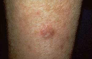
Several new therapies are under study to help physicians treat cutaneous diseases such as actinic keratoses (shown here on an elbow). Photo courtesy Dr. Roger I. Ceilley.
Dr. Jackson disclosed that he has received research, honoraria, consulting or other support from Abbott, Amgen, Biogen Idec, Centocor Ortho Biotech, Ferndale Laboratories, Galderma, Genentech, Medicis, Novartis, Promius Pharma, Quinnova Pharmaceuticals, Roche, and Stiefel.
SDEF and this news organization are owned by Elsevier.
A number of new therapies for cutaneous diseases are helping dermatologists improve patient care.
One such agent, ingenol mebutate, is a plant extract being used to treat actinic keratoses, Dr. J. Mark Jackson explained at the annual Hawaii Dermatology Seminar sponsored by Skin Disease Education Foundation.
An active component of radium weed, the extract is believed to induce primary necrosis by a neutrophil-mediated, antibody-dependent cellular toxicity. Ingenol mebutate is widely used in Australia to treat actinic keratoses, said Dr. Jackson, a clinical professor of medicine and dermatology at the University of Louisville in Kentucky.
Phase III trial results were released at press time by LEO Pharma, the manufacturer of ingenol mebutate. A 0.05% concentration of ingenol mebutate was used for 2 consecutive days in 250 patients with actinic keratoses on non-head locations. According to the company's press release, the gel met its primary clinical endpoint of complete lesion clearance. The study's full findings are expected to be presented at the upcoming meeting of the American Academy of Dermatology.
In the phase II, randomized, double-blind, placebo-controlled trial, 58 patients with 5 biopsy-proven actinic keratoses were treated with ingenol mebutate gel or placebo on days 1 and 2 or days 1 and 8 (Australasian J. Dermatol. 2009;50:16-22).
Complete clearance was achieved in 71% of actinic keratoses treated with the 0.05% formulation of ingenol mebutate, compared with 25% of those treated with the 0.01% formulation, 40% of those treated with the 0.0025% formulation, and 32% of those treated with the placebo gel.
Side effects were mild to moderate, including four cases of hypopigmentation at the treatment sites and one case of persistent scabbing and pain that extended beyond the study period in three of five actinic keratoses.
The use of itraconazole for treating palmoplantar pustulosis is another advance, he said. In a recent open-label study, six patients with palmoplantar pustulosis and no other evidence of psoriasis received 100 mg itraconazole daily for 1 month, followed by every other day for 1 month (Dermatol. Ther. 2009;22:85-9).
Three patients achieved complete response, with improvement after 2 weeks of treatment. The remaining three patients developed no new pustules but continued to have some disease activity. All patients experienced relapse, but retreatment controlled disease in two patients.
During a discussion of retapamulin, which is approved for the topical treatment of impetigo due to Staphylococcus aureus (methicillin-susceptible S. aureus only) and S. pyogenes in patients older than 9 months of age, Dr. Jackson noted that the agent can cause irritation to the nasal mucosa "and is, therefore, not a good treatment for many patients with nasal carriage of MRSA. If nasal carriage is resistant to mupirocin, you may try retapamulin, but you should warn the patient in advance to try a test area first."
Cutaneous reactions can occur in response to epidermal growth factor receptors (EGFRs), Dr. Jackson warned. A prospective study of 30 patients taking either cetuximab or erlotinib for cancer noted acneiform pustule and follicular eruptions in a seborrheic distribution 7-10 days after therapy (J. Am. Acad. Dermatol. 2006;55:429-37).
"Palmoplantar eruptions have also been reported," he said. "So have xerosis, pyogenic granulomas, and paronychia." Cutaneous reactions to EGFRs are dose dependent and recur with subsequent treatment.
Be aware of the procoagulant effects of thalidomide, which is approved for the treatment of chronic recurrent and severe erythema nodosum leprosum and is used for many other inflammatory dermatologic conditions, he said. A study of 25 patients found that deep vein thrombosis occurred in 20% of patients taking thalidomide for various inflammatory conditions (J. Dermatolog. Treat. 2007;18:335-40).
Patients at particular risk include those with systemic lupus erythematosus, a malignancy, antiphospholipid syndrome, smokers, women on oral contraceptives, patients with hyperhomocysteinemia, and patients withdrawing from antimalarials, he added.
Dr. Jackson advised that dermatologists should have a basic understanding of interstitial granulomatous dermatitis, first described in 1993. Also known as granulomatous dermatitis with associated arthritis, the condition "generally presents as erythematous, violaceous, hyperpigmented, annular plaques on the trunk and thighs," he said. "Sometimes there is an acral distribution."
Histopathology reveals dense inflammatory mixed cell infiltrate, necrobiosis, sparse mucin deposition, and no vasculitis.
Associated diseases include rheumatoid arthritis, systemic lupus erythematosus, and other autoimmune conditions. The disease can also be triggered by infections and certain medications, including ACE inhibitors, calcium channel blockers, beta blockers, and lipid lowering agents.
Treatment options include glucocorticoids, NSAIDs, and TNF-alpha inhibitors.
Several new therapies are under study to help physicians treat cutaneous diseases such as actinic keratoses (shown here on an elbow). Photo courtesy Dr. Roger I. Ceilley.
Dr. Jackson disclosed that he has received research, honoraria, consulting or other support from Abbott, Amgen, Biogen Idec, Centocor Ortho Biotech, Ferndale Laboratories, Galderma, Genentech, Medicis, Novartis, Promius Pharma, Quinnova Pharmaceuticals, Roche, and Stiefel.
SDEF and this news organization are owned by Elsevier.
Contributing to the Burners
What started in 1986 as a bonfire and the spontaneous burning of a wooden man has evolved into a week-long camping event that attracts up to 50,000 people from around the globe each year. Dr. Marc S. Nelson has attended the Burning Man event for the past 10 years as one of three medical chiefs who serve the community of sculpture-building and sculpture-burning campers.
Burning Man takes place in Nevada's Black Rock Desert, about 120 miles north of Reno, Nev. The event's official Web site (www.burningman.com
In the raging heat of late August and early September attendees bring water, food, and shelter with them to create Black Rock City, an area that houses large-scale art projects, themed campsites, and villages. Clothing is optional. Illicit drugs are commonplace. Most anything goes here, Dr. Nelson said. But attendees are expected to embrace a sense of community.
“One of the things that make Burning Man work is that you need to do something to contribute to the community,” said Dr. Nelson, an emergency medicine physician who practices at Kaiser Oakland in California. “That's why the city works so well, because people contribute and do things. It can be as simple as setting up an espresso bar where you can get free coffee in the morning. The way I contribute is by being a doctor.”
Dr. Nelson helps to oversee a staff of 300-400 volunteers who provide medical services to attendees—known as Burners—throughout the week at one of three dedicated tents. Dr. Nelson's tent is located near center camp, where he is equipped to give intravenous fluids, suture simple lacerations, and do an EKG. “There are two peripheral tents at 3 o'clock and 9 o'clock, which serve as first-aid stations,” he said.
He and the other medical volunteers treat everything from dehydration to snakebites to sunburns. They also help to coordinate transportation for the 120-mile journey to Reno, if more advanced care is needed.
“Much of what we do is triage work,” Dr. Nelson said. “Different levels of experience come into play when you start making decisions about what to do with somebody who comes in with abdominal pain and trying to decide, could this be a case of appendicitis, in which case you need to get them to Reno, or is this just a bellyache from having eaten too much the night before? Or we may see a laceration and we need to decide, does this need sutures, or is this something we can put a band-aid on?”
The medical team holds daily briefings to talk about sites where they may need to increase surveillance, supply issues, drugs floating around that may be causing bad reactions, or any other medical concerns that may arise, he said.
Deaths have occurred at Burning Man, though none occurred in 2009. “But we have to deal with victims of trauma” from plane crashes to people falling off of high scaffolding with bad fractures, Dr. Nelson said. “There aren't that many drug overdoses. But we do see some that are serious enough” to require intubation and helicopter transportation.
The uninhibited environment of Burning Man also presents a unique set of questions from the patients Dr. Nelson sees. “[It's] the kind of place where somebody could come up to you and say, 'I'm on Lasix. Is it okay for me to take ecstasy with that?' That's not the kind of question you'd get asked anywhere else. It's an interesting question. The obvious answer is, 'You shouldn't be taking ecstasy,' but it is rarely that simple and it is forces us to think outside the box, [such as] how would the two drugs interact? What advice should we give them about hydration?”
One year a Burner on dialysis wanted to know if he could skip going back to dialysis that evening so he could watch the burning of the man, the climax of the week-long event. “I don't have the kind of things I would normally have in an emergency department to help me make that kind of decision,” Dr. Nelson said. “So you have to start relying more on your clinical judgment to make decisions.”
When he's not on medical duty (the chiefs work 24-hour shifts but most other medical personnel work 8- or 12-hour shifts), Dr. Nelson enjoys wandering the grounds and meeting fellow Burners. “One of the best experiences I ever had one year was when I went into a camp and opened the door of a big tent,” he recalled. “Inside there was a group of about 30 people who had come over from Ireland. They had saved up all their money for a year and they actually re-created an Irish pub there, with beer, guitar playing, and dancing. I spent almost the whole night there, talking with them, dancing, and singing.”
The kinship he builds with other clinicians who volunteer at Burning Man “is a real bonding experience,” he said. “It's a big time commitment, and it's hard work. It's unlike anyplace most people have been to in their entire lives.”
Among the spectacles he looks forward to seeing every year are the art installations by the Flaming Lotus Girls, a group of artisans who create fire-breathing sculptures as big as a house in scale (www.flaminglotus.com
“A couple of years ago they created a dragon with moveable metal parts that spewed out fire,” Dr. Nelson said. “All the scales coming back from the dragon lit up with fire, and they all wrapped around an egg, so it was like a mother guarding an egg that was also on fire,” he added.
The communal feel of Burning Man draws Dr. Nelson back year after year. “That was the thing that struck me the first year after I came back, was this amazing sense of community, where you could walk into someone's camp, and they'd invite you to sit down and have dinner or take a shower if they had an RV.”
Burning Man organizers call it “an annual experiment in temporary community dedicated to radical self-expression.”
Dr. Marc S. Nelson volunteers as a medical chief at the Burning Man event in Nevada's Black Rock Desert.
Source Photos courtesy Dr. Marc S. Nelson
Melanoma Incidence Expected to Rise This Year
SAN DIEGO — Data on the estimated incidence of melanoma in the United States in 2010 from the National Cancer Institute's Surveillance, Epidemiology and End Results program will not be available until later this year, but Dr. Darrell S. Rigel does not expect the news to be good.
“Melanoma rates are rising significantly in the United States and in other parts of the world,” he said at a melanoma update sponsored by the Scripps Clinic. “Whatever criteria you use, it's clear that we're seeing more melanomas than we've seen in the past, and we'll probably continue to do so in the next 5-10 years.”
According to the most recent SEER data, in 2009 there were 68,720 newly diagnosed cases of invasive melanoma and 53,120 cases of in situ melanoma. Dr. Rigel and his associates at the New York University Interdisciplinary Melanoma Cooperative Group estimate that the projected lifetime risk of invasive melanoma was 1:58 in 2009, up from 1:65 in 2004.
“Should that rate of increase continue, the risk will be about 1:50 by the year 2015,” said Dr. Rigel, professor of dermatology and dermatologic surgery at New York University Medical Center. “We've been pretty close on these projections over the last few years. One in 50 is a lot. That's 2% of the population.”
Factor in the incidence of in situ melanoma, and the risk of any American getting any kind of melanoma jumps to 1:30, which would be 121,840 total cases in 2009.
“It's a significant problem,” he said.
Nine years ago, researchers who analyzed the melanoma incidence rates in the United States from 1960-1997 forecasted a subsequent growing incidence of melanoma. They concluded that the increase in melanoma incidence is real––“not due to improved diagnosis,” Dr. Rigel said––and predicted that the incidence would continue to rise for the next decade or more (J. Natl. Cancer Inst. 2001;93:67-83).
Results from studies published in the past decade suggest that the incidence of melanoma is also rising in other parts of the world. In Finland, for example, the incidence of melanoma increased from 1.5 cases to 12.8 cases per 100,000 men between 1953 and 2003, and from 1.8 cases to 10.4 cases per 100,000 women during the same time period (Int. J. Cancer 2006;119:380-4).
In central Greece, the incidence increased from 1.4 cases to 5.2 cases per 100,000 people between 1988 and 1998 (Int. J. Tissue React. 2005;27:173-9). Melanomas were most frequently located on the head and neck, extremities, and trunk. In Columbia, the incidence of melanoma increased from 2.7 cases to 13 cases per 100,000 people between 2003 and 2005 (Rev. Salud Pública 2007;9:595-601).
Dr. Rigel emphasized that results from the best available studies in the medical literature suggest that the rising incidence of melanoma cannot be explained by increased surveillance, awareness, or by changing histologic criteria. However, while the number of melanoma deaths continues to rise, 5-year survival rates are improving—from 86% between 1985 and 1989 to 92% between 1995 and 2002 (Cancer J. Clin. 2007;57:43-66).
“That seems incongruous,” Dr. Rigel said. “The only way that can be happening mathematically is that the incidence has to be rising even faster. That's a compelling reason to explain why the rising incidence is real. According to the World Health Organization, melanoma is rising faster than any other cancer worldwide, on a percentage basis.”
He went on to note that the current incidence of melanoma is probably underreported because data from SEER are collected primarily from hospitals.
Dr. Rigel disclosed that he receives grants and advising and consulting fees from a number of pharmaceutical companies, including Neutrogena, Johnson & Johnson, Procter & Gamble, and Beiersdorf.
ABCDEF Rule Guides Melanychia Diagnosis
SAN DIEGO — Of the many cases of longitudinal melanonychia—a longitudinal brown-black discoloration of the nail plate—only a fraction are subungual melanomas.
“With some of these you know they're going to require an extensive work-up or biopsy,” Dr. Arash Izadpanah said at a melanoma update sponsored by the Scripps Clinic. “Others are not so concerning, but it's not always intuitive whether these are benign or malignant.”
In 2000, researchers led by Dr. Eyal K. Levit created a modified ABCDEF rule for the clinical detection of subungual melanomas (J. Am. Acad. Dermatol. 2000;42:269-74).
In this mnemonic, A stands for age, with the peak incidence occurring in the fifth to seventh decade of life.
B stands for black-brown band with a width greater than 3 mm. “The size of the band is crucial,” said Dr. Izadpanah of the division of dermatology at Scripps in San Diego.
C stands for change in morphology such as color and width. Subungual melanomas may have blurred indistinct margins or a variation in band color.
D stands for involvement of digits, particularly the thumb, the great toe, and the index finger. “If there's dystrophy of the nail, unfortunately that's a late finding,” he said.
E stands for extension of pigment to the periungual folds, and F stands for family history of melanoma.
“The most important thing is using your clinical judgment,” he said.
Longitudinal melanonychia stems from either melanocytic activation or melanocytic hyperplasia. In melanocytic activation—the most common cause of longitudinal melanonychia in adults—“the melanocytes are there and something happens to get them producing melanosomes and pigment, but there is no actual increase in the number of melanocytes,” Dr. Izadpanah explained.
Melanocytic hyperplasia—the most common cause of longitudinal melanonychia in children—is marked by an increase in the number of matrix melanocytes. “This could be for benign or malignant reasons,” he said.
Certain racial groups are susceptible to melanocytic activation, including African Americans, Chinese, Japanese, and Native Americans. Other physiologic causes include pregnancy and trauma to the nail, Dr. Izadpanah said.
Dermatologic causes of melanocytic activation include psoriasis, lichen planus, amyloidosis, systemic lupus erythematosus, onychomycosis, and nonmelanocytic tumors.
Systemic causes of melanocytic activation include endocrine disorders such as Addison's disease and Cushing's syndrome, hyperthyroidism, and acromegaly. Other possible culprits include vitamin B12 deficiency, malnutrition, alcaptonuria, porphyria, graft-versus-host disease, AIDS, Laugier-Hunziker syndrome, and Peutz-Jeghers syndrome.
Iatrogenic causes of melanocytic activation include radiation and a host of commonly used medications, including antimalarials, minocycline, sulfonamides, ketoconazole, zidovudine, and chemotherapy agents.
Melanocytic hyperplasia can occur in the form of benign lesions such as a lentigo or a nevus, or in the form of malignant lesions such as melanoma in situ or invasive melanoma. “Longitudinal melanonychia is the first manifestation in 38%–76% of nail apparatus melanoma,” Dr. Izadpanah said.
Dermoscopy can be useful in evaluating melanonychia, “but the main limitation is that you're examining pigment that's deposited in the nail plate, when the action is really in the nail matrix, so you're indirectly seeing what's going on,” he said. “It's not as useful as it might be for melanocytic lesions of the skin.”
He went on to note that the majority of melanocytes that give rise to longitudinal melanonychia become active in the distal matrix, not in the proximal nail matrix. “That's important, because the proximal nail matrix gives rise to the vast majority of the nail plate,” he said. “So if you do a biopsy of the distal nail plate, you're probably not going to cause any significant lasting damage to the nail.”
Before a biopsy, visualize the source of melanocytic activity. This requires making a small cut in the proximal nail fold and lifting it for inspection. “In most cases you can remove the whole nail or the proximal part of the nail or do two punch biopsies: one to get part of the nail out, and a second, deeper one that reaches bone,” Dr. Izadpanah said.
Lesions greater than 3 mm in width require a transverse elliptical excision. The goal here is “to get the pigment but try to avoid damage to the proximal nail matrix, so as to not cause nail dystrophy,” he said.
Dr. Izadpanah had no relevant conflicts to disclose.
'Longitudinal melanonychia is the first manifestation in 38%–76% of nail apparatus melanoma.'
Source DR. IZADPANAH
Patients who have melanocytic lesion bands that are greater than 3 mm in width require a transverse elliptical excision.
Source Courtesy Dr. Arash Izadpanah
Contributing to the Burners
What started in 1986 as a bonfire and the spontaneous burning of a wooden man has evolved into a week-long camping event that attracts up to 50,000 people from around the globe each year. Dr. Marc S. Nelson has attended the Burning Man event for the past 10 years as one of three medical chiefs who serve the community of sculpture-building and sculpture-burning campers.
Burning Man takes place in Nevada's Black Rock Desert, about 120 miles north of Reno, Nev. The event's official Web site (www.burningman.com
In the raging heat of late August and early September attendees bring water, food, and shelter with them to create Black Rock City, an area that houses large-scale art projects, themed campsites, and villages. Clothing is optional. Illicit drugs are commonplace. Just about anything goes, Dr. Nelson said. But attendees are expected to embrace a sense of community.
“One of the things that makes Burning Man work is that you need to do something to contribute to the community,” said Dr. Nelson, an emergency medicine physician who practices at Kaiser Oakland in California. “That's why the city works so well, because people contribute and do things. It can be as simple as setting up an espresso bar where you can get free coffee in the morning. The way I contribute is by being a doctor.”
Dr. Nelson helps to oversee a staff of 300-400 volunteers who provide medical services to attendees—known as Burners—throughout the week at one of three dedicated tents. Dr. Nelson's tent is located near center camp, where he is equipped to give intravenous fluids, suture simple lacerations, and do an EKG. “There are two peripheral tents at 3 o'clock and 9 o'clock, which serve as first-aid stations,” he said.
He and the other medical volunteers treat everything from dehydration to snakebites to sunburns. They also help to coordinate transportation for the 120-mile journey to Reno, if more advanced care is needed.
“Much of what we do is triage work,” Dr. Nelson said. “Different levels of experience come into play when you start making decisions about what to do with somebody who comes in with abdominal pain and trying to decide, could this be a case of appendicitis, in which case you need to get them to Reno, or is this just a bellyache from having eaten too much the night before? Or we may see a laceration and we need to decide, does this need sutures, or is this something we can put a band-aid on?”
The medical team holds daily briefings to talk about sites where they may need to increase surveillance, supply issues, drugs floating around that may be causing bad reactions, or any other medical concerns that may arise, he said.
Deaths have occurred at Burning Man, though none occurred in 2009. “But we have to deal with victims of trauma,” ranging from plane crashes to people falling off high scaffolding and experiencing bad fractures, Dr. Nelson said.
“There aren't that many drug overdoses. But we do see some that are serious enough” to require intubation and helicopter transportation, he added.
The uninhibited environment of Burning Man also presents a unique set of questions from the patients Dr. Nelson sees. “[It's] the kind of place where somebody could come up to you and say, 'I'm on Lasix. Is it okay for me to take ecstasy with that?' That's not the kind of question you'd get asked anywhere else. It's an interesting question. The obvious answer is, 'You shouldn't be taking ecstasy,' but it is rarely that simple and it forces us to think outside the box, [such as] how would the two drugs interact? What advice should we give them about hydration?”
One year a Burner on dialysis wanted to know if he could skip going back to dialysis that evening so he could watch the burning of the man, the climax of the week-long event. “I don't have the kind of things I would normally have in an emergency department to help me make that kind of decision,” Dr. Nelson said. “So you have to start relying more on your clinical judgment to make decisions.”
When he's not on medical duty (the chiefs work 24-hour shifts but most other medical personnel work 8- or 12-hour shifts), Dr. Nelson enjoys wandering the grounds and meeting fellow Burners.
“One of the best experiences I ever had one year was when I went into a camp and opened the door of a big tent,” he recalled.
“Inside there was a group of about 30 people who had come over from Ireland. They had saved up all their money for a year and they actually re-created an Irish pub there, with beer, guitar playing, and dancing. I spent almost the whole night there, talking with them, dancing, and singing.”
The kinship he builds with other clinicians who volunteer at Burning Man “is a real bonding experience,” he said. “It's a big time commitment, and it's hard work. It's unlike anyplace most people have been to in their entire lives.”
Among the spectacles he looks forward to seeing every year are the art installations by the Flaming Lotus Girls, a group of artists who create fire-breathing sculptures as big as a house in scale (www.flaminglotus.com
“A couple of years ago they created a dragon with moveable metal parts that spewed out fire,” Dr. Nelson said. “All the scales coming back from the dragon lit up with fire, and they all wrapped around an egg, so it was like a mother guarding an egg that was also on fire,” he added.
The communal feel of Burning Man draws Dr. Nelson back year after year. “That was the thing that struck me the first year after I came back, was this amazing sense of community, where you could walk into someone's camp, and they'd invite you to sit down and have dinner or take a shower if they had an RV.”
Burning Man is “an annual experiment in temporary community dedicated to radical self-expression and radical self-reliance,” according to the event's Web site.
Dr. Marc S. Nelson volunteers as a medical chief at the Burning Man event in Nevada's Black Rock Desert.
Source Photos courtesy Dr. Marc S. Nelson
Incidence of Melanoma Expected to Rise in 2010
SAN DIEGO — Data on the estimated incidence of melanoma in the United States in 2010 from the National Cancer Institute's Surveillance, Epidemiology and End Results program will not be available until later this year, but Dr. Darrell S. Rigel does not expect the news to be good.
“Melanoma rates are rising significantly in the United States and in other parts of the world,” he said at a melanoma update sponsored by the Scripps Clinic.
“Whatever criteria you use, it's clear that we're seeing more melanomas than we've seen in the past, and we'll probably continue to do so in the next 5-10 years.”
According to the most recent SEER data, in 2009 there were 68,720 newly diagnosed cases of invasive melanoma and 53,120 cases of in situ melanoma. Dr. Rigel and his associates at the New York University Interdisciplinary Melanoma Cooperative Group estimate that the projected lifetime risk of invasive melanoma was 1:58 in 2009, up from 1:65 in 2004.
“Should that rate of increase continue, the risk will be about 1:50 by the year 2015,” said Dr. Rigel, who is a professor of dermatology and dermatologic surgery at New York University Medical Center. “We've been pretty close on these projections over the last few years. One in 50 is a lot. That's 2% of the population.”
Factor in the incidence of in situ melanoma, and the risk of any American getting any kind of melanoma jumps to 1:30, which would be 121,840 total cases in 2009.
“It's a significant problem,” he said.
Nine years ago, researchers who analyzed the melanoma incidence rates in the United States from 1960 through 1997 forecasted a subsequent growing incidence of melanoma. They concluded that the increase in melanoma incidence is real––“not due to improved diagnosis,” Dr. Rigel said––and predicted that the incidence would continue to rise for the next decade or more (J. Natl. Cancer Inst. 2001;93:67-83).
Results from studies published in the past decade suggest that the incidence of melanoma is also rising in other parts of the world.
In Finland, for example, the incidence of melanoma increased from 1.5 cases to 12.8 cases per 100,000 men between 1953 and 2003, and from 1.8 cases to 10.4 cases per 100,000 women during the same time period (Int. J. Cancer 2006;119:380-4). In central Greece, the incidence increased from 1.4 cases to 5.2 cases per 100,000 people between 1988 and 1998 (Int. J. Tissue React. 2005;27:173-9). And in Columbia, the incidence increased from 2.7 cases to 13 cases per 100,000 people between 2003 and 2005 (Rev. Salud Pública 2007;9:595-601).
Dr. Rigel emphasized that the results from the best available studies in the medical literature suggest that the rising incidence of melanoma cannot be explained by increased surveillance or awareness, or by changing histologic criteria.
However, while the number of melanoma deaths continues to rise, 5-year survival rates are improving—from 86% between 1985 and 1989 to 92% between 1995 and 2002 (Cancer J. Clin. 2007;57:43-66).
“That seems incongruous,” Dr. Rigel said. “The only way that can be happening mathematically is that the incidence has to be rising even faster. That's a compelling reason to explain why the rising incidence is real. According to the World Health Organization, melanoma is rising faster than any other cancer worldwide, on a percentage basis.”
Dr. Rigel went on to caution that the current incidence of melanoma is probably underreported because the data from SEER are collected primarily from hospitals.
“The biopsy may be done in an outpatient setting,” he explained. “It may go to an outpatient lab. It may be reexcised, and then it may go back to the same lab. It may never hit a hospital. That's why melanoma probably is significantly undercounted.”
According to the American Cancer Society, melanoma kills one American citizen per hour. “Some people pooh-pooh skin cancer,” Dr. Rigel said. “It's the most common cancer in women aged 25-29, and it's the No. 1 cancer killer in women aged 30-35. There are some subsets of the population that are particularly hurt by this disease.”
Disclosures: Dr. Rigel disclosed that he receives grants and advising and consulting fees from a number of pharmaceutical companies, including Neutrogena, Johnson & Johnson, Procter & Gamble, and Beiersdorf.