User login
Doug Brunk is a San Diego-based award-winning reporter who began covering health care in 1991. Before joining the company, he wrote for the health sciences division of Columbia University and was an associate editor at Contemporary Long Term Care magazine when it won a Jesse H. Neal Award. His work has been syndicated by the Los Angeles Times and he is the author of two books related to the University of Kentucky Wildcats men's basketball program. Doug has a master’s degree in magazine journalism from the S.I. Newhouse School of Public Communications at Syracuse University. Follow him on Twitter @dougbrunk.
Work-up, treatment of adolescent PCOS varies by specialty
SAN DIEGO – Inconsistent diagnostic and treatment practices for the diagnosis of adolescent polycystic ovary syndrome exist among clinicians in adolescent medicine, gynecology, and endocrinology, results from a single-center, retrospective study demonstrated.
The differences "could reflect provider comfort within their specialty and how their specialty approaches these symptoms," Sarah Powers said at the annual meeting of the North American Society for Pediatric and Adolescent Gynecology.

Many criteria exist for the diagnosis of polycystic ovary syndrome (PCOS), said Ms. Powers of Georgetown University in Washington. The National Institutes of Health criteria from 1990 state that, to make the diagnosis, menstrual irregularity and androgen excess must be present, as well as no other possible etiologies for the diagnosis. According to the Rotterdam criteria from 2003, patients must have two of three factors to meet the diagnosis: menstrual irregularity, evidence of androgen excess based on clinical or laboratory data, and polycystic ovaries on ultrasound. Meanwhile, the Androgen Excess and PCOS Society guideline from 2006 requires evidence of androgen excess and either menstrual irregularity or polycystic ovaries.
"Adolescents are an interesting subset of PCOS patients," Ms. Powers said. "In the first place, they are difficult to diagnose. Many signs of PCOS are physiologically common within the adolescent population, including menstrual irregularity, acne, and polycystic ovaries. Secondly, does the diagnosis even need to be made in teenagers? How long should we wait after menarche to make the diagnosis? If PCOS is diagnosed and treated early, does this make a difference for the patient?"
In addition, she continued, adolescents with PCOS "can present to a number of specialties, including adolescent medicine, gynecology, and endocrinology." In an effort to assess how these pediatric subspecialties would work up, diagnose, and treat patients with PCOS, Ms. Powers and her associates conducted a retrospective chart study of 261 postmenarchal females aged 11-18 years from Children’s National Medical Center who had been worked up for or diagnosed with PCOS between June 2009 and October 2011. They excluded patients with a coexisting diagnosis of other primary etiology for amenorrhea.
Of the 261 patients, 144 (55%) were seen by endocrinologists, 9 (3.5%) by gynecologists, and 108 (41.5%) by adolescent medicine specialists. The mean age of patients at the time of their initial work-up was 15 years. There were no significant differences between the three patient populations in age, age at first menses, body mass index, obesity rates, or type of insurance. However, a higher percentage of African American patients were seen by adolescent medicine specialists, compared with clinicians in the other specialties.
Ms. Powers reported that 49% of adolescent medicine specialists ordered lipid tests, compared with 22% of gynecologists and 16% of endocrinologists, a difference that reached statistical significance (P less than .0001). Meanwhile, 44% of gynecologists ordered fasting glucose measures, compared with 38% of adolescent medicine specialists and 22% of endocrinologists, a difference that was also statistically significant (P less than.05). The proportion of HbA1c and fasting insulin measures ordered were similar among all three groups.
A significantly greater proportion of adolescent medicine specialists (31%; P less than.05) ordered total T4 labs compared with their counterparts, while rates of ordering androgen labs were about the same in all three specialties. "More than 80% of patients worked up for PCOS had a total testosterone test ordered for them," Ms. Powers said. "Ordering measurements of the other sex hormones was much less consistent. Endocrinologists were more likely to order estradiol while adolescent medicine specialists ordered more prolactin, and gynecologists ordered more 17-hydroxyprogesterone."
In addition, the majority of gynecologists (89%) ordered pelvic ultrasounds, compared with 9% of adolescent medicine specialists and 24% of endocrinologists.
Of all the 261 patients who were worked up for PCOS, 187 (72%) had at least two of the Rotterdam criteria. "Only hirsutism was used as evidence of clinical hyperandrogenism," Ms. Powers said. "Acne was not used as a marker." Of these patients, 33% were diagnosed with PCOS and 38% were diagnosed with likely PCOS. "Some caveat was given: either likely PCOS, possible PCOS, or probable PCOS – some variation on that," she said. Upon chart review, irregular menses and clinical and biochemical evidence of hyperandrogenism were used most often as justification for a PCOS diagnosis.
As for the treatment of patients who met diagnostic criteria for PCOS, metformin was prescribed most often by endocrinologists (58%), compared with 30% of adolescent medicine specialists and 14% of gynecologists. At the same time, oral contraceptives were prescribed most often by adolescent medicine specialists (58%), compared with 43% of gynecologists and 24% of endocrinologists.
Ms. Powers acknowledged certain limitations of the study, including the relatively small number of patients seen by gynecologists. "There were also difficulties capturing patients using the current billing system," she said. "In the long term, it would be useful to collect prospective data to identify the best diagnostic criteria and lab tests for a work-up."
Ms. Powers said she had no relevant financial conflicts.
SAN DIEGO – Inconsistent diagnostic and treatment practices for the diagnosis of adolescent polycystic ovary syndrome exist among clinicians in adolescent medicine, gynecology, and endocrinology, results from a single-center, retrospective study demonstrated.
The differences "could reflect provider comfort within their specialty and how their specialty approaches these symptoms," Sarah Powers said at the annual meeting of the North American Society for Pediatric and Adolescent Gynecology.
Many criteria exist for the diagnosis of polycystic ovary syndrome (PCOS), said Ms. Powers of Georgetown University in Washington. The National Institutes of Health criteria from 1990 state that, to make the diagnosis, menstrual irregularity and androgen excess must be present, as well as no other possible etiologies for the diagnosis. According to the Rotterdam criteria from 2003, patients must have two of three factors to meet the diagnosis: menstrual irregularity, evidence of androgen excess based on clinical or laboratory data, and polycystic ovaries on ultrasound. Meanwhile, the Androgen Excess and PCOS Society guideline from 2006 requires evidence of androgen excess and either menstrual irregularity or polycystic ovaries.
"Adolescents are an interesting subset of PCOS patients," Ms. Powers said. "In the first place, they are difficult to diagnose. Many signs of PCOS are physiologically common within the adolescent population, including menstrual irregularity, acne, and polycystic ovaries. Secondly, does the diagnosis even need to be made in teenagers? How long should we wait after menarche to make the diagnosis? If PCOS is diagnosed and treated early, does this make a difference for the patient?"
In addition, she continued, adolescents with PCOS "can present to a number of specialties, including adolescent medicine, gynecology, and endocrinology." In an effort to assess how these pediatric subspecialties would work up, diagnose, and treat patients with PCOS, Ms. Powers and her associates conducted a retrospective chart study of 261 postmenarchal females aged 11-18 years from Children’s National Medical Center who had been worked up for or diagnosed with PCOS between June 2009 and October 2011. They excluded patients with a coexisting diagnosis of other primary etiology for amenorrhea.
Of the 261 patients, 144 (55%) were seen by endocrinologists, 9 (3.5%) by gynecologists, and 108 (41.5%) by adolescent medicine specialists. The mean age of patients at the time of their initial work-up was 15 years. There were no significant differences between the three patient populations in age, age at first menses, body mass index, obesity rates, or type of insurance. However, a higher percentage of African American patients were seen by adolescent medicine specialists, compared with clinicians in the other specialties.
Ms. Powers reported that 49% of adolescent medicine specialists ordered lipid tests, compared with 22% of gynecologists and 16% of endocrinologists, a difference that reached statistical significance (P less than .0001). Meanwhile, 44% of gynecologists ordered fasting glucose measures, compared with 38% of adolescent medicine specialists and 22% of endocrinologists, a difference that was also statistically significant (P less than.05). The proportion of HbA1c and fasting insulin measures ordered were similar among all three groups.
A significantly greater proportion of adolescent medicine specialists (31%; P less than.05) ordered total T4 labs compared with their counterparts, while rates of ordering androgen labs were about the same in all three specialties. "More than 80% of patients worked up for PCOS had a total testosterone test ordered for them," Ms. Powers said. "Ordering measurements of the other sex hormones was much less consistent. Endocrinologists were more likely to order estradiol while adolescent medicine specialists ordered more prolactin, and gynecologists ordered more 17-hydroxyprogesterone."
In addition, the majority of gynecologists (89%) ordered pelvic ultrasounds, compared with 9% of adolescent medicine specialists and 24% of endocrinologists.
Of all the 261 patients who were worked up for PCOS, 187 (72%) had at least two of the Rotterdam criteria. "Only hirsutism was used as evidence of clinical hyperandrogenism," Ms. Powers said. "Acne was not used as a marker." Of these patients, 33% were diagnosed with PCOS and 38% were diagnosed with likely PCOS. "Some caveat was given: either likely PCOS, possible PCOS, or probable PCOS – some variation on that," she said. Upon chart review, irregular menses and clinical and biochemical evidence of hyperandrogenism were used most often as justification for a PCOS diagnosis.
As for the treatment of patients who met diagnostic criteria for PCOS, metformin was prescribed most often by endocrinologists (58%), compared with 30% of adolescent medicine specialists and 14% of gynecologists. At the same time, oral contraceptives were prescribed most often by adolescent medicine specialists (58%), compared with 43% of gynecologists and 24% of endocrinologists.
Ms. Powers acknowledged certain limitations of the study, including the relatively small number of patients seen by gynecologists. "There were also difficulties capturing patients using the current billing system," she said. "In the long term, it would be useful to collect prospective data to identify the best diagnostic criteria and lab tests for a work-up."
Ms. Powers said she had no relevant financial conflicts.
AT THE NASPAG ANNUAL MEETING
Major finding: After evaluating adolescents for potential PCOS, 49% of adolescent medicine specialists ordered lipid tests, compared with 22% of gynecologists and 16% of endocrinologists, a difference that reached statistical significance (P less than .0001).
Data source: A retrospective study of 261 postmenarchal females aged 11-18 years from Children’s National Medical Center who had been worked up for or diagnosed with PCOS between June 2009 and October 2011.
Disclosures: Ms. Powers said she had no relevant financial conflicts.

Top 10 emergency medicine audit defense strategies
SAN DIEGO – If you or your emergency medicine group is audited, one strategy is to use the Marshfield Clinic Tool to help explain medical decision making – specifically how comorbidities, the differential diagnosis, and special studies affected the course of treatment.
This marks one of 10 audit defense strategies that Edward R. Gaines III, J.D., shared at a meeting on reimbursement and coding held by the American College of Emergency Physicians.
The other nine are as follows:
• Do not assume that the auditors have ED coding knowledge. Use the Emergency Medical Treatment and Labor Act (EMTALA) to help explain medical necessity in the context of emergency medicine. "A lot of medical directors and coders employed by the CMS Medicare administrative contractors and the Recovery Audit contractor auditors have no idea about EMTALA," said Mr. Gaines, who is the chief compliance officer of the Greensboro, N.C.–based Medical Management Professionals.
• Explain why you code with the presenting symptoms instead of with the final diagnosis. "Your auditor may be from a hospital background, and they’re all about the final diagnosis because that is how hospitals train their coders based on [American Hospital Association] inpatient standards," he said. See the government’s final ruling on this topic.
• Be thorough about the "nature of the presenting problem." "Explain how it impacts not only initial presentation but also comorbidities and the differential diagnosis," advised Mr. Gaines, who is also cofounder of the Emergency Department Practice Management Association.
• Insist on providers being part of the audit defense process. Use the audit "as an opportunity to educate the auditor and the client," he said.
• Make sure your documentation is true, accurate, and complete. This includes signatures on the E/M documentation, orders, and supervisory notes regarding nonphysician-practitioner and Physicians-at-Teaching-Hospitals documentation for residents. A free documentation template is available.
• Consult with counsel as appropriate, especially when large amounts of money are involved.
• Prepare a prompt and thorough response. "After the redetermination or reconsideration stages, you cannot submit any additional documentation evidence in the appeal, so you don’t want to find yourself limited," Mr. Gaines noted. "Submit complete and thorough documentation at the initial stages of the appeal."
• Don’t argue for the sake of argument. "If you missed the coding, just punt and maintain an expert, polite, and professional demeanor," he said. "That’s the best way to go to maintain credibility for the cases that you really want to argue about in the audit."
Mr. Gaines offered meeting attendees an 11th tip, a concept from Medicare case law known as the Treating Physician Rule. This rule states that deference should be given to the treatment decisions of the treating physician. "While deference is supposed to be given under the rule in Medicare cases, the deference does not rise to a presumption that the physician’s treatment was medically necessary and appropriate," Mr. Gaines stated. "The provider still has the obligation to show that services were medically necessary."
Mr. Gaines said that he had no relevant financial conflicts except as noted above.
EXPERT ANALYSIS FROM THE ACEP REIMBURSEMENT & CODING CONFERENCE

Make your practice's Facebook page a success
LAS VEGAS – Hoping to make your practice’s Facebook page a success? Engage with your Facebook followers.
According to Monique Ramsey, founder of Del Mar, Calif.–based Cosmetic Social Media, the best path to social media success involves fostering. "Think about who your consumer is and what she wants to learn about," she advised.
"Provide that information to her and let her share it with her friends, because then your name is attached to that piece of content. And note it may not have anything to do with your cosmetic surgery practice, but this kind of a post will keep people coming to your page and get higher visibility for your posts in the newsfeed," she added.

The goal is to have your posts reach more than the Facebook average, which is about 16% of your Facebook followers. With more than 1 billion people using Facebook, physicians need to be able to "cut through the noise," Ms. Ramsey explained at the annual meeting of the American Academy of Cosmetic Surgery.
It’s okay for 20% of your posts to be about your practice – perhaps advertising special events and promotions – but the remaining 80% should be informative and off the topic yet engaging, meaningful, and fun.
For example, Ms. Ramsey crafted questions for one of her client’s Facebook page intended to trigger engagement in the form of comments. One read "It’s National Wine Day. Are you a red, white or bubbly?" Another post example was crafted to encourage fans to hit the "like" button and featured two cars, sporting false eyelashes which read, "Bet you the lady car uses Latisse – hit your LIKE button if you do too!" Both questions created a spike in traffic, engagement rate, and conversation on her client’s Facebook page, she said. The latter resulted in a reach of over 10,000 people and an engagement rate of over 11% (2% is average).
"You’re trying to create an emotional connection with your Facebook followers," Ms. Ramsey said. "When you’re doing your job right, you will. You will feel like a friend to people. You want people to feel like they’re part of a community. You want to be a resource for them. You want to share your own content as well as other people’s content. Social media is very reciprocal, so give a little and get a lot."
Other tips she shared for optimizing a practice’s Facebook page include the following:
• Be yourself. "Credibility and trust are important," Ms. Ramsey said. "Be authentic. Be humble. Think about influencing conversations, not controlling conversations. You can influence behavior but you don’t have to control it."
• Change your cover image at least monthly. That’s because cover images "get, on average, nine times more engagement than a regular post," she said. "Celebrate your 100th fan, or use this space to advertise a promotion you have going on."
• Make sure your avatar is superb. Ninety percent of Facebook users see your content in their newsfeed, "so it really needs to be good and easy to see," she said. "People prefer to hear from a face of the practice rather than a logo or building because we humans communicate with people, not logos."
• Monitor replies to your posts. If someone is sending abusive messages, "you can hide those messages and block that person from your page. Or it might be better to leave it there and let your community come to your defense on your behalf. I have only had to take down two messages on behalf of clients. Both were from competing physicians."
Ms. Ramsey is the founder of Cosmetic Social Media and had no other financial conflicts to disclose.
LAS VEGAS – Hoping to make your practice’s Facebook page a success? Engage with your Facebook followers.
According to Monique Ramsey, founder of Del Mar, Calif.–based Cosmetic Social Media, the best path to social media success involves fostering. "Think about who your consumer is and what she wants to learn about," she advised.
"Provide that information to her and let her share it with her friends, because then your name is attached to that piece of content. And note it may not have anything to do with your cosmetic surgery practice, but this kind of a post will keep people coming to your page and get higher visibility for your posts in the newsfeed," she added.
The goal is to have your posts reach more than the Facebook average, which is about 16% of your Facebook followers. With more than 1 billion people using Facebook, physicians need to be able to "cut through the noise," Ms. Ramsey explained at the annual meeting of the American Academy of Cosmetic Surgery.
It’s okay for 20% of your posts to be about your practice – perhaps advertising special events and promotions – but the remaining 80% should be informative and off the topic yet engaging, meaningful, and fun.
For example, Ms. Ramsey crafted questions for one of her client’s Facebook page intended to trigger engagement in the form of comments. One read "It’s National Wine Day. Are you a red, white or bubbly?" Another post example was crafted to encourage fans to hit the "like" button and featured two cars, sporting false eyelashes which read, "Bet you the lady car uses Latisse – hit your LIKE button if you do too!" Both questions created a spike in traffic, engagement rate, and conversation on her client’s Facebook page, she said. The latter resulted in a reach of over 10,000 people and an engagement rate of over 11% (2% is average).
"You’re trying to create an emotional connection with your Facebook followers," Ms. Ramsey said. "When you’re doing your job right, you will. You will feel like a friend to people. You want people to feel like they’re part of a community. You want to be a resource for them. You want to share your own content as well as other people’s content. Social media is very reciprocal, so give a little and get a lot."
Other tips she shared for optimizing a practice’s Facebook page include the following:
• Be yourself. "Credibility and trust are important," Ms. Ramsey said. "Be authentic. Be humble. Think about influencing conversations, not controlling conversations. You can influence behavior but you don’t have to control it."
• Change your cover image at least monthly. That’s because cover images "get, on average, nine times more engagement than a regular post," she said. "Celebrate your 100th fan, or use this space to advertise a promotion you have going on."
• Make sure your avatar is superb. Ninety percent of Facebook users see your content in their newsfeed, "so it really needs to be good and easy to see," she said. "People prefer to hear from a face of the practice rather than a logo or building because we humans communicate with people, not logos."
• Monitor replies to your posts. If someone is sending abusive messages, "you can hide those messages and block that person from your page. Or it might be better to leave it there and let your community come to your defense on your behalf. I have only had to take down two messages on behalf of clients. Both were from competing physicians."
Ms. Ramsey is the founder of Cosmetic Social Media and had no other financial conflicts to disclose.
EXPERT ANALYSIS FROM THE AACS ANNUAL MEETING

Ovarian dermoid cysts recur in 11% of adolescents
SAN DIEGO – The total recurrence rate of ovarian dermoid cysts in a pediatric and adolescent population following cystectomy is 11%, and evidence of recurrence that requires a second operative management is 3%.
Moreover, the method of surgical intervention – laparoscopy or laparotomy – does not appear to influence the rate of cyst recurrence, Erin Rogers reported at the annual meeting of the North American Society for Pediatric and Adolescent Gynecology.

Dermoid cysts can represent up to 70% of benign ovarian tumors in women under age 30, said Ms. Rogers, a medical student at Queens University, Kingston, Ontario, Canada. "They have a slow growth rate and on average have been seen to grow at 1.8 mm per year."
The general presentation is an asymptomatic woman who has dermoid cysts that are discovered incidentally through imaging for other reasons. However, some patients can present with abdominal pain or an abdominal mass, she said.
The preferred way to image dermoid cysts is by ultrasound. Clinicians "manage them expectantly if they’re small and not growing that quickly," Ms. Rogers said. "Expectant management involves routine imaging every 6-12 months. Surgery is generally indicated if the cysts reach 4-5 cm in size or if they grow at a rate greater than 2 cm per year. Surgical management includes cystectomy or laparoscopy."
In a study conducted at the Hospital for Sick Children in Toronto, Ms. Rogers and her associates set out to determine the rate of dermoid cyst recurrence after cystectomy in a pediatric/adolescent population; evaluate if the mode of surgery impacts the rate of recurrence; and develop a postsurgical follow-up protocol for dermoid cysts. They retrospectively evaluated 66 patients under age 18 treated with dermoid cystectomy at the hospital between January 2003 and June 2012. Data collected included follow-up information, imaging, and demographic information. Recurrence was defined as any evidence of dermoid cyst on postoperative imaging.
The average age of patients at the time of surgery was 13 years, and the initial cyst size was an average of 8 cm. More than half of patients (61%) underwent laparoscopy, and the rest underwent laparotomy. The follow-up for patients "was quite varied," she said. "This is because the management of these patients is at the discretion of the managing physician." More than one-third of patients (39%) were followed with a single postoperative visit and no ultrasound imaging; 9% were followed with a single follow-up visit and ultrasound; and 53% were followed annually with a follow-up visit and ultrasound.
Seven of the 66 patients had a recurrence (11%). Of the patients with a recurrence, 5 were treated expectantly with imaging alone and 2 were followed with a second operation.
Recurrence occurred in 15% of patients treated with laparoscopy and 4% of patients treated with laparotomy, a difference that did not reach significance (P = .23). However, about 5% of patients who were treated initially with a laparoscopic procedure showed evidence of recurrence that required a second surgery, while none who were treated with a laparotomy showed evidence of recurrence that required a second surgery.
Based on the study results, Ms. Rogers and her associates propose that dermoid cysts treated with cystectomy should involve a single follow-up visit with an ultrasound 12 months postoperatively. "At that time, if there is no dermoid cyst on ultrasound, these patients can be discharged to the care of their family physician or pediatrician and advised to seek medical attention if they have any symptoms that may suggest recurrence," she said. If a dermoid cyst or a cyst of unknown origin is seen on ultrasound, "we suggest that they be followed with a second ultrasound in 3-6 months. If there is no evidence of a dermoid cyst at that time, that cyst can be presumed to have been a functional cyst, and patients can seek medical attention as needed. However, if a dermoid cyst is seen on ultrasound, we suggest they be treated expectantly, with repeat ultrasounds every 6-12 months. We can consider surgery if they become symptomatic or seem to grow at a rapid rate."
Ms. Rogers said that she had no relevant financial conflicts to disclose.
SAN DIEGO – The total recurrence rate of ovarian dermoid cysts in a pediatric and adolescent population following cystectomy is 11%, and evidence of recurrence that requires a second operative management is 3%.
Moreover, the method of surgical intervention – laparoscopy or laparotomy – does not appear to influence the rate of cyst recurrence, Erin Rogers reported at the annual meeting of the North American Society for Pediatric and Adolescent Gynecology.
Dermoid cysts can represent up to 70% of benign ovarian tumors in women under age 30, said Ms. Rogers, a medical student at Queens University, Kingston, Ontario, Canada. "They have a slow growth rate and on average have been seen to grow at 1.8 mm per year."
The general presentation is an asymptomatic woman who has dermoid cysts that are discovered incidentally through imaging for other reasons. However, some patients can present with abdominal pain or an abdominal mass, she said.
The preferred way to image dermoid cysts is by ultrasound. Clinicians "manage them expectantly if they’re small and not growing that quickly," Ms. Rogers said. "Expectant management involves routine imaging every 6-12 months. Surgery is generally indicated if the cysts reach 4-5 cm in size or if they grow at a rate greater than 2 cm per year. Surgical management includes cystectomy or laparoscopy."
In a study conducted at the Hospital for Sick Children in Toronto, Ms. Rogers and her associates set out to determine the rate of dermoid cyst recurrence after cystectomy in a pediatric/adolescent population; evaluate if the mode of surgery impacts the rate of recurrence; and develop a postsurgical follow-up protocol for dermoid cysts. They retrospectively evaluated 66 patients under age 18 treated with dermoid cystectomy at the hospital between January 2003 and June 2012. Data collected included follow-up information, imaging, and demographic information. Recurrence was defined as any evidence of dermoid cyst on postoperative imaging.
The average age of patients at the time of surgery was 13 years, and the initial cyst size was an average of 8 cm. More than half of patients (61%) underwent laparoscopy, and the rest underwent laparotomy. The follow-up for patients "was quite varied," she said. "This is because the management of these patients is at the discretion of the managing physician." More than one-third of patients (39%) were followed with a single postoperative visit and no ultrasound imaging; 9% were followed with a single follow-up visit and ultrasound; and 53% were followed annually with a follow-up visit and ultrasound.
Seven of the 66 patients had a recurrence (11%). Of the patients with a recurrence, 5 were treated expectantly with imaging alone and 2 were followed with a second operation.
Recurrence occurred in 15% of patients treated with laparoscopy and 4% of patients treated with laparotomy, a difference that did not reach significance (P = .23). However, about 5% of patients who were treated initially with a laparoscopic procedure showed evidence of recurrence that required a second surgery, while none who were treated with a laparotomy showed evidence of recurrence that required a second surgery.
Based on the study results, Ms. Rogers and her associates propose that dermoid cysts treated with cystectomy should involve a single follow-up visit with an ultrasound 12 months postoperatively. "At that time, if there is no dermoid cyst on ultrasound, these patients can be discharged to the care of their family physician or pediatrician and advised to seek medical attention if they have any symptoms that may suggest recurrence," she said. If a dermoid cyst or a cyst of unknown origin is seen on ultrasound, "we suggest that they be followed with a second ultrasound in 3-6 months. If there is no evidence of a dermoid cyst at that time, that cyst can be presumed to have been a functional cyst, and patients can seek medical attention as needed. However, if a dermoid cyst is seen on ultrasound, we suggest they be treated expectantly, with repeat ultrasounds every 6-12 months. We can consider surgery if they become symptomatic or seem to grow at a rapid rate."
Ms. Rogers said that she had no relevant financial conflicts to disclose.
AT THE NASPAG ANNUAL MEETING
Major finding: Of 66 patients who underwent dermoid cystectomy, 7 (11%) had a recurrence of ovarian dermoid cysts.
Data source: A retrospective evaluation of 66 patients under age 18 who were treated with dermoid cystectomy at the Hospital for Sick Children in Toronto between January 2003 and June 2012.
Disclosures: Ms. Rogers said that she had no relevant financial conflicts to disclose.

Risk factors identified for in-hospital death after stroke
SAN DIEGO – The top two significant risk factors of in-hospital mortality following acute stroke are having a hemorrhagic stroke and having hyperhomocysteinemia, results from a large database analysis demonstrated.
Using information from the Nationwide Inpatient Database for 2006 to 2010, Dr. Nassim Naderi of the department of neurology at the University of California, Irvine, and colleagues set out to evaluate the effect of patient characteristics, comorbidities, stroke type, and teaching status of the hospital on in-patient mortality following acute stroke. They included patients admitted for acute stroke, excluded those admitted for transient ischemic attack, and used multivariate regression analysis to identify risk factors predictive of in-hospital mortality.
During a poster session at the annual meeting of the American Academy of Neurology, the researchers reported results from 2,567,100 patients admitted with a diagnosis of acute stroke. Most (82%) were ischemic while the remainder were hemorrhagic. Slightly more than half of the patients (52%) were female, 70% were white, and their mean age was 71 years. Hypertension was the most common associated comorbidity (77%), followed by hyperlipidemia (41%), diabetes (31%), coronary artery disease (24%), and atrial fibrillation and smoking (both at 21%).
The overall rate of in-hospital mortality was 8.42%. By stroke type, 5.43% of those with ischemic stroke died in the hospital, compared with 22.35% of those with hemorrhagic stroke. Multivariate regression analysis revealed eight major risk factors of in-hospital mortality for acute stroke: having hemorrhagic stroke (adjusted odds ratio of 4.89), hyperhomocysteinemia (AOR 4.70), hyperlipidemia (AOR 1.90), heart failure (AOR 1.49), age over 65 (AOR 1.42), chronic kidney disease (AOR 1.31), liver disease (AOR 1.31), and cocaine use (AOR 1.22).
Another seven risk factors had a statistically significant impact on the odds of in-hospital mortality following acute stroke, but they were less clinically significant. These were white race (AOR 1.15), female gender (AOR 1.11), coronary artery disease (AOR 1.09), being treated in a teaching hospital (AOR 1.09), peripheral vascular disease (AOR 1.07), chronic lung disease (AOR 1.04), and alcohol abuse (AOR 1.04).
"The present finding suggests that patients at increased risk of mortality can be identified and additional treatment for prevention might be warranted," the researchers concluded in their poster.
Dr. Naderi and all other authors except one said they had no relevant financial disclosures. One author reported receiving personal compensation for activities and research support from numerous companies.
AT THE 2013 AAN ANNUAL MEETING
Major finding: The leading risk factor for in-hospital mortality following acute stroke was having a hemorrhagic stroke (adjusted odds ratio of 4.89).
Data source: An analysis of 2,567,100 patients from the Nationwide Inpatient Database who were admitted with a diagnosis of acute stroke between 2006 and 2010.
Disclosures: Dr. Naderi and all other authors except one said they had no disclosures. One author reported receiving personal compensation for activities and research support from numerous companies.
RF technology altering skin rejuvenation, fat reduction
LAS VEGAS – Current radiofrequency technologies, which yield measurable results in little or no down time, have altered the way clinicians and patients alike think about cervicofacial skin tightening and reduction of body fat, according to Dr. Paul J. Carniol.
"These devices do not replace surgery," he said at the annual meeting of the American Academy of Cosmetic Surgery. "They do not replace a facelift or liposuction, but they have a role for patients who tell you they don’t want to undergo a surgical procedure."
As radiofrequency (RF) technology evolves, its application in cosmetic dermatology "is just going to get better," predicted Dr. Carniol, a cosmetic, laser, and reconstructive plastic surgeon in Summit, N.J. "It’s something to keep your eye on because these devices have changed in the past few years, and they continue evolving."

He discussed his experience using the Pellevè Wrinkle Reduction System (Ellman International, Oceanside, N.Y.) for skin tightening and wrinkle reduction. This 4.0-MHz high-frequency RF device produces lateral thermal spread on the dermal layer of skin, reducing the risk of injury to surrounding tissue and structures. Therapeutic levels of heat delivered at intradermal temperatures that range from 39 to 53 C induce collagen contraction, neocollagenesis, and elastin uniformity, resulting in firmer, denser rejuvenated skin with restored elasticity. The initial wound healing response takes 3-4 weeks.
"It’s great for the patient who comes in and wants no down time, a little bit of tightening, and they want some global facial rejuvenation," Dr. Carniol said of the Pellevè system, which was cleared by the Food and Drug Administration in 2009 for the nonablative treatment of mild to moderate facial wrinkles and rhytids for Fitzpatrick skin types I-IV. The technology is contraindicated for patients with pacemakers, implantable cardioverter defibrillators, and any other implanted devices.
In one study, 93 patients were followed for 6 months after a single treatment with the Pellevè system. The response rate was determined by three independent, blinded assessors, including two facial plastic surgeons and one dermatologist (J. Drugs Dermatol. 2007;6:381-6). The response rates at 30, 90, and 180 days were 90.3%, 88.2%, and 87.1%, respectively.
In Dr. Carniol’s clinical experience, the ideal patients for the Pellevè system include those aged 35-60 years who present with modest to moderate laxity, but relatively elastic skin, moderate submental sagging, lax jaw lines and early jowls, moderate nasolabial folds, perioral and periorbital wrinkles, and/or relatively thin connective tissue layers. "You also want patients who aren’t that heavy," he said. "If someone is very overweight, the device is not going to work because the effect of all the excess tissue is too much."
Other poor candidates for the procedure include those with severely photodamaged skin, severe laxity or sagging, very deep wrinkles or muscle motion–induced rhytids, thick connective tissues, and/or unrealistic expectations. Post treatment, he said, "there is essentially no down time, which is one of the reasons that it’s such a great procedure, because your patients can take a break from work, come in, and return right to work."
His posttreatment instructions include washing the skin with tepid water and a gentle cleanser, use of nonirritating moisturizers, and use of sun block with UVA and UVB protection with an SPF of 30 or greater. The Pellevè treatment may be combined with fractionated resurfacing, but RF treatment "should be done at least 4 weeks prior to the laser treatment," he advised. "Pellevè may also be given with other treatments such as neurotoxins and fillers. Most physicians recommend this either at least 2 weeks prior to radiofrequency or at 1-2 weeks after injection after tissue reactions have subsided."
He also advised suspending the use of topical agents, which may cause erythema or irritation, for a week before and a week after Pellevè treatment; these agents include retinol/Retin-A (tretinoin)/isotretinoin, glycolic acid, and salicylic acid.
Dr. Carniol also discussed his experience with truSculpt (Cutera, Brisbane, Calif.), a monopolar RF device cleared for deep tissue heating and the temporary reduction in the appearance of cellulite. It features a large applicator that delivers energy 1.3-3 cm into the skin to targeted subcutaneous tissue before dispersing in underlying highly conductive fluids and tissues. The goal is for patients to be treated at the highest temperature that they can comfortably tolerate, typically between 42 and 46 C. Pulse durations of 4 minutes are recommended for each imprint. "Treatment should be uncomfortable, but not unbearable," he said. "Moderate hyperemia is expected and will persist for several hours."
The subcutaneous heating creates apoptosis, "a process whereby the cells resorb over a period of 3-12 weeks," explained Dr. Carniol. "It’s a very slow and gradual loss of fat cells compared with necrosis, which gives you instantaneous cell death and an acute inflammatory response and indurations."
The truSculpt device includes a proprietary contoured design to ensure uniform delivery and current. The electrode is available in four sizes: 15 cm2, 25 cm2, 30 cm2, and 40 cm2. This design "optimizes treatment uniformity over large exposure areas, which results in enhanced patient comfort and no hot spots or edge effects to cause pain," Dr. Carniol said.
Studies using before and after ultrasound images have demonstrated that the technology reduces treated areas of subcutaneous fat by up to 25%. "In general, I don’t think you see as much improvement with just one treatment," he noted. "I think it takes two to three treatments for best results."
In his clinical experience, the ideal patients for the procedure include those in generally good health with a body mass index between 22 and 28 kg/m2 who have focal areas that they want reduced. The device is indicated for use on all Fitzpatrick skin types and tanned skin.
Dr. Carniol disclosed that he has given presentations for Ellman and Cutera, but said that he has no relevant financial interests in either company.
LAS VEGAS – Current radiofrequency technologies, which yield measurable results in little or no down time, have altered the way clinicians and patients alike think about cervicofacial skin tightening and reduction of body fat, according to Dr. Paul J. Carniol.
"These devices do not replace surgery," he said at the annual meeting of the American Academy of Cosmetic Surgery. "They do not replace a facelift or liposuction, but they have a role for patients who tell you they don’t want to undergo a surgical procedure."
As radiofrequency (RF) technology evolves, its application in cosmetic dermatology "is just going to get better," predicted Dr. Carniol, a cosmetic, laser, and reconstructive plastic surgeon in Summit, N.J. "It’s something to keep your eye on because these devices have changed in the past few years, and they continue evolving."
He discussed his experience using the Pellevè Wrinkle Reduction System (Ellman International, Oceanside, N.Y.) for skin tightening and wrinkle reduction. This 4.0-MHz high-frequency RF device produces lateral thermal spread on the dermal layer of skin, reducing the risk of injury to surrounding tissue and structures. Therapeutic levels of heat delivered at intradermal temperatures that range from 39 to 53 C induce collagen contraction, neocollagenesis, and elastin uniformity, resulting in firmer, denser rejuvenated skin with restored elasticity. The initial wound healing response takes 3-4 weeks.
"It’s great for the patient who comes in and wants no down time, a little bit of tightening, and they want some global facial rejuvenation," Dr. Carniol said of the Pellevè system, which was cleared by the Food and Drug Administration in 2009 for the nonablative treatment of mild to moderate facial wrinkles and rhytids for Fitzpatrick skin types I-IV. The technology is contraindicated for patients with pacemakers, implantable cardioverter defibrillators, and any other implanted devices.
In one study, 93 patients were followed for 6 months after a single treatment with the Pellevè system. The response rate was determined by three independent, blinded assessors, including two facial plastic surgeons and one dermatologist (J. Drugs Dermatol. 2007;6:381-6). The response rates at 30, 90, and 180 days were 90.3%, 88.2%, and 87.1%, respectively.
In Dr. Carniol’s clinical experience, the ideal patients for the Pellevè system include those aged 35-60 years who present with modest to moderate laxity, but relatively elastic skin, moderate submental sagging, lax jaw lines and early jowls, moderate nasolabial folds, perioral and periorbital wrinkles, and/or relatively thin connective tissue layers. "You also want patients who aren’t that heavy," he said. "If someone is very overweight, the device is not going to work because the effect of all the excess tissue is too much."
Other poor candidates for the procedure include those with severely photodamaged skin, severe laxity or sagging, very deep wrinkles or muscle motion–induced rhytids, thick connective tissues, and/or unrealistic expectations. Post treatment, he said, "there is essentially no down time, which is one of the reasons that it’s such a great procedure, because your patients can take a break from work, come in, and return right to work."
His posttreatment instructions include washing the skin with tepid water and a gentle cleanser, use of nonirritating moisturizers, and use of sun block with UVA and UVB protection with an SPF of 30 or greater. The Pellevè treatment may be combined with fractionated resurfacing, but RF treatment "should be done at least 4 weeks prior to the laser treatment," he advised. "Pellevè may also be given with other treatments such as neurotoxins and fillers. Most physicians recommend this either at least 2 weeks prior to radiofrequency or at 1-2 weeks after injection after tissue reactions have subsided."
He also advised suspending the use of topical agents, which may cause erythema or irritation, for a week before and a week after Pellevè treatment; these agents include retinol/Retin-A (tretinoin)/isotretinoin, glycolic acid, and salicylic acid.
Dr. Carniol also discussed his experience with truSculpt (Cutera, Brisbane, Calif.), a monopolar RF device cleared for deep tissue heating and the temporary reduction in the appearance of cellulite. It features a large applicator that delivers energy 1.3-3 cm into the skin to targeted subcutaneous tissue before dispersing in underlying highly conductive fluids and tissues. The goal is for patients to be treated at the highest temperature that they can comfortably tolerate, typically between 42 and 46 C. Pulse durations of 4 minutes are recommended for each imprint. "Treatment should be uncomfortable, but not unbearable," he said. "Moderate hyperemia is expected and will persist for several hours."
The subcutaneous heating creates apoptosis, "a process whereby the cells resorb over a period of 3-12 weeks," explained Dr. Carniol. "It’s a very slow and gradual loss of fat cells compared with necrosis, which gives you instantaneous cell death and an acute inflammatory response and indurations."
The truSculpt device includes a proprietary contoured design to ensure uniform delivery and current. The electrode is available in four sizes: 15 cm2, 25 cm2, 30 cm2, and 40 cm2. This design "optimizes treatment uniformity over large exposure areas, which results in enhanced patient comfort and no hot spots or edge effects to cause pain," Dr. Carniol said.
Studies using before and after ultrasound images have demonstrated that the technology reduces treated areas of subcutaneous fat by up to 25%. "In general, I don’t think you see as much improvement with just one treatment," he noted. "I think it takes two to three treatments for best results."
In his clinical experience, the ideal patients for the procedure include those in generally good health with a body mass index between 22 and 28 kg/m2 who have focal areas that they want reduced. The device is indicated for use on all Fitzpatrick skin types and tanned skin.
Dr. Carniol disclosed that he has given presentations for Ellman and Cutera, but said that he has no relevant financial interests in either company.
EXPERT ANALYSIS FROM THE AACS ANNUAL MEETING
Eczema of the eyelids? Think chemical allergy
LAS VEGAS – If a patient presents with eczema of the eyelids, or swollen eyelids that don’t respond to topical steroids, think about sending them for chemical testing, advised Dr. Janet M. Neigel.
"The eyelids are red and scaly, a little swollen, and it just never goes away," she said in an interview at the annual meeting of the American Academy of Cosmetic Surgery.
Dr. Neigel, a cosmetic surgeon in West Orange, N.J., said that over the past 5 years, she has seen increasing numbers of patients present with eczema localized to the eyelids or eyeball area that recurs like pesky crabgrass.
"I treat them with topical steroids," Dr. Neigel said. "It will get better, but it always comes back. Some of this is seasonal. It may only happen in the winter, when the air is drier and their skin tends to get drier. In others it can be all year long," she said. "It seems to be more common in women, but I see men with this condition, too. In men, it tends to present as a reddish eye and tearing," she noted.
In the majority of cases, the culprit turns out to be an allergy to chemicals including gold, nickel, tin, rubber, preservatives in shampoos and laundry detergent, and formaldehyde resin, which is used in nail polish. "There was one patient who was allergic to the preservative in eyedrops," Dr. Neigel recalled. "She was on several different eyedrops trying to treat the swollen eye area, and it was just making the condition worse."
Another patient’s eczema cleared only after she removed her wedding ring, Dr. Neigel said. "So she couldn’t wear any gold jewelry. In somebody else it was tin and nickel, so she couldn’t wear any cheap jewelry."
Ointments commonly used for cosmetic procedures also can cause trouble. "There is cross-reactivity between neomycin, tobramycin, and Neosporin," Dr. Neigel said. "One patient was applying Neosporin every time she bumped herself on different parts of her body, and her eyelids were the only things flaring up."
Dr. Neigel speculated that the reaction in such cases is localized to the eyelid because "it’s the thinnest skin in the body. It’s the most sensitive, and for some reason, the patients I’m seeing only have reactions there," she noted. So, for patients with allergic conjunctivitis or tearing for a contact dermatitis–type presentation around the eyeball or the eyelids, send them for chemical testing, she advised. "There’s a good chance you might clear things up and figure out what they’re truly reacting to – get to the source instead of just treating the problem symptomatically," she said.
Dr. Neigel said she had no relevant financial disclosures.
AT THE AACS ANNUAL MEETING
Eczema of the eyelids? Think chemical allergy
LAS VEGAS – If a patient presents with eczema of the eyelids, or swollen eyelids that don’t respond to topical steroids, think about sending them for chemical testing, advised Dr. Janet M. Neigel.
"The eyelids are red and scaly, a little swollen, and it just never goes away," she said in an interview at the annual meeting of the American Academy of Cosmetic Surgery.
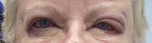
Dr. Neigel, a cosmetic surgeon in West Orange, N.J., said that over the past 5 years, she has seen increasing numbers of patients present with eczema localized to the eyelids or eyeball area that recurs like pesky crabgrass.
"I treat them with topical steroids," Dr. Neigel said. "It will get better, but it always comes back. Some of this is seasonal. It may only happen in the winter, when the air is drier and their skin tends to get drier. In others it can be all year long," she said. "It seems to be more common in women, but I see men with this condition, too. In men, it tends to present as a reddish eye and tearing," she noted.
In the majority of cases, the culprit turns out to be an allergy to chemicals including gold, nickel, tin, rubber, preservatives in shampoos and laundry detergent, and formaldehyde resin, which is used in nail polish. "There was one patient who was allergic to the preservative in eyedrops," Dr. Neigel recalled. "She was on several different eyedrops trying to treat the swollen eye area, and it was just making the condition worse."
Another patient’s eczema cleared only after she removed her wedding ring, Dr. Neigel said. "So she couldn’t wear any gold jewelry. In somebody else it was tin and nickel, so she couldn’t wear any cheap jewelry."
Ointments commonly used for cosmetic procedures also can cause trouble. "There is cross-reactivity between neomycin, tobramycin, and Neosporin," Dr. Neigel said. "One patient was applying Neosporin every time she bumped herself on different parts of her body, and her eyelids were the only things flaring up."
Dr. Neigel speculated that the reaction in such cases is localized to the eyelid because "it’s the thinnest skin in the body. It’s the most sensitive, and for some reason, the patients I’m seeing only have reactions there," she noted. So, for patients with allergic conjunctivitis or tearing for a contact dermatitis–type presentation around the eyeball or the eyelids, send them for chemical testing, she advised. "There’s a good chance you might clear things up and figure out what they’re truly reacting to – get to the source instead of just treating the problem symptomatically," she said.
Dr. Neigel said she had no relevant financial disclosures.
LAS VEGAS – If a patient presents with eczema of the eyelids, or swollen eyelids that don’t respond to topical steroids, think about sending them for chemical testing, advised Dr. Janet M. Neigel.
"The eyelids are red and scaly, a little swollen, and it just never goes away," she said in an interview at the annual meeting of the American Academy of Cosmetic Surgery.
Dr. Neigel, a cosmetic surgeon in West Orange, N.J., said that over the past 5 years, she has seen increasing numbers of patients present with eczema localized to the eyelids or eyeball area that recurs like pesky crabgrass.
"I treat them with topical steroids," Dr. Neigel said. "It will get better, but it always comes back. Some of this is seasonal. It may only happen in the winter, when the air is drier and their skin tends to get drier. In others it can be all year long," she said. "It seems to be more common in women, but I see men with this condition, too. In men, it tends to present as a reddish eye and tearing," she noted.
In the majority of cases, the culprit turns out to be an allergy to chemicals including gold, nickel, tin, rubber, preservatives in shampoos and laundry detergent, and formaldehyde resin, which is used in nail polish. "There was one patient who was allergic to the preservative in eyedrops," Dr. Neigel recalled. "She was on several different eyedrops trying to treat the swollen eye area, and it was just making the condition worse."
Another patient’s eczema cleared only after she removed her wedding ring, Dr. Neigel said. "So she couldn’t wear any gold jewelry. In somebody else it was tin and nickel, so she couldn’t wear any cheap jewelry."
Ointments commonly used for cosmetic procedures also can cause trouble. "There is cross-reactivity between neomycin, tobramycin, and Neosporin," Dr. Neigel said. "One patient was applying Neosporin every time she bumped herself on different parts of her body, and her eyelids were the only things flaring up."
Dr. Neigel speculated that the reaction in such cases is localized to the eyelid because "it’s the thinnest skin in the body. It’s the most sensitive, and for some reason, the patients I’m seeing only have reactions there," she noted. So, for patients with allergic conjunctivitis or tearing for a contact dermatitis–type presentation around the eyeball or the eyelids, send them for chemical testing, she advised. "There’s a good chance you might clear things up and figure out what they’re truly reacting to – get to the source instead of just treating the problem symptomatically," she said.
Dr. Neigel said she had no relevant financial disclosures.
AT THE AACS ANNUAL MEETING
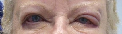
Fillers a popular option for periorbital rejuvenation
LAS VEGAS – Periorbital fillers are an attractive option to the myriad of surgical face-lift procedures available to patients seeking skin rejuvenation, according to Dr. Janet M. Neigel.
"The advantages to using fillers for periorbital rejuvenation are that they are a lunchtime treatment, there’s minimal risk, it results in a natural appearance, it’s minimally invasive, and it’s a good nonsurgical alternative, especially if a patient is not ready for surgery," Dr. Neigel said at the annual meeting of the American Academy of Cosmetic Surgery. "It’s also a great way to get to know your patients before they undergo a bigger procedure."

According to statistics from the American Society of Plastic Surgeons, clinicians performed 12.2 million nonsurgical cosmetic surgical procedures in the United States in 2011. Of these, 1.9 million were fillers.
"With aging there are certain structural changes to the face, including dermal thinning, fat loss, prolapse, redistribution, and skeletal bone remodeling," said Dr. Neigel, a West Orange, N.J.–based cosmetic surgeon who specializes in plastic and reconstructive surgery of the eyes and face. "The natural oval, upside down egg shape of the face changes more to a peanut-shaped face."
Prior to using any fillers, Dr. Neigel said she examines the intended treatment area of skin for hyperpigmentation and points out the relevant spots to patients. "Sometimes patients think that the use of fillers is going to make them have normal skin," she said. "With fillers you’ll improve the shadows, but you’re not going to be able to improve the hyperpigmentation."
Her go-to products for periorbital rejuvenation include the hyaluronic acids Juvederm, Restylane, and Belotero as well as the stimulatory fillers Sculptra (poly-l-lactic acid) and Radiesse (calcium hydroxylapatite). "Crow’s feet are difficult to deal with if they don’t respond completely to your neuromodulator of choice," Dr. Neigel noted. "Botox, Dysport, or Xeomin are the first injections of choice, but I have recently been using Belotero and have been very impressed with how it can improve the eyelid area. It’s used superficially in a layered manner."
When treating the lower-lid tear trough Dr. Neigel said she makes it a point to inject deep just over the periosteum and more inferiorly on the cheek. "Many times if you just inject above the orbital rim, you’re not going to address the full defect," she said. "Oftentimes you need to inject in multiple tissue planes. I use Restylane and Belotero around the eyes, and I inject in a retrograde fashion. I’m always nervous that Juvederm is going to cause too much water to be brought in and too much swelling. I reserve Juvederm for the lower face."
When treating the midface region, Dr. Neigel said that she most often uses Sculptra. "The goal in this area is to volumize the deep medial cheek fat, try to get anterior projection of the cheek and effacement of the lower-lid bags," she said. "We’re reflating the tissue, fighting some gravity, and filling Ristow’s space."
In her clinical experience, the most common complications from using stimulator fillers are ecchymosis and edema. "I have not seen any infections," she said. "I have seen several granulomas, papules, and nodules. If you get lumpiness, you can massage and most of the time it goes away, or you can use hyaluronidase. You can get a Tyndall effect, but I have not seen it in this area. You can also get overcorrection or undercorrection."
When nodules occur, "most of the time they’re not visible," she added. "You can palpate them, but as long as they’re not visible I leave them be."
Dr. Neigel disclosed that she is a consultant for Allergan, Medicis, and Valeant.
LAS VEGAS – Periorbital fillers are an attractive option to the myriad of surgical face-lift procedures available to patients seeking skin rejuvenation, according to Dr. Janet M. Neigel.
"The advantages to using fillers for periorbital rejuvenation are that they are a lunchtime treatment, there’s minimal risk, it results in a natural appearance, it’s minimally invasive, and it’s a good nonsurgical alternative, especially if a patient is not ready for surgery," Dr. Neigel said at the annual meeting of the American Academy of Cosmetic Surgery. "It’s also a great way to get to know your patients before they undergo a bigger procedure."
According to statistics from the American Society of Plastic Surgeons, clinicians performed 12.2 million nonsurgical cosmetic surgical procedures in the United States in 2011. Of these, 1.9 million were fillers.
"With aging there are certain structural changes to the face, including dermal thinning, fat loss, prolapse, redistribution, and skeletal bone remodeling," said Dr. Neigel, a West Orange, N.J.–based cosmetic surgeon who specializes in plastic and reconstructive surgery of the eyes and face. "The natural oval, upside down egg shape of the face changes more to a peanut-shaped face."
Prior to using any fillers, Dr. Neigel said she examines the intended treatment area of skin for hyperpigmentation and points out the relevant spots to patients. "Sometimes patients think that the use of fillers is going to make them have normal skin," she said. "With fillers you’ll improve the shadows, but you’re not going to be able to improve the hyperpigmentation."
Her go-to products for periorbital rejuvenation include the hyaluronic acids Juvederm, Restylane, and Belotero as well as the stimulatory fillers Sculptra (poly-l-lactic acid) and Radiesse (calcium hydroxylapatite). "Crow’s feet are difficult to deal with if they don’t respond completely to your neuromodulator of choice," Dr. Neigel noted. "Botox, Dysport, or Xeomin are the first injections of choice, but I have recently been using Belotero and have been very impressed with how it can improve the eyelid area. It’s used superficially in a layered manner."
When treating the lower-lid tear trough Dr. Neigel said she makes it a point to inject deep just over the periosteum and more inferiorly on the cheek. "Many times if you just inject above the orbital rim, you’re not going to address the full defect," she said. "Oftentimes you need to inject in multiple tissue planes. I use Restylane and Belotero around the eyes, and I inject in a retrograde fashion. I’m always nervous that Juvederm is going to cause too much water to be brought in and too much swelling. I reserve Juvederm for the lower face."
When treating the midface region, Dr. Neigel said that she most often uses Sculptra. "The goal in this area is to volumize the deep medial cheek fat, try to get anterior projection of the cheek and effacement of the lower-lid bags," she said. "We’re reflating the tissue, fighting some gravity, and filling Ristow’s space."
In her clinical experience, the most common complications from using stimulator fillers are ecchymosis and edema. "I have not seen any infections," she said. "I have seen several granulomas, papules, and nodules. If you get lumpiness, you can massage and most of the time it goes away, or you can use hyaluronidase. You can get a Tyndall effect, but I have not seen it in this area. You can also get overcorrection or undercorrection."
When nodules occur, "most of the time they’re not visible," she added. "You can palpate them, but as long as they’re not visible I leave them be."
Dr. Neigel disclosed that she is a consultant for Allergan, Medicis, and Valeant.
EXPERT ANALYSIS FROM THE AACS ANNUAL MEETING

Technology transforming the practice of psychiatry
KAUAI, HAWAII – The way Dr. James (Jay) H. Shore sees it, current advances in technology are transforming the practice of psychiatry more than at any other time in history.
"The information age holds this beautiful promise of crossing the quality chasm and getting to a patient-centered health care where we’re moving from centralization to redistributing the health care to where the patient is," he said at the annual meeting of the American College of Psychiatrists. "I would say that it is a promise that has yet to be achieved."

One of the most important advances over the past 20 years, he said, has been the maturation of convergence science – the concept of bringing disparate scientific disciplines and fields together to work on problems. "I think psychiatry has been on the leading edge of convergence science in the fact that we often turned to the humanities in the beginning of our field to find models and conceptualizations for how the brain works," explained Dr. Shore, an associate professor in the department of psychiatry at the University of Colorado Denver Anschutz Medical Campus. "Nowadays, teams of engineers, mathematicians, and bioscientists are working more closely with physicians."
He discussed three examples of ways in which convergence science is helping clinicians to better understand brain mechanisms. The first involves reconstructing visual experiences from brain activity evoked by natural movies. The effort, spearheaded by researchers at the University of California, Berkeley, and described in the journal Current Biology, uses blood oxygen level–dependent signals measured by functional magnetic resonance imaging to measure brain activity in subjects who viewed natural movie scenes (Current Biol. 2011;21:1641-6). "These subjects are having their visual lobes read by functional MRI connected to a computer program that is attempting to reconstruct what the MRI is reading in their brain," said Dr. Shore, who was not involved with the study. "The computer is drawing off of a database of 18 million images. Basically, what you have is a computer reading someone’s mind as they look at a visual stimulus."
The second technology he discussed is a brain-computer interface known as the BrainGate system, an investigational device that allows people with physical disabilities to increase their independence by controlling a computer with their thoughts. Clinical study participants undergo a surgical procedure where a sensor smaller than a contact lens is placed on the area of the brain responsible for movement. The sensor records electrical signals from the brain. These signals feed into a device, where they are interpreted by an operator using special software. To date, clinical studies have involved the use of BrainGate to check e-mail, change the channels of a television, and use a personal computer, said Dr. Shore, who is also with the Centers for American Indian and Alaska Native Health at the University of Colorado.
The third example of convergence science Dr. Shore discussed is the Blue Brain Project, a half-billion-dollar effort based in Switzerland and intended to construct a complete virtual human brain. "This is not an exercise in artificial intelligence, which uses software to mimic call and response," Dr. Shore said. "They started with a computer simulation model of a single neuron. By 2014, they are on target to have a fully functional artificial rat brain, and by 2023 they propose to have a fully functional human brain computer simulation. This project is really pushing us to understand mechanistically the relationships and networks within the brain."
In the current clinical realm, a variety of technologies are being used for patient care each day, including video conferencing, which "is part of standard psychiatric practice," Dr. Shore said. "The current trend is using smaller devices. I have colleagues who are providing care to patients through video conferencing on their mobile devices and computer tablets. That’s a mature application of technology."
He predicted that mobile telephone technology will drive future models of health care delivery. This is already playing out, he said, noting that an estimated 3,000 to 4,000 of the iPhone’s 13,000 apps are dedicated to some aspect of mental health care. "There are no good studies on these apps yet," Dr. Shore said. "From feasibility studies, we know that patients and providers are using them, but we have no idea how, or how they affect outcomes. It’s kind of like the Wild West out there with apps."
In another development, Dr. Shore discussed work occurring at the Telemedicine and Advanced Technology Research Center (TATRC), an office of the headquarters of the U.S. Army Medical Research and Materiel Command. TATRC has a research program bringing together academia and experts in the video game industry to create a video game "that will passively do neuropsychiatric screening assessment and tracking of the subject, things like reaction time, executive function, and task-shifting, for patients with deployment-related mental health issues such as traumatic brain injury or PTSD [post-traumatic stress disorder]," Dr. Shore said.
A separate group led by Dr. Peter Yellowlees at the University of California, Davis, has developed a virtual simulation of hallucinations intended to give patients, family members, students, and the general public a better understanding of schizophrenia.
"In clinical practice, there is a huge gap between technologies we’ve adapted and used, and the data that support them," he pointed out. "I don’t think this is any different from other fields of medicine. We’re seeing an increasing combination of technologies often being driven by our patients rather than by ourselves. We’re all going to be grappling with information overload.
"With all of these different ways to interact with patients and get information about their clinical status, the increasing ratio of signal to noise and how we sort this out is going to be a challenge."
One thing’s for sure, though: There is no slowing down advances in technology. "In a good way, for all of medicine these technologies are shifting the fulcrum and putting control of the care, information about the care, and guidance of the care back in the hands of our patients so they can be more engaged and active partners," Dr. Shore said. "I think we’ll continue to see this."
Dr. Shore said he had no relevant financial conflicts to disclose.
KAUAI, HAWAII – The way Dr. James (Jay) H. Shore sees it, current advances in technology are transforming the practice of psychiatry more than at any other time in history.
"The information age holds this beautiful promise of crossing the quality chasm and getting to a patient-centered health care where we’re moving from centralization to redistributing the health care to where the patient is," he said at the annual meeting of the American College of Psychiatrists. "I would say that it is a promise that has yet to be achieved."
One of the most important advances over the past 20 years, he said, has been the maturation of convergence science – the concept of bringing disparate scientific disciplines and fields together to work on problems. "I think psychiatry has been on the leading edge of convergence science in the fact that we often turned to the humanities in the beginning of our field to find models and conceptualizations for how the brain works," explained Dr. Shore, an associate professor in the department of psychiatry at the University of Colorado Denver Anschutz Medical Campus. "Nowadays, teams of engineers, mathematicians, and bioscientists are working more closely with physicians."
He discussed three examples of ways in which convergence science is helping clinicians to better understand brain mechanisms. The first involves reconstructing visual experiences from brain activity evoked by natural movies. The effort, spearheaded by researchers at the University of California, Berkeley, and described in the journal Current Biology, uses blood oxygen level–dependent signals measured by functional magnetic resonance imaging to measure brain activity in subjects who viewed natural movie scenes (Current Biol. 2011;21:1641-6). "These subjects are having their visual lobes read by functional MRI connected to a computer program that is attempting to reconstruct what the MRI is reading in their brain," said Dr. Shore, who was not involved with the study. "The computer is drawing off of a database of 18 million images. Basically, what you have is a computer reading someone’s mind as they look at a visual stimulus."
The second technology he discussed is a brain-computer interface known as the BrainGate system, an investigational device that allows people with physical disabilities to increase their independence by controlling a computer with their thoughts. Clinical study participants undergo a surgical procedure where a sensor smaller than a contact lens is placed on the area of the brain responsible for movement. The sensor records electrical signals from the brain. These signals feed into a device, where they are interpreted by an operator using special software. To date, clinical studies have involved the use of BrainGate to check e-mail, change the channels of a television, and use a personal computer, said Dr. Shore, who is also with the Centers for American Indian and Alaska Native Health at the University of Colorado.
The third example of convergence science Dr. Shore discussed is the Blue Brain Project, a half-billion-dollar effort based in Switzerland and intended to construct a complete virtual human brain. "This is not an exercise in artificial intelligence, which uses software to mimic call and response," Dr. Shore said. "They started with a computer simulation model of a single neuron. By 2014, they are on target to have a fully functional artificial rat brain, and by 2023 they propose to have a fully functional human brain computer simulation. This project is really pushing us to understand mechanistically the relationships and networks within the brain."
In the current clinical realm, a variety of technologies are being used for patient care each day, including video conferencing, which "is part of standard psychiatric practice," Dr. Shore said. "The current trend is using smaller devices. I have colleagues who are providing care to patients through video conferencing on their mobile devices and computer tablets. That’s a mature application of technology."
He predicted that mobile telephone technology will drive future models of health care delivery. This is already playing out, he said, noting that an estimated 3,000 to 4,000 of the iPhone’s 13,000 apps are dedicated to some aspect of mental health care. "There are no good studies on these apps yet," Dr. Shore said. "From feasibility studies, we know that patients and providers are using them, but we have no idea how, or how they affect outcomes. It’s kind of like the Wild West out there with apps."
In another development, Dr. Shore discussed work occurring at the Telemedicine and Advanced Technology Research Center (TATRC), an office of the headquarters of the U.S. Army Medical Research and Materiel Command. TATRC has a research program bringing together academia and experts in the video game industry to create a video game "that will passively do neuropsychiatric screening assessment and tracking of the subject, things like reaction time, executive function, and task-shifting, for patients with deployment-related mental health issues such as traumatic brain injury or PTSD [post-traumatic stress disorder]," Dr. Shore said.
A separate group led by Dr. Peter Yellowlees at the University of California, Davis, has developed a virtual simulation of hallucinations intended to give patients, family members, students, and the general public a better understanding of schizophrenia.
"In clinical practice, there is a huge gap between technologies we’ve adapted and used, and the data that support them," he pointed out. "I don’t think this is any different from other fields of medicine. We’re seeing an increasing combination of technologies often being driven by our patients rather than by ourselves. We’re all going to be grappling with information overload.
"With all of these different ways to interact with patients and get information about their clinical status, the increasing ratio of signal to noise and how we sort this out is going to be a challenge."
One thing’s for sure, though: There is no slowing down advances in technology. "In a good way, for all of medicine these technologies are shifting the fulcrum and putting control of the care, information about the care, and guidance of the care back in the hands of our patients so they can be more engaged and active partners," Dr. Shore said. "I think we’ll continue to see this."
Dr. Shore said he had no relevant financial conflicts to disclose.
EXPERT ANALYSIS FROM THE AMERICAN COLLEGE OF PSYCHIATRISTS ANNUAL MEETING