User login
FDA removes partial clinical hold on imetelstat
The US Food and Drug Administration (FDA) has removed the partial clinical hold on the investigator-sponsored trial of imetelstat in myelofibrosis, which the agency imposed in March.
The partial hold was placed because of a safety signal of liver toxicity in myelofibrosis patients receiving the drug.
The principal investigator of the trial, Ayalew Tefferi, MD, of Mayo Clinic in Rochester, Minnesota, provided follow-up information to the FDA regarding the reversibility of the hepatotoxicity.
Upon review of the additional data, the FDA allowed the myelofibrosis trial to proceed.
Imetelstat is a lipid-conjugated oligonucleotide that binds with high affinity to the RNA template of telomerase, thereby inhibiting telomerase activity.
Most cancers are characterized by short telomeres and a high level of telomerase activity, which makes telomerase a rational target for the treatment of cancer.
Imetelstat is also being investigated in essential thrombocythemia and other myeloid hematologic malignancies.
The trial in myelofibrosis enrolled 33 high-risk or intermediate-2 risk patients with either primary or secondary myelofibrosis. Dr Teferi presented the data at the annual meeting of the American Society of Hematology in 2013 as abstract 662.
At the time of the meeting, myelosuppression appeared to be the major dose-limiting toxicity.
Geron’s Investigational New Drug application to the FDA for imetelstat remains on full clinical hold. This affects clinical trials in essential thrombocythemia, polycythemia vera, and multiple myeloma. 
The US Food and Drug Administration (FDA) has removed the partial clinical hold on the investigator-sponsored trial of imetelstat in myelofibrosis, which the agency imposed in March.
The partial hold was placed because of a safety signal of liver toxicity in myelofibrosis patients receiving the drug.
The principal investigator of the trial, Ayalew Tefferi, MD, of Mayo Clinic in Rochester, Minnesota, provided follow-up information to the FDA regarding the reversibility of the hepatotoxicity.
Upon review of the additional data, the FDA allowed the myelofibrosis trial to proceed.
Imetelstat is a lipid-conjugated oligonucleotide that binds with high affinity to the RNA template of telomerase, thereby inhibiting telomerase activity.
Most cancers are characterized by short telomeres and a high level of telomerase activity, which makes telomerase a rational target for the treatment of cancer.
Imetelstat is also being investigated in essential thrombocythemia and other myeloid hematologic malignancies.
The trial in myelofibrosis enrolled 33 high-risk or intermediate-2 risk patients with either primary or secondary myelofibrosis. Dr Teferi presented the data at the annual meeting of the American Society of Hematology in 2013 as abstract 662.
At the time of the meeting, myelosuppression appeared to be the major dose-limiting toxicity.
Geron’s Investigational New Drug application to the FDA for imetelstat remains on full clinical hold. This affects clinical trials in essential thrombocythemia, polycythemia vera, and multiple myeloma.
The US Food and Drug Administration (FDA) has removed the partial clinical hold on the investigator-sponsored trial of imetelstat in myelofibrosis, which the agency imposed in March.
The partial hold was placed because of a safety signal of liver toxicity in myelofibrosis patients receiving the drug.
The principal investigator of the trial, Ayalew Tefferi, MD, of Mayo Clinic in Rochester, Minnesota, provided follow-up information to the FDA regarding the reversibility of the hepatotoxicity.
Upon review of the additional data, the FDA allowed the myelofibrosis trial to proceed.
Imetelstat is a lipid-conjugated oligonucleotide that binds with high affinity to the RNA template of telomerase, thereby inhibiting telomerase activity.
Most cancers are characterized by short telomeres and a high level of telomerase activity, which makes telomerase a rational target for the treatment of cancer.
Imetelstat is also being investigated in essential thrombocythemia and other myeloid hematologic malignancies.
The trial in myelofibrosis enrolled 33 high-risk or intermediate-2 risk patients with either primary or secondary myelofibrosis. Dr Teferi presented the data at the annual meeting of the American Society of Hematology in 2013 as abstract 662.
At the time of the meeting, myelosuppression appeared to be the major dose-limiting toxicity.
Geron’s Investigational New Drug application to the FDA for imetelstat remains on full clinical hold. This affects clinical trials in essential thrombocythemia, polycythemia vera, and multiple myeloma.
CAR T-cell therapy successfully used frontline as consolidation in CLL

©ASCO/Todd Buchanan
CHICAGO—Infusion of autologous CD19-targeted chimeric antigen receptor (CAR)-modified T cells appears to have promising anti-tumor activity and be well-tolerated as a consolidation to frontline chemotherapy in patients with high-risk chronic lymphocytic leukemia (CLL), researchers report.
In a phase 1 trial, the modified T cells benefitted 43% of patients, including 1 who achieved a complete response and 2 who achieved marrow responses only.
Jae Park, MD, of Memorial Sloan-Kettering Cancer Center in New York, reported the findings at the 2014 ASCO Annual Meeting as abstract 7020.
Dr Park and colleagues enrolled 7 CLL patients who had detectable minimal residual disease after achieving either a partial or complete response following 6 cycles of pentostatin, cyclophosphamide, and rituximab.
Patients then received cyclophosphamide conditioning therapy, followed by 3 escalating doses of CAR T cells in 3 dose cohorts.
Six patients had unmutated IgHV, and 2 patients had del 11q. Four patients had palpable lymphadenopathy, including 1 patient with bulky lymph nodes, prior to T-cell infusion.
After a median follow-up of 11 months, 1 patient achieved a complete response, and 2 patients achieved complete responses in the bone marrow but had progressive disease in the lymph nodes.
Three patients achieved a partial response, and 1 patient had progressive disease, but this patient had rapidly rising absolute lymphocyte count at the time of T-cell infusion, Dr Park noted.
The investigators observed no dose-limiting toxicity. Mild and self-limiting cytokine release syndrome occurred in 3 patients.
“There was a positive correlation between the development of cytokine release syndrome and the CAR T-cell persistence,” Dr Park said.
“Our findings suggest that the CD19-targeted CAR T cells are more effective in eradicating disease in the marrow versus lymph nodes,” he added. “And further studies are being conducted to better understand the mechanism of resistance.”
ASCO discussant Veronika Bachanova, MD, of the University of Minnesota, noted that all previous studies of CAR T-cell therapy in CLL were conducted with relapsed/refractory patients.
“The novelty of this study,” she commented, “is the use of CAR T cells upfront.”
She noted that it can take as long as 8 months to develop the CAR T cells for therapy, adding that “the vector design is of critical importance, since inhibitory signals influence therapy.”
©ASCO/Todd Buchanan
CHICAGO—Infusion of autologous CD19-targeted chimeric antigen receptor (CAR)-modified T cells appears to have promising anti-tumor activity and be well-tolerated as a consolidation to frontline chemotherapy in patients with high-risk chronic lymphocytic leukemia (CLL), researchers report.
In a phase 1 trial, the modified T cells benefitted 43% of patients, including 1 who achieved a complete response and 2 who achieved marrow responses only.
Jae Park, MD, of Memorial Sloan-Kettering Cancer Center in New York, reported the findings at the 2014 ASCO Annual Meeting as abstract 7020.
Dr Park and colleagues enrolled 7 CLL patients who had detectable minimal residual disease after achieving either a partial or complete response following 6 cycles of pentostatin, cyclophosphamide, and rituximab.
Patients then received cyclophosphamide conditioning therapy, followed by 3 escalating doses of CAR T cells in 3 dose cohorts.
Six patients had unmutated IgHV, and 2 patients had del 11q. Four patients had palpable lymphadenopathy, including 1 patient with bulky lymph nodes, prior to T-cell infusion.
After a median follow-up of 11 months, 1 patient achieved a complete response, and 2 patients achieved complete responses in the bone marrow but had progressive disease in the lymph nodes.
Three patients achieved a partial response, and 1 patient had progressive disease, but this patient had rapidly rising absolute lymphocyte count at the time of T-cell infusion, Dr Park noted.
The investigators observed no dose-limiting toxicity. Mild and self-limiting cytokine release syndrome occurred in 3 patients.
“There was a positive correlation between the development of cytokine release syndrome and the CAR T-cell persistence,” Dr Park said.
“Our findings suggest that the CD19-targeted CAR T cells are more effective in eradicating disease in the marrow versus lymph nodes,” he added. “And further studies are being conducted to better understand the mechanism of resistance.”
ASCO discussant Veronika Bachanova, MD, of the University of Minnesota, noted that all previous studies of CAR T-cell therapy in CLL were conducted with relapsed/refractory patients.
“The novelty of this study,” she commented, “is the use of CAR T cells upfront.”
She noted that it can take as long as 8 months to develop the CAR T cells for therapy, adding that “the vector design is of critical importance, since inhibitory signals influence therapy.”
©ASCO/Todd Buchanan
CHICAGO—Infusion of autologous CD19-targeted chimeric antigen receptor (CAR)-modified T cells appears to have promising anti-tumor activity and be well-tolerated as a consolidation to frontline chemotherapy in patients with high-risk chronic lymphocytic leukemia (CLL), researchers report.
In a phase 1 trial, the modified T cells benefitted 43% of patients, including 1 who achieved a complete response and 2 who achieved marrow responses only.
Jae Park, MD, of Memorial Sloan-Kettering Cancer Center in New York, reported the findings at the 2014 ASCO Annual Meeting as abstract 7020.
Dr Park and colleagues enrolled 7 CLL patients who had detectable minimal residual disease after achieving either a partial or complete response following 6 cycles of pentostatin, cyclophosphamide, and rituximab.
Patients then received cyclophosphamide conditioning therapy, followed by 3 escalating doses of CAR T cells in 3 dose cohorts.
Six patients had unmutated IgHV, and 2 patients had del 11q. Four patients had palpable lymphadenopathy, including 1 patient with bulky lymph nodes, prior to T-cell infusion.
After a median follow-up of 11 months, 1 patient achieved a complete response, and 2 patients achieved complete responses in the bone marrow but had progressive disease in the lymph nodes.
Three patients achieved a partial response, and 1 patient had progressive disease, but this patient had rapidly rising absolute lymphocyte count at the time of T-cell infusion, Dr Park noted.
The investigators observed no dose-limiting toxicity. Mild and self-limiting cytokine release syndrome occurred in 3 patients.
“There was a positive correlation between the development of cytokine release syndrome and the CAR T-cell persistence,” Dr Park said.
“Our findings suggest that the CD19-targeted CAR T cells are more effective in eradicating disease in the marrow versus lymph nodes,” he added. “And further studies are being conducted to better understand the mechanism of resistance.”
ASCO discussant Veronika Bachanova, MD, of the University of Minnesota, noted that all previous studies of CAR T-cell therapy in CLL were conducted with relapsed/refractory patients.
“The novelty of this study,” she commented, “is the use of CAR T cells upfront.”
She noted that it can take as long as 8 months to develop the CAR T cells for therapy, adding that “the vector design is of critical importance, since inhibitory signals influence therapy.”
Lenalidomide combination improves QOL in newly diagnosed MM

©ASCO/Scott Morgan
CHICAGO—Substituting lenalidomide for thalidomide in the standard treatment of newly diagnosed multiple myeloma (MM) improves quality of life and
lowers toxicity without significant loss of response, results of a phase 3 study suggest.
Combination melphalan, prednisone, and thalidomide (MPT) is considered a standard treatment option for transplant ineligible, newly diagnosed MM.
Early phase 1/2 studies suggested substituting lenalidomide for thalidomide might result in similar efficacy and less toxicity.
“However, neither feature can be confidently predicted, since long-term follow-up of the melphalan, prednisone, and lenalidomide (MPR) regimen is lacking, and myelosuppression may prove limiting, compromising drug dose and efficacy,” said A. Keith Stewart, MD, from the Mayo Clinic in Scottsdale, Arizona.
So Dr Stewart and his colleagues conducted a randomized, multicenter, phase 3 trial comparing MPT to MPR in untreated, symptomatic, transplant-ineligible MM patients. Dr Stewart presented the results at the 2014 ASCO Annual Meeting as abstract 8511.
The study included 306 patients with a median age of 76 years. The primary objective was to evaluate the difference in progression-free survival (PFS) between patients receiving MPT and those treated with MPR.
Patients received melphalan at 9 mg/m2 and prednisone at 100 mg orally on days 1-4, with thalidomide at 100 mg daily. Or they received melphalan at 5 mg/m2 and prednisone at 100 mg orally on days 1-4, with lenalidomide at 10 mg orally on days 1-21.
MPT or MPR therapy was continued for twelve 28-day cycles, followed by thalidomide at 100 mg or lenalidomide at 10 mg daily until relapse. Patients were required to have aspirin prophylaxis.
Secondary study objectives included overall survival, toxicities, response rates, depth of response, and quality of life change. Treatment arms were balanced for age, ISS stage, and other major prognostic factors.
The median follow-up was 40.7 months. The median time on therapy was 12 months overall and 23 months for the 46% of patients on maintenance therapy, with no differences by arm. Some 7% of patients remained on treatment through cycle 60.
The results showed similar response rates between the 2 arms. The partial response rate was 64% for the MPT group, compared with 60% for the MPR group, with no difference in very good partial response/complete response rates (18.8% vs 23%).
The median PFS was 21 months for patients receiving MPT and 18.7 months for those receiving MPR. The median overall survival was not significantly different between the arms—52.6% for the MPT arm and 47.7% for the MPR arm.
Toxicities of grade 3 or higher were significantly more likely in the MPT arm (73%) than in the MPR arm (58%), as was non-hematologic toxicity (59% and 40%, respectively).
The incidence of second primary malignancies was higher with MPT (3.47/100 person years) than with MPR (2.01/100 person years). And deep vein thrombosis or pulmonary embolism occurred in 8.8% of MPT patients and 6.7% MPR patients.
“The quality of life analysis favored MPR by induction end,” Dr Stewart said. “Response rates, PFS, and overall survival were similar between the 2 arms. However, there was significantly better quality of life at 12 months and lower toxicity with MPR.”
Dr Stewart noted, however, that in the US, “melphalan-based regimens are now seldom utilized due to availability of lenalidomide and bortezomib in newly diagnosed patients.”
©ASCO/Scott Morgan
CHICAGO—Substituting lenalidomide for thalidomide in the standard treatment of newly diagnosed multiple myeloma (MM) improves quality of life and
lowers toxicity without significant loss of response, results of a phase 3 study suggest.
Combination melphalan, prednisone, and thalidomide (MPT) is considered a standard treatment option for transplant ineligible, newly diagnosed MM.
Early phase 1/2 studies suggested substituting lenalidomide for thalidomide might result in similar efficacy and less toxicity.
“However, neither feature can be confidently predicted, since long-term follow-up of the melphalan, prednisone, and lenalidomide (MPR) regimen is lacking, and myelosuppression may prove limiting, compromising drug dose and efficacy,” said A. Keith Stewart, MD, from the Mayo Clinic in Scottsdale, Arizona.
So Dr Stewart and his colleagues conducted a randomized, multicenter, phase 3 trial comparing MPT to MPR in untreated, symptomatic, transplant-ineligible MM patients. Dr Stewart presented the results at the 2014 ASCO Annual Meeting as abstract 8511.
The study included 306 patients with a median age of 76 years. The primary objective was to evaluate the difference in progression-free survival (PFS) between patients receiving MPT and those treated with MPR.
Patients received melphalan at 9 mg/m2 and prednisone at 100 mg orally on days 1-4, with thalidomide at 100 mg daily. Or they received melphalan at 5 mg/m2 and prednisone at 100 mg orally on days 1-4, with lenalidomide at 10 mg orally on days 1-21.
MPT or MPR therapy was continued for twelve 28-day cycles, followed by thalidomide at 100 mg or lenalidomide at 10 mg daily until relapse. Patients were required to have aspirin prophylaxis.
Secondary study objectives included overall survival, toxicities, response rates, depth of response, and quality of life change. Treatment arms were balanced for age, ISS stage, and other major prognostic factors.
The median follow-up was 40.7 months. The median time on therapy was 12 months overall and 23 months for the 46% of patients on maintenance therapy, with no differences by arm. Some 7% of patients remained on treatment through cycle 60.
The results showed similar response rates between the 2 arms. The partial response rate was 64% for the MPT group, compared with 60% for the MPR group, with no difference in very good partial response/complete response rates (18.8% vs 23%).
The median PFS was 21 months for patients receiving MPT and 18.7 months for those receiving MPR. The median overall survival was not significantly different between the arms—52.6% for the MPT arm and 47.7% for the MPR arm.
Toxicities of grade 3 or higher were significantly more likely in the MPT arm (73%) than in the MPR arm (58%), as was non-hematologic toxicity (59% and 40%, respectively).
The incidence of second primary malignancies was higher with MPT (3.47/100 person years) than with MPR (2.01/100 person years). And deep vein thrombosis or pulmonary embolism occurred in 8.8% of MPT patients and 6.7% MPR patients.
“The quality of life analysis favored MPR by induction end,” Dr Stewart said. “Response rates, PFS, and overall survival were similar between the 2 arms. However, there was significantly better quality of life at 12 months and lower toxicity with MPR.”
Dr Stewart noted, however, that in the US, “melphalan-based regimens are now seldom utilized due to availability of lenalidomide and bortezomib in newly diagnosed patients.”
©ASCO/Scott Morgan
CHICAGO—Substituting lenalidomide for thalidomide in the standard treatment of newly diagnosed multiple myeloma (MM) improves quality of life and
lowers toxicity without significant loss of response, results of a phase 3 study suggest.
Combination melphalan, prednisone, and thalidomide (MPT) is considered a standard treatment option for transplant ineligible, newly diagnosed MM.
Early phase 1/2 studies suggested substituting lenalidomide for thalidomide might result in similar efficacy and less toxicity.
“However, neither feature can be confidently predicted, since long-term follow-up of the melphalan, prednisone, and lenalidomide (MPR) regimen is lacking, and myelosuppression may prove limiting, compromising drug dose and efficacy,” said A. Keith Stewart, MD, from the Mayo Clinic in Scottsdale, Arizona.
So Dr Stewart and his colleagues conducted a randomized, multicenter, phase 3 trial comparing MPT to MPR in untreated, symptomatic, transplant-ineligible MM patients. Dr Stewart presented the results at the 2014 ASCO Annual Meeting as abstract 8511.
The study included 306 patients with a median age of 76 years. The primary objective was to evaluate the difference in progression-free survival (PFS) between patients receiving MPT and those treated with MPR.
Patients received melphalan at 9 mg/m2 and prednisone at 100 mg orally on days 1-4, with thalidomide at 100 mg daily. Or they received melphalan at 5 mg/m2 and prednisone at 100 mg orally on days 1-4, with lenalidomide at 10 mg orally on days 1-21.
MPT or MPR therapy was continued for twelve 28-day cycles, followed by thalidomide at 100 mg or lenalidomide at 10 mg daily until relapse. Patients were required to have aspirin prophylaxis.
Secondary study objectives included overall survival, toxicities, response rates, depth of response, and quality of life change. Treatment arms were balanced for age, ISS stage, and other major prognostic factors.
The median follow-up was 40.7 months. The median time on therapy was 12 months overall and 23 months for the 46% of patients on maintenance therapy, with no differences by arm. Some 7% of patients remained on treatment through cycle 60.
The results showed similar response rates between the 2 arms. The partial response rate was 64% for the MPT group, compared with 60% for the MPR group, with no difference in very good partial response/complete response rates (18.8% vs 23%).
The median PFS was 21 months for patients receiving MPT and 18.7 months for those receiving MPR. The median overall survival was not significantly different between the arms—52.6% for the MPT arm and 47.7% for the MPR arm.
Toxicities of grade 3 or higher were significantly more likely in the MPT arm (73%) than in the MPR arm (58%), as was non-hematologic toxicity (59% and 40%, respectively).
The incidence of second primary malignancies was higher with MPT (3.47/100 person years) than with MPR (2.01/100 person years). And deep vein thrombosis or pulmonary embolism occurred in 8.8% of MPT patients and 6.7% MPR patients.
“The quality of life analysis favored MPR by induction end,” Dr Stewart said. “Response rates, PFS, and overall survival were similar between the 2 arms. However, there was significantly better quality of life at 12 months and lower toxicity with MPR.”
Dr Stewart noted, however, that in the US, “melphalan-based regimens are now seldom utilized due to availability of lenalidomide and bortezomib in newly diagnosed patients.”
Novel agent shows promising activity in heavily pretreated NHL

©ASCO/Rodney White
CHICAGO—The novel, oral selective inhibitor of nuclear transport known as selinexor (KPT-330) can safely be given as monotherapy to patients with heavily pretreated non-Hodgkin lymphoma (NHL), according to a presentation at the 2014 ASCO Annual Meeting.
“Selinexor has favorable pharmacokinetic and pharmacodynamic characteristics,” said presenter Martin Gutierrez, MD, of the John Theurer Cancer Center at Hackensack University Medical Center in New Jersey.
Selinexor is a slowly reversible, selective inhibitor of nuclear transport that inhibits XPO1, which is elevated in NHL, chronic lymphocytic leukemia (CLL), and other malignances.
“It shows single-agent anti-tumor activity across all NHL types, with durable cancer control of more than 9 months [and] marked activity across germ cell B (GCB), non-germ cell B, and double-hit diffuse large B-cell lymphoma (DLBCL),” Dr Gutierrez said.
He provided an update of the ongoing phase 1 dose-escalation study at the meeting as abstract 8518.
The study now includes 51 patients, half of whom have NHL. Their median age is 60 years.
The patients received selinexor across 8 dose levels, ranging from 3 mg/m2 to 60 mg/m2. Dosing at 60 mg/m2 twice weekly is ongoing, and the maximum-tolerated dose has not been reached.
Among the 43 evaluable patients, the disease control rate was 74%, the overall response rate was 28%, and the complete response rate was 5%.
“All patients who had their disease controlled had a reduction in lymph nodes and some degree of activity across all dose levels,” Dr Gutierrez said. “GCB and non-GCB patients responded similarly.”
The length of response was up to 632 days in the DLBCL group.
Most adverse events were gastrointestinal in nature, and most of them were grade 1 or 2. The most common adverse events were nausea, anorexia, and fatigue.
The investigators found a higher incidence of side effects in the first treatment cycle. The dosing schedule was interrupted and reduced to maintain steady state levels.
“The results suggest that an intermittent dosing schedule optimally induces a steady state with maximal induction of XPO1 mRNA,” Dr Gutierrez said.
There were 3 dose-limiting toxicities, including 1 multiple myeloma patient with grade 4 thrombocytopenia, 1 follicular lymphoma patient with grade 4 thrombocytopenia, and 1 CLL patient with grade 2 fatigue.
ASCO discussant Owen O’Connor, MD, from Columbia University Medical Center in New York, commented, “These clinical data are interesting with a provocative target. I applaud the investigators for doing a trial across a diversity of B- and T-cell lymphomas . . . . The results suggest a potential effect in a rare subset of lymphoma patients that have little treatment options.”
Frontline trials of selinexor are planned, including patients with Richter transformation and follicular lymphoma.
Selinexor recently received orphan drug status from the US Food and Drug Administration for the treatment of DLBCL.
©ASCO/Rodney White
CHICAGO—The novel, oral selective inhibitor of nuclear transport known as selinexor (KPT-330) can safely be given as monotherapy to patients with heavily pretreated non-Hodgkin lymphoma (NHL), according to a presentation at the 2014 ASCO Annual Meeting.
“Selinexor has favorable pharmacokinetic and pharmacodynamic characteristics,” said presenter Martin Gutierrez, MD, of the John Theurer Cancer Center at Hackensack University Medical Center in New Jersey.
Selinexor is a slowly reversible, selective inhibitor of nuclear transport that inhibits XPO1, which is elevated in NHL, chronic lymphocytic leukemia (CLL), and other malignances.
“It shows single-agent anti-tumor activity across all NHL types, with durable cancer control of more than 9 months [and] marked activity across germ cell B (GCB), non-germ cell B, and double-hit diffuse large B-cell lymphoma (DLBCL),” Dr Gutierrez said.
He provided an update of the ongoing phase 1 dose-escalation study at the meeting as abstract 8518.
The study now includes 51 patients, half of whom have NHL. Their median age is 60 years.
The patients received selinexor across 8 dose levels, ranging from 3 mg/m2 to 60 mg/m2. Dosing at 60 mg/m2 twice weekly is ongoing, and the maximum-tolerated dose has not been reached.
Among the 43 evaluable patients, the disease control rate was 74%, the overall response rate was 28%, and the complete response rate was 5%.
“All patients who had their disease controlled had a reduction in lymph nodes and some degree of activity across all dose levels,” Dr Gutierrez said. “GCB and non-GCB patients responded similarly.”
The length of response was up to 632 days in the DLBCL group.
Most adverse events were gastrointestinal in nature, and most of them were grade 1 or 2. The most common adverse events were nausea, anorexia, and fatigue.
The investigators found a higher incidence of side effects in the first treatment cycle. The dosing schedule was interrupted and reduced to maintain steady state levels.
“The results suggest that an intermittent dosing schedule optimally induces a steady state with maximal induction of XPO1 mRNA,” Dr Gutierrez said.
There were 3 dose-limiting toxicities, including 1 multiple myeloma patient with grade 4 thrombocytopenia, 1 follicular lymphoma patient with grade 4 thrombocytopenia, and 1 CLL patient with grade 2 fatigue.
ASCO discussant Owen O’Connor, MD, from Columbia University Medical Center in New York, commented, “These clinical data are interesting with a provocative target. I applaud the investigators for doing a trial across a diversity of B- and T-cell lymphomas . . . . The results suggest a potential effect in a rare subset of lymphoma patients that have little treatment options.”
Frontline trials of selinexor are planned, including patients with Richter transformation and follicular lymphoma.
Selinexor recently received orphan drug status from the US Food and Drug Administration for the treatment of DLBCL.
©ASCO/Rodney White
CHICAGO—The novel, oral selective inhibitor of nuclear transport known as selinexor (KPT-330) can safely be given as monotherapy to patients with heavily pretreated non-Hodgkin lymphoma (NHL), according to a presentation at the 2014 ASCO Annual Meeting.
“Selinexor has favorable pharmacokinetic and pharmacodynamic characteristics,” said presenter Martin Gutierrez, MD, of the John Theurer Cancer Center at Hackensack University Medical Center in New Jersey.
Selinexor is a slowly reversible, selective inhibitor of nuclear transport that inhibits XPO1, which is elevated in NHL, chronic lymphocytic leukemia (CLL), and other malignances.
“It shows single-agent anti-tumor activity across all NHL types, with durable cancer control of more than 9 months [and] marked activity across germ cell B (GCB), non-germ cell B, and double-hit diffuse large B-cell lymphoma (DLBCL),” Dr Gutierrez said.
He provided an update of the ongoing phase 1 dose-escalation study at the meeting as abstract 8518.
The study now includes 51 patients, half of whom have NHL. Their median age is 60 years.
The patients received selinexor across 8 dose levels, ranging from 3 mg/m2 to 60 mg/m2. Dosing at 60 mg/m2 twice weekly is ongoing, and the maximum-tolerated dose has not been reached.
Among the 43 evaluable patients, the disease control rate was 74%, the overall response rate was 28%, and the complete response rate was 5%.
“All patients who had their disease controlled had a reduction in lymph nodes and some degree of activity across all dose levels,” Dr Gutierrez said. “GCB and non-GCB patients responded similarly.”
The length of response was up to 632 days in the DLBCL group.
Most adverse events were gastrointestinal in nature, and most of them were grade 1 or 2. The most common adverse events were nausea, anorexia, and fatigue.
The investigators found a higher incidence of side effects in the first treatment cycle. The dosing schedule was interrupted and reduced to maintain steady state levels.
“The results suggest that an intermittent dosing schedule optimally induces a steady state with maximal induction of XPO1 mRNA,” Dr Gutierrez said.
There were 3 dose-limiting toxicities, including 1 multiple myeloma patient with grade 4 thrombocytopenia, 1 follicular lymphoma patient with grade 4 thrombocytopenia, and 1 CLL patient with grade 2 fatigue.
ASCO discussant Owen O’Connor, MD, from Columbia University Medical Center in New York, commented, “These clinical data are interesting with a provocative target. I applaud the investigators for doing a trial across a diversity of B- and T-cell lymphomas . . . . The results suggest a potential effect in a rare subset of lymphoma patients that have little treatment options.”
Frontline trials of selinexor are planned, including patients with Richter transformation and follicular lymphoma.
Selinexor recently received orphan drug status from the US Food and Drug Administration for the treatment of DLBCL.
Method allows for high-resolution cell imaging

filaments (red), microtubules
(green), and nuclei (blue)
National Institutes of Health
A new technique allows scientists to view the cell cytoskeleton with “unprecedented resolution,” according to a paper published in Nature Methods.
A group of researchers exploited the properties of a fluorescent molecule they developed to generate 2 probes for imaging the cytoskeleton.
The team said these probes can easily enter live cells, are non-toxic, have long-lasting signals, and offer better image resolution than current imaging techniques.
This research began when Kai Johnsson, PhD, of École Polytechnique Fédérale de Lausanne in Switzerland, and his colleagues developed a fluorescent molecule called silicon-rhodamine (SiR).
The molecule switches “on” only when it binds to the charged surface of a protein like those found on the cytoskeleton. When SiR switches on, it emits light at far-red wavelengths.
The challenge was getting SiR to bind specifically to the cytoskeleton’s proteins, actin and tubulin. To achieve this, the researchers fused SiR molecules with compounds that bind tubulin or actin.
The resulting hybrid molecules consist of a SiR molecule, which provides the fluorescent signal, and a molecule of a natural compound that can bind the target protein.
One such compound was docetaxel, an anticancer drug that binds tubulin, and the other was jasplakinolide, which specifically binds the cytoskeletal form of actin. Both compounds, which are used here in very low, non-toxic concentrations, can easily pass through the cell’s membrane and into the cell itself.
The probes, called SiR-tubulin and SiR-actin, were used to visualize the dynamics of the cytoskeleton in human skin cells. Because the light signal of the probes is emitted in the far-red spectrum, it is easy to isolate from background noise, which generates images of unprecedented resolution when used with super-resolution microscopy.
An additional advantage of the probes, according to the researchers, is their practicality.
“You just add them directly into your cell culture, and they are taken up by the cells,” Dr Johnsson said.
The probes don’t require any washing or preparation of the cells before administration or any subsequent washing steps, which helps in maintaining the stability of their environment and their natural biological functions.
“Up to now, no probes were available that would allow you to get high-quality images of microtubules and microfilaments in living cells without some kind of genetic modification,” Dr Johnsson said.
“With this work, we provide the biological community with 2 high-performing, high-contrast fluorogenic probes that emit in the non-phototoxic part of the light spectrum, and can be even used in tissues like whole-blood samples.”
filaments (red), microtubules
(green), and nuclei (blue)
National Institutes of Health
A new technique allows scientists to view the cell cytoskeleton with “unprecedented resolution,” according to a paper published in Nature Methods.
A group of researchers exploited the properties of a fluorescent molecule they developed to generate 2 probes for imaging the cytoskeleton.
The team said these probes can easily enter live cells, are non-toxic, have long-lasting signals, and offer better image resolution than current imaging techniques.
This research began when Kai Johnsson, PhD, of École Polytechnique Fédérale de Lausanne in Switzerland, and his colleagues developed a fluorescent molecule called silicon-rhodamine (SiR).
The molecule switches “on” only when it binds to the charged surface of a protein like those found on the cytoskeleton. When SiR switches on, it emits light at far-red wavelengths.
The challenge was getting SiR to bind specifically to the cytoskeleton’s proteins, actin and tubulin. To achieve this, the researchers fused SiR molecules with compounds that bind tubulin or actin.
The resulting hybrid molecules consist of a SiR molecule, which provides the fluorescent signal, and a molecule of a natural compound that can bind the target protein.
One such compound was docetaxel, an anticancer drug that binds tubulin, and the other was jasplakinolide, which specifically binds the cytoskeletal form of actin. Both compounds, which are used here in very low, non-toxic concentrations, can easily pass through the cell’s membrane and into the cell itself.
The probes, called SiR-tubulin and SiR-actin, were used to visualize the dynamics of the cytoskeleton in human skin cells. Because the light signal of the probes is emitted in the far-red spectrum, it is easy to isolate from background noise, which generates images of unprecedented resolution when used with super-resolution microscopy.
An additional advantage of the probes, according to the researchers, is their practicality.
“You just add them directly into your cell culture, and they are taken up by the cells,” Dr Johnsson said.
The probes don’t require any washing or preparation of the cells before administration or any subsequent washing steps, which helps in maintaining the stability of their environment and their natural biological functions.
“Up to now, no probes were available that would allow you to get high-quality images of microtubules and microfilaments in living cells without some kind of genetic modification,” Dr Johnsson said.
“With this work, we provide the biological community with 2 high-performing, high-contrast fluorogenic probes that emit in the non-phototoxic part of the light spectrum, and can be even used in tissues like whole-blood samples.”
filaments (red), microtubules
(green), and nuclei (blue)
National Institutes of Health
A new technique allows scientists to view the cell cytoskeleton with “unprecedented resolution,” according to a paper published in Nature Methods.
A group of researchers exploited the properties of a fluorescent molecule they developed to generate 2 probes for imaging the cytoskeleton.
The team said these probes can easily enter live cells, are non-toxic, have long-lasting signals, and offer better image resolution than current imaging techniques.
This research began when Kai Johnsson, PhD, of École Polytechnique Fédérale de Lausanne in Switzerland, and his colleagues developed a fluorescent molecule called silicon-rhodamine (SiR).
The molecule switches “on” only when it binds to the charged surface of a protein like those found on the cytoskeleton. When SiR switches on, it emits light at far-red wavelengths.
The challenge was getting SiR to bind specifically to the cytoskeleton’s proteins, actin and tubulin. To achieve this, the researchers fused SiR molecules with compounds that bind tubulin or actin.
The resulting hybrid molecules consist of a SiR molecule, which provides the fluorescent signal, and a molecule of a natural compound that can bind the target protein.
One such compound was docetaxel, an anticancer drug that binds tubulin, and the other was jasplakinolide, which specifically binds the cytoskeletal form of actin. Both compounds, which are used here in very low, non-toxic concentrations, can easily pass through the cell’s membrane and into the cell itself.
The probes, called SiR-tubulin and SiR-actin, were used to visualize the dynamics of the cytoskeleton in human skin cells. Because the light signal of the probes is emitted in the far-red spectrum, it is easy to isolate from background noise, which generates images of unprecedented resolution when used with super-resolution microscopy.
An additional advantage of the probes, according to the researchers, is their practicality.
“You just add them directly into your cell culture, and they are taken up by the cells,” Dr Johnsson said.
The probes don’t require any washing or preparation of the cells before administration or any subsequent washing steps, which helps in maintaining the stability of their environment and their natural biological functions.
“Up to now, no probes were available that would allow you to get high-quality images of microtubules and microfilaments in living cells without some kind of genetic modification,” Dr Johnsson said.
“With this work, we provide the biological community with 2 high-performing, high-contrast fluorogenic probes that emit in the non-phototoxic part of the light spectrum, and can be even used in tissues like whole-blood samples.”
FDA approves first antihemophilic Fc fusion protein for hemophilia A

The US Food and Drug Administration (FDA) has approved the recombinant factor VIII Fc fusion protein efmoroctocog alfa (Eloctate) to treat children and adults with hemophilia A.
Efmoroctocog alfa is intended to reduce the frequency of injections required to prevent or reduce the occurrence of bleeding in patients with hemophilia A.
The FDA approved efmoroctocog alfa based on results of the phase 3 A-LONG trial, which were reported last year.
The trial included 165 male patients age 12 or older.
Patients who received individualized prophylaxis with efmoroctocog alfa every 3 to 5 days reduced their annualized bleeding rate by 92% compared with patients who received episodic treatment.
Patients who received weekly prophylaxis with efmoroctocog alfa reduced their annualized bleeding rate by 76% compared to patients who received episodic treatment.
Overall, 1 injection of efmoroctocog alfa resolved 87.3% of bleeding episodes.
None of the patients in the study developed neutralizing antibodies, and there were no serious adverse events related to efmoroctocog alfa.
The study was published online in Blood last November.
Efmoroctocog alfa consists of the coagulation factor VIII molecule linked to an Fc protein fragment found in antibodies, which confers the product with a longer half-life than recombinant factor VIII.
Efmoroctocog alfa is specifically indicated for the control and prevention of bleeding episodes, the management of bleeding during surgical procedures, and prophylaxis against bleeding episodes.
The approval of efmoroctocog alfa fills an unmet medical need, according to Johnny Mahlangu, MD, of the Haemophilia Comprehensive Care Centre at the University of Witwatersrand in Johannesburg, South Africa.
He explained that the fusion protein allows patients with hemophilia A to go for longer intervals between prophylactic infusions while maintaining good control of bleeding episodes.
The product, which received orphan drug designation by the FDA, is manufactured by Biogen Idec, Inc, of Cambridge, Massachusetts.
Full prescribing information is available on the drug website. 
The US Food and Drug Administration (FDA) has approved the recombinant factor VIII Fc fusion protein efmoroctocog alfa (Eloctate) to treat children and adults with hemophilia A.
Efmoroctocog alfa is intended to reduce the frequency of injections required to prevent or reduce the occurrence of bleeding in patients with hemophilia A.
The FDA approved efmoroctocog alfa based on results of the phase 3 A-LONG trial, which were reported last year.
The trial included 165 male patients age 12 or older.
Patients who received individualized prophylaxis with efmoroctocog alfa every 3 to 5 days reduced their annualized bleeding rate by 92% compared with patients who received episodic treatment.
Patients who received weekly prophylaxis with efmoroctocog alfa reduced their annualized bleeding rate by 76% compared to patients who received episodic treatment.
Overall, 1 injection of efmoroctocog alfa resolved 87.3% of bleeding episodes.
None of the patients in the study developed neutralizing antibodies, and there were no serious adverse events related to efmoroctocog alfa.
The study was published online in Blood last November.
Efmoroctocog alfa consists of the coagulation factor VIII molecule linked to an Fc protein fragment found in antibodies, which confers the product with a longer half-life than recombinant factor VIII.
Efmoroctocog alfa is specifically indicated for the control and prevention of bleeding episodes, the management of bleeding during surgical procedures, and prophylaxis against bleeding episodes.
The approval of efmoroctocog alfa fills an unmet medical need, according to Johnny Mahlangu, MD, of the Haemophilia Comprehensive Care Centre at the University of Witwatersrand in Johannesburg, South Africa.
He explained that the fusion protein allows patients with hemophilia A to go for longer intervals between prophylactic infusions while maintaining good control of bleeding episodes.
The product, which received orphan drug designation by the FDA, is manufactured by Biogen Idec, Inc, of Cambridge, Massachusetts.
Full prescribing information is available on the drug website.
The US Food and Drug Administration (FDA) has approved the recombinant factor VIII Fc fusion protein efmoroctocog alfa (Eloctate) to treat children and adults with hemophilia A.
Efmoroctocog alfa is intended to reduce the frequency of injections required to prevent or reduce the occurrence of bleeding in patients with hemophilia A.
The FDA approved efmoroctocog alfa based on results of the phase 3 A-LONG trial, which were reported last year.
The trial included 165 male patients age 12 or older.
Patients who received individualized prophylaxis with efmoroctocog alfa every 3 to 5 days reduced their annualized bleeding rate by 92% compared with patients who received episodic treatment.
Patients who received weekly prophylaxis with efmoroctocog alfa reduced their annualized bleeding rate by 76% compared to patients who received episodic treatment.
Overall, 1 injection of efmoroctocog alfa resolved 87.3% of bleeding episodes.
None of the patients in the study developed neutralizing antibodies, and there were no serious adverse events related to efmoroctocog alfa.
The study was published online in Blood last November.
Efmoroctocog alfa consists of the coagulation factor VIII molecule linked to an Fc protein fragment found in antibodies, which confers the product with a longer half-life than recombinant factor VIII.
Efmoroctocog alfa is specifically indicated for the control and prevention of bleeding episodes, the management of bleeding during surgical procedures, and prophylaxis against bleeding episodes.
The approval of efmoroctocog alfa fills an unmet medical need, according to Johnny Mahlangu, MD, of the Haemophilia Comprehensive Care Centre at the University of Witwatersrand in Johannesburg, South Africa.
He explained that the fusion protein allows patients with hemophilia A to go for longer intervals between prophylactic infusions while maintaining good control of bleeding episodes.
The product, which received orphan drug designation by the FDA, is manufactured by Biogen Idec, Inc, of Cambridge, Massachusetts.
Full prescribing information is available on the drug website.
Voluntary recall of eculizumab issued

Credit: Globovision
The manufacturer of the recently approved eculizumab (Soliris) issued a voluntary US recall on June 2 of a single affected lot of the drug, although it included 8 additional lots in the recall.
The manufacturer, Alexion Pharmaceuticals, found visible particulates in the 300 mg/30 mL concentrated solution for intravenous infusion, which could cause an immune reaction and blood clots in patients receiving the drug.
The single affected Soliris lot, distributed in the US only, is #1007A. Also included in the U.S. recall are lots 10002-1, 00006-1, 10003A, 10004A, 10005A, 10005AR, 10006A, and 10008A.
All lots in the recall were manufactured using the same process component.
Alexion believes it has identified the part of the process that has resulted in the visible particles in the solution and has changed the process component.
An earlier recall of eculizumab, also for particulate matter, occurred in December 2013.
Alexion does not anticipate any interruption to the patient supply of eculizumab.
Eculizumab recently received full US Food and Drug Administration (FDA) approval to treat adult and pediatric patients with atypical hemolytic uremic syndrome (aHUS). It had received accelerated approval for this indication in 2011.
Eculizumab is also FDA-approved to treat patients with paroxysmal nocturnal hemoglobinuria.
For more information on the recall, visit the company website.
Healthcare professionals and patients should report adverse events or side effects related to Soliris to the FDA's MedWatch Safety Information and Adverse Event Reporting Program.
Credit: Globovision
The manufacturer of the recently approved eculizumab (Soliris) issued a voluntary US recall on June 2 of a single affected lot of the drug, although it included 8 additional lots in the recall.
The manufacturer, Alexion Pharmaceuticals, found visible particulates in the 300 mg/30 mL concentrated solution for intravenous infusion, which could cause an immune reaction and blood clots in patients receiving the drug.
The single affected Soliris lot, distributed in the US only, is #1007A. Also included in the U.S. recall are lots 10002-1, 00006-1, 10003A, 10004A, 10005A, 10005AR, 10006A, and 10008A.
All lots in the recall were manufactured using the same process component.
Alexion believes it has identified the part of the process that has resulted in the visible particles in the solution and has changed the process component.
An earlier recall of eculizumab, also for particulate matter, occurred in December 2013.
Alexion does not anticipate any interruption to the patient supply of eculizumab.
Eculizumab recently received full US Food and Drug Administration (FDA) approval to treat adult and pediatric patients with atypical hemolytic uremic syndrome (aHUS). It had received accelerated approval for this indication in 2011.
Eculizumab is also FDA-approved to treat patients with paroxysmal nocturnal hemoglobinuria.
For more information on the recall, visit the company website.
Healthcare professionals and patients should report adverse events or side effects related to Soliris to the FDA's MedWatch Safety Information and Adverse Event Reporting Program.
Credit: Globovision
The manufacturer of the recently approved eculizumab (Soliris) issued a voluntary US recall on June 2 of a single affected lot of the drug, although it included 8 additional lots in the recall.
The manufacturer, Alexion Pharmaceuticals, found visible particulates in the 300 mg/30 mL concentrated solution for intravenous infusion, which could cause an immune reaction and blood clots in patients receiving the drug.
The single affected Soliris lot, distributed in the US only, is #1007A. Also included in the U.S. recall are lots 10002-1, 00006-1, 10003A, 10004A, 10005A, 10005AR, 10006A, and 10008A.
All lots in the recall were manufactured using the same process component.
Alexion believes it has identified the part of the process that has resulted in the visible particles in the solution and has changed the process component.
An earlier recall of eculizumab, also for particulate matter, occurred in December 2013.
Alexion does not anticipate any interruption to the patient supply of eculizumab.
Eculizumab recently received full US Food and Drug Administration (FDA) approval to treat adult and pediatric patients with atypical hemolytic uremic syndrome (aHUS). It had received accelerated approval for this indication in 2011.
Eculizumab is also FDA-approved to treat patients with paroxysmal nocturnal hemoglobinuria.
For more information on the recall, visit the company website.
Healthcare professionals and patients should report adverse events or side effects related to Soliris to the FDA's MedWatch Safety Information and Adverse Event Reporting Program.
Research reveals previously unidentified proteins
Credit: Lu Wei
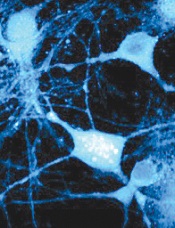
By cataloging more than 18,000 proteins, scientists have produced an “almost-complete” inventory of the human proteome.
They discovered protein fragments encoded by DNA outside of currently known genes and found that many known genes have become non-functional.
They also described an important function for messenger RNA (mRNA) and showed that protein profiles could predict drug sensitivity.
The team reported their findings in Nature.
They also made the information freely available in ProteomicsDB. This database includes information on the types, distribution, and abundance of proteins in various cells, tissues, and body fluids.
Through their protein cataloguing, the researchers showed that each mRNA determines the number of protein copies to be produced by a cell. And this “copying key” is specific to each protein.
“It appears that every mRNA molecule knows the unit amount for its protein so it knows whether to produce 10, 100, or 1000 copies,” said study author Bernhard Küster, PhD, of Technische Universitaet Muenchen in Germany.
“Since we now know this ratio for a large number of proteins, we can infer protein abundance from mRNA abundance in just about every tissue, and vice versa.”
The researchers were also surprised to discover hundreds of protein fragments that are encoded by DNA outside of currently known genes. The team believes these “new” proteins may possess novel biological properties and functions that could be exploited for therapeutic purposes.
On the other hand, Dr Küster and his colleagues have been unable to locate roughly 2000 proteins that should exist, according to the gene map. The team also found evidence suggesting that many known genes have become non-functional.
“We might be watching evolution in action here,” Dr Küster said. “The human organism deactivates superfluous genes and tests new gene prototypes at the same time.”
That being the case, the researchers noted that it might never be possible to determine exactly how many proteins there are in the human body.
Lastly, Dr Küster and his colleagues confirmed the findings of previous studies, which showed that specific protein patterns can predict the efficacy of a given drug.
The team evaluated 24 cancer drugs and found their effectiveness against 35 cancer cell lines was strongly correlated with their protein profiles.
“This edges us a little bit closer to the individualized treatment of patients,” Dr Küster said. “If we knew the protein profile of a tumor in detail, we might be able to administer drugs in a more targeted way.”
Credit: Lu Wei
By cataloging more than 18,000 proteins, scientists have produced an “almost-complete” inventory of the human proteome.
They discovered protein fragments encoded by DNA outside of currently known genes and found that many known genes have become non-functional.
They also described an important function for messenger RNA (mRNA) and showed that protein profiles could predict drug sensitivity.
The team reported their findings in Nature.
They also made the information freely available in ProteomicsDB. This database includes information on the types, distribution, and abundance of proteins in various cells, tissues, and body fluids.
Through their protein cataloguing, the researchers showed that each mRNA determines the number of protein copies to be produced by a cell. And this “copying key” is specific to each protein.
“It appears that every mRNA molecule knows the unit amount for its protein so it knows whether to produce 10, 100, or 1000 copies,” said study author Bernhard Küster, PhD, of Technische Universitaet Muenchen in Germany.
“Since we now know this ratio for a large number of proteins, we can infer protein abundance from mRNA abundance in just about every tissue, and vice versa.”
The researchers were also surprised to discover hundreds of protein fragments that are encoded by DNA outside of currently known genes. The team believes these “new” proteins may possess novel biological properties and functions that could be exploited for therapeutic purposes.
On the other hand, Dr Küster and his colleagues have been unable to locate roughly 2000 proteins that should exist, according to the gene map. The team also found evidence suggesting that many known genes have become non-functional.
“We might be watching evolution in action here,” Dr Küster said. “The human organism deactivates superfluous genes and tests new gene prototypes at the same time.”
That being the case, the researchers noted that it might never be possible to determine exactly how many proteins there are in the human body.
Lastly, Dr Küster and his colleagues confirmed the findings of previous studies, which showed that specific protein patterns can predict the efficacy of a given drug.
The team evaluated 24 cancer drugs and found their effectiveness against 35 cancer cell lines was strongly correlated with their protein profiles.
“This edges us a little bit closer to the individualized treatment of patients,” Dr Küster said. “If we knew the protein profile of a tumor in detail, we might be able to administer drugs in a more targeted way.”
Credit: Lu Wei
By cataloging more than 18,000 proteins, scientists have produced an “almost-complete” inventory of the human proteome.
They discovered protein fragments encoded by DNA outside of currently known genes and found that many known genes have become non-functional.
They also described an important function for messenger RNA (mRNA) and showed that protein profiles could predict drug sensitivity.
The team reported their findings in Nature.
They also made the information freely available in ProteomicsDB. This database includes information on the types, distribution, and abundance of proteins in various cells, tissues, and body fluids.
Through their protein cataloguing, the researchers showed that each mRNA determines the number of protein copies to be produced by a cell. And this “copying key” is specific to each protein.
“It appears that every mRNA molecule knows the unit amount for its protein so it knows whether to produce 10, 100, or 1000 copies,” said study author Bernhard Küster, PhD, of Technische Universitaet Muenchen in Germany.
“Since we now know this ratio for a large number of proteins, we can infer protein abundance from mRNA abundance in just about every tissue, and vice versa.”
The researchers were also surprised to discover hundreds of protein fragments that are encoded by DNA outside of currently known genes. The team believes these “new” proteins may possess novel biological properties and functions that could be exploited for therapeutic purposes.
On the other hand, Dr Küster and his colleagues have been unable to locate roughly 2000 proteins that should exist, according to the gene map. The team also found evidence suggesting that many known genes have become non-functional.
“We might be watching evolution in action here,” Dr Küster said. “The human organism deactivates superfluous genes and tests new gene prototypes at the same time.”
That being the case, the researchers noted that it might never be possible to determine exactly how many proteins there are in the human body.
Lastly, Dr Küster and his colleagues confirmed the findings of previous studies, which showed that specific protein patterns can predict the efficacy of a given drug.
The team evaluated 24 cancer drugs and found their effectiveness against 35 cancer cell lines was strongly correlated with their protein profiles.
“This edges us a little bit closer to the individualized treatment of patients,” Dr Küster said. “If we knew the protein profile of a tumor in detail, we might be able to administer drugs in a more targeted way.”
‘Nanodaisies’ deliver drug cocktail to leukemia cells
Credit: PNAS
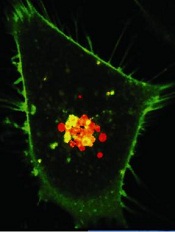
Biomedical engineers have reported that daisy-shaped, nanoscale structures can deliver a cocktail of drugs directly to cancer cells.
The “nanodaisies” effectively delivered a 2-drug combination in a range of cell lines, including the leukemia cell line HL-60.
The drug-delivery vehicles also proved effective in a mouse model of lung cancer.
Zhen Gu, PhD, of North Carolina State University and the University of North Carolina at Chapel Hill, and his colleagues detailed these results in Biomaterials.
“We found that this technique was much better than conventional drug-delivery techniques at inhibiting the growth of lung cancer tumors in mice,” Dr Gu said.
“And based on in vitro tests in 9 different cell lines, the technique is also promising for use against leukemia, breast, prostate, liver, ovarian, and brain cancers.”
To make the “nanodaisies,” the researchers begin with a solution that contains a polymer called polyethylene glycol (PEG). The PEG forms long strands that have much shorter strands branching off to either side.
The researchers directly link the anticancer drug camptothecin (CPT) onto the shorter strands and introduce the anticancer drug doxorubicin (Dox) into the solution.
PEG is hydrophilic, but CPT and Dox are hydrophobic. As a result, the CPT and Dox cluster together in the solution, wrapping the PEG around themselves. This results in a daisy-shaped drug cocktail, only 50 nanometers in diameter, that can (in theory) be injected into a cancer patient.
Once injected, the nanodaisies float through the bloodstream until they are absorbed by cancer cells. In fact, one of the reasons the researchers chose to use PEG is because it has chemical properties that prolong the life of the drugs in the bloodstream. Once in a cancer cell, the drugs are released.
“Both drugs attack the cell’s nucleus but via different mechanisms,” said study author Wanyi Tai, PhD, who was previously a researcher in Dr Gu’s lab but is now at the University of Washington in Seattle.
“Combined, the drugs are more effective than either drug is by itself,” Dr Gu added. “We are very optimistic about this technique and are hoping to begin preclinical testing in the near future.”
Credit: PNAS
Biomedical engineers have reported that daisy-shaped, nanoscale structures can deliver a cocktail of drugs directly to cancer cells.
The “nanodaisies” effectively delivered a 2-drug combination in a range of cell lines, including the leukemia cell line HL-60.
The drug-delivery vehicles also proved effective in a mouse model of lung cancer.
Zhen Gu, PhD, of North Carolina State University and the University of North Carolina at Chapel Hill, and his colleagues detailed these results in Biomaterials.
“We found that this technique was much better than conventional drug-delivery techniques at inhibiting the growth of lung cancer tumors in mice,” Dr Gu said.
“And based on in vitro tests in 9 different cell lines, the technique is also promising for use against leukemia, breast, prostate, liver, ovarian, and brain cancers.”
To make the “nanodaisies,” the researchers begin with a solution that contains a polymer called polyethylene glycol (PEG). The PEG forms long strands that have much shorter strands branching off to either side.
The researchers directly link the anticancer drug camptothecin (CPT) onto the shorter strands and introduce the anticancer drug doxorubicin (Dox) into the solution.
PEG is hydrophilic, but CPT and Dox are hydrophobic. As a result, the CPT and Dox cluster together in the solution, wrapping the PEG around themselves. This results in a daisy-shaped drug cocktail, only 50 nanometers in diameter, that can (in theory) be injected into a cancer patient.
Once injected, the nanodaisies float through the bloodstream until they are absorbed by cancer cells. In fact, one of the reasons the researchers chose to use PEG is because it has chemical properties that prolong the life of the drugs in the bloodstream. Once in a cancer cell, the drugs are released.
“Both drugs attack the cell’s nucleus but via different mechanisms,” said study author Wanyi Tai, PhD, who was previously a researcher in Dr Gu’s lab but is now at the University of Washington in Seattle.
“Combined, the drugs are more effective than either drug is by itself,” Dr Gu added. “We are very optimistic about this technique and are hoping to begin preclinical testing in the near future.”
Credit: PNAS
Biomedical engineers have reported that daisy-shaped, nanoscale structures can deliver a cocktail of drugs directly to cancer cells.
The “nanodaisies” effectively delivered a 2-drug combination in a range of cell lines, including the leukemia cell line HL-60.
The drug-delivery vehicles also proved effective in a mouse model of lung cancer.
Zhen Gu, PhD, of North Carolina State University and the University of North Carolina at Chapel Hill, and his colleagues detailed these results in Biomaterials.
“We found that this technique was much better than conventional drug-delivery techniques at inhibiting the growth of lung cancer tumors in mice,” Dr Gu said.
“And based on in vitro tests in 9 different cell lines, the technique is also promising for use against leukemia, breast, prostate, liver, ovarian, and brain cancers.”
To make the “nanodaisies,” the researchers begin with a solution that contains a polymer called polyethylene glycol (PEG). The PEG forms long strands that have much shorter strands branching off to either side.
The researchers directly link the anticancer drug camptothecin (CPT) onto the shorter strands and introduce the anticancer drug doxorubicin (Dox) into the solution.
PEG is hydrophilic, but CPT and Dox are hydrophobic. As a result, the CPT and Dox cluster together in the solution, wrapping the PEG around themselves. This results in a daisy-shaped drug cocktail, only 50 nanometers in diameter, that can (in theory) be injected into a cancer patient.
Once injected, the nanodaisies float through the bloodstream until they are absorbed by cancer cells. In fact, one of the reasons the researchers chose to use PEG is because it has chemical properties that prolong the life of the drugs in the bloodstream. Once in a cancer cell, the drugs are released.
“Both drugs attack the cell’s nucleus but via different mechanisms,” said study author Wanyi Tai, PhD, who was previously a researcher in Dr Gu’s lab but is now at the University of Washington in Seattle.
“Combined, the drugs are more effective than either drug is by itself,” Dr Gu added. “We are very optimistic about this technique and are hoping to begin preclinical testing in the near future.”
Study elucidates enzyme’s role in cell survival
Credit: Egelberg
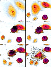
Investigators say they have solved the mystery of an enzyme’s role in cell survival, thereby offering clues as to how the immune system fights infection and pointing to possible strategies for treating cancers.
The enzyme, receptor-interacting protein kinase 1 (RIPK1), is known to play a pivotal role in survival after birth.
But the new research, published in Cell, reveals that RIPK1 inhibits the pathways that control apoptosis and necroptosis.
By removing different components of each pathway in different combinations, the investigators demonstrated that, after birth, RIPK1 helps cells maintain a balanced response to signals that promote either pathway.
“We are learning that, in disease, this balancing act can be perturbed to produce damage and cell death,” said study author Douglas Green, PhD, of the St Jude Children’s Research Hospital in Memphis, Tennessee.
The results resolve long-standing questions about RIPK1’s role in cell survival and provide clues about how the immune system might use these pathways to contain infections.
The findings have also prompted the researchers to launch an investigation into whether RIPK1 could be harnessed to kill cancer cells or provide insight into tumor development.
“This study fundamentally changes the way we think about RIPK1, a molecule that we care about because it is required for life,” Dr Green said. “The results helped us identify new pathways involved in regulating programmed cell death and suggest that we might be able to develop cancer therapies that target these pathways or engage them in other ways to advance treatment of a range of diseases.”
The report builds on previous research from Dr Green’s lab regarding regulation of the pathways that control apoptosis and necroptosis. The investigators knew that apoptosis is driven by the enzyme caspase-8, which forms a complex with FADD and other proteins.
And necroptosis involves a pathway orchestrated by the enzyme receptor-interacting protein kinase 3 (RIPK3). Before birth, RIPK1 works through RIPK3 to trigger cell death by necroptosis, but, until now, the enzyme’s primary role after birth was uncertain.
So the investigators bred mice lacking different combinations of genes for RIPK1, RIPK3, caspase-8, FADD and other components of both the apoptotic and necroptotic pathways.
Mice lacking RIPK1 died. Mice missing 2 genes—RIPK1 plus RIPK3 or RIPK1 plus caspase-8 or FADD—also died soon after birth.
However, mice survived and developed normally when the investigators removed 3 genes—RIPK1, RIPK3, and either caspase-8 or FADD.
“The fact that the mice survived was totally unexpected and made us rethink how these pathways work,” Dr Green said.
The results also demonstrated that other pathways must exist in cells to maintain a balanced response to signals pushing for cell death via apoptosis or necroptosis.
Evidence in this study, for example, suggested one possible new pathway that triggered necroptosis using interferon and other elements of the immune response to infections.
Credit: Egelberg
Investigators say they have solved the mystery of an enzyme’s role in cell survival, thereby offering clues as to how the immune system fights infection and pointing to possible strategies for treating cancers.
The enzyme, receptor-interacting protein kinase 1 (RIPK1), is known to play a pivotal role in survival after birth.
But the new research, published in Cell, reveals that RIPK1 inhibits the pathways that control apoptosis and necroptosis.
By removing different components of each pathway in different combinations, the investigators demonstrated that, after birth, RIPK1 helps cells maintain a balanced response to signals that promote either pathway.
“We are learning that, in disease, this balancing act can be perturbed to produce damage and cell death,” said study author Douglas Green, PhD, of the St Jude Children’s Research Hospital in Memphis, Tennessee.
The results resolve long-standing questions about RIPK1’s role in cell survival and provide clues about how the immune system might use these pathways to contain infections.
The findings have also prompted the researchers to launch an investigation into whether RIPK1 could be harnessed to kill cancer cells or provide insight into tumor development.
“This study fundamentally changes the way we think about RIPK1, a molecule that we care about because it is required for life,” Dr Green said. “The results helped us identify new pathways involved in regulating programmed cell death and suggest that we might be able to develop cancer therapies that target these pathways or engage them in other ways to advance treatment of a range of diseases.”
The report builds on previous research from Dr Green’s lab regarding regulation of the pathways that control apoptosis and necroptosis. The investigators knew that apoptosis is driven by the enzyme caspase-8, which forms a complex with FADD and other proteins.
And necroptosis involves a pathway orchestrated by the enzyme receptor-interacting protein kinase 3 (RIPK3). Before birth, RIPK1 works through RIPK3 to trigger cell death by necroptosis, but, until now, the enzyme’s primary role after birth was uncertain.
So the investigators bred mice lacking different combinations of genes for RIPK1, RIPK3, caspase-8, FADD and other components of both the apoptotic and necroptotic pathways.
Mice lacking RIPK1 died. Mice missing 2 genes—RIPK1 plus RIPK3 or RIPK1 plus caspase-8 or FADD—also died soon after birth.
However, mice survived and developed normally when the investigators removed 3 genes—RIPK1, RIPK3, and either caspase-8 or FADD.
“The fact that the mice survived was totally unexpected and made us rethink how these pathways work,” Dr Green said.
The results also demonstrated that other pathways must exist in cells to maintain a balanced response to signals pushing for cell death via apoptosis or necroptosis.
Evidence in this study, for example, suggested one possible new pathway that triggered necroptosis using interferon and other elements of the immune response to infections.
Credit: Egelberg
Investigators say they have solved the mystery of an enzyme’s role in cell survival, thereby offering clues as to how the immune system fights infection and pointing to possible strategies for treating cancers.
The enzyme, receptor-interacting protein kinase 1 (RIPK1), is known to play a pivotal role in survival after birth.
But the new research, published in Cell, reveals that RIPK1 inhibits the pathways that control apoptosis and necroptosis.
By removing different components of each pathway in different combinations, the investigators demonstrated that, after birth, RIPK1 helps cells maintain a balanced response to signals that promote either pathway.
“We are learning that, in disease, this balancing act can be perturbed to produce damage and cell death,” said study author Douglas Green, PhD, of the St Jude Children’s Research Hospital in Memphis, Tennessee.
The results resolve long-standing questions about RIPK1’s role in cell survival and provide clues about how the immune system might use these pathways to contain infections.
The findings have also prompted the researchers to launch an investigation into whether RIPK1 could be harnessed to kill cancer cells or provide insight into tumor development.
“This study fundamentally changes the way we think about RIPK1, a molecule that we care about because it is required for life,” Dr Green said. “The results helped us identify new pathways involved in regulating programmed cell death and suggest that we might be able to develop cancer therapies that target these pathways or engage them in other ways to advance treatment of a range of diseases.”
The report builds on previous research from Dr Green’s lab regarding regulation of the pathways that control apoptosis and necroptosis. The investigators knew that apoptosis is driven by the enzyme caspase-8, which forms a complex with FADD and other proteins.
And necroptosis involves a pathway orchestrated by the enzyme receptor-interacting protein kinase 3 (RIPK3). Before birth, RIPK1 works through RIPK3 to trigger cell death by necroptosis, but, until now, the enzyme’s primary role after birth was uncertain.
So the investigators bred mice lacking different combinations of genes for RIPK1, RIPK3, caspase-8, FADD and other components of both the apoptotic and necroptotic pathways.
Mice lacking RIPK1 died. Mice missing 2 genes—RIPK1 plus RIPK3 or RIPK1 plus caspase-8 or FADD—also died soon after birth.
However, mice survived and developed normally when the investigators removed 3 genes—RIPK1, RIPK3, and either caspase-8 or FADD.
“The fact that the mice survived was totally unexpected and made us rethink how these pathways work,” Dr Green said.
The results also demonstrated that other pathways must exist in cells to maintain a balanced response to signals pushing for cell death via apoptosis or necroptosis.
Evidence in this study, for example, suggested one possible new pathway that triggered necroptosis using interferon and other elements of the immune response to infections.