User login
Gut bacteria may help predict survival after allo-SCT

The diversity of gut microbiota in patients receiving allogeneic stem cell transplants (allo-SCTs) may be an important predictor of their survival, according to a study published in Blood.
Previous studies have suggested the intensive treatment given to allo-SCT recipients can destroy a significant portion of their gut microbiota and reduce its overall diversity.
And disturbances of the gut microbiota have been associated with complications such as bloodstream infections and graft-vs-host disease.
“While the link between gut microbiota and complications in allogeneic SCT has been previously established, until this point, it has remained unclear whether the gut bacteria of transplant recipients could predict their survival,” said study author Ying Taur, MD, of Memorial Sloan-Kettering Cancer Center in New York.
“This study sought to further explore the potential connection between transplantation, gut bacteria, and overall survival.”
To that end, the researchers collected fecal specimens from 80 patients undergoing allo-SCT and sequenced each sample’s bacterial DNA. Specimens were collected within 7 days of engraftment, the point at which the researchers speculated that microbiota diversity would be greatest following pre-transplant conditioning.
The team compared patient outcomes based on diversity of microbiota in their specimens, grouping subjects into high-, intermediate-, and low-microbiota-diversity categories.
At engraftment, 34 patients (42.5%) had low diversity, 20 (25%) had intermediate diversity, and 26 (32.5%) had high diversity. The analysis continued for up to 3 years or until death or last follow-up.
Following their analyses, the researchers found a strong connection between post-transplant gut microbiota diversity and outcomes, observing overall survival rates of 36%, 60%, and 67% among the low-, intermediate-, and high-diversity groups, respectively.
Furthermore, diversity was particularly associated with transplant-related outcomes. Patients with low microbiota diversity were approximately 5 times more likely to die of transplant-related causes within the follow-up period than those with more diverse gut bacteria.
“These results further underscore the significance of the gut microbiota in allogeneic stem cell transplant,” Dr Taur said. “A major question is whether we can improve outcomes by preserving diversity within the gut microbiota.”
“One possible strategy is to find ways to perform transplants in a manner that minimizes damage to the gut microbiota. Another approach would be to replenish the gut with beneficial microbes that are lost after this procedure is performed. We hope that this study will inspire additional research that will further examine the role and importance of the gut microbiota to stem cell transplant outcome.” 
The diversity of gut microbiota in patients receiving allogeneic stem cell transplants (allo-SCTs) may be an important predictor of their survival, according to a study published in Blood.
Previous studies have suggested the intensive treatment given to allo-SCT recipients can destroy a significant portion of their gut microbiota and reduce its overall diversity.
And disturbances of the gut microbiota have been associated with complications such as bloodstream infections and graft-vs-host disease.
“While the link between gut microbiota and complications in allogeneic SCT has been previously established, until this point, it has remained unclear whether the gut bacteria of transplant recipients could predict their survival,” said study author Ying Taur, MD, of Memorial Sloan-Kettering Cancer Center in New York.
“This study sought to further explore the potential connection between transplantation, gut bacteria, and overall survival.”
To that end, the researchers collected fecal specimens from 80 patients undergoing allo-SCT and sequenced each sample’s bacterial DNA. Specimens were collected within 7 days of engraftment, the point at which the researchers speculated that microbiota diversity would be greatest following pre-transplant conditioning.
The team compared patient outcomes based on diversity of microbiota in their specimens, grouping subjects into high-, intermediate-, and low-microbiota-diversity categories.
At engraftment, 34 patients (42.5%) had low diversity, 20 (25%) had intermediate diversity, and 26 (32.5%) had high diversity. The analysis continued for up to 3 years or until death or last follow-up.
Following their analyses, the researchers found a strong connection between post-transplant gut microbiota diversity and outcomes, observing overall survival rates of 36%, 60%, and 67% among the low-, intermediate-, and high-diversity groups, respectively.
Furthermore, diversity was particularly associated with transplant-related outcomes. Patients with low microbiota diversity were approximately 5 times more likely to die of transplant-related causes within the follow-up period than those with more diverse gut bacteria.
“These results further underscore the significance of the gut microbiota in allogeneic stem cell transplant,” Dr Taur said. “A major question is whether we can improve outcomes by preserving diversity within the gut microbiota.”
“One possible strategy is to find ways to perform transplants in a manner that minimizes damage to the gut microbiota. Another approach would be to replenish the gut with beneficial microbes that are lost after this procedure is performed. We hope that this study will inspire additional research that will further examine the role and importance of the gut microbiota to stem cell transplant outcome.”
The diversity of gut microbiota in patients receiving allogeneic stem cell transplants (allo-SCTs) may be an important predictor of their survival, according to a study published in Blood.
Previous studies have suggested the intensive treatment given to allo-SCT recipients can destroy a significant portion of their gut microbiota and reduce its overall diversity.
And disturbances of the gut microbiota have been associated with complications such as bloodstream infections and graft-vs-host disease.
“While the link between gut microbiota and complications in allogeneic SCT has been previously established, until this point, it has remained unclear whether the gut bacteria of transplant recipients could predict their survival,” said study author Ying Taur, MD, of Memorial Sloan-Kettering Cancer Center in New York.
“This study sought to further explore the potential connection between transplantation, gut bacteria, and overall survival.”
To that end, the researchers collected fecal specimens from 80 patients undergoing allo-SCT and sequenced each sample’s bacterial DNA. Specimens were collected within 7 days of engraftment, the point at which the researchers speculated that microbiota diversity would be greatest following pre-transplant conditioning.
The team compared patient outcomes based on diversity of microbiota in their specimens, grouping subjects into high-, intermediate-, and low-microbiota-diversity categories.
At engraftment, 34 patients (42.5%) had low diversity, 20 (25%) had intermediate diversity, and 26 (32.5%) had high diversity. The analysis continued for up to 3 years or until death or last follow-up.
Following their analyses, the researchers found a strong connection between post-transplant gut microbiota diversity and outcomes, observing overall survival rates of 36%, 60%, and 67% among the low-, intermediate-, and high-diversity groups, respectively.
Furthermore, diversity was particularly associated with transplant-related outcomes. Patients with low microbiota diversity were approximately 5 times more likely to die of transplant-related causes within the follow-up period than those with more diverse gut bacteria.
“These results further underscore the significance of the gut microbiota in allogeneic stem cell transplant,” Dr Taur said. “A major question is whether we can improve outcomes by preserving diversity within the gut microbiota.”
“One possible strategy is to find ways to perform transplants in a manner that minimizes damage to the gut microbiota. Another approach would be to replenish the gut with beneficial microbes that are lost after this procedure is performed. We hope that this study will inspire additional research that will further examine the role and importance of the gut microbiota to stem cell transplant outcome.”
CNS involvement predicts relapse but not survival in ARL, study shows
CHICAGO—Investigators have found evidence to suggest that identifying central nervous system (CNS) involvement at diagnosis does not impact overall survival for patients with AIDS-related lymphoma (ARL).
The research showed that ARL patients with CNS involvement at diagnosis were nearly 3 times as likely as their peers to have CNS relapse during cancer treatment.
But there was no significant difference between the 2 groups with regard to survival.
This may be due to the low overall incidence of CNS relapse, the use of insufficient treatments, and/or inadequate methods for identifying patients with CNS involvement, according to the investigators.
Stefan K. Barta, MD, of the Fox Chase Cancer Center in Philadelphia, Pennsylvania, and his colleagues conducted this research and presented the results at the 2014 ASCO Annual Meeting (abstract 8570).
In 2013, Dr Barta led the assembly of a database containing medical data from more than 1500 patients newly diagnosed with ARL who participated in clinical trials in Europe and the US from 1990 through 2010.
In the new study, he and his colleagues used the database to identify 880 patients with ARL whose data included complete information on CNS involvement at diagnosis and CNS relapse.
The team set out to find associations between CNS relapse and patient characteristics, including age, sex, CD4 count, lymphoma subtype, treatment history with combination antiretroviral therapies (cART), rituximab use, and the type of initial chemotherapy.
All of the patients had received either intrathecal therapy for CNS involvement or intrathecal prophylaxis with single-agent or 3-agent regimens. Sixty-nine percent of patients (n=607) had received cART, and 31% (n=276) had received rituximab-containing induction chemoimmunotherapy.
CNS involvement was identified in 13% of patients at diagnosis, including 27% of patients with Burkitt lymphoma or Burkitt-like lymphoma and 6% of patients with diffuse large B-cell lymphoma.
There was no difference in the prevalence of baseline CNS involvement between patients treated before and after the introduction of cART (13% each).
In all, 5.3% of patients experienced CNS relapse at a median of 4.2 months after diagnosis (range, 0.3-19.3 months). This included 12% of patients diagnosed with CNS involvement at baseline and 4% of patients who were not.
The median overall survival after CNS relapse was 1.6 months (range, 0-86.4 months). There was no significant difference in overall survival between patients with CNS involvement at diagnosis and those without it. The hazard ratio was 0.85 (P=0.32).
Multivariate analysis showed the only baseline characteristic significantly associated with the frequency of CNS relapse was CNS involvement, with a hazard ratio of 2.9 (P=0.01). None of the treatments had a significant impact on CNS relapse.
Dr Barta said these results suggest that current treatments are insufficient, and the approaches used to identify CNS involvement may be missing many patients who are at risk of CNS relapse.
“A lot of patients who relapsed probably had undetected CNS involvement at diagnosis,” he said. “We want to figure out if there are better strategies to identify patients at risk.”
CHICAGO—Investigators have found evidence to suggest that identifying central nervous system (CNS) involvement at diagnosis does not impact overall survival for patients with AIDS-related lymphoma (ARL).
The research showed that ARL patients with CNS involvement at diagnosis were nearly 3 times as likely as their peers to have CNS relapse during cancer treatment.
But there was no significant difference between the 2 groups with regard to survival.
This may be due to the low overall incidence of CNS relapse, the use of insufficient treatments, and/or inadequate methods for identifying patients with CNS involvement, according to the investigators.
Stefan K. Barta, MD, of the Fox Chase Cancer Center in Philadelphia, Pennsylvania, and his colleagues conducted this research and presented the results at the 2014 ASCO Annual Meeting (abstract 8570).
In 2013, Dr Barta led the assembly of a database containing medical data from more than 1500 patients newly diagnosed with ARL who participated in clinical trials in Europe and the US from 1990 through 2010.
In the new study, he and his colleagues used the database to identify 880 patients with ARL whose data included complete information on CNS involvement at diagnosis and CNS relapse.
The team set out to find associations between CNS relapse and patient characteristics, including age, sex, CD4 count, lymphoma subtype, treatment history with combination antiretroviral therapies (cART), rituximab use, and the type of initial chemotherapy.
All of the patients had received either intrathecal therapy for CNS involvement or intrathecal prophylaxis with single-agent or 3-agent regimens. Sixty-nine percent of patients (n=607) had received cART, and 31% (n=276) had received rituximab-containing induction chemoimmunotherapy.
CNS involvement was identified in 13% of patients at diagnosis, including 27% of patients with Burkitt lymphoma or Burkitt-like lymphoma and 6% of patients with diffuse large B-cell lymphoma.
There was no difference in the prevalence of baseline CNS involvement between patients treated before and after the introduction of cART (13% each).
In all, 5.3% of patients experienced CNS relapse at a median of 4.2 months after diagnosis (range, 0.3-19.3 months). This included 12% of patients diagnosed with CNS involvement at baseline and 4% of patients who were not.
The median overall survival after CNS relapse was 1.6 months (range, 0-86.4 months). There was no significant difference in overall survival between patients with CNS involvement at diagnosis and those without it. The hazard ratio was 0.85 (P=0.32).
Multivariate analysis showed the only baseline characteristic significantly associated with the frequency of CNS relapse was CNS involvement, with a hazard ratio of 2.9 (P=0.01). None of the treatments had a significant impact on CNS relapse.
Dr Barta said these results suggest that current treatments are insufficient, and the approaches used to identify CNS involvement may be missing many patients who are at risk of CNS relapse.
“A lot of patients who relapsed probably had undetected CNS involvement at diagnosis,” he said. “We want to figure out if there are better strategies to identify patients at risk.”
CHICAGO—Investigators have found evidence to suggest that identifying central nervous system (CNS) involvement at diagnosis does not impact overall survival for patients with AIDS-related lymphoma (ARL).
The research showed that ARL patients with CNS involvement at diagnosis were nearly 3 times as likely as their peers to have CNS relapse during cancer treatment.
But there was no significant difference between the 2 groups with regard to survival.
This may be due to the low overall incidence of CNS relapse, the use of insufficient treatments, and/or inadequate methods for identifying patients with CNS involvement, according to the investigators.
Stefan K. Barta, MD, of the Fox Chase Cancer Center in Philadelphia, Pennsylvania, and his colleagues conducted this research and presented the results at the 2014 ASCO Annual Meeting (abstract 8570).
In 2013, Dr Barta led the assembly of a database containing medical data from more than 1500 patients newly diagnosed with ARL who participated in clinical trials in Europe and the US from 1990 through 2010.
In the new study, he and his colleagues used the database to identify 880 patients with ARL whose data included complete information on CNS involvement at diagnosis and CNS relapse.
The team set out to find associations between CNS relapse and patient characteristics, including age, sex, CD4 count, lymphoma subtype, treatment history with combination antiretroviral therapies (cART), rituximab use, and the type of initial chemotherapy.
All of the patients had received either intrathecal therapy for CNS involvement or intrathecal prophylaxis with single-agent or 3-agent regimens. Sixty-nine percent of patients (n=607) had received cART, and 31% (n=276) had received rituximab-containing induction chemoimmunotherapy.
CNS involvement was identified in 13% of patients at diagnosis, including 27% of patients with Burkitt lymphoma or Burkitt-like lymphoma and 6% of patients with diffuse large B-cell lymphoma.
There was no difference in the prevalence of baseline CNS involvement between patients treated before and after the introduction of cART (13% each).
In all, 5.3% of patients experienced CNS relapse at a median of 4.2 months after diagnosis (range, 0.3-19.3 months). This included 12% of patients diagnosed with CNS involvement at baseline and 4% of patients who were not.
The median overall survival after CNS relapse was 1.6 months (range, 0-86.4 months). There was no significant difference in overall survival between patients with CNS involvement at diagnosis and those without it. The hazard ratio was 0.85 (P=0.32).
Multivariate analysis showed the only baseline characteristic significantly associated with the frequency of CNS relapse was CNS involvement, with a hazard ratio of 2.9 (P=0.01). None of the treatments had a significant impact on CNS relapse.
Dr Barta said these results suggest that current treatments are insufficient, and the approaches used to identify CNS involvement may be missing many patients who are at risk of CNS relapse.
“A lot of patients who relapsed probably had undetected CNS involvement at diagnosis,” he said. “We want to figure out if there are better strategies to identify patients at risk.”
How baseline death risk may impact transfusion outcome
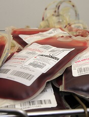
Credit: UAB Hospital
A new study suggests the risks and benefits of red blood cell (RBC) transfusions can vary considerably for patients with trauma and major bleeding, depending on the patients’ risk of death at baseline.
Patients with the highest predicted risk of death on arrival at a trauma center received the greatest benefit from RBC transfusions.
But among patients with the lowest predicted risk of death at baseline, transfusion was associated with a higher risk of death post-treatment.
Pablo Perel, MD, PhD, of the London School of Hygiene & Tropical Medicine in the UK, and his colleagues reported these findings in PLOS Medicine.
To conduct this study, the team used data from the CRASH-2 trial, which assessed the effect of tranexamic acid in trauma patients. The trial included 20,127 patients with significant bleeding who were treated at 274 hospitals in 40 countries.
Dr Perel and his colleagues used that data to evaluate the association between receiving an RBC transfusion and death from all causes at 28 days post-trauma. The findings were stratified by predicted risk of death based on clinical observations on arrival at the trauma center.
The researchers found that patients with the greatest predicted risk of dying—greater than 50%—had a smaller chance of death from all causes if they were transfused than if they were not. The odds ratio (OR) was 0.59.
For patients whose predicted risk of death ranged from 21% to 50%, there was no significant difference in their chance of dying whether they were transfused or not. The OR was 0.92.
But for patients with a lower risk of death at baseline, transfusion was associated with an increased risk of death.
Patients with a 6% to 20% risk of death at baseline had an OR of 2.31 if they received a transfusion. And for patients whose initial risk of death was below 6%, the OR for death associated with transfusion was 5.40.
In absolute figures, compared to no transfusion, RBC transfusion was associated with 5.1 more deaths per 100 patients in the group with the lowest predicted risk of death but with 11.9 fewer deaths per 100 patients in the group with the highest predicted risk of death.
The researchers noted that, although these data suggest RBC transfusion could be harmful for patients whose predicted risk of death is low, this study was observational. So the team cannot confirm a causal link, and a randomized trial investigating the association is warranted.
Credit: UAB Hospital
A new study suggests the risks and benefits of red blood cell (RBC) transfusions can vary considerably for patients with trauma and major bleeding, depending on the patients’ risk of death at baseline.
Patients with the highest predicted risk of death on arrival at a trauma center received the greatest benefit from RBC transfusions.
But among patients with the lowest predicted risk of death at baseline, transfusion was associated with a higher risk of death post-treatment.
Pablo Perel, MD, PhD, of the London School of Hygiene & Tropical Medicine in the UK, and his colleagues reported these findings in PLOS Medicine.
To conduct this study, the team used data from the CRASH-2 trial, which assessed the effect of tranexamic acid in trauma patients. The trial included 20,127 patients with significant bleeding who were treated at 274 hospitals in 40 countries.
Dr Perel and his colleagues used that data to evaluate the association between receiving an RBC transfusion and death from all causes at 28 days post-trauma. The findings were stratified by predicted risk of death based on clinical observations on arrival at the trauma center.
The researchers found that patients with the greatest predicted risk of dying—greater than 50%—had a smaller chance of death from all causes if they were transfused than if they were not. The odds ratio (OR) was 0.59.
For patients whose predicted risk of death ranged from 21% to 50%, there was no significant difference in their chance of dying whether they were transfused or not. The OR was 0.92.
But for patients with a lower risk of death at baseline, transfusion was associated with an increased risk of death.
Patients with a 6% to 20% risk of death at baseline had an OR of 2.31 if they received a transfusion. And for patients whose initial risk of death was below 6%, the OR for death associated with transfusion was 5.40.
In absolute figures, compared to no transfusion, RBC transfusion was associated with 5.1 more deaths per 100 patients in the group with the lowest predicted risk of death but with 11.9 fewer deaths per 100 patients in the group with the highest predicted risk of death.
The researchers noted that, although these data suggest RBC transfusion could be harmful for patients whose predicted risk of death is low, this study was observational. So the team cannot confirm a causal link, and a randomized trial investigating the association is warranted.
Credit: UAB Hospital
A new study suggests the risks and benefits of red blood cell (RBC) transfusions can vary considerably for patients with trauma and major bleeding, depending on the patients’ risk of death at baseline.
Patients with the highest predicted risk of death on arrival at a trauma center received the greatest benefit from RBC transfusions.
But among patients with the lowest predicted risk of death at baseline, transfusion was associated with a higher risk of death post-treatment.
Pablo Perel, MD, PhD, of the London School of Hygiene & Tropical Medicine in the UK, and his colleagues reported these findings in PLOS Medicine.
To conduct this study, the team used data from the CRASH-2 trial, which assessed the effect of tranexamic acid in trauma patients. The trial included 20,127 patients with significant bleeding who were treated at 274 hospitals in 40 countries.
Dr Perel and his colleagues used that data to evaluate the association between receiving an RBC transfusion and death from all causes at 28 days post-trauma. The findings were stratified by predicted risk of death based on clinical observations on arrival at the trauma center.
The researchers found that patients with the greatest predicted risk of dying—greater than 50%—had a smaller chance of death from all causes if they were transfused than if they were not. The odds ratio (OR) was 0.59.
For patients whose predicted risk of death ranged from 21% to 50%, there was no significant difference in their chance of dying whether they were transfused or not. The OR was 0.92.
But for patients with a lower risk of death at baseline, transfusion was associated with an increased risk of death.
Patients with a 6% to 20% risk of death at baseline had an OR of 2.31 if they received a transfusion. And for patients whose initial risk of death was below 6%, the OR for death associated with transfusion was 5.40.
In absolute figures, compared to no transfusion, RBC transfusion was associated with 5.1 more deaths per 100 patients in the group with the lowest predicted risk of death but with 11.9 fewer deaths per 100 patients in the group with the highest predicted risk of death.
The researchers noted that, although these data suggest RBC transfusion could be harmful for patients whose predicted risk of death is low, this study was observational. So the team cannot confirm a causal link, and a randomized trial investigating the association is warranted.
Cream provides relief for leg ulcers in SCD
MILAN—Results of a phase 1 study indicate that topical sodium nitrate is safe and effective for treating leg ulcers in patients with sickle cell disease (SCD).
The cream significantly decreased the size of leg ulcers overall, healed ulcers in 6 of the 18 patients studied, and reduced pain levels, seemingly independent of wound healing.
A few patients did experience short-lived burning at the treatment site, and some experienced a temporary, asymptomatic drop in blood pressure that resolved without intervention.
But the treatment was generally well-tolerated, according to study investigator Caterina P. Minniti, MD, of the National Heart, Lung and Blood Institute in Bethesda, Maryland.
She presented these results at the 19th Congress of the European Hematology Association (EHA) as abstract S663.
“The morbidity from chronic and recurrent leg ulcers in sickle cell disease and other hematologic disorders . . . remains a clinical and economic burden,” Dr Minniti noted.
“[C]urrent therapies have limited efficacy and usually are borrowed from the treatment of venous ulcers and diabetic ulcers. There isn’t really a concerted effort to treat sickle cell leg ulcers.”
With that in mind, she and her colleagues initiated a phase 1 dose-escalation trial of topical sodium nitrate in SCD patients.
Patient characteristics and treatment
The researchers enrolled 18 patients with a median age of 39 ± 12 (range, 20-59). The median number of ulcers per patient was 1.5 (range, 1-10), and the median ulcer age was 10 months (range, 2-300).
Manual assessment suggested the median ulcer size was 7.50 ± 4.65 cm2 (range, 2.09-16.50). Digital assessment suggested the median ulcer size was 5.97± 3.40 cm2 (range, 2.51-14.66).
The mean number of prior ulcer therapies was 8. This included surgical/sharp debridement (n=18), hyperbaric chamber (n=7), skin graft (n=6), MIST therapy (n=4), and oral/parenteral antibiotics (n=11).
For this study, patients had sodium nitrate cream applied twice a week for 4 weeks on 1 leg ulcer per subject. There were 5 cohorts of escalating treatment concentrations: 0.5%, 1%, 1.5%, 1.8%, and 2%.
Adverse events
There were no serious adverse events, and none of the patients discontinued treatment. One adverse event that was likely related to treatment was short-lived burning after cream application in 4 patients. But this resolved without intervention.
Another event that may have been related to treatment was asymptomatic, short-lived, diastolic blood pressure less than 50 mmHg in 5 subjects who received treatment at the highest concentrations (2 in the 2% cohort and 3 in the 1.8% cohort). On the other hand, 3 of these 5 subjects had documented diastolic blood pressure less than 50 mmHg prior to starting the treatment.
For the most part, there were no changes in laboratory or clinical parameters before and after the trial. However, the researchers did observe a significant decrease in white blood cell counts.
Effects on ulcer size
Among all patients, there was a significant decrease in ulcer size from the first treatment application to the end of the study, both according to digital photography and assessment by wound-care nurses (P<0.001 and P<0.0001, respectively).
Although patients in all of the treatment groups experienced a decrease in wound size, there was a correlation between the decrease and the concentration of treatment.
One patient in the 1%-concentration cohort had an ulcer that progressed, but all other patients saw improvements. The 4 patients who received the 1.8% concentration had a 69.7% decrease in ulcer size at week 5, and 1 ulcer had healed by the end of treatment.
The 3 patients who received the 2% concentration had an 88.3% decrease in ulcer size at week 5, and 2 ulcers had healed by that time. An additional 3 ulcers healed within weeks or months of the study end.
Effects on pain and blood flow
One of the most interesting findings of this study, according to Dr Minniti, was the effect of the cream on patients’ pain.
There was a significant decrease in patient-reported pain for treated ulcers (P<0.006) but not for untreated ulcers (P=0.38). And there was a significant correlation with pain score and nitrate concentration (P=0.006).
Patients’ weekly total usage of opioids decreased from baseline to the end of the study, but this difference was not significant (P=0.26).
“There was a trend toward significance,” Dr Minniti noted. “It’s very hard, in 1 month, to get off your long-acting opioid.”
Finally, Dr Minniti and her colleagues found that blood flow to the wound area changed before and after treatment. According to laser speckle contrast imaging, there was a significant increase in blood flow after treatment (P<0.0002).
Based on these results, the researchers have initiated a phase 1/2, randomized trial comparing topical sodium nitrate to placebo in SCD patients.
MILAN—Results of a phase 1 study indicate that topical sodium nitrate is safe and effective for treating leg ulcers in patients with sickle cell disease (SCD).
The cream significantly decreased the size of leg ulcers overall, healed ulcers in 6 of the 18 patients studied, and reduced pain levels, seemingly independent of wound healing.
A few patients did experience short-lived burning at the treatment site, and some experienced a temporary, asymptomatic drop in blood pressure that resolved without intervention.
But the treatment was generally well-tolerated, according to study investigator Caterina P. Minniti, MD, of the National Heart, Lung and Blood Institute in Bethesda, Maryland.
She presented these results at the 19th Congress of the European Hematology Association (EHA) as abstract S663.
“The morbidity from chronic and recurrent leg ulcers in sickle cell disease and other hematologic disorders . . . remains a clinical and economic burden,” Dr Minniti noted.
“[C]urrent therapies have limited efficacy and usually are borrowed from the treatment of venous ulcers and diabetic ulcers. There isn’t really a concerted effort to treat sickle cell leg ulcers.”
With that in mind, she and her colleagues initiated a phase 1 dose-escalation trial of topical sodium nitrate in SCD patients.
Patient characteristics and treatment
The researchers enrolled 18 patients with a median age of 39 ± 12 (range, 20-59). The median number of ulcers per patient was 1.5 (range, 1-10), and the median ulcer age was 10 months (range, 2-300).
Manual assessment suggested the median ulcer size was 7.50 ± 4.65 cm2 (range, 2.09-16.50). Digital assessment suggested the median ulcer size was 5.97± 3.40 cm2 (range, 2.51-14.66).
The mean number of prior ulcer therapies was 8. This included surgical/sharp debridement (n=18), hyperbaric chamber (n=7), skin graft (n=6), MIST therapy (n=4), and oral/parenteral antibiotics (n=11).
For this study, patients had sodium nitrate cream applied twice a week for 4 weeks on 1 leg ulcer per subject. There were 5 cohorts of escalating treatment concentrations: 0.5%, 1%, 1.5%, 1.8%, and 2%.
Adverse events
There were no serious adverse events, and none of the patients discontinued treatment. One adverse event that was likely related to treatment was short-lived burning after cream application in 4 patients. But this resolved without intervention.
Another event that may have been related to treatment was asymptomatic, short-lived, diastolic blood pressure less than 50 mmHg in 5 subjects who received treatment at the highest concentrations (2 in the 2% cohort and 3 in the 1.8% cohort). On the other hand, 3 of these 5 subjects had documented diastolic blood pressure less than 50 mmHg prior to starting the treatment.
For the most part, there were no changes in laboratory or clinical parameters before and after the trial. However, the researchers did observe a significant decrease in white blood cell counts.
Effects on ulcer size
Among all patients, there was a significant decrease in ulcer size from the first treatment application to the end of the study, both according to digital photography and assessment by wound-care nurses (P<0.001 and P<0.0001, respectively).
Although patients in all of the treatment groups experienced a decrease in wound size, there was a correlation between the decrease and the concentration of treatment.
One patient in the 1%-concentration cohort had an ulcer that progressed, but all other patients saw improvements. The 4 patients who received the 1.8% concentration had a 69.7% decrease in ulcer size at week 5, and 1 ulcer had healed by the end of treatment.
The 3 patients who received the 2% concentration had an 88.3% decrease in ulcer size at week 5, and 2 ulcers had healed by that time. An additional 3 ulcers healed within weeks or months of the study end.
Effects on pain and blood flow
One of the most interesting findings of this study, according to Dr Minniti, was the effect of the cream on patients’ pain.
There was a significant decrease in patient-reported pain for treated ulcers (P<0.006) but not for untreated ulcers (P=0.38). And there was a significant correlation with pain score and nitrate concentration (P=0.006).
Patients’ weekly total usage of opioids decreased from baseline to the end of the study, but this difference was not significant (P=0.26).
“There was a trend toward significance,” Dr Minniti noted. “It’s very hard, in 1 month, to get off your long-acting opioid.”
Finally, Dr Minniti and her colleagues found that blood flow to the wound area changed before and after treatment. According to laser speckle contrast imaging, there was a significant increase in blood flow after treatment (P<0.0002).
Based on these results, the researchers have initiated a phase 1/2, randomized trial comparing topical sodium nitrate to placebo in SCD patients.
MILAN—Results of a phase 1 study indicate that topical sodium nitrate is safe and effective for treating leg ulcers in patients with sickle cell disease (SCD).
The cream significantly decreased the size of leg ulcers overall, healed ulcers in 6 of the 18 patients studied, and reduced pain levels, seemingly independent of wound healing.
A few patients did experience short-lived burning at the treatment site, and some experienced a temporary, asymptomatic drop in blood pressure that resolved without intervention.
But the treatment was generally well-tolerated, according to study investigator Caterina P. Minniti, MD, of the National Heart, Lung and Blood Institute in Bethesda, Maryland.
She presented these results at the 19th Congress of the European Hematology Association (EHA) as abstract S663.
“The morbidity from chronic and recurrent leg ulcers in sickle cell disease and other hematologic disorders . . . remains a clinical and economic burden,” Dr Minniti noted.
“[C]urrent therapies have limited efficacy and usually are borrowed from the treatment of venous ulcers and diabetic ulcers. There isn’t really a concerted effort to treat sickle cell leg ulcers.”
With that in mind, she and her colleagues initiated a phase 1 dose-escalation trial of topical sodium nitrate in SCD patients.
Patient characteristics and treatment
The researchers enrolled 18 patients with a median age of 39 ± 12 (range, 20-59). The median number of ulcers per patient was 1.5 (range, 1-10), and the median ulcer age was 10 months (range, 2-300).
Manual assessment suggested the median ulcer size was 7.50 ± 4.65 cm2 (range, 2.09-16.50). Digital assessment suggested the median ulcer size was 5.97± 3.40 cm2 (range, 2.51-14.66).
The mean number of prior ulcer therapies was 8. This included surgical/sharp debridement (n=18), hyperbaric chamber (n=7), skin graft (n=6), MIST therapy (n=4), and oral/parenteral antibiotics (n=11).
For this study, patients had sodium nitrate cream applied twice a week for 4 weeks on 1 leg ulcer per subject. There were 5 cohorts of escalating treatment concentrations: 0.5%, 1%, 1.5%, 1.8%, and 2%.
Adverse events
There were no serious adverse events, and none of the patients discontinued treatment. One adverse event that was likely related to treatment was short-lived burning after cream application in 4 patients. But this resolved without intervention.
Another event that may have been related to treatment was asymptomatic, short-lived, diastolic blood pressure less than 50 mmHg in 5 subjects who received treatment at the highest concentrations (2 in the 2% cohort and 3 in the 1.8% cohort). On the other hand, 3 of these 5 subjects had documented diastolic blood pressure less than 50 mmHg prior to starting the treatment.
For the most part, there were no changes in laboratory or clinical parameters before and after the trial. However, the researchers did observe a significant decrease in white blood cell counts.
Effects on ulcer size
Among all patients, there was a significant decrease in ulcer size from the first treatment application to the end of the study, both according to digital photography and assessment by wound-care nurses (P<0.001 and P<0.0001, respectively).
Although patients in all of the treatment groups experienced a decrease in wound size, there was a correlation between the decrease and the concentration of treatment.
One patient in the 1%-concentration cohort had an ulcer that progressed, but all other patients saw improvements. The 4 patients who received the 1.8% concentration had a 69.7% decrease in ulcer size at week 5, and 1 ulcer had healed by the end of treatment.
The 3 patients who received the 2% concentration had an 88.3% decrease in ulcer size at week 5, and 2 ulcers had healed by that time. An additional 3 ulcers healed within weeks or months of the study end.
Effects on pain and blood flow
One of the most interesting findings of this study, according to Dr Minniti, was the effect of the cream on patients’ pain.
There was a significant decrease in patient-reported pain for treated ulcers (P<0.006) but not for untreated ulcers (P=0.38). And there was a significant correlation with pain score and nitrate concentration (P=0.006).
Patients’ weekly total usage of opioids decreased from baseline to the end of the study, but this difference was not significant (P=0.26).
“There was a trend toward significance,” Dr Minniti noted. “It’s very hard, in 1 month, to get off your long-acting opioid.”
Finally, Dr Minniti and her colleagues found that blood flow to the wound area changed before and after treatment. According to laser speckle contrast imaging, there was a significant increase in blood flow after treatment (P<0.0002).
Based on these results, the researchers have initiated a phase 1/2, randomized trial comparing topical sodium nitrate to placebo in SCD patients.
US ranked last in healthcare report

Credit: NIH
In a study comparing healthcare in 11 industrialized countries, the US ranked last on measures of health system quality, efficiency, access to care, equity, and healthy lives.
The other countries included in this study were Australia, Canada, France, Germany, the Netherlands, New Zealand, Norway, Sweden, Switzerland, and the UK.
While the results revealed room for improvement in every country, the US stood out for having the highest costs and lowest performance.
For instance, the US spent $8508 per person on healthcare in 2011, compared with $3406 in the UK, which ranked first overall.
Details on expenditures and rankings derived from this study are available in the Commonwealth Fund report, Mirror, Mirror on the Wall: How the Performance of the U.S. Health Care System Compares Internationally, 2014 Update.
“It is disappointing, but not surprising, that, despite our significant investment in healthcare, the US has continued to lag behind other countries,” said lead report author Karen Davis, of the Johns Hopkins Bloomberg School of Public Health in Baltimore, Maryland.
The report was also produced in 2004, 2006, 2007, and 2010, with the US ranking last in each of those years. Four countries were added to this year’s report: Switzerland and Sweden, which followed the UK at the top of the rankings, and Norway and France, which were in the middle of the pack.
Australia, Germany, the Netherlands, New Zealand, and Norway also placed in the middle, and Canada ranked just above the US.
In addition to ranking last overall, the US ranked last on infant mortality and on deaths that were potentially preventable with timely access to effective healthcare. The country ranked second-to-last on healthy life expectancy at age 60.
The US also ranked last on every measure of cost-related access. More than one-third (37%) of US adults reported forgoing a recommended test, treatment, or follow-up care because of cost.
With regard to healthcare quality, the US fell somewhere in the middle. On 2 of 4 measures of quality—effective care and patient-centered care—the US ranked near the top (3rd and 4th of 11 countries, respectively). But it did not perform as well with regard to providing safe or coordinated care.
The US ranked last in efficiency, due to low marks on the time and dollars spent dealing with insurance administration, lack of communication among healthcare providers, and duplicative medical testing.
Forty percent of US adults who had visited an emergency room reported they could have been treated by a regular doctor if one had been available. This is more than double the rate of patients in the UK (16%).
The US also ranked last in healthcare equity. About 4 of 10 (39%) adults with below-average incomes in the US reported a medical problem but did not visit a doctor in the past year because of costs, compared with less than 1 of 10 in the UK, Sweden, Canada, and Norway.
There were also large discrepancies in the length of time US adults waited for specialist, emergency, and after-hours care. And wait times were associated with patient income.
The data for this research were drawn from the Commonwealth Fund 2011 International Health Policy Survey of Sicker Adults, the Commonwealth Fund 2012 International Health Policy Survey of Primary Care Physicians, and the Commonwealth Fund 2013 International Health Policy Survey.
The 2011 survey targeted a representative sample of “sicker adults,” defined as those who rated their health status as fair or poor, received medical care for a serious chronic illness, serious injury, or disability in the past year, or were hospitalized or underwent surgery in the previous 2 years.
The 2012 survey looked at the experiences of primary care physicians. The 2013 survey focused on the experiences of nationally representative samples of adults ages 18 and older.
Additional data on health outcomes were drawn from the Organization for Economic Cooperation and Development and the World Health Organization.
Credit: NIH
In a study comparing healthcare in 11 industrialized countries, the US ranked last on measures of health system quality, efficiency, access to care, equity, and healthy lives.
The other countries included in this study were Australia, Canada, France, Germany, the Netherlands, New Zealand, Norway, Sweden, Switzerland, and the UK.
While the results revealed room for improvement in every country, the US stood out for having the highest costs and lowest performance.
For instance, the US spent $8508 per person on healthcare in 2011, compared with $3406 in the UK, which ranked first overall.
Details on expenditures and rankings derived from this study are available in the Commonwealth Fund report, Mirror, Mirror on the Wall: How the Performance of the U.S. Health Care System Compares Internationally, 2014 Update.
“It is disappointing, but not surprising, that, despite our significant investment in healthcare, the US has continued to lag behind other countries,” said lead report author Karen Davis, of the Johns Hopkins Bloomberg School of Public Health in Baltimore, Maryland.
The report was also produced in 2004, 2006, 2007, and 2010, with the US ranking last in each of those years. Four countries were added to this year’s report: Switzerland and Sweden, which followed the UK at the top of the rankings, and Norway and France, which were in the middle of the pack.
Australia, Germany, the Netherlands, New Zealand, and Norway also placed in the middle, and Canada ranked just above the US.
In addition to ranking last overall, the US ranked last on infant mortality and on deaths that were potentially preventable with timely access to effective healthcare. The country ranked second-to-last on healthy life expectancy at age 60.
The US also ranked last on every measure of cost-related access. More than one-third (37%) of US adults reported forgoing a recommended test, treatment, or follow-up care because of cost.
With regard to healthcare quality, the US fell somewhere in the middle. On 2 of 4 measures of quality—effective care and patient-centered care—the US ranked near the top (3rd and 4th of 11 countries, respectively). But it did not perform as well with regard to providing safe or coordinated care.
The US ranked last in efficiency, due to low marks on the time and dollars spent dealing with insurance administration, lack of communication among healthcare providers, and duplicative medical testing.
Forty percent of US adults who had visited an emergency room reported they could have been treated by a regular doctor if one had been available. This is more than double the rate of patients in the UK (16%).
The US also ranked last in healthcare equity. About 4 of 10 (39%) adults with below-average incomes in the US reported a medical problem but did not visit a doctor in the past year because of costs, compared with less than 1 of 10 in the UK, Sweden, Canada, and Norway.
There were also large discrepancies in the length of time US adults waited for specialist, emergency, and after-hours care. And wait times were associated with patient income.
The data for this research were drawn from the Commonwealth Fund 2011 International Health Policy Survey of Sicker Adults, the Commonwealth Fund 2012 International Health Policy Survey of Primary Care Physicians, and the Commonwealth Fund 2013 International Health Policy Survey.
The 2011 survey targeted a representative sample of “sicker adults,” defined as those who rated their health status as fair or poor, received medical care for a serious chronic illness, serious injury, or disability in the past year, or were hospitalized or underwent surgery in the previous 2 years.
The 2012 survey looked at the experiences of primary care physicians. The 2013 survey focused on the experiences of nationally representative samples of adults ages 18 and older.
Additional data on health outcomes were drawn from the Organization for Economic Cooperation and Development and the World Health Organization.
Credit: NIH
In a study comparing healthcare in 11 industrialized countries, the US ranked last on measures of health system quality, efficiency, access to care, equity, and healthy lives.
The other countries included in this study were Australia, Canada, France, Germany, the Netherlands, New Zealand, Norway, Sweden, Switzerland, and the UK.
While the results revealed room for improvement in every country, the US stood out for having the highest costs and lowest performance.
For instance, the US spent $8508 per person on healthcare in 2011, compared with $3406 in the UK, which ranked first overall.
Details on expenditures and rankings derived from this study are available in the Commonwealth Fund report, Mirror, Mirror on the Wall: How the Performance of the U.S. Health Care System Compares Internationally, 2014 Update.
“It is disappointing, but not surprising, that, despite our significant investment in healthcare, the US has continued to lag behind other countries,” said lead report author Karen Davis, of the Johns Hopkins Bloomberg School of Public Health in Baltimore, Maryland.
The report was also produced in 2004, 2006, 2007, and 2010, with the US ranking last in each of those years. Four countries were added to this year’s report: Switzerland and Sweden, which followed the UK at the top of the rankings, and Norway and France, which were in the middle of the pack.
Australia, Germany, the Netherlands, New Zealand, and Norway also placed in the middle, and Canada ranked just above the US.
In addition to ranking last overall, the US ranked last on infant mortality and on deaths that were potentially preventable with timely access to effective healthcare. The country ranked second-to-last on healthy life expectancy at age 60.
The US also ranked last on every measure of cost-related access. More than one-third (37%) of US adults reported forgoing a recommended test, treatment, or follow-up care because of cost.
With regard to healthcare quality, the US fell somewhere in the middle. On 2 of 4 measures of quality—effective care and patient-centered care—the US ranked near the top (3rd and 4th of 11 countries, respectively). But it did not perform as well with regard to providing safe or coordinated care.
The US ranked last in efficiency, due to low marks on the time and dollars spent dealing with insurance administration, lack of communication among healthcare providers, and duplicative medical testing.
Forty percent of US adults who had visited an emergency room reported they could have been treated by a regular doctor if one had been available. This is more than double the rate of patients in the UK (16%).
The US also ranked last in healthcare equity. About 4 of 10 (39%) adults with below-average incomes in the US reported a medical problem but did not visit a doctor in the past year because of costs, compared with less than 1 of 10 in the UK, Sweden, Canada, and Norway.
There were also large discrepancies in the length of time US adults waited for specialist, emergency, and after-hours care. And wait times were associated with patient income.
The data for this research were drawn from the Commonwealth Fund 2011 International Health Policy Survey of Sicker Adults, the Commonwealth Fund 2012 International Health Policy Survey of Primary Care Physicians, and the Commonwealth Fund 2013 International Health Policy Survey.
The 2011 survey targeted a representative sample of “sicker adults,” defined as those who rated their health status as fair or poor, received medical care for a serious chronic illness, serious injury, or disability in the past year, or were hospitalized or underwent surgery in the previous 2 years.
The 2012 survey looked at the experiences of primary care physicians. The 2013 survey focused on the experiences of nationally representative samples of adults ages 18 and older.
Additional data on health outcomes were drawn from the Organization for Economic Cooperation and Development and the World Health Organization.
Sequential decitabine, idarubicin combo synergistic in AML

Credit: Rhoda Baer
Researchers tested 5 anti-leukemia agents in combination with the methylation inhibitor decitabine and found that the sequential combination of decitabine and idarubicin worked synergistically to produce anti-leukemia effects.
The combination induced cell death in U937, HEL, and SKM-1 human cell lines and acute myeloid leukemia (AML) cells isolated from patients.
The researchers attributed the effects to demethylation of the Wnt/β-catenin pathway inhibitors and downregulation of the Wnt/β-catenin pathway nuclear targets.
The researchers noted that decitabine monotherapy has resulted in relatively low complete remission rates in AML and myelodysplastic syndromes (MDS). So they undertook to investigate combination therapies that would potentially improve efficacy.
Hongyan Tong, PhD, of Zhejiang University School of Medicine in Hangzhou, China, and colleagues reported their findings in the Journal of Translational Medicine.
The researchers chose 5 agents to combine, either simultaneously or sequentially, with decitabine—idarubicin, daunorubicin, aclarubicin, thalidomide, and homoharringtonine—and analyzed their effect on leukemia proliferation in the various AML cell lines mentioned above.
Using the U937 cell line first, the researchers found that when decitabine was combined simulataneously or sequentially with homharringtonine, aclarubicin, thalidomide, and daunorubicin, there was no synergistic effect. The confidence interval (CI) values of various doses were almost all over 0.8.
This was also true for the simultaneous combination of decitabine with idarubicin.
However, when they combined decitabine sequentially with idarubicin, the CI values on all 5 doses were under 0.8, indicating synergism.
In addition, when they administered decitabine twice in the sequence, the CI values were lower than a single administration.
They then confirmed the results in other AML cell lines (HEL and SKM-1) and in cells from AML patients.
Next, they confirmed the synergism of the sequential combination of decitabine and idarubicin in an AML mouse model and found that the combination inhibited tumor growth.
Tumor growth was inhibited significantly on days 4 (P<0.01), days 6 -16 (P<0.001), and started to wane by day 18 (P<0.05) after treatment.
The investigators determined that apoptosis was responsible for the combination’s decrease in leukemic cell viability. The apoptosis rates with the combination therapy were significantly increased in the U937, HEL, and SKM-1 cell lines compared with controls, (all P< 0.001).
In addition, the researchers observed that the tumor cells after treatment showed typical apoptosis characteristics, such as the absence of microvilli on cell membrane, nuclear and cell membrane blebbing, chromosome condensation, and the formation of apoptotic bodies.
The investigators used microarray expression to ascertain the differential gene expression profile of decitabine and idarubicin and found that the Wnt pathway was one of the major pathways disturbed.
Sequential treatment significantly upregulated the Wnt antagonist genes SFRP1, HDPR1, and DKK3. This in turn resulted in increased expression of these genes at the mRNA and protein levels.
In addition, treatment with idarubicin after decitabine caused significant down regulation of the expression of c-Myc, β-catenin, and cyclinD1 genes compared to treatment with decitabine or idarubicin alone.
The investigators concluded that the findings suggest clinical potential in sequential administration of decitabine and idarubicin in AML and high-risk MDS.
Credit: Rhoda Baer
Researchers tested 5 anti-leukemia agents in combination with the methylation inhibitor decitabine and found that the sequential combination of decitabine and idarubicin worked synergistically to produce anti-leukemia effects.
The combination induced cell death in U937, HEL, and SKM-1 human cell lines and acute myeloid leukemia (AML) cells isolated from patients.
The researchers attributed the effects to demethylation of the Wnt/β-catenin pathway inhibitors and downregulation of the Wnt/β-catenin pathway nuclear targets.
The researchers noted that decitabine monotherapy has resulted in relatively low complete remission rates in AML and myelodysplastic syndromes (MDS). So they undertook to investigate combination therapies that would potentially improve efficacy.
Hongyan Tong, PhD, of Zhejiang University School of Medicine in Hangzhou, China, and colleagues reported their findings in the Journal of Translational Medicine.
The researchers chose 5 agents to combine, either simultaneously or sequentially, with decitabine—idarubicin, daunorubicin, aclarubicin, thalidomide, and homoharringtonine—and analyzed their effect on leukemia proliferation in the various AML cell lines mentioned above.
Using the U937 cell line first, the researchers found that when decitabine was combined simulataneously or sequentially with homharringtonine, aclarubicin, thalidomide, and daunorubicin, there was no synergistic effect. The confidence interval (CI) values of various doses were almost all over 0.8.
This was also true for the simultaneous combination of decitabine with idarubicin.
However, when they combined decitabine sequentially with idarubicin, the CI values on all 5 doses were under 0.8, indicating synergism.
In addition, when they administered decitabine twice in the sequence, the CI values were lower than a single administration.
They then confirmed the results in other AML cell lines (HEL and SKM-1) and in cells from AML patients.
Next, they confirmed the synergism of the sequential combination of decitabine and idarubicin in an AML mouse model and found that the combination inhibited tumor growth.
Tumor growth was inhibited significantly on days 4 (P<0.01), days 6 -16 (P<0.001), and started to wane by day 18 (P<0.05) after treatment.
The investigators determined that apoptosis was responsible for the combination’s decrease in leukemic cell viability. The apoptosis rates with the combination therapy were significantly increased in the U937, HEL, and SKM-1 cell lines compared with controls, (all P< 0.001).
In addition, the researchers observed that the tumor cells after treatment showed typical apoptosis characteristics, such as the absence of microvilli on cell membrane, nuclear and cell membrane blebbing, chromosome condensation, and the formation of apoptotic bodies.
The investigators used microarray expression to ascertain the differential gene expression profile of decitabine and idarubicin and found that the Wnt pathway was one of the major pathways disturbed.
Sequential treatment significantly upregulated the Wnt antagonist genes SFRP1, HDPR1, and DKK3. This in turn resulted in increased expression of these genes at the mRNA and protein levels.
In addition, treatment with idarubicin after decitabine caused significant down regulation of the expression of c-Myc, β-catenin, and cyclinD1 genes compared to treatment with decitabine or idarubicin alone.
The investigators concluded that the findings suggest clinical potential in sequential administration of decitabine and idarubicin in AML and high-risk MDS.
Credit: Rhoda Baer
Researchers tested 5 anti-leukemia agents in combination with the methylation inhibitor decitabine and found that the sequential combination of decitabine and idarubicin worked synergistically to produce anti-leukemia effects.
The combination induced cell death in U937, HEL, and SKM-1 human cell lines and acute myeloid leukemia (AML) cells isolated from patients.
The researchers attributed the effects to demethylation of the Wnt/β-catenin pathway inhibitors and downregulation of the Wnt/β-catenin pathway nuclear targets.
The researchers noted that decitabine monotherapy has resulted in relatively low complete remission rates in AML and myelodysplastic syndromes (MDS). So they undertook to investigate combination therapies that would potentially improve efficacy.
Hongyan Tong, PhD, of Zhejiang University School of Medicine in Hangzhou, China, and colleagues reported their findings in the Journal of Translational Medicine.
The researchers chose 5 agents to combine, either simultaneously or sequentially, with decitabine—idarubicin, daunorubicin, aclarubicin, thalidomide, and homoharringtonine—and analyzed their effect on leukemia proliferation in the various AML cell lines mentioned above.
Using the U937 cell line first, the researchers found that when decitabine was combined simulataneously or sequentially with homharringtonine, aclarubicin, thalidomide, and daunorubicin, there was no synergistic effect. The confidence interval (CI) values of various doses were almost all over 0.8.
This was also true for the simultaneous combination of decitabine with idarubicin.
However, when they combined decitabine sequentially with idarubicin, the CI values on all 5 doses were under 0.8, indicating synergism.
In addition, when they administered decitabine twice in the sequence, the CI values were lower than a single administration.
They then confirmed the results in other AML cell lines (HEL and SKM-1) and in cells from AML patients.
Next, they confirmed the synergism of the sequential combination of decitabine and idarubicin in an AML mouse model and found that the combination inhibited tumor growth.
Tumor growth was inhibited significantly on days 4 (P<0.01), days 6 -16 (P<0.001), and started to wane by day 18 (P<0.05) after treatment.
The investigators determined that apoptosis was responsible for the combination’s decrease in leukemic cell viability. The apoptosis rates with the combination therapy were significantly increased in the U937, HEL, and SKM-1 cell lines compared with controls, (all P< 0.001).
In addition, the researchers observed that the tumor cells after treatment showed typical apoptosis characteristics, such as the absence of microvilli on cell membrane, nuclear and cell membrane blebbing, chromosome condensation, and the formation of apoptotic bodies.
The investigators used microarray expression to ascertain the differential gene expression profile of decitabine and idarubicin and found that the Wnt pathway was one of the major pathways disturbed.
Sequential treatment significantly upregulated the Wnt antagonist genes SFRP1, HDPR1, and DKK3. This in turn resulted in increased expression of these genes at the mRNA and protein levels.
In addition, treatment with idarubicin after decitabine caused significant down regulation of the expression of c-Myc, β-catenin, and cyclinD1 genes compared to treatment with decitabine or idarubicin alone.
The investigators concluded that the findings suggest clinical potential in sequential administration of decitabine and idarubicin in AML and high-risk MDS.
Genetic ‘barcode’ could help track malaria

Credit: James Gathany
A genetic “barcode” for malaria parasites could be used to track and contain the spread of the disease, according to research published in Nature Communications.
Investigators analyzed the DNA of more than 700 Plasmodium falciparum parasites taken from patients in East and West Africa, South East Asia, Oceania, and South America.
And this revealed several short genetic sequences that were distinct in the DNA of parasites from certain geographic regions.
The team used this information to design a genetic barcode of 23 single-nucleotide polymorphisms that can be used to identify the source of new malaria infections.
“Being able to determine the geographic origin of malaria parasites has enormous potential in containing drug-resistance and eliminating malaria,” said study author Taane Clark, DPhil, of the London School of Hygiene & Tropical Medicine in the UK.
“Our work represents a breakthrough in the genetic barcoding of P falciparum, as it reveals very specific and accurate sequences for different geographic settings. We are currently extending the barcode to include other populations, such as India, Central America, southern Africa, and the Caribbean, and plan to include genetic markers for other types malaria, such as P vivax.”
Previous candidates for malaria genetic barcodes have relied on identifying DNA markers found in the parasite’s cell nucleus, which shows too much genetic variation between individual parasites to be used accurately.
But Dr Clark and his colleagues studied the DNA found in 2 parts of the parasite’s cells outside of the nucleus—the mitochondria and the apicolasts, which are only inherited through maternal lines, so their genes remain much more stable over generations.
By identifying short sequences in the DNA of the parasite’s mitochondria and apicoplasts that were specific for different geographic locations, the investigators were able to design a genetic barcode that is 92% predictive, stable, and geographically informative over time.
“By taking finger-prick bloodspots from malaria patients and using rapid gene sequencing technologies on small amounts of parasite material, local agencies could use this new barcode to quickly and accurately identify where a form of the parasite may have come from and help in programs of malaria elimination and resistance containment,” said study author Cally Roper, PhD, also of the London School of Hygiene & Tropical Medicine.
The investigators noted, however, that this barcode is limited because their study lacks representation of the Indian sub-continent, Central America, southern Africa, and the Caribbean, owing to the scarcity of sequence data from these regions.
Additionally, there’s a need to study more samples from East Africa, a region of high genetic diversity, high migration, and poor predictive ability.
Credit: James Gathany
A genetic “barcode” for malaria parasites could be used to track and contain the spread of the disease, according to research published in Nature Communications.
Investigators analyzed the DNA of more than 700 Plasmodium falciparum parasites taken from patients in East and West Africa, South East Asia, Oceania, and South America.
And this revealed several short genetic sequences that were distinct in the DNA of parasites from certain geographic regions.
The team used this information to design a genetic barcode of 23 single-nucleotide polymorphisms that can be used to identify the source of new malaria infections.
“Being able to determine the geographic origin of malaria parasites has enormous potential in containing drug-resistance and eliminating malaria,” said study author Taane Clark, DPhil, of the London School of Hygiene & Tropical Medicine in the UK.
“Our work represents a breakthrough in the genetic barcoding of P falciparum, as it reveals very specific and accurate sequences for different geographic settings. We are currently extending the barcode to include other populations, such as India, Central America, southern Africa, and the Caribbean, and plan to include genetic markers for other types malaria, such as P vivax.”
Previous candidates for malaria genetic barcodes have relied on identifying DNA markers found in the parasite’s cell nucleus, which shows too much genetic variation between individual parasites to be used accurately.
But Dr Clark and his colleagues studied the DNA found in 2 parts of the parasite’s cells outside of the nucleus—the mitochondria and the apicolasts, which are only inherited through maternal lines, so their genes remain much more stable over generations.
By identifying short sequences in the DNA of the parasite’s mitochondria and apicoplasts that were specific for different geographic locations, the investigators were able to design a genetic barcode that is 92% predictive, stable, and geographically informative over time.
“By taking finger-prick bloodspots from malaria patients and using rapid gene sequencing technologies on small amounts of parasite material, local agencies could use this new barcode to quickly and accurately identify where a form of the parasite may have come from and help in programs of malaria elimination and resistance containment,” said study author Cally Roper, PhD, also of the London School of Hygiene & Tropical Medicine.
The investigators noted, however, that this barcode is limited because their study lacks representation of the Indian sub-continent, Central America, southern Africa, and the Caribbean, owing to the scarcity of sequence data from these regions.
Additionally, there’s a need to study more samples from East Africa, a region of high genetic diversity, high migration, and poor predictive ability.
Credit: James Gathany
A genetic “barcode” for malaria parasites could be used to track and contain the spread of the disease, according to research published in Nature Communications.
Investigators analyzed the DNA of more than 700 Plasmodium falciparum parasites taken from patients in East and West Africa, South East Asia, Oceania, and South America.
And this revealed several short genetic sequences that were distinct in the DNA of parasites from certain geographic regions.
The team used this information to design a genetic barcode of 23 single-nucleotide polymorphisms that can be used to identify the source of new malaria infections.
“Being able to determine the geographic origin of malaria parasites has enormous potential in containing drug-resistance and eliminating malaria,” said study author Taane Clark, DPhil, of the London School of Hygiene & Tropical Medicine in the UK.
“Our work represents a breakthrough in the genetic barcoding of P falciparum, as it reveals very specific and accurate sequences for different geographic settings. We are currently extending the barcode to include other populations, such as India, Central America, southern Africa, and the Caribbean, and plan to include genetic markers for other types malaria, such as P vivax.”
Previous candidates for malaria genetic barcodes have relied on identifying DNA markers found in the parasite’s cell nucleus, which shows too much genetic variation between individual parasites to be used accurately.
But Dr Clark and his colleagues studied the DNA found in 2 parts of the parasite’s cells outside of the nucleus—the mitochondria and the apicolasts, which are only inherited through maternal lines, so their genes remain much more stable over generations.
By identifying short sequences in the DNA of the parasite’s mitochondria and apicoplasts that were specific for different geographic locations, the investigators were able to design a genetic barcode that is 92% predictive, stable, and geographically informative over time.
“By taking finger-prick bloodspots from malaria patients and using rapid gene sequencing technologies on small amounts of parasite material, local agencies could use this new barcode to quickly and accurately identify where a form of the parasite may have come from and help in programs of malaria elimination and resistance containment,” said study author Cally Roper, PhD, also of the London School of Hygiene & Tropical Medicine.
The investigators noted, however, that this barcode is limited because their study lacks representation of the Indian sub-continent, Central America, southern Africa, and the Caribbean, owing to the scarcity of sequence data from these regions.
Additionally, there’s a need to study more samples from East Africa, a region of high genetic diversity, high migration, and poor predictive ability.
Regulator of hepcidin discovered

which secrete erythroferrone
Credit: Leon Kautz/UCLA
Researchers have discovered a new hormone, called erythroferrone, which regulates hepcidin, the main iron hormone that controls iron absorption and distribution in the body.
Using a mouse model, reseachers determined that erythroferrone is made by red blood-cell progenitors in the bone marrow, and its levels vary according to the demand for red blood cells.
“Modulating the activity of erythroferrone,” said study author Tomas Ganz, MD, PhD, of the David Geffen School of Medicine at UCLA, “could be a viable strategy for the treatment of iron disorders of both overabundance and scarcity.”
Higher levels of erythroferrone suppress hepcidin, thereby allowing more iron to be made available for red blood-cell production. Erythroferrone, or drugs acting like it, could suppress hepcidin and make more iron available for red blood-cell production.
The team began their inquiry by studying the impact of hemorrhage on the bone marrow. That led them to focus on a specific protein that was secreted in the blood. The protein belonged to a family of proteins involved in cell-to-cell communication.
They used recombinant DNA technology and found that the hormone suppressed the production of hepcidin, which affected iron metabolism.
Erythroferrone-deficient mice do not suppress hepcidin quickly after hemorrhage and consequently have delayed recovery from blood loss.
The investigators also found that erythroferrone expression is greatly increased in Hbbth3/+ mice with thalassemia intermedia, indicating that the hormone contributes to the suppression of hepcidin and iron overload characteristic of the disease.
“Overproduction of erythroferrone may be a major cause of iron overload in untransfused patients and may contribute to iron overload in transfused patients,” said study author Elizabeta Nemeth, PhD, also of the David Geffen School of Medicine at UCLA.
“The identification of erythroferrone can potentially allow researchers and drug developers to target the hormone for specific treatment to prevent iron overload in Cooley’s anemia,” she said.
The investigators reported their findings recently in Nature Genetics.
They noted that further research is needed to understand the role of the new hormone in various blood diseases and to study the mechanisms through which erythroferrone regulates hepcidin.
which secrete erythroferrone
Credit: Leon Kautz/UCLA
Researchers have discovered a new hormone, called erythroferrone, which regulates hepcidin, the main iron hormone that controls iron absorption and distribution in the body.
Using a mouse model, reseachers determined that erythroferrone is made by red blood-cell progenitors in the bone marrow, and its levels vary according to the demand for red blood cells.
“Modulating the activity of erythroferrone,” said study author Tomas Ganz, MD, PhD, of the David Geffen School of Medicine at UCLA, “could be a viable strategy for the treatment of iron disorders of both overabundance and scarcity.”
Higher levels of erythroferrone suppress hepcidin, thereby allowing more iron to be made available for red blood-cell production. Erythroferrone, or drugs acting like it, could suppress hepcidin and make more iron available for red blood-cell production.
The team began their inquiry by studying the impact of hemorrhage on the bone marrow. That led them to focus on a specific protein that was secreted in the blood. The protein belonged to a family of proteins involved in cell-to-cell communication.
They used recombinant DNA technology and found that the hormone suppressed the production of hepcidin, which affected iron metabolism.
Erythroferrone-deficient mice do not suppress hepcidin quickly after hemorrhage and consequently have delayed recovery from blood loss.
The investigators also found that erythroferrone expression is greatly increased in Hbbth3/+ mice with thalassemia intermedia, indicating that the hormone contributes to the suppression of hepcidin and iron overload characteristic of the disease.
“Overproduction of erythroferrone may be a major cause of iron overload in untransfused patients and may contribute to iron overload in transfused patients,” said study author Elizabeta Nemeth, PhD, also of the David Geffen School of Medicine at UCLA.
“The identification of erythroferrone can potentially allow researchers and drug developers to target the hormone for specific treatment to prevent iron overload in Cooley’s anemia,” she said.
The investigators reported their findings recently in Nature Genetics.
They noted that further research is needed to understand the role of the new hormone in various blood diseases and to study the mechanisms through which erythroferrone regulates hepcidin.
which secrete erythroferrone
Credit: Leon Kautz/UCLA
Researchers have discovered a new hormone, called erythroferrone, which regulates hepcidin, the main iron hormone that controls iron absorption and distribution in the body.
Using a mouse model, reseachers determined that erythroferrone is made by red blood-cell progenitors in the bone marrow, and its levels vary according to the demand for red blood cells.
“Modulating the activity of erythroferrone,” said study author Tomas Ganz, MD, PhD, of the David Geffen School of Medicine at UCLA, “could be a viable strategy for the treatment of iron disorders of both overabundance and scarcity.”
Higher levels of erythroferrone suppress hepcidin, thereby allowing more iron to be made available for red blood-cell production. Erythroferrone, or drugs acting like it, could suppress hepcidin and make more iron available for red blood-cell production.
The team began their inquiry by studying the impact of hemorrhage on the bone marrow. That led them to focus on a specific protein that was secreted in the blood. The protein belonged to a family of proteins involved in cell-to-cell communication.
They used recombinant DNA technology and found that the hormone suppressed the production of hepcidin, which affected iron metabolism.
Erythroferrone-deficient mice do not suppress hepcidin quickly after hemorrhage and consequently have delayed recovery from blood loss.
The investigators also found that erythroferrone expression is greatly increased in Hbbth3/+ mice with thalassemia intermedia, indicating that the hormone contributes to the suppression of hepcidin and iron overload characteristic of the disease.
“Overproduction of erythroferrone may be a major cause of iron overload in untransfused patients and may contribute to iron overload in transfused patients,” said study author Elizabeta Nemeth, PhD, also of the David Geffen School of Medicine at UCLA.
“The identification of erythroferrone can potentially allow researchers and drug developers to target the hormone for specific treatment to prevent iron overload in Cooley’s anemia,” she said.
The investigators reported their findings recently in Nature Genetics.
They noted that further research is needed to understand the role of the new hormone in various blood diseases and to study the mechanisms through which erythroferrone regulates hepcidin.
Blood sterilization processes harmful to platelets
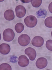
Some processes used to sterilize blood for transfusion are harmful to platelet function and could cause serious health issues in transfusion recipients, researchers say.
They found that some pathogen-reduction treatments impact platelets to the extent that they may be the cause of hemorrhages in recipients.
The pathogen reduction treatments “were developed more than 20 years ago, before we understood the importance of the genetic material contained in platelets,” explained study author Patrick Provost, PhD, of Université Laval and the CHU de Québec Research Center in Canada.
Platelets contain up to a third of the human genome in the form of ribonucleic acid (RNA), which enables them to synthesize over 1,000 proteins essential to the normal functioning of the human body.
The researchers studied the effects of 3 pathogen-reduction strategies—irradiation, riboflavin plus UVB light (Mirasol), and amotosalen plus UVA light (Intercept)—on platelet microRNAs, messenger RNAs (mRNAs), activation, and function.
They reported their findings in the journal Platelets.
The investigators collected 50 single-donor (apheresis) platelet concentrates (PCs) and subjected them to 5 treatments.
The control platelets were stored in donor plasma; additive solution platelets were stored in 65% storage solution and 35% donor plasma; the irradiation platelets were treated with 30Gy gamma irradiation and stored in donor plasma; the platelets treated with Mirasol were stored in donor plasma; and the platelets treated with Intercept were stored in the same solution as the additive solution group.
All treatments followed standard procedures or the manufacturer’s instructions.
After platelet isolation and RNA extraction, the investigators analyzed the levels of microRNA and mRNA levels of the platelets and assessed the impact of those levels on platelet activation and function.
MicroRNA profiles
They learned that platelets stored with additive solution or irradiation had significantly (P<0.05) reduced levels of one microRNA each, and only on day 7 of storage. Additive solution reduced the level of miR-223 and irradiation reduced the level of let-73.
Mirasol did not significantly reduce the level of any of the 11 tested micro RNAs.
And Intercept significantly reduced the level of 6 microRNAs on day 1, 1 microRNA on day 4, and 2 microRNAs on day 7. By day 7, let-7e was reduced by up to 70%.
The microRNA levels remained stable in the control sample for the entire 7-day storage period.
Platelet activation and function
Platelet counts in the Mirasol- and Intercept-treated platelets were significantly lower (P<0.001) on storage days 1, 4, and 7 compared with control platelets.
Pathogen-reduction treatments did not affect platelet microRNA synthesis, platelet microRNA function, nor did they induce the formation of cross-linked RNA adducts.
However, pathogen reduction caused platelet activation, which correlates with the observed reduction in platelet microRNAs.
The investigators measured CD62P expression, a marker of platelet activation, on the platelet surface. The additive solution platelets and Intercept-treated platelets, and to a lesser degree the irradiation group, had greater CD62P surface expression than the control group (P<0.05) on day 1.
The Mirasol group had similar activation to that of the control group.
On day 4, all treatment groups showed more activation than the control group (P<0.05). And on day 7, all groups had about the same activation level as the control group.
Pathogen reduction also impacted the aggregation response of platelets. Mirasol-treated platelets, which had the same aggregation response as that of controls on day 1, had no response on days 4 and 7.
And the aggregation response for Intercept-treated and additive solution platelets was already absent on day 1 and remained so on days 4 and 7.
Additive solution and Intercept also reduced platelet volume on day 1, which the investigators say could be explained by the platelet activation and release of microparticles induced by the treatments.
MicroRNA release
The investigators hypothesized that activated stored platelets could release microRNAs through microparticles in the supernatant. So they collected supernatant from each of the 5 groups and analyzed their total content of miR-223, which is one of the most abundant platelet microRNAs.
They discovered that the total amount of miR-223 was increased 30% to 86% in the microparticles released from additive solution and Intercept-treated platelets. They did not observe this increase in irradiation- or Mirasol-treated platelets compared to controls.
"The platelets end up depleted of RNA so, once transfused, they're unable to do what they normally would," Dr Provost said. Nevertheless, the clinical implications of the reduction in platelet activation and impaired platelet aggregation after Intercept treatment remain to be established.
The pathogen-reduction treatments are already marketed in some European countries, notably Switzerland, France, and Germany, and are under consideration in other countries, including Canada and the United States.
"In light of what we have demonstrated, the potentially harmful effects of these treatments should be carefully evaluated in the countries where they are not yet approved. It should also be re-evaluated in those countries where they are," Dr Provost said. 
Some processes used to sterilize blood for transfusion are harmful to platelet function and could cause serious health issues in transfusion recipients, researchers say.
They found that some pathogen-reduction treatments impact platelets to the extent that they may be the cause of hemorrhages in recipients.
The pathogen reduction treatments “were developed more than 20 years ago, before we understood the importance of the genetic material contained in platelets,” explained study author Patrick Provost, PhD, of Université Laval and the CHU de Québec Research Center in Canada.
Platelets contain up to a third of the human genome in the form of ribonucleic acid (RNA), which enables them to synthesize over 1,000 proteins essential to the normal functioning of the human body.
The researchers studied the effects of 3 pathogen-reduction strategies—irradiation, riboflavin plus UVB light (Mirasol), and amotosalen plus UVA light (Intercept)—on platelet microRNAs, messenger RNAs (mRNAs), activation, and function.
They reported their findings in the journal Platelets.
The investigators collected 50 single-donor (apheresis) platelet concentrates (PCs) and subjected them to 5 treatments.
The control platelets were stored in donor plasma; additive solution platelets were stored in 65% storage solution and 35% donor plasma; the irradiation platelets were treated with 30Gy gamma irradiation and stored in donor plasma; the platelets treated with Mirasol were stored in donor plasma; and the platelets treated with Intercept were stored in the same solution as the additive solution group.
All treatments followed standard procedures or the manufacturer’s instructions.
After platelet isolation and RNA extraction, the investigators analyzed the levels of microRNA and mRNA levels of the platelets and assessed the impact of those levels on platelet activation and function.
MicroRNA profiles
They learned that platelets stored with additive solution or irradiation had significantly (P<0.05) reduced levels of one microRNA each, and only on day 7 of storage. Additive solution reduced the level of miR-223 and irradiation reduced the level of let-73.
Mirasol did not significantly reduce the level of any of the 11 tested micro RNAs.
And Intercept significantly reduced the level of 6 microRNAs on day 1, 1 microRNA on day 4, and 2 microRNAs on day 7. By day 7, let-7e was reduced by up to 70%.
The microRNA levels remained stable in the control sample for the entire 7-day storage period.
Platelet activation and function
Platelet counts in the Mirasol- and Intercept-treated platelets were significantly lower (P<0.001) on storage days 1, 4, and 7 compared with control platelets.
Pathogen-reduction treatments did not affect platelet microRNA synthesis, platelet microRNA function, nor did they induce the formation of cross-linked RNA adducts.
However, pathogen reduction caused platelet activation, which correlates with the observed reduction in platelet microRNAs.
The investigators measured CD62P expression, a marker of platelet activation, on the platelet surface. The additive solution platelets and Intercept-treated platelets, and to a lesser degree the irradiation group, had greater CD62P surface expression than the control group (P<0.05) on day 1.
The Mirasol group had similar activation to that of the control group.
On day 4, all treatment groups showed more activation than the control group (P<0.05). And on day 7, all groups had about the same activation level as the control group.
Pathogen reduction also impacted the aggregation response of platelets. Mirasol-treated platelets, which had the same aggregation response as that of controls on day 1, had no response on days 4 and 7.
And the aggregation response for Intercept-treated and additive solution platelets was already absent on day 1 and remained so on days 4 and 7.
Additive solution and Intercept also reduced platelet volume on day 1, which the investigators say could be explained by the platelet activation and release of microparticles induced by the treatments.
MicroRNA release
The investigators hypothesized that activated stored platelets could release microRNAs through microparticles in the supernatant. So they collected supernatant from each of the 5 groups and analyzed their total content of miR-223, which is one of the most abundant platelet microRNAs.
They discovered that the total amount of miR-223 was increased 30% to 86% in the microparticles released from additive solution and Intercept-treated platelets. They did not observe this increase in irradiation- or Mirasol-treated platelets compared to controls.
"The platelets end up depleted of RNA so, once transfused, they're unable to do what they normally would," Dr Provost said. Nevertheless, the clinical implications of the reduction in platelet activation and impaired platelet aggregation after Intercept treatment remain to be established.
The pathogen-reduction treatments are already marketed in some European countries, notably Switzerland, France, and Germany, and are under consideration in other countries, including Canada and the United States.
"In light of what we have demonstrated, the potentially harmful effects of these treatments should be carefully evaluated in the countries where they are not yet approved. It should also be re-evaluated in those countries where they are," Dr Provost said.
Some processes used to sterilize blood for transfusion are harmful to platelet function and could cause serious health issues in transfusion recipients, researchers say.
They found that some pathogen-reduction treatments impact platelets to the extent that they may be the cause of hemorrhages in recipients.
The pathogen reduction treatments “were developed more than 20 years ago, before we understood the importance of the genetic material contained in platelets,” explained study author Patrick Provost, PhD, of Université Laval and the CHU de Québec Research Center in Canada.
Platelets contain up to a third of the human genome in the form of ribonucleic acid (RNA), which enables them to synthesize over 1,000 proteins essential to the normal functioning of the human body.
The researchers studied the effects of 3 pathogen-reduction strategies—irradiation, riboflavin plus UVB light (Mirasol), and amotosalen plus UVA light (Intercept)—on platelet microRNAs, messenger RNAs (mRNAs), activation, and function.
They reported their findings in the journal Platelets.
The investigators collected 50 single-donor (apheresis) platelet concentrates (PCs) and subjected them to 5 treatments.
The control platelets were stored in donor plasma; additive solution platelets were stored in 65% storage solution and 35% donor plasma; the irradiation platelets were treated with 30Gy gamma irradiation and stored in donor plasma; the platelets treated with Mirasol were stored in donor plasma; and the platelets treated with Intercept were stored in the same solution as the additive solution group.
All treatments followed standard procedures or the manufacturer’s instructions.
After platelet isolation and RNA extraction, the investigators analyzed the levels of microRNA and mRNA levels of the platelets and assessed the impact of those levels on platelet activation and function.
MicroRNA profiles
They learned that platelets stored with additive solution or irradiation had significantly (P<0.05) reduced levels of one microRNA each, and only on day 7 of storage. Additive solution reduced the level of miR-223 and irradiation reduced the level of let-73.
Mirasol did not significantly reduce the level of any of the 11 tested micro RNAs.
And Intercept significantly reduced the level of 6 microRNAs on day 1, 1 microRNA on day 4, and 2 microRNAs on day 7. By day 7, let-7e was reduced by up to 70%.
The microRNA levels remained stable in the control sample for the entire 7-day storage period.
Platelet activation and function
Platelet counts in the Mirasol- and Intercept-treated platelets were significantly lower (P<0.001) on storage days 1, 4, and 7 compared with control platelets.
Pathogen-reduction treatments did not affect platelet microRNA synthesis, platelet microRNA function, nor did they induce the formation of cross-linked RNA adducts.
However, pathogen reduction caused platelet activation, which correlates with the observed reduction in platelet microRNAs.
The investigators measured CD62P expression, a marker of platelet activation, on the platelet surface. The additive solution platelets and Intercept-treated platelets, and to a lesser degree the irradiation group, had greater CD62P surface expression than the control group (P<0.05) on day 1.
The Mirasol group had similar activation to that of the control group.
On day 4, all treatment groups showed more activation than the control group (P<0.05). And on day 7, all groups had about the same activation level as the control group.
Pathogen reduction also impacted the aggregation response of platelets. Mirasol-treated platelets, which had the same aggregation response as that of controls on day 1, had no response on days 4 and 7.
And the aggregation response for Intercept-treated and additive solution platelets was already absent on day 1 and remained so on days 4 and 7.
Additive solution and Intercept also reduced platelet volume on day 1, which the investigators say could be explained by the platelet activation and release of microparticles induced by the treatments.
MicroRNA release
The investigators hypothesized that activated stored platelets could release microRNAs through microparticles in the supernatant. So they collected supernatant from each of the 5 groups and analyzed their total content of miR-223, which is one of the most abundant platelet microRNAs.
They discovered that the total amount of miR-223 was increased 30% to 86% in the microparticles released from additive solution and Intercept-treated platelets. They did not observe this increase in irradiation- or Mirasol-treated platelets compared to controls.
"The platelets end up depleted of RNA so, once transfused, they're unable to do what they normally would," Dr Provost said. Nevertheless, the clinical implications of the reduction in platelet activation and impaired platelet aggregation after Intercept treatment remain to be established.
The pathogen-reduction treatments are already marketed in some European countries, notably Switzerland, France, and Germany, and are under consideration in other countries, including Canada and the United States.
"In light of what we have demonstrated, the potentially harmful effects of these treatments should be carefully evaluated in the countries where they are not yet approved. It should also be re-evaluated in those countries where they are," Dr Provost said.
One in four children with ALL misses maintenance doses

Credit: Bill Branson
Forgetting to take medication is the number one reason for non-adherence to maintenance therapy in children with acute lymphoblastic leukemia (ALL), according to a new study by the Children’s Oncology Group.
And neglecting to take maintenance medication 10% of the time increases the patient’s risk of relapse threefold.
In a study of 298 children receiving 6-mercaptopurine (6MP) as part of maintenance therapy, African Americans and Asians had significantly lower adherence rates than non-Hispanic whites, at 46%, 28%, and 14%, respectively.
Researchers discovered a number of other race-specific characteristics to explain the disparity, including low maternal education, households with a single parent and multiple children, low-income households, and households in which mothers were not the full-time caregivers.
The investigators had studied adherence in Hispanic children in an earlier study and they were not included here.
“While we don’t yet know why children of different races have significantly different survival rates for ALL,” said senior study author Smita Bhatia, MD, MPH, of City of Hope in Duarte, California, “we know that their adherence to their maintenance medication is a critical factor in their survival.”
And so the researchers explored potential sociodemographic differences that impact adherence to 6MP and reported their findings in Blood.
They enrolled 298 children, with a median age of 6 years at study entry (range, 2-20 years). All were in first continuous remission and receiving maintenance therapy that included 6MP.
One-hundred fifty-nine patients were non-Hispanic whites (the referent group), 71 were Asians, and 68 African Americans.
The researchers recorded adherence for up to 5 months per patient using an electronic monitoring device (MEMS®TrackCapTM) that recorded the date and time the pill bottle was opened. These data were downloaded at the end of the adherence-monitoring period.
They also measured erythrocyte TGN levels of the patients on a monthly basis to determine the association between bottle opening and taking the 6MP. Erythrocyte TGN levels reflect 6MP exposure.
Demographics
The researchers found that disease characteristics were comparable across the racial groups, but sociodemographic characteristics varied significantly.
African American families (64%) reported annual household incomes of less than $50,000 compared with 44% of non-Hispanic white and 33% of Asian families.
African American parents had significantly less formal education than non-Hispanic white and Asian parents. Sixty-six percent of African American fathers and 61% of African American mothers reported having less than a college degree.
This compared with 48% and 31% of non-Hispanic white and Asian fathers, respectively, and 46% and 32% of non-Hispanic white and Asian mothers, respectively.
More African American households (37%) were headed by single parents, compared with non-Hispanic white (9%) and Asian (4%) households.
And only 27% of African American children had their mothers as full-time caregivers, compared with 38% of non-Hispanic white children and 52% of Asian children.
Overall adherence
The investigators found that adherence for the entire population declined over the course of the 5 months, from 94.8% to 91.3% (P<0.0001).
Adherence rates were significantly lower in Asians and African Americans than in non-Hispanic whites, and in patients from low-income households.
Adherence rates were significantly higher in patients from single-parent/single-child households (96.9%) and in households where the mothers were full-time caregivers (94.8%).
Adherence by race
In Asian households, adherence was significantly higher with mothers as full-time caregivers (95.6%) compared with all other configurations of caregivers. And adherence rates in households with income of $50,000 or more were also higher (93.9%) than in households with income under $50,000 (84.2%).
In African American households, those with low maternal education had significantly lower adherence rates, 74.6%, than in those households in which mothers held a college degree, 94.6%. And adherence rates were higher in households with single parents and a single child (94.2%) compared with those households with a single parent and multiple children (80.5%) or even from nuclear families (85.5%).
In non-Hispanic white households, paternal education higher than a postgraduate degree resulted in adherence of 97.2%, compared with households in which the father did not have a postgraduate degree, (95.3%). Again, adherence rates were higher in households with single parents and a single child (97.8%) compared with those from single parents with multiple children (94.0%) or from nuclear families (95.6%).
For all racial groups, forgetfulness was the most common reason for missing doses—non-Hispanic whites, 79%; Asians, 90%; and African Americans, 75%.
“Our data demonstrate that one in four children in remission from ALL does not take the medicine needed to remain cancer free,” said Dr Bhatia, “and in an overwhelming majority, the primary reason why is that they forget to take their pills each day,” said Dr. Bhatia.
“These results are the basis for further studies that will examine how physicians can successfully intervene, using technology, for example, to ensure that children do not experience an increased risk of relapse because they did not take their oral chemotherapy.”
Credit: Bill Branson
Forgetting to take medication is the number one reason for non-adherence to maintenance therapy in children with acute lymphoblastic leukemia (ALL), according to a new study by the Children’s Oncology Group.
And neglecting to take maintenance medication 10% of the time increases the patient’s risk of relapse threefold.
In a study of 298 children receiving 6-mercaptopurine (6MP) as part of maintenance therapy, African Americans and Asians had significantly lower adherence rates than non-Hispanic whites, at 46%, 28%, and 14%, respectively.
Researchers discovered a number of other race-specific characteristics to explain the disparity, including low maternal education, households with a single parent and multiple children, low-income households, and households in which mothers were not the full-time caregivers.
The investigators had studied adherence in Hispanic children in an earlier study and they were not included here.
“While we don’t yet know why children of different races have significantly different survival rates for ALL,” said senior study author Smita Bhatia, MD, MPH, of City of Hope in Duarte, California, “we know that their adherence to their maintenance medication is a critical factor in their survival.”
And so the researchers explored potential sociodemographic differences that impact adherence to 6MP and reported their findings in Blood.
They enrolled 298 children, with a median age of 6 years at study entry (range, 2-20 years). All were in first continuous remission and receiving maintenance therapy that included 6MP.
One-hundred fifty-nine patients were non-Hispanic whites (the referent group), 71 were Asians, and 68 African Americans.
The researchers recorded adherence for up to 5 months per patient using an electronic monitoring device (MEMS®TrackCapTM) that recorded the date and time the pill bottle was opened. These data were downloaded at the end of the adherence-monitoring period.
They also measured erythrocyte TGN levels of the patients on a monthly basis to determine the association between bottle opening and taking the 6MP. Erythrocyte TGN levels reflect 6MP exposure.
Demographics
The researchers found that disease characteristics were comparable across the racial groups, but sociodemographic characteristics varied significantly.
African American families (64%) reported annual household incomes of less than $50,000 compared with 44% of non-Hispanic white and 33% of Asian families.
African American parents had significantly less formal education than non-Hispanic white and Asian parents. Sixty-six percent of African American fathers and 61% of African American mothers reported having less than a college degree.
This compared with 48% and 31% of non-Hispanic white and Asian fathers, respectively, and 46% and 32% of non-Hispanic white and Asian mothers, respectively.
More African American households (37%) were headed by single parents, compared with non-Hispanic white (9%) and Asian (4%) households.
And only 27% of African American children had their mothers as full-time caregivers, compared with 38% of non-Hispanic white children and 52% of Asian children.
Overall adherence
The investigators found that adherence for the entire population declined over the course of the 5 months, from 94.8% to 91.3% (P<0.0001).
Adherence rates were significantly lower in Asians and African Americans than in non-Hispanic whites, and in patients from low-income households.
Adherence rates were significantly higher in patients from single-parent/single-child households (96.9%) and in households where the mothers were full-time caregivers (94.8%).
Adherence by race
In Asian households, adherence was significantly higher with mothers as full-time caregivers (95.6%) compared with all other configurations of caregivers. And adherence rates in households with income of $50,000 or more were also higher (93.9%) than in households with income under $50,000 (84.2%).
In African American households, those with low maternal education had significantly lower adherence rates, 74.6%, than in those households in which mothers held a college degree, 94.6%. And adherence rates were higher in households with single parents and a single child (94.2%) compared with those households with a single parent and multiple children (80.5%) or even from nuclear families (85.5%).
In non-Hispanic white households, paternal education higher than a postgraduate degree resulted in adherence of 97.2%, compared with households in which the father did not have a postgraduate degree, (95.3%). Again, adherence rates were higher in households with single parents and a single child (97.8%) compared with those from single parents with multiple children (94.0%) or from nuclear families (95.6%).
For all racial groups, forgetfulness was the most common reason for missing doses—non-Hispanic whites, 79%; Asians, 90%; and African Americans, 75%.
“Our data demonstrate that one in four children in remission from ALL does not take the medicine needed to remain cancer free,” said Dr Bhatia, “and in an overwhelming majority, the primary reason why is that they forget to take their pills each day,” said Dr. Bhatia.
“These results are the basis for further studies that will examine how physicians can successfully intervene, using technology, for example, to ensure that children do not experience an increased risk of relapse because they did not take their oral chemotherapy.”
Credit: Bill Branson
Forgetting to take medication is the number one reason for non-adherence to maintenance therapy in children with acute lymphoblastic leukemia (ALL), according to a new study by the Children’s Oncology Group.
And neglecting to take maintenance medication 10% of the time increases the patient’s risk of relapse threefold.
In a study of 298 children receiving 6-mercaptopurine (6MP) as part of maintenance therapy, African Americans and Asians had significantly lower adherence rates than non-Hispanic whites, at 46%, 28%, and 14%, respectively.
Researchers discovered a number of other race-specific characteristics to explain the disparity, including low maternal education, households with a single parent and multiple children, low-income households, and households in which mothers were not the full-time caregivers.
The investigators had studied adherence in Hispanic children in an earlier study and they were not included here.
“While we don’t yet know why children of different races have significantly different survival rates for ALL,” said senior study author Smita Bhatia, MD, MPH, of City of Hope in Duarte, California, “we know that their adherence to their maintenance medication is a critical factor in their survival.”
And so the researchers explored potential sociodemographic differences that impact adherence to 6MP and reported their findings in Blood.
They enrolled 298 children, with a median age of 6 years at study entry (range, 2-20 years). All were in first continuous remission and receiving maintenance therapy that included 6MP.
One-hundred fifty-nine patients were non-Hispanic whites (the referent group), 71 were Asians, and 68 African Americans.
The researchers recorded adherence for up to 5 months per patient using an electronic monitoring device (MEMS®TrackCapTM) that recorded the date and time the pill bottle was opened. These data were downloaded at the end of the adherence-monitoring period.
They also measured erythrocyte TGN levels of the patients on a monthly basis to determine the association between bottle opening and taking the 6MP. Erythrocyte TGN levels reflect 6MP exposure.
Demographics
The researchers found that disease characteristics were comparable across the racial groups, but sociodemographic characteristics varied significantly.
African American families (64%) reported annual household incomes of less than $50,000 compared with 44% of non-Hispanic white and 33% of Asian families.
African American parents had significantly less formal education than non-Hispanic white and Asian parents. Sixty-six percent of African American fathers and 61% of African American mothers reported having less than a college degree.
This compared with 48% and 31% of non-Hispanic white and Asian fathers, respectively, and 46% and 32% of non-Hispanic white and Asian mothers, respectively.
More African American households (37%) were headed by single parents, compared with non-Hispanic white (9%) and Asian (4%) households.
And only 27% of African American children had their mothers as full-time caregivers, compared with 38% of non-Hispanic white children and 52% of Asian children.
Overall adherence
The investigators found that adherence for the entire population declined over the course of the 5 months, from 94.8% to 91.3% (P<0.0001).
Adherence rates were significantly lower in Asians and African Americans than in non-Hispanic whites, and in patients from low-income households.
Adherence rates were significantly higher in patients from single-parent/single-child households (96.9%) and in households where the mothers were full-time caregivers (94.8%).
Adherence by race
In Asian households, adherence was significantly higher with mothers as full-time caregivers (95.6%) compared with all other configurations of caregivers. And adherence rates in households with income of $50,000 or more were also higher (93.9%) than in households with income under $50,000 (84.2%).
In African American households, those with low maternal education had significantly lower adherence rates, 74.6%, than in those households in which mothers held a college degree, 94.6%. And adherence rates were higher in households with single parents and a single child (94.2%) compared with those households with a single parent and multiple children (80.5%) or even from nuclear families (85.5%).
In non-Hispanic white households, paternal education higher than a postgraduate degree resulted in adherence of 97.2%, compared with households in which the father did not have a postgraduate degree, (95.3%). Again, adherence rates were higher in households with single parents and a single child (97.8%) compared with those from single parents with multiple children (94.0%) or from nuclear families (95.6%).
For all racial groups, forgetfulness was the most common reason for missing doses—non-Hispanic whites, 79%; Asians, 90%; and African Americans, 75%.
“Our data demonstrate that one in four children in remission from ALL does not take the medicine needed to remain cancer free,” said Dr Bhatia, “and in an overwhelming majority, the primary reason why is that they forget to take their pills each day,” said Dr. Bhatia.
“These results are the basis for further studies that will examine how physicians can successfully intervene, using technology, for example, to ensure that children do not experience an increased risk of relapse because they did not take their oral chemotherapy.”