User login
Team reports new method of chemo delivery

Credit: Kathy Atkinson
Researchers have devised a novel way to deliver chemotherapy drugs “on demand,” according to a paper published in Proceedings of the National Academy of Sciences.
The team loaded a biocompatible hydrogel with a chemotherapy drug and used ultrasound to trigger the gel to release the drug.
Like many other injectable gels, this one gradually releases a low level of the drug by diffusion over time. But the new hydrogel differs from others in a key way.
Researchers previously applied ultrasound to gels to temporarily increase doses of drug, but that approach was a one-shot deal, as the ultrasound was used to destroy those gels.
In the current study, the researchers used ultrasound to temporarily disrupt the gel so that it released short, high-dose bursts of the drug. But when they stopped the ultrasound, the hydrogels self-healed.
By closing back up, they were ready to go for the next “on demand” drug burst, providing a way to administer drugs with a greater level of control than was possible before.
The researchers also demonstrated in lab cultures and in mouse models of breast cancer that the pulsed, ultrasound-triggered hydrogel approach to drug delivery was more effective at stopping the growth of tumor cells than traditional, sustained-release drug therapy.
“Our approach counters the whole idea of sustained drug release and offers a double whammy,” said study author David J. Mooney, PhD, of the Harvard School of Engineering and Applied Sciences in Boston.
“We have shown that we can use the hydrogels repeatedly and turn the drug pulses on and off at will, and that the drug bursts in concert with the baseline low-level drug delivery seems to be particularly effective in killing cancer cells.”
Self-healing hydrogel
Key to the researchers’ success in designing a hydrogel that self-heals was choosing the right kind of hydrogel with the right kind of drug and applying the right intensity of ultrasound.
“We were able to trigger our system with a level of ultrasound that was much lower than high-intensity focused ultrasound that is used clinically to heat and destroy tumors,” said study author Cathal Kearney, PhD, of the Royal College of Surgeons in Ireland. “The careful selection of materials and properties make it a reversible process.”
The team carried out the majority of their work for this study with a gel made out of alginate, a natural polysaccharide from algae that is held together with calcium ions.
In a series of tests, they found that, with the right level of ultrasound, the bonds break up and enable the gel to release its drug cargo. But as long as the gel is in the presence of more calcium, the bonds reform and the gel self-heals.
Drug testing
Once the researchers knew the gel would self-heal, they tested out a drug they suspected it would hold well: the chemotherapy drug mitoxantrone.
Sure enough, the ultrasound triggered the gel to release the blue-colored drug, as indicated by the newly blue color of the surrounding medium. Just a single ultrasound dose was effective, and the gel reformed after it was disrupted, making multiple cycles possible.
Next, the team tested the treatment in mouse models of breast cancer. They injected the drug-laden gel close to the tumors.
Over the course of 6 months, the mice that received a low-level, sustained release of the drug with a daily concentrated pulse of ultrasound (2.5 minutes) fared significantly better than mice treated the same but without ultrasound.
In contrast to controls, the tumors in the ultrasound-treated mice did not grow substantially. And the mice survived for an additional 80 days.
Potential applications
The researchers believe their technique could help improve cancer treatment and other therapies requiring drugs to be delivered at the right place and the right time—from post-surgery pain medications to protein-based drugs that require daily injections.
It requires an initial injection of the hydrogel, but the approach could be a much less traumatic, minimally invasive, and more effective method of drug delivery than current methods, Dr Mooney said.
The researchers also found their hydrogel can release cargo other than drugs, including proteins and condensed plasmid DNA. This lays the groundwork for using these hydrogels for tissue regeneration and gene therapy.
Dr Mooney said he and his colleagues plan to explore these potential applications, as well as the possibility of unleashing 2 different drugs independently from the same hydrogel. 
Credit: Kathy Atkinson
Researchers have devised a novel way to deliver chemotherapy drugs “on demand,” according to a paper published in Proceedings of the National Academy of Sciences.
The team loaded a biocompatible hydrogel with a chemotherapy drug and used ultrasound to trigger the gel to release the drug.
Like many other injectable gels, this one gradually releases a low level of the drug by diffusion over time. But the new hydrogel differs from others in a key way.
Researchers previously applied ultrasound to gels to temporarily increase doses of drug, but that approach was a one-shot deal, as the ultrasound was used to destroy those gels.
In the current study, the researchers used ultrasound to temporarily disrupt the gel so that it released short, high-dose bursts of the drug. But when they stopped the ultrasound, the hydrogels self-healed.
By closing back up, they were ready to go for the next “on demand” drug burst, providing a way to administer drugs with a greater level of control than was possible before.
The researchers also demonstrated in lab cultures and in mouse models of breast cancer that the pulsed, ultrasound-triggered hydrogel approach to drug delivery was more effective at stopping the growth of tumor cells than traditional, sustained-release drug therapy.
“Our approach counters the whole idea of sustained drug release and offers a double whammy,” said study author David J. Mooney, PhD, of the Harvard School of Engineering and Applied Sciences in Boston.
“We have shown that we can use the hydrogels repeatedly and turn the drug pulses on and off at will, and that the drug bursts in concert with the baseline low-level drug delivery seems to be particularly effective in killing cancer cells.”
Self-healing hydrogel
Key to the researchers’ success in designing a hydrogel that self-heals was choosing the right kind of hydrogel with the right kind of drug and applying the right intensity of ultrasound.
“We were able to trigger our system with a level of ultrasound that was much lower than high-intensity focused ultrasound that is used clinically to heat and destroy tumors,” said study author Cathal Kearney, PhD, of the Royal College of Surgeons in Ireland. “The careful selection of materials and properties make it a reversible process.”
The team carried out the majority of their work for this study with a gel made out of alginate, a natural polysaccharide from algae that is held together with calcium ions.
In a series of tests, they found that, with the right level of ultrasound, the bonds break up and enable the gel to release its drug cargo. But as long as the gel is in the presence of more calcium, the bonds reform and the gel self-heals.
Drug testing
Once the researchers knew the gel would self-heal, they tested out a drug they suspected it would hold well: the chemotherapy drug mitoxantrone.
Sure enough, the ultrasound triggered the gel to release the blue-colored drug, as indicated by the newly blue color of the surrounding medium. Just a single ultrasound dose was effective, and the gel reformed after it was disrupted, making multiple cycles possible.
Next, the team tested the treatment in mouse models of breast cancer. They injected the drug-laden gel close to the tumors.
Over the course of 6 months, the mice that received a low-level, sustained release of the drug with a daily concentrated pulse of ultrasound (2.5 minutes) fared significantly better than mice treated the same but without ultrasound.
In contrast to controls, the tumors in the ultrasound-treated mice did not grow substantially. And the mice survived for an additional 80 days.
Potential applications
The researchers believe their technique could help improve cancer treatment and other therapies requiring drugs to be delivered at the right place and the right time—from post-surgery pain medications to protein-based drugs that require daily injections.
It requires an initial injection of the hydrogel, but the approach could be a much less traumatic, minimally invasive, and more effective method of drug delivery than current methods, Dr Mooney said.
The researchers also found their hydrogel can release cargo other than drugs, including proteins and condensed plasmid DNA. This lays the groundwork for using these hydrogels for tissue regeneration and gene therapy.
Dr Mooney said he and his colleagues plan to explore these potential applications, as well as the possibility of unleashing 2 different drugs independently from the same hydrogel.
Credit: Kathy Atkinson
Researchers have devised a novel way to deliver chemotherapy drugs “on demand,” according to a paper published in Proceedings of the National Academy of Sciences.
The team loaded a biocompatible hydrogel with a chemotherapy drug and used ultrasound to trigger the gel to release the drug.
Like many other injectable gels, this one gradually releases a low level of the drug by diffusion over time. But the new hydrogel differs from others in a key way.
Researchers previously applied ultrasound to gels to temporarily increase doses of drug, but that approach was a one-shot deal, as the ultrasound was used to destroy those gels.
In the current study, the researchers used ultrasound to temporarily disrupt the gel so that it released short, high-dose bursts of the drug. But when they stopped the ultrasound, the hydrogels self-healed.
By closing back up, they were ready to go for the next “on demand” drug burst, providing a way to administer drugs with a greater level of control than was possible before.
The researchers also demonstrated in lab cultures and in mouse models of breast cancer that the pulsed, ultrasound-triggered hydrogel approach to drug delivery was more effective at stopping the growth of tumor cells than traditional, sustained-release drug therapy.
“Our approach counters the whole idea of sustained drug release and offers a double whammy,” said study author David J. Mooney, PhD, of the Harvard School of Engineering and Applied Sciences in Boston.
“We have shown that we can use the hydrogels repeatedly and turn the drug pulses on and off at will, and that the drug bursts in concert with the baseline low-level drug delivery seems to be particularly effective in killing cancer cells.”
Self-healing hydrogel
Key to the researchers’ success in designing a hydrogel that self-heals was choosing the right kind of hydrogel with the right kind of drug and applying the right intensity of ultrasound.
“We were able to trigger our system with a level of ultrasound that was much lower than high-intensity focused ultrasound that is used clinically to heat and destroy tumors,” said study author Cathal Kearney, PhD, of the Royal College of Surgeons in Ireland. “The careful selection of materials and properties make it a reversible process.”
The team carried out the majority of their work for this study with a gel made out of alginate, a natural polysaccharide from algae that is held together with calcium ions.
In a series of tests, they found that, with the right level of ultrasound, the bonds break up and enable the gel to release its drug cargo. But as long as the gel is in the presence of more calcium, the bonds reform and the gel self-heals.
Drug testing
Once the researchers knew the gel would self-heal, they tested out a drug they suspected it would hold well: the chemotherapy drug mitoxantrone.
Sure enough, the ultrasound triggered the gel to release the blue-colored drug, as indicated by the newly blue color of the surrounding medium. Just a single ultrasound dose was effective, and the gel reformed after it was disrupted, making multiple cycles possible.
Next, the team tested the treatment in mouse models of breast cancer. They injected the drug-laden gel close to the tumors.
Over the course of 6 months, the mice that received a low-level, sustained release of the drug with a daily concentrated pulse of ultrasound (2.5 minutes) fared significantly better than mice treated the same but without ultrasound.
In contrast to controls, the tumors in the ultrasound-treated mice did not grow substantially. And the mice survived for an additional 80 days.
Potential applications
The researchers believe their technique could help improve cancer treatment and other therapies requiring drugs to be delivered at the right place and the right time—from post-surgery pain medications to protein-based drugs that require daily injections.
It requires an initial injection of the hydrogel, but the approach could be a much less traumatic, minimally invasive, and more effective method of drug delivery than current methods, Dr Mooney said.
The researchers also found their hydrogel can release cargo other than drugs, including proteins and condensed plasmid DNA. This lays the groundwork for using these hydrogels for tissue regeneration and gene therapy.
Dr Mooney said he and his colleagues plan to explore these potential applications, as well as the possibility of unleashing 2 different drugs independently from the same hydrogel.
Team reports new method of chemo delivery
Credit: Kathy Atkinson
Researchers have devised a novel way to deliver chemotherapy drugs “on demand,” according to a paper published in Proceedings of the National Academy of Sciences.
The team loaded a biocompatible hydrogel with a chemotherapy drug and used ultrasound to trigger the gel to release the drug.
Like many other injectable gels, this one gradually releases a low level of the drug by diffusion over time. But the new hydrogel differs from others in a key way.
Researchers previously applied ultrasound to gels to temporarily increase doses of drug, but that approach was a one-shot deal, as the ultrasound was used to destroy those gels.
In the current study, the researchers used ultrasound to temporarily disrupt the gel so that it released short, high-dose bursts of the drug. But when they stopped the ultrasound, the hydrogels self-healed.
By closing back up, they were ready to go for the next “on demand” drug burst, providing a way to administer drugs with a greater level of control than was possible before.
The researchers also demonstrated in lab cultures and in mouse models of breast cancer that the pulsed, ultrasound-triggered hydrogel approach to drug delivery was more effective at stopping the growth of tumor cells than traditional, sustained-release drug therapy.
“Our approach counters the whole idea of sustained drug release and offers a double whammy,” said study author David J. Mooney, PhD, of the Harvard School of Engineering and Applied Sciences in Boston.
“We have shown that we can use the hydrogels repeatedly and turn the drug pulses on and off at will, and that the drug bursts in concert with the baseline low-level drug delivery seems to be particularly effective in killing cancer cells.”
Self-healing hydrogel
Key to the researchers’ success in designing a hydrogel that self-heals was choosing the right kind of hydrogel with the right kind of drug and applying the right intensity of ultrasound.
“We were able to trigger our system with a level of ultrasound that was much lower than high-intensity focused ultrasound that is used clinically to heat and destroy tumors,” said study author Cathal Kearney, PhD, of the Royal College of Surgeons in Ireland. “The careful selection of materials and properties make it a reversible process.”
The team carried out the majority of their work for this study with a gel made out of alginate, a natural polysaccharide from algae that is held together with calcium ions.
In a series of tests, they found that, with the right level of ultrasound, the bonds break up and enable the gel to release its drug cargo. But as long as the gel is in the presence of more calcium, the bonds reform and the gel self-heals.
Drug testing
Once the researchers knew the gel would self-heal, they tested out a drug they suspected it would hold well: the chemotherapy drug mitoxantrone.
Sure enough, the ultrasound triggered the gel to release the blue-colored drug, as indicated by the newly blue color of the surrounding medium. Just a single ultrasound dose was effective, and the gel reformed after it was disrupted, making multiple cycles possible.
Next, the team tested the treatment in mouse models of breast cancer. They injected the drug-laden gel close to the tumors.
Over the course of 6 months, the mice that received a low-level, sustained release of the drug with a daily concentrated pulse of ultrasound (2.5 minutes) fared significantly better than mice treated the same but without ultrasound.
In contrast to controls, the tumors in the ultrasound-treated mice did not grow substantially. And the mice survived for an additional 80 days.
Potential applications
The researchers believe their technique could help improve cancer treatment and other therapies requiring drugs to be delivered at the right place and the right time—from post-surgery pain medications to protein-based drugs that require daily injections.
It requires an initial injection of the hydrogel, but the approach could be a much less traumatic, minimally invasive, and more effective method of drug delivery than current methods, Dr Mooney said.
The researchers also found their hydrogel can release cargo other than drugs, including proteins and condensed plasmid DNA. This lays the groundwork for using these hydrogels for tissue regeneration and gene therapy.
Dr Mooney said he and his colleagues plan to explore these potential applications, as well as the possibility of unleashing 2 different drugs independently from the same hydrogel.
Credit: Kathy Atkinson
Researchers have devised a novel way to deliver chemotherapy drugs “on demand,” according to a paper published in Proceedings of the National Academy of Sciences.
The team loaded a biocompatible hydrogel with a chemotherapy drug and used ultrasound to trigger the gel to release the drug.
Like many other injectable gels, this one gradually releases a low level of the drug by diffusion over time. But the new hydrogel differs from others in a key way.
Researchers previously applied ultrasound to gels to temporarily increase doses of drug, but that approach was a one-shot deal, as the ultrasound was used to destroy those gels.
In the current study, the researchers used ultrasound to temporarily disrupt the gel so that it released short, high-dose bursts of the drug. But when they stopped the ultrasound, the hydrogels self-healed.
By closing back up, they were ready to go for the next “on demand” drug burst, providing a way to administer drugs with a greater level of control than was possible before.
The researchers also demonstrated in lab cultures and in mouse models of breast cancer that the pulsed, ultrasound-triggered hydrogel approach to drug delivery was more effective at stopping the growth of tumor cells than traditional, sustained-release drug therapy.
“Our approach counters the whole idea of sustained drug release and offers a double whammy,” said study author David J. Mooney, PhD, of the Harvard School of Engineering and Applied Sciences in Boston.
“We have shown that we can use the hydrogels repeatedly and turn the drug pulses on and off at will, and that the drug bursts in concert with the baseline low-level drug delivery seems to be particularly effective in killing cancer cells.”
Self-healing hydrogel
Key to the researchers’ success in designing a hydrogel that self-heals was choosing the right kind of hydrogel with the right kind of drug and applying the right intensity of ultrasound.
“We were able to trigger our system with a level of ultrasound that was much lower than high-intensity focused ultrasound that is used clinically to heat and destroy tumors,” said study author Cathal Kearney, PhD, of the Royal College of Surgeons in Ireland. “The careful selection of materials and properties make it a reversible process.”
The team carried out the majority of their work for this study with a gel made out of alginate, a natural polysaccharide from algae that is held together with calcium ions.
In a series of tests, they found that, with the right level of ultrasound, the bonds break up and enable the gel to release its drug cargo. But as long as the gel is in the presence of more calcium, the bonds reform and the gel self-heals.
Drug testing
Once the researchers knew the gel would self-heal, they tested out a drug they suspected it would hold well: the chemotherapy drug mitoxantrone.
Sure enough, the ultrasound triggered the gel to release the blue-colored drug, as indicated by the newly blue color of the surrounding medium. Just a single ultrasound dose was effective, and the gel reformed after it was disrupted, making multiple cycles possible.
Next, the team tested the treatment in mouse models of breast cancer. They injected the drug-laden gel close to the tumors.
Over the course of 6 months, the mice that received a low-level, sustained release of the drug with a daily concentrated pulse of ultrasound (2.5 minutes) fared significantly better than mice treated the same but without ultrasound.
In contrast to controls, the tumors in the ultrasound-treated mice did not grow substantially. And the mice survived for an additional 80 days.
Potential applications
The researchers believe their technique could help improve cancer treatment and other therapies requiring drugs to be delivered at the right place and the right time—from post-surgery pain medications to protein-based drugs that require daily injections.
It requires an initial injection of the hydrogel, but the approach could be a much less traumatic, minimally invasive, and more effective method of drug delivery than current methods, Dr Mooney said.
The researchers also found their hydrogel can release cargo other than drugs, including proteins and condensed plasmid DNA. This lays the groundwork for using these hydrogels for tissue regeneration and gene therapy.
Dr Mooney said he and his colleagues plan to explore these potential applications, as well as the possibility of unleashing 2 different drugs independently from the same hydrogel.
Credit: Kathy Atkinson
Researchers have devised a novel way to deliver chemotherapy drugs “on demand,” according to a paper published in Proceedings of the National Academy of Sciences.
The team loaded a biocompatible hydrogel with a chemotherapy drug and used ultrasound to trigger the gel to release the drug.
Like many other injectable gels, this one gradually releases a low level of the drug by diffusion over time. But the new hydrogel differs from others in a key way.
Researchers previously applied ultrasound to gels to temporarily increase doses of drug, but that approach was a one-shot deal, as the ultrasound was used to destroy those gels.
In the current study, the researchers used ultrasound to temporarily disrupt the gel so that it released short, high-dose bursts of the drug. But when they stopped the ultrasound, the hydrogels self-healed.
By closing back up, they were ready to go for the next “on demand” drug burst, providing a way to administer drugs with a greater level of control than was possible before.
The researchers also demonstrated in lab cultures and in mouse models of breast cancer that the pulsed, ultrasound-triggered hydrogel approach to drug delivery was more effective at stopping the growth of tumor cells than traditional, sustained-release drug therapy.
“Our approach counters the whole idea of sustained drug release and offers a double whammy,” said study author David J. Mooney, PhD, of the Harvard School of Engineering and Applied Sciences in Boston.
“We have shown that we can use the hydrogels repeatedly and turn the drug pulses on and off at will, and that the drug bursts in concert with the baseline low-level drug delivery seems to be particularly effective in killing cancer cells.”
Self-healing hydrogel
Key to the researchers’ success in designing a hydrogel that self-heals was choosing the right kind of hydrogel with the right kind of drug and applying the right intensity of ultrasound.
“We were able to trigger our system with a level of ultrasound that was much lower than high-intensity focused ultrasound that is used clinically to heat and destroy tumors,” said study author Cathal Kearney, PhD, of the Royal College of Surgeons in Ireland. “The careful selection of materials and properties make it a reversible process.”
The team carried out the majority of their work for this study with a gel made out of alginate, a natural polysaccharide from algae that is held together with calcium ions.
In a series of tests, they found that, with the right level of ultrasound, the bonds break up and enable the gel to release its drug cargo. But as long as the gel is in the presence of more calcium, the bonds reform and the gel self-heals.
Drug testing
Once the researchers knew the gel would self-heal, they tested out a drug they suspected it would hold well: the chemotherapy drug mitoxantrone.
Sure enough, the ultrasound triggered the gel to release the blue-colored drug, as indicated by the newly blue color of the surrounding medium. Just a single ultrasound dose was effective, and the gel reformed after it was disrupted, making multiple cycles possible.
Next, the team tested the treatment in mouse models of breast cancer. They injected the drug-laden gel close to the tumors.
Over the course of 6 months, the mice that received a low-level, sustained release of the drug with a daily concentrated pulse of ultrasound (2.5 minutes) fared significantly better than mice treated the same but without ultrasound.
In contrast to controls, the tumors in the ultrasound-treated mice did not grow substantially. And the mice survived for an additional 80 days.
Potential applications
The researchers believe their technique could help improve cancer treatment and other therapies requiring drugs to be delivered at the right place and the right time—from post-surgery pain medications to protein-based drugs that require daily injections.
It requires an initial injection of the hydrogel, but the approach could be a much less traumatic, minimally invasive, and more effective method of drug delivery than current methods, Dr Mooney said.
The researchers also found their hydrogel can release cargo other than drugs, including proteins and condensed plasmid DNA. This lays the groundwork for using these hydrogels for tissue regeneration and gene therapy.
Dr Mooney said he and his colleagues plan to explore these potential applications, as well as the possibility of unleashing 2 different drugs independently from the same hydrogel.
Predicting problems in families of cancer patients

Credit: Rhoda Baer
A new analysis suggests family dysfunction is the greatest predictor of emotional and behavioral problems among children who have a parent with cancer.
Other variables, such as the child’s age, did not predict the risk as accurately.
And illness-related factors, such as the parent’s prognosis, did not appear to have an impact at all.
Birgit Möller, PhD, of the University Medical Center Hamburg-Eppendorf in Germany, and her colleagues reported these findings in Cancer.
The researchers evaluated 235 families in which at least 1 parent was diagnosed with cancer. This included 402 parents and 324 children aged 11 to 21 years. Parents and children completed questionnaires that assessed emotional and behavioral health.
Responses suggested that children of cancer patients have higher-than-average levels of emotional and behavioral symptoms.
The overall mean values for emotional and behavioral problems—from both the parents’ and children’s perspectives—were significantly higher in the study population than the average values from a representative non-cancer population.
General family functioning was the strongest predictor of children’s symptom status from both the parents’ and child’s perspectives.
The effects of the child’s age and gender on behavioral and emotional symptoms varied according to the subject asked. But none of the respondents reported an association between child adjustment and illness-related factors such as poor prognoses or recurrent illness.
Dr Möller noted that screening for child mental health problems, family dysfunction, and parental depression can be easily adopted into cancer care so that families in need of support can be identified.
“Additional training of oncologists, interdisciplinary approaches, and family-based mental health liaison services are recommended to meet the needs of minor
children and their families and to minimize negative long-term effects in children,” she said.
Dr Möller and her team have developed a preventive counseling program—called the Children of Somatically Ill Parents (COSIP) program—that focuses on family communication, involvement of family members, flexible problem solving, mutual support, and parenting issues.
Credit: Rhoda Baer
A new analysis suggests family dysfunction is the greatest predictor of emotional and behavioral problems among children who have a parent with cancer.
Other variables, such as the child’s age, did not predict the risk as accurately.
And illness-related factors, such as the parent’s prognosis, did not appear to have an impact at all.
Birgit Möller, PhD, of the University Medical Center Hamburg-Eppendorf in Germany, and her colleagues reported these findings in Cancer.
The researchers evaluated 235 families in which at least 1 parent was diagnosed with cancer. This included 402 parents and 324 children aged 11 to 21 years. Parents and children completed questionnaires that assessed emotional and behavioral health.
Responses suggested that children of cancer patients have higher-than-average levels of emotional and behavioral symptoms.
The overall mean values for emotional and behavioral problems—from both the parents’ and children’s perspectives—were significantly higher in the study population than the average values from a representative non-cancer population.
General family functioning was the strongest predictor of children’s symptom status from both the parents’ and child’s perspectives.
The effects of the child’s age and gender on behavioral and emotional symptoms varied according to the subject asked. But none of the respondents reported an association between child adjustment and illness-related factors such as poor prognoses or recurrent illness.
Dr Möller noted that screening for child mental health problems, family dysfunction, and parental depression can be easily adopted into cancer care so that families in need of support can be identified.
“Additional training of oncologists, interdisciplinary approaches, and family-based mental health liaison services are recommended to meet the needs of minor
children and their families and to minimize negative long-term effects in children,” she said.
Dr Möller and her team have developed a preventive counseling program—called the Children of Somatically Ill Parents (COSIP) program—that focuses on family communication, involvement of family members, flexible problem solving, mutual support, and parenting issues.
Credit: Rhoda Baer
A new analysis suggests family dysfunction is the greatest predictor of emotional and behavioral problems among children who have a parent with cancer.
Other variables, such as the child’s age, did not predict the risk as accurately.
And illness-related factors, such as the parent’s prognosis, did not appear to have an impact at all.
Birgit Möller, PhD, of the University Medical Center Hamburg-Eppendorf in Germany, and her colleagues reported these findings in Cancer.
The researchers evaluated 235 families in which at least 1 parent was diagnosed with cancer. This included 402 parents and 324 children aged 11 to 21 years. Parents and children completed questionnaires that assessed emotional and behavioral health.
Responses suggested that children of cancer patients have higher-than-average levels of emotional and behavioral symptoms.
The overall mean values for emotional and behavioral problems—from both the parents’ and children’s perspectives—were significantly higher in the study population than the average values from a representative non-cancer population.
General family functioning was the strongest predictor of children’s symptom status from both the parents’ and child’s perspectives.
The effects of the child’s age and gender on behavioral and emotional symptoms varied according to the subject asked. But none of the respondents reported an association between child adjustment and illness-related factors such as poor prognoses or recurrent illness.
Dr Möller noted that screening for child mental health problems, family dysfunction, and parental depression can be easily adopted into cancer care so that families in need of support can be identified.
“Additional training of oncologists, interdisciplinary approaches, and family-based mental health liaison services are recommended to meet the needs of minor
children and their families and to minimize negative long-term effects in children,” she said.
Dr Möller and her team have developed a preventive counseling program—called the Children of Somatically Ill Parents (COSIP) program—that focuses on family communication, involvement of family members, flexible problem solving, mutual support, and parenting issues.
Inhibitor shows promise for hematologic disorders

Photo courtesy of EHA
MILAN—The IDH2 inhibitor AG-221 is well-tolerated and exhibits durable clinical activity in patients with hematologic disorders, results of a phase 1 study suggest.
The drug prompted responses in patients with myelodysplastic syndromes (MDS), acute myeloid leukemia (AML), or chronic myelomonocytic leukemia (CMML).
Fourteen of 25 patients achieved a response, and 12 of those responses are ongoing.
Most adverse events (AEs) were grade 1 or 2 in nature. However, 4 patients did have serious AEs that were possibly related to treatment.
Stéphane de Botton, MD, PhD, of Institut Gustave Roussy in Villejuif, France, presented these results at the 19th Annual Congress of the European Hematology Association (EHA) as abstract LB2434.
Dr de Botton and his colleagues enrolled 35 patients who had a median age of 68 years (range, 48-81).
Twenty-seven patients had relapsed/refractory AML, 4 had relapsed/refractory MDS, 2 had untreated AML, 1 had CMML, and 1 had granulocytic sarcoma. Thirty-one patients had R140Q IDH2 mutations, and 4 had R172K IDH2 mutations.
The patients received AG-221 at 30 mg BID (n=7), 50 mg BID (n=7), 75 mg BID (n=6), 100 mg QD (n=5), 100 mg BID (n=5), or 150 mg QD (n=5). Patients completed a median of 1 cycle of treatment (range, <1-5+) and a mean of 2 cycles.
Safety data
“AG-221 was remarkable well-tolerated, and the [maximum tolerated dose] has not been reached,” Dr de Botton said. “The majority of adverse events were grade 1 and 2.”
Eighteen patients were evaluable for safety. AEs of all grades included nausea (n=4), pyrexia (n=4), thrombocytopenia (n=4), anemia (n=3), dizziness (n=3), febrile neutropenia (n=3), peripheral edema (n=3), sepsis (n=3), cough (n=2), diarrhea (n=2), fatigue (n=2), leukocytosis (n=2), neutropenia (n=2), petechiae (n=2), and rash (n=2).
Grade 3 or higher AEs included thrombocytopenia (n=3), anemia (n=1), febrile neutropenia (n=3), sepsis (n=3), diarrhea (n=1), fatigue (n=1), leukocytosis (n=2), neutropenia (n=1), and rash (n=1). Dr de Botton noted that diarrhea and rash were not expected events.
Four patients had serious AEs possibly related to treatment. One patient had grade 3 confusion and grade 5 respiratory failure. One patient had grade 3 leukocytosis, grade 3 anorexia, and grade 1 nausea. One patient had grade 3 diarrhea. And 1 patient had grade 3 leukocytosis.
Seven patients died within 30 days of study drug termination: 4 in the 30-mg cohort, 2 in the 50-mg cohort, and 1 in the 100-mg-BID cohort.
Five deaths were due to complications of disease-related sepsis (all in cycle 1), 1 complication of a humeral fracture, and 1 complication of a stroke.
Activity and response data
The researchers observed high AG-221 accumulation after multiple doses. And results were “really very similar” between the 30-mg-BID cohort and the 100-mg-QD cohort, Dr de Botton noted.
He also said AG-221 was “very efficient” at inhibiting 2-HG in the plasma. 2-HG was inhibited up to 100% in subjects with R140Q mutations and up to 60% in subjects with R172K mutations.
Twenty-five patients were evaluable for response. The remaining 10 patients did not have day-28 marrow assessments, either due to early termination (n=7) or receiving less than 28 days of treatment although they were still on the study (n=3).
In all, there were 6 complete responses (CRs), 2 CRs with incomplete platelet recovery (CRps), 1 CR with incomplete hematologic recovery (CRi), and 5 partial responses (PRs). Five patients had stable disease (SD), and 6 had progressive disease (PD).
The most responses occurred in the 50-mg group, which had 3 CRs, 1 CRi, and 1 PR. This was followed by the 30-mg group, which had 2 CRs, 1 CRp, and 1 PR.
“The majority of responses occurred in cycle 1,” Dr de Botton noted, “except in the first cohort [30 mg], where responses occurred late, at the end of cycle 3 and cycle 4.”
Twelve of the 14 responses are ongoing. Of the 8 patients who achieved a CR or CRp, 5 have lasted more than 2.5 months (range, 1-4+ months). And the 5 patients with SD remain on study.
This study is sponsored by Celgene Corporation and Agios Pharmaceuticals Inc., the companies developing AG-221. 
Photo courtesy of EHA
MILAN—The IDH2 inhibitor AG-221 is well-tolerated and exhibits durable clinical activity in patients with hematologic disorders, results of a phase 1 study suggest.
The drug prompted responses in patients with myelodysplastic syndromes (MDS), acute myeloid leukemia (AML), or chronic myelomonocytic leukemia (CMML).
Fourteen of 25 patients achieved a response, and 12 of those responses are ongoing.
Most adverse events (AEs) were grade 1 or 2 in nature. However, 4 patients did have serious AEs that were possibly related to treatment.
Stéphane de Botton, MD, PhD, of Institut Gustave Roussy in Villejuif, France, presented these results at the 19th Annual Congress of the European Hematology Association (EHA) as abstract LB2434.
Dr de Botton and his colleagues enrolled 35 patients who had a median age of 68 years (range, 48-81).
Twenty-seven patients had relapsed/refractory AML, 4 had relapsed/refractory MDS, 2 had untreated AML, 1 had CMML, and 1 had granulocytic sarcoma. Thirty-one patients had R140Q IDH2 mutations, and 4 had R172K IDH2 mutations.
The patients received AG-221 at 30 mg BID (n=7), 50 mg BID (n=7), 75 mg BID (n=6), 100 mg QD (n=5), 100 mg BID (n=5), or 150 mg QD (n=5). Patients completed a median of 1 cycle of treatment (range, <1-5+) and a mean of 2 cycles.
Safety data
“AG-221 was remarkable well-tolerated, and the [maximum tolerated dose] has not been reached,” Dr de Botton said. “The majority of adverse events were grade 1 and 2.”
Eighteen patients were evaluable for safety. AEs of all grades included nausea (n=4), pyrexia (n=4), thrombocytopenia (n=4), anemia (n=3), dizziness (n=3), febrile neutropenia (n=3), peripheral edema (n=3), sepsis (n=3), cough (n=2), diarrhea (n=2), fatigue (n=2), leukocytosis (n=2), neutropenia (n=2), petechiae (n=2), and rash (n=2).
Grade 3 or higher AEs included thrombocytopenia (n=3), anemia (n=1), febrile neutropenia (n=3), sepsis (n=3), diarrhea (n=1), fatigue (n=1), leukocytosis (n=2), neutropenia (n=1), and rash (n=1). Dr de Botton noted that diarrhea and rash were not expected events.
Four patients had serious AEs possibly related to treatment. One patient had grade 3 confusion and grade 5 respiratory failure. One patient had grade 3 leukocytosis, grade 3 anorexia, and grade 1 nausea. One patient had grade 3 diarrhea. And 1 patient had grade 3 leukocytosis.
Seven patients died within 30 days of study drug termination: 4 in the 30-mg cohort, 2 in the 50-mg cohort, and 1 in the 100-mg-BID cohort.
Five deaths were due to complications of disease-related sepsis (all in cycle 1), 1 complication of a humeral fracture, and 1 complication of a stroke.
Activity and response data
The researchers observed high AG-221 accumulation after multiple doses. And results were “really very similar” between the 30-mg-BID cohort and the 100-mg-QD cohort, Dr de Botton noted.
He also said AG-221 was “very efficient” at inhibiting 2-HG in the plasma. 2-HG was inhibited up to 100% in subjects with R140Q mutations and up to 60% in subjects with R172K mutations.
Twenty-five patients were evaluable for response. The remaining 10 patients did not have day-28 marrow assessments, either due to early termination (n=7) or receiving less than 28 days of treatment although they were still on the study (n=3).
In all, there were 6 complete responses (CRs), 2 CRs with incomplete platelet recovery (CRps), 1 CR with incomplete hematologic recovery (CRi), and 5 partial responses (PRs). Five patients had stable disease (SD), and 6 had progressive disease (PD).
The most responses occurred in the 50-mg group, which had 3 CRs, 1 CRi, and 1 PR. This was followed by the 30-mg group, which had 2 CRs, 1 CRp, and 1 PR.
“The majority of responses occurred in cycle 1,” Dr de Botton noted, “except in the first cohort [30 mg], where responses occurred late, at the end of cycle 3 and cycle 4.”
Twelve of the 14 responses are ongoing. Of the 8 patients who achieved a CR or CRp, 5 have lasted more than 2.5 months (range, 1-4+ months). And the 5 patients with SD remain on study.
This study is sponsored by Celgene Corporation and Agios Pharmaceuticals Inc., the companies developing AG-221.
Photo courtesy of EHA
MILAN—The IDH2 inhibitor AG-221 is well-tolerated and exhibits durable clinical activity in patients with hematologic disorders, results of a phase 1 study suggest.
The drug prompted responses in patients with myelodysplastic syndromes (MDS), acute myeloid leukemia (AML), or chronic myelomonocytic leukemia (CMML).
Fourteen of 25 patients achieved a response, and 12 of those responses are ongoing.
Most adverse events (AEs) were grade 1 or 2 in nature. However, 4 patients did have serious AEs that were possibly related to treatment.
Stéphane de Botton, MD, PhD, of Institut Gustave Roussy in Villejuif, France, presented these results at the 19th Annual Congress of the European Hematology Association (EHA) as abstract LB2434.
Dr de Botton and his colleagues enrolled 35 patients who had a median age of 68 years (range, 48-81).
Twenty-seven patients had relapsed/refractory AML, 4 had relapsed/refractory MDS, 2 had untreated AML, 1 had CMML, and 1 had granulocytic sarcoma. Thirty-one patients had R140Q IDH2 mutations, and 4 had R172K IDH2 mutations.
The patients received AG-221 at 30 mg BID (n=7), 50 mg BID (n=7), 75 mg BID (n=6), 100 mg QD (n=5), 100 mg BID (n=5), or 150 mg QD (n=5). Patients completed a median of 1 cycle of treatment (range, <1-5+) and a mean of 2 cycles.
Safety data
“AG-221 was remarkable well-tolerated, and the [maximum tolerated dose] has not been reached,” Dr de Botton said. “The majority of adverse events were grade 1 and 2.”
Eighteen patients were evaluable for safety. AEs of all grades included nausea (n=4), pyrexia (n=4), thrombocytopenia (n=4), anemia (n=3), dizziness (n=3), febrile neutropenia (n=3), peripheral edema (n=3), sepsis (n=3), cough (n=2), diarrhea (n=2), fatigue (n=2), leukocytosis (n=2), neutropenia (n=2), petechiae (n=2), and rash (n=2).
Grade 3 or higher AEs included thrombocytopenia (n=3), anemia (n=1), febrile neutropenia (n=3), sepsis (n=3), diarrhea (n=1), fatigue (n=1), leukocytosis (n=2), neutropenia (n=1), and rash (n=1). Dr de Botton noted that diarrhea and rash were not expected events.
Four patients had serious AEs possibly related to treatment. One patient had grade 3 confusion and grade 5 respiratory failure. One patient had grade 3 leukocytosis, grade 3 anorexia, and grade 1 nausea. One patient had grade 3 diarrhea. And 1 patient had grade 3 leukocytosis.
Seven patients died within 30 days of study drug termination: 4 in the 30-mg cohort, 2 in the 50-mg cohort, and 1 in the 100-mg-BID cohort.
Five deaths were due to complications of disease-related sepsis (all in cycle 1), 1 complication of a humeral fracture, and 1 complication of a stroke.
Activity and response data
The researchers observed high AG-221 accumulation after multiple doses. And results were “really very similar” between the 30-mg-BID cohort and the 100-mg-QD cohort, Dr de Botton noted.
He also said AG-221 was “very efficient” at inhibiting 2-HG in the plasma. 2-HG was inhibited up to 100% in subjects with R140Q mutations and up to 60% in subjects with R172K mutations.
Twenty-five patients were evaluable for response. The remaining 10 patients did not have day-28 marrow assessments, either due to early termination (n=7) or receiving less than 28 days of treatment although they were still on the study (n=3).
In all, there were 6 complete responses (CRs), 2 CRs with incomplete platelet recovery (CRps), 1 CR with incomplete hematologic recovery (CRi), and 5 partial responses (PRs). Five patients had stable disease (SD), and 6 had progressive disease (PD).
The most responses occurred in the 50-mg group, which had 3 CRs, 1 CRi, and 1 PR. This was followed by the 30-mg group, which had 2 CRs, 1 CRp, and 1 PR.
“The majority of responses occurred in cycle 1,” Dr de Botton noted, “except in the first cohort [30 mg], where responses occurred late, at the end of cycle 3 and cycle 4.”
Twelve of the 14 responses are ongoing. Of the 8 patients who achieved a CR or CRp, 5 have lasted more than 2.5 months (range, 1-4+ months). And the 5 patients with SD remain on study.
This study is sponsored by Celgene Corporation and Agios Pharmaceuticals Inc., the companies developing AG-221.
Engineered protein targets EBV lymphoma
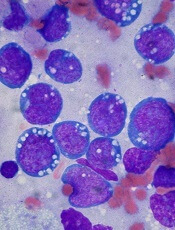
Credit: Ed Uthman
Preclinical research suggests a newly engineered protein can suppress tumor growth and extend survival in a mouse model of lymphoma.
The molecule, called BINDI (BHRF1-inhibiting design acting intracellularly), was designed to trigger the self-destruction of cancer cells infected with the Epstein-Barr virus (EBV).
EBV can disrupt the body’s clearance of old, abnormal, infected, and damaged cells. And BINDI works by overriding this interference.
Erik Procko, PhD, of the University of Washington in Seattle, and his colleagues described results observed with BINDI in Cell.
The researchers used computational design and experimental optimization to generate BINDI. The protein was designed to recognize and attach itself to an EBV protein called BHRF1 and to ignore similar proteins. BHRF1 keeps cancer cells alive, but, when bound to BINDI, it can no longer fend off cell death.
By examining the crystal structure of BINDI, the researchers saw that it nearly matched their computationally designed architecture for the protein molecule.
Furthermore, experiments showed that BINDI could prompt EBV-infected cancer cell lines to shrivel, disassemble their components, and burst into small pieces.
The researchers also tested BINDI in a mouse model of EBV-positive lymphoma. They delivered BINDI into cancer cells via an antibody-targeted nanocarrier designed to deliver protein cargo to intracellular cancer targets.
And BINDI behaved as ordered. It suppressed tumor growth and enabled the mice to live longer than control mice.
The researchers said this work demonstrates the potential to develop new classes of more effective, intracellular protein drugs, as current protein therapeutics are limited to extracellular targets.
Credit: Ed Uthman
Preclinical research suggests a newly engineered protein can suppress tumor growth and extend survival in a mouse model of lymphoma.
The molecule, called BINDI (BHRF1-inhibiting design acting intracellularly), was designed to trigger the self-destruction of cancer cells infected with the Epstein-Barr virus (EBV).
EBV can disrupt the body’s clearance of old, abnormal, infected, and damaged cells. And BINDI works by overriding this interference.
Erik Procko, PhD, of the University of Washington in Seattle, and his colleagues described results observed with BINDI in Cell.
The researchers used computational design and experimental optimization to generate BINDI. The protein was designed to recognize and attach itself to an EBV protein called BHRF1 and to ignore similar proteins. BHRF1 keeps cancer cells alive, but, when bound to BINDI, it can no longer fend off cell death.
By examining the crystal structure of BINDI, the researchers saw that it nearly matched their computationally designed architecture for the protein molecule.
Furthermore, experiments showed that BINDI could prompt EBV-infected cancer cell lines to shrivel, disassemble their components, and burst into small pieces.
The researchers also tested BINDI in a mouse model of EBV-positive lymphoma. They delivered BINDI into cancer cells via an antibody-targeted nanocarrier designed to deliver protein cargo to intracellular cancer targets.
And BINDI behaved as ordered. It suppressed tumor growth and enabled the mice to live longer than control mice.
The researchers said this work demonstrates the potential to develop new classes of more effective, intracellular protein drugs, as current protein therapeutics are limited to extracellular targets.
Credit: Ed Uthman
Preclinical research suggests a newly engineered protein can suppress tumor growth and extend survival in a mouse model of lymphoma.
The molecule, called BINDI (BHRF1-inhibiting design acting intracellularly), was designed to trigger the self-destruction of cancer cells infected with the Epstein-Barr virus (EBV).
EBV can disrupt the body’s clearance of old, abnormal, infected, and damaged cells. And BINDI works by overriding this interference.
Erik Procko, PhD, of the University of Washington in Seattle, and his colleagues described results observed with BINDI in Cell.
The researchers used computational design and experimental optimization to generate BINDI. The protein was designed to recognize and attach itself to an EBV protein called BHRF1 and to ignore similar proteins. BHRF1 keeps cancer cells alive, but, when bound to BINDI, it can no longer fend off cell death.
By examining the crystal structure of BINDI, the researchers saw that it nearly matched their computationally designed architecture for the protein molecule.
Furthermore, experiments showed that BINDI could prompt EBV-infected cancer cell lines to shrivel, disassemble their components, and burst into small pieces.
The researchers also tested BINDI in a mouse model of EBV-positive lymphoma. They delivered BINDI into cancer cells via an antibody-targeted nanocarrier designed to deliver protein cargo to intracellular cancer targets.
And BINDI behaved as ordered. It suppressed tumor growth and enabled the mice to live longer than control mice.
The researchers said this work demonstrates the potential to develop new classes of more effective, intracellular protein drugs, as current protein therapeutics are limited to extracellular targets.
Tool may predict cancer patients’ risk of financial stress

patient and her father
Credit: Rhoda Baer
A new questionnaire can measure a cancer patient’s risk for financial stress, according to a paper published in Cancer.
Researchers developed the 11-item questionnaire, called the COmprehensive Score for financial Toxicity (COST), through conversations with more than 150 cancer patients.
The team used the term “financial toxicity” to describe the expense, anxiety, and loss of confidence confronting patients who face big, unpredictable costs of cancer treatment.
And the researchers said financial toxicity can be considered another side effect of cancer care.
“Few physicians discuss this increasingly significant side effect with their patients,” said study author Jonas de Souza, MD, of the University of Chicago Medicine in Illinois.
“Physicians aren’t trained to do this. It makes them, as well as patients, feel uncomfortable. [However,] we believe that a thoughtful, concise tool that could help predict a patient’s risk for financial toxicity might open the lines of communication. This gives us a way to launch that discussion.”
Development of the COST questionnaire began with a literature review and a series of extensive interviews. Dr de Souza and his colleagues spoke with 20 patients and 6 cancer professionals, as well as nurses and social workers, and this produced a list of 147 questions.
The researchers pared the list down to 58 questions. Then, they asked 35 patients to help them decide which of the remaining questions were the most important. And the patients narrowed the list down to 30.
“In the end, 155 patients led us, with some judicious editing, to a set of 11 statements,” Dr de Souza said. “This was sufficiently brief to prevent annoying those responding to the questions but thorough enough to get us the information we need.”
All 11 entries are short and easy to understand, according to the researchers. For example, item 2 states, “My out-of-pocket medical expenses are more than I thought they would be.” And item 7 states, “I am able to meet my monthly expenses.”
For each question, patients choose from 5 potential responses: “not at all”, “a little bit,” “somewhat,” “quite a bit,” or “very much.”
Learning how a patient responds may help caregivers determine who is likely to need education, financial counseling, or referral to a support network. The quiz may also predict who is likely to have problems and require interventions.
All patients who helped develop the study had been in treatment for at least 2 months and had received bills. Excluding the top 10% and the bottom 10%, patients in the study earned between $37,000 and $111,000. The median annual income for these patients was about $63,000.
The researchers expected that financial toxicity would correlate with income.
“But, in our small sample, that did not hold up,” Dr de Souza said. “People with less education seemed to have more financial distress, but variations in income did not make much difference. We need bigger studies to confirm that, but at least we now have a tool we can use to study this.”
The researchers are now conducting a larger study to validate these findings and correlate the newly developed scale with quality of life and anxiety in cancer patients.
“We need to assess outcomes that are important for patients,” Dr de Souza said. “[T]his is another important piece of information in the shared-decision-making process.”
patient and her father
Credit: Rhoda Baer
A new questionnaire can measure a cancer patient’s risk for financial stress, according to a paper published in Cancer.
Researchers developed the 11-item questionnaire, called the COmprehensive Score for financial Toxicity (COST), through conversations with more than 150 cancer patients.
The team used the term “financial toxicity” to describe the expense, anxiety, and loss of confidence confronting patients who face big, unpredictable costs of cancer treatment.
And the researchers said financial toxicity can be considered another side effect of cancer care.
“Few physicians discuss this increasingly significant side effect with their patients,” said study author Jonas de Souza, MD, of the University of Chicago Medicine in Illinois.
“Physicians aren’t trained to do this. It makes them, as well as patients, feel uncomfortable. [However,] we believe that a thoughtful, concise tool that could help predict a patient’s risk for financial toxicity might open the lines of communication. This gives us a way to launch that discussion.”
Development of the COST questionnaire began with a literature review and a series of extensive interviews. Dr de Souza and his colleagues spoke with 20 patients and 6 cancer professionals, as well as nurses and social workers, and this produced a list of 147 questions.
The researchers pared the list down to 58 questions. Then, they asked 35 patients to help them decide which of the remaining questions were the most important. And the patients narrowed the list down to 30.
“In the end, 155 patients led us, with some judicious editing, to a set of 11 statements,” Dr de Souza said. “This was sufficiently brief to prevent annoying those responding to the questions but thorough enough to get us the information we need.”
All 11 entries are short and easy to understand, according to the researchers. For example, item 2 states, “My out-of-pocket medical expenses are more than I thought they would be.” And item 7 states, “I am able to meet my monthly expenses.”
For each question, patients choose from 5 potential responses: “not at all”, “a little bit,” “somewhat,” “quite a bit,” or “very much.”
Learning how a patient responds may help caregivers determine who is likely to need education, financial counseling, or referral to a support network. The quiz may also predict who is likely to have problems and require interventions.
All patients who helped develop the study had been in treatment for at least 2 months and had received bills. Excluding the top 10% and the bottom 10%, patients in the study earned between $37,000 and $111,000. The median annual income for these patients was about $63,000.
The researchers expected that financial toxicity would correlate with income.
“But, in our small sample, that did not hold up,” Dr de Souza said. “People with less education seemed to have more financial distress, but variations in income did not make much difference. We need bigger studies to confirm that, but at least we now have a tool we can use to study this.”
The researchers are now conducting a larger study to validate these findings and correlate the newly developed scale with quality of life and anxiety in cancer patients.
“We need to assess outcomes that are important for patients,” Dr de Souza said. “[T]his is another important piece of information in the shared-decision-making process.”
patient and her father
Credit: Rhoda Baer
A new questionnaire can measure a cancer patient’s risk for financial stress, according to a paper published in Cancer.
Researchers developed the 11-item questionnaire, called the COmprehensive Score for financial Toxicity (COST), through conversations with more than 150 cancer patients.
The team used the term “financial toxicity” to describe the expense, anxiety, and loss of confidence confronting patients who face big, unpredictable costs of cancer treatment.
And the researchers said financial toxicity can be considered another side effect of cancer care.
“Few physicians discuss this increasingly significant side effect with their patients,” said study author Jonas de Souza, MD, of the University of Chicago Medicine in Illinois.
“Physicians aren’t trained to do this. It makes them, as well as patients, feel uncomfortable. [However,] we believe that a thoughtful, concise tool that could help predict a patient’s risk for financial toxicity might open the lines of communication. This gives us a way to launch that discussion.”
Development of the COST questionnaire began with a literature review and a series of extensive interviews. Dr de Souza and his colleagues spoke with 20 patients and 6 cancer professionals, as well as nurses and social workers, and this produced a list of 147 questions.
The researchers pared the list down to 58 questions. Then, they asked 35 patients to help them decide which of the remaining questions were the most important. And the patients narrowed the list down to 30.
“In the end, 155 patients led us, with some judicious editing, to a set of 11 statements,” Dr de Souza said. “This was sufficiently brief to prevent annoying those responding to the questions but thorough enough to get us the information we need.”
All 11 entries are short and easy to understand, according to the researchers. For example, item 2 states, “My out-of-pocket medical expenses are more than I thought they would be.” And item 7 states, “I am able to meet my monthly expenses.”
For each question, patients choose from 5 potential responses: “not at all”, “a little bit,” “somewhat,” “quite a bit,” or “very much.”
Learning how a patient responds may help caregivers determine who is likely to need education, financial counseling, or referral to a support network. The quiz may also predict who is likely to have problems and require interventions.
All patients who helped develop the study had been in treatment for at least 2 months and had received bills. Excluding the top 10% and the bottom 10%, patients in the study earned between $37,000 and $111,000. The median annual income for these patients was about $63,000.
The researchers expected that financial toxicity would correlate with income.
“But, in our small sample, that did not hold up,” Dr de Souza said. “People with less education seemed to have more financial distress, but variations in income did not make much difference. We need bigger studies to confirm that, but at least we now have a tool we can use to study this.”
The researchers are now conducting a larger study to validate these findings and correlate the newly developed scale with quality of life and anxiety in cancer patients.
“We need to assess outcomes that are important for patients,” Dr de Souza said. “[T]his is another important piece of information in the shared-decision-making process.”
Low-dose T-cell transfer can fight CMV

CMV infection
A new study indicates that transferring low doses of immune cells may be sufficient to protect against cytomegalovirus (CMV) infection in patients receiving allogeneic stem cell transplant.
The researchers noted that the adoptive transfer of donor-derived, virus-specific, memory T cells is a promising strategy for treating and preventing CMV infection.
But transferring too many of these cells can increase the risk of graft-vs-host disease.
So Christian Stemberger, PhD, of Technische Universitaet Muenchen in Germany, and his colleagues set out to establish a lower limit for successful adoptive T-cell therapy.
The team reported their results—observed in mice and 2 human patients—in Blood.
The researchers first conducted low-dose CD8+ T-cell transfers in the murine Listeria monocytogenes infection model.
And they found these MHC-Streptamer-enriched, antigen-specific, CD62Lhi, CD8+ memory T cells proliferated, differentiated, and protected against Listeria monocytogenes infections.
“The most astonishing result was that the offspring cells of just 1 transferred donor cell were enough to completely protect the animals,” Dr Stemberger said.
Next, the researchers used virus-specific T cells to treat 2 critically ill pediatric patients—1 with severe combined immunodeficiency (SCID) and the other with B-cell acute lymphoblastic leukemia.
The patients had received allogeneic transplants and subsequently developed CMV infections.
The patients received low-dose transfers of Streptamer-enriched, CMV-specific, CD8+ T cells—3750 cells per kilogram of body weight for the SCID patient and 5130 cells per kilogram of body weight for the leukemia patient.
In both patients, the researchers observed “strong, pathogen-specific T-cell expansion.” The CMV-specific, CD8+ T cells proliferated, and the CMV viral load decreased. Furthermore, neither patient developed graft-vs-host disease.
“It is a great advantage that even just a few cells can provide protection [from CMV],” said study author Michael Neuenhahn, Dr med, also of Technische Universitaet Muenchen.
“This means that the cells can be used for preventive treatment in low doses that are gentler on the organism.”
The researchers are now testing the potential of the CMV-specific, CD8+ T cells in a clinical study.
CMV infection
A new study indicates that transferring low doses of immune cells may be sufficient to protect against cytomegalovirus (CMV) infection in patients receiving allogeneic stem cell transplant.
The researchers noted that the adoptive transfer of donor-derived, virus-specific, memory T cells is a promising strategy for treating and preventing CMV infection.
But transferring too many of these cells can increase the risk of graft-vs-host disease.
So Christian Stemberger, PhD, of Technische Universitaet Muenchen in Germany, and his colleagues set out to establish a lower limit for successful adoptive T-cell therapy.
The team reported their results—observed in mice and 2 human patients—in Blood.
The researchers first conducted low-dose CD8+ T-cell transfers in the murine Listeria monocytogenes infection model.
And they found these MHC-Streptamer-enriched, antigen-specific, CD62Lhi, CD8+ memory T cells proliferated, differentiated, and protected against Listeria monocytogenes infections.
“The most astonishing result was that the offspring cells of just 1 transferred donor cell were enough to completely protect the animals,” Dr Stemberger said.
Next, the researchers used virus-specific T cells to treat 2 critically ill pediatric patients—1 with severe combined immunodeficiency (SCID) and the other with B-cell acute lymphoblastic leukemia.
The patients had received allogeneic transplants and subsequently developed CMV infections.
The patients received low-dose transfers of Streptamer-enriched, CMV-specific, CD8+ T cells—3750 cells per kilogram of body weight for the SCID patient and 5130 cells per kilogram of body weight for the leukemia patient.
In both patients, the researchers observed “strong, pathogen-specific T-cell expansion.” The CMV-specific, CD8+ T cells proliferated, and the CMV viral load decreased. Furthermore, neither patient developed graft-vs-host disease.
“It is a great advantage that even just a few cells can provide protection [from CMV],” said study author Michael Neuenhahn, Dr med, also of Technische Universitaet Muenchen.
“This means that the cells can be used for preventive treatment in low doses that are gentler on the organism.”
The researchers are now testing the potential of the CMV-specific, CD8+ T cells in a clinical study.
CMV infection
A new study indicates that transferring low doses of immune cells may be sufficient to protect against cytomegalovirus (CMV) infection in patients receiving allogeneic stem cell transplant.
The researchers noted that the adoptive transfer of donor-derived, virus-specific, memory T cells is a promising strategy for treating and preventing CMV infection.
But transferring too many of these cells can increase the risk of graft-vs-host disease.
So Christian Stemberger, PhD, of Technische Universitaet Muenchen in Germany, and his colleagues set out to establish a lower limit for successful adoptive T-cell therapy.
The team reported their results—observed in mice and 2 human patients—in Blood.
The researchers first conducted low-dose CD8+ T-cell transfers in the murine Listeria monocytogenes infection model.
And they found these MHC-Streptamer-enriched, antigen-specific, CD62Lhi, CD8+ memory T cells proliferated, differentiated, and protected against Listeria monocytogenes infections.
“The most astonishing result was that the offspring cells of just 1 transferred donor cell were enough to completely protect the animals,” Dr Stemberger said.
Next, the researchers used virus-specific T cells to treat 2 critically ill pediatric patients—1 with severe combined immunodeficiency (SCID) and the other with B-cell acute lymphoblastic leukemia.
The patients had received allogeneic transplants and subsequently developed CMV infections.
The patients received low-dose transfers of Streptamer-enriched, CMV-specific, CD8+ T cells—3750 cells per kilogram of body weight for the SCID patient and 5130 cells per kilogram of body weight for the leukemia patient.
In both patients, the researchers observed “strong, pathogen-specific T-cell expansion.” The CMV-specific, CD8+ T cells proliferated, and the CMV viral load decreased. Furthermore, neither patient developed graft-vs-host disease.
“It is a great advantage that even just a few cells can provide protection [from CMV],” said study author Michael Neuenhahn, Dr med, also of Technische Universitaet Muenchen.
“This means that the cells can be used for preventive treatment in low doses that are gentler on the organism.”
The researchers are now testing the potential of the CMV-specific, CD8+ T cells in a clinical study.
Ruxolitinib improves disease control in PV

©ASCO/Phil McCarten
CHICAGO—The JAK1/2 inhibitor ruxolitinib may be a “valuable new treatment option” for patients with polycythemia vera (PV) who cannot tolerate or are resistant to hydroxyurea, according to a speaker at the 2014 ASCO Annual Meeting.
Results of the phase 3 RESPONSE trial showed that ruxolitinib can reduce spleen size in these patients and improve hematocrit control without the need for phlebotomy.
And the drug was generally well-tolerated.
“Patients with PV may not have their disease controlled with existing therapies, increasing their risk for cardiovascular complications,” said Srdan Verstovsek, MD, PhD, of MD Anderson Cancer Center in Houston, Texas.
“In the RESPONSE trial, patients treated with ruxolitinib showed better disease control and improved symptom management compared to [patients who received] current therapies.”
Dr Verstovsek presented results from the trial at ASCO as abstract 7026. RESPONSE was funded by Incyte Corporation, the company developing ruxolitinib.
The study has enrolled 222 patients with PV that is resistant to or intolerant of hydroxyurea. Patients were randomized to receive either ruxolitinib at a starting dose of 10 mg twice-daily or best available therapy (BAT). The ruxolitinib dose was adjusted as needed throughout the study.
At week 32, 77% of patients on ruxolitinib and 20% on BAT achieved hematocrit control or spleen reduction.
The primary endpoint of the study was the proportion of patients whose hematocrit was controlled without phlebotomy from week 8 through 32 and whose spleen volume was reduced by 35% or more from baseline as assessed by imaging at 32 weeks.
Twenty-one percent of patients in the ruxolitinib arm achieved this endpoint, compared to 1% of patients in the BAT arm.
“Ninety-one percent of patients who achieved the primary end point had a confirmed response at 48 weeks,” Dr Verstovsek said.
He also noted that nearly half of ruxolitinib-treated patients had a 50% or greater reduction in debilitating PV symptoms, compared to 5% of those on BAT.
Patients treated with ruxolitinib also experienced a reduction in night sweats and itchiness. In addition, a greater proportion of patients in the ruxolitinib arm achieved complete hematologic response when compared to the BAT arm—24% and 9%, respectively.
Ruxolitinib was generally well-tolerated, Dr Verstovsek said, noting that 85% of patients were still receiving the drug at a median follow-up of 81 weeks.
However, 3.6% of patients on ruxolitinib discontinued treatment due to adverse events, compared to 1.8% of patients on BAT.
Most adverse events observed in the ruxolitinib arm were grade 1 or 2 and were consistent with those previously seen in ruxolitinib studies in PV and myelofibrosis.
Few patients developed grade 3 or 4 cytopenias. Within the first 32 weeks of treatment, grade 3 or 4 hematologic adverse events in the ruxolitinib treatment arm were anemia (1.8%) and thrombocytopenia (5.5%). Fewer patients treated with ruxolitinib experienced thromboembolic events when compared to those who received BAT.
The most common non-hematologic adverse events were headache, diarrhea, and fatigue, which were mainly grade 1 or 2. Dr Verstovsek noted that herpes zoster infection was higher in the ruxolitinib arm than in the BAT arm.
Ruxolitinib is currently approved in more than 60 countries for patients with myelofibrosis. Global regulatory filings for PV are underway based on the RESPONSE data.
©ASCO/Phil McCarten
CHICAGO—The JAK1/2 inhibitor ruxolitinib may be a “valuable new treatment option” for patients with polycythemia vera (PV) who cannot tolerate or are resistant to hydroxyurea, according to a speaker at the 2014 ASCO Annual Meeting.
Results of the phase 3 RESPONSE trial showed that ruxolitinib can reduce spleen size in these patients and improve hematocrit control without the need for phlebotomy.
And the drug was generally well-tolerated.
“Patients with PV may not have their disease controlled with existing therapies, increasing their risk for cardiovascular complications,” said Srdan Verstovsek, MD, PhD, of MD Anderson Cancer Center in Houston, Texas.
“In the RESPONSE trial, patients treated with ruxolitinib showed better disease control and improved symptom management compared to [patients who received] current therapies.”
Dr Verstovsek presented results from the trial at ASCO as abstract 7026. RESPONSE was funded by Incyte Corporation, the company developing ruxolitinib.
The study has enrolled 222 patients with PV that is resistant to or intolerant of hydroxyurea. Patients were randomized to receive either ruxolitinib at a starting dose of 10 mg twice-daily or best available therapy (BAT). The ruxolitinib dose was adjusted as needed throughout the study.
At week 32, 77% of patients on ruxolitinib and 20% on BAT achieved hematocrit control or spleen reduction.
The primary endpoint of the study was the proportion of patients whose hematocrit was controlled without phlebotomy from week 8 through 32 and whose spleen volume was reduced by 35% or more from baseline as assessed by imaging at 32 weeks.
Twenty-one percent of patients in the ruxolitinib arm achieved this endpoint, compared to 1% of patients in the BAT arm.
“Ninety-one percent of patients who achieved the primary end point had a confirmed response at 48 weeks,” Dr Verstovsek said.
He also noted that nearly half of ruxolitinib-treated patients had a 50% or greater reduction in debilitating PV symptoms, compared to 5% of those on BAT.
Patients treated with ruxolitinib also experienced a reduction in night sweats and itchiness. In addition, a greater proportion of patients in the ruxolitinib arm achieved complete hematologic response when compared to the BAT arm—24% and 9%, respectively.
Ruxolitinib was generally well-tolerated, Dr Verstovsek said, noting that 85% of patients were still receiving the drug at a median follow-up of 81 weeks.
However, 3.6% of patients on ruxolitinib discontinued treatment due to adverse events, compared to 1.8% of patients on BAT.
Most adverse events observed in the ruxolitinib arm were grade 1 or 2 and were consistent with those previously seen in ruxolitinib studies in PV and myelofibrosis.
Few patients developed grade 3 or 4 cytopenias. Within the first 32 weeks of treatment, grade 3 or 4 hematologic adverse events in the ruxolitinib treatment arm were anemia (1.8%) and thrombocytopenia (5.5%). Fewer patients treated with ruxolitinib experienced thromboembolic events when compared to those who received BAT.
The most common non-hematologic adverse events were headache, diarrhea, and fatigue, which were mainly grade 1 or 2. Dr Verstovsek noted that herpes zoster infection was higher in the ruxolitinib arm than in the BAT arm.
Ruxolitinib is currently approved in more than 60 countries for patients with myelofibrosis. Global regulatory filings for PV are underway based on the RESPONSE data.
©ASCO/Phil McCarten
CHICAGO—The JAK1/2 inhibitor ruxolitinib may be a “valuable new treatment option” for patients with polycythemia vera (PV) who cannot tolerate or are resistant to hydroxyurea, according to a speaker at the 2014 ASCO Annual Meeting.
Results of the phase 3 RESPONSE trial showed that ruxolitinib can reduce spleen size in these patients and improve hematocrit control without the need for phlebotomy.
And the drug was generally well-tolerated.
“Patients with PV may not have their disease controlled with existing therapies, increasing their risk for cardiovascular complications,” said Srdan Verstovsek, MD, PhD, of MD Anderson Cancer Center in Houston, Texas.
“In the RESPONSE trial, patients treated with ruxolitinib showed better disease control and improved symptom management compared to [patients who received] current therapies.”
Dr Verstovsek presented results from the trial at ASCO as abstract 7026. RESPONSE was funded by Incyte Corporation, the company developing ruxolitinib.
The study has enrolled 222 patients with PV that is resistant to or intolerant of hydroxyurea. Patients were randomized to receive either ruxolitinib at a starting dose of 10 mg twice-daily or best available therapy (BAT). The ruxolitinib dose was adjusted as needed throughout the study.
At week 32, 77% of patients on ruxolitinib and 20% on BAT achieved hematocrit control or spleen reduction.
The primary endpoint of the study was the proportion of patients whose hematocrit was controlled without phlebotomy from week 8 through 32 and whose spleen volume was reduced by 35% or more from baseline as assessed by imaging at 32 weeks.
Twenty-one percent of patients in the ruxolitinib arm achieved this endpoint, compared to 1% of patients in the BAT arm.
“Ninety-one percent of patients who achieved the primary end point had a confirmed response at 48 weeks,” Dr Verstovsek said.
He also noted that nearly half of ruxolitinib-treated patients had a 50% or greater reduction in debilitating PV symptoms, compared to 5% of those on BAT.
Patients treated with ruxolitinib also experienced a reduction in night sweats and itchiness. In addition, a greater proportion of patients in the ruxolitinib arm achieved complete hematologic response when compared to the BAT arm—24% and 9%, respectively.
Ruxolitinib was generally well-tolerated, Dr Verstovsek said, noting that 85% of patients were still receiving the drug at a median follow-up of 81 weeks.
However, 3.6% of patients on ruxolitinib discontinued treatment due to adverse events, compared to 1.8% of patients on BAT.
Most adverse events observed in the ruxolitinib arm were grade 1 or 2 and were consistent with those previously seen in ruxolitinib studies in PV and myelofibrosis.
Few patients developed grade 3 or 4 cytopenias. Within the first 32 weeks of treatment, grade 3 or 4 hematologic adverse events in the ruxolitinib treatment arm were anemia (1.8%) and thrombocytopenia (5.5%). Fewer patients treated with ruxolitinib experienced thromboembolic events when compared to those who received BAT.
The most common non-hematologic adverse events were headache, diarrhea, and fatigue, which were mainly grade 1 or 2. Dr Verstovsek noted that herpes zoster infection was higher in the ruxolitinib arm than in the BAT arm.
Ruxolitinib is currently approved in more than 60 countries for patients with myelofibrosis. Global regulatory filings for PV are underway based on the RESPONSE data.
Treating HIV+ lymphoma patients

©ASCO/Brian Powers
CHICAGO—Hepatitis C reactivation does not worsen survival outcomes for HIV-positive patients diagnosed with lymphoma, new research indicates.
The study showed these patients can tolerate chemotherapy without adverse outcomes and are therefore eligible for aggressive treatment.
They should be closely monitored, however, according to study investigator Stefan K. Barta, MD, of Fox Chase Cancer Center in Philadelphia, Pennsylvania.
Dr Barta and his colleagues presented results observed in HIV-positive lymphoma patients at the 2014 ASCO Annual Meeting as abstract 8578.
The team noted that more than a quarter of HIV-positive patients are also infected with the hepatitis C virus (HCV), which may complicate treatment and care decisions after a cancer diagnosis.
“Patients undergoing chemotherapy can experience reactivation of the hepatitis C virus, which, in turn, can lead to liver failure,” Dr Barta said. “This means we have to dose-reduce chemotherapy, which could negatively affect outcomes.”
In addition, HIV-positive patients often take a host of other medications, including anti-retrovirals, which makes them especially vulnerable to side effects.
However, Dr Barta said these potential risks shouldn’t deter oncologists from treating these patients with chemotherapy because he and his colleagues found that reactivation of HCV did not worsen survival outcomes in this population.
The researchers analyzed the medical records of 190 HIV-positive patients who had been diagnosed with lymphoma at the Albert Einstein Cancer Center in Bronx, New York, from 1997 to 2013. Patients with primary central nervous system lymphomas were excluded.
Fifty-three (28%) eligible patients were also infected with HCV. The virus reactivated in 17 of those patients, or about one-third of the patient population infected with HCV, during treatment.
Patients infected with HCV had an overall survival of 59.7 months, compared to 88.6 months for patients with neither HCV nor hepatitis B virus (HBV).
However, that survival advantage vanished when the researchers adjusted for variables including age, sex, race, CD4 count, the presence of cirrhosis, type of lymphoma, and levels of lactate dehydrogenase (LDH).
The multivariate analysis showed that co-infection with HCV was not associated with lower overall survival in lymphoma patients.
At the same time, the researchers did find worse overall survival outcomes associated with low CD4 count (below 100 cells/cubic millimeter), a diagnosis of non-Hodgkin lymphoma, advanced stage disease, LDH levels over 190, or cirrhosis.
Dr Barta said he hopes this research will open cancer trials up to an understudied patient population. HIV-positive patients with HCV are often excluded from these trials because of concerns about liver failure and toxicity arising from the interaction of retroviral medications with chemotherapy.
“This is really important for a large proportion of patients,” he said. “We want to assure researchers that these patients, as long as they have adequate liver function, should also be enrolled in clinical trials.”
©ASCO/Brian Powers
CHICAGO—Hepatitis C reactivation does not worsen survival outcomes for HIV-positive patients diagnosed with lymphoma, new research indicates.
The study showed these patients can tolerate chemotherapy without adverse outcomes and are therefore eligible for aggressive treatment.
They should be closely monitored, however, according to study investigator Stefan K. Barta, MD, of Fox Chase Cancer Center in Philadelphia, Pennsylvania.
Dr Barta and his colleagues presented results observed in HIV-positive lymphoma patients at the 2014 ASCO Annual Meeting as abstract 8578.
The team noted that more than a quarter of HIV-positive patients are also infected with the hepatitis C virus (HCV), which may complicate treatment and care decisions after a cancer diagnosis.
“Patients undergoing chemotherapy can experience reactivation of the hepatitis C virus, which, in turn, can lead to liver failure,” Dr Barta said. “This means we have to dose-reduce chemotherapy, which could negatively affect outcomes.”
In addition, HIV-positive patients often take a host of other medications, including anti-retrovirals, which makes them especially vulnerable to side effects.
However, Dr Barta said these potential risks shouldn’t deter oncologists from treating these patients with chemotherapy because he and his colleagues found that reactivation of HCV did not worsen survival outcomes in this population.
The researchers analyzed the medical records of 190 HIV-positive patients who had been diagnosed with lymphoma at the Albert Einstein Cancer Center in Bronx, New York, from 1997 to 2013. Patients with primary central nervous system lymphomas were excluded.
Fifty-three (28%) eligible patients were also infected with HCV. The virus reactivated in 17 of those patients, or about one-third of the patient population infected with HCV, during treatment.
Patients infected with HCV had an overall survival of 59.7 months, compared to 88.6 months for patients with neither HCV nor hepatitis B virus (HBV).
However, that survival advantage vanished when the researchers adjusted for variables including age, sex, race, CD4 count, the presence of cirrhosis, type of lymphoma, and levels of lactate dehydrogenase (LDH).
The multivariate analysis showed that co-infection with HCV was not associated with lower overall survival in lymphoma patients.
At the same time, the researchers did find worse overall survival outcomes associated with low CD4 count (below 100 cells/cubic millimeter), a diagnosis of non-Hodgkin lymphoma, advanced stage disease, LDH levels over 190, or cirrhosis.
Dr Barta said he hopes this research will open cancer trials up to an understudied patient population. HIV-positive patients with HCV are often excluded from these trials because of concerns about liver failure and toxicity arising from the interaction of retroviral medications with chemotherapy.
“This is really important for a large proportion of patients,” he said. “We want to assure researchers that these patients, as long as they have adequate liver function, should also be enrolled in clinical trials.”
©ASCO/Brian Powers
CHICAGO—Hepatitis C reactivation does not worsen survival outcomes for HIV-positive patients diagnosed with lymphoma, new research indicates.
The study showed these patients can tolerate chemotherapy without adverse outcomes and are therefore eligible for aggressive treatment.
They should be closely monitored, however, according to study investigator Stefan K. Barta, MD, of Fox Chase Cancer Center in Philadelphia, Pennsylvania.
Dr Barta and his colleagues presented results observed in HIV-positive lymphoma patients at the 2014 ASCO Annual Meeting as abstract 8578.
The team noted that more than a quarter of HIV-positive patients are also infected with the hepatitis C virus (HCV), which may complicate treatment and care decisions after a cancer diagnosis.
“Patients undergoing chemotherapy can experience reactivation of the hepatitis C virus, which, in turn, can lead to liver failure,” Dr Barta said. “This means we have to dose-reduce chemotherapy, which could negatively affect outcomes.”
In addition, HIV-positive patients often take a host of other medications, including anti-retrovirals, which makes them especially vulnerable to side effects.
However, Dr Barta said these potential risks shouldn’t deter oncologists from treating these patients with chemotherapy because he and his colleagues found that reactivation of HCV did not worsen survival outcomes in this population.
The researchers analyzed the medical records of 190 HIV-positive patients who had been diagnosed with lymphoma at the Albert Einstein Cancer Center in Bronx, New York, from 1997 to 2013. Patients with primary central nervous system lymphomas were excluded.
Fifty-three (28%) eligible patients were also infected with HCV. The virus reactivated in 17 of those patients, or about one-third of the patient population infected with HCV, during treatment.
Patients infected with HCV had an overall survival of 59.7 months, compared to 88.6 months for patients with neither HCV nor hepatitis B virus (HBV).
However, that survival advantage vanished when the researchers adjusted for variables including age, sex, race, CD4 count, the presence of cirrhosis, type of lymphoma, and levels of lactate dehydrogenase (LDH).
The multivariate analysis showed that co-infection with HCV was not associated with lower overall survival in lymphoma patients.
At the same time, the researchers did find worse overall survival outcomes associated with low CD4 count (below 100 cells/cubic millimeter), a diagnosis of non-Hodgkin lymphoma, advanced stage disease, LDH levels over 190, or cirrhosis.
Dr Barta said he hopes this research will open cancer trials up to an understudied patient population. HIV-positive patients with HCV are often excluded from these trials because of concerns about liver failure and toxicity arising from the interaction of retroviral medications with chemotherapy.
“This is really important for a large proportion of patients,” he said. “We want to assure researchers that these patients, as long as they have adequate liver function, should also be enrolled in clinical trials.”
FDA warns of VTE related to testosterone products
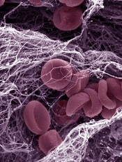
Credit: Kevin MacKenzie
The US Food and Drug Administration (FDA) is taking additional action to warn the public about the risk of venous thromboembolism (VTE) associated with the use of testosterone products.
The agency is now requiring manufacturers to include a general warning in the drug labeling of all approved testosterone products about the risk of VTE these products pose.
The risk is already included in the labeling as a possible consequence of polycythemia, which sometimes occurs with testosterone treatment.
But there have been post-market reports of VTE unrelated to polycythemia.
So the FDA has mandated the label change to provide a more general warning and ensure the risk of VTE is described consistently in the labeling of all approved testosterone products.
This new warning is not related to FDA’s ongoing evaluation of the possible risk of stroke, heart attack, and death in patients taking testosterone products.
The agency is currently evaluating the potential risk of these events, which are described in the Drug Safety Communication posted on January 31, 2014.
Testosterone products are FDA-approved for use in men who lack testosterone or have low testosterone levels in conjunction with an associated medical condition.
The FDA is asking healthcare professionals and consumers to report any adverse reactions related to testosterone products to the agency’s MedWatch Safety Information and Adverse Event Reporting program.
Credit: Kevin MacKenzie
The US Food and Drug Administration (FDA) is taking additional action to warn the public about the risk of venous thromboembolism (VTE) associated with the use of testosterone products.
The agency is now requiring manufacturers to include a general warning in the drug labeling of all approved testosterone products about the risk of VTE these products pose.
The risk is already included in the labeling as a possible consequence of polycythemia, which sometimes occurs with testosterone treatment.
But there have been post-market reports of VTE unrelated to polycythemia.
So the FDA has mandated the label change to provide a more general warning and ensure the risk of VTE is described consistently in the labeling of all approved testosterone products.
This new warning is not related to FDA’s ongoing evaluation of the possible risk of stroke, heart attack, and death in patients taking testosterone products.
The agency is currently evaluating the potential risk of these events, which are described in the Drug Safety Communication posted on January 31, 2014.
Testosterone products are FDA-approved for use in men who lack testosterone or have low testosterone levels in conjunction with an associated medical condition.
The FDA is asking healthcare professionals and consumers to report any adverse reactions related to testosterone products to the agency’s MedWatch Safety Information and Adverse Event Reporting program.
Credit: Kevin MacKenzie
The US Food and Drug Administration (FDA) is taking additional action to warn the public about the risk of venous thromboembolism (VTE) associated with the use of testosterone products.
The agency is now requiring manufacturers to include a general warning in the drug labeling of all approved testosterone products about the risk of VTE these products pose.
The risk is already included in the labeling as a possible consequence of polycythemia, which sometimes occurs with testosterone treatment.
But there have been post-market reports of VTE unrelated to polycythemia.
So the FDA has mandated the label change to provide a more general warning and ensure the risk of VTE is described consistently in the labeling of all approved testosterone products.
This new warning is not related to FDA’s ongoing evaluation of the possible risk of stroke, heart attack, and death in patients taking testosterone products.
The agency is currently evaluating the potential risk of these events, which are described in the Drug Safety Communication posted on January 31, 2014.
Testosterone products are FDA-approved for use in men who lack testosterone or have low testosterone levels in conjunction with an associated medical condition.
The FDA is asking healthcare professionals and consumers to report any adverse reactions related to testosterone products to the agency’s MedWatch Safety Information and Adverse Event Reporting program.