User login
CHMP recommends apixaban for VTE

Credit: Andre E.X. Brown
The European Medicines Agency’s Committee for Medicinal Products for Human Use (CHMP) has recommended that apixaban (Eliquis) be granted marketing authorization for the treatment and prevention of venous thromboembolism (VTE) in adults.
The European Commission will take the CHMP’s opinion into account when deciding whether to approve the drug for this indication in all 28 European Union member states, plus Iceland and Norway.
The CHMP’s recommendation of apixaban was based on results from the AMPLIFY and AMPLIFY-EXTENSION studies.
The phase 3 AMPLIFY trial enrolled 5395 patients with confirmed, symptomatic deep vein thrombosis (DVT) or pulmonary embolism (PE) requiring treatment for 6 months.
About half of patients (n=2691) were randomized to apixaban, and the other half (n=2704) were randomized to standard of care, which was initial enoxaparin treatment overlapped by warfarin therapy.
The primary efficacy outcome was the composite endpoint of recurrent, symptomatic VTE or VTE-related death. This occurred in 59 patients in the apixaban arm (2.3%) and 71 patients (2.7%) in the standard-therapy arm (P<0.0001 for noninferiority).
The composite endpoint of major bleeding and clinically relevant, nonmajor bleeding occurred in 4.3% of patients in the apixaban arm and 9.7% of patients in the standard-therapy arm (P<0.001).
The phase 3 AMPLIFY-EXTENSION trial included 2486 patients with prior VTE who had completed 6 to 12 months of anticoagulation treatment for DVT or PE.
Eight hundred and forty-two patients were randomized to apixaban at 2.5 mg, 815 were randomized to apixaban at 5 mg, and 829 were randomized to placebo.
The study’s primary efficacy endpoint was recurrent VTE or all-cause death. During the 12-month active study period, these events occurred in 32 patients (3.8%) in the 2.5-mg arm, 34 patients (4.2%) in the 5-mg arm, and 96 patients (11.6%) in the placebo arm. Both apixaban doses were significantly superior to placebo (P<0.001).
The primary safety endpoint was the incidence of major bleeding, which occurred in 2 patients (0.2%) in the 2.5-mg arm, 1 patient (0.1%) in the 5-mg arm, and 4 patients (0.5%) in the placebo arm. 
Credit: Andre E.X. Brown
The European Medicines Agency’s Committee for Medicinal Products for Human Use (CHMP) has recommended that apixaban (Eliquis) be granted marketing authorization for the treatment and prevention of venous thromboembolism (VTE) in adults.
The European Commission will take the CHMP’s opinion into account when deciding whether to approve the drug for this indication in all 28 European Union member states, plus Iceland and Norway.
The CHMP’s recommendation of apixaban was based on results from the AMPLIFY and AMPLIFY-EXTENSION studies.
The phase 3 AMPLIFY trial enrolled 5395 patients with confirmed, symptomatic deep vein thrombosis (DVT) or pulmonary embolism (PE) requiring treatment for 6 months.
About half of patients (n=2691) were randomized to apixaban, and the other half (n=2704) were randomized to standard of care, which was initial enoxaparin treatment overlapped by warfarin therapy.
The primary efficacy outcome was the composite endpoint of recurrent, symptomatic VTE or VTE-related death. This occurred in 59 patients in the apixaban arm (2.3%) and 71 patients (2.7%) in the standard-therapy arm (P<0.0001 for noninferiority).
The composite endpoint of major bleeding and clinically relevant, nonmajor bleeding occurred in 4.3% of patients in the apixaban arm and 9.7% of patients in the standard-therapy arm (P<0.001).
The phase 3 AMPLIFY-EXTENSION trial included 2486 patients with prior VTE who had completed 6 to 12 months of anticoagulation treatment for DVT or PE.
Eight hundred and forty-two patients were randomized to apixaban at 2.5 mg, 815 were randomized to apixaban at 5 mg, and 829 were randomized to placebo.
The study’s primary efficacy endpoint was recurrent VTE or all-cause death. During the 12-month active study period, these events occurred in 32 patients (3.8%) in the 2.5-mg arm, 34 patients (4.2%) in the 5-mg arm, and 96 patients (11.6%) in the placebo arm. Both apixaban doses were significantly superior to placebo (P<0.001).
The primary safety endpoint was the incidence of major bleeding, which occurred in 2 patients (0.2%) in the 2.5-mg arm, 1 patient (0.1%) in the 5-mg arm, and 4 patients (0.5%) in the placebo arm.
Credit: Andre E.X. Brown
The European Medicines Agency’s Committee for Medicinal Products for Human Use (CHMP) has recommended that apixaban (Eliquis) be granted marketing authorization for the treatment and prevention of venous thromboembolism (VTE) in adults.
The European Commission will take the CHMP’s opinion into account when deciding whether to approve the drug for this indication in all 28 European Union member states, plus Iceland and Norway.
The CHMP’s recommendation of apixaban was based on results from the AMPLIFY and AMPLIFY-EXTENSION studies.
The phase 3 AMPLIFY trial enrolled 5395 patients with confirmed, symptomatic deep vein thrombosis (DVT) or pulmonary embolism (PE) requiring treatment for 6 months.
About half of patients (n=2691) were randomized to apixaban, and the other half (n=2704) were randomized to standard of care, which was initial enoxaparin treatment overlapped by warfarin therapy.
The primary efficacy outcome was the composite endpoint of recurrent, symptomatic VTE or VTE-related death. This occurred in 59 patients in the apixaban arm (2.3%) and 71 patients (2.7%) in the standard-therapy arm (P<0.0001 for noninferiority).
The composite endpoint of major bleeding and clinically relevant, nonmajor bleeding occurred in 4.3% of patients in the apixaban arm and 9.7% of patients in the standard-therapy arm (P<0.001).
The phase 3 AMPLIFY-EXTENSION trial included 2486 patients with prior VTE who had completed 6 to 12 months of anticoagulation treatment for DVT or PE.
Eight hundred and forty-two patients were randomized to apixaban at 2.5 mg, 815 were randomized to apixaban at 5 mg, and 829 were randomized to placebo.
The study’s primary efficacy endpoint was recurrent VTE or all-cause death. During the 12-month active study period, these events occurred in 32 patients (3.8%) in the 2.5-mg arm, 34 patients (4.2%) in the 5-mg arm, and 96 patients (11.6%) in the placebo arm. Both apixaban doses were significantly superior to placebo (P<0.001).
The primary safety endpoint was the incidence of major bleeding, which occurred in 2 patients (0.2%) in the 2.5-mg arm, 1 patient (0.1%) in the 5-mg arm, and 4 patients (0.5%) in the placebo arm.
VSTs can target up to 5 viruses

for Cell and Gene Therapy
at Baylor College of Medicine
New virus-specific T-cells (VSTs) can effectively target multiple viruses, thereby clearing and preventing infections in transplant recipients, researchers have reported in Science Translational Medicine.
VSTs have proven effective in previous studies, but they are typically costly, can take months to produce, and often target a single virus.
The new VSTs took about 10 days to produce, are more cost-effective than standard therapy, and can target up to 5 viruses, according to the researchers.
Ann Leen, PhD, of the Baylor College of Medicine in Houston, and her colleagues developed the technique to produce the VSTs, which can target Epstein-Barr virus (EBV), adenovirus (ADV), cytomegalovirus (CMV), BK virus (BKV), and human herpesvirus 6 (HHV6).
“Unlike conventional antiviral drugs, our therapy improves virus-specific T-cell immunity—the root cause of post-transplant viral infections—providing both an effective and safe strategy to treat viruses,” Dr Leen said.
“Additionally, we can readily produce both individualized products and T-cell banks for third-party use, facilitating the extension of T-cell therapy to a standard of care for transplant recipients.”
The researchers generated 48 VST cell lines by stimulating peripheral blood mononuclear cells from allogeneic donors with overlapping peptide libraries that incorporate EBV, CMV, ADV, BKV, and HHV6 antigens.
The team then tested these VSTs in 11 patients who had received allogeneic transplants to treat leukemia, lymphoma, sickle cell disease, and other hematologic and immunodeficient disorders.
Eight of the patients had up to 4 active infections with the targeted viruses. Three patients received the VSTs to prevent infection.
The VSTs produced an overall 94% virological and clinical response that was sustained long-term. Of the 3 patients who received VSTs prophylactically, all remained free of infection for more than 3 months after infusion.
There were no immediate infusion-related toxicities. One patient developed de novo graft-vs-host disease of the skin that improved with topical steroids.
Two patients who received VSTs as prophylaxis developed transplant-associated microangiopathy, but the researchers considered this to be unrelated to VST infusion.
They noted that this is the first time BKV and HHV6 reactivations have been controlled using VSTs. Dr Leen’s team had previously reported promising results with VSTs targeting EBV, CMV, and ADV.
“This study translated improved manufacturing techniques developed in Dr Leen’s laboratory to the clinic and showed that virus-specific T cells produced with the new method could target new viruses and be ready for clinical use after 10 days,” said Helen Heslop, MD, also of Baylor College of Medicine.
“These advances mean that this therapy could be available for more patients to treat viral infections and provide long-lasting protection.”
for Cell and Gene Therapy
at Baylor College of Medicine
New virus-specific T-cells (VSTs) can effectively target multiple viruses, thereby clearing and preventing infections in transplant recipients, researchers have reported in Science Translational Medicine.
VSTs have proven effective in previous studies, but they are typically costly, can take months to produce, and often target a single virus.
The new VSTs took about 10 days to produce, are more cost-effective than standard therapy, and can target up to 5 viruses, according to the researchers.
Ann Leen, PhD, of the Baylor College of Medicine in Houston, and her colleagues developed the technique to produce the VSTs, which can target Epstein-Barr virus (EBV), adenovirus (ADV), cytomegalovirus (CMV), BK virus (BKV), and human herpesvirus 6 (HHV6).
“Unlike conventional antiviral drugs, our therapy improves virus-specific T-cell immunity—the root cause of post-transplant viral infections—providing both an effective and safe strategy to treat viruses,” Dr Leen said.
“Additionally, we can readily produce both individualized products and T-cell banks for third-party use, facilitating the extension of T-cell therapy to a standard of care for transplant recipients.”
The researchers generated 48 VST cell lines by stimulating peripheral blood mononuclear cells from allogeneic donors with overlapping peptide libraries that incorporate EBV, CMV, ADV, BKV, and HHV6 antigens.
The team then tested these VSTs in 11 patients who had received allogeneic transplants to treat leukemia, lymphoma, sickle cell disease, and other hematologic and immunodeficient disorders.
Eight of the patients had up to 4 active infections with the targeted viruses. Three patients received the VSTs to prevent infection.
The VSTs produced an overall 94% virological and clinical response that was sustained long-term. Of the 3 patients who received VSTs prophylactically, all remained free of infection for more than 3 months after infusion.
There were no immediate infusion-related toxicities. One patient developed de novo graft-vs-host disease of the skin that improved with topical steroids.
Two patients who received VSTs as prophylaxis developed transplant-associated microangiopathy, but the researchers considered this to be unrelated to VST infusion.
They noted that this is the first time BKV and HHV6 reactivations have been controlled using VSTs. Dr Leen’s team had previously reported promising results with VSTs targeting EBV, CMV, and ADV.
“This study translated improved manufacturing techniques developed in Dr Leen’s laboratory to the clinic and showed that virus-specific T cells produced with the new method could target new viruses and be ready for clinical use after 10 days,” said Helen Heslop, MD, also of Baylor College of Medicine.
“These advances mean that this therapy could be available for more patients to treat viral infections and provide long-lasting protection.”
for Cell and Gene Therapy
at Baylor College of Medicine
New virus-specific T-cells (VSTs) can effectively target multiple viruses, thereby clearing and preventing infections in transplant recipients, researchers have reported in Science Translational Medicine.
VSTs have proven effective in previous studies, but they are typically costly, can take months to produce, and often target a single virus.
The new VSTs took about 10 days to produce, are more cost-effective than standard therapy, and can target up to 5 viruses, according to the researchers.
Ann Leen, PhD, of the Baylor College of Medicine in Houston, and her colleagues developed the technique to produce the VSTs, which can target Epstein-Barr virus (EBV), adenovirus (ADV), cytomegalovirus (CMV), BK virus (BKV), and human herpesvirus 6 (HHV6).
“Unlike conventional antiviral drugs, our therapy improves virus-specific T-cell immunity—the root cause of post-transplant viral infections—providing both an effective and safe strategy to treat viruses,” Dr Leen said.
“Additionally, we can readily produce both individualized products and T-cell banks for third-party use, facilitating the extension of T-cell therapy to a standard of care for transplant recipients.”
The researchers generated 48 VST cell lines by stimulating peripheral blood mononuclear cells from allogeneic donors with overlapping peptide libraries that incorporate EBV, CMV, ADV, BKV, and HHV6 antigens.
The team then tested these VSTs in 11 patients who had received allogeneic transplants to treat leukemia, lymphoma, sickle cell disease, and other hematologic and immunodeficient disorders.
Eight of the patients had up to 4 active infections with the targeted viruses. Three patients received the VSTs to prevent infection.
The VSTs produced an overall 94% virological and clinical response that was sustained long-term. Of the 3 patients who received VSTs prophylactically, all remained free of infection for more than 3 months after infusion.
There were no immediate infusion-related toxicities. One patient developed de novo graft-vs-host disease of the skin that improved with topical steroids.
Two patients who received VSTs as prophylaxis developed transplant-associated microangiopathy, but the researchers considered this to be unrelated to VST infusion.
They noted that this is the first time BKV and HHV6 reactivations have been controlled using VSTs. Dr Leen’s team had previously reported promising results with VSTs targeting EBV, CMV, and ADV.
“This study translated improved manufacturing techniques developed in Dr Leen’s laboratory to the clinic and showed that virus-specific T cells produced with the new method could target new viruses and be ready for clinical use after 10 days,” said Helen Heslop, MD, also of Baylor College of Medicine.
“These advances mean that this therapy could be available for more patients to treat viral infections and provide long-lasting protection.”
System can attenuate thrombin generation

Credit: Kevin MacKenzie
Results of a phase 1 trial suggest the REG2 anticoagulation system can attenuate thrombin generation and then restore thrombin following reversal.
The REG2 system consists of pegnivacogin, a subcutaneously administered aptamer factor IXa inhibitor, and its intravenous active control agent, anivamersen.
REG2 is formulated to provide a controllable level of anticoagulation for up to 2 weeks for sub-acute uses, especially in cases where a patient may be unable to swallow an oral anticoagulant.
Results observed with REG2 were published in the Journal of Thrombosis and Thrombolysis. The study was sponsored by Regado Biosciences, Inc, the company developing the REG2 system.
The study included 32 healthy volunteers who were enrolled sequentially into 4 cohorts. Patients in cohorts 1-3 were randomized (3:1) to ascending doses of pegnivacogin—from 0.5 mg/kg to 3 mg/kg—or placebo, with no anivamersen administered.
In cohort 4, all patients received 2 mg/kg of pegnivacogin and were randomized (1:1) to reversal by 1 dose or multiple doses over time of 1 mg/kg of anivamersen.
The researchers drew blood samples in the presence or absence of corn trypsin inhibitor at specified times within each dosing cohort. They initiated thrombin generation with tissue factor and measured thrombin generation kinetics using the calibrated automated thrombogram (CAT).
The team found that REG2 attenuated thrombin generation in a dose-dependent manner. All parameters of the CAT assay, except for lag time, showed a dose- and concentration-dependent response to pegnivacogin.
Specifically, there was a decrease in velocity index (VIx) from baseline to 24 hours after pegnivacogin administration. The largest percent change in VIx was in cohort 3 (3 mg/kg). There was a 90% decrease at 24 hours post-dose. This remained stable up to 48 hours in cohort 1 and up to 96 hours in cohorts 2 and 3.
Similarly, mean peak thrombin generation (PTG) values decreased as pegnivacogin dose and plasma concentrations increased. Cohort 3 had an 80% decrease in PTG at 24 hours post-dose. The percent change in mean PTG was consistent and preserved for at least 24 hours for all pegnivacogin doses. In cohorts 2 and 3, it was stable for at least 96 hours.
Endogenous thrombin potential (ETP) levels also decreased after pegnivacogin administration. Again, the largest decrease occurred in cohort 3, which exhibited a 65% decrease at 24 hours. Beyond that, the percent change in ETP was stable within each dose group, up to the last recorded measurement at 96 hours.
Peak time (PTm) was positively correlated with pegnivacogin concentration and relative activated partial thromboplastin time values. The correlation between PTm and quartile of pegnivacogin concentration was statistically significant (P=0.04) but weaker than that of ETP (P=0.001), PTG (P<0.001), or VIx (P<0.001). The changes in PTm persisted for at least 96 hours after pegnivacogin administration.
Finally, the researchers found that anivamersen reversed the effects of pegnivacogin, restoring thrombin generation without rebound effect.
Credit: Kevin MacKenzie
Results of a phase 1 trial suggest the REG2 anticoagulation system can attenuate thrombin generation and then restore thrombin following reversal.
The REG2 system consists of pegnivacogin, a subcutaneously administered aptamer factor IXa inhibitor, and its intravenous active control agent, anivamersen.
REG2 is formulated to provide a controllable level of anticoagulation for up to 2 weeks for sub-acute uses, especially in cases where a patient may be unable to swallow an oral anticoagulant.
Results observed with REG2 were published in the Journal of Thrombosis and Thrombolysis. The study was sponsored by Regado Biosciences, Inc, the company developing the REG2 system.
The study included 32 healthy volunteers who were enrolled sequentially into 4 cohorts. Patients in cohorts 1-3 were randomized (3:1) to ascending doses of pegnivacogin—from 0.5 mg/kg to 3 mg/kg—or placebo, with no anivamersen administered.
In cohort 4, all patients received 2 mg/kg of pegnivacogin and were randomized (1:1) to reversal by 1 dose or multiple doses over time of 1 mg/kg of anivamersen.
The researchers drew blood samples in the presence or absence of corn trypsin inhibitor at specified times within each dosing cohort. They initiated thrombin generation with tissue factor and measured thrombin generation kinetics using the calibrated automated thrombogram (CAT).
The team found that REG2 attenuated thrombin generation in a dose-dependent manner. All parameters of the CAT assay, except for lag time, showed a dose- and concentration-dependent response to pegnivacogin.
Specifically, there was a decrease in velocity index (VIx) from baseline to 24 hours after pegnivacogin administration. The largest percent change in VIx was in cohort 3 (3 mg/kg). There was a 90% decrease at 24 hours post-dose. This remained stable up to 48 hours in cohort 1 and up to 96 hours in cohorts 2 and 3.
Similarly, mean peak thrombin generation (PTG) values decreased as pegnivacogin dose and plasma concentrations increased. Cohort 3 had an 80% decrease in PTG at 24 hours post-dose. The percent change in mean PTG was consistent and preserved for at least 24 hours for all pegnivacogin doses. In cohorts 2 and 3, it was stable for at least 96 hours.
Endogenous thrombin potential (ETP) levels also decreased after pegnivacogin administration. Again, the largest decrease occurred in cohort 3, which exhibited a 65% decrease at 24 hours. Beyond that, the percent change in ETP was stable within each dose group, up to the last recorded measurement at 96 hours.
Peak time (PTm) was positively correlated with pegnivacogin concentration and relative activated partial thromboplastin time values. The correlation between PTm and quartile of pegnivacogin concentration was statistically significant (P=0.04) but weaker than that of ETP (P=0.001), PTG (P<0.001), or VIx (P<0.001). The changes in PTm persisted for at least 96 hours after pegnivacogin administration.
Finally, the researchers found that anivamersen reversed the effects of pegnivacogin, restoring thrombin generation without rebound effect.
Credit: Kevin MacKenzie
Results of a phase 1 trial suggest the REG2 anticoagulation system can attenuate thrombin generation and then restore thrombin following reversal.
The REG2 system consists of pegnivacogin, a subcutaneously administered aptamer factor IXa inhibitor, and its intravenous active control agent, anivamersen.
REG2 is formulated to provide a controllable level of anticoagulation for up to 2 weeks for sub-acute uses, especially in cases where a patient may be unable to swallow an oral anticoagulant.
Results observed with REG2 were published in the Journal of Thrombosis and Thrombolysis. The study was sponsored by Regado Biosciences, Inc, the company developing the REG2 system.
The study included 32 healthy volunteers who were enrolled sequentially into 4 cohorts. Patients in cohorts 1-3 were randomized (3:1) to ascending doses of pegnivacogin—from 0.5 mg/kg to 3 mg/kg—or placebo, with no anivamersen administered.
In cohort 4, all patients received 2 mg/kg of pegnivacogin and were randomized (1:1) to reversal by 1 dose or multiple doses over time of 1 mg/kg of anivamersen.
The researchers drew blood samples in the presence or absence of corn trypsin inhibitor at specified times within each dosing cohort. They initiated thrombin generation with tissue factor and measured thrombin generation kinetics using the calibrated automated thrombogram (CAT).
The team found that REG2 attenuated thrombin generation in a dose-dependent manner. All parameters of the CAT assay, except for lag time, showed a dose- and concentration-dependent response to pegnivacogin.
Specifically, there was a decrease in velocity index (VIx) from baseline to 24 hours after pegnivacogin administration. The largest percent change in VIx was in cohort 3 (3 mg/kg). There was a 90% decrease at 24 hours post-dose. This remained stable up to 48 hours in cohort 1 and up to 96 hours in cohorts 2 and 3.
Similarly, mean peak thrombin generation (PTG) values decreased as pegnivacogin dose and plasma concentrations increased. Cohort 3 had an 80% decrease in PTG at 24 hours post-dose. The percent change in mean PTG was consistent and preserved for at least 24 hours for all pegnivacogin doses. In cohorts 2 and 3, it was stable for at least 96 hours.
Endogenous thrombin potential (ETP) levels also decreased after pegnivacogin administration. Again, the largest decrease occurred in cohort 3, which exhibited a 65% decrease at 24 hours. Beyond that, the percent change in ETP was stable within each dose group, up to the last recorded measurement at 96 hours.
Peak time (PTm) was positively correlated with pegnivacogin concentration and relative activated partial thromboplastin time values. The correlation between PTm and quartile of pegnivacogin concentration was statistically significant (P=0.04) but weaker than that of ETP (P=0.001), PTG (P<0.001), or VIx (P<0.001). The changes in PTm persisted for at least 96 hours after pegnivacogin administration.
Finally, the researchers found that anivamersen reversed the effects of pegnivacogin, restoring thrombin generation without rebound effect.
Study reveals new risk factors for bloodstream infections
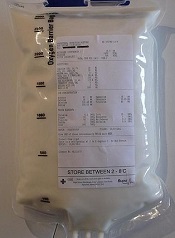
Current guidelines to help prevent bloodstream infections during intravenous feeding may need revisions to strengthen protection for patients, a new study suggests.
These guidelines restrict how long a single bag of parenteral nutrition containing lipids can be used, due to the ability of lipids to encourage the growth of microorganisms.
But researchers found the guidelines do not account for other independent factors that can affect the growth of potentially deadly microorganisms.
Their findings appear in the Journal of Parenteral and Enteral Nutrition.
Peter David Austin, of the University of Southampton in the UK, and his colleagues looked at the growth of Escherichia coli and Enterococcus durans in parenteral nutrition to identify factors that can affect microbial growth.
They assessed the growth of E coli and E durans in quadruplicate in 12 different patients who received parenteral nutrition, with and without lipid, in varying glucose concentrations.
Results showed that glucose concentration, the proportion of glucose to lipid, and osmolarity all affected microbial growth, in addition to the presence of lipids.
The researchers therefore recommend these additional factors be considered when making clinical and policy decisions to limit the potential growth of microorganisms in parenteral nutrition.
Current guidelines to help prevent bloodstream infections during intravenous feeding may need revisions to strengthen protection for patients, a new study suggests.
These guidelines restrict how long a single bag of parenteral nutrition containing lipids can be used, due to the ability of lipids to encourage the growth of microorganisms.
But researchers found the guidelines do not account for other independent factors that can affect the growth of potentially deadly microorganisms.
Their findings appear in the Journal of Parenteral and Enteral Nutrition.
Peter David Austin, of the University of Southampton in the UK, and his colleagues looked at the growth of Escherichia coli and Enterococcus durans in parenteral nutrition to identify factors that can affect microbial growth.
They assessed the growth of E coli and E durans in quadruplicate in 12 different patients who received parenteral nutrition, with and without lipid, in varying glucose concentrations.
Results showed that glucose concentration, the proportion of glucose to lipid, and osmolarity all affected microbial growth, in addition to the presence of lipids.
The researchers therefore recommend these additional factors be considered when making clinical and policy decisions to limit the potential growth of microorganisms in parenteral nutrition.
Current guidelines to help prevent bloodstream infections during intravenous feeding may need revisions to strengthen protection for patients, a new study suggests.
These guidelines restrict how long a single bag of parenteral nutrition containing lipids can be used, due to the ability of lipids to encourage the growth of microorganisms.
But researchers found the guidelines do not account for other independent factors that can affect the growth of potentially deadly microorganisms.
Their findings appear in the Journal of Parenteral and Enteral Nutrition.
Peter David Austin, of the University of Southampton in the UK, and his colleagues looked at the growth of Escherichia coli and Enterococcus durans in parenteral nutrition to identify factors that can affect microbial growth.
They assessed the growth of E coli and E durans in quadruplicate in 12 different patients who received parenteral nutrition, with and without lipid, in varying glucose concentrations.
Results showed that glucose concentration, the proportion of glucose to lipid, and osmolarity all affected microbial growth, in addition to the presence of lipids.
The researchers therefore recommend these additional factors be considered when making clinical and policy decisions to limit the potential growth of microorganisms in parenteral nutrition.
Group finds master regulator of MYC

Credit: Juha Klefstrom
New research indicates that an unexpected partnership between the MYC oncogene and a non-coding RNA called PVT1 could be the key to understanding how MYC fuels cancers.
The researchers knew that MYC amplifications cause cancer, but MYC does not amplify alone. It often pairs with adjacent chromosomal regions.
“We wanted to know if the neighboring genes played a role,” said study author Anindya Bagchi, PhD, of the University of Minnesota in Minneapolis.
“We took a chance and were surprised to find this unexpected and counter-intuitive partnership between MYC and its neighbor, PVT1. Not only do these genes amplify together, PVT1 helps boost the MYC protein’s ability to carry out its dangerous activities in the cell.”
The researchers reported this finding in Nature.
Dr Bagchi and his team focused on a region of the genome, 8q24, which contains the MYC gene and is commonly expressed in cancer. The team separated MYC from the neighboring region containing the non-coding RNA PVT1.
Using chromosome engineering, the researchers developed mouse strains in 3 separate iterations: MYC only, the rest of the region containing PVT1 but without MYC, and the pairing of MYC with the regional genes.
The expected outcome, if MYC was the sole driver of the cancer, was tumor growth on the MYC line as well as the paired line. However, the researchers found growth only on the paired line. This suggests MYC is not acting alone and needs help from adjacent genes.
“The discovery of this partnership gives us a stronger understanding of how MYC amplification is fueled,” said David Largaespada, PhD, also of the University of Minnesota.
“When cancer promotes a cell to make more MYC, it also increases the PVT1 in the cell, which, in turn, boosts the amount of MYC. It’s a cycle, and now we’ve identified it, we can look for ways to uncouple this dangerous partnership.”
Testing this theory of uncoupling, the researchers looked closely at several breast and colorectal cancers that are driven by MYC. For example, in colorectal cancer lab models, where a mutation in the beta-catenin gene drives MYC to cancerous levels, eliminating PVT1 from these cells made the tumors nearly disappear.
“Finding the cooperation between MYC and PVT1 could be a game changer,” said Yuen-Yi Tseng, a graduate student at the University of Minnesota.
“We used to think MYC amplification is the major issue but ignored that other co-amplified genes, such as PVT1, can be significant. In this study, we show that PVT1 can be a key regulator of MYC protein, which can shift the paradigm in our understanding of MYC-amplified cancers.”
MYC has been notoriously elusive as a drug target. By uncoupling MYC and PVT1, the researchers suspect they could disable the cancer growth and limit MYC to precancerous levels. This would make PVT1 an ideal drug target to potentially control a major cancer gene.
“This is a thrilling discovery, but there are more questions that follow,” Dr Bagchi said. “Two major areas present themselves now for research. Will breaking the nexus between MYC and PVT1 perform the same in any MYC-driven cancer, even those not driven by this specific genetic location?”
“And how is PVT1 stabilizing or boosting MYC within the cells? This relationship will be a key to developing any drugs to target this mechanism.”
Credit: Juha Klefstrom
New research indicates that an unexpected partnership between the MYC oncogene and a non-coding RNA called PVT1 could be the key to understanding how MYC fuels cancers.
The researchers knew that MYC amplifications cause cancer, but MYC does not amplify alone. It often pairs with adjacent chromosomal regions.
“We wanted to know if the neighboring genes played a role,” said study author Anindya Bagchi, PhD, of the University of Minnesota in Minneapolis.
“We took a chance and were surprised to find this unexpected and counter-intuitive partnership between MYC and its neighbor, PVT1. Not only do these genes amplify together, PVT1 helps boost the MYC protein’s ability to carry out its dangerous activities in the cell.”
The researchers reported this finding in Nature.
Dr Bagchi and his team focused on a region of the genome, 8q24, which contains the MYC gene and is commonly expressed in cancer. The team separated MYC from the neighboring region containing the non-coding RNA PVT1.
Using chromosome engineering, the researchers developed mouse strains in 3 separate iterations: MYC only, the rest of the region containing PVT1 but without MYC, and the pairing of MYC with the regional genes.
The expected outcome, if MYC was the sole driver of the cancer, was tumor growth on the MYC line as well as the paired line. However, the researchers found growth only on the paired line. This suggests MYC is not acting alone and needs help from adjacent genes.
“The discovery of this partnership gives us a stronger understanding of how MYC amplification is fueled,” said David Largaespada, PhD, also of the University of Minnesota.
“When cancer promotes a cell to make more MYC, it also increases the PVT1 in the cell, which, in turn, boosts the amount of MYC. It’s a cycle, and now we’ve identified it, we can look for ways to uncouple this dangerous partnership.”
Testing this theory of uncoupling, the researchers looked closely at several breast and colorectal cancers that are driven by MYC. For example, in colorectal cancer lab models, where a mutation in the beta-catenin gene drives MYC to cancerous levels, eliminating PVT1 from these cells made the tumors nearly disappear.
“Finding the cooperation between MYC and PVT1 could be a game changer,” said Yuen-Yi Tseng, a graduate student at the University of Minnesota.
“We used to think MYC amplification is the major issue but ignored that other co-amplified genes, such as PVT1, can be significant. In this study, we show that PVT1 can be a key regulator of MYC protein, which can shift the paradigm in our understanding of MYC-amplified cancers.”
MYC has been notoriously elusive as a drug target. By uncoupling MYC and PVT1, the researchers suspect they could disable the cancer growth and limit MYC to precancerous levels. This would make PVT1 an ideal drug target to potentially control a major cancer gene.
“This is a thrilling discovery, but there are more questions that follow,” Dr Bagchi said. “Two major areas present themselves now for research. Will breaking the nexus between MYC and PVT1 perform the same in any MYC-driven cancer, even those not driven by this specific genetic location?”
“And how is PVT1 stabilizing or boosting MYC within the cells? This relationship will be a key to developing any drugs to target this mechanism.”
Credit: Juha Klefstrom
New research indicates that an unexpected partnership between the MYC oncogene and a non-coding RNA called PVT1 could be the key to understanding how MYC fuels cancers.
The researchers knew that MYC amplifications cause cancer, but MYC does not amplify alone. It often pairs with adjacent chromosomal regions.
“We wanted to know if the neighboring genes played a role,” said study author Anindya Bagchi, PhD, of the University of Minnesota in Minneapolis.
“We took a chance and were surprised to find this unexpected and counter-intuitive partnership between MYC and its neighbor, PVT1. Not only do these genes amplify together, PVT1 helps boost the MYC protein’s ability to carry out its dangerous activities in the cell.”
The researchers reported this finding in Nature.
Dr Bagchi and his team focused on a region of the genome, 8q24, which contains the MYC gene and is commonly expressed in cancer. The team separated MYC from the neighboring region containing the non-coding RNA PVT1.
Using chromosome engineering, the researchers developed mouse strains in 3 separate iterations: MYC only, the rest of the region containing PVT1 but without MYC, and the pairing of MYC with the regional genes.
The expected outcome, if MYC was the sole driver of the cancer, was tumor growth on the MYC line as well as the paired line. However, the researchers found growth only on the paired line. This suggests MYC is not acting alone and needs help from adjacent genes.
“The discovery of this partnership gives us a stronger understanding of how MYC amplification is fueled,” said David Largaespada, PhD, also of the University of Minnesota.
“When cancer promotes a cell to make more MYC, it also increases the PVT1 in the cell, which, in turn, boosts the amount of MYC. It’s a cycle, and now we’ve identified it, we can look for ways to uncouple this dangerous partnership.”
Testing this theory of uncoupling, the researchers looked closely at several breast and colorectal cancers that are driven by MYC. For example, in colorectal cancer lab models, where a mutation in the beta-catenin gene drives MYC to cancerous levels, eliminating PVT1 from these cells made the tumors nearly disappear.
“Finding the cooperation between MYC and PVT1 could be a game changer,” said Yuen-Yi Tseng, a graduate student at the University of Minnesota.
“We used to think MYC amplification is the major issue but ignored that other co-amplified genes, such as PVT1, can be significant. In this study, we show that PVT1 can be a key regulator of MYC protein, which can shift the paradigm in our understanding of MYC-amplified cancers.”
MYC has been notoriously elusive as a drug target. By uncoupling MYC and PVT1, the researchers suspect they could disable the cancer growth and limit MYC to precancerous levels. This would make PVT1 an ideal drug target to potentially control a major cancer gene.
“This is a thrilling discovery, but there are more questions that follow,” Dr Bagchi said. “Two major areas present themselves now for research. Will breaking the nexus between MYC and PVT1 perform the same in any MYC-driven cancer, even those not driven by this specific genetic location?”
“And how is PVT1 stabilizing or boosting MYC within the cells? This relationship will be a key to developing any drugs to target this mechanism.”
Review uncovers patient safety violations

Credit: CDC
Patient safety violations emerged as a common theme in a review of “first warning” letters issued to clinical trial sponsors, investigators, and internal review boards (IRBs).
The US Food and Drug Administration (FDA) issued the 84 warning letters in response to trial violations uncovered during site visits.
The review of these letters showed that 55% of investigators failed to protect subjects’ safety and report adverse events to the IRB, 24% of sponsors failed to submit adverse event data to the FDA, and 22% of IRBs failed to “address risk minimization and protect vulnerable study subjects.”
Yashashri C. Shetty and Aafreen A. Saiyed, both of King Edward Memorial Hospital in Mumbai, detailed these findings in the Journal of Medical Ethics.
The researchers reviewed the content of 84 first-warning letters issued by the FDA following site visits between 2005 and 2012.
Sponsor violations
Forty-six warning letters were issued to trial sponsors. Their most common violations were failure to follow the monitoring schedule (58.69%) and failure to obtain investigator agreement (34.78%).
The same proportion of sponsors (30.43%) failed to secure investigators’ compliance and failed to maintain records, both of study data and for shipping product to the investigator.
Other violations included failure to submit an Investigational Device Exemption or Investigational New Drug application to the FDA (28.26%) and failure to review, evaluate, and submit adverse drug event reports to the FDA (23.91%). Two sponsors did not allow FDA inspection, and 1 did not obtain IRB approval.
Investigator infractions
Twenty warning letters were issued to investigators. Ninety-five percent of the letters said investigators were guilty of deviating from the investigational plan. Fifty-five percent of letters said the researchers failed to protect subject safety and report adverse events to the IRB.
Violations regarding records—largely, the failure to maintain and produce them for inspection—were documented in 40% of the letters. And informed consent issues were highlighted in 35%.
Other violations included those related to the product under investigation (15%), failure to obtain IRB approval (10%), and failure to personally supervise the study (30%).
IRB transgressions
Eighteen warning letters were issued to IRBs. The most common violation (61.11%) was failure to follow written procedures for continuing review.
Other common violations (55.56%) were those related to membership and meetings—failure to maintain minutes, inappropriate membership, quorum issues, misuse of expedited review, and the lack of a layperson in meetings.
The remaining violations included failure to follow regulatory requirements (44.4%), failure to follow standard operating procedures and maintain documentation (44.44%), failure to address risk minimization and protect vulnerable subjects (22.22%), conflicts of interest and informed consent issues (27.78%), and failure to appoint a qualified investigator (5.55%).
Comparing past and present
The study authors compared their findings with previously published research in the same arena, dating back as far as 1997. They found that regulatory compliance generally improved over the years, but supervision worsened.
And 2 new serious violations cropped up in the interim: failure to get the green light from an IRB before pressing ahead and submitting false data to the FDA and/or sponsors.
In a bid to boost compliance with good clinical practice, the authors suggest that every regulatory agency charged with overseeing clinical trials should pay main participating centers a visit and regularly publish the details of their findings.
Credit: CDC
Patient safety violations emerged as a common theme in a review of “first warning” letters issued to clinical trial sponsors, investigators, and internal review boards (IRBs).
The US Food and Drug Administration (FDA) issued the 84 warning letters in response to trial violations uncovered during site visits.
The review of these letters showed that 55% of investigators failed to protect subjects’ safety and report adverse events to the IRB, 24% of sponsors failed to submit adverse event data to the FDA, and 22% of IRBs failed to “address risk minimization and protect vulnerable study subjects.”
Yashashri C. Shetty and Aafreen A. Saiyed, both of King Edward Memorial Hospital in Mumbai, detailed these findings in the Journal of Medical Ethics.
The researchers reviewed the content of 84 first-warning letters issued by the FDA following site visits between 2005 and 2012.
Sponsor violations
Forty-six warning letters were issued to trial sponsors. Their most common violations were failure to follow the monitoring schedule (58.69%) and failure to obtain investigator agreement (34.78%).
The same proportion of sponsors (30.43%) failed to secure investigators’ compliance and failed to maintain records, both of study data and for shipping product to the investigator.
Other violations included failure to submit an Investigational Device Exemption or Investigational New Drug application to the FDA (28.26%) and failure to review, evaluate, and submit adverse drug event reports to the FDA (23.91%). Two sponsors did not allow FDA inspection, and 1 did not obtain IRB approval.
Investigator infractions
Twenty warning letters were issued to investigators. Ninety-five percent of the letters said investigators were guilty of deviating from the investigational plan. Fifty-five percent of letters said the researchers failed to protect subject safety and report adverse events to the IRB.
Violations regarding records—largely, the failure to maintain and produce them for inspection—were documented in 40% of the letters. And informed consent issues were highlighted in 35%.
Other violations included those related to the product under investigation (15%), failure to obtain IRB approval (10%), and failure to personally supervise the study (30%).
IRB transgressions
Eighteen warning letters were issued to IRBs. The most common violation (61.11%) was failure to follow written procedures for continuing review.
Other common violations (55.56%) were those related to membership and meetings—failure to maintain minutes, inappropriate membership, quorum issues, misuse of expedited review, and the lack of a layperson in meetings.
The remaining violations included failure to follow regulatory requirements (44.4%), failure to follow standard operating procedures and maintain documentation (44.44%), failure to address risk minimization and protect vulnerable subjects (22.22%), conflicts of interest and informed consent issues (27.78%), and failure to appoint a qualified investigator (5.55%).
Comparing past and present
The study authors compared their findings with previously published research in the same arena, dating back as far as 1997. They found that regulatory compliance generally improved over the years, but supervision worsened.
And 2 new serious violations cropped up in the interim: failure to get the green light from an IRB before pressing ahead and submitting false data to the FDA and/or sponsors.
In a bid to boost compliance with good clinical practice, the authors suggest that every regulatory agency charged with overseeing clinical trials should pay main participating centers a visit and regularly publish the details of their findings.
Credit: CDC
Patient safety violations emerged as a common theme in a review of “first warning” letters issued to clinical trial sponsors, investigators, and internal review boards (IRBs).
The US Food and Drug Administration (FDA) issued the 84 warning letters in response to trial violations uncovered during site visits.
The review of these letters showed that 55% of investigators failed to protect subjects’ safety and report adverse events to the IRB, 24% of sponsors failed to submit adverse event data to the FDA, and 22% of IRBs failed to “address risk minimization and protect vulnerable study subjects.”
Yashashri C. Shetty and Aafreen A. Saiyed, both of King Edward Memorial Hospital in Mumbai, detailed these findings in the Journal of Medical Ethics.
The researchers reviewed the content of 84 first-warning letters issued by the FDA following site visits between 2005 and 2012.
Sponsor violations
Forty-six warning letters were issued to trial sponsors. Their most common violations were failure to follow the monitoring schedule (58.69%) and failure to obtain investigator agreement (34.78%).
The same proportion of sponsors (30.43%) failed to secure investigators’ compliance and failed to maintain records, both of study data and for shipping product to the investigator.
Other violations included failure to submit an Investigational Device Exemption or Investigational New Drug application to the FDA (28.26%) and failure to review, evaluate, and submit adverse drug event reports to the FDA (23.91%). Two sponsors did not allow FDA inspection, and 1 did not obtain IRB approval.
Investigator infractions
Twenty warning letters were issued to investigators. Ninety-five percent of the letters said investigators were guilty of deviating from the investigational plan. Fifty-five percent of letters said the researchers failed to protect subject safety and report adverse events to the IRB.
Violations regarding records—largely, the failure to maintain and produce them for inspection—were documented in 40% of the letters. And informed consent issues were highlighted in 35%.
Other violations included those related to the product under investigation (15%), failure to obtain IRB approval (10%), and failure to personally supervise the study (30%).
IRB transgressions
Eighteen warning letters were issued to IRBs. The most common violation (61.11%) was failure to follow written procedures for continuing review.
Other common violations (55.56%) were those related to membership and meetings—failure to maintain minutes, inappropriate membership, quorum issues, misuse of expedited review, and the lack of a layperson in meetings.
The remaining violations included failure to follow regulatory requirements (44.4%), failure to follow standard operating procedures and maintain documentation (44.44%), failure to address risk minimization and protect vulnerable subjects (22.22%), conflicts of interest and informed consent issues (27.78%), and failure to appoint a qualified investigator (5.55%).
Comparing past and present
The study authors compared their findings with previously published research in the same arena, dating back as far as 1997. They found that regulatory compliance generally improved over the years, but supervision worsened.
And 2 new serious violations cropped up in the interim: failure to get the green light from an IRB before pressing ahead and submitting false data to the FDA and/or sponsors.
In a bid to boost compliance with good clinical practice, the authors suggest that every regulatory agency charged with overseeing clinical trials should pay main participating centers a visit and regularly publish the details of their findings.
BET inhibitor proves active in murine lymphoma

A bromodomain and extraterminal (BET) inhibitor known as RVX2135 has shown preclinical activity against Myc-driven lymphoma.
Both in vitro and in vivo, RVX2135 inhibited proliferation and prompted apoptosis in lymphoma cells.
Investigation revealed that RVX2135 induces effects similar to those of histone deacetylase (HDAC) inhibitors. Furthermore, RVX2135 and the HDAC inhibitor vorinostat demonstrated synergy in lymphoma-bearing mice.
Jonas Nilsson, PhD, of the University of Gothenburg in Sweden, and his colleagues reported these results in Proceedings of the National Academy of Sciences.
The researchers first evaluated the in vitro antiproliferative effects of RVX2135 and another BET inhibitor called JQ1. They tested the inhibitors on lymphoma cells from Myc-transgenic mice and found that both restricted proliferation and induced apoptosis in a dose-dependent manner.
Next, the team tested RVX2135 in 2 mouse models of lymphoma. The inhibitor was most effective in mice transplanted with dispersed lymphoma from a λ-Myc mouse (ID 2749).
In fact, RVX2135 doubled both the median and overall survival of mice carrying 2749 lymphoma, when compared to vehicle-treated controls.
Dr Nilsson and his colleagues then investigated the mechanism behind these effects. They found that RVX2135 induces a complex transcriptional program without specifically inactivating transgenic Myc transcription.
By examining the genes induced by BET inhibition, the researchers discovered that RVX2135 activates the same genes as those activated by HDAC inhibitors.
So the team tested the HDAC inhibitor vorinostat in combination with RVX2135. And the combination increased survival in mice with 2749 lymphoma, when compared to either inhibitor alone.
“It was also possible to reduce the dose of HDAC inhibitors when used in combination with RVX2135, and this reduced adverse effects,” Dr Nilsson said.
“We see this as a breakthrough in the clinical development of this type of treatment. [W]e believe that the prospects for success with combination treatments are good.”
A bromodomain and extraterminal (BET) inhibitor known as RVX2135 has shown preclinical activity against Myc-driven lymphoma.
Both in vitro and in vivo, RVX2135 inhibited proliferation and prompted apoptosis in lymphoma cells.
Investigation revealed that RVX2135 induces effects similar to those of histone deacetylase (HDAC) inhibitors. Furthermore, RVX2135 and the HDAC inhibitor vorinostat demonstrated synergy in lymphoma-bearing mice.
Jonas Nilsson, PhD, of the University of Gothenburg in Sweden, and his colleagues reported these results in Proceedings of the National Academy of Sciences.
The researchers first evaluated the in vitro antiproliferative effects of RVX2135 and another BET inhibitor called JQ1. They tested the inhibitors on lymphoma cells from Myc-transgenic mice and found that both restricted proliferation and induced apoptosis in a dose-dependent manner.
Next, the team tested RVX2135 in 2 mouse models of lymphoma. The inhibitor was most effective in mice transplanted with dispersed lymphoma from a λ-Myc mouse (ID 2749).
In fact, RVX2135 doubled both the median and overall survival of mice carrying 2749 lymphoma, when compared to vehicle-treated controls.
Dr Nilsson and his colleagues then investigated the mechanism behind these effects. They found that RVX2135 induces a complex transcriptional program without specifically inactivating transgenic Myc transcription.
By examining the genes induced by BET inhibition, the researchers discovered that RVX2135 activates the same genes as those activated by HDAC inhibitors.
So the team tested the HDAC inhibitor vorinostat in combination with RVX2135. And the combination increased survival in mice with 2749 lymphoma, when compared to either inhibitor alone.
“It was also possible to reduce the dose of HDAC inhibitors when used in combination with RVX2135, and this reduced adverse effects,” Dr Nilsson said.
“We see this as a breakthrough in the clinical development of this type of treatment. [W]e believe that the prospects for success with combination treatments are good.”
A bromodomain and extraterminal (BET) inhibitor known as RVX2135 has shown preclinical activity against Myc-driven lymphoma.
Both in vitro and in vivo, RVX2135 inhibited proliferation and prompted apoptosis in lymphoma cells.
Investigation revealed that RVX2135 induces effects similar to those of histone deacetylase (HDAC) inhibitors. Furthermore, RVX2135 and the HDAC inhibitor vorinostat demonstrated synergy in lymphoma-bearing mice.
Jonas Nilsson, PhD, of the University of Gothenburg in Sweden, and his colleagues reported these results in Proceedings of the National Academy of Sciences.
The researchers first evaluated the in vitro antiproliferative effects of RVX2135 and another BET inhibitor called JQ1. They tested the inhibitors on lymphoma cells from Myc-transgenic mice and found that both restricted proliferation and induced apoptosis in a dose-dependent manner.
Next, the team tested RVX2135 in 2 mouse models of lymphoma. The inhibitor was most effective in mice transplanted with dispersed lymphoma from a λ-Myc mouse (ID 2749).
In fact, RVX2135 doubled both the median and overall survival of mice carrying 2749 lymphoma, when compared to vehicle-treated controls.
Dr Nilsson and his colleagues then investigated the mechanism behind these effects. They found that RVX2135 induces a complex transcriptional program without specifically inactivating transgenic Myc transcription.
By examining the genes induced by BET inhibition, the researchers discovered that RVX2135 activates the same genes as those activated by HDAC inhibitors.
So the team tested the HDAC inhibitor vorinostat in combination with RVX2135. And the combination increased survival in mice with 2749 lymphoma, when compared to either inhibitor alone.
“It was also possible to reduce the dose of HDAC inhibitors when used in combination with RVX2135, and this reduced adverse effects,” Dr Nilsson said.
“We see this as a breakthrough in the clinical development of this type of treatment. [W]e believe that the prospects for success with combination treatments are good.”
An easier route for cell therapy
Credit: PNAS
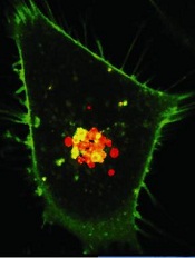
Laser technology can help ensure the delivery of drug and gene therapy at the cellular level without damaging surrounding tissue, according to research published in Nature Scientific Reports.
Investigators paired crystalline magnetic carbon nanoparticles and continuous wave near-infrared laser beams in what is called photothermal delivery.
And they used this delivery method to introduce impermeable dyes and small DNA molecules into human cancer cells.
This work grew out of a previous study in which the researchers used a 50 to 100 milliwatt laser and the same carbon nanoparticle, which absorbs the beam, to heat up and destroy cancer cells in the lab.
“In [the current study, we] used a lower-power, 20 to 30 milliwatt, continuous wave near-infrared laser and the nanoparticle to permeate the cell membrane without killing the cells,” said Ali Koymen, PhD, of the University of Texas at Arlington.
“This method stretches the desired cell membrane to allow for delivery and has the added bonus of creating a fluid flow that speeds the movement of what is being delivered.”
The investigators noted that, currently, the predominant practice is using viruses for delivery to cells. Unfortunately, the scope of what can be delivered with viruses is severely limited, and virus interaction can lead to inflammatory responses and other complications.
Researchers looking to create a path into the cell without employing a virus have experimented with using UV-visible light laser beams alone. But that method damages surrounding cells and has a relatively shallow level of effectiveness.
Dr Koymen and his colleagues said a significant advantage of their method is that the near-infrared light absorption of the nanoparticle can be used to selectively amplify the interaction of low-power laser with targeted tissue, and laser-induced damage to non-targeted cells can be avoided.
The magnetic properties of the nanoparticles also mean they can be localized with an external magnetic field. Therefore, a smaller concentration can be used effectively.
Credit: PNAS
Laser technology can help ensure the delivery of drug and gene therapy at the cellular level without damaging surrounding tissue, according to research published in Nature Scientific Reports.
Investigators paired crystalline magnetic carbon nanoparticles and continuous wave near-infrared laser beams in what is called photothermal delivery.
And they used this delivery method to introduce impermeable dyes and small DNA molecules into human cancer cells.
This work grew out of a previous study in which the researchers used a 50 to 100 milliwatt laser and the same carbon nanoparticle, which absorbs the beam, to heat up and destroy cancer cells in the lab.
“In [the current study, we] used a lower-power, 20 to 30 milliwatt, continuous wave near-infrared laser and the nanoparticle to permeate the cell membrane without killing the cells,” said Ali Koymen, PhD, of the University of Texas at Arlington.
“This method stretches the desired cell membrane to allow for delivery and has the added bonus of creating a fluid flow that speeds the movement of what is being delivered.”
The investigators noted that, currently, the predominant practice is using viruses for delivery to cells. Unfortunately, the scope of what can be delivered with viruses is severely limited, and virus interaction can lead to inflammatory responses and other complications.
Researchers looking to create a path into the cell without employing a virus have experimented with using UV-visible light laser beams alone. But that method damages surrounding cells and has a relatively shallow level of effectiveness.
Dr Koymen and his colleagues said a significant advantage of their method is that the near-infrared light absorption of the nanoparticle can be used to selectively amplify the interaction of low-power laser with targeted tissue, and laser-induced damage to non-targeted cells can be avoided.
The magnetic properties of the nanoparticles also mean they can be localized with an external magnetic field. Therefore, a smaller concentration can be used effectively.
Credit: PNAS
Laser technology can help ensure the delivery of drug and gene therapy at the cellular level without damaging surrounding tissue, according to research published in Nature Scientific Reports.
Investigators paired crystalline magnetic carbon nanoparticles and continuous wave near-infrared laser beams in what is called photothermal delivery.
And they used this delivery method to introduce impermeable dyes and small DNA molecules into human cancer cells.
This work grew out of a previous study in which the researchers used a 50 to 100 milliwatt laser and the same carbon nanoparticle, which absorbs the beam, to heat up and destroy cancer cells in the lab.
“In [the current study, we] used a lower-power, 20 to 30 milliwatt, continuous wave near-infrared laser and the nanoparticle to permeate the cell membrane without killing the cells,” said Ali Koymen, PhD, of the University of Texas at Arlington.
“This method stretches the desired cell membrane to allow for delivery and has the added bonus of creating a fluid flow that speeds the movement of what is being delivered.”
The investigators noted that, currently, the predominant practice is using viruses for delivery to cells. Unfortunately, the scope of what can be delivered with viruses is severely limited, and virus interaction can lead to inflammatory responses and other complications.
Researchers looking to create a path into the cell without employing a virus have experimented with using UV-visible light laser beams alone. But that method damages surrounding cells and has a relatively shallow level of effectiveness.
Dr Koymen and his colleagues said a significant advantage of their method is that the near-infrared light absorption of the nanoparticle can be used to selectively amplify the interaction of low-power laser with targeted tissue, and laser-induced damage to non-targeted cells can be avoided.
The magnetic properties of the nanoparticles also mean they can be localized with an external magnetic field. Therefore, a smaller concentration can be used effectively.
Thalidomide offers short-term efficacy in HHT
MILAN—Results of a phase 2 trial suggest thalidomide can elicit solid—though not necessarily durable—responses among patients with hereditary hemorrhagic telangiectasia (HHT) suffering from severe, recurrent epistaxis.
All 29 evaluable patients responded to thalidomide, with 4 achieving a complete response.
Unfortunately, 11 patients relapsed at a median of 43 weeks. But re-treatment was possible for a few patients and did prompt additional responses.
“Our results strongly support the hypothesis that low-dose thalidomide is safe and very effective for the treatment of severe epistaxis in HHT patients who did not benefit from other available modalities of treatment,” said Rosangela Invernizzi, MD, of the University of Pavia in Italy.
“However, the effect of thalidomide is not permanent, and maintenance therapy may be required.”
Dr Invernizzi presented these discoveries at the 19th Congress of the European Hematology Association (EHA) as abstract S692.
She and her colleagues enrolled 31 HHT patients on this phase 2 study. The patients had a median age of 64 years (range, 44-84). Nine had grade 2 epistaxis, and 22 had grade 3.
Previous treatments included argon plasma coagulation (n=19), electrocautery (n=12), embolization (n=7), laser coagulation (n=2), septodermoplasty (n=2), and arterial ligation (n=1). Eighteen patients had received less than 10 units of red blood cells, and 13 had received 10 or more units.
For this study, patients received thalidomide at 50 mg a day for 4 weeks. Complete responders received 8 additional weeks of treatment at the same dosage. Partial responders received 16 additional weeks of treatment at the same dosage.
For non-responders, the dose was increased by 50 mg per day every 4 weeks until they attained a complete or partial response (with a maximum dose of 200 mg). If patients did not respond, they received 24 additional weeks of treatment. Thalidomide courses could be repeated 3 times at the most.
Response and relapse
The median follow-up was 67 weeks (range, 3-130 weeks). All of the 29 evaluable patients achieved a response, with 4 complete responses (14%) and 25 partial responses (86%).
Twenty-four patients responded within 4 weeks of treatment initiation, and 5 responded within 8 weeks. The minimum dose of thalidomide was 50 mg, and the maximum was 100 mg.
“A significant decrease of all epistaxis parameters—frequency [P=0.001], intensity [P<0.0001], and duration [P=0.0001]—was recorded,” Dr Invernizzi noted.
“As a consequence, thalidomide treatment significantly increased hemoglobin levels [P=0.02] and abolished or greatly decreased transfusion need [P=0.0003] and improved the quality of life.”
However, 11 patients relapsed at a median of 43 weeks. Patients who relapsed within 52 weeks of ending thalidomide could be treated again for 8 weeks at the same maximum dose employed during induction.
Four patients received re-treatment and achieved partial responses. One of these patients relapsed again at 13 weeks. But, for the other 3 patients, clinical improvement persisted at more than 17 weeks, more than 26 weeks, and more than 27 weeks.
Safety and tolerability
Dr Invernizzi said there were no noticeable side effects associated with re-treatment of relapsed patients.
The most common adverse events among all patients were gastrointestinal symptoms, such as constipation, vomiting, and dry mouth (n=15); drowsiness (n=9); and constitutional symptoms, such as asthenia, malaise, and peripheral edema (n=7).
There were 2 patients with psychiatric symptoms (confusion, depression) and 2 patients with thyroid dysfunction.
There was 1 cardiovascular event, (bradycardia, heart failure), 1 patient with low hematologic counts, 1 patient with neurologic symptoms (peripheral neuropathy, dizziness, tremor), 1 dermatologic event (toxic cutaneous rashes, skin dryness), and 1 infection.
MILAN—Results of a phase 2 trial suggest thalidomide can elicit solid—though not necessarily durable—responses among patients with hereditary hemorrhagic telangiectasia (HHT) suffering from severe, recurrent epistaxis.
All 29 evaluable patients responded to thalidomide, with 4 achieving a complete response.
Unfortunately, 11 patients relapsed at a median of 43 weeks. But re-treatment was possible for a few patients and did prompt additional responses.
“Our results strongly support the hypothesis that low-dose thalidomide is safe and very effective for the treatment of severe epistaxis in HHT patients who did not benefit from other available modalities of treatment,” said Rosangela Invernizzi, MD, of the University of Pavia in Italy.
“However, the effect of thalidomide is not permanent, and maintenance therapy may be required.”
Dr Invernizzi presented these discoveries at the 19th Congress of the European Hematology Association (EHA) as abstract S692.
She and her colleagues enrolled 31 HHT patients on this phase 2 study. The patients had a median age of 64 years (range, 44-84). Nine had grade 2 epistaxis, and 22 had grade 3.
Previous treatments included argon plasma coagulation (n=19), electrocautery (n=12), embolization (n=7), laser coagulation (n=2), septodermoplasty (n=2), and arterial ligation (n=1). Eighteen patients had received less than 10 units of red blood cells, and 13 had received 10 or more units.
For this study, patients received thalidomide at 50 mg a day for 4 weeks. Complete responders received 8 additional weeks of treatment at the same dosage. Partial responders received 16 additional weeks of treatment at the same dosage.
For non-responders, the dose was increased by 50 mg per day every 4 weeks until they attained a complete or partial response (with a maximum dose of 200 mg). If patients did not respond, they received 24 additional weeks of treatment. Thalidomide courses could be repeated 3 times at the most.
Response and relapse
The median follow-up was 67 weeks (range, 3-130 weeks). All of the 29 evaluable patients achieved a response, with 4 complete responses (14%) and 25 partial responses (86%).
Twenty-four patients responded within 4 weeks of treatment initiation, and 5 responded within 8 weeks. The minimum dose of thalidomide was 50 mg, and the maximum was 100 mg.
“A significant decrease of all epistaxis parameters—frequency [P=0.001], intensity [P<0.0001], and duration [P=0.0001]—was recorded,” Dr Invernizzi noted.
“As a consequence, thalidomide treatment significantly increased hemoglobin levels [P=0.02] and abolished or greatly decreased transfusion need [P=0.0003] and improved the quality of life.”
However, 11 patients relapsed at a median of 43 weeks. Patients who relapsed within 52 weeks of ending thalidomide could be treated again for 8 weeks at the same maximum dose employed during induction.
Four patients received re-treatment and achieved partial responses. One of these patients relapsed again at 13 weeks. But, for the other 3 patients, clinical improvement persisted at more than 17 weeks, more than 26 weeks, and more than 27 weeks.
Safety and tolerability
Dr Invernizzi said there were no noticeable side effects associated with re-treatment of relapsed patients.
The most common adverse events among all patients were gastrointestinal symptoms, such as constipation, vomiting, and dry mouth (n=15); drowsiness (n=9); and constitutional symptoms, such as asthenia, malaise, and peripheral edema (n=7).
There were 2 patients with psychiatric symptoms (confusion, depression) and 2 patients with thyroid dysfunction.
There was 1 cardiovascular event, (bradycardia, heart failure), 1 patient with low hematologic counts, 1 patient with neurologic symptoms (peripheral neuropathy, dizziness, tremor), 1 dermatologic event (toxic cutaneous rashes, skin dryness), and 1 infection.
MILAN—Results of a phase 2 trial suggest thalidomide can elicit solid—though not necessarily durable—responses among patients with hereditary hemorrhagic telangiectasia (HHT) suffering from severe, recurrent epistaxis.
All 29 evaluable patients responded to thalidomide, with 4 achieving a complete response.
Unfortunately, 11 patients relapsed at a median of 43 weeks. But re-treatment was possible for a few patients and did prompt additional responses.
“Our results strongly support the hypothesis that low-dose thalidomide is safe and very effective for the treatment of severe epistaxis in HHT patients who did not benefit from other available modalities of treatment,” said Rosangela Invernizzi, MD, of the University of Pavia in Italy.
“However, the effect of thalidomide is not permanent, and maintenance therapy may be required.”
Dr Invernizzi presented these discoveries at the 19th Congress of the European Hematology Association (EHA) as abstract S692.
She and her colleagues enrolled 31 HHT patients on this phase 2 study. The patients had a median age of 64 years (range, 44-84). Nine had grade 2 epistaxis, and 22 had grade 3.
Previous treatments included argon plasma coagulation (n=19), electrocautery (n=12), embolization (n=7), laser coagulation (n=2), septodermoplasty (n=2), and arterial ligation (n=1). Eighteen patients had received less than 10 units of red blood cells, and 13 had received 10 or more units.
For this study, patients received thalidomide at 50 mg a day for 4 weeks. Complete responders received 8 additional weeks of treatment at the same dosage. Partial responders received 16 additional weeks of treatment at the same dosage.
For non-responders, the dose was increased by 50 mg per day every 4 weeks until they attained a complete or partial response (with a maximum dose of 200 mg). If patients did not respond, they received 24 additional weeks of treatment. Thalidomide courses could be repeated 3 times at the most.
Response and relapse
The median follow-up was 67 weeks (range, 3-130 weeks). All of the 29 evaluable patients achieved a response, with 4 complete responses (14%) and 25 partial responses (86%).
Twenty-four patients responded within 4 weeks of treatment initiation, and 5 responded within 8 weeks. The minimum dose of thalidomide was 50 mg, and the maximum was 100 mg.
“A significant decrease of all epistaxis parameters—frequency [P=0.001], intensity [P<0.0001], and duration [P=0.0001]—was recorded,” Dr Invernizzi noted.
“As a consequence, thalidomide treatment significantly increased hemoglobin levels [P=0.02] and abolished or greatly decreased transfusion need [P=0.0003] and improved the quality of life.”
However, 11 patients relapsed at a median of 43 weeks. Patients who relapsed within 52 weeks of ending thalidomide could be treated again for 8 weeks at the same maximum dose employed during induction.
Four patients received re-treatment and achieved partial responses. One of these patients relapsed again at 13 weeks. But, for the other 3 patients, clinical improvement persisted at more than 17 weeks, more than 26 weeks, and more than 27 weeks.
Safety and tolerability
Dr Invernizzi said there were no noticeable side effects associated with re-treatment of relapsed patients.
The most common adverse events among all patients were gastrointestinal symptoms, such as constipation, vomiting, and dry mouth (n=15); drowsiness (n=9); and constitutional symptoms, such as asthenia, malaise, and peripheral edema (n=7).
There were 2 patients with psychiatric symptoms (confusion, depression) and 2 patients with thyroid dysfunction.
There was 1 cardiovascular event, (bradycardia, heart failure), 1 patient with low hematologic counts, 1 patient with neurologic symptoms (peripheral neuropathy, dizziness, tremor), 1 dermatologic event (toxic cutaneous rashes, skin dryness), and 1 infection.
Long-term complications after ALL minimal, study shows

Credit: Bill Branson
Children recently diagnosed with standard-risk acute lymphoblastic leukemia (ALL) are likely to have few long-term complications into adulthood, investigators have reported in The Lancet Oncology.
The team said this is because current therapies are less harsh than their predecessors.
Newer protocols have eliminated radiation and restricted the use of chemotherapeutics that may cause subsequent malignancies and other chronic health conditions.
Previous research had only assessed the very long-term outcomes of children treated with older protocols, leaving physicians to piece together information from this outdated data and anecdotal evidence.
“This is one of the first studies to show that, in addition to their excellent probability of survival, long-term survivors of standard-risk childhood ALL are at low risk for complications of their therapy once they enter adulthood,” said study author Paul Nathan, MD, of The Hospital for Sick Children in Toronto, Canada.
He and his colleagues used longitudinal data from the Childhood Cancer Survivor Study, a North American study of 5-year survivors of childhood cancer diagnosed between 1970 and 1986.
The team analyzed 556 patients from this study who were older than 1 year of age and younger than 10 at the time of diagnosis and who had received treatment consistent with current therapies for standard-risk ALL.
Patients were followed from 5 years following diagnosis to a median of 18 years. The survivor group was compared with a group of their siblings who had not had cancer, as well as the general population.
Twenty-eight patients in the survivor group died. Sixteen of these deaths were from causes other than relapse.
Survivors were at a slightly increased risk of death due to non-relapse causes, when compared to controls. However, no individual cause was increased.
Six survivors developed another malignancy. The risk for most chronic health disorders did not differ between survivors and siblings. But survivors appeared to have a moderately increased risk for osteoporosis or osteopenia, short stature, and cataracts.
Survivors and their siblings had similar socioeconomic outcomes, including education, rates of marriage, independent living, and household income.
While these results suggest the prognosis is favorable, Dr Nathan noted that ALL survivors should receive ongoing care from a knowledgeable primary-care practitioner.
“Among kids with standard-risk ALL, we expect most to survive,” Dr Nathan said. “Now we can say with more certainty that they will probably do well in the long-term as well. This information will be very useful for oncologists counselling newly diagnosed patients and their families and will be quite reassuring to parents.”
Credit: Bill Branson
Children recently diagnosed with standard-risk acute lymphoblastic leukemia (ALL) are likely to have few long-term complications into adulthood, investigators have reported in The Lancet Oncology.
The team said this is because current therapies are less harsh than their predecessors.
Newer protocols have eliminated radiation and restricted the use of chemotherapeutics that may cause subsequent malignancies and other chronic health conditions.
Previous research had only assessed the very long-term outcomes of children treated with older protocols, leaving physicians to piece together information from this outdated data and anecdotal evidence.
“This is one of the first studies to show that, in addition to their excellent probability of survival, long-term survivors of standard-risk childhood ALL are at low risk for complications of their therapy once they enter adulthood,” said study author Paul Nathan, MD, of The Hospital for Sick Children in Toronto, Canada.
He and his colleagues used longitudinal data from the Childhood Cancer Survivor Study, a North American study of 5-year survivors of childhood cancer diagnosed between 1970 and 1986.
The team analyzed 556 patients from this study who were older than 1 year of age and younger than 10 at the time of diagnosis and who had received treatment consistent with current therapies for standard-risk ALL.
Patients were followed from 5 years following diagnosis to a median of 18 years. The survivor group was compared with a group of their siblings who had not had cancer, as well as the general population.
Twenty-eight patients in the survivor group died. Sixteen of these deaths were from causes other than relapse.
Survivors were at a slightly increased risk of death due to non-relapse causes, when compared to controls. However, no individual cause was increased.
Six survivors developed another malignancy. The risk for most chronic health disorders did not differ between survivors and siblings. But survivors appeared to have a moderately increased risk for osteoporosis or osteopenia, short stature, and cataracts.
Survivors and their siblings had similar socioeconomic outcomes, including education, rates of marriage, independent living, and household income.
While these results suggest the prognosis is favorable, Dr Nathan noted that ALL survivors should receive ongoing care from a knowledgeable primary-care practitioner.
“Among kids with standard-risk ALL, we expect most to survive,” Dr Nathan said. “Now we can say with more certainty that they will probably do well in the long-term as well. This information will be very useful for oncologists counselling newly diagnosed patients and their families and will be quite reassuring to parents.”
Credit: Bill Branson
Children recently diagnosed with standard-risk acute lymphoblastic leukemia (ALL) are likely to have few long-term complications into adulthood, investigators have reported in The Lancet Oncology.
The team said this is because current therapies are less harsh than their predecessors.
Newer protocols have eliminated radiation and restricted the use of chemotherapeutics that may cause subsequent malignancies and other chronic health conditions.
Previous research had only assessed the very long-term outcomes of children treated with older protocols, leaving physicians to piece together information from this outdated data and anecdotal evidence.
“This is one of the first studies to show that, in addition to their excellent probability of survival, long-term survivors of standard-risk childhood ALL are at low risk for complications of their therapy once they enter adulthood,” said study author Paul Nathan, MD, of The Hospital for Sick Children in Toronto, Canada.
He and his colleagues used longitudinal data from the Childhood Cancer Survivor Study, a North American study of 5-year survivors of childhood cancer diagnosed between 1970 and 1986.
The team analyzed 556 patients from this study who were older than 1 year of age and younger than 10 at the time of diagnosis and who had received treatment consistent with current therapies for standard-risk ALL.
Patients were followed from 5 years following diagnosis to a median of 18 years. The survivor group was compared with a group of their siblings who had not had cancer, as well as the general population.
Twenty-eight patients in the survivor group died. Sixteen of these deaths were from causes other than relapse.
Survivors were at a slightly increased risk of death due to non-relapse causes, when compared to controls. However, no individual cause was increased.
Six survivors developed another malignancy. The risk for most chronic health disorders did not differ between survivors and siblings. But survivors appeared to have a moderately increased risk for osteoporosis or osteopenia, short stature, and cataracts.
Survivors and their siblings had similar socioeconomic outcomes, including education, rates of marriage, independent living, and household income.
While these results suggest the prognosis is favorable, Dr Nathan noted that ALL survivors should receive ongoing care from a knowledgeable primary-care practitioner.
“Among kids with standard-risk ALL, we expect most to survive,” Dr Nathan said. “Now we can say with more certainty that they will probably do well in the long-term as well. This information will be very useful for oncologists counselling newly diagnosed patients and their families and will be quite reassuring to parents.”