User login
Fusion proteins enable expansion of HSPCs
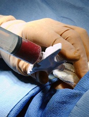
Credit: Chad McNeeley
A new technique has allowed researchers to expand hematopoietic stem and progenitor cells (HSPCs) from multiple sources.
The team developed fusion proteins and introduced them to HSPCs ex vivo.
This approach expanded cell populations whether HSPCs were derived from cord blood, bone marrow, or peripheral blood.
The potential clinical applications for this work range from immunodeficiency disorders to hematologic and other malignancies, according to the researchers.
Yosef Refaeli, PhD, of Taiga Biotechnologies, Inc. in Aurora, Colorado, and his colleagues described this work in PLOS ONE.
The team first cultured human or murine HSPCs with a pair of fusion proteins—the protein transduction domain of the HIV-1 transactivation protein (Tat) and either the Open Reading Frame for human MYC or a truncated form of human Bcl-2 that was deleted for the unstructured loop domain.
With this technique, the researchers were able to elicit an 87-fold expansion of HSPCs from mouse bone marrow, a 16.6-fold expansion of HSPCs from human cord blood, a 13.6-fold expansion of HSPCs from human peripheral blood cells mobilized by G-CSF, and a 10-fold expansion of HSPCs from human bone marrow.
The team then tested the biological function of the expanded cells, and they found the cells gave rise to BFU-E, CFU-M, CFU-G, and CFU-GM colonies in vitro.
When transplanted into irradiated mice, the expanded cells gave rise to mature hematopoietic populations and a self-renewing cell population that was able to support hematopoiesis upon serial transplantation.
The researchers said these results suggest their technique may be an attractive approach to expand human HSPCs ex vivo for clinical use.
The team’s goal now is to move the technology from the lab into clinical trials. Taiga Biotechnologies is in the process of setting up clinical trials testing this approach. 
Credit: Chad McNeeley
A new technique has allowed researchers to expand hematopoietic stem and progenitor cells (HSPCs) from multiple sources.
The team developed fusion proteins and introduced them to HSPCs ex vivo.
This approach expanded cell populations whether HSPCs were derived from cord blood, bone marrow, or peripheral blood.
The potential clinical applications for this work range from immunodeficiency disorders to hematologic and other malignancies, according to the researchers.
Yosef Refaeli, PhD, of Taiga Biotechnologies, Inc. in Aurora, Colorado, and his colleagues described this work in PLOS ONE.
The team first cultured human or murine HSPCs with a pair of fusion proteins—the protein transduction domain of the HIV-1 transactivation protein (Tat) and either the Open Reading Frame for human MYC or a truncated form of human Bcl-2 that was deleted for the unstructured loop domain.
With this technique, the researchers were able to elicit an 87-fold expansion of HSPCs from mouse bone marrow, a 16.6-fold expansion of HSPCs from human cord blood, a 13.6-fold expansion of HSPCs from human peripheral blood cells mobilized by G-CSF, and a 10-fold expansion of HSPCs from human bone marrow.
The team then tested the biological function of the expanded cells, and they found the cells gave rise to BFU-E, CFU-M, CFU-G, and CFU-GM colonies in vitro.
When transplanted into irradiated mice, the expanded cells gave rise to mature hematopoietic populations and a self-renewing cell population that was able to support hematopoiesis upon serial transplantation.
The researchers said these results suggest their technique may be an attractive approach to expand human HSPCs ex vivo for clinical use.
The team’s goal now is to move the technology from the lab into clinical trials. Taiga Biotechnologies is in the process of setting up clinical trials testing this approach.
Credit: Chad McNeeley
A new technique has allowed researchers to expand hematopoietic stem and progenitor cells (HSPCs) from multiple sources.
The team developed fusion proteins and introduced them to HSPCs ex vivo.
This approach expanded cell populations whether HSPCs were derived from cord blood, bone marrow, or peripheral blood.
The potential clinical applications for this work range from immunodeficiency disorders to hematologic and other malignancies, according to the researchers.
Yosef Refaeli, PhD, of Taiga Biotechnologies, Inc. in Aurora, Colorado, and his colleagues described this work in PLOS ONE.
The team first cultured human or murine HSPCs with a pair of fusion proteins—the protein transduction domain of the HIV-1 transactivation protein (Tat) and either the Open Reading Frame for human MYC or a truncated form of human Bcl-2 that was deleted for the unstructured loop domain.
With this technique, the researchers were able to elicit an 87-fold expansion of HSPCs from mouse bone marrow, a 16.6-fold expansion of HSPCs from human cord blood, a 13.6-fold expansion of HSPCs from human peripheral blood cells mobilized by G-CSF, and a 10-fold expansion of HSPCs from human bone marrow.
The team then tested the biological function of the expanded cells, and they found the cells gave rise to BFU-E, CFU-M, CFU-G, and CFU-GM colonies in vitro.
When transplanted into irradiated mice, the expanded cells gave rise to mature hematopoietic populations and a self-renewing cell population that was able to support hematopoiesis upon serial transplantation.
The researchers said these results suggest their technique may be an attractive approach to expand human HSPCs ex vivo for clinical use.
The team’s goal now is to move the technology from the lab into clinical trials. Taiga Biotechnologies is in the process of setting up clinical trials testing this approach.
Studies advance understanding of plasma cells
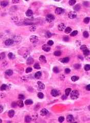
Murine studies have revealed signaling molecules that appear to play crucial roles in plasma cell development and survival.
Investigators found the adaptor protein DOK3 promotes the differentiation of plasma cells, and the protein-tyrosine phosphatase SHP1 enables plasma cell migration.
The team said these discoveries advance our understanding of plasma cells and may help us improve treatment for multiple myeloma and autoimmune disorders.
In PNAS, Kong-Peng Lam, PhD, of the Bioprocessing Technology Institute in Singapore, and his colleagues reported their discovery that DOK3 plays an important role in the formation of plasma cells.
The team found that calcium signaling inhibits the expression of PDL1 and PDL2, membrane proteins that are essential for plasma cell formation.
But DOK3 can promote the production of plasma cells by reducing the effects of calcium signaling on these proteins. And conversely, the absence of DOK3 results in defective plasma cell formation.
In Nature Communications, Dr Lam and his colleagues reported their discovery that SHP1 signaling is important for the long-term survival of plasma cells.
The researchers found that, in the absence of SHP1, plasma cells fail to migrate from the spleen to the bone marrow. This can impair the body’s immune response and increase susceptibility to infections and diseases.
However, Dr Lam and his colleagues were able to rectify the defective immune response caused by SHP1 deletion with antibody injections, a result that might aid the development of therapeutics for autoimmune disorders.
On the other hand, targeting SHP1 might be a strategy to treat multiple myeloma, in which the accumulation of cancerous plasma cells in the bone marrow is undesirable.
“These findings allow better understanding of plasma cells and their role in the immune system,” Dr Lam said. “The identification of these targets not only paves the way for development of therapeutics for those with autoimmune diseases and multiple myeloma but also impacts the development of immunological agents for combating infections.”
Murine studies have revealed signaling molecules that appear to play crucial roles in plasma cell development and survival.
Investigators found the adaptor protein DOK3 promotes the differentiation of plasma cells, and the protein-tyrosine phosphatase SHP1 enables plasma cell migration.
The team said these discoveries advance our understanding of plasma cells and may help us improve treatment for multiple myeloma and autoimmune disorders.
In PNAS, Kong-Peng Lam, PhD, of the Bioprocessing Technology Institute in Singapore, and his colleagues reported their discovery that DOK3 plays an important role in the formation of plasma cells.
The team found that calcium signaling inhibits the expression of PDL1 and PDL2, membrane proteins that are essential for plasma cell formation.
But DOK3 can promote the production of plasma cells by reducing the effects of calcium signaling on these proteins. And conversely, the absence of DOK3 results in defective plasma cell formation.
In Nature Communications, Dr Lam and his colleagues reported their discovery that SHP1 signaling is important for the long-term survival of plasma cells.
The researchers found that, in the absence of SHP1, plasma cells fail to migrate from the spleen to the bone marrow. This can impair the body’s immune response and increase susceptibility to infections and diseases.
However, Dr Lam and his colleagues were able to rectify the defective immune response caused by SHP1 deletion with antibody injections, a result that might aid the development of therapeutics for autoimmune disorders.
On the other hand, targeting SHP1 might be a strategy to treat multiple myeloma, in which the accumulation of cancerous plasma cells in the bone marrow is undesirable.
“These findings allow better understanding of plasma cells and their role in the immune system,” Dr Lam said. “The identification of these targets not only paves the way for development of therapeutics for those with autoimmune diseases and multiple myeloma but also impacts the development of immunological agents for combating infections.”
Murine studies have revealed signaling molecules that appear to play crucial roles in plasma cell development and survival.
Investigators found the adaptor protein DOK3 promotes the differentiation of plasma cells, and the protein-tyrosine phosphatase SHP1 enables plasma cell migration.
The team said these discoveries advance our understanding of plasma cells and may help us improve treatment for multiple myeloma and autoimmune disorders.
In PNAS, Kong-Peng Lam, PhD, of the Bioprocessing Technology Institute in Singapore, and his colleagues reported their discovery that DOK3 plays an important role in the formation of plasma cells.
The team found that calcium signaling inhibits the expression of PDL1 and PDL2, membrane proteins that are essential for plasma cell formation.
But DOK3 can promote the production of plasma cells by reducing the effects of calcium signaling on these proteins. And conversely, the absence of DOK3 results in defective plasma cell formation.
In Nature Communications, Dr Lam and his colleagues reported their discovery that SHP1 signaling is important for the long-term survival of plasma cells.
The researchers found that, in the absence of SHP1, plasma cells fail to migrate from the spleen to the bone marrow. This can impair the body’s immune response and increase susceptibility to infections and diseases.
However, Dr Lam and his colleagues were able to rectify the defective immune response caused by SHP1 deletion with antibody injections, a result that might aid the development of therapeutics for autoimmune disorders.
On the other hand, targeting SHP1 might be a strategy to treat multiple myeloma, in which the accumulation of cancerous plasma cells in the bone marrow is undesirable.
“These findings allow better understanding of plasma cells and their role in the immune system,” Dr Lam said. “The identification of these targets not only paves the way for development of therapeutics for those with autoimmune diseases and multiple myeloma but also impacts the development of immunological agents for combating infections.”
Tests use cell density to diagnose sickle cell disease

and a sickled cell
Credit: Betty Pace
Two new tests can diagnose sickle cell disease (SCD) quickly, simply, and cheaply, researchers have reported in PNAS.
The tests separate red blood cells based on density and can provide an SCD diagnosis in less than 12 minutes for as little as 50 cents.
When run against clinical samples, both tests showed high sensitivity and specificity for SCD. And one of the tests could distinguish between the two main subclasses of SCD.
Ashok A. Kumar, PhD, of Harvard University in Cambridge, Massachusetts, and his colleagues developed the tests by connecting two ideas scientists have understood for decades.
The first is the notion that blood cells affected by SCD are denser than normal cells, and the second is that many polymers, when mixed in water, automatically separate into layers ordered by density.
Dr Kumar and his colleagues discovered the layers could be used to separate red blood cells by density.
So the researchers designed 2 tests to separate red blood cells into multiple bins of density. The presence or absence of cells in the bins distinguished individuals with the most prevalent forms of SCD (Hb SS or Hb SC) from individuals with either normal hemoglobin (Hb AA) or sickle-cell trait (Hb AS).
“We wanted to make the test[s] as simple as possible,” Dr Kumar noted. “The idea was to make [them] something you could run from just a finger prick. Because these gradients assemble on their own, that meant we could make them in whatever volume we wanted, even a small capillary tube.”
The design the team settled on is barely larger than a toothpick. Running the tests is as simple as uncapping a tube, pricking a patient’s finger, and allowing the blood to wick into the tube.
The researchers evaluated the tests in 59 subjects, 33 who were negative for SCD and 26 who were positive for SCD. Both tests identified SCD-positive samples with a sensitivity greater than 90% and a specificity greater than 88%.
The simpler test, called SCD-AMPS-2, involves 2 phases. It identified Hb SS and Hb SC with a sensitivity of 90% (73%–98%) and a specificity of 97% (86%–100%).
The higher-resolution test, called SCD-AMPS-3, involves 3 phases. It identified the 2 types of SCD with a sensitivity of 91% (78%–98%) and a specificity of 88% (74%–98%). This test could also distinguish between Hb SS and Hb SC.
Despite these promising results, Dr Kumar said additional testing will be needed to determine whether the tests are truly accurate enough to use in the field.
and a sickled cell
Credit: Betty Pace
Two new tests can diagnose sickle cell disease (SCD) quickly, simply, and cheaply, researchers have reported in PNAS.
The tests separate red blood cells based on density and can provide an SCD diagnosis in less than 12 minutes for as little as 50 cents.
When run against clinical samples, both tests showed high sensitivity and specificity for SCD. And one of the tests could distinguish between the two main subclasses of SCD.
Ashok A. Kumar, PhD, of Harvard University in Cambridge, Massachusetts, and his colleagues developed the tests by connecting two ideas scientists have understood for decades.
The first is the notion that blood cells affected by SCD are denser than normal cells, and the second is that many polymers, when mixed in water, automatically separate into layers ordered by density.
Dr Kumar and his colleagues discovered the layers could be used to separate red blood cells by density.
So the researchers designed 2 tests to separate red blood cells into multiple bins of density. The presence or absence of cells in the bins distinguished individuals with the most prevalent forms of SCD (Hb SS or Hb SC) from individuals with either normal hemoglobin (Hb AA) or sickle-cell trait (Hb AS).
“We wanted to make the test[s] as simple as possible,” Dr Kumar noted. “The idea was to make [them] something you could run from just a finger prick. Because these gradients assemble on their own, that meant we could make them in whatever volume we wanted, even a small capillary tube.”
The design the team settled on is barely larger than a toothpick. Running the tests is as simple as uncapping a tube, pricking a patient’s finger, and allowing the blood to wick into the tube.
The researchers evaluated the tests in 59 subjects, 33 who were negative for SCD and 26 who were positive for SCD. Both tests identified SCD-positive samples with a sensitivity greater than 90% and a specificity greater than 88%.
The simpler test, called SCD-AMPS-2, involves 2 phases. It identified Hb SS and Hb SC with a sensitivity of 90% (73%–98%) and a specificity of 97% (86%–100%).
The higher-resolution test, called SCD-AMPS-3, involves 3 phases. It identified the 2 types of SCD with a sensitivity of 91% (78%–98%) and a specificity of 88% (74%–98%). This test could also distinguish between Hb SS and Hb SC.
Despite these promising results, Dr Kumar said additional testing will be needed to determine whether the tests are truly accurate enough to use in the field.
and a sickled cell
Credit: Betty Pace
Two new tests can diagnose sickle cell disease (SCD) quickly, simply, and cheaply, researchers have reported in PNAS.
The tests separate red blood cells based on density and can provide an SCD diagnosis in less than 12 minutes for as little as 50 cents.
When run against clinical samples, both tests showed high sensitivity and specificity for SCD. And one of the tests could distinguish between the two main subclasses of SCD.
Ashok A. Kumar, PhD, of Harvard University in Cambridge, Massachusetts, and his colleagues developed the tests by connecting two ideas scientists have understood for decades.
The first is the notion that blood cells affected by SCD are denser than normal cells, and the second is that many polymers, when mixed in water, automatically separate into layers ordered by density.
Dr Kumar and his colleagues discovered the layers could be used to separate red blood cells by density.
So the researchers designed 2 tests to separate red blood cells into multiple bins of density. The presence or absence of cells in the bins distinguished individuals with the most prevalent forms of SCD (Hb SS or Hb SC) from individuals with either normal hemoglobin (Hb AA) or sickle-cell trait (Hb AS).
“We wanted to make the test[s] as simple as possible,” Dr Kumar noted. “The idea was to make [them] something you could run from just a finger prick. Because these gradients assemble on their own, that meant we could make them in whatever volume we wanted, even a small capillary tube.”
The design the team settled on is barely larger than a toothpick. Running the tests is as simple as uncapping a tube, pricking a patient’s finger, and allowing the blood to wick into the tube.
The researchers evaluated the tests in 59 subjects, 33 who were negative for SCD and 26 who were positive for SCD. Both tests identified SCD-positive samples with a sensitivity greater than 90% and a specificity greater than 88%.
The simpler test, called SCD-AMPS-2, involves 2 phases. It identified Hb SS and Hb SC with a sensitivity of 90% (73%–98%) and a specificity of 97% (86%–100%).
The higher-resolution test, called SCD-AMPS-3, involves 3 phases. It identified the 2 types of SCD with a sensitivity of 91% (78%–98%) and a specificity of 88% (74%–98%). This test could also distinguish between Hb SS and Hb SC.
Despite these promising results, Dr Kumar said additional testing will be needed to determine whether the tests are truly accurate enough to use in the field.
Method could improve malaria diagnosis
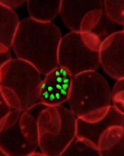
red blood cell; Credit: St Jude
Children’s Research Hospital
Researchers have found they can diagnose malaria using magnetic fields to detect a byproduct of malarial metabolism.
They used magnetic resonance relaxometry (MRR) to detect a parasitic waste product called hemozoin in malaria-infected red blood cells from mice and humans.
The team said MRR is more sensitive than other methods of detecting malaria, can be carried out using a portable benchtop system, and costs less than 10 cents per test.
Jongyoon Han, PhD, of the Massachusetts Institute of Technology in Cambridge, and his colleagues described the technique in Nature Medicine.
When malaria parasites infect red blood cells, they feed on the nutrient-rich hemoglobin. As hemoglobin breaks down, it releases iron, which can be toxic, so the parasite converts the iron into hemozoin—a weakly paramagnetic crystallite.
Those crystals interfere with the normal magnetic spins of hydrogen atoms. When exposed to a powerful magnetic field, hydrogen atoms align their spins in the same direction.
When a second, smaller field perturbs the atoms, they should all change their spins in synchrony. But if another magnetic particle, such as hemozoin, is present, this synchrony is disrupted through a process called relaxation. The more magnetic particles present, the more quickly the synchrony is disrupted.
“What we are trying to really measure is how the hydrogen’s nuclear magnetic resonance is affected by the proximity of other magnetic particles,” Dr Han said.
This MRR technique enables malaria diagnosis because hemozoin crystals are produced in all 4 stages of malaria infection and are generated by all known species of the Plasmodium parasite. Furthermore, the amount of hemozoin can reveal how severe the infection is, or whether it is responding to treatment.
Dr Han and his colleagues found they could use MRR to detect Plasmodium falciparum infection to as low as 0.0002% parasitemia in 750 nl of cultured blood in less than 5 minutes.
They also detected Plasmodium berghei in mice, allowing for reliable estimation of parasitemia to as low as 0.0001%.
The device the researchers used in this study is small enough to sit on a table or lab bench, but they are working on a portable version the size of a small electronic tablet.
“This system can be built at a very low cost, relative to the million-dollar MRI machines used in a hospital,” said study author Weng Kung Peng, PhD, of the Singapore-MIT Alliance for Research and Technology Centre in Singapore.
“Furthermore, since this technique does not rely on expensive labeling with chemical reagents, we are able to get each diagnostic test done at a cost of less than 10 cents.”
The researchers are launching a company to make this technology available at an affordable price. The team is also running field tests in Southeast Asia and exploring powering the device on solar energy.
red blood cell; Credit: St Jude
Children’s Research Hospital
Researchers have found they can diagnose malaria using magnetic fields to detect a byproduct of malarial metabolism.
They used magnetic resonance relaxometry (MRR) to detect a parasitic waste product called hemozoin in malaria-infected red blood cells from mice and humans.
The team said MRR is more sensitive than other methods of detecting malaria, can be carried out using a portable benchtop system, and costs less than 10 cents per test.
Jongyoon Han, PhD, of the Massachusetts Institute of Technology in Cambridge, and his colleagues described the technique in Nature Medicine.
When malaria parasites infect red blood cells, they feed on the nutrient-rich hemoglobin. As hemoglobin breaks down, it releases iron, which can be toxic, so the parasite converts the iron into hemozoin—a weakly paramagnetic crystallite.
Those crystals interfere with the normal magnetic spins of hydrogen atoms. When exposed to a powerful magnetic field, hydrogen atoms align their spins in the same direction.
When a second, smaller field perturbs the atoms, they should all change their spins in synchrony. But if another magnetic particle, such as hemozoin, is present, this synchrony is disrupted through a process called relaxation. The more magnetic particles present, the more quickly the synchrony is disrupted.
“What we are trying to really measure is how the hydrogen’s nuclear magnetic resonance is affected by the proximity of other magnetic particles,” Dr Han said.
This MRR technique enables malaria diagnosis because hemozoin crystals are produced in all 4 stages of malaria infection and are generated by all known species of the Plasmodium parasite. Furthermore, the amount of hemozoin can reveal how severe the infection is, or whether it is responding to treatment.
Dr Han and his colleagues found they could use MRR to detect Plasmodium falciparum infection to as low as 0.0002% parasitemia in 750 nl of cultured blood in less than 5 minutes.
They also detected Plasmodium berghei in mice, allowing for reliable estimation of parasitemia to as low as 0.0001%.
The device the researchers used in this study is small enough to sit on a table or lab bench, but they are working on a portable version the size of a small electronic tablet.
“This system can be built at a very low cost, relative to the million-dollar MRI machines used in a hospital,” said study author Weng Kung Peng, PhD, of the Singapore-MIT Alliance for Research and Technology Centre in Singapore.
“Furthermore, since this technique does not rely on expensive labeling with chemical reagents, we are able to get each diagnostic test done at a cost of less than 10 cents.”
The researchers are launching a company to make this technology available at an affordable price. The team is also running field tests in Southeast Asia and exploring powering the device on solar energy.
red blood cell; Credit: St Jude
Children’s Research Hospital
Researchers have found they can diagnose malaria using magnetic fields to detect a byproduct of malarial metabolism.
They used magnetic resonance relaxometry (MRR) to detect a parasitic waste product called hemozoin in malaria-infected red blood cells from mice and humans.
The team said MRR is more sensitive than other methods of detecting malaria, can be carried out using a portable benchtop system, and costs less than 10 cents per test.
Jongyoon Han, PhD, of the Massachusetts Institute of Technology in Cambridge, and his colleagues described the technique in Nature Medicine.
When malaria parasites infect red blood cells, they feed on the nutrient-rich hemoglobin. As hemoglobin breaks down, it releases iron, which can be toxic, so the parasite converts the iron into hemozoin—a weakly paramagnetic crystallite.
Those crystals interfere with the normal magnetic spins of hydrogen atoms. When exposed to a powerful magnetic field, hydrogen atoms align their spins in the same direction.
When a second, smaller field perturbs the atoms, they should all change their spins in synchrony. But if another magnetic particle, such as hemozoin, is present, this synchrony is disrupted through a process called relaxation. The more magnetic particles present, the more quickly the synchrony is disrupted.
“What we are trying to really measure is how the hydrogen’s nuclear magnetic resonance is affected by the proximity of other magnetic particles,” Dr Han said.
This MRR technique enables malaria diagnosis because hemozoin crystals are produced in all 4 stages of malaria infection and are generated by all known species of the Plasmodium parasite. Furthermore, the amount of hemozoin can reveal how severe the infection is, or whether it is responding to treatment.
Dr Han and his colleagues found they could use MRR to detect Plasmodium falciparum infection to as low as 0.0002% parasitemia in 750 nl of cultured blood in less than 5 minutes.
They also detected Plasmodium berghei in mice, allowing for reliable estimation of parasitemia to as low as 0.0001%.
The device the researchers used in this study is small enough to sit on a table or lab bench, but they are working on a portable version the size of a small electronic tablet.
“This system can be built at a very low cost, relative to the million-dollar MRI machines used in a hospital,” said study author Weng Kung Peng, PhD, of the Singapore-MIT Alliance for Research and Technology Centre in Singapore.
“Furthermore, since this technique does not rely on expensive labeling with chemical reagents, we are able to get each diagnostic test done at a cost of less than 10 cents.”
The researchers are launching a company to make this technology available at an affordable price. The team is also running field tests in Southeast Asia and exploring powering the device on solar energy.
Antibody gets orphan status for CTCL in Europe
The European Commission has granted orphan drug designation to IPH4102 for the treatment of cutaneous T-cell lymphoma (CTCL).
IPH4102 is a cytotoxic anti-KIR3DL2 monoclonal antibody (mAb) that targets CTCL cells.
Orphan status provides Innate Pharma, the company developing IPH4102, with benefits such as tax incentives, market exclusivity for 10 years, possibilities for additional research funding, and additional guidance from the European Medicines Agency during clinical development.
Preclinical results with IPH4102 were presented in a poster at the 2014 T-cell Lymphoma Forum. The research was conducted by investigators from Innate Pharma and INSERM at Hôpital Saint Louis in Paris.
The researchers generated 3 mAbs that bind selectively to KIR3DL2 and evaluated their efficacy against KIR3DL2-expressing tumors and Sézary cell lines.
IPH4102 was among the 3 mAbs and emerged as the most promising drug candidate.
Experiments revealed that anti-KIR3DL2 mAbs can kill KIR3DL2+ cell lines through allo-antibody-dependent cell cytotoxicity, even at low tumor antigen density.
The mAbs also improved survival in KIR3DL2+ xenograft models. Survival in mAb-treated mice ranged from 30.5 days to 54.5 days, compared to 19 days in controls.
Finally, the mAbs mediated killing of primary Sézary cells with autologous natural killer cells nearly as efficiently as alemtuzumab.
The investigators said these results suggest anti-KIR3DL2 mAbs are a feasible treatment option for CTCL patients. They plan to prove this hypothesis with a phase 1 trial of IPH4102, which is expected to begin in 2015.
The European Commission has granted orphan drug designation to IPH4102 for the treatment of cutaneous T-cell lymphoma (CTCL).
IPH4102 is a cytotoxic anti-KIR3DL2 monoclonal antibody (mAb) that targets CTCL cells.
Orphan status provides Innate Pharma, the company developing IPH4102, with benefits such as tax incentives, market exclusivity for 10 years, possibilities for additional research funding, and additional guidance from the European Medicines Agency during clinical development.
Preclinical results with IPH4102 were presented in a poster at the 2014 T-cell Lymphoma Forum. The research was conducted by investigators from Innate Pharma and INSERM at Hôpital Saint Louis in Paris.
The researchers generated 3 mAbs that bind selectively to KIR3DL2 and evaluated their efficacy against KIR3DL2-expressing tumors and Sézary cell lines.
IPH4102 was among the 3 mAbs and emerged as the most promising drug candidate.
Experiments revealed that anti-KIR3DL2 mAbs can kill KIR3DL2+ cell lines through allo-antibody-dependent cell cytotoxicity, even at low tumor antigen density.
The mAbs also improved survival in KIR3DL2+ xenograft models. Survival in mAb-treated mice ranged from 30.5 days to 54.5 days, compared to 19 days in controls.
Finally, the mAbs mediated killing of primary Sézary cells with autologous natural killer cells nearly as efficiently as alemtuzumab.
The investigators said these results suggest anti-KIR3DL2 mAbs are a feasible treatment option for CTCL patients. They plan to prove this hypothesis with a phase 1 trial of IPH4102, which is expected to begin in 2015.
The European Commission has granted orphan drug designation to IPH4102 for the treatment of cutaneous T-cell lymphoma (CTCL).
IPH4102 is a cytotoxic anti-KIR3DL2 monoclonal antibody (mAb) that targets CTCL cells.
Orphan status provides Innate Pharma, the company developing IPH4102, with benefits such as tax incentives, market exclusivity for 10 years, possibilities for additional research funding, and additional guidance from the European Medicines Agency during clinical development.
Preclinical results with IPH4102 were presented in a poster at the 2014 T-cell Lymphoma Forum. The research was conducted by investigators from Innate Pharma and INSERM at Hôpital Saint Louis in Paris.
The researchers generated 3 mAbs that bind selectively to KIR3DL2 and evaluated their efficacy against KIR3DL2-expressing tumors and Sézary cell lines.
IPH4102 was among the 3 mAbs and emerged as the most promising drug candidate.
Experiments revealed that anti-KIR3DL2 mAbs can kill KIR3DL2+ cell lines through allo-antibody-dependent cell cytotoxicity, even at low tumor antigen density.
The mAbs also improved survival in KIR3DL2+ xenograft models. Survival in mAb-treated mice ranged from 30.5 days to 54.5 days, compared to 19 days in controls.
Finally, the mAbs mediated killing of primary Sézary cells with autologous natural killer cells nearly as efficiently as alemtuzumab.
The investigators said these results suggest anti-KIR3DL2 mAbs are a feasible treatment option for CTCL patients. They plan to prove this hypothesis with a phase 1 trial of IPH4102, which is expected to begin in 2015.
FDA approves generic decitabine for MDS

Credit: Bill Branson
The US Food and Drug Administration (FDA) has approved decitabine for injection, a generic version of Dacogen, to treat patients with myelodysplastic syndromes (MDS).
Decitabine is indicated for previously treated and untreated patients with de novo and secondary MDS of all French-American-British subtypes—refractory anemia, refractory anemia with ringed sideroblasts, refractory anemia with excess blasts, refractory anemia with excess blasts in transformation, and chronic myelomonocytic leukemia—as well as intermediate-1, intermediate-2, and high-risk International Prognostic Scoring System groups.
Decitabine will be marketed in 20 mL single-dose glass vials containing 50 mg decitabine—the same size and strength as the brand name drug. The dosing regimen is identical as well.
InnoPharma developed the generic formulation of decitabine and entered into an agreement with Sandoz Inc. Sandoz will sell, market, and distribute decitabine in the US. InnoPharma is set to be acquired by Pfizer Inc., but the transaction is subject to US regulatory approval.
The FDA approved another generic form of decitabine for the treatment of MDS in July 2013. That drug is a product of Dr Reddy’s Laboratories Limited.
Dacogen has been FDA-approved to treat MDS since May 2006. Dacogen is a registered trademark used by Eisai Inc. under license from Astex Pharmaceuticals Inc.
Credit: Bill Branson
The US Food and Drug Administration (FDA) has approved decitabine for injection, a generic version of Dacogen, to treat patients with myelodysplastic syndromes (MDS).
Decitabine is indicated for previously treated and untreated patients with de novo and secondary MDS of all French-American-British subtypes—refractory anemia, refractory anemia with ringed sideroblasts, refractory anemia with excess blasts, refractory anemia with excess blasts in transformation, and chronic myelomonocytic leukemia—as well as intermediate-1, intermediate-2, and high-risk International Prognostic Scoring System groups.
Decitabine will be marketed in 20 mL single-dose glass vials containing 50 mg decitabine—the same size and strength as the brand name drug. The dosing regimen is identical as well.
InnoPharma developed the generic formulation of decitabine and entered into an agreement with Sandoz Inc. Sandoz will sell, market, and distribute decitabine in the US. InnoPharma is set to be acquired by Pfizer Inc., but the transaction is subject to US regulatory approval.
The FDA approved another generic form of decitabine for the treatment of MDS in July 2013. That drug is a product of Dr Reddy’s Laboratories Limited.
Dacogen has been FDA-approved to treat MDS since May 2006. Dacogen is a registered trademark used by Eisai Inc. under license from Astex Pharmaceuticals Inc.
Credit: Bill Branson
The US Food and Drug Administration (FDA) has approved decitabine for injection, a generic version of Dacogen, to treat patients with myelodysplastic syndromes (MDS).
Decitabine is indicated for previously treated and untreated patients with de novo and secondary MDS of all French-American-British subtypes—refractory anemia, refractory anemia with ringed sideroblasts, refractory anemia with excess blasts, refractory anemia with excess blasts in transformation, and chronic myelomonocytic leukemia—as well as intermediate-1, intermediate-2, and high-risk International Prognostic Scoring System groups.
Decitabine will be marketed in 20 mL single-dose glass vials containing 50 mg decitabine—the same size and strength as the brand name drug. The dosing regimen is identical as well.
InnoPharma developed the generic formulation of decitabine and entered into an agreement with Sandoz Inc. Sandoz will sell, market, and distribute decitabine in the US. InnoPharma is set to be acquired by Pfizer Inc., but the transaction is subject to US regulatory approval.
The FDA approved another generic form of decitabine for the treatment of MDS in July 2013. That drug is a product of Dr Reddy’s Laboratories Limited.
Dacogen has been FDA-approved to treat MDS since May 2006. Dacogen is a registered trademark used by Eisai Inc. under license from Astex Pharmaceuticals Inc.
Group exploits autophagy to fight myeloma
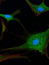
Credit: Sarah Pfau
Researchers have devised a strategy that leverages autophagy to work against multiple myeloma (MM).
Their method involves targeting the p62 protein, which plays a role in disposing of unwanted cellular proteins during autophagy.
The team used anticancer agents to induce autophagy in MM cells and found that simultaneously blocking p62 expression, either pharmacologically or with shRNA, could prompt apoptosis in the cells, both in vitro and in vivo.
Steven Grant, MD, of Virginia Commonwealth University’s Massey Cancer Center, and his colleagues described this work in Molecular and Cellular Biology.
“Therapies that are designed to block the early stages of autophagy do not offer the possibility of exploiting its potentially lethal effects,” Dr Grant said. “Our strategy turns autophagy from a protective process into a toxic one, and these results suggest it could increase the effectiveness of a variety of cancer therapies that induce autophagy.”
The researchers conducted several experiments in MM cell lines and mouse models of the disease. They used an anticancer agent—obatoclax or bortezomib—to induce autophagy and a cyclin-dependent kinase (CDK) inhibitor—flavopiridol or dinaciclib—or shRNA to target p62.
And they discovered that blocking p62 disrupted autophagy and killed far more MM cells than administering anticancer agents alone.
Critical to the success of this strategy is Bik, a protein that plays a significant role in governing cell death and survival. With anticancer treatment, Bik accumulates in MM cells until it triggers apoptosis.
Normally, the MM cells would initiate autophagy to survive, and p62 would rid the cells of Bik by loading the proteins into autophagosomes for disposal.
However, the researchers found that blocking p62 production results in an inefficient form of autophagy, and the accumulation of Bik eventually causes the MM cells to undergo apoptosis.
This research builds upon more than a decade of work by Dr Grant’s lab, in which the team investigated novel treatment strategies and combination therapies that selectively kill MM cells and other blood cancer cells.
“We are now working to identify additional CDK inhibitors that can be used to disrupt autophagy,” Dr Grant said. “The ultimate goal will be to translate these findings into a clinical trial to test the therapy in patients with various blood cancers.”
The technology in his study has been made available for licensing through the Virginia Commonwealth University Office of Research.
Credit: Sarah Pfau
Researchers have devised a strategy that leverages autophagy to work against multiple myeloma (MM).
Their method involves targeting the p62 protein, which plays a role in disposing of unwanted cellular proteins during autophagy.
The team used anticancer agents to induce autophagy in MM cells and found that simultaneously blocking p62 expression, either pharmacologically or with shRNA, could prompt apoptosis in the cells, both in vitro and in vivo.
Steven Grant, MD, of Virginia Commonwealth University’s Massey Cancer Center, and his colleagues described this work in Molecular and Cellular Biology.
“Therapies that are designed to block the early stages of autophagy do not offer the possibility of exploiting its potentially lethal effects,” Dr Grant said. “Our strategy turns autophagy from a protective process into a toxic one, and these results suggest it could increase the effectiveness of a variety of cancer therapies that induce autophagy.”
The researchers conducted several experiments in MM cell lines and mouse models of the disease. They used an anticancer agent—obatoclax or bortezomib—to induce autophagy and a cyclin-dependent kinase (CDK) inhibitor—flavopiridol or dinaciclib—or shRNA to target p62.
And they discovered that blocking p62 disrupted autophagy and killed far more MM cells than administering anticancer agents alone.
Critical to the success of this strategy is Bik, a protein that plays a significant role in governing cell death and survival. With anticancer treatment, Bik accumulates in MM cells until it triggers apoptosis.
Normally, the MM cells would initiate autophagy to survive, and p62 would rid the cells of Bik by loading the proteins into autophagosomes for disposal.
However, the researchers found that blocking p62 production results in an inefficient form of autophagy, and the accumulation of Bik eventually causes the MM cells to undergo apoptosis.
This research builds upon more than a decade of work by Dr Grant’s lab, in which the team investigated novel treatment strategies and combination therapies that selectively kill MM cells and other blood cancer cells.
“We are now working to identify additional CDK inhibitors that can be used to disrupt autophagy,” Dr Grant said. “The ultimate goal will be to translate these findings into a clinical trial to test the therapy in patients with various blood cancers.”
The technology in his study has been made available for licensing through the Virginia Commonwealth University Office of Research.
Credit: Sarah Pfau
Researchers have devised a strategy that leverages autophagy to work against multiple myeloma (MM).
Their method involves targeting the p62 protein, which plays a role in disposing of unwanted cellular proteins during autophagy.
The team used anticancer agents to induce autophagy in MM cells and found that simultaneously blocking p62 expression, either pharmacologically or with shRNA, could prompt apoptosis in the cells, both in vitro and in vivo.
Steven Grant, MD, of Virginia Commonwealth University’s Massey Cancer Center, and his colleagues described this work in Molecular and Cellular Biology.
“Therapies that are designed to block the early stages of autophagy do not offer the possibility of exploiting its potentially lethal effects,” Dr Grant said. “Our strategy turns autophagy from a protective process into a toxic one, and these results suggest it could increase the effectiveness of a variety of cancer therapies that induce autophagy.”
The researchers conducted several experiments in MM cell lines and mouse models of the disease. They used an anticancer agent—obatoclax or bortezomib—to induce autophagy and a cyclin-dependent kinase (CDK) inhibitor—flavopiridol or dinaciclib—or shRNA to target p62.
And they discovered that blocking p62 disrupted autophagy and killed far more MM cells than administering anticancer agents alone.
Critical to the success of this strategy is Bik, a protein that plays a significant role in governing cell death and survival. With anticancer treatment, Bik accumulates in MM cells until it triggers apoptosis.
Normally, the MM cells would initiate autophagy to survive, and p62 would rid the cells of Bik by loading the proteins into autophagosomes for disposal.
However, the researchers found that blocking p62 production results in an inefficient form of autophagy, and the accumulation of Bik eventually causes the MM cells to undergo apoptosis.
This research builds upon more than a decade of work by Dr Grant’s lab, in which the team investigated novel treatment strategies and combination therapies that selectively kill MM cells and other blood cancer cells.
“We are now working to identify additional CDK inhibitors that can be used to disrupt autophagy,” Dr Grant said. “The ultimate goal will be to translate these findings into a clinical trial to test the therapy in patients with various blood cancers.”
The technology in his study has been made available for licensing through the Virginia Commonwealth University Office of Research.
Program improves depression treatment in cancer

Credit: NIH
Results of a large study suggest major depression is common—but largely untreated—among cancer patients in Scotland.
And 2 additional studies of Scottish patients showed that a program specifically designed for individuals with cancer can treat depression and improve quality of life more effectively than current methods of care.
These studies appear in The Lancet, The Lancet Oncology, and The Lancet Psychiatry.
In The Lancet Psychiatry, researchers recounted their analysis of data from 21,151 patients treated at cancer clinics in Scotland. The team found that major depression was substantially more common in cancer patients than in the general population.
Major depression was most common in patients with lung cancer (13%) and lowest in those with genitourinary cancer (6%). Moreover, nearly three-quarters (73%) of depressed cancer patients were not receiving treatment.
To address the problem of inadequate treatment, researchers initiated the SMaRT Oncology-2 trial. They reported the results in The Lancet.
The team evaluated a new treatment program called “Depression Care for People with Cancer” (DCPC). DCPC is delivered by specially trained cancer nurses and psychiatrists, working in collaboration with the patient’s cancer team and general practitioner, and is given as part of cancer care. It is a systematic treatment program that includes both antidepressants and psychological therapy.
The trial included 500 adults with major depression and a cancer with a good prognosis (predicted survival of more than 12 months).
Patients were randomized to receive either DCPC or “usual care,” which was provided by a patient’s general practitioner and might have included prescribing antidepressants or referring the patient to mental health services for assessment or psychological treatment.
Results showed that DCPC was more effective than usual care in reducing depression. At 6 months, 62% of patients who received DCPC responded to treatment (experiencing at least a 50% reduction in the severity of their depression), compared with 17% of those who received the usual care (P<0.0001). This benefit was sustained at 12 months.
In addition, DCPC improved anxiety, pain, fatigue, functioning, and overall quality of life (all P<0.05). The researchers also noted that the cost of providing DCPC was modest (£613 per patient).
“The huge benefit that DCPC delivers for patients with cancer and depression shows what we can achieve for patients if we take as much care with the treatment of their depression as we do with the treatment of their cancer,” said study author Michael Sharpe, MD, of the University of Oxford in the UK.
To see if patients with a poor-prognosis cancer could also benefit from DCPC, researchers initiated the SMaRT Oncology-3 trial. They reported the results in The Lancet Oncology.
The team tested a version of DCPC adapted for cancer patients with a poor prognosis. The trial included 142 patients with lung cancer and major depression.
Patients who received the modified version of DCPC had a significantly greater improvement in depression than those who received the usual care during 32 weeks of follow-up (P=0.0003). DCPC also improved patients’ anxiety (P=0.046), functioning (P=0.0019), and quality of life (P=0.018).
“Patients with lung cancer often have a poor prognosis,” said study author Jane Walker, MBChB, PhD, of the University of Oxford and Sobell House Hospice in Oxford, UK.
“If they also have major depression, that can blight the time they have left to live. This trial shows that we can effectively treat depression in patients with poor-prognosis cancers, like lung cancer, and really improve patients’ lives.”
Credit: NIH
Results of a large study suggest major depression is common—but largely untreated—among cancer patients in Scotland.
And 2 additional studies of Scottish patients showed that a program specifically designed for individuals with cancer can treat depression and improve quality of life more effectively than current methods of care.
These studies appear in The Lancet, The Lancet Oncology, and The Lancet Psychiatry.
In The Lancet Psychiatry, researchers recounted their analysis of data from 21,151 patients treated at cancer clinics in Scotland. The team found that major depression was substantially more common in cancer patients than in the general population.
Major depression was most common in patients with lung cancer (13%) and lowest in those with genitourinary cancer (6%). Moreover, nearly three-quarters (73%) of depressed cancer patients were not receiving treatment.
To address the problem of inadequate treatment, researchers initiated the SMaRT Oncology-2 trial. They reported the results in The Lancet.
The team evaluated a new treatment program called “Depression Care for People with Cancer” (DCPC). DCPC is delivered by specially trained cancer nurses and psychiatrists, working in collaboration with the patient’s cancer team and general practitioner, and is given as part of cancer care. It is a systematic treatment program that includes both antidepressants and psychological therapy.
The trial included 500 adults with major depression and a cancer with a good prognosis (predicted survival of more than 12 months).
Patients were randomized to receive either DCPC or “usual care,” which was provided by a patient’s general practitioner and might have included prescribing antidepressants or referring the patient to mental health services for assessment or psychological treatment.
Results showed that DCPC was more effective than usual care in reducing depression. At 6 months, 62% of patients who received DCPC responded to treatment (experiencing at least a 50% reduction in the severity of their depression), compared with 17% of those who received the usual care (P<0.0001). This benefit was sustained at 12 months.
In addition, DCPC improved anxiety, pain, fatigue, functioning, and overall quality of life (all P<0.05). The researchers also noted that the cost of providing DCPC was modest (£613 per patient).
“The huge benefit that DCPC delivers for patients with cancer and depression shows what we can achieve for patients if we take as much care with the treatment of their depression as we do with the treatment of their cancer,” said study author Michael Sharpe, MD, of the University of Oxford in the UK.
To see if patients with a poor-prognosis cancer could also benefit from DCPC, researchers initiated the SMaRT Oncology-3 trial. They reported the results in The Lancet Oncology.
The team tested a version of DCPC adapted for cancer patients with a poor prognosis. The trial included 142 patients with lung cancer and major depression.
Patients who received the modified version of DCPC had a significantly greater improvement in depression than those who received the usual care during 32 weeks of follow-up (P=0.0003). DCPC also improved patients’ anxiety (P=0.046), functioning (P=0.0019), and quality of life (P=0.018).
“Patients with lung cancer often have a poor prognosis,” said study author Jane Walker, MBChB, PhD, of the University of Oxford and Sobell House Hospice in Oxford, UK.
“If they also have major depression, that can blight the time they have left to live. This trial shows that we can effectively treat depression in patients with poor-prognosis cancers, like lung cancer, and really improve patients’ lives.”
Credit: NIH
Results of a large study suggest major depression is common—but largely untreated—among cancer patients in Scotland.
And 2 additional studies of Scottish patients showed that a program specifically designed for individuals with cancer can treat depression and improve quality of life more effectively than current methods of care.
These studies appear in The Lancet, The Lancet Oncology, and The Lancet Psychiatry.
In The Lancet Psychiatry, researchers recounted their analysis of data from 21,151 patients treated at cancer clinics in Scotland. The team found that major depression was substantially more common in cancer patients than in the general population.
Major depression was most common in patients with lung cancer (13%) and lowest in those with genitourinary cancer (6%). Moreover, nearly three-quarters (73%) of depressed cancer patients were not receiving treatment.
To address the problem of inadequate treatment, researchers initiated the SMaRT Oncology-2 trial. They reported the results in The Lancet.
The team evaluated a new treatment program called “Depression Care for People with Cancer” (DCPC). DCPC is delivered by specially trained cancer nurses and psychiatrists, working in collaboration with the patient’s cancer team and general practitioner, and is given as part of cancer care. It is a systematic treatment program that includes both antidepressants and psychological therapy.
The trial included 500 adults with major depression and a cancer with a good prognosis (predicted survival of more than 12 months).
Patients were randomized to receive either DCPC or “usual care,” which was provided by a patient’s general practitioner and might have included prescribing antidepressants or referring the patient to mental health services for assessment or psychological treatment.
Results showed that DCPC was more effective than usual care in reducing depression. At 6 months, 62% of patients who received DCPC responded to treatment (experiencing at least a 50% reduction in the severity of their depression), compared with 17% of those who received the usual care (P<0.0001). This benefit was sustained at 12 months.
In addition, DCPC improved anxiety, pain, fatigue, functioning, and overall quality of life (all P<0.05). The researchers also noted that the cost of providing DCPC was modest (£613 per patient).
“The huge benefit that DCPC delivers for patients with cancer and depression shows what we can achieve for patients if we take as much care with the treatment of their depression as we do with the treatment of their cancer,” said study author Michael Sharpe, MD, of the University of Oxford in the UK.
To see if patients with a poor-prognosis cancer could also benefit from DCPC, researchers initiated the SMaRT Oncology-3 trial. They reported the results in The Lancet Oncology.
The team tested a version of DCPC adapted for cancer patients with a poor prognosis. The trial included 142 patients with lung cancer and major depression.
Patients who received the modified version of DCPC had a significantly greater improvement in depression than those who received the usual care during 32 weeks of follow-up (P=0.0003). DCPC also improved patients’ anxiety (P=0.046), functioning (P=0.0019), and quality of life (P=0.018).
“Patients with lung cancer often have a poor prognosis,” said study author Jane Walker, MBChB, PhD, of the University of Oxford and Sobell House Hospice in Oxford, UK.
“If they also have major depression, that can blight the time they have left to live. This trial shows that we can effectively treat depression in patients with poor-prognosis cancers, like lung cancer, and really improve patients’ lives.”
Drug granted orphan status for PNH in EU

A novel compound has received orphan status in the Europe Union to treat paroxysmal nocturnal hemoglobinuria (PNH), a life-threatening disease that causes severe anemia and confers a high risk of thrombosis.
The compound, AMY-101, works by inhibiting C3, a central component of the complement immune system.
AMY-101 was developed by John Lambris, PhD, of the University of Pennsylvania, and subsequently licensed to Amyndas Pharmaceuticals.
AMY-101’s orphan status provides Amyndas with benefits such as tax incentives, market exclusivity for 10 years, possibilities for additional research funding, and additional guidance from the European Medicines Agency during clinical development.
How AMY-101 works
PNH is caused by the defective expression of regulatory proteins on the surface of blood cells, which leaves them vulnerable to complement attack. This can lead to hemolysis, which results in severe anemia and contributes to a high risk of thrombosis.
The monoclonal antibody eculizumab is often successful in treating PNH, but roughly a third of patients do not respond well to the drug and still require blood transfusions to manage their anemia.
Research has suggested this lack of response is due to fragments of complement C3 proteins on the surface of the patients’ red blood cells, which are eventually attacked by immune cells.
In an attempt to overcome this problem, Dr Lambris and his colleagues developed AMY-101. The drug is designed to inhibit the complement cascade, thereby preventing hemolysis and immune cell recognition.
The researchers have investigated the effects of AMY-101 on self-attack and the resulting hemolysis in human PNH cells and found the drug to be active.
These results have not been published, but the group has published results with a C3 inhibitor known as Cp40, and AMY-101 is based on Cp40.
The researchers reported in Blood that Cp40 and its long-acting form, PEG-Cp40, effectively inhibited hemolysis and efficiently prevented the deposition of C3 fragments on red blood cells from patients with PNH.
A novel compound has received orphan status in the Europe Union to treat paroxysmal nocturnal hemoglobinuria (PNH), a life-threatening disease that causes severe anemia and confers a high risk of thrombosis.
The compound, AMY-101, works by inhibiting C3, a central component of the complement immune system.
AMY-101 was developed by John Lambris, PhD, of the University of Pennsylvania, and subsequently licensed to Amyndas Pharmaceuticals.
AMY-101’s orphan status provides Amyndas with benefits such as tax incentives, market exclusivity for 10 years, possibilities for additional research funding, and additional guidance from the European Medicines Agency during clinical development.
How AMY-101 works
PNH is caused by the defective expression of regulatory proteins on the surface of blood cells, which leaves them vulnerable to complement attack. This can lead to hemolysis, which results in severe anemia and contributes to a high risk of thrombosis.
The monoclonal antibody eculizumab is often successful in treating PNH, but roughly a third of patients do not respond well to the drug and still require blood transfusions to manage their anemia.
Research has suggested this lack of response is due to fragments of complement C3 proteins on the surface of the patients’ red blood cells, which are eventually attacked by immune cells.
In an attempt to overcome this problem, Dr Lambris and his colleagues developed AMY-101. The drug is designed to inhibit the complement cascade, thereby preventing hemolysis and immune cell recognition.
The researchers have investigated the effects of AMY-101 on self-attack and the resulting hemolysis in human PNH cells and found the drug to be active.
These results have not been published, but the group has published results with a C3 inhibitor known as Cp40, and AMY-101 is based on Cp40.
The researchers reported in Blood that Cp40 and its long-acting form, PEG-Cp40, effectively inhibited hemolysis and efficiently prevented the deposition of C3 fragments on red blood cells from patients with PNH.
A novel compound has received orphan status in the Europe Union to treat paroxysmal nocturnal hemoglobinuria (PNH), a life-threatening disease that causes severe anemia and confers a high risk of thrombosis.
The compound, AMY-101, works by inhibiting C3, a central component of the complement immune system.
AMY-101 was developed by John Lambris, PhD, of the University of Pennsylvania, and subsequently licensed to Amyndas Pharmaceuticals.
AMY-101’s orphan status provides Amyndas with benefits such as tax incentives, market exclusivity for 10 years, possibilities for additional research funding, and additional guidance from the European Medicines Agency during clinical development.
How AMY-101 works
PNH is caused by the defective expression of regulatory proteins on the surface of blood cells, which leaves them vulnerable to complement attack. This can lead to hemolysis, which results in severe anemia and contributes to a high risk of thrombosis.
The monoclonal antibody eculizumab is often successful in treating PNH, but roughly a third of patients do not respond well to the drug and still require blood transfusions to manage their anemia.
Research has suggested this lack of response is due to fragments of complement C3 proteins on the surface of the patients’ red blood cells, which are eventually attacked by immune cells.
In an attempt to overcome this problem, Dr Lambris and his colleagues developed AMY-101. The drug is designed to inhibit the complement cascade, thereby preventing hemolysis and immune cell recognition.
The researchers have investigated the effects of AMY-101 on self-attack and the resulting hemolysis in human PNH cells and found the drug to be active.
These results have not been published, but the group has published results with a C3 inhibitor known as Cp40, and AMY-101 is based on Cp40.
The researchers reported in Blood that Cp40 and its long-acting form, PEG-Cp40, effectively inhibited hemolysis and efficiently prevented the deposition of C3 fragments on red blood cells from patients with PNH.
Genes aid classification of polycythemia vera

Credit: AFIP
New research has revealed a molecular method for classifying patients with polycythemia vera (PV).
Investigators identified 102 genes that can be used to distinguish patients with aggressive PV from those with indolent disease.
The 2 patient groups exhibited significant differences with regard to leukemic transformation, disease duration, survival, hemoglobin level, thrombosis, splenomegaly, splenectomy, and chemotherapy exposure.
Jerry L. Spivak, MD, of the Johns Hopkins University School of Medicine in Baltimore, and his colleagues conducted this research and recounted the results in NEJM.
The researchers analyzed gene expression in CD34+ cells from 19 patients with PV and compared the results to healthy control subjects of the same sex.
Males with PV had roughly twice as many differentially regulated genes as females with PV—571 and 253, respectively.
The investigators subtracted the genes with sex-specific expression and were left with 102 genes that were differentially regulated (68 upregulated and 34 downregulated) between PV patients and controls.
And the team found they could use these genes to separate patients with indolent PV from those with aggressive disease, as the expression of the genes differed markedly between the 2 groups.
The 2 groups also differed significantly with regard to a number of clinical characteristics. The median disease duration was 14 years for patients with aggressive disease and 6 years for those with indolent disease (P=0.05).
The number of patients who transformed to acute leukemia was 4 and 1, respectively (P=0.04). And the number of patients who were still alive at the time of analysis was 1 and 11, respectively (P=0.001).
There were also significant differences with regard to hemoglobin level (P=0.007), the incidence of thromboembolic events (P=0.04), the frequency of palpable splenomegaly (P=0.03), the rate of splenectomy (P=0.007), and chemotherapy exposure (P=0.03).
However, there were no significant differences between the 2 groups with regard to age, JAK2 V617F neutrophil allele burden, white cell count, or platelet count.
Credit: AFIP
New research has revealed a molecular method for classifying patients with polycythemia vera (PV).
Investigators identified 102 genes that can be used to distinguish patients with aggressive PV from those with indolent disease.
The 2 patient groups exhibited significant differences with regard to leukemic transformation, disease duration, survival, hemoglobin level, thrombosis, splenomegaly, splenectomy, and chemotherapy exposure.
Jerry L. Spivak, MD, of the Johns Hopkins University School of Medicine in Baltimore, and his colleagues conducted this research and recounted the results in NEJM.
The researchers analyzed gene expression in CD34+ cells from 19 patients with PV and compared the results to healthy control subjects of the same sex.
Males with PV had roughly twice as many differentially regulated genes as females with PV—571 and 253, respectively.
The investigators subtracted the genes with sex-specific expression and were left with 102 genes that were differentially regulated (68 upregulated and 34 downregulated) between PV patients and controls.
And the team found they could use these genes to separate patients with indolent PV from those with aggressive disease, as the expression of the genes differed markedly between the 2 groups.
The 2 groups also differed significantly with regard to a number of clinical characteristics. The median disease duration was 14 years for patients with aggressive disease and 6 years for those with indolent disease (P=0.05).
The number of patients who transformed to acute leukemia was 4 and 1, respectively (P=0.04). And the number of patients who were still alive at the time of analysis was 1 and 11, respectively (P=0.001).
There were also significant differences with regard to hemoglobin level (P=0.007), the incidence of thromboembolic events (P=0.04), the frequency of palpable splenomegaly (P=0.03), the rate of splenectomy (P=0.007), and chemotherapy exposure (P=0.03).
However, there were no significant differences between the 2 groups with regard to age, JAK2 V617F neutrophil allele burden, white cell count, or platelet count.
Credit: AFIP
New research has revealed a molecular method for classifying patients with polycythemia vera (PV).
Investigators identified 102 genes that can be used to distinguish patients with aggressive PV from those with indolent disease.
The 2 patient groups exhibited significant differences with regard to leukemic transformation, disease duration, survival, hemoglobin level, thrombosis, splenomegaly, splenectomy, and chemotherapy exposure.
Jerry L. Spivak, MD, of the Johns Hopkins University School of Medicine in Baltimore, and his colleagues conducted this research and recounted the results in NEJM.
The researchers analyzed gene expression in CD34+ cells from 19 patients with PV and compared the results to healthy control subjects of the same sex.
Males with PV had roughly twice as many differentially regulated genes as females with PV—571 and 253, respectively.
The investigators subtracted the genes with sex-specific expression and were left with 102 genes that were differentially regulated (68 upregulated and 34 downregulated) between PV patients and controls.
And the team found they could use these genes to separate patients with indolent PV from those with aggressive disease, as the expression of the genes differed markedly between the 2 groups.
The 2 groups also differed significantly with regard to a number of clinical characteristics. The median disease duration was 14 years for patients with aggressive disease and 6 years for those with indolent disease (P=0.05).
The number of patients who transformed to acute leukemia was 4 and 1, respectively (P=0.04). And the number of patients who were still alive at the time of analysis was 1 and 11, respectively (P=0.001).
There were also significant differences with regard to hemoglobin level (P=0.007), the incidence of thromboembolic events (P=0.04), the frequency of palpable splenomegaly (P=0.03), the rate of splenectomy (P=0.007), and chemotherapy exposure (P=0.03).
However, there were no significant differences between the 2 groups with regard to age, JAK2 V617F neutrophil allele burden, white cell count, or platelet count.