User login
A new treatment strategy for high-risk MDS/AML

Credit: Rhoda Baer
Preclinical research has revealed a potential therapeutic strategy for high-risk acute myeloid leukemia (AML) and myelodysplastic syndrome (MDS).
In experiments with human cells and mouse models of del(5q) AML/MDS, researchers found that an NF-κB signaling network fueled the survival and growth of leukemic cells.
But the team could inhibit this network by targeting p62, thereby impeding leukemic cell expansion and inducing apoptosis.
Daniel Starczynowski PhD, of Cincinnati Children’s Hospital Medical Center in Ohio, and his colleagues described this work in Cell Reports.
“Unfortunately, a large portion of del(5q) AML and MDS patients have an increased number of bone marrow blasts and additional chromosomal mutations,” Dr Starczynowski said.
“These patients have very poor prognosis because the disease is very resistant to available treatments such as chemotherapy and radiation. Finding new therapies is important, and this study identifies new therapeutic possibilities.”
Dr Starczynowski and his colleagues began this research by focusing on miR-146a, a microRNA previously shown to be involved in the pathogenesis of del(5q) MDS/AML.
The team found the loss of miR-146a in leukemic cells results in derepression of TRAF6, a mediator of NF-κB activation, which implicates this molecular complex in the aggressive nature of del(5q) MDS/AML.
So the researchers theorized that inhibiting the TRAF6/NF-κB axis might help treat aggressive forms of del(5q) MDS/AML with low miR-146a expression. The problem was that past attempts to directly inhibit NF-κB had not exactly proven successful.
Fortunately, chromosome deletions that target tumor suppressor genes also involve multiple neighboring genes. So the team examined the expression of all genes residing within chromosome 5q from del(5q) and control CD34+ cells, with the goal of finding a more suitable therapeutic target.
To determine which of the genes they identified are necessary for del(5q) leukemic cell function, the researchers knocked down each gene and examined leukemic progenitor function. They found that only knockdown of SQSTM1/p62 resulted in reduced colony formation.
So the team tested inhibition/knockdown of p62 as an experimental treatment strategy in miR-146alow MDS/AML cell lines, primary del(5q) AML samples, and mouse models of AML/MDS.
They found that targeting p62 reduced the number of leukemic cell colonies by 80% in human AML/MDS cells. And in mice, targeting p62 prevented the expansion of leukemic cells and significantly delayed mortality.
These results suggest that interfering with the p62-TRAF6 signaling complex represents a therapeutic option in miR-146a-deficient and aggressive del(5q) MDS/AML.
However, Dr Starczynowski noted that additional research is needed to further verify these findings and learn more about the molecular processes involved. 
Credit: Rhoda Baer
Preclinical research has revealed a potential therapeutic strategy for high-risk acute myeloid leukemia (AML) and myelodysplastic syndrome (MDS).
In experiments with human cells and mouse models of del(5q) AML/MDS, researchers found that an NF-κB signaling network fueled the survival and growth of leukemic cells.
But the team could inhibit this network by targeting p62, thereby impeding leukemic cell expansion and inducing apoptosis.
Daniel Starczynowski PhD, of Cincinnati Children’s Hospital Medical Center in Ohio, and his colleagues described this work in Cell Reports.
“Unfortunately, a large portion of del(5q) AML and MDS patients have an increased number of bone marrow blasts and additional chromosomal mutations,” Dr Starczynowski said.
“These patients have very poor prognosis because the disease is very resistant to available treatments such as chemotherapy and radiation. Finding new therapies is important, and this study identifies new therapeutic possibilities.”
Dr Starczynowski and his colleagues began this research by focusing on miR-146a, a microRNA previously shown to be involved in the pathogenesis of del(5q) MDS/AML.
The team found the loss of miR-146a in leukemic cells results in derepression of TRAF6, a mediator of NF-κB activation, which implicates this molecular complex in the aggressive nature of del(5q) MDS/AML.
So the researchers theorized that inhibiting the TRAF6/NF-κB axis might help treat aggressive forms of del(5q) MDS/AML with low miR-146a expression. The problem was that past attempts to directly inhibit NF-κB had not exactly proven successful.
Fortunately, chromosome deletions that target tumor suppressor genes also involve multiple neighboring genes. So the team examined the expression of all genes residing within chromosome 5q from del(5q) and control CD34+ cells, with the goal of finding a more suitable therapeutic target.
To determine which of the genes they identified are necessary for del(5q) leukemic cell function, the researchers knocked down each gene and examined leukemic progenitor function. They found that only knockdown of SQSTM1/p62 resulted in reduced colony formation.
So the team tested inhibition/knockdown of p62 as an experimental treatment strategy in miR-146alow MDS/AML cell lines, primary del(5q) AML samples, and mouse models of AML/MDS.
They found that targeting p62 reduced the number of leukemic cell colonies by 80% in human AML/MDS cells. And in mice, targeting p62 prevented the expansion of leukemic cells and significantly delayed mortality.
These results suggest that interfering with the p62-TRAF6 signaling complex represents a therapeutic option in miR-146a-deficient and aggressive del(5q) MDS/AML.
However, Dr Starczynowski noted that additional research is needed to further verify these findings and learn more about the molecular processes involved.
Credit: Rhoda Baer
Preclinical research has revealed a potential therapeutic strategy for high-risk acute myeloid leukemia (AML) and myelodysplastic syndrome (MDS).
In experiments with human cells and mouse models of del(5q) AML/MDS, researchers found that an NF-κB signaling network fueled the survival and growth of leukemic cells.
But the team could inhibit this network by targeting p62, thereby impeding leukemic cell expansion and inducing apoptosis.
Daniel Starczynowski PhD, of Cincinnati Children’s Hospital Medical Center in Ohio, and his colleagues described this work in Cell Reports.
“Unfortunately, a large portion of del(5q) AML and MDS patients have an increased number of bone marrow blasts and additional chromosomal mutations,” Dr Starczynowski said.
“These patients have very poor prognosis because the disease is very resistant to available treatments such as chemotherapy and radiation. Finding new therapies is important, and this study identifies new therapeutic possibilities.”
Dr Starczynowski and his colleagues began this research by focusing on miR-146a, a microRNA previously shown to be involved in the pathogenesis of del(5q) MDS/AML.
The team found the loss of miR-146a in leukemic cells results in derepression of TRAF6, a mediator of NF-κB activation, which implicates this molecular complex in the aggressive nature of del(5q) MDS/AML.
So the researchers theorized that inhibiting the TRAF6/NF-κB axis might help treat aggressive forms of del(5q) MDS/AML with low miR-146a expression. The problem was that past attempts to directly inhibit NF-κB had not exactly proven successful.
Fortunately, chromosome deletions that target tumor suppressor genes also involve multiple neighboring genes. So the team examined the expression of all genes residing within chromosome 5q from del(5q) and control CD34+ cells, with the goal of finding a more suitable therapeutic target.
To determine which of the genes they identified are necessary for del(5q) leukemic cell function, the researchers knocked down each gene and examined leukemic progenitor function. They found that only knockdown of SQSTM1/p62 resulted in reduced colony formation.
So the team tested inhibition/knockdown of p62 as an experimental treatment strategy in miR-146alow MDS/AML cell lines, primary del(5q) AML samples, and mouse models of AML/MDS.
They found that targeting p62 reduced the number of leukemic cell colonies by 80% in human AML/MDS cells. And in mice, targeting p62 prevented the expansion of leukemic cells and significantly delayed mortality.
These results suggest that interfering with the p62-TRAF6 signaling complex represents a therapeutic option in miR-146a-deficient and aggressive del(5q) MDS/AML.
However, Dr Starczynowski noted that additional research is needed to further verify these findings and learn more about the molecular processes involved.
Regimen confers PFS benefit in newly diagnosed MM

Credit: CDC
In the phase 3 FIRST trial, a regimen of continuous lenalidomide and low-dose dexamethasone conferred the greatest progression-free survival (PFS) benefit among patients with newly diagnosed multiple myeloma (MM).
Patients who received this regimen had a significantly longer median PFS than patients who received a fixed course of lenalidomide plus low-dose dexamethasone or a combination of melphalan, prednisone, and thalidomide.
Results of this study appear in The New England Journal of Medicine. The research was previously presented at the 2013 ASH Annual Meeting. The study was supported by Intergroupe Francophone du Myélome and Celgene Corporation, the makers of lenalidomide.
Thierry Facon, MD, of Hôpital Claude Huriez in Lille, France, and his colleagues enrolled 1623 patients on this study. They were newly diagnosed with MM and not eligible for stem cell transplant.
Patients were randomized to receive lenalidomide and dexamethasone (Rd) in 28-day cycles until disease progression (n=535), to 18 cycles of lenalidomide and dexamethasone (Rd18) for 72 weeks (n=541), or to melphalan, prednisone, and thalidomide (MPT) for 72 weeks (n=547).
Response rates were significantly better with continuous Rd (75%) and with Rd18 (73%) than with MPT (62%, P<0.001 for both comparisons). Complete response rates were 15%, 14%, and 9%, respectively.
The median duration of response was 35.0 months with continuous Rd compared with 22.3 months for MPT (hazard ratio [HR]=0.63, P<0.001) and 22.1 months for Rd18 (HR=0.60, P<0.001).
The median time to disease progression was 32.5 months for patients receiving continuous Rd compared with 23.9 months (HR=0.68, P<0.001) for MPT and 21.9 months for Rd18 (HR=0.62, P<0.001).
The median PFS was 25.5 months with continuous Rd, 20.7 months with Rd18, and 21.2 months with MPT. This resulted in a 28% reduction in the risk of progression or death for patients treated with continuous Rd compared with those treated with MPT (HR=0.72, P<0.001) and a 30% reduction compared with Rd18 (HR=0.70, P<0.001).
The pre-planned interim analysis of overall survival demonstrated a 22% reduction in the risk of death for continuous Rd vs MPT (HR=0.78, P=0.02), but the difference did not cross the pre-specified superiority boundary (P<0.0096).
At the time of the analysis (May 24, 2013), 23% of patients in the continuous Rd arm were still on therapy.
Grade 3/4 adverse events that occurred in at least 8% of patients in the continuous Rd arm, Rd18 arm, or MPT arm included neutropenia (28%, 26%, 45%, respectively), anemia (18%, 16%, 19%), thrombocytopenia (8%, 8%,11%), febrile neutropenia (1%, 3%, 3%), leukopenia (5%, 6%, 10%), infection (29%, 22%, 17%), pneumonia (8%, 8%, 6%), deep vein thrombosis and/or pulmonary embolism (8%, 6%, 5%), asthenia (8%, 6%, 6%), fatigue (7%, 9%, 6%), and peripheral sensory neuropathy (1%, <1%, 9%).
Grade 3/4 cardiac disorders occurred in 12% of patients in the continuous Rd arm, 7% in the Rd18 arm, and 9% in the MPT arm.
The incidence of invasive second primary malignancies was 3% in patients taking continuous Rd, 6% in patients taking Rd18, and 5% in those taking MPT. The overall incidence of solid tumors was identical in the continuous Rd and MPT arms (3%) and 5% in the Rd18 arm. 
Credit: CDC
In the phase 3 FIRST trial, a regimen of continuous lenalidomide and low-dose dexamethasone conferred the greatest progression-free survival (PFS) benefit among patients with newly diagnosed multiple myeloma (MM).
Patients who received this regimen had a significantly longer median PFS than patients who received a fixed course of lenalidomide plus low-dose dexamethasone or a combination of melphalan, prednisone, and thalidomide.
Results of this study appear in The New England Journal of Medicine. The research was previously presented at the 2013 ASH Annual Meeting. The study was supported by Intergroupe Francophone du Myélome and Celgene Corporation, the makers of lenalidomide.
Thierry Facon, MD, of Hôpital Claude Huriez in Lille, France, and his colleagues enrolled 1623 patients on this study. They were newly diagnosed with MM and not eligible for stem cell transplant.
Patients were randomized to receive lenalidomide and dexamethasone (Rd) in 28-day cycles until disease progression (n=535), to 18 cycles of lenalidomide and dexamethasone (Rd18) for 72 weeks (n=541), or to melphalan, prednisone, and thalidomide (MPT) for 72 weeks (n=547).
Response rates were significantly better with continuous Rd (75%) and with Rd18 (73%) than with MPT (62%, P<0.001 for both comparisons). Complete response rates were 15%, 14%, and 9%, respectively.
The median duration of response was 35.0 months with continuous Rd compared with 22.3 months for MPT (hazard ratio [HR]=0.63, P<0.001) and 22.1 months for Rd18 (HR=0.60, P<0.001).
The median time to disease progression was 32.5 months for patients receiving continuous Rd compared with 23.9 months (HR=0.68, P<0.001) for MPT and 21.9 months for Rd18 (HR=0.62, P<0.001).
The median PFS was 25.5 months with continuous Rd, 20.7 months with Rd18, and 21.2 months with MPT. This resulted in a 28% reduction in the risk of progression or death for patients treated with continuous Rd compared with those treated with MPT (HR=0.72, P<0.001) and a 30% reduction compared with Rd18 (HR=0.70, P<0.001).
The pre-planned interim analysis of overall survival demonstrated a 22% reduction in the risk of death for continuous Rd vs MPT (HR=0.78, P=0.02), but the difference did not cross the pre-specified superiority boundary (P<0.0096).
At the time of the analysis (May 24, 2013), 23% of patients in the continuous Rd arm were still on therapy.
Grade 3/4 adverse events that occurred in at least 8% of patients in the continuous Rd arm, Rd18 arm, or MPT arm included neutropenia (28%, 26%, 45%, respectively), anemia (18%, 16%, 19%), thrombocytopenia (8%, 8%,11%), febrile neutropenia (1%, 3%, 3%), leukopenia (5%, 6%, 10%), infection (29%, 22%, 17%), pneumonia (8%, 8%, 6%), deep vein thrombosis and/or pulmonary embolism (8%, 6%, 5%), asthenia (8%, 6%, 6%), fatigue (7%, 9%, 6%), and peripheral sensory neuropathy (1%, <1%, 9%).
Grade 3/4 cardiac disorders occurred in 12% of patients in the continuous Rd arm, 7% in the Rd18 arm, and 9% in the MPT arm.
The incidence of invasive second primary malignancies was 3% in patients taking continuous Rd, 6% in patients taking Rd18, and 5% in those taking MPT. The overall incidence of solid tumors was identical in the continuous Rd and MPT arms (3%) and 5% in the Rd18 arm.
Credit: CDC
In the phase 3 FIRST trial, a regimen of continuous lenalidomide and low-dose dexamethasone conferred the greatest progression-free survival (PFS) benefit among patients with newly diagnosed multiple myeloma (MM).
Patients who received this regimen had a significantly longer median PFS than patients who received a fixed course of lenalidomide plus low-dose dexamethasone or a combination of melphalan, prednisone, and thalidomide.
Results of this study appear in The New England Journal of Medicine. The research was previously presented at the 2013 ASH Annual Meeting. The study was supported by Intergroupe Francophone du Myélome and Celgene Corporation, the makers of lenalidomide.
Thierry Facon, MD, of Hôpital Claude Huriez in Lille, France, and his colleagues enrolled 1623 patients on this study. They were newly diagnosed with MM and not eligible for stem cell transplant.
Patients were randomized to receive lenalidomide and dexamethasone (Rd) in 28-day cycles until disease progression (n=535), to 18 cycles of lenalidomide and dexamethasone (Rd18) for 72 weeks (n=541), or to melphalan, prednisone, and thalidomide (MPT) for 72 weeks (n=547).
Response rates were significantly better with continuous Rd (75%) and with Rd18 (73%) than with MPT (62%, P<0.001 for both comparisons). Complete response rates were 15%, 14%, and 9%, respectively.
The median duration of response was 35.0 months with continuous Rd compared with 22.3 months for MPT (hazard ratio [HR]=0.63, P<0.001) and 22.1 months for Rd18 (HR=0.60, P<0.001).
The median time to disease progression was 32.5 months for patients receiving continuous Rd compared with 23.9 months (HR=0.68, P<0.001) for MPT and 21.9 months for Rd18 (HR=0.62, P<0.001).
The median PFS was 25.5 months with continuous Rd, 20.7 months with Rd18, and 21.2 months with MPT. This resulted in a 28% reduction in the risk of progression or death for patients treated with continuous Rd compared with those treated with MPT (HR=0.72, P<0.001) and a 30% reduction compared with Rd18 (HR=0.70, P<0.001).
The pre-planned interim analysis of overall survival demonstrated a 22% reduction in the risk of death for continuous Rd vs MPT (HR=0.78, P=0.02), but the difference did not cross the pre-specified superiority boundary (P<0.0096).
At the time of the analysis (May 24, 2013), 23% of patients in the continuous Rd arm were still on therapy.
Grade 3/4 adverse events that occurred in at least 8% of patients in the continuous Rd arm, Rd18 arm, or MPT arm included neutropenia (28%, 26%, 45%, respectively), anemia (18%, 16%, 19%), thrombocytopenia (8%, 8%,11%), febrile neutropenia (1%, 3%, 3%), leukopenia (5%, 6%, 10%), infection (29%, 22%, 17%), pneumonia (8%, 8%, 6%), deep vein thrombosis and/or pulmonary embolism (8%, 6%, 5%), asthenia (8%, 6%, 6%), fatigue (7%, 9%, 6%), and peripheral sensory neuropathy (1%, <1%, 9%).
Grade 3/4 cardiac disorders occurred in 12% of patients in the continuous Rd arm, 7% in the Rd18 arm, and 9% in the MPT arm.
The incidence of invasive second primary malignancies was 3% in patients taking continuous Rd, 6% in patients taking Rd18, and 5% in those taking MPT. The overall incidence of solid tumors was identical in the continuous Rd and MPT arms (3%) and 5% in the Rd18 arm.
Deaths from childhood cancer on decline in UK

Credit: Logan Tuttle
The rate of children dying from cancer in the UK has dropped 22% in the last decade, according to new figures published by Cancer Research UK.
From 2001 to 2003, 328 children died from cancer each year. But from 2010 to 2012, the annual death toll from childhood cancers decreased to 258.
The steepest decline in mortality was among leukemia patients. Death rates across all forms of leukemia combined dropped by 47%, from 102 to 53 deaths each year.
For acute lymphoblastic leukemia, the annual mortality rate decreased by 52%, falling from 63 to 29 deaths per year. For acute myeloid leukemia, the death rate fell by 33%, from 30 to 20 deaths per year. And for chronic myeloid leukemia, the death rate decreased by 74%, from 2 deaths per year to 1.
Annual mortality rates decreased for lymphoma patients as well. For all lymphomas, the death rate decreased by 31%, falling from 15 to 11 deaths per year. And for non-Hodgkin lymphomas, the rate dropped 35%, from 14 to 10 deaths per year.
Much of this success is due to new combinations of chemotherapy drugs, but efforts to improve imaging and radiotherapy techniques has also played a part, according to Cancer Research UK.
“It’s very encouraging to see that fewer children are dying of cancer, but a lot more needs to be done,” said Pam Kearns, director of the Cancer Research UK Clinical Trials Unit in Birmingham.
“Many children who survive cancer will live with the long-term side effects of their treatment that can have an impact throughout their adult lives, so it’s vital that we find kinder and even more effective treatments for them.”
Around 1600 children are diagnosed with cancer every year in the UK. Overall survival for childhood cancer has tripled since the 1960s. The proportion of children surviving their cancer for at least 10 years increased from 24% in 1966-1970 to 76% in 2001-2005.
Note: The above figures are age-standardized mortality rates, which take the age and size of the population into account, providing a figure for the number of children who die from cancer per million individuals. Looking at the numbers of children dying from the disease does not adjust for the increasing size of the UK population over the last 10 years, so changes in the numbers of deaths will not match the changes in rates.
Credit: Logan Tuttle
The rate of children dying from cancer in the UK has dropped 22% in the last decade, according to new figures published by Cancer Research UK.
From 2001 to 2003, 328 children died from cancer each year. But from 2010 to 2012, the annual death toll from childhood cancers decreased to 258.
The steepest decline in mortality was among leukemia patients. Death rates across all forms of leukemia combined dropped by 47%, from 102 to 53 deaths each year.
For acute lymphoblastic leukemia, the annual mortality rate decreased by 52%, falling from 63 to 29 deaths per year. For acute myeloid leukemia, the death rate fell by 33%, from 30 to 20 deaths per year. And for chronic myeloid leukemia, the death rate decreased by 74%, from 2 deaths per year to 1.
Annual mortality rates decreased for lymphoma patients as well. For all lymphomas, the death rate decreased by 31%, falling from 15 to 11 deaths per year. And for non-Hodgkin lymphomas, the rate dropped 35%, from 14 to 10 deaths per year.
Much of this success is due to new combinations of chemotherapy drugs, but efforts to improve imaging and radiotherapy techniques has also played a part, according to Cancer Research UK.
“It’s very encouraging to see that fewer children are dying of cancer, but a lot more needs to be done,” said Pam Kearns, director of the Cancer Research UK Clinical Trials Unit in Birmingham.
“Many children who survive cancer will live with the long-term side effects of their treatment that can have an impact throughout their adult lives, so it’s vital that we find kinder and even more effective treatments for them.”
Around 1600 children are diagnosed with cancer every year in the UK. Overall survival for childhood cancer has tripled since the 1960s. The proportion of children surviving their cancer for at least 10 years increased from 24% in 1966-1970 to 76% in 2001-2005.
Note: The above figures are age-standardized mortality rates, which take the age and size of the population into account, providing a figure for the number of children who die from cancer per million individuals. Looking at the numbers of children dying from the disease does not adjust for the increasing size of the UK population over the last 10 years, so changes in the numbers of deaths will not match the changes in rates.
Credit: Logan Tuttle
The rate of children dying from cancer in the UK has dropped 22% in the last decade, according to new figures published by Cancer Research UK.
From 2001 to 2003, 328 children died from cancer each year. But from 2010 to 2012, the annual death toll from childhood cancers decreased to 258.
The steepest decline in mortality was among leukemia patients. Death rates across all forms of leukemia combined dropped by 47%, from 102 to 53 deaths each year.
For acute lymphoblastic leukemia, the annual mortality rate decreased by 52%, falling from 63 to 29 deaths per year. For acute myeloid leukemia, the death rate fell by 33%, from 30 to 20 deaths per year. And for chronic myeloid leukemia, the death rate decreased by 74%, from 2 deaths per year to 1.
Annual mortality rates decreased for lymphoma patients as well. For all lymphomas, the death rate decreased by 31%, falling from 15 to 11 deaths per year. And for non-Hodgkin lymphomas, the rate dropped 35%, from 14 to 10 deaths per year.
Much of this success is due to new combinations of chemotherapy drugs, but efforts to improve imaging and radiotherapy techniques has also played a part, according to Cancer Research UK.
“It’s very encouraging to see that fewer children are dying of cancer, but a lot more needs to be done,” said Pam Kearns, director of the Cancer Research UK Clinical Trials Unit in Birmingham.
“Many children who survive cancer will live with the long-term side effects of their treatment that can have an impact throughout their adult lives, so it’s vital that we find kinder and even more effective treatments for them.”
Around 1600 children are diagnosed with cancer every year in the UK. Overall survival for childhood cancer has tripled since the 1960s. The proportion of children surviving their cancer for at least 10 years increased from 24% in 1966-1970 to 76% in 2001-2005.
Note: The above figures are age-standardized mortality rates, which take the age and size of the population into account, providing a figure for the number of children who die from cancer per million individuals. Looking at the numbers of children dying from the disease does not adjust for the increasing size of the UK population over the last 10 years, so changes in the numbers of deaths will not match the changes in rates.
A step toward safer, effective malaria treatment

Walter and Eliza Hall Institute
In uncovering how an antibiotic fights malaria, investigators may have enabled the development of safer and more effective antimalarial drugs.
The group has determined how the antibiotic emetine blocks the molecular machinery that produces the proteins required for malaria parasite survival.
Although emetine is known to be effective against malaria, the drug is not used for this purpose because it confers significant side effects.
By gaining new insight into how emetine works, the investigators have forged the way ahead for designing drugs that can provide the efficacy of emetine without the adverse effects.
Wilson Wong, PhD, of the Walter and Eliza Hall Institute in Victoria, Australia, and his colleagues described this research in eLife.
For this work, the team used Cryo-EM, a technique that allows researchers to visualize the structure of protein complexes from cellular material without having to crystallize them. This method revealed how emetine binds to the ribosome of the malaria parasite Plasmodium falciparum.
“The ribosome is responsible for constructing all proteins inside the cell, based on the DNA ‘blueprint,’” Dr Wong said. “Antibiotics such as emetine kill the malaria parasite by binding to its ribosome and preventing the parasite from building the proteins it needs to produce energy, grow, reproduce, and evade the immune system.”
The investigators discovered that emetine interacts with the E-site of the ribosomal small subunit and shares a similar binding site with the antibiotic pactamycin. And emetine delivers its antimalaria effect by blocking mRNA/tRNA translocation.
“Our structure is an exciting discovery, as it gives a clear path forward in developing new drugs to tackle this deadly disease,” Dr Wong said. “We have found features of the parasitic ribosome that are not found in the human form. Drug makers could exploit these features in order to specifically target the production of proteins within the malaria parasite.”
“We are now working with our colleagues from the institute’s ACRF Chemical Biology division to develop new molecules based on emetine and pactamycin. Knowing exactly how these antibiotics work will enable development of new antimalarial drugs that replicate the active component of these antibiotics, while changing the parts that make it toxic to patients.”
Walter and Eliza Hall Institute
In uncovering how an antibiotic fights malaria, investigators may have enabled the development of safer and more effective antimalarial drugs.
The group has determined how the antibiotic emetine blocks the molecular machinery that produces the proteins required for malaria parasite survival.
Although emetine is known to be effective against malaria, the drug is not used for this purpose because it confers significant side effects.
By gaining new insight into how emetine works, the investigators have forged the way ahead for designing drugs that can provide the efficacy of emetine without the adverse effects.
Wilson Wong, PhD, of the Walter and Eliza Hall Institute in Victoria, Australia, and his colleagues described this research in eLife.
For this work, the team used Cryo-EM, a technique that allows researchers to visualize the structure of protein complexes from cellular material without having to crystallize them. This method revealed how emetine binds to the ribosome of the malaria parasite Plasmodium falciparum.
“The ribosome is responsible for constructing all proteins inside the cell, based on the DNA ‘blueprint,’” Dr Wong said. “Antibiotics such as emetine kill the malaria parasite by binding to its ribosome and preventing the parasite from building the proteins it needs to produce energy, grow, reproduce, and evade the immune system.”
The investigators discovered that emetine interacts with the E-site of the ribosomal small subunit and shares a similar binding site with the antibiotic pactamycin. And emetine delivers its antimalaria effect by blocking mRNA/tRNA translocation.
“Our structure is an exciting discovery, as it gives a clear path forward in developing new drugs to tackle this deadly disease,” Dr Wong said. “We have found features of the parasitic ribosome that are not found in the human form. Drug makers could exploit these features in order to specifically target the production of proteins within the malaria parasite.”
“We are now working with our colleagues from the institute’s ACRF Chemical Biology division to develop new molecules based on emetine and pactamycin. Knowing exactly how these antibiotics work will enable development of new antimalarial drugs that replicate the active component of these antibiotics, while changing the parts that make it toxic to patients.”
Walter and Eliza Hall Institute
In uncovering how an antibiotic fights malaria, investigators may have enabled the development of safer and more effective antimalarial drugs.
The group has determined how the antibiotic emetine blocks the molecular machinery that produces the proteins required for malaria parasite survival.
Although emetine is known to be effective against malaria, the drug is not used for this purpose because it confers significant side effects.
By gaining new insight into how emetine works, the investigators have forged the way ahead for designing drugs that can provide the efficacy of emetine without the adverse effects.
Wilson Wong, PhD, of the Walter and Eliza Hall Institute in Victoria, Australia, and his colleagues described this research in eLife.
For this work, the team used Cryo-EM, a technique that allows researchers to visualize the structure of protein complexes from cellular material without having to crystallize them. This method revealed how emetine binds to the ribosome of the malaria parasite Plasmodium falciparum.
“The ribosome is responsible for constructing all proteins inside the cell, based on the DNA ‘blueprint,’” Dr Wong said. “Antibiotics such as emetine kill the malaria parasite by binding to its ribosome and preventing the parasite from building the proteins it needs to produce energy, grow, reproduce, and evade the immune system.”
The investigators discovered that emetine interacts with the E-site of the ribosomal small subunit and shares a similar binding site with the antibiotic pactamycin. And emetine delivers its antimalaria effect by blocking mRNA/tRNA translocation.
“Our structure is an exciting discovery, as it gives a clear path forward in developing new drugs to tackle this deadly disease,” Dr Wong said. “We have found features of the parasitic ribosome that are not found in the human form. Drug makers could exploit these features in order to specifically target the production of proteins within the malaria parasite.”
“We are now working with our colleagues from the institute’s ACRF Chemical Biology division to develop new molecules based on emetine and pactamycin. Knowing exactly how these antibiotics work will enable development of new antimalarial drugs that replicate the active component of these antibiotics, while changing the parts that make it toxic to patients.”
Study explains BCR-ABL-independent imatinib resistance

A new study helps explain why some chronic myeloid leukemia (CML) patients develop resistance to imatinib despite the absence of BCR-ABL mutations.
Researchers discovered that a signaling pathway associated with cell division and growth acts as an alternative survival signal underlying imatinib resistance.
But blocking this pathway with an inhibitor known as trametinib can prevent resistance to imatinib and increase survival in mice.
The researchers recounted these discoveries in Science Translational Medicine.
Michael R. Green, MD, PhD, of the University of Massachusetts Medical School in Worcester, and his colleagues began this research with a large-scale RNA interference screen. This revealed a set of genes that promote imatinib sensitivity.
The team then set out to identify the regulatory pathways through which these genes promote imatinib sensitivity. They found that knocking down the genes in BCR-ABL+ cells results in sustained RAF/MEK/ERK signaling after treatment with imatinib.
Further investigation revealed it is PRKCH, which encodes the protein kinase C family member PKCη, that increases RAF/MEK/ERK signaling through phosphorylation and activation of CRAF.
Dr Green and his colleagues also found that PRKCH is upregulated in CML cell lines and patient samples that exhibit BCR-ABL-independent imatinib resistance. Experiments in mice revealed that PRKCH modulates the proliferation of BCR-ABL+ cells, CML progression, and imatinib sensitivity.
Furthermore, imatinib-resistant murine and human CML stem cells contained high levels of PRKCH. And experiments confirmed that high PRKCH expression contributed to the imatinib resistance observed in these cells.
Fortunately, the researchers discovered that combining imatinib with the MEK inhibitor trametinib can overcome BCR-ABL-independent imatinib resistance in CML cells. The combination also prolonged survival in mouse models of imatinib-resistant CML.
Dr Green and his colleagues said these results reveal a mechanism of BCR-ABL-independent imatinib resistance that can be targeted with therapy. And, as treatment with trametinib and imatinib kills CML stem cells but spares normal hematopoietic stem cells, it may be a feasible treatment option for CML patients.
A new study helps explain why some chronic myeloid leukemia (CML) patients develop resistance to imatinib despite the absence of BCR-ABL mutations.
Researchers discovered that a signaling pathway associated with cell division and growth acts as an alternative survival signal underlying imatinib resistance.
But blocking this pathway with an inhibitor known as trametinib can prevent resistance to imatinib and increase survival in mice.
The researchers recounted these discoveries in Science Translational Medicine.
Michael R. Green, MD, PhD, of the University of Massachusetts Medical School in Worcester, and his colleagues began this research with a large-scale RNA interference screen. This revealed a set of genes that promote imatinib sensitivity.
The team then set out to identify the regulatory pathways through which these genes promote imatinib sensitivity. They found that knocking down the genes in BCR-ABL+ cells results in sustained RAF/MEK/ERK signaling after treatment with imatinib.
Further investigation revealed it is PRKCH, which encodes the protein kinase C family member PKCη, that increases RAF/MEK/ERK signaling through phosphorylation and activation of CRAF.
Dr Green and his colleagues also found that PRKCH is upregulated in CML cell lines and patient samples that exhibit BCR-ABL-independent imatinib resistance. Experiments in mice revealed that PRKCH modulates the proliferation of BCR-ABL+ cells, CML progression, and imatinib sensitivity.
Furthermore, imatinib-resistant murine and human CML stem cells contained high levels of PRKCH. And experiments confirmed that high PRKCH expression contributed to the imatinib resistance observed in these cells.
Fortunately, the researchers discovered that combining imatinib with the MEK inhibitor trametinib can overcome BCR-ABL-independent imatinib resistance in CML cells. The combination also prolonged survival in mouse models of imatinib-resistant CML.
Dr Green and his colleagues said these results reveal a mechanism of BCR-ABL-independent imatinib resistance that can be targeted with therapy. And, as treatment with trametinib and imatinib kills CML stem cells but spares normal hematopoietic stem cells, it may be a feasible treatment option for CML patients.
A new study helps explain why some chronic myeloid leukemia (CML) patients develop resistance to imatinib despite the absence of BCR-ABL mutations.
Researchers discovered that a signaling pathway associated with cell division and growth acts as an alternative survival signal underlying imatinib resistance.
But blocking this pathway with an inhibitor known as trametinib can prevent resistance to imatinib and increase survival in mice.
The researchers recounted these discoveries in Science Translational Medicine.
Michael R. Green, MD, PhD, of the University of Massachusetts Medical School in Worcester, and his colleagues began this research with a large-scale RNA interference screen. This revealed a set of genes that promote imatinib sensitivity.
The team then set out to identify the regulatory pathways through which these genes promote imatinib sensitivity. They found that knocking down the genes in BCR-ABL+ cells results in sustained RAF/MEK/ERK signaling after treatment with imatinib.
Further investigation revealed it is PRKCH, which encodes the protein kinase C family member PKCη, that increases RAF/MEK/ERK signaling through phosphorylation and activation of CRAF.
Dr Green and his colleagues also found that PRKCH is upregulated in CML cell lines and patient samples that exhibit BCR-ABL-independent imatinib resistance. Experiments in mice revealed that PRKCH modulates the proliferation of BCR-ABL+ cells, CML progression, and imatinib sensitivity.
Furthermore, imatinib-resistant murine and human CML stem cells contained high levels of PRKCH. And experiments confirmed that high PRKCH expression contributed to the imatinib resistance observed in these cells.
Fortunately, the researchers discovered that combining imatinib with the MEK inhibitor trametinib can overcome BCR-ABL-independent imatinib resistance in CML cells. The combination also prolonged survival in mouse models of imatinib-resistant CML.
Dr Green and his colleagues said these results reveal a mechanism of BCR-ABL-independent imatinib resistance that can be targeted with therapy. And, as treatment with trametinib and imatinib kills CML stem cells but spares normal hematopoietic stem cells, it may be a feasible treatment option for CML patients.
Patch solves problem plaguing malaria vaccination
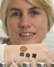
patches on her hand
Credit: Tómas Tyner
Delivering malaria vaccines via a skin patch can streamline the vaccination process, according to preclinical research published in Scientific Reports.
The patch has arrays of tiny silicon microneedles that painlessly create temporary pores in the outermost layer of the skin, permitting the vaccine to flow into the skin.
Researchers used the patch to vaccinate mice with a live adenovirus engineered to deliver a protein from the malaria parasite Plasmodium yoelii.
The team found the patch could overcome one of the main problems with this type of vaccine—namely, although it induces high levels of immunity to malaria, it can also induce a strong immune response to the adenovirus itself.
This anti-adenovirus immunity prevents its repeated use as a vaccine, as the immune system recognizes the adenovirus and prevents it from delivering the malaria protein. So another vaccine type or adenovirus strain needs to be used in the booster immunization.
“What’s exciting from this work is that administration of this vaccine with the microneedle patch did not induce this strong anti-adenovirus immunity, even though very potent immunity to the malaria antigen is generated,” said study author Anne Moore, PhD, of University College Cork in Ireland.
This suggests the patch can facilitate the repeated use of the same adenovirus vaccine, thereby potentially reducing manufacturing costs of multiple vaccines.
In their experiments with mice, Dr Moore and her colleagues demonstrated that using the microneedle patch in the primary immunization does indeed permit repeated use of the same adenovirus vaccine. And this immunization method induced potent and highly protective immune responses against malaria.
Specifically, the researchers delivered a vaccine known as HAdV5-PyMSP142 to mice. They found the patch induced equivalent or enhanced antibody responses but decreased anti-vector responses when compared to intradermal delivery of the vaccine.
The addition of a heterologous vaccine known as MVA-PyMSP142 also produced greater antibody responses in mice primed with HAdV5-PyMSP142 via the patch, compared to those vaccinated intradermally.
The researchers observed the highest protection against blood-stage malaria when mice were vaccinated first with the patch and then intradermally.
In an attempt to commercialize this research, Dr Moore is heading to Silicon Valley next week to meet with venture capitalists and technology companies.
patches on her hand
Credit: Tómas Tyner
Delivering malaria vaccines via a skin patch can streamline the vaccination process, according to preclinical research published in Scientific Reports.
The patch has arrays of tiny silicon microneedles that painlessly create temporary pores in the outermost layer of the skin, permitting the vaccine to flow into the skin.
Researchers used the patch to vaccinate mice with a live adenovirus engineered to deliver a protein from the malaria parasite Plasmodium yoelii.
The team found the patch could overcome one of the main problems with this type of vaccine—namely, although it induces high levels of immunity to malaria, it can also induce a strong immune response to the adenovirus itself.
This anti-adenovirus immunity prevents its repeated use as a vaccine, as the immune system recognizes the adenovirus and prevents it from delivering the malaria protein. So another vaccine type or adenovirus strain needs to be used in the booster immunization.
“What’s exciting from this work is that administration of this vaccine with the microneedle patch did not induce this strong anti-adenovirus immunity, even though very potent immunity to the malaria antigen is generated,” said study author Anne Moore, PhD, of University College Cork in Ireland.
This suggests the patch can facilitate the repeated use of the same adenovirus vaccine, thereby potentially reducing manufacturing costs of multiple vaccines.
In their experiments with mice, Dr Moore and her colleagues demonstrated that using the microneedle patch in the primary immunization does indeed permit repeated use of the same adenovirus vaccine. And this immunization method induced potent and highly protective immune responses against malaria.
Specifically, the researchers delivered a vaccine known as HAdV5-PyMSP142 to mice. They found the patch induced equivalent or enhanced antibody responses but decreased anti-vector responses when compared to intradermal delivery of the vaccine.
The addition of a heterologous vaccine known as MVA-PyMSP142 also produced greater antibody responses in mice primed with HAdV5-PyMSP142 via the patch, compared to those vaccinated intradermally.
The researchers observed the highest protection against blood-stage malaria when mice were vaccinated first with the patch and then intradermally.
In an attempt to commercialize this research, Dr Moore is heading to Silicon Valley next week to meet with venture capitalists and technology companies.
patches on her hand
Credit: Tómas Tyner
Delivering malaria vaccines via a skin patch can streamline the vaccination process, according to preclinical research published in Scientific Reports.
The patch has arrays of tiny silicon microneedles that painlessly create temporary pores in the outermost layer of the skin, permitting the vaccine to flow into the skin.
Researchers used the patch to vaccinate mice with a live adenovirus engineered to deliver a protein from the malaria parasite Plasmodium yoelii.
The team found the patch could overcome one of the main problems with this type of vaccine—namely, although it induces high levels of immunity to malaria, it can also induce a strong immune response to the adenovirus itself.
This anti-adenovirus immunity prevents its repeated use as a vaccine, as the immune system recognizes the adenovirus and prevents it from delivering the malaria protein. So another vaccine type or adenovirus strain needs to be used in the booster immunization.
“What’s exciting from this work is that administration of this vaccine with the microneedle patch did not induce this strong anti-adenovirus immunity, even though very potent immunity to the malaria antigen is generated,” said study author Anne Moore, PhD, of University College Cork in Ireland.
This suggests the patch can facilitate the repeated use of the same adenovirus vaccine, thereby potentially reducing manufacturing costs of multiple vaccines.
In their experiments with mice, Dr Moore and her colleagues demonstrated that using the microneedle patch in the primary immunization does indeed permit repeated use of the same adenovirus vaccine. And this immunization method induced potent and highly protective immune responses against malaria.
Specifically, the researchers delivered a vaccine known as HAdV5-PyMSP142 to mice. They found the patch induced equivalent or enhanced antibody responses but decreased anti-vector responses when compared to intradermal delivery of the vaccine.
The addition of a heterologous vaccine known as MVA-PyMSP142 also produced greater antibody responses in mice primed with HAdV5-PyMSP142 via the patch, compared to those vaccinated intradermally.
The researchers observed the highest protection against blood-stage malaria when mice were vaccinated first with the patch and then intradermally.
In an attempt to commercialize this research, Dr Moore is heading to Silicon Valley next week to meet with venture capitalists and technology companies.
Journals may fail to correct major errors in articles

Credit: CDC/James Gathany
A review of errata reports from medical publications revealed that nearly a quarter of the errors may have changed the way study data were interpreted.
And about half of the errors were not corrected in the original text, or the errata report did not specify whether a correction was made.
This suggests authors and journals must be more vigilant and consistent in identifying and reporting errors, said Paul Hauptman, MD, of Saint Louis University in Missouri.
He and his colleagues conducted this research and reported the results in The American Journal of Medicine.
The researchers reviewed 20 prominent English language journals in the fields of general medicine and cardiology. And they identified 577 error reports over an 18-month period.
More than 24% of these reports included an error the team rated as major. Major errors were associated with material changes in the interpretation of data in text, figures, or tables, or with significant alterations in the article’s conclusions.
One of the examples of a major error report in the study comes from a paper about depression. In the original article, the incidence of new depression cases was misquoted by a factor of 10. The article initially reported 15.8% of women with new depression, rather than the real figure of 1.58%, which was later corrected.
“As the volume of research publications continues to rise, the scientific community needs to examine how it manages its mistakes,” said Eric Armbrecht, PhD, of the Saint Louis University Center for Outcomes Research.
“Transparency, consistency, and clarity are essential. Our study found that these are not common among some of the top medical journals.”
In fact, the researchers found that 51% of the errors they identified were not corrected in the original text, or the errata report did not specify whether a correction was made.
The team noted that this study did not provide any definitive explanations for why such errors appear in publications, although one possibility is that most authors don’t read and edit the final version of their manuscripts prior to publication.
“It’s noteworthy that although final approval of an article may fall to the first or corresponding author, the criteria put forth by [the International Committee of Medical Journal Editors] specifies that each author must provide final approval of the version to be published,” Dr Hauptman said.
He added that, at this point, it’s not possible to measure how frequently a journal reader incorporates errata into clinical care or the extent to which patient outcomes may be affected.
Credit: CDC/James Gathany
A review of errata reports from medical publications revealed that nearly a quarter of the errors may have changed the way study data were interpreted.
And about half of the errors were not corrected in the original text, or the errata report did not specify whether a correction was made.
This suggests authors and journals must be more vigilant and consistent in identifying and reporting errors, said Paul Hauptman, MD, of Saint Louis University in Missouri.
He and his colleagues conducted this research and reported the results in The American Journal of Medicine.
The researchers reviewed 20 prominent English language journals in the fields of general medicine and cardiology. And they identified 577 error reports over an 18-month period.
More than 24% of these reports included an error the team rated as major. Major errors were associated with material changes in the interpretation of data in text, figures, or tables, or with significant alterations in the article’s conclusions.
One of the examples of a major error report in the study comes from a paper about depression. In the original article, the incidence of new depression cases was misquoted by a factor of 10. The article initially reported 15.8% of women with new depression, rather than the real figure of 1.58%, which was later corrected.
“As the volume of research publications continues to rise, the scientific community needs to examine how it manages its mistakes,” said Eric Armbrecht, PhD, of the Saint Louis University Center for Outcomes Research.
“Transparency, consistency, and clarity are essential. Our study found that these are not common among some of the top medical journals.”
In fact, the researchers found that 51% of the errors they identified were not corrected in the original text, or the errata report did not specify whether a correction was made.
The team noted that this study did not provide any definitive explanations for why such errors appear in publications, although one possibility is that most authors don’t read and edit the final version of their manuscripts prior to publication.
“It’s noteworthy that although final approval of an article may fall to the first or corresponding author, the criteria put forth by [the International Committee of Medical Journal Editors] specifies that each author must provide final approval of the version to be published,” Dr Hauptman said.
He added that, at this point, it’s not possible to measure how frequently a journal reader incorporates errata into clinical care or the extent to which patient outcomes may be affected.
Credit: CDC/James Gathany
A review of errata reports from medical publications revealed that nearly a quarter of the errors may have changed the way study data were interpreted.
And about half of the errors were not corrected in the original text, or the errata report did not specify whether a correction was made.
This suggests authors and journals must be more vigilant and consistent in identifying and reporting errors, said Paul Hauptman, MD, of Saint Louis University in Missouri.
He and his colleagues conducted this research and reported the results in The American Journal of Medicine.
The researchers reviewed 20 prominent English language journals in the fields of general medicine and cardiology. And they identified 577 error reports over an 18-month period.
More than 24% of these reports included an error the team rated as major. Major errors were associated with material changes in the interpretation of data in text, figures, or tables, or with significant alterations in the article’s conclusions.
One of the examples of a major error report in the study comes from a paper about depression. In the original article, the incidence of new depression cases was misquoted by a factor of 10. The article initially reported 15.8% of women with new depression, rather than the real figure of 1.58%, which was later corrected.
“As the volume of research publications continues to rise, the scientific community needs to examine how it manages its mistakes,” said Eric Armbrecht, PhD, of the Saint Louis University Center for Outcomes Research.
“Transparency, consistency, and clarity are essential. Our study found that these are not common among some of the top medical journals.”
In fact, the researchers found that 51% of the errors they identified were not corrected in the original text, or the errata report did not specify whether a correction was made.
The team noted that this study did not provide any definitive explanations for why such errors appear in publications, although one possibility is that most authors don’t read and edit the final version of their manuscripts prior to publication.
“It’s noteworthy that although final approval of an article may fall to the first or corresponding author, the criteria put forth by [the International Committee of Medical Journal Editors] specifies that each author must provide final approval of the version to be published,” Dr Hauptman said.
He added that, at this point, it’s not possible to measure how frequently a journal reader incorporates errata into clinical care or the extent to which patient outcomes may be affected.
Cellular RNA can template DNA repair in yeast
DNA has been repaired by
transcript RNA within the cells
Credit: Georgia Tech/Rob Felt
Scientists have shown that RNA produced within yeast cells can serve as a template for repairing the most devastating DNA damage—a break in both strands of a DNA helix.
The group believes their study is the first to show that a cell’s own RNA can be used for DNA recombination and repair.
If the phenomenon extends to human cells, it could potentially lead to new therapeutic or preventative strategies for genetic diseases.
The scientists described the phenomenon in Nature.
“We have found that genetic information can flow from RNA to DNA in a homology-driven manner, from cellular RNA to a homologous DNA sequence,” said study author Francesca Storici, PhD, of the Georgia Institute of Technology in Atlanta.
“This process is moving the genetic information in the opposite direction from which it normally flows. We have shown that when an endogenous RNA molecule can anneal to broken homologous DNA without being removed, the RNA can repair the damaged DNA. This finding reveals the existence of a novel mechanism of genetic recombination.”
Dr Storici’s team previously showed that synthetic RNA introduced into cells—including human cells—could repair DNA damage. But the process was inefficient, and there were questions about whether it could occur naturally.
To find out whether cells could use endogenous RNA transcripts to repair DNA damage, she and her colleagues devised experiments using the yeast Saccharomyces cerevisiae.
The team developed a strategy for distinguishing repair by endogenous RNA from repair by the normal DNA-based mechanisms in the budding yeast cells, including using mutants that lacked the ability to convert the RNA into a DNA copy.
They then induced a DNA double-strand break in the yeast genome and observed whether the organism could survive and grow by repairing the damage using only transcript RNA within the cells.
The DNA region that generates the transcript was constructed to contain a marker gene interrupted by an intron. Following intron removal during transcription, the transcript RNA sequence has no intron, while the DNA region that generates the transcript retains the intron.
Only the repair templated by the transcript devoid of the intron can restore the function of a homologous marker gene in which the DNA double-strand break is induced.
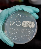
Dr Storici and her colleagues measured success by counting the number of yeast colonies growing on a Petri dish, indicating that the repair had been made by endogenous RNA.
They conducted testing on two types of breaks, one in the DNA from which the RNA transcript had been made, and the other in a homologous sequence from a different location in the DNA.
The team found that proximity of the RNA to the broken DNA increased the efficiency of the repair, and the repair occurred via a homologous recombination process. Dr Storici believes the repair mechanism may operate in cells beyond yeast, and many types of RNA can be used.
“We are showing that the flow of genetic information from RNA to DNA is not restricted to retro-elements and telomeres but occurs with a generic cellular transcript, making it more of a general phenomenon than had been anticipated,” she explained. “Potentially, any RNA in the cell could have this function.”
For the future, Dr Storici hopes to learn more about the mechanism, including what regulates it. She also wants to determine whether it takes place in human cells. If so, that could have implications for treating or preventing diseases caused by genetic damage.
“Cells synthesize lots of RNA transcripts during their life spans,” Dr Storici said. “Therefore, RNA may have an unanticipated impact on genomic stability and plasticity. We need to understand in which situations cells would activate RNA-DNA recombination. Better understanding this molecular process could also help us manipulate mechanisms for therapy, allowing us to treat a disease or prevent it altogether.”
DNA has been repaired by
transcript RNA within the cells
Credit: Georgia Tech/Rob Felt
Scientists have shown that RNA produced within yeast cells can serve as a template for repairing the most devastating DNA damage—a break in both strands of a DNA helix.
The group believes their study is the first to show that a cell’s own RNA can be used for DNA recombination and repair.
If the phenomenon extends to human cells, it could potentially lead to new therapeutic or preventative strategies for genetic diseases.
The scientists described the phenomenon in Nature.
“We have found that genetic information can flow from RNA to DNA in a homology-driven manner, from cellular RNA to a homologous DNA sequence,” said study author Francesca Storici, PhD, of the Georgia Institute of Technology in Atlanta.
“This process is moving the genetic information in the opposite direction from which it normally flows. We have shown that when an endogenous RNA molecule can anneal to broken homologous DNA without being removed, the RNA can repair the damaged DNA. This finding reveals the existence of a novel mechanism of genetic recombination.”
Dr Storici’s team previously showed that synthetic RNA introduced into cells—including human cells—could repair DNA damage. But the process was inefficient, and there were questions about whether it could occur naturally.
To find out whether cells could use endogenous RNA transcripts to repair DNA damage, she and her colleagues devised experiments using the yeast Saccharomyces cerevisiae.
The team developed a strategy for distinguishing repair by endogenous RNA from repair by the normal DNA-based mechanisms in the budding yeast cells, including using mutants that lacked the ability to convert the RNA into a DNA copy.
They then induced a DNA double-strand break in the yeast genome and observed whether the organism could survive and grow by repairing the damage using only transcript RNA within the cells.
The DNA region that generates the transcript was constructed to contain a marker gene interrupted by an intron. Following intron removal during transcription, the transcript RNA sequence has no intron, while the DNA region that generates the transcript retains the intron.
Only the repair templated by the transcript devoid of the intron can restore the function of a homologous marker gene in which the DNA double-strand break is induced.
Dr Storici and her colleagues measured success by counting the number of yeast colonies growing on a Petri dish, indicating that the repair had been made by endogenous RNA.
They conducted testing on two types of breaks, one in the DNA from which the RNA transcript had been made, and the other in a homologous sequence from a different location in the DNA.
The team found that proximity of the RNA to the broken DNA increased the efficiency of the repair, and the repair occurred via a homologous recombination process. Dr Storici believes the repair mechanism may operate in cells beyond yeast, and many types of RNA can be used.
“We are showing that the flow of genetic information from RNA to DNA is not restricted to retro-elements and telomeres but occurs with a generic cellular transcript, making it more of a general phenomenon than had been anticipated,” she explained. “Potentially, any RNA in the cell could have this function.”
For the future, Dr Storici hopes to learn more about the mechanism, including what regulates it. She also wants to determine whether it takes place in human cells. If so, that could have implications for treating or preventing diseases caused by genetic damage.
“Cells synthesize lots of RNA transcripts during their life spans,” Dr Storici said. “Therefore, RNA may have an unanticipated impact on genomic stability and plasticity. We need to understand in which situations cells would activate RNA-DNA recombination. Better understanding this molecular process could also help us manipulate mechanisms for therapy, allowing us to treat a disease or prevent it altogether.”
DNA has been repaired by
transcript RNA within the cells
Credit: Georgia Tech/Rob Felt
Scientists have shown that RNA produced within yeast cells can serve as a template for repairing the most devastating DNA damage—a break in both strands of a DNA helix.
The group believes their study is the first to show that a cell’s own RNA can be used for DNA recombination and repair.
If the phenomenon extends to human cells, it could potentially lead to new therapeutic or preventative strategies for genetic diseases.
The scientists described the phenomenon in Nature.
“We have found that genetic information can flow from RNA to DNA in a homology-driven manner, from cellular RNA to a homologous DNA sequence,” said study author Francesca Storici, PhD, of the Georgia Institute of Technology in Atlanta.
“This process is moving the genetic information in the opposite direction from which it normally flows. We have shown that when an endogenous RNA molecule can anneal to broken homologous DNA without being removed, the RNA can repair the damaged DNA. This finding reveals the existence of a novel mechanism of genetic recombination.”
Dr Storici’s team previously showed that synthetic RNA introduced into cells—including human cells—could repair DNA damage. But the process was inefficient, and there were questions about whether it could occur naturally.
To find out whether cells could use endogenous RNA transcripts to repair DNA damage, she and her colleagues devised experiments using the yeast Saccharomyces cerevisiae.
The team developed a strategy for distinguishing repair by endogenous RNA from repair by the normal DNA-based mechanisms in the budding yeast cells, including using mutants that lacked the ability to convert the RNA into a DNA copy.
They then induced a DNA double-strand break in the yeast genome and observed whether the organism could survive and grow by repairing the damage using only transcript RNA within the cells.
The DNA region that generates the transcript was constructed to contain a marker gene interrupted by an intron. Following intron removal during transcription, the transcript RNA sequence has no intron, while the DNA region that generates the transcript retains the intron.
Only the repair templated by the transcript devoid of the intron can restore the function of a homologous marker gene in which the DNA double-strand break is induced.
Dr Storici and her colleagues measured success by counting the number of yeast colonies growing on a Petri dish, indicating that the repair had been made by endogenous RNA.
They conducted testing on two types of breaks, one in the DNA from which the RNA transcript had been made, and the other in a homologous sequence from a different location in the DNA.
The team found that proximity of the RNA to the broken DNA increased the efficiency of the repair, and the repair occurred via a homologous recombination process. Dr Storici believes the repair mechanism may operate in cells beyond yeast, and many types of RNA can be used.
“We are showing that the flow of genetic information from RNA to DNA is not restricted to retro-elements and telomeres but occurs with a generic cellular transcript, making it more of a general phenomenon than had been anticipated,” she explained. “Potentially, any RNA in the cell could have this function.”
For the future, Dr Storici hopes to learn more about the mechanism, including what regulates it. She also wants to determine whether it takes place in human cells. If so, that could have implications for treating or preventing diseases caused by genetic damage.
“Cells synthesize lots of RNA transcripts during their life spans,” Dr Storici said. “Therefore, RNA may have an unanticipated impact on genomic stability and plasticity. We need to understand in which situations cells would activate RNA-DNA recombination. Better understanding this molecular process could also help us manipulate mechanisms for therapy, allowing us to treat a disease or prevent it altogether.”
Preclinical results support blood transfusions containing fibronectin

Credit: UAB Hospital
The protein fibronectin is instrumental in stopping bleeding and preventing life-threatening blood clots, according to preclinical research published in The Journal of Clinical Investigation.
Experiments in mice revealed that plasma fibronectin (pFn) can switch from supporting hemostasis to inhibiting thrombosis.
The researchers said these findings suggest transfusions containing pFn may control bleeding in humans, particularly in association with anticoagulant therapy.
“Most treatments that help the body stop bleeding can actually cause blood clots, and many treatments to prevent excessive blood clots increase [the] risk of bleeding out,” said study author Heyu Ni, MD, of St Michael’s Hospital in Toronto, Ontario, Canada.
“But when given to mice after an injury or to mice treated with blood thinners—which frequently lead to bleeding complications—fibronectin seems to offer a win-win solution.”
Dr Ni and his colleagues began this research by generating mice deficient in both fibrinogen (Fg) and von Willebrand factor (VWF), as well as mice deficient in both VWF and pFn. They then compared these mice to untreated wild-type mice and anticoagulant-treated wild-type mice.
The team discovered that pFn is vital for controlling bleeding in the context of Fg deficiency and supports hemostasis in the context of VWF deficiency. pFn deposition occurred before platelet accumulation (the first wave of hemostasis) in both wild-type and deficient mice.
However, pFn switched its function with regard to platelet aggregation based on the presence or absence of fibrin. pFn inhibited platelet aggregation when fibrin was absent and promoted aggregation when linked with fibrin.
The researchers found that pFn can control the diameter of fibrin fibers, increase the mechanical strength of a blood clot, and be actively involved in thrombosis.
The team said these results provide a solution for the controversy surrounding pFn’s function in hemostasis and establish pFn as a unique regulatory factor in thrombosis. The findings also suggest transfusions containing pFn may prove particularly effective in controlling bleeding.
“Fibrinogen has been shown to help the body stop bleeding, but our research indicates that less-refined blood products that include fibronectin and fibrinogen may help stop bleeding even more effectively,” Dr Ni said. “And, as an added bonus, fibronectin likely also reduces the risk of life-threatening blood clots from forming.”
“There is a lot of work to be done, but we might find that the less expensive and less processed form of donor blood may be more effective for transfusions. We’ve shown that fibronectin might play a role in improving results from transfusions and should not be discarded during blood product processing. It may be also an important protein in transfusions for stopping bleeding, particularly for patients who receive blood thinners during surgeries.”
Credit: UAB Hospital
The protein fibronectin is instrumental in stopping bleeding and preventing life-threatening blood clots, according to preclinical research published in The Journal of Clinical Investigation.
Experiments in mice revealed that plasma fibronectin (pFn) can switch from supporting hemostasis to inhibiting thrombosis.
The researchers said these findings suggest transfusions containing pFn may control bleeding in humans, particularly in association with anticoagulant therapy.
“Most treatments that help the body stop bleeding can actually cause blood clots, and many treatments to prevent excessive blood clots increase [the] risk of bleeding out,” said study author Heyu Ni, MD, of St Michael’s Hospital in Toronto, Ontario, Canada.
“But when given to mice after an injury or to mice treated with blood thinners—which frequently lead to bleeding complications—fibronectin seems to offer a win-win solution.”
Dr Ni and his colleagues began this research by generating mice deficient in both fibrinogen (Fg) and von Willebrand factor (VWF), as well as mice deficient in both VWF and pFn. They then compared these mice to untreated wild-type mice and anticoagulant-treated wild-type mice.
The team discovered that pFn is vital for controlling bleeding in the context of Fg deficiency and supports hemostasis in the context of VWF deficiency. pFn deposition occurred before platelet accumulation (the first wave of hemostasis) in both wild-type and deficient mice.
However, pFn switched its function with regard to platelet aggregation based on the presence or absence of fibrin. pFn inhibited platelet aggregation when fibrin was absent and promoted aggregation when linked with fibrin.
The researchers found that pFn can control the diameter of fibrin fibers, increase the mechanical strength of a blood clot, and be actively involved in thrombosis.
The team said these results provide a solution for the controversy surrounding pFn’s function in hemostasis and establish pFn as a unique regulatory factor in thrombosis. The findings also suggest transfusions containing pFn may prove particularly effective in controlling bleeding.
“Fibrinogen has been shown to help the body stop bleeding, but our research indicates that less-refined blood products that include fibronectin and fibrinogen may help stop bleeding even more effectively,” Dr Ni said. “And, as an added bonus, fibronectin likely also reduces the risk of life-threatening blood clots from forming.”
“There is a lot of work to be done, but we might find that the less expensive and less processed form of donor blood may be more effective for transfusions. We’ve shown that fibronectin might play a role in improving results from transfusions and should not be discarded during blood product processing. It may be also an important protein in transfusions for stopping bleeding, particularly for patients who receive blood thinners during surgeries.”
Credit: UAB Hospital
The protein fibronectin is instrumental in stopping bleeding and preventing life-threatening blood clots, according to preclinical research published in The Journal of Clinical Investigation.
Experiments in mice revealed that plasma fibronectin (pFn) can switch from supporting hemostasis to inhibiting thrombosis.
The researchers said these findings suggest transfusions containing pFn may control bleeding in humans, particularly in association with anticoagulant therapy.
“Most treatments that help the body stop bleeding can actually cause blood clots, and many treatments to prevent excessive blood clots increase [the] risk of bleeding out,” said study author Heyu Ni, MD, of St Michael’s Hospital in Toronto, Ontario, Canada.
“But when given to mice after an injury or to mice treated with blood thinners—which frequently lead to bleeding complications—fibronectin seems to offer a win-win solution.”
Dr Ni and his colleagues began this research by generating mice deficient in both fibrinogen (Fg) and von Willebrand factor (VWF), as well as mice deficient in both VWF and pFn. They then compared these mice to untreated wild-type mice and anticoagulant-treated wild-type mice.
The team discovered that pFn is vital for controlling bleeding in the context of Fg deficiency and supports hemostasis in the context of VWF deficiency. pFn deposition occurred before platelet accumulation (the first wave of hemostasis) in both wild-type and deficient mice.
However, pFn switched its function with regard to platelet aggregation based on the presence or absence of fibrin. pFn inhibited platelet aggregation when fibrin was absent and promoted aggregation when linked with fibrin.
The researchers found that pFn can control the diameter of fibrin fibers, increase the mechanical strength of a blood clot, and be actively involved in thrombosis.
The team said these results provide a solution for the controversy surrounding pFn’s function in hemostasis and establish pFn as a unique regulatory factor in thrombosis. The findings also suggest transfusions containing pFn may prove particularly effective in controlling bleeding.
“Fibrinogen has been shown to help the body stop bleeding, but our research indicates that less-refined blood products that include fibronectin and fibrinogen may help stop bleeding even more effectively,” Dr Ni said. “And, as an added bonus, fibronectin likely also reduces the risk of life-threatening blood clots from forming.”
“There is a lot of work to be done, but we might find that the less expensive and less processed form of donor blood may be more effective for transfusions. We’ve shown that fibronectin might play a role in improving results from transfusions and should not be discarded during blood product processing. It may be also an important protein in transfusions for stopping bleeding, particularly for patients who receive blood thinners during surgeries.”
Many new additions in ESC’s latest guidelines for PE
The European Society of Cardiology (ESC) has published new guidelines for managing patients with pulmonary embolism (PE).
The guidelines now include recommendations for all new oral anticoagulants, a risk-stratification algorithm, new advice on managing PE in specific patient populations, and age-specific D-dimer cutoffs, among other additions.
The guidelines were presented at the ESC Congress 2014 and published in the European Heart Journal and on the ESC website.
Previous ESC Guidelines on acute PE were published in 2000 and 2008.
“These are the first major international guidelines with a complete set of recommendations on the use of new oral anticoagulants in VTE [venous thromboembolism],” said guidelines author Stavros Konstantinides, MD, PhD, of the Johannes Gutenberg University Mainz in Germany.
“For each drug, we provide detailed recommendations on how and when to use it, and whether it should be first-line treatment or an alternative to standard treatment.”
Managing specific patients
For the first time, the guidelines include formal recommendations for managing PE in pregnancy and in cancer patients.
The guidelines also highlight recently identified risk factors for VTE, including in vitro fertilization, which increases the risk of VTE in early pregnancy.
And the guidelines include a new chapter on the diagnosis and treatment of chronic thromboembolic pulmonary hypertension.
Risk stratification
Another first with the new guidelines is the inclusion of an algorithm for risk stratification. It incorporates all available tools and provides recommendations for managing patients according to risk.
“Patients with PE or suspected PE who are in shock are at high risk, but at least 95% of patients are at intermediate or low risk, and defining how to manage them has not been clear,” Dr Konstantinides said.
“Previously, we used echocardiography and/or a CT scan to evaluate the right ventricle but did not combine this information with clinical data. These topics have advanced in the past 6 years, and now we can integrate clinical scores of severity, imaging with echo and CT, and biomarkers to define levels of risk.”
“And, more importantly, we now have solid evidence to give recommendations on rescue rather than primary thrombolysis in patients at intermediate risk of early adverse outcome. We are also now able to recommend how to identify low-risk patients [who] may be considered for early discharge despite a confirmed PE episode.”
D-dimer cutoffs
Another new addition to the guidelines is age-adjusted D-dimer cutoffs, which have been introduced to identify patients of all ages who do not require anticoagulation.
Until now, anticoagulation could be withheld in patients with D-dimer levels less than 500 µg/L, but D-dimer rises naturally with age.
The guidelines reference evidence suggesting that, for patients older than 50, the cutoff may now be their age times 10. For example, in a 65-year-old, the cutoff would be 650 µg/L.
“These guidelines provide the most comprehensive recommendations ever for the diagnosis and treatment of PE,” Dr Konstantinides concluded. “Clinicians can confidently risk-stratify their patients with suspected PE and provide appropriate treatment including the new oral anticoagulants.”
The European Society of Cardiology (ESC) has published new guidelines for managing patients with pulmonary embolism (PE).
The guidelines now include recommendations for all new oral anticoagulants, a risk-stratification algorithm, new advice on managing PE in specific patient populations, and age-specific D-dimer cutoffs, among other additions.
The guidelines were presented at the ESC Congress 2014 and published in the European Heart Journal and on the ESC website.
Previous ESC Guidelines on acute PE were published in 2000 and 2008.
“These are the first major international guidelines with a complete set of recommendations on the use of new oral anticoagulants in VTE [venous thromboembolism],” said guidelines author Stavros Konstantinides, MD, PhD, of the Johannes Gutenberg University Mainz in Germany.
“For each drug, we provide detailed recommendations on how and when to use it, and whether it should be first-line treatment or an alternative to standard treatment.”
Managing specific patients
For the first time, the guidelines include formal recommendations for managing PE in pregnancy and in cancer patients.
The guidelines also highlight recently identified risk factors for VTE, including in vitro fertilization, which increases the risk of VTE in early pregnancy.
And the guidelines include a new chapter on the diagnosis and treatment of chronic thromboembolic pulmonary hypertension.
Risk stratification
Another first with the new guidelines is the inclusion of an algorithm for risk stratification. It incorporates all available tools and provides recommendations for managing patients according to risk.
“Patients with PE or suspected PE who are in shock are at high risk, but at least 95% of patients are at intermediate or low risk, and defining how to manage them has not been clear,” Dr Konstantinides said.
“Previously, we used echocardiography and/or a CT scan to evaluate the right ventricle but did not combine this information with clinical data. These topics have advanced in the past 6 years, and now we can integrate clinical scores of severity, imaging with echo and CT, and biomarkers to define levels of risk.”
“And, more importantly, we now have solid evidence to give recommendations on rescue rather than primary thrombolysis in patients at intermediate risk of early adverse outcome. We are also now able to recommend how to identify low-risk patients [who] may be considered for early discharge despite a confirmed PE episode.”
D-dimer cutoffs
Another new addition to the guidelines is age-adjusted D-dimer cutoffs, which have been introduced to identify patients of all ages who do not require anticoagulation.
Until now, anticoagulation could be withheld in patients with D-dimer levels less than 500 µg/L, but D-dimer rises naturally with age.
The guidelines reference evidence suggesting that, for patients older than 50, the cutoff may now be their age times 10. For example, in a 65-year-old, the cutoff would be 650 µg/L.
“These guidelines provide the most comprehensive recommendations ever for the diagnosis and treatment of PE,” Dr Konstantinides concluded. “Clinicians can confidently risk-stratify their patients with suspected PE and provide appropriate treatment including the new oral anticoagulants.”
The European Society of Cardiology (ESC) has published new guidelines for managing patients with pulmonary embolism (PE).
The guidelines now include recommendations for all new oral anticoagulants, a risk-stratification algorithm, new advice on managing PE in specific patient populations, and age-specific D-dimer cutoffs, among other additions.
The guidelines were presented at the ESC Congress 2014 and published in the European Heart Journal and on the ESC website.
Previous ESC Guidelines on acute PE were published in 2000 and 2008.
“These are the first major international guidelines with a complete set of recommendations on the use of new oral anticoagulants in VTE [venous thromboembolism],” said guidelines author Stavros Konstantinides, MD, PhD, of the Johannes Gutenberg University Mainz in Germany.
“For each drug, we provide detailed recommendations on how and when to use it, and whether it should be first-line treatment or an alternative to standard treatment.”
Managing specific patients
For the first time, the guidelines include formal recommendations for managing PE in pregnancy and in cancer patients.
The guidelines also highlight recently identified risk factors for VTE, including in vitro fertilization, which increases the risk of VTE in early pregnancy.
And the guidelines include a new chapter on the diagnosis and treatment of chronic thromboembolic pulmonary hypertension.
Risk stratification
Another first with the new guidelines is the inclusion of an algorithm for risk stratification. It incorporates all available tools and provides recommendations for managing patients according to risk.
“Patients with PE or suspected PE who are in shock are at high risk, but at least 95% of patients are at intermediate or low risk, and defining how to manage them has not been clear,” Dr Konstantinides said.
“Previously, we used echocardiography and/or a CT scan to evaluate the right ventricle but did not combine this information with clinical data. These topics have advanced in the past 6 years, and now we can integrate clinical scores of severity, imaging with echo and CT, and biomarkers to define levels of risk.”
“And, more importantly, we now have solid evidence to give recommendations on rescue rather than primary thrombolysis in patients at intermediate risk of early adverse outcome. We are also now able to recommend how to identify low-risk patients [who] may be considered for early discharge despite a confirmed PE episode.”
D-dimer cutoffs
Another new addition to the guidelines is age-adjusted D-dimer cutoffs, which have been introduced to identify patients of all ages who do not require anticoagulation.
Until now, anticoagulation could be withheld in patients with D-dimer levels less than 500 µg/L, but D-dimer rises naturally with age.
The guidelines reference evidence suggesting that, for patients older than 50, the cutoff may now be their age times 10. For example, in a 65-year-old, the cutoff would be 650 µg/L.
“These guidelines provide the most comprehensive recommendations ever for the diagnosis and treatment of PE,” Dr Konstantinides concluded. “Clinicians can confidently risk-stratify their patients with suspected PE and provide appropriate treatment including the new oral anticoagulants.”