User login
Inhibitor could transform AML therapy, speaker says

Credit: University of Colorado
BARCELONA—An agent that inhibits isocitrate dehydrogenase (IDH) 1 shows the potential to transform therapy for certain patients with acute myeloid leukemia (AML), according to a speaker at the 26th EORTC-NCI-AACR Symposium on Molecular Targets and Cancer Therapeutics.
The drug, known as AG-120, demonstrated clinical activity and was considered to be well-tolerated in a phase 1 study of patients with advanced, IDH1 mutant-positive AML.
Daniel Pollyea, MD, of the University of Colorado School of Medicine in Aurora, presented data on AG-120 at the symposium as abstract LBA1. The research was sponsored by Agios Pharmaceuticals, makers of AG-120.
“This is the first study in humans of an inhibitor of mutant IDH1 and the first demonstration of clinical activity of AG-120 in AML patients whose cancers have the IDH1 mutation,” Dr Pollyea said. “Although the data are early, we are encouraged to see evidence of clinical activity, as the primary objectives of phase 1 studies are to determine safety and tolerability.”
Dr Pollyea noted that mutations in IDH1 lead to a cascade of metabolic events that contribute to malignancy. Mutant IDH1 produces an excess amount of 2-hydroxyglutarate (2-HG), which prevents cells from maturing into normal, functioning cells, and this leads to malignancy.
In this study, the researchers found that AG-120 reduced 2-HG levels in diseased cells to normal levels, allowing them to mature into normal cells.
The trial included 17 patients with relapsed and/or refractory AML, who had received a median of 2 prior treatments. Patients were scheduled to
receive AG-120 in 1 of 4 dose groups: 100 mg twice a day, 300 mg once a day, 500 mg once a day, and 800 mg once a day over continuous 28-day cycles.
Fourteen patients were evaluable for response, and 7 responded. Four patients achieved a complete response, 2 had a complete response in the marrow, and 1 had a partial response.
Responses occurred at all the dose levels tested. In the 4 patients who achieved a complete response, there was early evidence of durability, ranging from 15 days to 5 months. All responding patients remain on AG-120, and 1 patient with stable disease remains on the drug.
“AML is a devastating disease that has historically been very difficult to treat, and these findings suggest that AG-120 has the potential to transform therapy for patients with IDH1-mutant positive AML,” Dr Pollyea said.
He and his colleagues also found that AG-120 was generally well-tolerated. The majority of adverse events were grade 1 and 2. The most common of these were nausea, fatigue, and dyspnea.
Eight patients experienced serious adverse events, but these were primarily related to disease progression.
One patient experienced a dose-limiting toxicity of asymptomatic grade 3 QT prolongation at the highest dose tested to date, which improved to grade 1 with dose reduction. This patient is in complete remission and remains on AG-120.
The maximum-tolerated dose of AG-120 has not been reached.
There were 6 patient deaths, all unrelated to AG-120. Five deaths occurred after patients discontinued treatment due to progressive disease, and 1 patient died due to disease-related intracranial hemorrhage while on treatment.
Dr Pollyea and his colleagues are continuing this study with the aim of fully understanding the safety of the drug, determining the maximum-tolerated dose, and assessing its efficacy in treating AML and myelodysplastic syndromes. 
Credit: University of Colorado
BARCELONA—An agent that inhibits isocitrate dehydrogenase (IDH) 1 shows the potential to transform therapy for certain patients with acute myeloid leukemia (AML), according to a speaker at the 26th EORTC-NCI-AACR Symposium on Molecular Targets and Cancer Therapeutics.
The drug, known as AG-120, demonstrated clinical activity and was considered to be well-tolerated in a phase 1 study of patients with advanced, IDH1 mutant-positive AML.
Daniel Pollyea, MD, of the University of Colorado School of Medicine in Aurora, presented data on AG-120 at the symposium as abstract LBA1. The research was sponsored by Agios Pharmaceuticals, makers of AG-120.
“This is the first study in humans of an inhibitor of mutant IDH1 and the first demonstration of clinical activity of AG-120 in AML patients whose cancers have the IDH1 mutation,” Dr Pollyea said. “Although the data are early, we are encouraged to see evidence of clinical activity, as the primary objectives of phase 1 studies are to determine safety and tolerability.”
Dr Pollyea noted that mutations in IDH1 lead to a cascade of metabolic events that contribute to malignancy. Mutant IDH1 produces an excess amount of 2-hydroxyglutarate (2-HG), which prevents cells from maturing into normal, functioning cells, and this leads to malignancy.
In this study, the researchers found that AG-120 reduced 2-HG levels in diseased cells to normal levels, allowing them to mature into normal cells.
The trial included 17 patients with relapsed and/or refractory AML, who had received a median of 2 prior treatments. Patients were scheduled to
receive AG-120 in 1 of 4 dose groups: 100 mg twice a day, 300 mg once a day, 500 mg once a day, and 800 mg once a day over continuous 28-day cycles.
Fourteen patients were evaluable for response, and 7 responded. Four patients achieved a complete response, 2 had a complete response in the marrow, and 1 had a partial response.
Responses occurred at all the dose levels tested. In the 4 patients who achieved a complete response, there was early evidence of durability, ranging from 15 days to 5 months. All responding patients remain on AG-120, and 1 patient with stable disease remains on the drug.
“AML is a devastating disease that has historically been very difficult to treat, and these findings suggest that AG-120 has the potential to transform therapy for patients with IDH1-mutant positive AML,” Dr Pollyea said.
He and his colleagues also found that AG-120 was generally well-tolerated. The majority of adverse events were grade 1 and 2. The most common of these were nausea, fatigue, and dyspnea.
Eight patients experienced serious adverse events, but these were primarily related to disease progression.
One patient experienced a dose-limiting toxicity of asymptomatic grade 3 QT prolongation at the highest dose tested to date, which improved to grade 1 with dose reduction. This patient is in complete remission and remains on AG-120.
The maximum-tolerated dose of AG-120 has not been reached.
There were 6 patient deaths, all unrelated to AG-120. Five deaths occurred after patients discontinued treatment due to progressive disease, and 1 patient died due to disease-related intracranial hemorrhage while on treatment.
Dr Pollyea and his colleagues are continuing this study with the aim of fully understanding the safety of the drug, determining the maximum-tolerated dose, and assessing its efficacy in treating AML and myelodysplastic syndromes.
Credit: University of Colorado
BARCELONA—An agent that inhibits isocitrate dehydrogenase (IDH) 1 shows the potential to transform therapy for certain patients with acute myeloid leukemia (AML), according to a speaker at the 26th EORTC-NCI-AACR Symposium on Molecular Targets and Cancer Therapeutics.
The drug, known as AG-120, demonstrated clinical activity and was considered to be well-tolerated in a phase 1 study of patients with advanced, IDH1 mutant-positive AML.
Daniel Pollyea, MD, of the University of Colorado School of Medicine in Aurora, presented data on AG-120 at the symposium as abstract LBA1. The research was sponsored by Agios Pharmaceuticals, makers of AG-120.
“This is the first study in humans of an inhibitor of mutant IDH1 and the first demonstration of clinical activity of AG-120 in AML patients whose cancers have the IDH1 mutation,” Dr Pollyea said. “Although the data are early, we are encouraged to see evidence of clinical activity, as the primary objectives of phase 1 studies are to determine safety and tolerability.”
Dr Pollyea noted that mutations in IDH1 lead to a cascade of metabolic events that contribute to malignancy. Mutant IDH1 produces an excess amount of 2-hydroxyglutarate (2-HG), which prevents cells from maturing into normal, functioning cells, and this leads to malignancy.
In this study, the researchers found that AG-120 reduced 2-HG levels in diseased cells to normal levels, allowing them to mature into normal cells.
The trial included 17 patients with relapsed and/or refractory AML, who had received a median of 2 prior treatments. Patients were scheduled to
receive AG-120 in 1 of 4 dose groups: 100 mg twice a day, 300 mg once a day, 500 mg once a day, and 800 mg once a day over continuous 28-day cycles.
Fourteen patients were evaluable for response, and 7 responded. Four patients achieved a complete response, 2 had a complete response in the marrow, and 1 had a partial response.
Responses occurred at all the dose levels tested. In the 4 patients who achieved a complete response, there was early evidence of durability, ranging from 15 days to 5 months. All responding patients remain on AG-120, and 1 patient with stable disease remains on the drug.
“AML is a devastating disease that has historically been very difficult to treat, and these findings suggest that AG-120 has the potential to transform therapy for patients with IDH1-mutant positive AML,” Dr Pollyea said.
He and his colleagues also found that AG-120 was generally well-tolerated. The majority of adverse events were grade 1 and 2. The most common of these were nausea, fatigue, and dyspnea.
Eight patients experienced serious adverse events, but these were primarily related to disease progression.
One patient experienced a dose-limiting toxicity of asymptomatic grade 3 QT prolongation at the highest dose tested to date, which improved to grade 1 with dose reduction. This patient is in complete remission and remains on AG-120.
The maximum-tolerated dose of AG-120 has not been reached.
There were 6 patient deaths, all unrelated to AG-120. Five deaths occurred after patients discontinued treatment due to progressive disease, and 1 patient died due to disease-related intracranial hemorrhage while on treatment.
Dr Pollyea and his colleagues are continuing this study with the aim of fully understanding the safety of the drug, determining the maximum-tolerated dose, and assessing its efficacy in treating AML and myelodysplastic syndromes.
Comparison of mice and men may lead to better use of mouse models

Credit: Aaron Logan
Scientists have long known that mice are not perfect models for studying conditions in humans, but new research reveals why certain processes and systems in
mice are so different from those in humans.
As part of the mouse ENCODE project, researchers discovered that a significant number of mouse genes do not behave like their human counterparts.
This suggests scientists will need to rethink at least some roles of the lab mouse as a model organism.
“There are a substantial number of mouse genes that are regulated in ways different from similar genes in humans,” said Bing Ren, PhD, of the University of California, San Diego.
“The differences are not random. They are clustered along certain pathways, such as in genes regulating the immune system.”
The findings, part of a series of related papers being published in Nature, Science, and other journals (see below), are derived from the ongoing mouse ENCODE (Encyclopedia of DNA Elements) project.
This multi-institution effort was launched in 2007 to build a comprehensive list of functional elements of the mouse genome. It complements the earlier human ENCODE project, which published its functional catalogue in 2012.
“Both the original human and mouse genome projects gave us the sequence of genetic letters that comprise each organism but no idea how they worked or worked together to create and sustain life,” Dr Ren said.
“The human ENCODE project was designed to answer some of those questions. The mouse ENCODE project is its complement. It’s intended to provide scientists with comprehensive annotation of what mouse genes do, information that may ultimately be used for human therapeutic purposes.”
Only half of human genomic DNA aligns to mouse genomic DNA. But protein-coding genes, which provide the actionable instructions to build a living organism, are more strongly conserved across the two species.
Mice and humans share approximately 70% of the same protein-coding gene sequences, though these genes constitute just 1.5% of their respective genomes.
Dr Ren said scientists had assumed that significant conservation would occur at the deeper level of gene regulation as well, that similar genes in humans and mice would be expressed in similar ways.
Using the same high throughput technologies applied in the human ENCODE project, he and his colleagues analyzed 100 different mouse cell types and tissues.
They found that, while much conservation did exist, the expression profiles of some distinct biological pathways in mouse samples diverged considerably from human samples.
Put another way, core genomic programs were largely conserved between the species, but genes and their underlying regulatory programs had changed significantly over time. Each species had evolved to find different ways to do some of the same things.
The findings are not entirely unexpected. Dr Ren said previous studies had documented rapidly evolving transcription factors in a handful of cell types and model organisms, but the ability to more systematically discern how humans and mice differ in genomic function marks an important milestone.
“One benefit is that, while mice have proved to be substantially different than humans in some ways, we now have a better idea of where exactly they are different, where we will need to take into account those differences, perhaps finding or developing a better model, and where the mouse continues to be a very good model indeed,” Dr Ren concluded.
Visit the following links for the mouse ENCODE papers published yesterday in Nature and Science:
A comparative encyclopedia of DNA elements in the mouse genome
Conservation of trans-acting circuitry during mammalian regulatory evolution
Principles of regulatory information conservation between mouse and human
Topologically associating domains are stable units of replication-timing regulation
Mouse regulatory DNA landscapes reveal global principles of cis-regulatory evolution
Related studies are set to appear in PNAS, Genome Research, Genome Biology, Nature Communications, and Blood.
Credit: Aaron Logan
Scientists have long known that mice are not perfect models for studying conditions in humans, but new research reveals why certain processes and systems in
mice are so different from those in humans.
As part of the mouse ENCODE project, researchers discovered that a significant number of mouse genes do not behave like their human counterparts.
This suggests scientists will need to rethink at least some roles of the lab mouse as a model organism.
“There are a substantial number of mouse genes that are regulated in ways different from similar genes in humans,” said Bing Ren, PhD, of the University of California, San Diego.
“The differences are not random. They are clustered along certain pathways, such as in genes regulating the immune system.”
The findings, part of a series of related papers being published in Nature, Science, and other journals (see below), are derived from the ongoing mouse ENCODE (Encyclopedia of DNA Elements) project.
This multi-institution effort was launched in 2007 to build a comprehensive list of functional elements of the mouse genome. It complements the earlier human ENCODE project, which published its functional catalogue in 2012.
“Both the original human and mouse genome projects gave us the sequence of genetic letters that comprise each organism but no idea how they worked or worked together to create and sustain life,” Dr Ren said.
“The human ENCODE project was designed to answer some of those questions. The mouse ENCODE project is its complement. It’s intended to provide scientists with comprehensive annotation of what mouse genes do, information that may ultimately be used for human therapeutic purposes.”
Only half of human genomic DNA aligns to mouse genomic DNA. But protein-coding genes, which provide the actionable instructions to build a living organism, are more strongly conserved across the two species.
Mice and humans share approximately 70% of the same protein-coding gene sequences, though these genes constitute just 1.5% of their respective genomes.
Dr Ren said scientists had assumed that significant conservation would occur at the deeper level of gene regulation as well, that similar genes in humans and mice would be expressed in similar ways.
Using the same high throughput technologies applied in the human ENCODE project, he and his colleagues analyzed 100 different mouse cell types and tissues.
They found that, while much conservation did exist, the expression profiles of some distinct biological pathways in mouse samples diverged considerably from human samples.
Put another way, core genomic programs were largely conserved between the species, but genes and their underlying regulatory programs had changed significantly over time. Each species had evolved to find different ways to do some of the same things.
The findings are not entirely unexpected. Dr Ren said previous studies had documented rapidly evolving transcription factors in a handful of cell types and model organisms, but the ability to more systematically discern how humans and mice differ in genomic function marks an important milestone.
“One benefit is that, while mice have proved to be substantially different than humans in some ways, we now have a better idea of where exactly they are different, where we will need to take into account those differences, perhaps finding or developing a better model, and where the mouse continues to be a very good model indeed,” Dr Ren concluded.
Visit the following links for the mouse ENCODE papers published yesterday in Nature and Science:
A comparative encyclopedia of DNA elements in the mouse genome
Conservation of trans-acting circuitry during mammalian regulatory evolution
Principles of regulatory information conservation between mouse and human
Topologically associating domains are stable units of replication-timing regulation
Mouse regulatory DNA landscapes reveal global principles of cis-regulatory evolution
Related studies are set to appear in PNAS, Genome Research, Genome Biology, Nature Communications, and Blood.
Credit: Aaron Logan
Scientists have long known that mice are not perfect models for studying conditions in humans, but new research reveals why certain processes and systems in
mice are so different from those in humans.
As part of the mouse ENCODE project, researchers discovered that a significant number of mouse genes do not behave like their human counterparts.
This suggests scientists will need to rethink at least some roles of the lab mouse as a model organism.
“There are a substantial number of mouse genes that are regulated in ways different from similar genes in humans,” said Bing Ren, PhD, of the University of California, San Diego.
“The differences are not random. They are clustered along certain pathways, such as in genes regulating the immune system.”
The findings, part of a series of related papers being published in Nature, Science, and other journals (see below), are derived from the ongoing mouse ENCODE (Encyclopedia of DNA Elements) project.
This multi-institution effort was launched in 2007 to build a comprehensive list of functional elements of the mouse genome. It complements the earlier human ENCODE project, which published its functional catalogue in 2012.
“Both the original human and mouse genome projects gave us the sequence of genetic letters that comprise each organism but no idea how they worked or worked together to create and sustain life,” Dr Ren said.
“The human ENCODE project was designed to answer some of those questions. The mouse ENCODE project is its complement. It’s intended to provide scientists with comprehensive annotation of what mouse genes do, information that may ultimately be used for human therapeutic purposes.”
Only half of human genomic DNA aligns to mouse genomic DNA. But protein-coding genes, which provide the actionable instructions to build a living organism, are more strongly conserved across the two species.
Mice and humans share approximately 70% of the same protein-coding gene sequences, though these genes constitute just 1.5% of their respective genomes.
Dr Ren said scientists had assumed that significant conservation would occur at the deeper level of gene regulation as well, that similar genes in humans and mice would be expressed in similar ways.
Using the same high throughput technologies applied in the human ENCODE project, he and his colleagues analyzed 100 different mouse cell types and tissues.
They found that, while much conservation did exist, the expression profiles of some distinct biological pathways in mouse samples diverged considerably from human samples.
Put another way, core genomic programs were largely conserved between the species, but genes and their underlying regulatory programs had changed significantly over time. Each species had evolved to find different ways to do some of the same things.
The findings are not entirely unexpected. Dr Ren said previous studies had documented rapidly evolving transcription factors in a handful of cell types and model organisms, but the ability to more systematically discern how humans and mice differ in genomic function marks an important milestone.
“One benefit is that, while mice have proved to be substantially different than humans in some ways, we now have a better idea of where exactly they are different, where we will need to take into account those differences, perhaps finding or developing a better model, and where the mouse continues to be a very good model indeed,” Dr Ren concluded.
Visit the following links for the mouse ENCODE papers published yesterday in Nature and Science:
A comparative encyclopedia of DNA elements in the mouse genome
Conservation of trans-acting circuitry during mammalian regulatory evolution
Principles of regulatory information conservation between mouse and human
Topologically associating domains are stable units of replication-timing regulation
Mouse regulatory DNA landscapes reveal global principles of cis-regulatory evolution
Related studies are set to appear in PNAS, Genome Research, Genome Biology, Nature Communications, and Blood.
Immunologist Herman Eisen dies at 96

Photo courtesy of MIT
Herman Eisen, MD, a respected immunologist, has died at the age of 96.
Over a 70-year career, Dr Eisen contributed a great deal to his own field and other fields of research.
He published work describing affinity maturation, clarified the basis for certain allergic reactions, and provided new insight into multiple myeloma and other cancers.
Dr Eisen was a professor emeritus of biology at the Massachusetts Institute of Technology (MIT) and a founding member of the MIT Center for Cancer Research (now the Koch Institute for Integrative Cancer Research).
He joined MIT in 1973 and retired in 1989, but only in the official sense. As a professor emeritus, he maintained an active lab and continued to research, publish, and advise students and postdocs at MIT until his passing.
Early years
Born in 1918 in Brooklyn, New York, Dr Eisen developed a keen interest in science at an early age, when a high school chemistry class helped frame his perception of the world as a collection of atoms and molecules.
Dr Eisen began premedical studies at New York University (NYU) in 1934, but halfway through his undergraduate career, he developed tuberculosis and left school. Though this kept him out of school for one year, the illness sparked a curiosity about the immune system that would endure for the rest of his life.
Dr Eisen returned to NYU to complete his bachelor’s degree, then enrolled at the university’s medical school. He graduated with an MD in 1943 and then worked as an assistant in the pathology department at the Columbia University College of Physicians and Surgeons before going back to NYU for his residency.
Research interests
Dr Eisen had a strong interest in basic science research, particularly in trying to better understand the body’s immune system. Though career options for physician-scientists had historically been limited, the federal government began to increase its funding of biomedical research through the National Institutes of Health (NIH) following World War II.
Seizing these new opportunities, Dr Eisen became one of the first recipients of an NIH fellowship, which supported his research on sulfonamide-induced antibodies at NYU. These investigations helped him and colleague Fred Karush, PhD, determine the number of antigen-binding sites on antibodies.
After his 2-year NIH fellowship, Dr Eisen worked briefly at the Sloan Kettering Institute before returning once again to NYU as a faculty member.
Inspired by the work of his recently deceased role model, Karl Landsteiner, MD, Dr Eisen studied immune reactions of the skin. In doing so, he clarified the basis for certain allergic responses and showed that only those chemicals capable of forming covalent bonds to skin proteins could cause a characteristic itchy rash.
In 1955, Washington University in St Louis recruited Dr Eisen to join the faculty of its School of Medicine. There, he served as dermatologist-in-chief for 5 years before moving to the Department of Microbiology to serve as chair.
While at Washington University, Dr Eisen published groundbreaking research in which he described affinity maturation: the process by which activated B cells produce antibodies with an increasingly higher affinity for invading pathogens after infection. This process is fundamental to the development of potent immune responses.
“Our understanding of affinity maturation begins with Herman’s papers,” said Arup K. Chakraborty, PhD, director of MIT’s Institute for Medical Engineering and Science.
“Understanding this evolutionary process is critical for vaccine design, and affinity maturation is also mimicked in countless academic laboratories and companies to design antibody-based therapies.”
Cancer research
In response to the National Cancer Act of 1971, MIT tasked Nobel Prize-winning biology professor Salvador Luria, MD, with establishing and leading a new MIT Center for Cancer Research. Wanting to include cancer immunology as a focus of this new center, Dr Luria approached Dr Eisen about joining as a founding faculty member.
Dr Eisen accepted the role and arrived at MIT in 1973 as a professor in the Department of Biology. He brought his immunology expertise to MIT’s new cancer center to study how cancer cells evade the body’s natural immune response.
Much of his work focused on studying myeloma tumors in mice and screening their associated proteins. He found that if he used myeloma proteins from one mouse to immunize other mice from the same strain, they were resistant when challenged with cancer cells.
Dr Eisen and his lab went on to study how CD8 T cells develop into cytotoxic T cells and long-lived memory T cells. Therapeutic vaccines that exploit CD8 responses have not yet been developed for humans.
Dr Eisen was working to understand and overcome the barriers to creating effective CD8 vaccines, and his research on the subject was of particular importance to the advancement of cancer immunology.
“Herman’s lifelong pursuit of science, even to the very last day of his life, has been an inspiration to many of us,” said Jianzhu Chen, PhD, of the Koch Institute. “He was a great human being with a great attitude and a clear mind. He will be missed greatly.”
Other colleagues remember Dr Eisen not only as a respected immunologist, but as a hardworking collaborator and a man of integrity. He continued to be an active scientist and had been working with Dr Chakraborty on a paper until his passing.
“Herman was a giant in the field of immunology, with many seminal discoveries,” Dr Chakraborty said. “He was also the kindest and most generous and moral person I have known. Until the end, he was working on scientific problems with junior colleagues and students who benefited from his wisdom. I am lucky to have worked with this great scientist and wonderful human being.”
Dr Eisen was elected to the American Academy of Arts and Sciences in 1965, the National Academy of Sciences in 1969, and the Institute of Medicine of the National Academies in 1974.
He received numerous other awards and honors throughout his career, including the Behring-Heidelberger Award from the American Association of Immunologists, an Outstanding Investigator Award from the National Cancer Institute, and the Lifetime Service Award from the American Association of Immunologists, of which he served as president from 1968 to 1969.
Dr Eisen passed away on November 2. He is survived by his wife Natalie and their children, Ellen, Jane, Jim, Tom, and Matthew, as well as 12 grandchildren.
Photo courtesy of MIT
Herman Eisen, MD, a respected immunologist, has died at the age of 96.
Over a 70-year career, Dr Eisen contributed a great deal to his own field and other fields of research.
He published work describing affinity maturation, clarified the basis for certain allergic reactions, and provided new insight into multiple myeloma and other cancers.
Dr Eisen was a professor emeritus of biology at the Massachusetts Institute of Technology (MIT) and a founding member of the MIT Center for Cancer Research (now the Koch Institute for Integrative Cancer Research).
He joined MIT in 1973 and retired in 1989, but only in the official sense. As a professor emeritus, he maintained an active lab and continued to research, publish, and advise students and postdocs at MIT until his passing.
Early years
Born in 1918 in Brooklyn, New York, Dr Eisen developed a keen interest in science at an early age, when a high school chemistry class helped frame his perception of the world as a collection of atoms and molecules.
Dr Eisen began premedical studies at New York University (NYU) in 1934, but halfway through his undergraduate career, he developed tuberculosis and left school. Though this kept him out of school for one year, the illness sparked a curiosity about the immune system that would endure for the rest of his life.
Dr Eisen returned to NYU to complete his bachelor’s degree, then enrolled at the university’s medical school. He graduated with an MD in 1943 and then worked as an assistant in the pathology department at the Columbia University College of Physicians and Surgeons before going back to NYU for his residency.
Research interests
Dr Eisen had a strong interest in basic science research, particularly in trying to better understand the body’s immune system. Though career options for physician-scientists had historically been limited, the federal government began to increase its funding of biomedical research through the National Institutes of Health (NIH) following World War II.
Seizing these new opportunities, Dr Eisen became one of the first recipients of an NIH fellowship, which supported his research on sulfonamide-induced antibodies at NYU. These investigations helped him and colleague Fred Karush, PhD, determine the number of antigen-binding sites on antibodies.
After his 2-year NIH fellowship, Dr Eisen worked briefly at the Sloan Kettering Institute before returning once again to NYU as a faculty member.
Inspired by the work of his recently deceased role model, Karl Landsteiner, MD, Dr Eisen studied immune reactions of the skin. In doing so, he clarified the basis for certain allergic responses and showed that only those chemicals capable of forming covalent bonds to skin proteins could cause a characteristic itchy rash.
In 1955, Washington University in St Louis recruited Dr Eisen to join the faculty of its School of Medicine. There, he served as dermatologist-in-chief for 5 years before moving to the Department of Microbiology to serve as chair.
While at Washington University, Dr Eisen published groundbreaking research in which he described affinity maturation: the process by which activated B cells produce antibodies with an increasingly higher affinity for invading pathogens after infection. This process is fundamental to the development of potent immune responses.
“Our understanding of affinity maturation begins with Herman’s papers,” said Arup K. Chakraborty, PhD, director of MIT’s Institute for Medical Engineering and Science.
“Understanding this evolutionary process is critical for vaccine design, and affinity maturation is also mimicked in countless academic laboratories and companies to design antibody-based therapies.”
Cancer research
In response to the National Cancer Act of 1971, MIT tasked Nobel Prize-winning biology professor Salvador Luria, MD, with establishing and leading a new MIT Center for Cancer Research. Wanting to include cancer immunology as a focus of this new center, Dr Luria approached Dr Eisen about joining as a founding faculty member.
Dr Eisen accepted the role and arrived at MIT in 1973 as a professor in the Department of Biology. He brought his immunology expertise to MIT’s new cancer center to study how cancer cells evade the body’s natural immune response.
Much of his work focused on studying myeloma tumors in mice and screening their associated proteins. He found that if he used myeloma proteins from one mouse to immunize other mice from the same strain, they were resistant when challenged with cancer cells.
Dr Eisen and his lab went on to study how CD8 T cells develop into cytotoxic T cells and long-lived memory T cells. Therapeutic vaccines that exploit CD8 responses have not yet been developed for humans.
Dr Eisen was working to understand and overcome the barriers to creating effective CD8 vaccines, and his research on the subject was of particular importance to the advancement of cancer immunology.
“Herman’s lifelong pursuit of science, even to the very last day of his life, has been an inspiration to many of us,” said Jianzhu Chen, PhD, of the Koch Institute. “He was a great human being with a great attitude and a clear mind. He will be missed greatly.”
Other colleagues remember Dr Eisen not only as a respected immunologist, but as a hardworking collaborator and a man of integrity. He continued to be an active scientist and had been working with Dr Chakraborty on a paper until his passing.
“Herman was a giant in the field of immunology, with many seminal discoveries,” Dr Chakraborty said. “He was also the kindest and most generous and moral person I have known. Until the end, he was working on scientific problems with junior colleagues and students who benefited from his wisdom. I am lucky to have worked with this great scientist and wonderful human being.”
Dr Eisen was elected to the American Academy of Arts and Sciences in 1965, the National Academy of Sciences in 1969, and the Institute of Medicine of the National Academies in 1974.
He received numerous other awards and honors throughout his career, including the Behring-Heidelberger Award from the American Association of Immunologists, an Outstanding Investigator Award from the National Cancer Institute, and the Lifetime Service Award from the American Association of Immunologists, of which he served as president from 1968 to 1969.
Dr Eisen passed away on November 2. He is survived by his wife Natalie and their children, Ellen, Jane, Jim, Tom, and Matthew, as well as 12 grandchildren.
Photo courtesy of MIT
Herman Eisen, MD, a respected immunologist, has died at the age of 96.
Over a 70-year career, Dr Eisen contributed a great deal to his own field and other fields of research.
He published work describing affinity maturation, clarified the basis for certain allergic reactions, and provided new insight into multiple myeloma and other cancers.
Dr Eisen was a professor emeritus of biology at the Massachusetts Institute of Technology (MIT) and a founding member of the MIT Center for Cancer Research (now the Koch Institute for Integrative Cancer Research).
He joined MIT in 1973 and retired in 1989, but only in the official sense. As a professor emeritus, he maintained an active lab and continued to research, publish, and advise students and postdocs at MIT until his passing.
Early years
Born in 1918 in Brooklyn, New York, Dr Eisen developed a keen interest in science at an early age, when a high school chemistry class helped frame his perception of the world as a collection of atoms and molecules.
Dr Eisen began premedical studies at New York University (NYU) in 1934, but halfway through his undergraduate career, he developed tuberculosis and left school. Though this kept him out of school for one year, the illness sparked a curiosity about the immune system that would endure for the rest of his life.
Dr Eisen returned to NYU to complete his bachelor’s degree, then enrolled at the university’s medical school. He graduated with an MD in 1943 and then worked as an assistant in the pathology department at the Columbia University College of Physicians and Surgeons before going back to NYU for his residency.
Research interests
Dr Eisen had a strong interest in basic science research, particularly in trying to better understand the body’s immune system. Though career options for physician-scientists had historically been limited, the federal government began to increase its funding of biomedical research through the National Institutes of Health (NIH) following World War II.
Seizing these new opportunities, Dr Eisen became one of the first recipients of an NIH fellowship, which supported his research on sulfonamide-induced antibodies at NYU. These investigations helped him and colleague Fred Karush, PhD, determine the number of antigen-binding sites on antibodies.
After his 2-year NIH fellowship, Dr Eisen worked briefly at the Sloan Kettering Institute before returning once again to NYU as a faculty member.
Inspired by the work of his recently deceased role model, Karl Landsteiner, MD, Dr Eisen studied immune reactions of the skin. In doing so, he clarified the basis for certain allergic responses and showed that only those chemicals capable of forming covalent bonds to skin proteins could cause a characteristic itchy rash.
In 1955, Washington University in St Louis recruited Dr Eisen to join the faculty of its School of Medicine. There, he served as dermatologist-in-chief for 5 years before moving to the Department of Microbiology to serve as chair.
While at Washington University, Dr Eisen published groundbreaking research in which he described affinity maturation: the process by which activated B cells produce antibodies with an increasingly higher affinity for invading pathogens after infection. This process is fundamental to the development of potent immune responses.
“Our understanding of affinity maturation begins with Herman’s papers,” said Arup K. Chakraborty, PhD, director of MIT’s Institute for Medical Engineering and Science.
“Understanding this evolutionary process is critical for vaccine design, and affinity maturation is also mimicked in countless academic laboratories and companies to design antibody-based therapies.”
Cancer research
In response to the National Cancer Act of 1971, MIT tasked Nobel Prize-winning biology professor Salvador Luria, MD, with establishing and leading a new MIT Center for Cancer Research. Wanting to include cancer immunology as a focus of this new center, Dr Luria approached Dr Eisen about joining as a founding faculty member.
Dr Eisen accepted the role and arrived at MIT in 1973 as a professor in the Department of Biology. He brought his immunology expertise to MIT’s new cancer center to study how cancer cells evade the body’s natural immune response.
Much of his work focused on studying myeloma tumors in mice and screening their associated proteins. He found that if he used myeloma proteins from one mouse to immunize other mice from the same strain, they were resistant when challenged with cancer cells.
Dr Eisen and his lab went on to study how CD8 T cells develop into cytotoxic T cells and long-lived memory T cells. Therapeutic vaccines that exploit CD8 responses have not yet been developed for humans.
Dr Eisen was working to understand and overcome the barriers to creating effective CD8 vaccines, and his research on the subject was of particular importance to the advancement of cancer immunology.
“Herman’s lifelong pursuit of science, even to the very last day of his life, has been an inspiration to many of us,” said Jianzhu Chen, PhD, of the Koch Institute. “He was a great human being with a great attitude and a clear mind. He will be missed greatly.”
Other colleagues remember Dr Eisen not only as a respected immunologist, but as a hardworking collaborator and a man of integrity. He continued to be an active scientist and had been working with Dr Chakraborty on a paper until his passing.
“Herman was a giant in the field of immunology, with many seminal discoveries,” Dr Chakraborty said. “He was also the kindest and most generous and moral person I have known. Until the end, he was working on scientific problems with junior colleagues and students who benefited from his wisdom. I am lucky to have worked with this great scientist and wonderful human being.”
Dr Eisen was elected to the American Academy of Arts and Sciences in 1965, the National Academy of Sciences in 1969, and the Institute of Medicine of the National Academies in 1974.
He received numerous other awards and honors throughout his career, including the Behring-Heidelberger Award from the American Association of Immunologists, an Outstanding Investigator Award from the National Cancer Institute, and the Lifetime Service Award from the American Association of Immunologists, of which he served as president from 1968 to 1969.
Dr Eisen passed away on November 2. He is survived by his wife Natalie and their children, Ellen, Jane, Jim, Tom, and Matthew, as well as 12 grandchildren.
Discovery reveals potential approach to treat CML
Credit: UCSD School of Medicine
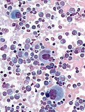
By analyzing structural changes that occur during Abl kinase activation, researchers have gained new insight into this process.
The team discovered a mechanism that links the allosteric regulation of the SH2 domain to two critical phosphorylation events.
As allosteric SH2-kinase domain interactions have proven essential for leukemogenesis caused by Bcr-Abl, the researchers believe this finding has implications for treating chronic myeloid leukemia (CML).
Oliver Hantschel, PhD, of the École polytechnique fédérale de Lausanne (EPFL) in Lausanne, Switzerland, and his colleagues described this work in Nature Communications.
The team made small, strategic mutations to Abl kinase that caused its 3D structure to change. Then, they tested each mutant version of the enzyme to see if its function would change.
The researchers built on previous studies showing that Abl kinase is indirectly controlled by the SH2 region. Normally, the SH2 region regulates the activation loop by opening and closing it. But under the Philadelphia chromosome translocation, that regulation is lost.
The team discovered that when the Philadelphia mutation takes effect, the SH2 region changes the Abl activation loop to a fully open conformation. This enables the trans-autophosphorylation of the activation loop and requires prior phosphorylation of the SH2-kinase linker.
This discovery provides the first-ever picture of the molecular events surrounding the hyperactivity of Abl kinase, the researchers said.
They also found that by disrupting the SH2-kinase interaction, it’s possible to modulate the activity of Abl kinase, which could potentially stop the growth of leukemia.
Since the SH2 region is common to other kinases, the researchers think it’s likely the effect could extend to malignancies other than CML as well, particularly those characterized by abnormal kinase activity.
Finally, the team expects this approach could overcome the problem of drug resistance in CML, as it might offer an alternative way to inhibit Abl kinase, and mutations of rapidly growing cancer cells may be less likely to occur.
Credit: UCSD School of Medicine
By analyzing structural changes that occur during Abl kinase activation, researchers have gained new insight into this process.
The team discovered a mechanism that links the allosteric regulation of the SH2 domain to two critical phosphorylation events.
As allosteric SH2-kinase domain interactions have proven essential for leukemogenesis caused by Bcr-Abl, the researchers believe this finding has implications for treating chronic myeloid leukemia (CML).
Oliver Hantschel, PhD, of the École polytechnique fédérale de Lausanne (EPFL) in Lausanne, Switzerland, and his colleagues described this work in Nature Communications.
The team made small, strategic mutations to Abl kinase that caused its 3D structure to change. Then, they tested each mutant version of the enzyme to see if its function would change.
The researchers built on previous studies showing that Abl kinase is indirectly controlled by the SH2 region. Normally, the SH2 region regulates the activation loop by opening and closing it. But under the Philadelphia chromosome translocation, that regulation is lost.
The team discovered that when the Philadelphia mutation takes effect, the SH2 region changes the Abl activation loop to a fully open conformation. This enables the trans-autophosphorylation of the activation loop and requires prior phosphorylation of the SH2-kinase linker.
This discovery provides the first-ever picture of the molecular events surrounding the hyperactivity of Abl kinase, the researchers said.
They also found that by disrupting the SH2-kinase interaction, it’s possible to modulate the activity of Abl kinase, which could potentially stop the growth of leukemia.
Since the SH2 region is common to other kinases, the researchers think it’s likely the effect could extend to malignancies other than CML as well, particularly those characterized by abnormal kinase activity.
Finally, the team expects this approach could overcome the problem of drug resistance in CML, as it might offer an alternative way to inhibit Abl kinase, and mutations of rapidly growing cancer cells may be less likely to occur.
Credit: UCSD School of Medicine
By analyzing structural changes that occur during Abl kinase activation, researchers have gained new insight into this process.
The team discovered a mechanism that links the allosteric regulation of the SH2 domain to two critical phosphorylation events.
As allosteric SH2-kinase domain interactions have proven essential for leukemogenesis caused by Bcr-Abl, the researchers believe this finding has implications for treating chronic myeloid leukemia (CML).
Oliver Hantschel, PhD, of the École polytechnique fédérale de Lausanne (EPFL) in Lausanne, Switzerland, and his colleagues described this work in Nature Communications.
The team made small, strategic mutations to Abl kinase that caused its 3D structure to change. Then, they tested each mutant version of the enzyme to see if its function would change.
The researchers built on previous studies showing that Abl kinase is indirectly controlled by the SH2 region. Normally, the SH2 region regulates the activation loop by opening and closing it. But under the Philadelphia chromosome translocation, that regulation is lost.
The team discovered that when the Philadelphia mutation takes effect, the SH2 region changes the Abl activation loop to a fully open conformation. This enables the trans-autophosphorylation of the activation loop and requires prior phosphorylation of the SH2-kinase linker.
This discovery provides the first-ever picture of the molecular events surrounding the hyperactivity of Abl kinase, the researchers said.
They also found that by disrupting the SH2-kinase interaction, it’s possible to modulate the activity of Abl kinase, which could potentially stop the growth of leukemia.
Since the SH2 region is common to other kinases, the researchers think it’s likely the effect could extend to malignancies other than CML as well, particularly those characterized by abnormal kinase activity.
Finally, the team expects this approach could overcome the problem of drug resistance in CML, as it might offer an alternative way to inhibit Abl kinase, and mutations of rapidly growing cancer cells may be less likely to occur.
Combo proves active in newly diagnosed MM

Credit: Esther Dyson
Combining the proteasome inhibitor ixazomib with lenalidomide and dexamethasone shows promise for treating patients with newly diagnosed multiple myeloma (MM), according to researchers.
In a phase 1/2 study, the all-oral combination produced a 92% overall response rate and a 27% complete response rate.
Drug-related adverse events occurred in 100% of patients, with events of grade 3 or higher occurring in 63% of patients.
These results appear in The Lancet Oncology. The study was funded by Millennium Pharmaceuticals, the company developing ixazomib.
“Ixazomib is an investigational, oral proteasome inhibitor with promising anti-myeloma effects and low rates of peripheral neuropathy,” said study author Shaji Kumar, MD, of the Mayo Clinic in Rochester, Minnesota.
“While it is well known that a combination of bortezomib, lenalidomide, and dexamethasone is highly effective in treating newly diagnosed multiple myeloma, we wanted to study the safety, tolerability, and activity of ixazomib in combination with lenalidomide and dexamethasone in newly diagnosed multiple myeloma.”
Dr Kumar and colleagues enrolled 65 patients—15 for phase 1 and 50 for phase 2 of the study—who were newly diagnosed with MM and 18 years of age or older. Patients had measurable disease, ECOG performance status of 0 to 2, and no grade 2 or higher peripheral neuropathy.
They received ixazomib (on days 1, 8, and 15) plus lenalidomide at 25 mg (on days 1 to 21) and dexamethasone at 40 mg (on days 1, 8, 15, and 22) for up to twelve 28-day cycles, followed by maintenance therapy with ixazomib alone.
In phase 1, patients received escalating doses of ixazomib, from 1.68 mg/m2 to 3.95 mg/m2, to establish the recommended dose for phase 2. The researchers established 2.97 mg/m2 as the maximum-tolerated dose and recommended the phase 2 dose be 2.23 mg/m2. This was converted to a 4.0 mg fixed dose based on population pharmacokinetic results.
Adverse events
The researchers said the combination was well tolerated, and most toxic effects were managed through dose modifications.
All patients reported at least one treatment-emergent adverse event, and 75% reported at least one treatment-emergent event that was grade 3 or higher.
Fifty-seven percent of patients had adverse events that led to dose reductions, including 53% of patients in the dose-escalation cohort and 58% of patients in the phase 2 cohort.
The most common adverse events resulting in dose reductions included skin and subcutaneous tissue disorders (20%), fatigue (14%), diarrhea (8%), peripheral neuropathy not elsewhere classified (8%), insomnia (6%), and increased body weight (6%). Five patients had adverse events leading to treatment discontinuation.
Two patients in the phase 2 cohort died while on study. One patient died of respiratory syncytial viral pneumonia that was thought to be treatment-related. The other patient died from cardiorespiratory arrest, which was considered not related to treatment.
Response and survival rates
Of the 64 response-evaluable patients, 92% responded to treatment. Fifty-eight percent had a partial response or better, and 27% had a complete response.
Responses deepened with an increasing number of treatment cycles. In the 25 patients continuing with maintenance therapy, 5 (20%) had an improvement in the depth of response during this phase.
The median duration of response has not been reached, but patients maintained responses for up to 2 years.
At last follow-up, 9 patients had progressed or died. The estimated 1-year progression-free survival was 88%, and 2-year progression-free survival was 67%.
The median overall survival has not been reached, but the estimated 1-year overall survival was 94%.
“The all-oral combination of weekly ixazomib plus lenalidomide and dexamethasone was generally well-tolerated and appeared active in patients with newly diagnosed multiple myeloma,” Dr Kumar said. “Our results support the development of a phase 3 trial studying this combination for multiple myeloma.”
Credit: Esther Dyson
Combining the proteasome inhibitor ixazomib with lenalidomide and dexamethasone shows promise for treating patients with newly diagnosed multiple myeloma (MM), according to researchers.
In a phase 1/2 study, the all-oral combination produced a 92% overall response rate and a 27% complete response rate.
Drug-related adverse events occurred in 100% of patients, with events of grade 3 or higher occurring in 63% of patients.
These results appear in The Lancet Oncology. The study was funded by Millennium Pharmaceuticals, the company developing ixazomib.
“Ixazomib is an investigational, oral proteasome inhibitor with promising anti-myeloma effects and low rates of peripheral neuropathy,” said study author Shaji Kumar, MD, of the Mayo Clinic in Rochester, Minnesota.
“While it is well known that a combination of bortezomib, lenalidomide, and dexamethasone is highly effective in treating newly diagnosed multiple myeloma, we wanted to study the safety, tolerability, and activity of ixazomib in combination with lenalidomide and dexamethasone in newly diagnosed multiple myeloma.”
Dr Kumar and colleagues enrolled 65 patients—15 for phase 1 and 50 for phase 2 of the study—who were newly diagnosed with MM and 18 years of age or older. Patients had measurable disease, ECOG performance status of 0 to 2, and no grade 2 or higher peripheral neuropathy.
They received ixazomib (on days 1, 8, and 15) plus lenalidomide at 25 mg (on days 1 to 21) and dexamethasone at 40 mg (on days 1, 8, 15, and 22) for up to twelve 28-day cycles, followed by maintenance therapy with ixazomib alone.
In phase 1, patients received escalating doses of ixazomib, from 1.68 mg/m2 to 3.95 mg/m2, to establish the recommended dose for phase 2. The researchers established 2.97 mg/m2 as the maximum-tolerated dose and recommended the phase 2 dose be 2.23 mg/m2. This was converted to a 4.0 mg fixed dose based on population pharmacokinetic results.
Adverse events
The researchers said the combination was well tolerated, and most toxic effects were managed through dose modifications.
All patients reported at least one treatment-emergent adverse event, and 75% reported at least one treatment-emergent event that was grade 3 or higher.
Fifty-seven percent of patients had adverse events that led to dose reductions, including 53% of patients in the dose-escalation cohort and 58% of patients in the phase 2 cohort.
The most common adverse events resulting in dose reductions included skin and subcutaneous tissue disorders (20%), fatigue (14%), diarrhea (8%), peripheral neuropathy not elsewhere classified (8%), insomnia (6%), and increased body weight (6%). Five patients had adverse events leading to treatment discontinuation.
Two patients in the phase 2 cohort died while on study. One patient died of respiratory syncytial viral pneumonia that was thought to be treatment-related. The other patient died from cardiorespiratory arrest, which was considered not related to treatment.
Response and survival rates
Of the 64 response-evaluable patients, 92% responded to treatment. Fifty-eight percent had a partial response or better, and 27% had a complete response.
Responses deepened with an increasing number of treatment cycles. In the 25 patients continuing with maintenance therapy, 5 (20%) had an improvement in the depth of response during this phase.
The median duration of response has not been reached, but patients maintained responses for up to 2 years.
At last follow-up, 9 patients had progressed or died. The estimated 1-year progression-free survival was 88%, and 2-year progression-free survival was 67%.
The median overall survival has not been reached, but the estimated 1-year overall survival was 94%.
“The all-oral combination of weekly ixazomib plus lenalidomide and dexamethasone was generally well-tolerated and appeared active in patients with newly diagnosed multiple myeloma,” Dr Kumar said. “Our results support the development of a phase 3 trial studying this combination for multiple myeloma.”
Credit: Esther Dyson
Combining the proteasome inhibitor ixazomib with lenalidomide and dexamethasone shows promise for treating patients with newly diagnosed multiple myeloma (MM), according to researchers.
In a phase 1/2 study, the all-oral combination produced a 92% overall response rate and a 27% complete response rate.
Drug-related adverse events occurred in 100% of patients, with events of grade 3 or higher occurring in 63% of patients.
These results appear in The Lancet Oncology. The study was funded by Millennium Pharmaceuticals, the company developing ixazomib.
“Ixazomib is an investigational, oral proteasome inhibitor with promising anti-myeloma effects and low rates of peripheral neuropathy,” said study author Shaji Kumar, MD, of the Mayo Clinic in Rochester, Minnesota.
“While it is well known that a combination of bortezomib, lenalidomide, and dexamethasone is highly effective in treating newly diagnosed multiple myeloma, we wanted to study the safety, tolerability, and activity of ixazomib in combination with lenalidomide and dexamethasone in newly diagnosed multiple myeloma.”
Dr Kumar and colleagues enrolled 65 patients—15 for phase 1 and 50 for phase 2 of the study—who were newly diagnosed with MM and 18 years of age or older. Patients had measurable disease, ECOG performance status of 0 to 2, and no grade 2 or higher peripheral neuropathy.
They received ixazomib (on days 1, 8, and 15) plus lenalidomide at 25 mg (on days 1 to 21) and dexamethasone at 40 mg (on days 1, 8, 15, and 22) for up to twelve 28-day cycles, followed by maintenance therapy with ixazomib alone.
In phase 1, patients received escalating doses of ixazomib, from 1.68 mg/m2 to 3.95 mg/m2, to establish the recommended dose for phase 2. The researchers established 2.97 mg/m2 as the maximum-tolerated dose and recommended the phase 2 dose be 2.23 mg/m2. This was converted to a 4.0 mg fixed dose based on population pharmacokinetic results.
Adverse events
The researchers said the combination was well tolerated, and most toxic effects were managed through dose modifications.
All patients reported at least one treatment-emergent adverse event, and 75% reported at least one treatment-emergent event that was grade 3 or higher.
Fifty-seven percent of patients had adverse events that led to dose reductions, including 53% of patients in the dose-escalation cohort and 58% of patients in the phase 2 cohort.
The most common adverse events resulting in dose reductions included skin and subcutaneous tissue disorders (20%), fatigue (14%), diarrhea (8%), peripheral neuropathy not elsewhere classified (8%), insomnia (6%), and increased body weight (6%). Five patients had adverse events leading to treatment discontinuation.
Two patients in the phase 2 cohort died while on study. One patient died of respiratory syncytial viral pneumonia that was thought to be treatment-related. The other patient died from cardiorespiratory arrest, which was considered not related to treatment.
Response and survival rates
Of the 64 response-evaluable patients, 92% responded to treatment. Fifty-eight percent had a partial response or better, and 27% had a complete response.
Responses deepened with an increasing number of treatment cycles. In the 25 patients continuing with maintenance therapy, 5 (20%) had an improvement in the depth of response during this phase.
The median duration of response has not been reached, but patients maintained responses for up to 2 years.
At last follow-up, 9 patients had progressed or died. The estimated 1-year progression-free survival was 88%, and 2-year progression-free survival was 67%.
The median overall survival has not been reached, but the estimated 1-year overall survival was 94%.
“The all-oral combination of weekly ixazomib plus lenalidomide and dexamethasone was generally well-tolerated and appeared active in patients with newly diagnosed multiple myeloma,” Dr Kumar said. “Our results support the development of a phase 3 trial studying this combination for multiple myeloma.”
Potential treatment method induces severe side effects

Previous studies have shown that inhibiting the activity of the Malt1 protein can kill lymphoma cells.
Now, research published in Cell Reports has revealed that it also causes the immune system to malfunction.
Malt1 carries out a variety of tasks in lymphocytes, including acting as a protease that breaks down messenger substances and controls their quantity.
Until now, researchers were unsure about the role the protease function plays in immune cell development.
Several years ago, Jürgen Ruland, PhD, of Technische Universität München in Germany, and his colleagues turned their attention to this question.
Via cell culture experiments, the researchers found that blocking the protease function of Malt1 kills lymphoma cells. The team decided to test this strategy in an animal model to shed light on the exact function of Malt1 protease.
“It’s only possible to study complex interactions in the immune system, which comprises a finely orchestrated interplay of various cell types, in an intact organism, not in cell cultures,” Dr Ruland noted. “The processes are too complex to recreate in cells outside the body.”
The mice the researchers used were genetically modified so their Malt1 protein could no longer act as a protease but was still able to carry out all of its other functions.
The team was surprised to find that these mice developed severe signs of inflammation. Moreover, the immune system attacked and destroyed key neurons that coordinate movements.
The researchers were able to explain how this serious malfunction occurred and, in doing so, discovered an unexpected function of Malt1.
They found that, in the absence of the protease function, the mice were unable to produce regulatory T cells, and this caused their immune responses to spin
out of control.
The team also found that normal lymphocytes can be activated without the protease function of Malt1, but they release messenger substances uncontrollably, which causes inflammation.
“Our study showed that Malt1 protease is surprisingly important for the development of regulatory T cells and for damping the immune response in general,” Dr Ruland said. “Since the blockade of the protease function in the organism produces undesirable effects, new alternatives should urgently be sought for the treatment of lymphoma.”
Previous studies have shown that inhibiting the activity of the Malt1 protein can kill lymphoma cells.
Now, research published in Cell Reports has revealed that it also causes the immune system to malfunction.
Malt1 carries out a variety of tasks in lymphocytes, including acting as a protease that breaks down messenger substances and controls their quantity.
Until now, researchers were unsure about the role the protease function plays in immune cell development.
Several years ago, Jürgen Ruland, PhD, of Technische Universität München in Germany, and his colleagues turned their attention to this question.
Via cell culture experiments, the researchers found that blocking the protease function of Malt1 kills lymphoma cells. The team decided to test this strategy in an animal model to shed light on the exact function of Malt1 protease.
“It’s only possible to study complex interactions in the immune system, which comprises a finely orchestrated interplay of various cell types, in an intact organism, not in cell cultures,” Dr Ruland noted. “The processes are too complex to recreate in cells outside the body.”
The mice the researchers used were genetically modified so their Malt1 protein could no longer act as a protease but was still able to carry out all of its other functions.
The team was surprised to find that these mice developed severe signs of inflammation. Moreover, the immune system attacked and destroyed key neurons that coordinate movements.
The researchers were able to explain how this serious malfunction occurred and, in doing so, discovered an unexpected function of Malt1.
They found that, in the absence of the protease function, the mice were unable to produce regulatory T cells, and this caused their immune responses to spin
out of control.
The team also found that normal lymphocytes can be activated without the protease function of Malt1, but they release messenger substances uncontrollably, which causes inflammation.
“Our study showed that Malt1 protease is surprisingly important for the development of regulatory T cells and for damping the immune response in general,” Dr Ruland said. “Since the blockade of the protease function in the organism produces undesirable effects, new alternatives should urgently be sought for the treatment of lymphoma.”
Previous studies have shown that inhibiting the activity of the Malt1 protein can kill lymphoma cells.
Now, research published in Cell Reports has revealed that it also causes the immune system to malfunction.
Malt1 carries out a variety of tasks in lymphocytes, including acting as a protease that breaks down messenger substances and controls their quantity.
Until now, researchers were unsure about the role the protease function plays in immune cell development.
Several years ago, Jürgen Ruland, PhD, of Technische Universität München in Germany, and his colleagues turned their attention to this question.
Via cell culture experiments, the researchers found that blocking the protease function of Malt1 kills lymphoma cells. The team decided to test this strategy in an animal model to shed light on the exact function of Malt1 protease.
“It’s only possible to study complex interactions in the immune system, which comprises a finely orchestrated interplay of various cell types, in an intact organism, not in cell cultures,” Dr Ruland noted. “The processes are too complex to recreate in cells outside the body.”
The mice the researchers used were genetically modified so their Malt1 protein could no longer act as a protease but was still able to carry out all of its other functions.
The team was surprised to find that these mice developed severe signs of inflammation. Moreover, the immune system attacked and destroyed key neurons that coordinate movements.
The researchers were able to explain how this serious malfunction occurred and, in doing so, discovered an unexpected function of Malt1.
They found that, in the absence of the protease function, the mice were unable to produce regulatory T cells, and this caused their immune responses to spin
out of control.
The team also found that normal lymphocytes can be activated without the protease function of Malt1, but they release messenger substances uncontrollably, which causes inflammation.
“Our study showed that Malt1 protease is surprisingly important for the development of regulatory T cells and for damping the immune response in general,” Dr Ruland said. “Since the blockade of the protease function in the organism produces undesirable effects, new alternatives should urgently be sought for the treatment of lymphoma.”
Reintroducing heparin in patients with a history of HIT
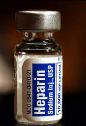
Two tests used to measure antibodies in patients with a history of heparin-induced thrombocytopenia (HIT) can produce radically different results, new research shows.
The anti-PF4/heparin IgG-specific enzyme-immunoassay can show that HIT antibody levels are high, while the functional platelet serotonin-release assay indicates that levels are low.
And researchers said the functional assay’s results may be the better indicator of a patient’s readiness for re-exposure to heparin.
Theodore Warkentin, MD, of McMaster University in Hamilton, Ontario, Canada, and his colleagues expressed this viewpoint and detailed the research supporting it in Blood.
When patients with a history of HIT require urgent heart surgery, physicians must test for the presence of HIT antibodies to determine whether the patient can be re-exposed to heparin during the procedure.
Hematologists use two types of tests to measure HIT antibodies—a functional platelet serotonin-release assay and an anti-PF4/heparin IgG-specific enzyme-immunoassay. If the more widely used immunoassay indicates the presence of HIT antibodies in a patient, surgery is usually delayed or plasma exchange is performed to lower the antibodies.
Practitioners have historically understood the two assays to provide similar conclusions, but a case report suggested otherwise.
A 76-year-old female with kidney cancer and previous HIT required urgent cardiac surgery to remove a tumor that had spread to her heart. After both her initial functional and immunoassays indicated the presence of HIT antibodies, her doctors deemed her ineligible for surgery.
But after repeated plasma exchange, the researchers performed both the functional and immunoassays on the patient again, and, this time, they observed strikingly different results.
“We were surprised to see that levels of HIT antibodies in this patient fell very quickly according to the functional assay, yet the antibodies detected by the immunoassay remained high,” Dr Warkentin said.
“This suggested to us that, while physicians in many situations may be waiting for the immunoassay to indicate lower antibody levels, patients in urgent need of heart surgery may be ready much earlier than the results suggest.”
To better understand the dissociation between the results of the two tests, Dr Warkentin’s team developed a model comparing functional and immunoassay results among 15 HIT blood samples. The samples were sequentially diluted and tested with both assays to mimic the effects of repeated plasma exchange.
The researchers observed that HIT antibody levels as measured by the functional assay decreased rather quickly, while the immunoassay continued to indicate high levels.
This suggests the sensitivity of the immunoassay may provide an overly conservative estimate of HIT antibody levels and their clinical relevance. The analysis illustrates how quickly platelet-activating properties can decline in a patient, either naturally or by using plasma exchange.
The observations also support the use of repeated plasma exchange as a therapeutic strategy prior to planned heparin re-exposure among patients with a recent HIT episode who require urgent cardiac surgery.
“Based on these findings, physicians should consider utilizing both of these tests when preparing a patient with a history of HIT for urgent heart surgery, considering the functional assay result as the stronger indicator of a patient’s readiness,” Dr Warkentin said.
“For these patients, [plasma exchange] can be a useful option to help rapidly reduce their remaining HIT antibody levels, minimize their risk of developing clots, and get them into the operating room sooner.”
Two tests used to measure antibodies in patients with a history of heparin-induced thrombocytopenia (HIT) can produce radically different results, new research shows.
The anti-PF4/heparin IgG-specific enzyme-immunoassay can show that HIT antibody levels are high, while the functional platelet serotonin-release assay indicates that levels are low.
And researchers said the functional assay’s results may be the better indicator of a patient’s readiness for re-exposure to heparin.
Theodore Warkentin, MD, of McMaster University in Hamilton, Ontario, Canada, and his colleagues expressed this viewpoint and detailed the research supporting it in Blood.
When patients with a history of HIT require urgent heart surgery, physicians must test for the presence of HIT antibodies to determine whether the patient can be re-exposed to heparin during the procedure.
Hematologists use two types of tests to measure HIT antibodies—a functional platelet serotonin-release assay and an anti-PF4/heparin IgG-specific enzyme-immunoassay. If the more widely used immunoassay indicates the presence of HIT antibodies in a patient, surgery is usually delayed or plasma exchange is performed to lower the antibodies.
Practitioners have historically understood the two assays to provide similar conclusions, but a case report suggested otherwise.
A 76-year-old female with kidney cancer and previous HIT required urgent cardiac surgery to remove a tumor that had spread to her heart. After both her initial functional and immunoassays indicated the presence of HIT antibodies, her doctors deemed her ineligible for surgery.
But after repeated plasma exchange, the researchers performed both the functional and immunoassays on the patient again, and, this time, they observed strikingly different results.
“We were surprised to see that levels of HIT antibodies in this patient fell very quickly according to the functional assay, yet the antibodies detected by the immunoassay remained high,” Dr Warkentin said.
“This suggested to us that, while physicians in many situations may be waiting for the immunoassay to indicate lower antibody levels, patients in urgent need of heart surgery may be ready much earlier than the results suggest.”
To better understand the dissociation between the results of the two tests, Dr Warkentin’s team developed a model comparing functional and immunoassay results among 15 HIT blood samples. The samples were sequentially diluted and tested with both assays to mimic the effects of repeated plasma exchange.
The researchers observed that HIT antibody levels as measured by the functional assay decreased rather quickly, while the immunoassay continued to indicate high levels.
This suggests the sensitivity of the immunoassay may provide an overly conservative estimate of HIT antibody levels and their clinical relevance. The analysis illustrates how quickly platelet-activating properties can decline in a patient, either naturally or by using plasma exchange.
The observations also support the use of repeated plasma exchange as a therapeutic strategy prior to planned heparin re-exposure among patients with a recent HIT episode who require urgent cardiac surgery.
“Based on these findings, physicians should consider utilizing both of these tests when preparing a patient with a history of HIT for urgent heart surgery, considering the functional assay result as the stronger indicator of a patient’s readiness,” Dr Warkentin said.
“For these patients, [plasma exchange] can be a useful option to help rapidly reduce their remaining HIT antibody levels, minimize their risk of developing clots, and get them into the operating room sooner.”
Two tests used to measure antibodies in patients with a history of heparin-induced thrombocytopenia (HIT) can produce radically different results, new research shows.
The anti-PF4/heparin IgG-specific enzyme-immunoassay can show that HIT antibody levels are high, while the functional platelet serotonin-release assay indicates that levels are low.
And researchers said the functional assay’s results may be the better indicator of a patient’s readiness for re-exposure to heparin.
Theodore Warkentin, MD, of McMaster University in Hamilton, Ontario, Canada, and his colleagues expressed this viewpoint and detailed the research supporting it in Blood.
When patients with a history of HIT require urgent heart surgery, physicians must test for the presence of HIT antibodies to determine whether the patient can be re-exposed to heparin during the procedure.
Hematologists use two types of tests to measure HIT antibodies—a functional platelet serotonin-release assay and an anti-PF4/heparin IgG-specific enzyme-immunoassay. If the more widely used immunoassay indicates the presence of HIT antibodies in a patient, surgery is usually delayed or plasma exchange is performed to lower the antibodies.
Practitioners have historically understood the two assays to provide similar conclusions, but a case report suggested otherwise.
A 76-year-old female with kidney cancer and previous HIT required urgent cardiac surgery to remove a tumor that had spread to her heart. After both her initial functional and immunoassays indicated the presence of HIT antibodies, her doctors deemed her ineligible for surgery.
But after repeated plasma exchange, the researchers performed both the functional and immunoassays on the patient again, and, this time, they observed strikingly different results.
“We were surprised to see that levels of HIT antibodies in this patient fell very quickly according to the functional assay, yet the antibodies detected by the immunoassay remained high,” Dr Warkentin said.
“This suggested to us that, while physicians in many situations may be waiting for the immunoassay to indicate lower antibody levels, patients in urgent need of heart surgery may be ready much earlier than the results suggest.”
To better understand the dissociation between the results of the two tests, Dr Warkentin’s team developed a model comparing functional and immunoassay results among 15 HIT blood samples. The samples were sequentially diluted and tested with both assays to mimic the effects of repeated plasma exchange.
The researchers observed that HIT antibody levels as measured by the functional assay decreased rather quickly, while the immunoassay continued to indicate high levels.
This suggests the sensitivity of the immunoassay may provide an overly conservative estimate of HIT antibody levels and their clinical relevance. The analysis illustrates how quickly platelet-activating properties can decline in a patient, either naturally or by using plasma exchange.
The observations also support the use of repeated plasma exchange as a therapeutic strategy prior to planned heparin re-exposure among patients with a recent HIT episode who require urgent cardiac surgery.
“Based on these findings, physicians should consider utilizing both of these tests when preparing a patient with a history of HIT for urgent heart surgery, considering the functional assay result as the stronger indicator of a patient’s readiness,” Dr Warkentin said.
“For these patients, [plasma exchange] can be a useful option to help rapidly reduce their remaining HIT antibody levels, minimize their risk of developing clots, and get them into the operating room sooner.”
FDA grants CAR T-cell therapy orphan designation
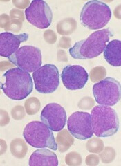
The US Food and Drug Administration (FDA) has granted orphan drug designation for the chimeric antigen receptor (CAR) T-cell therapy JCAR015 to treat acute lymphoblastic leukemia (ALL).
The designation will provide the product’s developer, Juno Therapeutics, with multiple benefits, including the availability of grant money, certain tax credits, and 7 years of market exclusivity, as well as the possibility of an expedited regulatory process.
JCAR015 consists of autologous T cells expressing a CD19-specific, CD28/CD3z CAR. The treatment has shown promise in an ongoing phase 1 trial of patients with B-cell ALL.
Initial results from this study were published in Science Translational Medicine last year and in February. Updated results were presented at the AACR Annual Meeting in April.
At that point, the researchers had enrolled 22 adult patients with relapsed or refractory B-ALL who were minimal residual disease-positive or were in first complete remission at enrollment. Patients in complete remission were monitored and only received JCAR015 if they relapsed.
The remaining patients received re-induction chemotherapy (physician’s choice), followed by an infusion of JCAR015. After treatment, patients could receive allogeneic transplant, a different salvage therapy, or monitoring.
Eighty-two percent of patients achieved a complete response to JCAR015. The average time to complete response was about 24.5 days.
Twelve of the responders were eligible for transplant. Of the 8 patients who ultimately underwent transplant and survived, 1 relapsed, but the rest remained in remission.
Ten patients had died at the time of the AACR presentation. Six deaths were a result of disease relapse or progression, and 2 patients died of complications from stem cell transplant.
The 2 remaining deaths prompted a temporary suspension of enrollment in this trial.
Those deaths were related to complications from cytokine release syndrome. One patient died of cardiovascular disease, and the other died following “persistent seizure activity.”
So researchers at the Memorial Sloan-Kettering Cancer Center, where the trial is being conducted, reviewed these cases.
The results prompted them to amend trial enrollment criteria and dosing recommendations. Now, patients with cardiac disease are ineligible to receive JCAR015.
And the T-cell dose a patient receives will depend on the extent of his or her disease. The hope is that this will reduce the risk of cytokine release syndrome and any resulting seizures.
The researchers also noted that the monoclonal antibody tocilizumab has proven effective in treating cytokine release syndrome.
In addition to this trial, JCAR015 is under investigation in another phase 1 trial of patients with relapsed and refractory non-Hodgkin lymphoma.
The US Food and Drug Administration (FDA) has granted orphan drug designation for the chimeric antigen receptor (CAR) T-cell therapy JCAR015 to treat acute lymphoblastic leukemia (ALL).
The designation will provide the product’s developer, Juno Therapeutics, with multiple benefits, including the availability of grant money, certain tax credits, and 7 years of market exclusivity, as well as the possibility of an expedited regulatory process.
JCAR015 consists of autologous T cells expressing a CD19-specific, CD28/CD3z CAR. The treatment has shown promise in an ongoing phase 1 trial of patients with B-cell ALL.
Initial results from this study were published in Science Translational Medicine last year and in February. Updated results were presented at the AACR Annual Meeting in April.
At that point, the researchers had enrolled 22 adult patients with relapsed or refractory B-ALL who were minimal residual disease-positive or were in first complete remission at enrollment. Patients in complete remission were monitored and only received JCAR015 if they relapsed.
The remaining patients received re-induction chemotherapy (physician’s choice), followed by an infusion of JCAR015. After treatment, patients could receive allogeneic transplant, a different salvage therapy, or monitoring.
Eighty-two percent of patients achieved a complete response to JCAR015. The average time to complete response was about 24.5 days.
Twelve of the responders were eligible for transplant. Of the 8 patients who ultimately underwent transplant and survived, 1 relapsed, but the rest remained in remission.
Ten patients had died at the time of the AACR presentation. Six deaths were a result of disease relapse or progression, and 2 patients died of complications from stem cell transplant.
The 2 remaining deaths prompted a temporary suspension of enrollment in this trial.
Those deaths were related to complications from cytokine release syndrome. One patient died of cardiovascular disease, and the other died following “persistent seizure activity.”
So researchers at the Memorial Sloan-Kettering Cancer Center, where the trial is being conducted, reviewed these cases.
The results prompted them to amend trial enrollment criteria and dosing recommendations. Now, patients with cardiac disease are ineligible to receive JCAR015.
And the T-cell dose a patient receives will depend on the extent of his or her disease. The hope is that this will reduce the risk of cytokine release syndrome and any resulting seizures.
The researchers also noted that the monoclonal antibody tocilizumab has proven effective in treating cytokine release syndrome.
In addition to this trial, JCAR015 is under investigation in another phase 1 trial of patients with relapsed and refractory non-Hodgkin lymphoma.
The US Food and Drug Administration (FDA) has granted orphan drug designation for the chimeric antigen receptor (CAR) T-cell therapy JCAR015 to treat acute lymphoblastic leukemia (ALL).
The designation will provide the product’s developer, Juno Therapeutics, with multiple benefits, including the availability of grant money, certain tax credits, and 7 years of market exclusivity, as well as the possibility of an expedited regulatory process.
JCAR015 consists of autologous T cells expressing a CD19-specific, CD28/CD3z CAR. The treatment has shown promise in an ongoing phase 1 trial of patients with B-cell ALL.
Initial results from this study were published in Science Translational Medicine last year and in February. Updated results were presented at the AACR Annual Meeting in April.
At that point, the researchers had enrolled 22 adult patients with relapsed or refractory B-ALL who were minimal residual disease-positive or were in first complete remission at enrollment. Patients in complete remission were monitored and only received JCAR015 if they relapsed.
The remaining patients received re-induction chemotherapy (physician’s choice), followed by an infusion of JCAR015. After treatment, patients could receive allogeneic transplant, a different salvage therapy, or monitoring.
Eighty-two percent of patients achieved a complete response to JCAR015. The average time to complete response was about 24.5 days.
Twelve of the responders were eligible for transplant. Of the 8 patients who ultimately underwent transplant and survived, 1 relapsed, but the rest remained in remission.
Ten patients had died at the time of the AACR presentation. Six deaths were a result of disease relapse or progression, and 2 patients died of complications from stem cell transplant.
The 2 remaining deaths prompted a temporary suspension of enrollment in this trial.
Those deaths were related to complications from cytokine release syndrome. One patient died of cardiovascular disease, and the other died following “persistent seizure activity.”
So researchers at the Memorial Sloan-Kettering Cancer Center, where the trial is being conducted, reviewed these cases.
The results prompted them to amend trial enrollment criteria and dosing recommendations. Now, patients with cardiac disease are ineligible to receive JCAR015.
And the T-cell dose a patient receives will depend on the extent of his or her disease. The hope is that this will reduce the risk of cytokine release syndrome and any resulting seizures.
The researchers also noted that the monoclonal antibody tocilizumab has proven effective in treating cytokine release syndrome.
In addition to this trial, JCAR015 is under investigation in another phase 1 trial of patients with relapsed and refractory non-Hodgkin lymphoma.
Agent can reverse anticoagulant effect of apixaban

Credit: Andre E.X. Brown
CHICAGO—An experimental agent known as andexanet alfa can reverse the anticoagulant effect of apixaban in healthy volunteers, a phase 3 study suggests.
All subjects who received andexanet alfa after treatment with apixaban exhibited at least a 90% reversal of anti‐factor Xa activity, and thrombin generation was restored to baseline levels.
None of the subjects experienced serious adverse events or thrombotic events, and none developed antibodies to factor X or Xa.
Mark Crowther, MD, of McMaster University in Hamilton, Ontario, Canada, presented these results at the American Heart Association 2014 Scientific Sessions. Dr Crowther is a consultant for Portola Pharmaceuticals, the makers of andexanet alfa.
Andexanet alfa is a recombinant, modified factor Xa molecule. It is being developed as an antidote for patients receiving a factor Xa inhibitor who suffer a major bleeding episode or who may require emergency surgery.
In the phase 3 ANNEXA-A study, researchers evaluated the safety and efficacy of andexanet alfa in reversing apixaban-induced anticoagulation in healthy volunteers.
Dr Crowther presented results from the first part of the study, which included 33 subjects ages 50 to 73. The subjects received apixaban at 5 mg twice daily for 4 days. Then, they were randomized in a 3:1 ratio to receive andexanet alfa as a 400 mg intravenous bolus (n=24) or placebo (n=9).
Two to 5 minutes after subjects received andexanet alfa, the anticoagulant activity of apixaban was reversed by approximately 94% compared to placebo (P<0.0001). And the effect lasted 1 to 2 hours.
Every subject treated with andexanet alfa had between 90% and 96% reversal of the anticoagulant activity of apixaban. The reversal of anti-factor Xa activity was correlated with a significant reduction in the level of free, unbound apixaban in the plasma (P<0.0001).
In addition, andexanet alfa restored thrombin generation to baseline levels. And there was no rebound effect on thrombin generation after andexanet and/or apixaban were cleared.
Three subjects experienced mild infusion reactions after receiving andexanet alfa. But there were no serious adverse events, thrombotic events, or antibodies to factor X or Xa reported.
Now, researchers are going ahead with the second part of the ANNEXA-A study, hoping to demonstrate
that prolonged reversal can be sustained with a continuous infusion of andexanet alfa after the bolus dose.
Thirty-two healthy volunteers will receive apixaban at 5 mg twice daily for 4 days and then be randomized to placebo or andexanet alfa administered as a 400 mg intravenous bolus followed by a continuous infusion of 4 mg/min for 120 minutes. Data from this part of the study are expected to be available in early 2015.
Credit: Andre E.X. Brown
CHICAGO—An experimental agent known as andexanet alfa can reverse the anticoagulant effect of apixaban in healthy volunteers, a phase 3 study suggests.
All subjects who received andexanet alfa after treatment with apixaban exhibited at least a 90% reversal of anti‐factor Xa activity, and thrombin generation was restored to baseline levels.
None of the subjects experienced serious adverse events or thrombotic events, and none developed antibodies to factor X or Xa.
Mark Crowther, MD, of McMaster University in Hamilton, Ontario, Canada, presented these results at the American Heart Association 2014 Scientific Sessions. Dr Crowther is a consultant for Portola Pharmaceuticals, the makers of andexanet alfa.
Andexanet alfa is a recombinant, modified factor Xa molecule. It is being developed as an antidote for patients receiving a factor Xa inhibitor who suffer a major bleeding episode or who may require emergency surgery.
In the phase 3 ANNEXA-A study, researchers evaluated the safety and efficacy of andexanet alfa in reversing apixaban-induced anticoagulation in healthy volunteers.
Dr Crowther presented results from the first part of the study, which included 33 subjects ages 50 to 73. The subjects received apixaban at 5 mg twice daily for 4 days. Then, they were randomized in a 3:1 ratio to receive andexanet alfa as a 400 mg intravenous bolus (n=24) or placebo (n=9).
Two to 5 minutes after subjects received andexanet alfa, the anticoagulant activity of apixaban was reversed by approximately 94% compared to placebo (P<0.0001). And the effect lasted 1 to 2 hours.
Every subject treated with andexanet alfa had between 90% and 96% reversal of the anticoagulant activity of apixaban. The reversal of anti-factor Xa activity was correlated with a significant reduction in the level of free, unbound apixaban in the plasma (P<0.0001).
In addition, andexanet alfa restored thrombin generation to baseline levels. And there was no rebound effect on thrombin generation after andexanet and/or apixaban were cleared.
Three subjects experienced mild infusion reactions after receiving andexanet alfa. But there were no serious adverse events, thrombotic events, or antibodies to factor X or Xa reported.
Now, researchers are going ahead with the second part of the ANNEXA-A study, hoping to demonstrate
that prolonged reversal can be sustained with a continuous infusion of andexanet alfa after the bolus dose.
Thirty-two healthy volunteers will receive apixaban at 5 mg twice daily for 4 days and then be randomized to placebo or andexanet alfa administered as a 400 mg intravenous bolus followed by a continuous infusion of 4 mg/min for 120 minutes. Data from this part of the study are expected to be available in early 2015.
Credit: Andre E.X. Brown
CHICAGO—An experimental agent known as andexanet alfa can reverse the anticoagulant effect of apixaban in healthy volunteers, a phase 3 study suggests.
All subjects who received andexanet alfa after treatment with apixaban exhibited at least a 90% reversal of anti‐factor Xa activity, and thrombin generation was restored to baseline levels.
None of the subjects experienced serious adverse events or thrombotic events, and none developed antibodies to factor X or Xa.
Mark Crowther, MD, of McMaster University in Hamilton, Ontario, Canada, presented these results at the American Heart Association 2014 Scientific Sessions. Dr Crowther is a consultant for Portola Pharmaceuticals, the makers of andexanet alfa.
Andexanet alfa is a recombinant, modified factor Xa molecule. It is being developed as an antidote for patients receiving a factor Xa inhibitor who suffer a major bleeding episode or who may require emergency surgery.
In the phase 3 ANNEXA-A study, researchers evaluated the safety and efficacy of andexanet alfa in reversing apixaban-induced anticoagulation in healthy volunteers.
Dr Crowther presented results from the first part of the study, which included 33 subjects ages 50 to 73. The subjects received apixaban at 5 mg twice daily for 4 days. Then, they were randomized in a 3:1 ratio to receive andexanet alfa as a 400 mg intravenous bolus (n=24) or placebo (n=9).
Two to 5 minutes after subjects received andexanet alfa, the anticoagulant activity of apixaban was reversed by approximately 94% compared to placebo (P<0.0001). And the effect lasted 1 to 2 hours.
Every subject treated with andexanet alfa had between 90% and 96% reversal of the anticoagulant activity of apixaban. The reversal of anti-factor Xa activity was correlated with a significant reduction in the level of free, unbound apixaban in the plasma (P<0.0001).
In addition, andexanet alfa restored thrombin generation to baseline levels. And there was no rebound effect on thrombin generation after andexanet and/or apixaban were cleared.
Three subjects experienced mild infusion reactions after receiving andexanet alfa. But there were no serious adverse events, thrombotic events, or antibodies to factor X or Xa reported.
Now, researchers are going ahead with the second part of the ANNEXA-A study, hoping to demonstrate
that prolonged reversal can be sustained with a continuous infusion of andexanet alfa after the bolus dose.
Thirty-two healthy volunteers will receive apixaban at 5 mg twice daily for 4 days and then be randomized to placebo or andexanet alfa administered as a 400 mg intravenous bolus followed by a continuous infusion of 4 mg/min for 120 minutes. Data from this part of the study are expected to be available in early 2015.
Group silences microRNA to treat DLBCL

Scientists believe they’ve devised a way to use antimiRs as anticancer drugs by showing that a specific antimiR could treat diffuse large B-cell lymphoma (DLBCL) in mice.
In a letter to Nature, the group explained that microRNAs known as oncomiRs can play a causal role in the onset and maintenance of cancer when they are overexpressed.
So inhibiting oncomiRs using antisense oligomers, or antimiRs, has seemed a promising therapeutic strategy. But physiological and cellular barriers have prevented targeted delivery.
Frank Slack, PhD, of Beth Israel Deaconess Medical Center in Boston, Massachusetts, and his colleagues have devised a new antimiR delivery platform and shown that it can inhibit DLBCL growth in vivo.
The team created a mouse model to study miR-155, an oncomiR that, when overexpressed, leads to DLBCL.
“We hypothesized that we could inhibit the function of miR-155 by way of an antisense molecule that would bind to miR-155,” Dr Slack said. “[However,] there are a number of significant obstacles to reaching the tumor cell target. Some roadblocks are clearance through the kidneys and accumulation in the liver, which absorbs any systemically injected agent.”
“Furthermore, even if you are able to reach your targeted cells, the molecules must cross cell membranes and escape degradation from a process known as endocytosis. If you can picture our antisense molecule being a warhead, we had to find the right ‘rocket’ to actually transport it to its target.”
The “rocket” turned out to be a peptide with a low-pH induced transmembrane structure (pHLIP), meaning it inserts into cell membranes only when cells are low in pH. And tumor cells provided the ideal environment.
“When we attached our antisense warhead to the pHLIP peptide, not only did it successfully insert itself into the tumor cell, but it also dragged the antisense molecule itself into the cell,” Dr Slack said. “Now the ‘warhead’ could deploy and actually inhibit microRNA function and control cancer growth.”
In the miR-155/DLBCL mouse models, pHLIP-anti155 slowed tumor growth, suppressed the metastatic spread of neoplastic lymphocytes to other organs, reduced the onset of splenomegaly, and delayed the development of conspicuous lymphadenopathy, when compared to control mice.
Responses with pHLIP-anti155 were similar to those observed in mice that received doxorubicin or CHOP, but pHLIP-anti155 proved less toxic than the other treatments.
“With this delivery platform, we should also be able to transform other RNAs into druggable targets,” Dr Slack said, adding that low pH is also an issue in kidney disease, myocardial infarction, stroke, and infection, among other conditions. So this type of therapy could have wide applications.
Scientists believe they’ve devised a way to use antimiRs as anticancer drugs by showing that a specific antimiR could treat diffuse large B-cell lymphoma (DLBCL) in mice.
In a letter to Nature, the group explained that microRNAs known as oncomiRs can play a causal role in the onset and maintenance of cancer when they are overexpressed.
So inhibiting oncomiRs using antisense oligomers, or antimiRs, has seemed a promising therapeutic strategy. But physiological and cellular barriers have prevented targeted delivery.
Frank Slack, PhD, of Beth Israel Deaconess Medical Center in Boston, Massachusetts, and his colleagues have devised a new antimiR delivery platform and shown that it can inhibit DLBCL growth in vivo.
The team created a mouse model to study miR-155, an oncomiR that, when overexpressed, leads to DLBCL.
“We hypothesized that we could inhibit the function of miR-155 by way of an antisense molecule that would bind to miR-155,” Dr Slack said. “[However,] there are a number of significant obstacles to reaching the tumor cell target. Some roadblocks are clearance through the kidneys and accumulation in the liver, which absorbs any systemically injected agent.”
“Furthermore, even if you are able to reach your targeted cells, the molecules must cross cell membranes and escape degradation from a process known as endocytosis. If you can picture our antisense molecule being a warhead, we had to find the right ‘rocket’ to actually transport it to its target.”
The “rocket” turned out to be a peptide with a low-pH induced transmembrane structure (pHLIP), meaning it inserts into cell membranes only when cells are low in pH. And tumor cells provided the ideal environment.
“When we attached our antisense warhead to the pHLIP peptide, not only did it successfully insert itself into the tumor cell, but it also dragged the antisense molecule itself into the cell,” Dr Slack said. “Now the ‘warhead’ could deploy and actually inhibit microRNA function and control cancer growth.”
In the miR-155/DLBCL mouse models, pHLIP-anti155 slowed tumor growth, suppressed the metastatic spread of neoplastic lymphocytes to other organs, reduced the onset of splenomegaly, and delayed the development of conspicuous lymphadenopathy, when compared to control mice.
Responses with pHLIP-anti155 were similar to those observed in mice that received doxorubicin or CHOP, but pHLIP-anti155 proved less toxic than the other treatments.
“With this delivery platform, we should also be able to transform other RNAs into druggable targets,” Dr Slack said, adding that low pH is also an issue in kidney disease, myocardial infarction, stroke, and infection, among other conditions. So this type of therapy could have wide applications.
Scientists believe they’ve devised a way to use antimiRs as anticancer drugs by showing that a specific antimiR could treat diffuse large B-cell lymphoma (DLBCL) in mice.
In a letter to Nature, the group explained that microRNAs known as oncomiRs can play a causal role in the onset and maintenance of cancer when they are overexpressed.
So inhibiting oncomiRs using antisense oligomers, or antimiRs, has seemed a promising therapeutic strategy. But physiological and cellular barriers have prevented targeted delivery.
Frank Slack, PhD, of Beth Israel Deaconess Medical Center in Boston, Massachusetts, and his colleagues have devised a new antimiR delivery platform and shown that it can inhibit DLBCL growth in vivo.
The team created a mouse model to study miR-155, an oncomiR that, when overexpressed, leads to DLBCL.
“We hypothesized that we could inhibit the function of miR-155 by way of an antisense molecule that would bind to miR-155,” Dr Slack said. “[However,] there are a number of significant obstacles to reaching the tumor cell target. Some roadblocks are clearance through the kidneys and accumulation in the liver, which absorbs any systemically injected agent.”
“Furthermore, even if you are able to reach your targeted cells, the molecules must cross cell membranes and escape degradation from a process known as endocytosis. If you can picture our antisense molecule being a warhead, we had to find the right ‘rocket’ to actually transport it to its target.”
The “rocket” turned out to be a peptide with a low-pH induced transmembrane structure (pHLIP), meaning it inserts into cell membranes only when cells are low in pH. And tumor cells provided the ideal environment.
“When we attached our antisense warhead to the pHLIP peptide, not only did it successfully insert itself into the tumor cell, but it also dragged the antisense molecule itself into the cell,” Dr Slack said. “Now the ‘warhead’ could deploy and actually inhibit microRNA function and control cancer growth.”
In the miR-155/DLBCL mouse models, pHLIP-anti155 slowed tumor growth, suppressed the metastatic spread of neoplastic lymphocytes to other organs, reduced the onset of splenomegaly, and delayed the development of conspicuous lymphadenopathy, when compared to control mice.
Responses with pHLIP-anti155 were similar to those observed in mice that received doxorubicin or CHOP, but pHLIP-anti155 proved less toxic than the other treatments.
“With this delivery platform, we should also be able to transform other RNAs into druggable targets,” Dr Slack said, adding that low pH is also an issue in kidney disease, myocardial infarction, stroke, and infection, among other conditions. So this type of therapy could have wide applications.