User login
Vaccine candidate fails to prevent malaria in humans
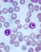
Plasmodium vivax
Image by Mae Melvin
Results of a phase 1 trial suggest a vaccine candidate does not prevent Plasmodium vivax malaria.
The vaccine, VMP001/AS01B, did not prevent malaria in any of the 30 subjects tested, although it did significantly delay parasitemia in 59% of subjects.
Lieutenant Colonel Jason W. Bennett, of the Walter Reed Army Institute
of Research (WRAIR) in Silver Spring, Maryland, and his colleagues reported these results in PLOS Neglected Tropical Diseases.
The vaccine antigen VMP001 is an Escherichia coli-produced, synthetic, chimeric, recombinant protein that incorporates the 3 major domains of circumsporozoite protein but is distinct from the native molecule. The VMP001 antigen was adjuvanted with AS01B, a proprietary liposome-based adjuvant system from GlaxoSmithKline.
For this phase 1 study, the investigators tested VMP001/AS01B in 30 healthy volunteers. The subjects received 3 intramuscular injections of VMP001 at 15 μg (n=10), 30 μg (n=10), or 60 μg (n=10), respectively, all formulated in 500 μL of AS01B at each immunization.
Fourteen days after the third immunization, the subjects were exposed to mosquitoes carrying P vivax. Six non-vaccinated control subjects were exposed to the mosquitoes as well.
The investigators said VMP001/AS01B was well tolerated and generated robust immune responses. However, it did not protect the subjects from malaria infection.
Still, the vaccine induced a “small but significant” delay in time to parasitemia in 59% of vaccinated subjects, when compared to controls.
Colonel Robert Paris, director of the US Military Malaria Research Program at WRAIR, believes the investigators can use the results of this study to design a better vaccine for P vivax malaria.
“Findings from the analysis of the immune response of vaccinated subjects have given us clues to improve vaccine candidates,” he said. “[S]tudies are now underway at WRAIR to develop next-generation vivax vaccines.” ![]()
Combining inhibitors to treat AML

Preclinical research has revealed a treatment approach that could prove effective against acute myeloid leukemia (AML).
Researchers tested the IAP inhibitor birinapant in combination with p38 inhibitors and observed antileukemic activity in mouse models of AML and samples from patients with the disease.
Combination treatment proved more effective than either agent alone, and the combination was less toxic than single-agent chemotherapy.
Najoua Lalaoui, PhD, of the Walter and Eliza Hall Institute of Medical Research in Parkville, Victoria, Australia, and her colleagues conducted this research and relayed the results in an article published in Cancer Cell.
The researchers generated several mouse models of AML—MLL-ENL ± NRasG12D, MLL-AF9 ± NrasG12D, AML1-ETO9a + NrasG12D, CBFβ-MYH11 + NrasG12D, NUP98-HoxA9, and HoxA9/Meis1.
In these models, the team tested birinapant with 1 of 2 p38 inhibitors—LY2228820 or SCIO-469—or with the MK2 inhibitor PF-3644022. They said each combination “dramatically” increased cell death, when compared to birinapant alone, in most models. The exceptions were AML1-ETO9a + NrasG12D and CBFβ-MYH11 + NrasG12D.
Next, the researchers tested LY2228820 plus birinapant in samples from 8 AML patients. The samples had FLT3-ITD mutations (patients 1, 2, 4, 6, and 7), a FLT3 D835 missense mutation (patient 4), nucleophosmin exon-12 mutations (patients 2 and 4), an MLL translocation (patient 3), inv(3) (patient 1), and inv(16) (patient 8).
All 8 samples were sensitive to birinapant alone. And although LY2228820 alone did not induce cell death in any of the samples, the drug had a synergistic effect with birinapant in 4 of the samples (patients 2, 3, 4, and 7).
The researchers also found that peripheral blood mononuclear cells from healthy donors proved more resistant to combination LY2228820 (at 10 µM) and birinapant (at 500 nM) than to cytarabine (10 µM), daunorubicin (at 0.4 µM), or idarubicin (at 0.4 µM).
In addition, 4 weeks of treatment with birinapant and LY2228820 was well-tolerated in mice without tumors.
Finally, the researchers tested birinapant and LY2228820, either alone or in combination, in mouse models of MLL-ENL, MLL-AF9, and NRasG12D mutant/MLL-AF9/Luc AML.
Combination treatment prolonged survival in all 3 models, when compared with mice that received single agents or no treatment. However, unlike in the MLL-ENL and MLL-AF9 models, the combination was unable to cure NRasG12D mutant/MLL-AF9/Luc mice of their leukemia.
“Our findings have made us hopeful that a combination of birinapant and a p38 inhibitor may be more effective in treating AML than current therapies and also have less toxicity for patients,” Dr Lalaoui said.
“We tested forms of AML that are highly resistant to chemotherapy and found that birinapant and p38 inhibitors could even kill these cancer cells, which is great news.”
Birinapant is being developed by TetraLogic Pharmaceuticals Corporation, and some of the researchers involved in this work reported relationships with the company. ![]()
Drug granted orphan designation for hemolytic anemia

The European Commission (EC) has granted orphan drug designation for TNT009 to treat autoimmune hemolytic anemia, including cold agglutinin disease.
TNT009 is a monoclonal antibody that selectively inhibits the classical complement pathway by targeting C1s, a serine protease within the C1-complex in the complement pathway.
The drug thereby prevents downstream disease processes involving phagocytosis, inflammation, and cell lysis.
TNT009 is being developed by True North Therapeutics.
The drug is currently in development for the treatment of autoimmune hemolytic anemia, which is characterized by the premature destruction of healthy red blood cells by autoantibodies.
In cold agglutinin disease, this destruction of red blood cells results in anemia, fatigue, and potentially fatal thrombosis.
TNT009 is also being evaluated in patients with bullous pemphigoid and end-stage renal disease.
Top-line results from a phase 1b trial of TNT009 are expected in mid-2016.
About orphan designation
The EC grants orphan designation to products intended to treat, prevent, or diagnose a life-threatening condition affecting up to 5 in 10,000 people in the European Union. The product must provide significant benefit to those affected by the condition.
Orphan drug designation from the EC provides companies with certain development incentives, including protocol assistance, a type of scientific advice specific for orphan drugs, and 10 years of market exclusivity once the drug is on the market. ![]()
EC grants venetoclax orphan designation for AML

The European Commission has granted orphan drug designation for the oral BCL-2 inhibitor venetoclax to treat acute myeloid leukemia (AML).
The EC grants orphan designation to products intended to treat, prevent, or diagnose a life-threatening condition affecting up to 5 in 10,000 people in the
European Union. The product must provide significant benefit to those affected by the condition.
Orphan drug designation from the EC provides companies with certain development incentives, including protocol assistance, a type of scientific advice specific for orphan drugs, and 10 years of market exclusivity once the drug is on the market.
Venetoclax is being developed by AbbVie in partnership with Genentech and Roche.
Phase 2 study
Results from a phase 2 study of venetoclax in AML were presented at ASH 2014. At that time, the trial had enrolled 32 patients, 30 of whom had relapsed or refractory disease. Patients had a median age of 71 (range, 19 to 84), and half were male.
The overall response rate was 15.5%, with 1 patient achieving a complete response (CR) and 4 achieving a CR with incomplete count recovery (CRi).
The researchers noted that 3 of the patients who had a CR/CRi had IDH mutations. Two of these patients also achieved minimal residual disease negativity.
The median bone marrow blast count in evaluable patients decreased 36% after treatment, and 6 patients (19%) had at least a 50% reduction in bone marrow blasts.
Common adverse events following treatment (occurring in at least 25% of patients) included nausea, diarrhea, fatigue, neutropenia, and vomiting.
Grade 3/4 adverse events (occurring in 3 or more patients) included febrile neutropenia, anemia, and pneumonia. No patient died as a result of treatment-related adverse events. ![]()
CHMP recommends fusion protein for hemophilia B

The European Medicines Agency’s Committee for Medicinal Products for Human Use (CHMP) has recommended that albutrepenonacog alfa (Idelvion) receive marketing authorization to treat patients with hemophilia B.
Albutrepenonacog alfa is a recombinant fusion protein linking coagulation factor IX with albumin.
The CHMP’s recommendation will be reviewed by the European Commission, which usually follows the CHMP’s advice.
In 2010, the European Commission granted albutrepenonacog alfa orphan designation as a treatment for hemophilia B.
Albutrepenonacog alfa is being developed by CSL Behring. The product is approved for use in Canada. Regulatory agencies in the US, Australia, Switzerland, and Japan are reviewing applications for the drug.
Phase 3 trial
The CHMP’s recommendation to approve albutrepenonacog alfa is based on the PROLONG-9FP clinical development program. PROLONG-9FP includes phase 1, 2, and 3 studies evaluating the safety and efficacy of albutrepenonacog alfa in adults and children (ages 1 to 61) with hemophilia B.
Data from the phase 3 study were recently published in Blood. This study included 63 previously treated male patients with severe hemophilia B. They had a mean age of 33 (range, 12 to 61).
The patients were divided into 2 groups. Group 1 (n=40) received routine prophylaxis with albutrepenonacog alfa once every 7 days for 26 weeks, followed by a 7-, 10- or 14-day prophylaxis regimen for a mean of 50, 38, or 51 weeks, respectively.
Group 2 received on-demand treatment with albutrepenonacog alfa for bleeding episodes for 26 weeks (n=23) and then switched to a 7-day prophylaxis regimen for a mean of 45 weeks (n=19).
The median annualized bleeding rate (ABR) was 2.0 in the prophylaxis arm (group 1) and 23.5 in the on-demand treatment arm (group 2). The median spontaneous ABRs were 0.0 and 17.0, respectively.
For patients in group 2, there was a significant reduction in median ABRs when patients switched from on-demand treatment to prophylaxis—19.22 and 1.58, respectively (P<0.0001). And there was a significant reduction in median spontaneous ABRs—15.43 and 0.00, respectively (P<0.0001).
Overall, 98.6% of bleeding episodes were treated successfully, including 93.6% that were treated with a single injection of albutrepenonacog alfa.
None of the patients developed inhibitors or experienced thromboembolic events, anaphylaxis, or life-threatening adverse events (AEs).
There were 347 treatment-emergent AEs reported in 54 (85.7%) patients. The most common were nasopharyngitis (25.4%), headache (23.8%), arthralgia (4.3%), and influenza (11.1%).
Eleven mild/moderate AEs in 5 patients (7.9%) were considered possibly related to albutrepenonacog alfa. Two patients discontinued treatment due to AEs—1 with hypersensitivity and 1 with headache. ![]()
Pre-labor cesarean delivery linked to ALL

Photo by Nina Matthews
Researchers have found a potential correlation between pre-labor cesarean delivery (PLCD) and acute lymphoblastic leukemia (ALL).
The team analyzed data from 13 studies and found a 23% increase in the risk of ALL in children born by PLCD.
However, there was no link between PLCD and acute myeloid leukemia (AML), and there was no correlation between emergency cesareans and ALL or AML.
Erin Marcotte, PhD, of the University of Minnesota, and her colleagues reported these results in The Lancet Haematology.
The researchers analyzed data from the Childhood Leukemia International Consortium. They looked at 33,571 subjects overall, including 23,351 control subjects, 8780 cases of ALL, and 1332 cases of AML.
The analyses were controlled for a number of outside factors, including breastfeeding, parental education levels, and ethnicity.
“Our goal was to determine if there was an association between cesarean deliveries and ALL [and] to identify potential new targets for research into cancer prevention if there is a correlation,” Dr Marcotte said.
“While the link between overall cesarean delivery and childhood leukemia was not statistically significant, it was notable to find an association between pre-labor cesarean delivery and ALL.”
The data suggested AML was not associated with cesarean delivery. The odds ratios (ORs) were 0.99 for all cesarean deliveries, 0.83 for PLCD, and 1.05 for emergency cesarean delivery.
The ORs for ALL were 1.06 overall, 1.02 for emergency cesarean delivery, and 1.23 (P=0.018) for PLCD.
The reason for the increased risk of ALL with PLCD is not known. The researchers said several mechanisms may be at play, including the stress response in the fetus caused by labor and the colonization of microbiota a newborn experiences during a vaginal delivery that is missed during a cesarean birth.
“The most plausible explanation for the association between ALL and pre-labor cesarean delivery is in the cortisol, or stress-related, mechanism,” Dr Marcotte said. “Because ALL is not associated with all cesarean deliveries, it seems less likely the microbiota colonization is a significant factor in this phenomenon. We believe further investigation into this cortisol-mechanism link is warranted due to these findings.”
The researchers said the strength of association in these findings is comparable to other studies looking at cesarean delivery rates and other childhood outcomes, including Type I diabetes and asthma. They believe further investigation into this study’s findings is needed, utilizing more detailed and reliable delivery information.
“This association deserves a closer look to better determine what’s behind the link,” said Logan Spector, PhD, of the University of Minnesota.
“Cortisol exposure is plausible since similar compounds are used to treat ALL. We also know that some are born with cells that are on the path to becoming leukemia. Thus, our working hypothesis is that cortisol exposure at birth may eliminate these pre-leukemic cells.” ![]()
FDA authorizes CDC’s test for Zika virus
Photo by Jeremy L. Grisham
The US Food and Drug Administration (FDA) has issued an Emergency Use Authorization (EUA) for a laboratory test used to detect the Zika virus.
The test, which was developed by the Centers for Disease Control and Prevention (CDC), is called the Zika IgM Antibody Capture Enzyme-Linked Immunosorbent Assay (Zika MAC-ELISA).
It will be distributed to certain laboratories in the US and abroad over the next 2 weeks.
The test will not be available in US hospitals or other primary care settings.
The Zika MAC-ELISA test can detect human immunoglobulin M (IgM) antibodies to the Zika virus. These antibodies appear in the blood of an infected person 4 to 5 days after the start of the illness, and they remain in the blood for about 12 weeks.
The Zika MAC-ELISA test is intended for use in sera or cerebrospinal fluid when submitted with a patient-matched serum sample from individuals meeting CDC Zika clinical and epidemiological criteria for testing. This includes people with a history of symptoms associated with Zika and/or people who have recently traveled to an area that has active Zika transmission.
Results of Zika MAC-ELISA tests require careful interpretation. The test can give false-positive results when someone has been infected with a virus closely related to Zika (such as dengue virus).
When positive or inconclusive results occur, additional testing (plaque reduction neutralization test) to confirm the presence of antibodies to Zika virus will be performed by the CDC or a CDC-authorized laboratory.
Moreover, a negative test result does not necessarily mean a person has not been infected with Zika virus. If a sample is collected just after a person becomes ill, there may not be enough IgM antibodies for the test to measure, resulting in a false-negative.
Similarly, if the sample was collected more than 12 weeks after illness, it is possible that the body has successfully fought the virus and IgM antibody levels have dropped below the detectable limit.
The CDC will begin distributing the Zika MAC-ELISA test over the next 2 weeks to qualified laboratories in the Laboratory Response Network, an integrated network of domestic and international laboratories that can respond to public health emergencies.
About the EUA
An EUA allows the use of unapproved medical products or unapproved uses of approved medical products in an emergency. The products must be used to diagnose, treat, or prevent serious or life-threatening conditions caused by chemical, biological, radiological, or nuclear threat agents, when there are no adequate alternatives.
As there are no commercially available diagnostic tests approved by the FDA for the detection of Zika virus infection, the agency decided an EUA was crucial to ensure timely access to a diagnostic tool.
The FDA issued the EUA for the Zika MAC-ELISA test based on data submitted by the CDC and on the US Secretary of Health and Human Services’ declaration that circumstances exist to justify the emergency use of in vitro diagnostic tests for the detection of Zika virus and Zika virus infection. This EUA will end when the Secretary’s declaration ends, unless the FDA revokes it sooner. ![]()
Photo by Jeremy L. Grisham
The US Food and Drug Administration (FDA) has issued an Emergency Use Authorization (EUA) for a laboratory test used to detect the Zika virus.
The test, which was developed by the Centers for Disease Control and Prevention (CDC), is called the Zika IgM Antibody Capture Enzyme-Linked Immunosorbent Assay (Zika MAC-ELISA).
It will be distributed to certain laboratories in the US and abroad over the next 2 weeks.
The test will not be available in US hospitals or other primary care settings.
The Zika MAC-ELISA test can detect human immunoglobulin M (IgM) antibodies to the Zika virus. These antibodies appear in the blood of an infected person 4 to 5 days after the start of the illness, and they remain in the blood for about 12 weeks.
The Zika MAC-ELISA test is intended for use in sera or cerebrospinal fluid when submitted with a patient-matched serum sample from individuals meeting CDC Zika clinical and epidemiological criteria for testing. This includes people with a history of symptoms associated with Zika and/or people who have recently traveled to an area that has active Zika transmission.
Results of Zika MAC-ELISA tests require careful interpretation. The test can give false-positive results when someone has been infected with a virus closely related to Zika (such as dengue virus).
When positive or inconclusive results occur, additional testing (plaque reduction neutralization test) to confirm the presence of antibodies to Zika virus will be performed by the CDC or a CDC-authorized laboratory.
Moreover, a negative test result does not necessarily mean a person has not been infected with Zika virus. If a sample is collected just after a person becomes ill, there may not be enough IgM antibodies for the test to measure, resulting in a false-negative.
Similarly, if the sample was collected more than 12 weeks after illness, it is possible that the body has successfully fought the virus and IgM antibody levels have dropped below the detectable limit.
The CDC will begin distributing the Zika MAC-ELISA test over the next 2 weeks to qualified laboratories in the Laboratory Response Network, an integrated network of domestic and international laboratories that can respond to public health emergencies.
About the EUA
An EUA allows the use of unapproved medical products or unapproved uses of approved medical products in an emergency. The products must be used to diagnose, treat, or prevent serious or life-threatening conditions caused by chemical, biological, radiological, or nuclear threat agents, when there are no adequate alternatives.
As there are no commercially available diagnostic tests approved by the FDA for the detection of Zika virus infection, the agency decided an EUA was crucial to ensure timely access to a diagnostic tool.
The FDA issued the EUA for the Zika MAC-ELISA test based on data submitted by the CDC and on the US Secretary of Health and Human Services’ declaration that circumstances exist to justify the emergency use of in vitro diagnostic tests for the detection of Zika virus and Zika virus infection. This EUA will end when the Secretary’s declaration ends, unless the FDA revokes it sooner.
FDA approves obinutuzumab for FL

The US Food and Drug Administration (FDA) has approved obinutuzumab (Gazyva) for certain patients with previously treated follicular lymphoma (FL).
Obinutuzumab is a glycoengineered, humanized, monoclonal antibody that selectively binds to the extracellular domain of the CD20 antigen on B cells.
The drug was previously approved by the FDA for use in combination with chlorambucil to treat patients with previously untreated chronic lymphocytic leukemia.
Now, obinutuzumab is approved for use in combination with bendamustine, followed by obinutuzumab alone, to treat patients with FL who did not respond to a rituximab-containing regimen or whose FL returned after such treatment.
The recommended dose and schedule for the regimen is:
- Obinutuzumab at 1000 mg by intravenous infusion on days 1, 8, and 15 of cycle 1; on day 1 of cycles 2-6 (28-day cycles); then every 2 months for 2 years.
- Bendamustine at 90 mg/m2 by intravenous infusion on days 1 and 2 of cycles 1-6.
Full prescribing information for obinutuzumab is available on the FDA website or at www.Gazyva.com.
Phase 3 study
The approval for obinutuzumab in FL is based on results from the phase 3 GADOLIN study. The trial included 413 patients with rituximab-refractory non-Hodgkin lymphoma, including 321 patients with FL, 46 with marginal zone lymphoma, and 28 with small lymphocytic lymphoma.
The patients were randomized to receive bendamustine alone (control arm) or a combination of bendamustine and obinutuzumab followed by obinutuzumab maintenance (every 2 months for 2 years or until progression).
The primary endpoint of the study was progression-free survival (PFS), as assessed by an independent review committee (IRC). The secondary endpoints were PFS as assessed by investigator review, best overall response, complete response (CR), partial response (PR), duration of response, overall survival, and safety profile.
Among patients with FL, the obinutuzumab regimen improved PFS compared to bendamustine alone, as assessed by IRC (hazard ratio [HR]=0.48, P<0.0001). The median PFS was not reached in patients receiving the obinutuzumab regimen but was 13.8 months in those receiving bendamustine alone.
Investigator-assessed PFS was consistent with IRC-assessed PFS. Investigators said the median PFS with the obinutuzumab regimen was more than double that with bendamustine alone—29.2 months vs 13.7 months (HR=0.48, P<0.0001).
Best overall response for patients receiving the obinutuzumab regimen was 78.7% (15.5% CR, 63.2% PR), compared to 74.7% for those receiving bendamustine alone (18.7% CR, 56% PR), as assessed by the IRC.
The median duration of response was not reached for patients receiving the obinutuzumab regimen and was 11.6 months for those receiving bendamustine alone.
The median overall survival has not yet been reached in either study arm.
The most common grade 3/4 adverse events observed in patients receiving the obinutuzumab regimen were neutropenia (33%), infusion reactions (11%), and thrombocytopenia (10%).
The most common adverse events of any grade were infusion reactions (69%), neutropenia (35%), nausea (54%), fatigue (39%), cough (26%), diarrhea (27%), constipation (19%), fever (18%), thrombocytopenia (15%), vomiting (22%), upper respiratory tract infection (13%), decreased appetite (18%), joint or muscle pain (12%), sinusitis (12%), anemia (12%), general weakness (11%), and urinary tract infection (10%).
About obinutuzumab
Obinutuzumab is being studied in a large clinical program, including the phase 3 GOYA and GALLIUM studies.
In GOYA, researchers are comparing obinutuzumab head-to-head with rituximab plus CHOP chemotherapy in first-line diffuse large B-cell lymphoma. In GALLIUM, researchers are comparing obinutuzumab plus chemotherapy head-to-head with rituximab plus chemotherapy in first-line indolent non-Hodgkin lymphoma.
Additional combination studies investigating obinutuzumab with other approved or investigational medicines, including cancer immunotherapies and small-molecule inhibitors, are planned or underway across a range of blood cancers.
Obinutuzumab was discovered by Roche Glycart AG, a wholly owned, independent research unit of Roche. In the US, obinutuzumab is part of a collaboration between Genentech and Biogen.
Genentech has a patient assistance program, Genentech Access Solutions, that can help qualifying patients access obinutuzumab and other Genentech medications.
The program is designed to help people navigate the access and reimbursement process and provide assistance to eligible patients in the US who are uninsured or cannot afford the out-of-pocket costs for their medicine. For more information, visit www.Genentech-Access.com.
DNA delivery vehicles may circumvent drug resistance in AML

Image courtesy of PNAS
DNA origami nanostructures may be used to overcome drug resistance in acute myeloid leukemia (AML), according to preclinical research published in the journal Small.
Researchers found they could create these nanostructures in 10 minutes and load them with the anthracycline daunorubicin.
When the team introduced the structures to daunorubicin-resistant AML cells, the drug delivery vehicles entered the cells via endocytosis.
This allowed the drug to bypass defenses in the cell membrane that are effective against the free drug.
Once the nanostructures broke down, daunorubicin flooded the cells and killed them off.
Other research groups have used this delivery technique to overcome drug resistance in solid tumors, but this is the first time researchers have shown the same technique works on drug-resistant leukemia cells.
To create the DNA origami nanostructures, the researchers used the genome of a common bacteriophage and synthetic strands that were designed to fold up the bacteriophage DNA.
Although the folded-up shape performs a function, the DNA itself does not, explained Patrick Halley, a graduate student at The Ohio State University in Columbus.
“[T]he DNA capsule doesn’t do anything except hold a shape,” Halley said. “It’s just a static, rigid structure that carries things. It doesn’t encode any proteins or do anything else that we normally think of DNA as doing.”
The researchers tested the DNA origami nanostructures in AML cell lines that had developed resistance to daunorubicin. When molecules of daunorubicin enter these cells, the cells recognize the drug molecules and eject them through openings in the cell wall.
“Cancer cells have novel ways of resisting drugs, like these ‘pumps,’ and the exciting part of packaging the drug this way is that we can circumvent those defenses so that the drug accumulates in the cancer cell and causes it to die,” said John Byrd, MD, of The Ohio State University.
“Potentially, we can also tailor these structures to make them deliver drugs selectively to cancer cells and not to other parts of the body where they can cause side effects.”
In tests, the resistant AML cells effectively absorbed molecules of daunorubicin when they were hidden inside the rod-shaped nanostructures.
The researchers tracked the nanostructures inside the cells using fluorescent tags. Each structure measures about 15 nanometers wide and 100 nanometers long, and each has 4 hollow, open-ended interior compartments.
Study author Christopher Lucas, PhD, of The Ohio State University, said the design of the nanostructures maximizes the surface area available to carry the drug.
“The way daunorubicin works is it tucks into the cancer cell’s DNA and prevents it from replicating,” Dr Lucas said. “So we designed a capsule structure that would have lots of accessible DNA base-pairs for it to tuck into. When the capsule breaks down, the drug molecules are freed to flood the cell.”
The researchers said they designed the nanostructures to be strong and stable so they wouldn’t fully disintegrate and release the bulk of the drug until it was too late for the cells to eject them.
And that’s what the team observed with a fluorescence microscope. The cells drew the nanostructures into the organelles that would normally digest them (if they were food).
When the nanostructures broke down, the drug flooded the cells and caused them to disintegrate. Most cells died within the first 15 hours after consuming the nanostructures.
“DNA origami nanostructures have a lot of potential for drug delivery, not just for making effective drug delivery vehicles, but enabling new ways to study drug delivery,” said Carlos Castro, PhD, of The Ohio State University.
“For instance, we can vary the shape or mechanical stiffness of a structure very precisely and see how that affects entry into cells.”
Dr Castro said he hopes to create a streamlined and economically viable process for building DNA origami nanostructures as part of a modular drug delivery system.
Dr Byrd said the technique should work on most any form of drug-resistant cancer if further research shows it can be translated to animal models.
AAs have lower rate of most blood cancers than NHWs

receiving treatment
Photo by Rhoda Baer
A new report suggests African Americans (AAs) have significantly lower rates of most hematologic malignancies than non-Hispanic white (NHW) individuals in the US.
AAs of both sexes had significantly lower rates of leukemia, Hodgkin lymphoma (HL), and non-Hodgkin lymphoma (NHL) than NHWs, but the rate of myeloma was significantly higher among AAs.
The death rates for these malignancies followed the same patterns, with the exception of HL. There was no significant difference in HL mortality between AAs and NHWs of either sex.
These findings can be found in the report, “Cancer Statistics for African Americans, 2016,” appearing in CA: A Cancer Journal for Clinicians.
To compile this report, the researchers used data from the Surveillance, Epidemiology, and End Results program and the Centers for Disease Control and Prevention’s National Program of Cancer Registries.
Incidence
For part of the report, the researchers compared the incidence of cancers between AAs and NHWs (divided by gender) for the period from 2008 to 2012.
Among females, the incidence of leukemia was 8.6 per 100,000 in AAs and 10.7 per 100,000 in NHWs (P<0.05). Among males, the incidence was 13.2 per 100,000 in AAs and 17.7 per 100,000 in NHWs (P<0.05).
The incidence of HL in females was 2.4 per 100,000 in AAs and 2.7 per 100,000 in NHWs (P<0.05). The incidence of HL in males was 3.2 per 100,000 in AAs and 3.4 per 100,000 in NHWs (P<0.05).
The incidence of NHL in females was 12.0 per 100,000 in AAs and 16.6 per 100,000 in NHWs (P<0.05). The incidence of NHL in males was 17.2 per 100,000 in AAs and 24.1 per 100,000 in NHWs (P<0.05).
The incidence of myeloma in females was 11.1 per 100,000 in AAs and 4.3 per 100,000 in NHWs (P<0.05). The incidence of myeloma in males was 14.8 per 100,000 in AAs and 7.0 per 100,000 in NHWs (P<0.05).
Mortality
The researchers also compared cancer mortality between AAs and NHWs (divided by gender) for the period from 2008 to 2012.
The death rate for female leukemia patients was 4.8 per 100,000 in AAs and 5.4 per 100,000 in NHWs (P<0.05). The death rate for male leukemia patients was 8.1 per 100,000 in AAs and 9.9 per 100,000 in NHWs (P<0.05).
The death rate for female HL patients was 0.3 per 100,000 for both AAs and NHWs. The death rate for male HL patients was 0.4 per 100,000 for AAs and 0.5 per 100,000 in NHWs (not significant).
The death rate for female NHL patients was 3.6 per 100,000 in AAs and 5.0 per 100,000 in NHWs (P<0.05). The death rate for male NHL patients was 5.9 per 100,000 in AAs and 8.3 per 100,000 in NHWs (P<0.05).
The death rate for female myeloma patients was 5.4 per 100,000 in AAs and 2.4 per 100,000 in NHWs (P<0.05). The death rate for male myeloma patients was 7.8 per 100,000 in AAs and 4.0 per 100,000 in NHWs (P<0.05).
The researchers noted that the reasons for the higher rates of myeloma and myeloma death among AAs are, at present, unknown.