User login
Study implicates circular RNAs in leukemia, other cancers

Image courtesy of The Armed
Forces Institute of Pathology
A class of circular RNAs may play a key role in the development and progression of certain leukemias and other cancers, according to research published in Cell.
Investigators found that cancer-associated chromosomal translocations give rise to fusion circular RNAs (f-circRNAs).
And these f-circRNAs aid cellular transformation, promote cell viability, confer treatment resistance, and exhibit tumor-promoting properties in vivo.
“Cancer is essentially a disease of mutated or broken genes, so that motivated us to examine whether circular RNAs, like proteins, can be affected by these chromosomal breaks,” said study author Pier Paolo Pandolfi, MD, PhD, of Beth Israel Deaconess Medical Center in Boston, Massachusetts.
“Our work paves the way to discovering many more of these unusual RNAs and how they contribute to cancer, which could reveal new mechanisms and druggable pathways involved in tumor progression.”
Curious about the possibility of circular RNAs contributing to cancer, Dr Pandolfi and his colleagues set out to see if they could detect relevant changes in tumors known to harbor distinct fusion proteins.
The team examined acute promyelocytic leukemia, which often carries a translocation between the PML and RARα genes, and acute myeloid leukemia, which can harbor a translocation between the MLL and AF9 genes.
The investigators found f-circRNAs corresponding to different exons associated with the PML-RARα gene fusion and the MLL-AF9 gene fusion. Normally, multiple circular RNAs can be generated from a single gene, so the team was not surprised to find different f-circRNAs emerging from the same fusion gene.
Dr Pandolfi and his colleagues also uncovered f-circRNAs in solid tumors—in Ewing sarcoma and lung cancer.
The team identified the f-circRNAs using 2 distinct methods—PCR-based amplification and sequencing-based approaches. They said this suggests f-circRNAs are bona fide biological entities, rather than experimental artifacts.
“Our ability to readily detect these fusion-circular RNAs—and their normal, non-fused counterparts—will be enhanced by advances in sequencing technology and analytic methods,” said study author Jlenia Guarnerio, PhD, also of Beth Israel Deaconess Medical Center.
“Indeed, as we look ahead to cataloguing them comprehensively across all cancers and to deeply understanding their mechanisms of action, we will need to propel these new methodologies even further.”
To determine whether f-circRNAs play a functional role in cancer, the investigators introduced the RNAs into cells. This caused the cells to increase their proliferation and tendency to overgrow—features shared by tumor cells.
On the other hand, when the team blocked f-circRNA activity, the cells’ normal behaviors were restored.
Dr Pandolfi and his colleagues also conducted experiments using a mouse model of leukemia. They focused on a specific f-circRNA associated with the MLL-AF9 fusion gene, called f-circM9.
Although f-circM9 could not trigger leukemia on its own, it appeared to work with other cancer-promoting signals—such as the MLL-AF9 fusion protein—to cause leukemia.
Additional experiments suggested that f-circM9 may also help tumor cells persist despite treatment with anticancer drugs.
“These results are particularly exciting because they suggest that drugs directed at fusion-circular RNAs could be a powerful strategy to pursue for future therapeutic development in cancer,” Dr Pandolfi said.
“[However,] our knowledge of circular RNAs is really in its infancy. We know that, normally, they can bind proteins as well as DNA and microRNAs, but much more needs to be done to understand how fusion-circular RNAs work. We have only scratched the surface of these RNAs and their roles in cancer and other diseases.” ![]()
FDA grants product orphan designation for AML

Image by Lance Liotta
The US Food and Drug Administration (FDA) has granted orphan designation for the radioimmunoconjugate Iomab-B to be used as a conditioning agent for patients with relapsed or refractory acute myeloid leukemia (AML) who are undergoing hematopoietic stem cell transplant (HSCT).
Iomab-B is a radioimmunoconjugate consisting of BC8, a novel murine monoclonal antibody, and the radioisotope iodine-131.
BC8 targets CD45, a pan-leukocytic antigen widely expressed on white blood cells. This makes BC8 potentially useful in targeting white blood cells in preparation for HSCT.
When labeled with radioactive isotopes, BC8 carries radioactivity directly to the site of cancerous growth and bone marrow, while avoiding the effects of radiation on most healthy tissues, according to Actinium Pharmaceuticals, Inc., the company developing Iomab-B.
Actinium said Iomab-B has been tested as a myeloconditioning/myeloablative agent in more than 250 patients with incurable hematologic malignancies.
The company has released data from a phase 1/2 trial of Iomab-B in patients with relapsed/refractory AML who are older than 50.
The data show that patients who received Iomab-B before HSCT (n=27) had higher rates of survival at 1 and 2 years than patients who underwent HSCT with conventional myeloablative conditioning (n=10) or chemotherapy (n=61).
One-year survival rates were 30% in the Iomab-B arm and 10% each in the conventional conditioning and chemotherapy arms. Two-year survival rates were 19%, 0%, and 0%, respectively.
Now, Actinium is planning a phase 3 trial of Iomab-B in relapsed/refractory AML patients over the age of 55.
About orphan designation
The FDA grants orphan designation to drugs intended to treat diseases or conditions affecting fewer than 200,000 patients in the US.
The designation provides the drug’s sponsor with various development incentives, including opportunities to apply for research-related tax credits and grant funding, assistance in designing clinical trials, and 7 years of US market exclusivity if the drug is approved. ![]()
Drug bests placebo in iron deficiency anemia trial

Top-line results from a phase 3 trial suggest the oral, iron-based drug ferric citrate is more effective than placebo for treating iron deficiency anemia in adults with stage 3-5, non-dialysis-dependent chronic kidney disease.
Fifty-two percent of patients who received ferric citrate achieved at least a 1 g/dL increase in hemoglobin over a 16-week period, compared to 19% of patients who received placebo.
Researchers said the safety profile of ferric citrate in this trial was consistent with that in previous studies.
Keryx Biopharmaceuticals, Inc., the company developing ferric citrate, recently announced these results.
Patients and treatment
In this phase 3 study, researchers compared treatment with ferric citrate to placebo in 234 patients who previously had not adequately responded to or tolerated current oral iron therapies. The patients were not allowed to receive any iron (intravenous or oral) or erythropoiesis-stimulating agents during this study.
The patients were randomized 1:1 to receive ferric citrate (n=117) or placebo (n=115). Two patients in the placebo arm discontinued the study and were not included in the efficacy analysis. One discontinued after randomization prior to receiving placebo, and the other discontinued after taking a dose of placebo but before having laboratory values drawn.
The study had a 16-week, randomized, double-blind, placebo-controlled efficacy period, followed by an 8-week, open-label safety extension period. During the extension period, all patients remaining in the study, including the placebo arm, received ferric citrate.
During the efficacy period, ferric citrate was administered at a starting dose of 3 tablets per day, with food, and could be titrated every 4 weeks by an additional 3 tablets, for up to 12 tablets per day. The mean dose of ferric citrate was 5 tablets per day.
Baseline laboratory values were similar between the treatment arms. The mean hemoglobin was 10.4 g/dL in both arms.
The mean transferrin saturation was 20.2% in the ferric citrate arm and 19.6% in the placebo arm. The mean ferritin was 85.9 ng/mL and 81.7 ng/mL, respectively. And the mean serum phosphate was 4.2 mg/dL and 4.1 mg/dL, respectively.
Efficacy results
The study achieved its primary endpoint, with 52.1% (61/117) of patients who received ferric citrate achieving a 1g/dL or greater rise in hemoglobin at any time point during the 16-week efficacy period, compared to 19.1% (22/115) of patients in the placebo arm (P<0.001).
The researchers also observed significant differences in all pre-specified secondary efficacy endpoints.
The mean change in hemoglobin was 0.75 g/dL in the ferric citrate arm and -0.08 g/dL in the placebo arm (P<0.001). The mean change in transferrin saturation was 17.8% and -0.6%, respectively (P<0.001).
The mean change in ferritin was 162.5 ng/mL and -7.7 ng/mL, respectively (P<0.001). And the mean change in serum phosphate was -0.43 mg/dL and -0.22 mg/dL, respectively (P=0.02).
The proportion of patients with a durable response during the efficacy period was 48.7% in the ferric citrate arm and 14.8% in the placebo arm (P<0.001).
A durable response was defined as a mean change in hemoglobin from baseline of at least 0.75 g/dL over any 4-week time period during the efficacy period, provided that an increase of at least 1.0 g/dL had occurred during that 4-week period.
Safety results
During the efficacy period, the majority of adverse events (AEs) were mild to moderate. The most common AEs—in the ferric citrate and placebo arms, respectively—were diarrhea (20.5% vs 16.4%), constipation (18.8% vs 12.9%), discolored feces (14.5% vs 0%), and nausea (11.1% vs 2.6%).
Hypophosphatemia was reported in 4 patients—1 in the ferric citrate arm and 3 in the placebo arm.
Twenty-six percent (31/117) of ferric citrate-treated patients and 30% (35/116) of patients receiving placebo discontinued treatment during the efficacy period. Twelve patients treated with ferric citrate discontinued due to an AE, as did 10 patients who received placebo.
During the efficacy period, the rate of serious AEs was balanced between the ferric citrate and placebo arms, at 12% and 10%, respectively. None of the serious AEs were deemed drug-related.
Over the course of the study, there were 2 deaths reported. Both occurred in patients receiving ferric citrate, but neither were considered drug-related. ![]()
AHA and ACC update guidelines for DAPT

Photo courtesy of AstraZeneca
The American College of Cardiology (ACC) and American Heart Association (AHA) have released updated guidelines for the use of dual antiplatelet therapy (DAPT) in patients with coronary artery disease.
DAPT, the combination of aspirin and a P2Y12 inhibitor (clopidogrel, prasugrel, or ticagrelor), is used to reduce the risks of future heart attack and coronary stent thrombosis in this patient population.
Overall, the new guidelines, which update recommendations from 6 previous guidelines, recommend an individualized approach to DAPT.
The guidelines were published in the Journal of the American College of Cardiology.
The new recommendations are based on the current use of coronary stents that present a lower risk of thrombosis than some older stents.
The recommendations are also based on the findings of recent studies investigating the duration of DAPT in patients with coronary artery disease, specifically those with myocardial infarction and those undergoing coronary stent implantation.
Studies examining shorter duration (3 to 6 months) of DAPT compared with a standard 12 months of DAPT in select, generally lower-risk patients did not show an increased risk of stent thrombosis. And, in some cases, a shorter treatment duration was associated with less bleeding.
Other studies investigating extending DAPT for an additional 18 or 36 months (beyond a year) showed a decrease in the risk of heart attack and stent thrombosis at the expense of an increase in bleeding risk.
Overview of recommendations
In general, the recommendations regarding DAPT duration consist of a Class I recommendation of “should be given” for a minimum period of time (usually 6 to 12 months), and a Class IIb recommendation of “may be considered” for continuation beyond that time.
Shorter duration of DAPT is recommended for patients at lower ischemic risk with high bleeding risk, whereas longer duration of DAPT may be reasonable for patients at higher ischemic risk with lower bleeding risk.
These recommendations for duration of DAPT apply to newer-generation stents and, in general, only to those not treated with oral anticoagulant therapy.
An aspirin dose of 81 mg daily (range, 75-100 mg) is now recommended in patients treated with DAPT. Regardless of the duration of DAPT, aspirin is almost always continued indefinitely in patients with coronary artery disease.
The updated guidelines also address DAPT after coronary artery bypass grafting and issues regarding the timing of non-cardiac surgery in patients treated with coronary stent implantation and DAPT.
Decisions about the timing of surgery and whether to discontinue DAPT after coronary stent implantation involve weighing the particular surgical procedure and the risks of delaying the procedure, the risks of ischemia and stent thrombosis, and the risk and consequences of bleeding, and are therefore best individualized, according to the guidelines.
The new guidelines update recommendations on the duration of DAPT across 6 previously published guidelines:
- The 2011 ACCF/AHA/SCAI Guideline for Percutaneous Coronary Intervention
- The 2011 ACCF/AHA Guideline for Coronary Artery Bypass Graft Surgery
- The 2012 ACCF/AHA/ACP/AATS/PCNA/SCAI/STS Guideline for the Diagnosis and Management of Patients With Stable Ischemic Heart Disease
- The 2013 ACC/AHA Guideline for the Management of ST-Elevation Myocardial Infarction
- The 2014 ACC/AHA Guideline for Non-ST-Elevation Acute Coronary Syndromes
- The 2014 ACC/AHA Guideline on Perioperative Cardiovascular Evaluation and Management of Patients Undergoing Noncardiac Surgery.
FDA approves drug to treat VOD after HSCT
![]()
Photo by Chad McNeeley
The US Food and Drug Administration (FDA) has approved the use of defibrotide sodium (Defitelio).
The product can now be used to treat adult and pediatric patients who develop hepatic veno-occlusive disease (VOD), also known as sinusoidal obstruction syndrome, with renal or pulmonary dysfunction after receiving a hematopoietic stem cell transplant (HSCT).
Defibrotide sodium is the first FDA-approved therapy for patients with this rare, potentially fatal complication.
Defibrotide sodium is a product of Jazz Pharmaceuticals, Inc. The company said shipments of the drug to distribution channels will begin within a week.
The recommended dose and schedule for defibrotide sodium is 6.25 mg/kg every 6 hours, given as a 2-hour intravenous infusion, for at least 21 days, and continued until VOD resolution or up to 60 days of treatment.
In vitro defibrotide sodium has profibrinolytic activity. The use of defibrotide sodium is contraindicated in patients receiving concurrent anticoagulants or fibrinolytic therapies. Hemorrhage and hypersensitivity reactions are the major potential adverse reactions.
The FDA previously granted the defibrotide sodium application priority review status, and the drug received orphan drug designation from the FDA for the treatment of hepatic VOD.
Full prescribing information for defibrotide sodium can be found on the FDA website.
Trial results
The FDA’s approval of defibrotide sodium is supported by data in 528 patients treated on 3 studies: a phase 2 trial, a phase 3 trial, and an expanded access study. Data from the expanded access study were presented at the 2015 BMT Tandem Meetings, and data from the phase 3 trial were published in Blood earlier this year.
The 528 patients all had hepatic VOD with multi-organ dysfunction after HSCT. They received defibrotide sodium at 6.25 mg/kg intravenously every 6 hours until resolution of VOD.
The approval was based on survival at day +100 after HSCT. The day +100 survival rates for Study 1 (phase 3, n=102), Study 2 (phase 2, n=75), and Study 3 (expanded access, n=351) were 38%, 44%, and 45%, respectively.
Based on published reports and analyses of patient-level data, the day +100 survival rates were 21% to 31% for patients with hepatic VOD with renal or pulmonary dysfunction who received supportive care or interventions other than defibrotide sodium.
The safety of defibrotide sodium to support approval is based on data from 176 patients in the clinical development program for the treatment of VOD with renal and/or pulmonary dysfunction following HSCT.
The most common adverse events (incidence ≥10% and independent of causality) were hypotension, diarrhea, vomiting, nausea, and epistaxis. The most common serious adverse events (incidence ≥5% and independent of causality) were hypotension (11%) and pulmonary alveolar hemorrhage (7%). ![]()
Mouse model replicates aggressive AML subtype

Researchers have developed a mouse model of an aggressive type of acute myeloid leukemia (AML) that, they believe, accurately replicates the human form of the disease.
The model replicates AML with co-occurring mutations in FLT3 and DNMT3A.
The researchers said they found that mice with Flt3-ITD and inducible deletion of Dnmt3a developed a rapidly lethal, completely penetrant, and transplantable AML of normal karyotype.
The team described this work in Cancer Discovery.
“Our goal was to create a model that was faithful to the human form of the disease that can be used for preclinical testing of potential cures,” said study author H. Leighton Grimes, PhD, of Cincinnati Children’s Hospital Medical Center in Ohio.
“Previous models were slow, difficult to analyze, and did not accurately represent the human disease. This model is rapid, fully penetrant, and completely spontaneous. We hope that it will open the way for other researchers to join us in attacking this particularly lethal AML subtype.”
Dr Grimes and his colleagues said they were able to look at the disease in a new way with the help of a powerful new core facility utilizing analytical tools related to single-cell RNA sequencing. The team used complementary single-cell analyses to identify the core leukemia-causing stem cells of the tumor.
“Before, researchers were comparing the gene expression patterns of one AML subtype to either normal cells or other AML subtypes,” said study author Sara Meyer, PhD, a fellow in the Grimes lab.
“That approach made it difficult to tease out the specific impact of Dnmt3a mutation. Instead, we isolated the variables and studied only human and murine AML with Flt3 mutation. Comparing Flt3-mutant AML with and without Dnmt3a mutation allowed us to more finely identify those patterns that were specific to the Dnmt3a mutation.”
With that more detailed understanding, the researchers gained new insights into the contributions of the Dnmt3a mutation to the disease.
First, their work confirms suspicions that low-level Dnmt3a activity is cancer-causing. Moreover, they discovered that reduced Dnmt3a function allows genes that are normally expressed only at early development stages of blood cell formation to continue expression at later stages, leading to the development of AML.
The researchers also found that, in mouse tumor cells, rescuing expression of Dnmt3a reversed the leukemia phenotypes and gene expression. But they said more research is warranted to determine if rescuing normal levels of DNMT3A function is a viable method for treating human AML.
The team also identified several potential treatment targets that are unique to this type of AML. In future studies, they plan to proceed with testing potential therapies. ![]()
FDA OKs use of test to screen blood donations for Zika virus
The US Food and Drug Administration (FDA) is allowing the use of an investigational test to screen blood donations for Zika virus.
The test, known as the cobas® Zika test, has not been granted FDA clearance or approval, but it may be used under an investigational new drug application protocol for screening donated blood in areas with active, mosquito-borne transmission of Zika virus.
This means the test can be used by US blood screening laboratories, but the laboratories will need to be enrolled in and contracted into a clinical trial for the test, as specified and agreed with the FDA’s Center for Biologics Evaluation and Research.
By authorizing use of the cobas® Zika test, the FDA is allowing blood establishments in Puerto Rico—a US territory with local, mosquito-borne transmission of the Zika virus—to resume collecting donations of whole blood and blood components.
“The availability of an investigational test to screen donated blood for Zika virus is an important step forward in maintaining the safety of the nation’s blood supply, especially for those US territories already experiencing active transmission,” said Peter Marks, MD, PhD, director of the FDA’s Center for Biologics Evaluation and Research.
“In the future, should Zika virus transmission occur in other areas, blood collection establishments will be able to continue to collect blood and use the investigational screening test, minimizing disruption to the blood supply.”
About the test
The cobas® Zika test is a qualitative in vitro nucleic acid screening test for the direct detection of Zika virus RNA in plasma specimens from individual human
blood donors.
The test is based on fully automated sample preparation (nucleic acid extraction and purification), followed by PCR amplification and detection.
The cobas® Zika test is manufactured by Roche and is intended for use with Roche’s cobas® 6800/8800 Systems.
The cobas® 6800/8800 Systems consist of the sample supply module, the transfer module, the processing module, and the analytic module. Automated data management is performed by the cobas® 6800/8800 software, which assigns test results for all tests as non-reactive, reactive, or invalid.
“The cobas® Zika test has been specifically designed utilizing the generic cobas® omni Utility Channel on the cobas® 6800/8800 Systems,” said Roland Diggelmann, chief operating officer of Roche Diagnostics.
“These fully automated, high-volume systems provide solutions for blood services to detect the virus and ensure that potentially infected blood units are not made available for transfusion.”
Test availability
Initially, the cobas® Zika test will be deployed to screen blood donations collected locally in Puerto Rico. It is expected that this testing will enable the
reinstatement of the blood services in Puerto Rico and reduce the reliance of blood importation from other areas in the US.
The second stage of deployment for the cobas® Zika test will be to prepare for screening of blood donations collected by blood services in the southern US.
In addition, Roche said it is working with regulators around the world to determine the path forward to implement the cobas® Zika test for blood screening.
Implications for Puerto Rico
On February 16, the FDA issued a guidance for US blood establishments to reduce the risk of transfusion-transmitted Zika virus. In the guidance, the FDA recommended that areas with active transmission of Zika virus obtain whole blood and blood components from areas without active transmission of the virus.
As a result, local blood collection in Puerto Rico was suspended. On March 7, the Department of Health and Human Services announced that it arranged for shipments of blood products from the continental US to Puerto Rico.
The FDA guidance also states that establishments in areas with active Zika transmission may collect locally if a licensed or investigational test for screening donated blood is available.
Once screening of blood donations for Zika virus using the cobas® Zika test begins, blood establishments in Puerto Rico may resume collecting donations of whole blood and blood components. However, the FDA’s recommendations for Zika blood donor deferrals remain in place. ![]()
The US Food and Drug Administration (FDA) is allowing the use of an investigational test to screen blood donations for Zika virus.
The test, known as the cobas® Zika test, has not been granted FDA clearance or approval, but it may be used under an investigational new drug application protocol for screening donated blood in areas with active, mosquito-borne transmission of Zika virus.
This means the test can be used by US blood screening laboratories, but the laboratories will need to be enrolled in and contracted into a clinical trial for the test, as specified and agreed with the FDA’s Center for Biologics Evaluation and Research.
By authorizing use of the cobas® Zika test, the FDA is allowing blood establishments in Puerto Rico—a US territory with local, mosquito-borne transmission of the Zika virus—to resume collecting donations of whole blood and blood components.
“The availability of an investigational test to screen donated blood for Zika virus is an important step forward in maintaining the safety of the nation’s blood supply, especially for those US territories already experiencing active transmission,” said Peter Marks, MD, PhD, director of the FDA’s Center for Biologics Evaluation and Research.
“In the future, should Zika virus transmission occur in other areas, blood collection establishments will be able to continue to collect blood and use the investigational screening test, minimizing disruption to the blood supply.”
About the test
The cobas® Zika test is a qualitative in vitro nucleic acid screening test for the direct detection of Zika virus RNA in plasma specimens from individual human
blood donors.
The test is based on fully automated sample preparation (nucleic acid extraction and purification), followed by PCR amplification and detection.
The cobas® Zika test is manufactured by Roche and is intended for use with Roche’s cobas® 6800/8800 Systems.
The cobas® 6800/8800 Systems consist of the sample supply module, the transfer module, the processing module, and the analytic module. Automated data management is performed by the cobas® 6800/8800 software, which assigns test results for all tests as non-reactive, reactive, or invalid.
“The cobas® Zika test has been specifically designed utilizing the generic cobas® omni Utility Channel on the cobas® 6800/8800 Systems,” said Roland Diggelmann, chief operating officer of Roche Diagnostics.
“These fully automated, high-volume systems provide solutions for blood services to detect the virus and ensure that potentially infected blood units are not made available for transfusion.”
Test availability
Initially, the cobas® Zika test will be deployed to screen blood donations collected locally in Puerto Rico. It is expected that this testing will enable the
reinstatement of the blood services in Puerto Rico and reduce the reliance of blood importation from other areas in the US.
The second stage of deployment for the cobas® Zika test will be to prepare for screening of blood donations collected by blood services in the southern US.
In addition, Roche said it is working with regulators around the world to determine the path forward to implement the cobas® Zika test for blood screening.
Implications for Puerto Rico
On February 16, the FDA issued a guidance for US blood establishments to reduce the risk of transfusion-transmitted Zika virus. In the guidance, the FDA recommended that areas with active transmission of Zika virus obtain whole blood and blood components from areas without active transmission of the virus.
As a result, local blood collection in Puerto Rico was suspended. On March 7, the Department of Health and Human Services announced that it arranged for shipments of blood products from the continental US to Puerto Rico.
The FDA guidance also states that establishments in areas with active Zika transmission may collect locally if a licensed or investigational test for screening donated blood is available.
Once screening of blood donations for Zika virus using the cobas® Zika test begins, blood establishments in Puerto Rico may resume collecting donations of whole blood and blood components. However, the FDA’s recommendations for Zika blood donor deferrals remain in place.
Tool predicts risks of DAPT with ‘modest accuracy’

Researchers believe a new tool could help physicians predict the risks and benefits of extended dual antiplatelet therapy (DAPT) in patients who have undergone percutaneous coronary intervention (PCI).
The team said the tool, known as the DAPT Score, exhibited “modest accuracy” for determining which patients were at high risk for late ischemic events and would therefore benefit most from longer-term DAPT therapy.
The DAPT Score also proved somewhat accurate for identifying patients who were at high risk of late bleeding events and might be harmed by continuing DAPT for more than a year after PCI.
Still, the researchers said the scoring system requires further validation and prospective evaluation to assess its potential effects on patient care.
Robert W. Yeh, MD, of Beth Israel Deaconess Medical Center in Boston, Massachusetts, and his colleagues reported these results in JAMA.
“Dual antiplatelet therapy is standard for patients following coronary stent procedures, but we haven’t had good tools to help us determine how long we should be treating individual patients,” Dr Yeh said.
So he and his colleagues set out to identify factors that would predict whether the expected benefit of reduced ischemia would outweigh the expected increase in bleeding associated with continuing DAPT for more than a year after PCI.
The team used 11,648 patients treated on the DAPT study to create the DAPT Score. Patients in this trial had a drug-eluting stent placed, then received 12 months of open-label thienopyridine plus aspirin. After that, they were randomized to 18 months of continued thienopyridine plus aspirin or placebo plus aspirin.
The DAPT Score was designed to distinguish ischemic and bleeding risk 12 to 30 months after PCI. Patients are given a numerical score (-2 to 10) based on certain risk factors. They receive:
- 1 point each for myocardial infarction at presentation, prior myocardial infarction or PCI, diabetes, stent diameter less than 3 mm, smoking, and paclitaxel-eluting stent
- 2 points each for history of congestive heart failure/low ejection fraction and vein graft intervention
- −1 point for age 65 to younger than 75
- −2 points for age 75 or older.
The researchers validated the DAPT Score in 8136 patients from the PROTECT trial. In this trial, researchers assessed the effect of DAPT on the incidence of stent thrombosis at 3 years in patients randomized to receive the Endeavor zotarolimus-eluting stent or the Cypher sirolimus-eluting stent.
After stent placement, patients were assigned to receive aspirin indefinitely and clopidogrel/ticlopidine for at least 3 months and up to 12 months.
Results in DAPT cohort
In the DAPT cohort, ischemia occurred in 348 patients (3.0%), and bleeding occurred in 215 (1.8%).
The researchers said the derivation cohort models predicting ischemia and bleeding had moderate discrimination, with c statistics of 0.70 and 0.68, respectively. After bootstrap internal validation, optimism-corrected c statistics were 0.68 and 0.66, respectively.
The researchers also compared patients with high DAPT scores (≥2 points) to those with lower scores (<2). As expected, continued DAPT was associated with larger reductions in ischemia and smaller increases in bleeding in the high-score group (n=5917) than in the low-score group (n=5731).
In the high-score group, the incidence of ischemia was 2.7% for continued DAPT and 5.7% for placebo plus aspirin (P<0.001). In the low-score group, the incidence was 1.7% for continued DAPT and 2.3% for placebo plus aspirin (P=0.07; interaction P<0.001).
In the high-score group, the incidence of bleeding was 1.8% for continued DAPT and 1.4% for placebo plus aspirin (P=0.26). In the low-score group, the incidence was 3.0% for continued DAPT and 1.4% for placebo plus aspirin (P<0.001; interaction P=0.02).
Results in PROTECT cohort
In the PROTECT cohort, ischemia occurred in 79 patients (1.0%) and bleeding in 37 patients (0.5%). Again, the models predicting ischemia and bleeding had moderate discrimination, with c statistics of 0.64 for both outcomes.
The rate of ischemia from 12 through 30 months after PCI was greater among the high-score patients (n=2848) than the low-score patients (n=5288). The rates were 1.5% and 0.7%, respectively. The hazard ratio was 2.01 (P=0.002).
Rates of moderate or severe bleeding were not significantly different by DAPT Score. The rates were 0.4% in the high-score patients and 0.5% in the low-score patients. The hazard ratio was 0.69 (P=0.31).
The researchers said that, based on these results, use of the DAPT Score should be cautious pending further validation.
“We haven’t prospectively validated the use of the score, and it’s only applicable to patients similar to those who were randomized in the DAPT study, so we still need to be cautious,” Dr Yeh said. “Nevertheless, we think it represents a significant step forward in understanding benefits and risks of treatment.”
PET probe could aid treatment for leukemia
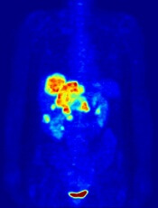
Image by Jens Langner
A PET probe known as [18F]CFA could be used to aid the treatment of leukemias and other cancers, according to research published in PNAS.
Investigators say [18F]CFA can detect the activity of deoxycytidine kinase (dCK) in humans more effectively than existing probes.
dCK is a rate-limiting enzyme in the cytosolic deoxyribonucleoside salvage pathway and is considered an important therapeutic and PET-imaging target in certain cancers.
Research has shown that dCK is highly expressed in acute leukemia cells and activated lymphocytes. And the enzyme plays an integral role in allowing drugs such as clofarabine, cytarabine, and fludarabine to treat certain leukemias.
“This enzyme is essential for the therapeutic activity of an entire class of anticancer drugs and even for some antiviral drugs,” said study author Caius Radu, MD, of the University of California, Los Angeles.
“It can take an inactive drug and activate it. If you trick a cancer cell or virus to activate the drug, it would be toxic for the cancer cell or viral genome.”
Until recently, PET technology was only able to clearly detect dCK in mice due to the metabolic instability of the available probes and cross-reactivity with a dCK-related enzyme in humans.
However, Dr Radu and his colleagues showed that [18F]CFA can clearly detect dCK in humans.
The team found that [18F]CFA accumulation in leukemia cells correlated with dCK expression, and they were able to inhibit [18F]CFA accumulation with a dCK inhibitor.
Experiments with [18F]CFA PET/CT in humans showed probe accumulation in tissues with high dCK expression, such as hematopoietic bone marrow and secondary lymphoid organs.
“We are able to clearly see tissues, including tumor tissues, with high dCK activity that we haven’t seen before in humans using any of the other probes previously developed for this enzyme,” Dr Radu said.
He added that, since activated immune cells increase their expression of dCK, [18F]CFA could also be used to monitor the effectiveness of immunotherapeutic interventions.
The investigators hope to begin clinical trials with [18F]CFA in the near future.
Dr Radu and his team invented [18F]CFA and its analogs, which were patented by the University of California and have been licensed to Sofie Biosciences, a company founded by Dr Radu and his team. The University of California also patented additional intellectual property for small-molecule dCK inhibitors.
Blood culture panel cleared by FDA

Staphylococcus infection
Photo by Bill Branson
The US Food and Drug Administration (FDA) has granted 510(k) clearance for a blood culture panel that detects sepsis caused by methicillin-resistant Staphylococcus aureus (MRSA) and other Staphylococcus species.
The Staph ID/R Blood Culture Panel is a product of Great Basin Scientific, Inc.
It is an automated, DNA multiplex assay used to identify Staphylococcus species directly from positive blood cultures in about 2 hours.
The panel also detects the mecA gene, a drug-resistance marker that confers resistance to methicillin and other beta-lactams and creates MRSA.
In addition, the Staph ID/R Blood Culture Panel identifies coagulase-negative staphylococci.
According to the US Centers for Disease Control and Prevention, 20% to 50% of all positive blood cultures are likely false positives due to contamination caused by coagulase-negative staphylococci, many of which are part of the normal flora of human skin and are not dangerous.
The Staph ID/R Blood Culture Panel is run on the Great Basin Analyzer. The company says the assay requires less than a minute of hands-on time and no results interpretation due to electronic results reporting.