User login
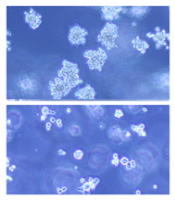
Method could make injectable drugs safer

Photo by Bill Branson
A new drug-making technique could reduce the risk of hemolysis, thrombosis, and other serious side effects that can occur with injectable drugs, according to researchers.
The team used the technique to remove potentially harmful additives—surfactants—from 12 common injectable drugs.
Jonathan F. Lovell, PhD, of University at Buffalo in New York, and his colleagues described the technique in Nature Communications.
Pharmaceutical companies use surfactants to dissolve a medicine into a liquid solution, making it suitable for injection. Unfortunately, solutions loaded with surfactants and other nonessential ingredients may increase the risk of thrombosis, hemolysis, anaphylactic shock, and other side effects.
Researchers have tried to address this problem in two ways, each with varying degrees of success.
Some have taken the “top-down” approach, in which they shrink drug particles to nanoscale sizes to eliminate excess additives. While promising, the method doesn’t work well with injectable medicine because the drug particles are still too large to safely inject.
Other researchers have worked from the “bottom up,” using nanotechnology to build new drugs from scratch. This can yield the desired results, but developing new drug formulations takes years, and drugs are coupled with new additives that can produce new side effects.
The technique described in Nature Communications differs from both of these approaches.
Dr Lovell and his colleagues dissolved 12 drugs—vitamin K1, cyclosporine, cabazitaxel, and others—one at a time into a surfactant called Pluronic. Then, by lowering the solution’s temperature to 4° C, they were able to remove the excess Pluronic via a membrane.
The end result was drugs that contain 100 to 1000 times less excess additives.
“For the drugs we looked at, this is as close as anyone has gotten to introducing pure, injectable medicine into the body,” Dr Lovell said. “Essentially, it’s a new way to package drugs.”
The findings are significant, he said, because they show that many injectable drug formulations may be improved through an easy-to-adopt process. He and his colleagues are now working to refine the method further. ![]()
Mutation in mice may have affected research results

Researchers say they have discovered a mutation in a subline of C57BL/6 mice that could compromise results from previous studies.
“We found an unexpected mutation with potentially important consequences in strains of mice that had been separately engineered in labs in California and Japan,” said Shiv Pillai, MD, PhD, of The Ragon Institute of MGH, MIT and Harvard in Cambridge, Massachusetts.
“[W]e traced the problem to a subline of B6 mice from one specific company that have been sold in Asia, North America, Europe, and Israel. We have notified this company—Harlan Laboratories, which is now part of Envigo—of our findings, and they have been very responsive and will use approaches we have provided to check all of their colonies.”
Dr Pillai and his colleagues described their discovery of the mutation in Cell Reports.
The team noted that, in immunological studies, mutant mice are backcrossed for many generations into B6 mice so that all genes other than the mutant gene are derived from the B6 strain. This allows the comparison of data from laboratories in different parts of the world and simplifies creating mice with mutations in several genes.
A 2009 study out of Dr Pillai’s lab showed that 2 strains of mice engineered to lack the Siae or Cmah genes—both of which code for enzymes involved with sialic acid proteins—also had significant defects in the development of B cells, which were assumed to be the result of the knockout genes.
However, when Siae-deficient mice were further backcrossed with a different group of C57BL/6 mice, the result was a strain of Siae-knockout mice that did not have the B-cell development defects. A newly engineered strain of mice with a different Siae mutation also had normal B-cell development.
Detailed genetic sequencing of the first knockout line revealed a previously unsuspected mutation in a gene called Dock2, located on a chromosome 11, instead of chromosome 9 where Siae is located.
The same mutation was previously reported in 2 colonies of a different knockout mouse developed in Japan.
Dr Pillai’s team also found the Dock2 mutation in a completely different group of mice from the University of California, San Diego—where their Siae-mutant strain had been developed—and realized that 3 different engineered strains with the same unwanted mutation had probably acquired it from a common source. The team eventually traced it back to a subline of C57BL/6 mice from Harlan/Envigo.
Since most research papers using C57BL/6 mice or other such “background” strains do not indicate the specific subline, the researchers said they have no way of knowing how many studies might be affected by their findings.
But they hope the publication of these results will alert other research teams to the potential need to review their results.
“While embryonic stem cells from C57BL/6 mice have recently become available, which allows the generation of knockout strains with less backcrossing, B6 mice are used for many different kinds of experiments, including as controls,” Dr Pillai said.
“Researchers who have used them need to re-genotype the mice to look for the Dock2 mutation and, if they find it, check to see whether their results are preserved if the Dock2 mutation is bred out.” ![]()
Breast cancer drug could treat AML
(below) palbociclib treatment
Images courtesy of Iris Uras
and Vetmeduni Vienna
Palbociclib, a CDK4/6 kinase inhibitor approved to treat breast cancer, could also be used to treat acute myeloid leukemia (AML), according to research published in Blood.
The drug induced apoptosis in FLT3-mutant AML cells and inhibited tumor growth in mouse models of FLT3-ITD+ AML.
Palbociclib also demonstrated synergy with a range of FLT3 inhibitors.
The researchers said these results can be explained by the fact that CDK6 is “absolutely required” for the viability of FLT3-dependent leukemic cells and FLT3-ITD-induced leukemogenesis.
“We found a novel therapeutic window that attacks the dependency of a cancer cell on its growth regulator,” said study author Iris Uras, PhD, of Vetmeduni Vienna in Austria.
Dr Uras and her colleagues first found that palbociclib acts specifically on FLT3-ITD+ AML cells, inhibiting their viability in a dose-dependent manner. Palbociclib induced cell-cycle arrest and apoptosis in these cells.
Palbociclib also arrested tumor growth in mouse models of FLT3-ITD+ AML, significantly decreasing tumor size when compared to untreated controls (P<0.05).
Further investigation revealed that CDK6—but not CDK4—directly regulates FLT3 expression.
CDK6 acts as a transcriptional regulator of FLT3 and the serine threonine kinase PIM1, which also plays a role in leukemogenesis. As palbociclib inhibits CDK6, it downregulates FLT3 and reduces PIM1 transcription.
To build upon these findings, the researchers tested palbociclib in combination with FLT3 inhibitors.
They observed “pronounced in vitro synergy” between palbociclib and TCS-359, tandutinib, and quizartinib in FLT3-ITD+ AML cell lines and samples from patients with FLT3-ITD+ AML.
“We are attacking FLT3 from two sides there—blocking its expression and inhibiting its activity,” Dr Uras said. “A combination therapy could be a breakthrough for many patients suffering from leukemia.” ![]()
SMAC mimetics could treat relapsed/refractory ALL

Patients with high-risk, relapsed/refractory acute lymphoblastic leukemia (ALL) may be sensitive to treatment with SMAC mimetics, according to researchers.
One SMAC mimetic in particular, birinapant, demonstrated varied activity in samples from ALL patients, but samples from patients with resistant disease were the most sensitive to the drug.
Birinapant also had “marked antileukemic effects” in some mice with ALL.
The researchers found this antileukemic activity was dependent on simultaneous activation of apoptosis and necroptosis.
The team reported these findings in Science Translational Medicine.
“Our research reveals that an alternative cell-death program, necroptosis, can be activated in human ALL cells,” said study author Beat Bornhauser, PhD, of the Children’s Hospital Zurich in Switzerland.
“This enables leukemia cells that barely respond to existing chemotherapeutic drugs to be killed off.”
In vitro and in vivo activity
The researchers tested the efficacy of SMAC mimetics in a set of 51 patient-derived B-cell precursor ALL xenografts, which was enriched for samples from relapsed and drug-resistant disease.
The response to birinapant varied greatly, but samples from high-risk or relapsed patients tended to be highly sensitive to the drug.
The researchers observed similar response profiles with the SMAC mimetic LCL161, although this drug proved less potent than birinapant.
The team also evaluated the antileukemic activity of SMAC mimetics in mouse models of ALL.
Birinapant delayed disease progression and induced complete responses in sensitive ALL cases. LCL161, on the other hand, did not display any in vivo activity.
Determining the mechanism of activity
The researchers used CRISPR-Cas9 to determine how SMAC mimetics fight ALL, and they discovered that the drugs trigger both apoptosis and necroptosis.
If the genes responsible for apoptosis were disabled via genome editing, leukemia cells died due to necroptosis after SMAC mimetics had been administered. If necroptotic genes were disabled, apoptosis led to cell death.
Only the simultaneous deactivation of apoptotic and necroptotic genes resulted in the complete resistance of leukemic cells to SMAC mimetics.
Therefore, the researchers concluded that simultaneous activation of apoptosis and necroptosis is responsible for the strong anti-leukemic effect of SMAC mimetics.
“SMAC mimetics have great potential to eliminate leukemia cells in patients that aren’t sensitive to established chemotherapeutic drugs,” Dr Bornhauser said. “They are effectively a double-edged sword. They kill cells that block apoptosis through necroptosis.”
The researchers are now looking for suitable biomarkers to identify patients who might benefit from treatment with SMAC mimetics in clinical trials. ![]()
Upfront ASCT still preferred for young MM patients
![]()
Photo by Chad McNeeley
CHICAGO—An interim analysis of a large, phase 3 study has confirmed that upfront autologous stem cell transplantation (ASCT) is still the preferred
treatment for newly diagnosed, young multiple myeloma (MM) patients, even in the age of novel agents such as bortezomib.
Investigators compared 4 cycles of bortezomib-melphalan-prednisone (VMP) with high-dose melphalan (HDM) and single or double ASCT, depending upon the policy of the treating institution.
At a median follow-up of 24 months, the 3-year progression-free survival (PFS) was significantly better for patients who had received ASCT.
Michele Cavo, MD, of Seràgnoli Institute of Hematology in Bologna, Italy, reported the results of this first interim analysis of the European Myeloma Network trial (EMN/HO95 MM) at a press briefing preceding the 2016 ASCO Annual Meeting. More details will be presented at the meeting itself (abstract 8000).
Study investigators enrolled 1503 patients from February 2011 through April 2014. They performed the specified interim analysis in January 2016.
Patients were 65 years or younger, and all received bortezomib-based induction therapy followed by stem cell collection. Investigators then randomized 1266 patients to receive either VMP (n=754) or HDM plus single or double ASCT (n=512).
Patients underwent a second randomization to either 2 cycles of bortezomib-based consolidation or no consolidation therapy.
All patients received lenalidomide maintenance until disease progression. The primary endpoint was PFS after the first randomization.
Results
PFS was significantly longer in patients who had received a transplant, with a hazard ratio (HR) of 0.76, 95% confidence interval (CI) of 0.61-0.94, and P value of 0.01.
This benefit held true for patients with revised ISS stage III (HR=0.52, 95% CI 0.32-0.84, P=0.01).
And patients with high-risk cytogenetics also retained the benefit (HR=0.72, 95% CI 0.54-0.97, P=0.03). High-risk was defined as t(4;14), del(17p), del(1p), or gain of 1q.
Investigators also performed a multivariate analysis and found randomization to the HDM arm to be an independent predictor of prolonged PFS (HR=0.61, 95% CI 0.45-0.82, P=0.001).
There was no significant difference between the 2 arms in terms of stringent complete response and complete response.
However, when very good partial response was included in the best-response analysis, patients in the transplant arm fared significantly better (P<0.0001) than patients in the VMP arm—84% and 74%, respectively.
Investigators have not yet completed the interim data analysis related to the second randomization. The study is ongoing, and future analyses will include overall survival, toxicity, quality of life, and other measures.
This study was funded by the Haemato Oncology Foundation for Adults in the Netherlands (HOVON). ![]()
HU improves lung function in young SCD patients

Photo courtesy of St. Jude
Children’s Research Hospital
SAN FRANCISCO—A new study has shown that hydroxyurea (HU) can improve lung function in young patients with sickle cell disease (SCD).
“Persons with sickle cell disease experience an annual decline in lung function that starts in childhood,” said study investigator Anya McLaren, MD, of The Hospital for Sick Children in Toronto, Ontario, Canada.
“This study is the first of its kind to look at the effect of hydroxyurea on lung function. We found that hydroxyurea improves annual pulmonary function decline in children with sickle cell disease by more than one-third.”
The study was presented at the ATS 2016 International Conference as abstract 7225.
For this study, Dr McLaren and her colleagues evaluated the effects of HU in 94 SCD patients. The patients’ average age at baseline was 11 (range, 6 to 20), 96% of patients had HbSS genotype, and 47% were male.
The patients were followed for 4 years after HU initiation. The investigators assessed lung function before and after HU initiation in a few ways.
They used the forced expiratory volume (FEV) test, which measures how much air a person can exhale during a forced breath. The amount of air can be measured during the first second of the forced breath (FEV1) and at later time points.
Forced vital capacity (FVC) is the total amount of air exhaled during the FEV test. If the FEV1/FVC ratio is less than 80%, it indicates that an obstructive defect is present.
The investigators also assessed FEF25-75, or the forced expiratory flow at 25%–75% of FVC. This measurement helps determine if there is an obstruction in the airway.
In addition, the team measured total lung capacity.
Results
The investigators found no significant change in total lung capacity, FVC, or FEV1/FVC predicted measurements after patients began receiving HU.
However, there were significant improvements in both FEV1 and FEF25-75 after treatment.
The annual rate of decline in predicted FEV1 and FEF25-75 before patients started HU was -1.98%/year (95% CI -2.57 to -1.39) and -3.59%/year (95% CI -4.43 to -2.75), respectively.
After HU treatment began, there was a significant (P<0.05) improvement in the annual decline, to -1.28%/year (95% CI -1.79 to -0.76) and -2.88%/year (95% CI -3.49 to -2.28), respectively.
The investigators noted that changes in FEV1 and FEF25-75 were independent of a patient’s age at baseline and the time from HU therapy initiation.
Dr McLaren pointed out that HU is underused in SCD patients, likely because clinicians are concerned about patient non-compliance and afraid of potential side effects, particularly carcinogenesis. But some of those fears may be unfounded, she said.
“Long-term observational studies suggest beneficial effects [of HU] without excessive damage to bone marrow, deleterious effects on growth and development, altered fertility, accumulation of mutations, or increased carcinogenicity,” Dr McLaren said.
“Evidence that lung function may be better preserved while on hydroxyurea may encourage compliance and adherence to this medication for patients with sickle cell disease. In combination with the established safety data, it hopefully will promote physician recommendations for hydroxyurea initiation and encouragement of compliance.” ![]()
Creating ‘real’ HSCs in the lab

in the bone marrow
Scientists believe they have come one step closer to creating hematopoietic stem cells (HSCs) that are just like the real thing.
“Our work focuses on finding a way to generate a supply of these life-saving hematopoietic stem cells in the lab so that they are a perfect match to the patient in need of a transplant,” said Hanna Mikkola, MD, PhD, of the University of California, Los Angeles.
“One big challenge is that when we try to create hematopoietic stem cells from pluripotent stem cells in the lab, they don’t acquire the same abilities of the real hematopoietic stem cells found in the body.”
Dr Mikkola and her colleagues described their attempts to overcome this challenge in Nature Cell Biology.
The researchers tried to create HSCs from pluripotent stem cells, but when they compared the lab-created cells to HSCs found in the body, they found that HOXA genes weren’t activated in the lab-created cells.
The team also discovered that HOXA genes help HSCs maintain their stem-cell attributes, such as the ability to self-renew.
“Without the ability to self-renew, hematopoietic stem cells cannot be used for transplantation therapies,” said Vincenzo Calvanese, PhD, an assistant project scientist in Dr Mikkola’s lab.
“Our findings show that the activation of HOXA genes can be used as a marker for hematopoietic stem cells that have acquired the capacity to renew themselves.”
The researchers’ next challenge was to pinpoint the naturally occurring process that activates HOXA genes so they could try to replicate the process in the lab.
They found that mimicking the effects of retinoic acid acts like a switch that turns on the HOXA genes during HSC development.
“Inducing retinoic acid activity at a very specific time in cell development makes our lab-created cells more similar to the real hematopoietic stem cells found in the body,” said Diana Dou, a graduate student in Dr Mikkola’s lab.
“This is an important step forward as we work to develop hematopoietic stem cells for transplantation therapies for life-threatening blood diseases.”
The researchers’ next step will be to refine the process they’ve developed in order to produce lab-created HSCs that have—and maintain—all the functions of human HSCs. ![]()
in the bone marrow
Scientists believe they have come one step closer to creating hematopoietic stem cells (HSCs) that are just like the real thing.
“Our work focuses on finding a way to generate a supply of these life-saving hematopoietic stem cells in the lab so that they are a perfect match to the patient in need of a transplant,” said Hanna Mikkola, MD, PhD, of the University of California, Los Angeles.
“One big challenge is that when we try to create hematopoietic stem cells from pluripotent stem cells in the lab, they don’t acquire the same abilities of the real hematopoietic stem cells found in the body.”
Dr Mikkola and her colleagues described their attempts to overcome this challenge in Nature Cell Biology.
The researchers tried to create HSCs from pluripotent stem cells, but when they compared the lab-created cells to HSCs found in the body, they found that HOXA genes weren’t activated in the lab-created cells.
The team also discovered that HOXA genes help HSCs maintain their stem-cell attributes, such as the ability to self-renew.
“Without the ability to self-renew, hematopoietic stem cells cannot be used for transplantation therapies,” said Vincenzo Calvanese, PhD, an assistant project scientist in Dr Mikkola’s lab.
“Our findings show that the activation of HOXA genes can be used as a marker for hematopoietic stem cells that have acquired the capacity to renew themselves.”
The researchers’ next challenge was to pinpoint the naturally occurring process that activates HOXA genes so they could try to replicate the process in the lab.
They found that mimicking the effects of retinoic acid acts like a switch that turns on the HOXA genes during HSC development.
“Inducing retinoic acid activity at a very specific time in cell development makes our lab-created cells more similar to the real hematopoietic stem cells found in the body,” said Diana Dou, a graduate student in Dr Mikkola’s lab.
“This is an important step forward as we work to develop hematopoietic stem cells for transplantation therapies for life-threatening blood diseases.”
The researchers’ next step will be to refine the process they’ve developed in order to produce lab-created HSCs that have—and maintain—all the functions of human HSCs.
EPO may not benefit preterm infants long-term

Photo by Petr Kratochvil
Giving very preterm infants high-dose recombinant human erythropoietin (EPO) at birth does not improve neurodevelopmental outcomes at 2 years, according to a study published in JAMA.
Researchers found no significant differences between infants who received EPO and those who did not when it came to cognitive development, motor development, cerebral palsy, hearing or visual impairment, and anthropometric growth parameters.
Giancarlo Natalucci, MD, of the University of Zurich in Switzerland, and his colleagues conducted this study in 448 preterm infants who were born between 26 weeks’ gestation and 31 weeks 6 days’ gestation.
The subjects’ average gestational age was 29 weeks, and their average birth weight was 1210 g (2.7 lbs).
The infants were randomized to receive high-dose EPO (n=228) or placebo (saline, n=220) intravenously within 3 hours of birth, at 12 to 18 hours, and at 36 to 42 hours after birth.
Neurodevelopmental outcome data were available for 81% of the infants (n=365) at an average age of 23.6 months.
Cognitive development, as assessed with the Mental Development Index (MDI), was not significantly different between the EPO group and the placebo group. In an intent-to-treat analysis, the mean MDI was 93.5 in the EPO group and 94.5 in the placebo group (P=0.056). In the per-protocol analysis, the mean MDI was 93.9 and 94.5, respectively (P=0.70).
The researchers also found no significant differences between the treatment groups for secondary outcomes such as motor development, cerebral palsy, hearing or visual impairment, and anthropometric growth parameters.
The team assessed motor development using the psychomotor development index (PDI). In the intent-to-treat analysis, the mean PDI was 89.5 in the EPO group and 92.1 in the placebo group (P=0.15). In the per-protocol analysis, the mean PDI was 89.2 and 92.8, respectively (P=0.06).
In the intent-to-treat analysis, the incidence of cerebral palsy was 4% in the EPO group and 5% in the placebo group (P>0.99). In the per-protocol analysis, it was 5% for both groups (P=0.41).
In the intent-to-treat analysis, severe hearing impairment occurred in 1 EPO-treated patient and no placebo-treated patients (P>0.99). Severe visual impairment occurred in 2 and 0, respectively (P=0.50). The incidences were the same in the per-protocol analysis.
And there were no significant differences between the treatment groups (per-protocol or intent-to-treat) when it came to growth parameters such as head circumference, weight, or length.
The researchers said these results suggest that EPO may not have a neuroprotective role in very preterm infants, but follow-up is required to assess cognitive and physical problems that may not become evident until later in life.
Method may produce better CAR-NKTs

Photo by Aaron Logan
Researchers say they have discovered a method for expanding natural killer T cells (NKTs) that ensures their persistence, thereby making NKTs more attractive as chimeric antigen receptor (CAR) carriers for cancer immunotherapy.
When transduced with a CD19-specific CAR, the researchers’ persistent NKTs produced sustained tumor regression in a mouse model of B-cell lymphoma.
The team described this work in the Journal of Clinical Investigation.
“NKT technology is quite powerful and offers a significant potential for treatment of cancer,” said study author Leonid Metelitsa, MD, PhD, of Baylor College of Medicine in Houston, Texas.
“But for it to be most effective, we have to find the best way to expand the cells ex vivo while preserving their ability to persist once delivered back to patients. If they can persist in the body for a long time, they have much longer therapeutic activity, and this is essential for fighting cancer.”
Molecule affects persistence
The researchers noted that central memory T cells are known for their ability to proliferate and persist, and these cells are characterized by expression of the surface molecule CD62L.
In this study, the team found that NKT cells freshly derived from blood did not express CD62L, or it was expressed at a very low level. However, after the researchers expanded NKT cells, they found that CD62L was expressed at higher levels.
“We consistently identified a subset of cells present at very high numbers after the first 12 days of expansion, and critical to this subset of cells was the presence of CD62L,” Dr Metelitsa said. “In fact, they became the majority of the new cells.”
In addition, Dr Metelitsa said that CD62L-positive NKT cells were responsible for further propagation of NKTs in culture, which is important for achieving large numbers of cells. However, extensive culture led to the eventual decline of CD62L expression in NKTs.
To test the role of CD62L in NKT-cell persistence, the researchers delivered CD62L-positive and CD62L-negative NKTs to immune-deficient NSG mice. They found that CD62L-positive NKTs persisted 5 times longer than CD62L-negative NKTs.
CAR-NKTs fight lymphoma
Next, the researchers transduced CD62L-positive and CD62L-negative NKTs with a CD19-specific CAR and delivered these cells to mice with B-cell lymphoma.
The team found that both CD62L-positve and CD62L-negative CAR-NKTs prolonged the survival of mice, when compared to controls (P<0.001).
However, only the CD62L-positive CAR-NKTs induced sustained tumor regression. Seven of 9 mice that received CD62L-positive CAR-NKTs lived, and 5 were tumor-free for at least 3 months. But all 10 mice that received CD62L-negative CAR-NKTs ultimately succumbed to tumor progression (P<0.001).
Costimulation improves NKTs/CAR-NKTs
The researchers then turned their focus to costimulation of NKTs in order to maintain the subset with a high percentage of CD62L-positive cells in prolonged culture. Costimulation involves the interaction of receptors on NKTs with activating molecules on an antigen-presenting cell to increase the NKTs’ immune functions.
“We have known that costimulation is an important part of immune response and immunotherapy, but, in this case, we did not know which costimulatory molecules would be important for the expansion and persistence of CD62L-positive NKT cells,” said Gengwen Tian, MD, of the Baylor College of Medicine.
After testing more than 100 combinations, the researchers discovered that combining an antigen-presenting molecule—CD1d—with 3 costimulatory molecules—CD86, 4-1BBL, and OX40L—induced prolonged persistence and better therapeutic activity of NKTs and CAR-NKTs in mouse models.
“When we developed an antigen-presenting cell clone that expressed CD1d with all of these costimulatory molecules at certain levels, NKT cells maintained a high percentage of CD62L even in a prolonged culture,” Dr Metelitsa said.
The researchers conducted in vivo testing of CAR-NKT cells that were expanded with the original method or with the costimulation method. And they found that costimulated cells had significantly higher therapeutic activity in mouse models of neuroblastoma and lymphoma.
“Our goal now is to optimize our NKT cell expansion protocol so that we can obtain FDA approval to initiate clinical trials,” Dr Metelitsa said.
HIV patients undertreated for lymphoma, other cancers
cultured lymphocyte
Image courtesy of the CDC
A new study suggests that cancer patients with HIV are less likely to receive cancer treatment, regardless of insurance status and comorbidities.
Patients with HIV were less likely than their HIV-free peers to receive treatment for Hodgkin lymphoma, diffuse large B-cell lymphoma, and 7 solid tumor malignancies.
Gita Suneja, MD, of the University of Utah in Salt Lake City, and her colleagues reported these findings in Cancer.
The team used the National Cancer Data Base to study non-elderly US adults diagnosed with 10 common cancers from 2003 to 2011. There were a total of 10,265 HIV-infected patients and 2,219,232 HIV-free patients.
The researchers examined associations between HIV status and lack of cancer treatment, taking into account insurance status and comorbidities.
The results showed a lack of treatment among HIV patients for all of the cancers studied except anal cancer (relative risk [RR]=1.20, P=0.333).
So HIV-infected patients were more likely to lack cancer treatment for:
- Hodgkin lymphoma (RR=1.92, P<0.001)
- Diffuse large B-cell lymphoma (RR=1.82, P<0.001)
- Head and neck cancer (RR=1.48, P=0.013)
- Upper gastrointestinal tract cancer (RR=2.62, P<0.001)
- Colorectal cancer (RR=1.70, P=0.006)
- Lung cancer (RR=2.46, P<0.001)
- Breast cancer (RR=2.14, P=0.015)
- Cervical cancer (RR=2.81, P<0.001)
- Prostate cancer (RR=2.16, P<0.001).
The researchers said factors that predicted a lack of cancer treatment among HIV-infected individuals varied between those with solid tumors and those with lymphomas.
Advanced stage at the time of cancer diagnosis (stage IV vs stage I) meant HIV patients with solid tumors were less likely to receive cancer treatment, but lymphoma patients were more likely to receive cancer treatment.
Having a higher modified Charlson-Deyo comorbidity score (1 or 2+ vs 0) predicted a lack of cancer treatment for HIV-infected patients with lymphoma but not those with solid tumors.
And older age (45-64 vs 18-44) was associated with a lack of treatment for HIV-infected patients regardless of cancer type, but this was only significant for lymphoma patients.
For the entire cohort, black race (vs white) and a lack of private insurance (Medicaid, Medicare, uninsured, or unknown insurance status) were significant predictors for a lack of cancer treatment among HIV patients.
Still, the researchers noted that, even among privately insured patients, HIV-infected individuals were less likely to receive cancer treatment.
Dr Suneja and her colleagues said several factors may contribute to these findings. For one, HIV-infected patients have historically been excluded from cancer clinical trials, thereby limiting the applicability of trial results for this population.
In addition, cancer treatment guidelines specific to HIV-infected patients are not available for most cancer types. And clinicians may lack experience treating HIV-infected patients with cancer.
Furthermore, the psychosocial and economic challenges associated with the dual management of cancer and HIV treatment may make adherence to treatment a challenge.